![Absorption, Metabolism and Excretion of …dmd.aspetjournals.org/content/dmd/40/4/815.full.pdfAbsorption, Metabolism and Excretion of [14C]Mirabegron (YM178),a Potent and Selective](https://static.fdocuments.in/doc/165x107/5b3dd4207f8b9a986e8de445/absorption-metabolism-and-excretion-of-dmd-metabolism-and-excretion-of-14cmirabegron.jpg)
Absorption and metabolism of Astragali Radix...
56
DMD #8300 1 Absorption and metabolism of Astragali Radix decoction: in silico, in vitro and a case study in vivo Feng Xu, Yue Zhang, Shengyuan Xiao, Xiaowei Lu, Donghui Yang, Xiaoda Yang, Changling Li, Mingying Shang, Pengfei Tu, Shaoqing Cai The State Key Laboratory of Natural and Biomimetic Drugs, School of Pharmaceutical Sciences (F.X., Y.Z., X.L., D.Y., X.Y., C.L., M.S., P.T, S.C.), Peking University Health Science Center, Beijing, China School of Bioscience and Biotechnology (S.X.), Beijing Institute of Technology, Beijing, China DMD Fast Forward. Published on February 28, 2006 as doi:10.1124/dmd.105.008300 Copyright 2006 by the American Society for Pharmacology and Experimental Therapeutics. This article has not been copyedited and formatted. The final version may differ from this version. DMD Fast Forward. Published on February 28, 2006 as DOI: 10.1124/dmd.105.008300 at ASPET Journals on May 10, 2018 dmd.aspetjournals.org Downloaded from
Transcript of Absorption and metabolism of Astragali Radix...

DMD #8300
1
Absorption and metabolism of Astragali Radix decoction:
in silico, in vitro and a case study in vivo
Feng Xu, Yue Zhang, Shengyuan Xiao, Xiaowei Lu, Donghui Yang, Xiaoda Yang,
Changling Li, Mingying Shang, Pengfei Tu, Shaoqing Cai
The State Key Laboratory of Natural and Biomimetic Drugs, School of
Pharmaceutical Sciences (F.X., Y.Z., X.L., D.Y., X.Y., C.L., M.S., P.T, S.C.), Peking
University Health Science Center, Beijing, China
School of Bioscience and Biotechnology (S.X.), Beijing Institute of Technology,
Beijing, China
DMD Fast Forward. Published on February 28, 2006 as doi:10.1124/dmd.105.008300
Copyright 2006 by the American Society for Pharmacology and Experimental Therapeutics.
This article has not been copyedited and formatted. The final version may differ from this version.DMD Fast Forward. Published on February 28, 2006 as DOI: 10.1124/dmd.105.008300
at ASPE
T Journals on M
ay 10, 2018dm
d.aspetjournals.orgD
ownloaded from
DMD #8300
2
Running title: Absorption and metabolism of Astragali Radix decoction
Address correspondence to: Dr. Shaoqing Cai, Department of Natural Medicines,
School of Pharmaceutical Sciences, Peking University Health Science Center. No. 38,
Xueyuan Road, Haidian District, Beijing, China, 100083. Tel. 86-10-82801693, Fax.
86-10-82801693, E-mail: [email protected]
Text pages: 49
Tables: 1
Figures: 4
References: 37
Abstract words: 243
Introduction words: 668
Discussion words: 1411
Abbreviations: HPLC-DAD-ESI-MSn, high-performance liquid
chromatography-diode array detection-electrospray ion trap tandem mass
spectrometry; UV, ultraviolet; ClogP, the LogP value calculated by ClogP 4.0 program,
P is partition coefficient of the molecule; C1: calycosin-7-O-β-D-glucuronide; C2:
(6aR,11aR)-3-hydroxy-9,10-dimethoxypterocarpan-3-O-β-D-glucuronide; C3:
7,2′-dihydroxy-3′,4′-dimethoxyisoflavan-2′-O-β-D-glucuronide; C4:
7,2′-dihydroxy-3′,4′,5′-trimethoxyisoflavan-7-O-β-D-glucuronide; C5:
7,2′-dihydroxy-3′,4′-dimethoxyisoflavan-7-O-β-D-glucuronide; C6:
(6aR,11aR)-3-hydroxy-9,10-dimethoxypterocarpan-3-O-β-D-glucoside-6″-O-malonat
e; C7: 7,2′-dihydroxy-3′,4′-dimethoxyisoflavan-7-O-β-D-glucoside-6″-O-malonate;
C8: calycosin-7-O-β-D-glucoside; C9: formononetin-7-O-β-D-glucoside; C10:
(6aR,11aR)-3-hydroxy-9,10-dimethoxypterocarpan-3-O-β-D-sambubioside; C11:
(6aR,11aR)-3-hydroxy-9,10-dimethoxypterocarpane-3-O-β-D-glucoside; C12:
7,3′-dihydroxy-2′,4′-dimethoxyisoflavan-7-O-β-D-glucoside; C13: calycosin; C14:
formononetin; C15: (6aR,11aR)-3-hydroxy-9,10-dimethoxypterocarpan; C16:
7,2′-dihydroxy-3′,4′-dimethoxyisoflavan; C17: formononetin-7-O-β-D-glucuronide;
C18: calycosin sulphate; C19: 7,2′-dihydroxy-3′,4′,6′-trimethoxyisoflavan; C20:
daidzein-7-O-β-D-glucuronide; C21:
7,3′-dihydroxy-2′,4′-dimethoxyisoflavan-7-O-β-D-glucosyl-3′-O-β-D-glucuronide
This article has not been copyedited and formatted. The final version may differ from this version.DMD Fast Forward. Published on February 28, 2006 as DOI: 10.1124/dmd.105.008300
at ASPE
T Journals on M
ay 10, 2018dm
d.aspetjournals.orgD
ownloaded from

DMD #8300
3
Abstract
To profile absorption of Astragali Radix decoction and identify its oral
absorbable constituents and their metabolites, four complementary in silico, in vitro
and in vivo methods, i.e., computational chemistry prediction method, Caco-2 cell
monolayer model experiment, improved rat everted gut sac experiment and healthy
human volunteer experiment were used. According to in silico computation result, 26
compounds of Astragali Radix could be regarded as oral available compounds,
including 12 flavonoids. In the in vitro and in vivo experiments, 21 compounds were
tentatively identified by high-performance liquid chromatography-diode array
detection-electrospray ion trap tandem mass spectrometry data, which involved
calycosin, formononetin, (6aR,11aR)-3-hydroxy-9,10-dimethoxypterocarpan,
7,2′-dihydroxy-3′,4′-dimethoxyisoflavan, calycosin-7-O-β-D-glucoside,
formononetin-7-O-β-D-glucoside,
7,2′-dihydroxy-3′,4′-dimethoxyisoflavan-7-O-β-D-glucoside-6″-O-malonate,
(6aR,11aR)-3-hydroxy-9,10-dimethoxypterocarpan-3-O-β-D-glucoside and phase II
metabolites calycosin-7-O-β-D-glucuronide, formononetin-7-O-β-D-glucuronide,
(6aR,11aR)-3-hydroxy-9,10-dimethoxypterocarpan-3-O-β-D-glucuronide,
7,2′-dihydroxy-3′,4′-dimethoxyisoflavan-7-O-β-D-glucuronide and calycosin sulphate.
Calycosin and formononetin were proved absorbable by four methods;
(6aR,11aR)-3-hydroxy-9,10-dimethoxypterocarpan and
7,2′-dihydroxy-3′,4′-dimethoxyisoflavan were proved absorbable by three methods;
formononetin-7-O-β-D-glucoside and
This article has not been copyedited and formatted. The final version may differ from this version.DMD Fast Forward. Published on February 28, 2006 as DOI: 10.1124/dmd.105.008300
at ASPE
T Journals on M
ay 10, 2018dm
d.aspetjournals.orgD
ownloaded from
DMD #8300
4
(6aR,11aR)-3-hydroxy-9,10-dimethoxypterocarpane-3-O-β-D-glucoside were proved
absorbable by two methods. The existence of calycosin-7-O-β-D-glucuronide,
formononetin-7-O-β-D-glucuronide,
(6aR,11aR)-3-hydroxy-9,10-dimethoxypterocarpan-3-O-β-D-glucuronide,
7,2′-dihydroxy-3′,4′-dimethoxyisoflavan-7-O-β-D-glucuronide and calycosin sulphate
was proved by two or three methods. We found that besides isoflavones, pterocarpans
and isoflavans also could be metabolized by the intestine during absorption, and the
major metabolites were glucuronides. In conclusion, the present study demonstrated
that the flavonoids in Astragali Radix decoction, including isoflavones, pterocarpans
and isoflavans, could be absorbed and metabolized by intestine. These absorbable
compounds, which were reported to have various bioactivities related to the curative
effects of Astragali Radix decoction, could be regarded as an important component
part of the effective constituents of Astragali Radix decoction.
This article has not been copyedited and formatted. The final version may differ from this version.DMD Fast Forward. Published on February 28, 2006 as DOI: 10.1124/dmd.105.008300
at ASPE
T Journals on M
ay 10, 2018dm
d.aspetjournals.orgD
ownloaded from

DMD #8300
5
Astragali Radix, a commonly used traditional Chinese drug, which is called
Huangqi in Chinese, is derived from the dried roots of Astragalus membranaceus
(Fisch.) Bunge or Astragalus membranaceus (Fisch.) Bunge var. mongholicus (Bunge)
Hsiao (The Pharmacopoeia Commission of People’s Republic of China, 1997). It has
been used as a qi-tonifying drug in China for about 2000 years (first recorded in
Shennong Bencao Jing, a materia medica book edited in 1st century). In general, its
traditional usage is to be prepared as a decoction alone or to be prepared as a
decoction together with other crude drugs (such as Chuanxiong Rhizoma, Angelica
sinensis Radix ) for oral administration.
Pharmacological studies indicate that Astragali Radix has various bioactivities,
such as hypotensive (Hikino et al., 1976), inducing vasodilatation (Zhang et al., 2005),
antioxidative (Shirataki et al., 1997), immunostimulating (Lee et al., 2003), antiviral
(Anonymous, 2003), inducing cancer cell apoptosis (Cheng et al., 2004), reducing the
capillary hyperpermeability and alleviating the dyskinesia caused by cerebral
ischemia (Quan and Du, 1998), inhibiting cyclooxygenase-2 (Kim et al., 2001),
promoting the motility of human spermatozoa (Liu et al., 2004), enhancing
cardiovascular function, protecting the myocardium in diabetic nephropathy (Chen et
al., 2001), anti-aging (Wang et al., 2003), hepatoprotective effect (Zhu et al., 2001),
inhibiting sterol biosynthesis (Sung, 1999) and antibacterial (Hu et al., 2005).
Clinical researches show that Astragali Radix can improve cardiovascular function,
restore and strengthen immune response, and enhance vitality. Indications supported
by clinical trials include acute myocardial infarction, impaired immunity, viral
This article has not been copyedited and formatted. The final version may differ from this version.DMD Fast Forward. Published on February 28, 2006 as DOI: 10.1124/dmd.105.008300
at ASPE
T Journals on M
ay 10, 2018dm
d.aspetjournals.orgD
ownloaded from

DMD #8300
6
infections and adjunctive cancer treatment (Anonymous, 2003).
Regarding the chemical constituents of Astragali Radix, more than 100 compounds
have been isolated and identified up to now, such as flavonoids (Subarnas et al., 1991;
Lin et al., 2000), triterpene saponins (Kitagawa et al., 1983), polysaccharides, amino
acids, phytosterols and phenolic acids.
As mentioned above, many pharmacological, clinical and phytochemical
investigations on Astragali Radix have been conducted so far. However, researchers
still don’t know what its effective constituents are, how many compounds are
absorbed into our blood after oral administration of the decoction, and what the fate of
the decoction in our body is. The reasons include: (i) in clinical studies, the materials
to be tested are usually the whole prescription or its different solvent extracts, and the
researchers are always not clear about the chemical compositions of them; (ii) in
phytochemical researches, the amounts of compounds isolated are always so small
that it is impossible to carry out in vivo pharmacological or clinical experiments; (iii)
although many researchers have ascribed immunomodulating activity to
polysaccharides, antioxidative action to flavonoids and triterpene saponins, and
hypotensive to γ–aminobutyic acid, these conclusions were mainly drawn from in
vitro pharmacological experiments or the compounds investigated were always
administrated by injection instead of per oral; (iv) no research on the intestinal
absorption and metabolism of Astragali Radix decoction has been conducted to date.
As a result, the knowledge about the absorption and metabolism of Astragali Radix
decoction is very poor, and it is hard to correlate the compounds isolated from
This article has not been copyedited and formatted. The final version may differ from this version.DMD Fast Forward. Published on February 28, 2006 as DOI: 10.1124/dmd.105.008300
at ASPE
T Journals on M
ay 10, 2018dm
d.aspetjournals.orgD
ownloaded from

DMD #8300
7
Astragali Radix with the curative effects of Astragali Radix decoction.
In the present work, our aim was to profile the absorption of Astragali Radix
decoction and identify its absorbable constituents and their metabolites.
We established a chemical database containing 124 compounds of Astragali Radix
for estimating the drug-like properties of these compounds and predicting their oral
absorption properties.
Two in vitro models, namely Caco-2 cell monolayer model and improved rat
everted gut sac model, were used to profile the absorption of Astragali Radix
decoction, with the aid of high-performance liquid chromatography-diode array
detection-electrospray ion trap tandem mass spectrometry (HPLC-DAD-ESI-MSn)1.
The absorbable compounds and their metabolites in the urine samples of a healthy
male volunteer orally dosed Astragali Radix decoction were also identified.
It was found that the flavonoids in Astragali Radix decoction, including
isoflavones, pterocarpans and isoflavans, could be absorbed and metabolized by
intestine and their main metabolites were glucuronides. They could be regarded as an
important component part of the effective constituents of Astragali Radix decoction.
This article has not been copyedited and formatted. The final version may differ from this version.DMD Fast Forward. Published on February 28, 2006 as DOI: 10.1124/dmd.105.008300
at ASPE
T Journals on M
ay 10, 2018dm
d.aspetjournals.orgD
ownloaded from

DMD #8300
8
Materials and Methods
Materials and Chemicals. Astragali Radix was purchased from Hunyuan County
in Shanxi Province of China, which was authenticated by Prof. Shaoqing Cai as the
dried roots of Astragalus membranaceus (Fisch.) Bunge var. mongholicus (Bunge)
Hsiao), and its voucher specimen (No. 2688) was deposited in the Herbarium of
Pharmacognosy, School of Pharmaceutical Sciences, Peking University Health
Science Center. Calycosin and calycosin-7-O-β-D-glucoside (purity>95%) were
isolated from the roots of Astragalus membranaceus var. mongholicus by one of the
author, Prof. Pengfei Tu. HPLC grade acetonitrile was purchased from Fisher
Scientific (Loughborough, UK). Pure water was purchased from Hangzhou Wahaha
Group Co., Ltd. (Hangzhou, China). NaHCO3 and NaCl of analytical grade were
obtained from Beijing Beihua Fine Chemicals Co., Ltd. (Beijing, China). Medium 199
powder (GibcoTM, with Earle’s salts and L-glutamine, without NaHCO3, Category No.
31100035) was purchased from Invitrogen Corp. (Carlsbad, California, USA). Caco-2
cells were obtained from American Type Culture Collection (Rockville, MD, USA).
Hanks’ balanced salt solution, Earle’s balanced salt solution, fetal calf serum and
other culture media and supplements were obtained from Gibco (Grand Island, NY,
USA). Transwells were purchased from Corning Costar (Cambridge, MA, USA).
Instrumentation. An Agilent 1100 LC-MSD-Trap-SL system (Agilent
Technologies, Palo Alto, CA, USA) consists of a degasser, an autosampler, a column
thermostat, a quaternary pump, a diode-array detector and an electrospray ion trap
mass spectrometer. The column used was a Zorbax SB-C18 (4.6 × 250mm, 5µm)
This article has not been copyedited and formatted. The final version may differ from this version.DMD Fast Forward. Published on February 28, 2006 as DOI: 10.1124/dmd.105.008300
at ASPE
T Journals on M
ay 10, 2018dm
d.aspetjournals.orgD
ownloaded from
DMD #8300
9
HPLC column with a Zorbax SB-C18 (4.6 × 12.5 mm, 5 µm) guard column (Agilent
Technologies, Palo Alto, CA, USA).
Construction of the chemical database of Astragali Radix. The database was
created by us using ChemFinder Ultra 8.0 (CambridgeSoft Corporation, Cambridge,
MA, USA) under Microsoft Windows 2000 Professional system (Microsoft
Corporation China Ltd., Beijing, China). The database contains one table with 26
searchable fields, including Structure, Molecular identification, Formula, Molecular
weight, Accurate mass, Name, Chinese name, Original plant, Physical properties,
Chemical abstracts service registry number, Simplified molecular input line entry
specification, ClogP, Topological molecular polar surface area, Number of hydrogen
bond acceptors, Number of hydrogen bond donors, Number of rotatable bonds,
Number of violations of “rule of 5”, UV, Mass spectrum data, Infrared data,
1H-nuclear magnetic resonance data, 13C-nuclear magnetic resonance data, References,
Biological activities, Biological activities references and Notes. Records of 124
compounds were inputted into the database according to the phytochemical and
pharmacological literatures of Astragali Radix and the Combined Chemical
Dictionary on CD-ROM version 8.1 (Chapman & Hall/CRC, FL, USA). Molecular
weight and accurate mass were calculated by ChemDraw Ultra 8.0 (CambridgeSoft
Corporation, Cambridge, MA, USA). ClogP was calculated by ClogP4.0 program
(BioByte Corporation, Claremont, CA, USA). The number of hydrogen bond
acceptors, the number of hydrogen bond donors, the number of violations of “rule of
5”, topological molecular polar surface area and the number of rotatable bonds were
This article has not been copyedited and formatted. The final version may differ from this version.DMD Fast Forward. Published on February 28, 2006 as DOI: 10.1124/dmd.105.008300
at ASPE
T Journals on M
ay 10, 2018dm
d.aspetjournals.orgD
ownloaded from

DMD #8300
10
calculated by the free Molinspiration Property Calculation Services on the Internet
(http://www.molinspiration.com/cgi-bin/properties).
In silico prediction of oral absorption properties of Astragali Radix
constituents. Two simple counting methods were used to predict oral absorption
properties of Astragali Radix constituents. Firstly, the “rule of 5” descriptors (Lipinski
et al., 1997), i.e., molecular weight, ClogP, the number of hydrogen bond acceptors,
the number of hydrogen bond donors, were used to estimate oral absorption properties
of the constituents. The molecules which met the “rule of 5” were considered as good
oral absorbable compounds. Secondly, the molecules which contained 10 or fewer
rotatable bonds and topological molecular polar surface area (Ertl et al., 2000) of
which were equal to or less than 140 Å2 were regarded as good oral absorbable
compounds (Veber et al., 2002). The “rule of 5” set a lower limit of molecular polarity,
and the rule that the number of rotatable bonds ≤10 and topological molecular polar
surface area ≤ 140 Å2 set an upper limit of molecular polarity. Therefore, we
considered that the compounds met both rules were oral available compounds.
Preparation of freeze-drying powder of Astragali Radix decoction. Astragali
Radix was cut into decoction pieces about 1 cm long. Two hundred grams decoction
pieces were weighed and soaked with 2.0 L water for 30 min. Then, the decoction
pieces were boiled for 30 min and the decoction was filtrated out by absorbent cotton
inserted in a funnel. Subsequently, the dregs were boiled twice again for 30 min with
1.6 L and 1.2 L water successively, and the decoctions were filtrated out with the
above method. Afterwards, the decoctions of three times were merged and condensed
This article has not been copyedited and formatted. The final version may differ from this version.DMD Fast Forward. Published on February 28, 2006 as DOI: 10.1124/dmd.105.008300
at ASPE
T Journals on M
ay 10, 2018dm
d.aspetjournals.orgD
ownloaded from

DMD #8300
11
by a Heidolph Laborota 4001 rotatory evaporator (Heidolph Instruments GmbH&Co.,
Schwabach, Germany) under reduced pressure. Finally, the concentrate was
lyophilized to 70.6 g powder by a Labconco Freezone® 6 freeze dryer (Labconco
Corporation, Kansas, MO, USA). Thus, each gram powder was equivalent to 2.83 g
crude drug of Astragali Radix. The powder was stored in a desiccator at room
temperature for later use.
Animals. Adult male Sprague-Dawley rats weighing between 250 to 340 g (The
Experimental Animal Center of Peking University Health Science Center, Beijing,
China) were used in the everted gut sac experiment. The animals were handled in
accordance with the Guide for the Care and Use of Laboratory Animals of the U.S.
National Institutes of Health. The rats were fasted for 24 h before the date of the
experiment.
In vitro improved rat everted gut sac experiment. One package of Medium 199
powder (9.5 g) was dissolved in 1.00 L distilled water with the addition of 2.2 g
NaHCO3, then the Medium 199 solution was gassed with 95% O2 + 5% CO2 at 37°C.
Twelve rats were euthanized by cervical dislocation, and the entire small intestine was
quickly taken out and flushed three times by saline using a 20 ml syringe at room
temperature. The intestine was instantly put in oxygenated Medium 199 solution.
With the help of a smooth plastic rod (4.0 mm diameter), the intestine was gently
everted as quickly as possible to make the serosal side towards the inside and the
mucosal side towards the outside, and then the everted intestine was slid into the
medium solution. Afterwards, one end of the intestine was tied with suture, and the
This article has not been copyedited and formatted. The final version may differ from this version.DMD Fast Forward. Published on February 28, 2006 as DOI: 10.1124/dmd.105.008300
at ASPE
T Journals on M
ay 10, 2018dm
d.aspetjournals.orgD
ownloaded from

DMD #8300
12
intestine was filled with 10 ml Medium 199 solution. Then, the intestine was sealed in
the other end with suture (Barthe et al., 1998).
In control group, each of six everted gut sacs was put into an Erlenmeyer flask (250
ml) containing 100 ml pregassed (95%O2 + 5%CO2) Medium 199 solution at 37°C. In
test group, each of six everted gut sacs was put into an Erlenmeyer flask (250 ml),
which contained 100 ml pregassed (95%O2 + 5%CO2) Astragali Radix Medium 199
solution at 37°C with a concentration of 100 mg crude drug per milliliter.
The flasks of both test and control groups were plugged with rubber stoppers,
which had two holes for gas (95%O2 + 5%CO2) in and out. Then, the sacs were
incubated at 37°C in a DSY-2-4 type electroheating constant temperature water bath
(Beijing Guohua Medical Apparatus and Instrument Factory, Beijing, China) for one
hour, aerated by gas (95%O2 + 5%CO2). Afterwards the sacs were removed and
blotted dry with gauze. Then the sacs were cut open and the serosal side solutions,
which should contain absorbable constituents of Astragali Radix decoction or their
metabolites, were drained into small tubes. At the same time, the mucosal side
solutions which contained Astragali Radix decoction were also sampled for analysis.
Both mucosal side and serosal side solutions were stored at −40°C in a MDF-U5410
Sanyo medical freezer (Sanyo Electric Co., Ltd, Osaka, Japan) until analyzed.
In vitro Caco-2 cell monolayer model experiment. The cell monolayer was
prepared as the method described previously (Yang et al., 2004). The monolayer with
transepithelial electrical resistance values less than 800 ohms × cm2 were not used.
The monolayers were washed three times with Hanks’ balanced salt solution, pH 7.4,
This article has not been copyedited and formatted. The final version may differ from this version.DMD Fast Forward. Published on February 28, 2006 as DOI: 10.1124/dmd.105.008300
at ASPE
T Journals on M
ay 10, 2018dm
d.aspetjournals.orgD
ownloaded from
DMD #8300
13
at 37 °C. In test group, an Astragali Radix Earle’s balanced salt solution at a
concentration of 100 mg crude drug per milliliter was added to the apical side. In
control group, an Earle’s balanced salt solution was added to the apical side. In both
groups, Hanks’ balanced salt solutions were added to the basolateral sides. Then, the
samples were shaken (37°C, 50 rpm) and 200 µl aliquots were taken from the
basolateral sides at 0, 45, 90, 135, 180 min. The apical solutions at 0 min were also
sampled for analysis. All experiments were repeated three times. All of the apical
solutions and basolateral solutions were stored at −40°C in a MDF-U5410 Sanyo
medical freezer (Sanyo Electric Co., Ltd, Osaka, Japan) until analyzed.
In vivo human experiment. In 10 days, the diet of a volunteer (a 28-year-old
healthy Chinese male) was fixed throughout but water was allowed ad libitum. The
diet didn’t contain soybean, vegetables and fruits. The volunteer has three meals a day.
Each meal consists of a piece of 400g bread which contains vitamin B1, vitamin B2,
nicotinic acid, carotene, Zn, Fe and Ca (Mankattan Beijing Co., Ltd., Beijing, China),
an egg and 20 g pure honey (Beijing Baihua Bee Product Co., Ltd., Beijing, China).
The volunteer was prohibited from smoking and drinking alcoholic beverages. In day
3 and day 4, total volume urine samples were collected as blank urine samples. From
day 5 to day 10, the volunteer took Astragali Radix decoction orally before meal at a
dosage of 60 g crude drug twice a day, and the total volume urine samples were
collected as drug-containing urine samples. This study was approved by ethics
committee of Health Science Center of Peking University, and was carried out
following good clinical practice guidelines and was in accordance with the guidelines
This article has not been copyedited and formatted. The final version may differ from this version.DMD Fast Forward. Published on February 28, 2006 as DOI: 10.1124/dmd.105.008300
at ASPE
T Journals on M
ay 10, 2018dm
d.aspetjournals.orgD
ownloaded from

DMD #8300
14
of the Declaration of Helsinki and all its amendments, and the subject had given his
written consent.
Each of two blank urine samples and six drug-containing urine samples was
evaporated to dryness under vacuum at 37°C by a Heidolph Laborota 4001 rotatory
evaporator (Heidolph Instruments GmbH&Co., Schwabach, Germany). These eight
dried samples were weighed by an electronic balance (Ohaus China, Shanghai, China),
and 1.00 g of each was transferred to a centrifuge tube. Then the dried samples were
extracted with 6ml methanol (HPLC grade, Fisher Scientific, Loughborough, UK) by
sonication for 5 min using a TP-150 ultrasonic cleaner (Tianpong Electricity New
Technology Co., Beijing, China). The extracts were centrifugated at 4800 rpm for 20
min using a Anke TDL-5-A centrifuge (Shanghai Anting Experimental Instrument
Factory, Shanghai, China), and then the supernatant were applied to
HPLC-DAD-ESI-MSn analysis.
Sample analysis. All of the serosal, mucosal, apical, basolateral side solutions and
uric samples were filtered through 0.45 µm micropore membranes (Tianjin Tengda
Filter Factory, Tianjin, China) and were injected into the instrument for HPLC-DAD
analysis directly. According to HPLC-DAD analytical results, representative samples
were chosen. Then, representative samples and a calycosin and
calycosin-7-O-β-D-glucoside methanol solution were further analyzed by
HPLC-DAD-ESI-MSn.
The samples were analyzed by a gradient method at a flow rate of 1.000 ml/min.
The mobile phase consisted of water (A) and acetonitrile (B). The gradient was 0.0%
This article has not been copyedited and formatted. The final version may differ from this version.DMD Fast Forward. Published on February 28, 2006 as DOI: 10.1124/dmd.105.008300
at ASPE
T Journals on M
ay 10, 2018dm
d.aspetjournals.orgD
ownloaded from

DMD #8300
15
B at 0.00~5.00 min, 0.0~15.0% B at 5.00~20.00min, 15.0% B at 20.00~25.00 min,
20.0% B at 25.00~30.00 min, 20.0%~30.0% B at 30.00~35.00 min, 30.0% B at
35.00~40.00min, 30.0%~50.0% B at 40.00~50.00min, 50.0%~100.0% B at
50.00~60.00 min. The injection volume was 10.00~30.00 µl. The column temperature
was 30.0°C. In HPLC-DAD-ESI-MSn analysis, HPLC chromatograms were recorded
at 210, 230, 254, 280 and 365 nm, and UV and visible light spectra were obtained by
scanning from 190 nm to 800 nm. The HPLC effluent was split, and about 200 µl/min
was introduced into the mass spectrometer.
The mass spectrometer was operated in alternating negative ion and positive ion
electrospray mode and full scans were acquired from m/z 50 to 1500. The temperature
of drying gas (N2) was 325°C at a flow-rate of 7.00 L/min and a nebulizing pressure
of 20.00 psi. A data-dependent program was used in the HPLC-DAD-ESI-MSn
analysis so that the two most abundant ions in each scan were selected and subjected
to MS2 and MS3 analyses. The collision-induced dissociation energy was varied
automatically from 0.3eV to 2.0 eV in smart fragmentation mode. The isolation width
of precursor ions was 4.0 mass units. All data were collected and processed by
Agilent 1100 ChemStation 09.03 version and LC/MSD Trap Software 4.2 version
(Agilent Technologies, Palo Alto, CA, USA).
This article has not been copyedited and formatted. The final version may differ from this version.DMD Fast Forward. Published on February 28, 2006 as DOI: 10.1124/dmd.105.008300
at ASPE
T Journals on M
ay 10, 2018dm
d.aspetjournals.orgD
ownloaded from
DMD #8300
16
Results
I. The Results of in silico Absorption Prediction, in vitro and in vivo Absorption
Experiments of Astragali Radix Decoction
In silico prediction of oral available constituents of Astragali Radix. Totally 124
compounds have been isolated from Astragali Radix (including Astragalus
membranaceus (Fisch.) Bunge and Astragalus membranaceus (Fisch.) Bunge var.
mongholicus (Bunge) Hsiao.) up to now (refer to Supplemental data – Table s1).
According to calculation results (refer to Supplemental data – Table s1), 68
compounds met the “rule of 5”, and 74 compounds met the rule that topological
molecular polar surface area ≤ 140 Å2 and the number of rotatable bonds ≤ 10.
Sixty-two compounds met both rules.
Except 17 amino acids and 4 essential oil ingredients identified by gas
chromatography-mass spectrometry, 41 compounds could be regarded as good oral
available compounds. Among them, 26 compounds were isolated from Astragalus
membranaceus var. mongholicus, including 12 flavonoids, 5 phenolic acids, 5
nitrogen-containing compounds, 3 lignanoids and 1 coumarin, and their structures
were shown in Supplemental data – Fig. s1.
Absorption in the improved rat everted gut sac model. Fig. 1 shows the base
peak chromatograms of the samples detected in negative ion mode. Eighteen peaks
(C1~C18, denoting compound 1~18) were tentatively identified by
HPLC-DAD-ESI-MSn data. The peak areas of compound 1, 2, 3, 4 and 5 in the
serosal side solution were larger than those of compound 1, 2, 3, 4 and 5 in the
This article has not been copyedited and formatted. The final version may differ from this version.DMD Fast Forward. Published on February 28, 2006 as DOI: 10.1124/dmd.105.008300
at ASPE
T Journals on M
ay 10, 2018dm
d.aspetjournals.orgD
ownloaded from

DMD #8300
17
mucosal side solution. This indicated that either compounds 1, 2, 3, 4, 5 might be
metabolites or they might be absorbed by active transport. Besides, compound 17 and
compound 18 were detected in both serosal side solution and mucosal side solution.
These identified compounds can be classified into three groups according to their
retention time and structure type. (i) Four flavonoid aglycons: calycosin (C13);
formononetin (C14); (6aR,11aR)-3-hydroxy-9,10-dimethoxypterocarpan (C15) and
7,2′-dihydroxy-3′,4′-dimethoxyisoflavan (C16). (ii) Seven flavonoid glycosides:
calycosin-7-O-β-D-glucoside (C8); formononetin-7-O-β-D-glucoside (C9);
(6aR,11aR)-3-hydroxy-9,10-dimethoxypterocarpan-3-O-β-D-glucoside (C11);
(6aR,11aR)-3-hydroxy-9,10-dimethoxypterocarpan-3-O-β-D-sambubioside (C10);
(6aR,11aR)-3-hydroxy-9,10-dimethoxypterocarpan-3-O-β-D-glucoside-6″-O-malonat
e (C6); 7,3′-dihydroxy-2′,4′-dimethoxyisoflavan-7-O-β-D-glucoside (C12) and
7,2′-dihydroxy-3′,4′-dimethoxyisoflavan-7-O-β-D-glucoside-6″-O-malonate (C7). (iii)
Seven flavonoid metabolites: calycosin-7-O-β-D-glucuronide (C1); calycosin sulphate
(C18); formononetin-7-O-β-D-glucuronide (C17);
(6aR,11aR)-3-hydroxy-9,10-dimethoxypterocarpan-3-O-β-D-glucuronide (C2);
7,2′-dihydroxy-3′,4′-dimethoxyisoflavan-2′-O-β-D-glucuronide (C3);
7,2′-dihydroxy-3′,4′,5′-trimethoxyisoflavan-7-O-β-D-glucuronide (C4) and
7,2′-dihydroxy-3′,4′-dimethoxyisoflavan-7-O-β-D-glucuronide (C5).
Absorption in the Caco-2 monolayer model. Fig. 2 shows the base peak
chromatograms of the samples detected in negative ion mode. Six peaks (C18, C13,
C14, C19, C15, C16) were tentatively identified by HPLC-DAD-ESI-MSn data. The
This article has not been copyedited and formatted. The final version may differ from this version.DMD Fast Forward. Published on February 28, 2006 as DOI: 10.1124/dmd.105.008300
at ASPE
T Journals on M
ay 10, 2018dm
d.aspetjournals.orgD
ownloaded from

DMD #8300
18
peak area of C18 in basolateral solution was larger than that in apical solution. This
indicated that either C18 might be a metabolite or it might be absorbed by active
transport. Five of these compounds (C13, C14, C15, C16, C18) were identical with
those identified in improved rat everted gut sac experiment. C19 was identified as
7,2′-dihydroxy-3′,4′,6′-trimethoxyisoflavan.
Absorption and metabolism in human. The extracted ion chromatograms at m/z
363, 475, 477, 443, 459, 429, 639, 283, 267 of blank urine and drug-containing urine
detected in negative ion mode are shown in Fig. 3. Seven peaks (C20, C1, C17, C21,
C18, C2, C5) were tentatively identified by HPLC-DAD-ESI-MSn data. Besides these
seven compounds, calycosin (C13) and formononetin (C14) were also detected and
identified by their HPLC retention times and UV spectra. In nine compounds, seven
compounds (C1, C2, C5, C13, C14, C17, C18) were identical with those identified in
rat everted gut sac experiment. The others were identified as
daidzein-7-O-β-D-glucuronide (C20) and
7,3′-dihydroxy-2′,4′-dimethoxyisoflavan-7-O-β-D-glucosyl-3′-O-β-D-glucuronide
(C21).
II. The Results of Identification of Absorbable Compounds and Their
Metabolites.
The molecular weight of a compound in our study was confirmed by its molecular
ion, quasi-molecular ion, dimer ion and adduct ion in negative and positive ion mass
spectra. Then, we searched for this molecular weight in the chemical database of
Astragali Radix and in the Combined Chemical Dictionary on CD-ROM version 8.1
This article has not been copyedited and formatted. The final version may differ from this version.DMD Fast Forward. Published on February 28, 2006 as DOI: 10.1124/dmd.105.008300
at ASPE
T Journals on M
ay 10, 2018dm
d.aspetjournals.orgD
ownloaded from
DMD #8300
19
to find relevant compounds. The structure type of the compound was judged by its
UV spectrum. The structure of the compound was elucidated based on its MS2 and
MS3 data, and when possible, by direct comparison with the data of standard
compounds and the data in the literatures (Lin et al., 2000; Xiao et al., 2004). As for
metabolites, we first confirmed their structure types by MS2 and MS3 data, and then
identified their aglycons by above-mentioned method. Altogether 21 compounds were
identified tentatively by HPLC-DAD-ESI-MSn data, and their structures were shown
in Fig. 4.
Identification of absorbable constituents of Astragali Radix decoction
Compound 13 (C13): C13 had a retention time of 42.1~42.9 min on HPLC. It
showed [M−H]− at m/z 283 in negative ion mass spectrum, and [M+H]+ at m/z 285,
[M+Na]+ at m/z 307, [2M+Na]+ at m/z 591 in positive ion mass spectrum. So, its
molecular weight was inferred to be 284 Da. Its UV spectrum exhibited maximum
absorption at 200, 220, 250, 290 nm and shoulder peak at 310 nm with weak band I
(310 nm) and strong band II (250 nm), which suggested that it was an isoflavone. The
negative MS2 spectrum of m/z 283 gave a fragment ion at m/z 268, and loss of 15 Da
from the precursor ion indicated that there was a methyl in the molecule. In the
chemical database of Astragali Radix, only calycosin was found to have a molecular
weight of 284 Da. In addition, the HPLC retention time, UV spectrum, MSn data of
C13 were identical with those of standard compound of calycosin. So, C13 was
identified as calycosin unequivocally.
Compound 14 (C14): C14 had a retention time of 50.8~51.3 min on HPLC. It
This article has not been copyedited and formatted. The final version may differ from this version.DMD Fast Forward. Published on February 28, 2006 as DOI: 10.1124/dmd.105.008300
at ASPE
T Journals on M
ay 10, 2018dm
d.aspetjournals.orgD
ownloaded from

DMD #8300
20
showed [M−H]− at m/z 267 in negative ion mass spectrum, and [M+H]+ at m/z 269,
[M+Na]+ at m/z 291, [2M+Na]+ at m/z 559 in positive ion mass spectrum. So, its
molecular weight was inferred to be 268 Da. Its UV spectrum exhibited maximum
absorption at 250, 288 nm and shoulder peak at 300 nm with weak band I (300 nm)
and strong band II (250 nm), which suggested that it was an isoflavone. The negative
MS2 spectrum of m/z 267 showed fragment ions at m/z 252 and m/z 224; sequential
loss of 15 Da and 28 Da from the precursor ion indicated the presence of a methyl and
a carbonyl in the molecule. The positive MS2 spectrum of m/z 291 showed a fragment
ion at m/z 273. The fragment ion at m/z 273, loss of 18 Da from ion m/z 291,
suggested the presence of a hydroxyl group. In the chemical database of Astragali
Radix, only formononetin had a molecular weight of 268 Da. Based on these data,
C14 was identified as formononetin tentatively.
Compound 15 (C15): C15 had a retention time of 51.9~52.3 min on HPLC. It
showed [M−H]− at m/z 299 in negative ion mass spectrum, and [M+H]+ at m/z 301,
[M+Na]+ at m/z 323, [2M+Na]+ at m/z 623 in positive ion mass spectrum. So, its
molecular weight was inferred to be 300 Da. Its UV spectrum exhibited maximum
absorption at 208, 280 nm, shoulder peak at 225 nm and minimum absorption at 250
nm, which suggested that it was an isoflavan or a pterocarpan. The negative MS2
spectrum of m/z 299 showed fragment ions at m/z 284, 269, 241; sequential loss of 15
Da, 15 Da and 28 Da from m/z 299 indicated the presence of two methyl groups and a
C-O fragment in the molecule. In the chemical database of Astragali Radix, three
compounds, i.e., (6aR,11aR)-3-hydroxy-9,10-dimethoxypterocarpan,
This article has not been copyedited and formatted. The final version may differ from this version.DMD Fast Forward. Published on February 28, 2006 as DOI: 10.1124/dmd.105.008300
at ASPE
T Journals on M
ay 10, 2018dm
d.aspetjournals.orgD
ownloaded from

DMD #8300
21
(6aR,11aR)-3,9-dimethoxy-10-hydroxypterocarpan and
3,4′,5-trihydroxy-7-methoxyflavone, have the molecular weight of 300 Da, but only
(6aR,11aR)-3-hydroxy-9,10-dimethoxypterocarpan was a pterocarpan isolated from
Astragalus membranaceus var. mongholicus. Based on these data, C15 was identified
as (6aR,11aR)-3-hydroxy-9,10-dimethoxypterocarpan.
Compound 16 (C16): C16 had a retention time of 52.4~52.7 min on HPLC. It
showed [M−H]− at m/z 301 in negative ion mass spectrum, indicating that its
molecular weight was 302 Da. Its UV spectrum exhibited maximum absorption at 204,
280 nm, shoulder peak at 225 nm and minimum absorption at 250 nm, suggesting that
it was an isoflavan or a pterocarpan. The negative MS2 spectrum of m/z 301 gave
fragment ions at m/z 286, 153, 147, 135, 121, 109. The fragment ion at m/z 286, loss
of 15 Da from the precursor ion indicated that there was a methyl in the molecule.
The fragment ions at m/z 147 and m/z 153 were a pair of complementary ions, m/z 153
was from B ring of isoflavan, m/z 147 was from A ring and C ring. The characteristic
A ring ions at m/z 121, 109 were generated by Retro Diels-Alder fragmentation in C
ring. The fragmentation ion at m/z 135 was also from A ring and C ring by loss of B
ring and C3 from the quasi-molecular ion at m/z 301. These data indicated that C16
was an isoflavan, and there was only one hydroxyl substituent on A ring. In the
chemical database of Astragali Radix, three compounds, i.e.,
7,2′-dihydroxy-3′,4′-dimethoxyisoflavan, quercetin and
(3R)-8,2'-dihydroxy-7,4'-dimethoxyisoflavan, have the molecular weight of 302 Da,
but only 7,2′-dihydroxy-3′,4′-dimethoxyisoflavan was an isoflavan with one
This article has not been copyedited and formatted. The final version may differ from this version.DMD Fast Forward. Published on February 28, 2006 as DOI: 10.1124/dmd.105.008300
at ASPE
T Journals on M
ay 10, 2018dm
d.aspetjournals.orgD
ownloaded from

DMD #8300
22
substituent on A ring isolated from Astragalus membranaceus var. mongholicus.
Based on these data, C16 was identified as 7,2′-dihydroxy-3′,4′-dimethoxyisoflavan
tentatively.
Compound 19 (C19): C19 had a retention time of 51.4~51.7 min on HPLC. It
showed [M−H]− at m/z 331 in negative ion mass spectrum, indicating that its
molecular weight was 332 Da. Its UV spectrum exhibited maximum absorption at 204,
280 nm, shoulder peak at 225 nm and minimum absorption at 250 nm, suggesting that
it was an isoflavan or a pterocarpan. The molecular weight of C19 was only 30 Da
higher than that of 7,2′-dihydroxy-3′,4′-dimethoxyisoflavan (C16), suggesting that it
might be a methoxyl derivative of C16. The negative MS2 spectrum of m/z 331 gave
fragment ions at m/z 316, 301, 299, 313, 295, 209, 194, 147, 135, 109. The fragment
ions at m/z 316, 301, sequential loss of 15 Da, 15 Da from m/z 331 indicated the
presence of two methyl groups. The fragment ions at m/z 313, 295, sequential loss of
18 Da, 18 Da from m/z 331 indicated the presence of two hydroxy groups. The
fragment ion m/z 299, loss of 32 Da from m/z 331 indicated that there was a 2′ or 6′
methoxyl in the molecule. The characteristic B ring ions at m/z 209, 194 were
generated by Retro Diels-Alder fragmentation in C ring. The fragmentation ion m/z
147 was from A ring and C ring. The fragmentation ion at m/z 135 was also from A
ring and C ring by loss of B ring and C3 from the quasi-molecular ion at m/z 301. The
fragment ion at m/z 109 was from A ring, indicating that there was only one hydroxy
in A ring. These data indicated that C19 was an isoflavan with at least two methyl,
two hydroxy and one 2′ or 6′ methoxyl in the molecule. In the chemical database of
This article has not been copyedited and formatted. The final version may differ from this version.DMD Fast Forward. Published on February 28, 2006 as DOI: 10.1124/dmd.105.008300
at ASPE
T Journals on M
ay 10, 2018dm
d.aspetjournals.orgD
ownloaded from

DMD #8300
23
Astragali Radix, no compound has a molecular weight of 332 Da. Based on these data
and compared with the structure of 7,2′-dihydroxy-3′,4′-dimethoxyisoflavan (C16)
C19 was tentatively identified as a 6′ methoxyl substitution derivative of C16, i.e.,
7,2′-dihydroxy-3′,4′,6′-trimethoxyisoflavan, and this compound was found in
Astragali Radix for the first time.
Compound 8 (C8): C8 had a retention time of 30.9~31.7 min on HPLC. It showed
[M−H]− at m/z 445, [M+HCOOH−H]− at m/z 491, [M+CH3COOH−H]− at m/z 505 and
aglycon ion at m/z 283 in negative ion mass spectrum. It showed [M+H]+ at m/z 447,
[M+Na]+ at m/z 469, [M+K]+ at m/z 485, [2M+Na]+ at m/z 915 in positive ion mass
spectrum. So, its molecular weight was inferred to be 446 Da. Its UV spectrum
exhibited maximum absorption at 200, 220, 250, 258 nm and shoulder peak at 286 nm
with weak band I (300~400 nm) and strong band II (250 nm), suggesting that it was
an isoflavone. The positive MS2 spectrum of m/z 447 gave a fragment ion at m/z 285;
loss of 162 Da indicated that it was a glucoside. The positive MS3 spectrum of m/z
285 showed fragment ions at m/z 270, 267, 257, 253, 225, 137. The fragment ions at
m/z 270, 253 and 225, sequential loss of 15 Da, 17 Da, 28 Da from aglycon ion m/z
285, suggested the presence of a methyl, a hydroxyl and a carbonyl group. The
fragment ion at m/z 137 was derived from A ring of isoflavone. These data were
identical with those of calycosin-7-O-β-D-glucoside reported in the literature (Xiao et
al., 2004). In addition, the HPLC retention time, UV spectrum, MSn data of C8 were
identical with those of standard compound of calycosin-7-O-β-D-glucoside. So, C8
was identified as calycosin-7-O-β-D-glucoside unequivocally.
This article has not been copyedited and formatted. The final version may differ from this version.DMD Fast Forward. Published on February 28, 2006 as DOI: 10.1124/dmd.105.008300
at ASPE
T Journals on M
ay 10, 2018dm
d.aspetjournals.orgD
ownloaded from
DMD #8300
24
Compound 9 (C9): C9 had a retention time of 38.6~38.9 min on HPLC. It showed
[M−H]− at m/z 429, [M+HCOOH−H]− at m/z 475, [M+CH3COOH−H]− at m/z 489,
[M+Cl]− at m/z 465, [2M−H]− at m/z 859 and aglycon ion at m/z 267 in negative ion
mass spectrum. It showed [M+H]+ at m/z 431, [M+Na]+ at m/z 453 and [M+K]+ at m/z
469 in positive ion mass spectrum. So, its molecular weight was inferred to be 430 Da.
Its UV spectrum exhibited maximum absorption at 254 nm and shoulder peak at 300
nm with weak band I (300~400 nm) and strong band II (254 nm), suggesting that it
was an isoflavone. The negative MS2 spectrum of m/z 489 showed ions at m/z 429,
267; loss of 162 Da indicated that it was a glucoside. The negative MS3 spectrum of
m/z 267 showed fragment ions at m/z 252, 223; sequential loss of 15 Da, 29 Da from
aglycon ion m/z 267 suggested the presence of a methyl and a carbonyl group. Only
formononetin-7-O-β-D-glucoside had a molecular weight of 430 Da in the chemical
database of Astragali Radix. Based on thses data, C9 was identified as
formononetin-7-O-β-D-glucoside.
Compound 11 (C11): C11 had a retention time of 40.2~40.9 min on HPLC. It
showed [M−H]− at m/z 461, [M+HCOOH−H]− at m/z 507, [M+CH3COOH−H]− at m/z
521 and [2M−H]− at m/z 923 in negative ion mass spectrum. It showed [M+H]+ at m/z
463, [M+NH4]+ at m/z 480, [M+Na]+ at m/z 485 and [M+K]+ at m/z 501 in positive
ion mass spectrum. So, its molecular weight was inferred to be 462 Da. Its UV
spectrum exhibited maximum absorption at 206, 284 nm, shoulder peak at 230 nm
and minimum absorption at 250 nm, suggesting that it was an isoflavan or a
pterocarpan. The negative MS2 spectrum of m/z 461 showed fragment ions at m/z 299,
This article has not been copyedited and formatted. The final version may differ from this version.DMD Fast Forward. Published on February 28, 2006 as DOI: 10.1124/dmd.105.008300
at ASPE
T Journals on M
ay 10, 2018dm
d.aspetjournals.orgD
ownloaded from

DMD #8300
25
284; sequential loss of 162 Da and 15 Da indicated the presence of a glucosyl and a
methyl group. The negative MS3 spectrum of m/z 299 showed fragment ions at m/z
284, 269; sequential loss of 15 Da, 15 Da indicated the presence of two methyl groups
in the molecule. In the chemical database of Astragali Radix,
(6aR,11aR)-3-hydroxy-9,10-dimethoxypterocarpan-3-O-β-D-glucoside,
rhamnocitrin-3-O-β-D-glucoside and pratensein-7-O-β-D-glucoside have the
molecular weight of 462 Da, but only
(6aR,11aR)-3-hydroxy-9,10-dimethoxypterocarpan-3-O-β-D-glucoside was a
pterocarpan isolated from Astragalus membranaceus var. mongholicus. Based on
these data, C11 was identified as
(6aR,11aR)-3-hydroxy-9,10-dimethoxypterocarpan-3-O-β-D-glucoside.
Compound 12 (C12): C12 had a retention time of 40.8~41.8 min on HPLC. It
showed [M−H]− at m/z 463 in negative ion mass spectrum, and [M+NH4]+ at m/z 482,
[M+Na]+ at m/z 487 in positive ion mass spectrum. This indicated that its molecular
weight was 464 Da. Its UV spectrum exhibited maximum absorption at 204, 280 nm,
shoulder peak at 226 nm and minimum absorption at 250 nm, suggesting that it was
an isoflavan or a pterocarpan. The negative MS2 spectrum of m/z 463 showed
fragment ions at m/z 301, 286, 271; sequential loss of 162 Da, 15 Da and 15 Da
indicated the presence of a glucosyl and two methyl groups. The negative MS3
spectrum of m/z 301 gave fragment ions at m/z 286, 254, 179, 164, 153, 147, 135, 121,
109. The fragment ions at m/z 121 and m/z 179 were a pair of complementary ions
generated by Retro Diels-Alder fragmentation in C ring. The fragment ions at m/z 147
This article has not been copyedited and formatted. The final version may differ from this version.DMD Fast Forward. Published on February 28, 2006 as DOI: 10.1124/dmd.105.008300
at ASPE
T Journals on M
ay 10, 2018dm
d.aspetjournals.orgD
ownloaded from

DMD #8300
26
and m/z 153 were a pair of complementary ions, m/z 153 was from B ring, m/z 147
was from A ring and C ring. The ion at m/z 109 was from A ring. The fragmentation
ion at m/z 135 was also from A ring and C ring by loss of B ring and C3 from the
quasi-molecular ion at m/z 301. The fragmentation ion at m/z 254, loss of 32 Da from
m/z 286, indicated the presence of a 2′ or 6′ methoxyl in the molecule. The positive
MS2 spectrum of m/z 482 showed fragmentation ions at m/z 465, 303. The positive
MS3 spectrum of m/z 303 gave fragmentation ions at m/z 193, 181, 167, 149, 123.
These positive ion mass data also indicated that C12 was an isoflavan glucoside. In
the chemical database of Astragali Radix,
7,3′-dihydroxy-2′,4′-dimethoxyisoflavan-7-O-β-D-glucoside,
quercetin-3-O-β-D-glucoside and
7,2′-dihydroxy-3′,4′-dimethoxylisoflavan-7-O-β-D-glucoside have a molecular
weight of 464 Da, but only
7,3′-dihydroxy-2′,4′-dimethoxyisoflavan-7-O-β-D-glucoside was an isoflavan with a
2′ methoxyl. Based on these data, C12 was identified as
7,3′-dihydroxy-2′,4′-dimethoxyisoflavan-7-O-β-D-glucoside tentatively.
Compound 10 (C10): C10 had a retention time of 39.0~39.4 min on HPLC. It
showed [M−H]− at m/z 593 in negative ion mass spectrum. It showed [M+NH4]+ at
m/z 612 and [M+Na]+ at m/z 617 in positive ion mass spectrum. So, its molecular
weight was confirmed to be 594 Da. Its UV spectrum exhibited maximum absorption
at 207, 280 nm, shoulder peak at 230 nm and minimum absorption at 250 nm,
suggesting that it was an isoflavan or a pterocarpan. The negative MS2 spectrum of
This article has not been copyedited and formatted. The final version may differ from this version.DMD Fast Forward. Published on February 28, 2006 as DOI: 10.1124/dmd.105.008300
at ASPE
T Journals on M
ay 10, 2018dm
d.aspetjournals.orgD
ownloaded from

DMD #8300
27
m/z 593 showed fragment ions at m/z 461, 299, 284, 269, 241; sequential losses of
132 Da, 162 Da, 15 Da, 15 Da and 28 Da indicated the presence of a pentosyl, a
glucosyl, two methyls and a C-O fragment. The positive MS2 spectrum of m/z 617
showed fragment ions at m/z 485, 323, 317, indicating that the glucosyl was directly
attached to the aglycon, and the pentosyl was linked to the glucosyl group. There were
no compounds whose molecular weight was 594 Da in the chemical database of
Astragali Radix. By searching the constituents of Astragalus plants in the Combined
Chemical Dictionary on CD-ROM version 8.1, we found that the common
glucosyl-pentosyl residue in Astragalus plants was sambubiose. Based on these data,
C10 was identified as
(6aR,11aR)-3-hydroxy-9,10-dimethoxypterocarpan-3-O-β-D-sambubioside tentatively,
and this compound was found in Astragali Radix for the first time.
Compound 6 (C6): C6 had a retention time of 24.5~24.9 min on HPLC. It showed
[M−H−CO2]− at m/z 503 and aglycon ion at m/z 299 in negative ion mass spectrum. It
showed [M+H]+ at m/z 549, [M+NH4]+ at m/z 566, [M+Na]+ at m/z 571, [M+K]+ at
m/z 587 in positive ion mass spectrum. So, its molecular weight was inferred to be
548 Da. Its UV spectrum exhibited maximum absorption at 280 nm and minimum
absorption at 250 nm, suggesting that it was an isoflavan or a pterocarpan. The
positive MS2 spectrum of m/z 571 showed fragment ions at m/z 527, 485 and m/z 323;
loss of 44 Da, 86 Da from the ion at m/z 571 and loss of 162 Da from the ion at m/z
485 indicated the presence of a malonyl and a glucosyl in the molecule. The negative
MS2 spectrum of m/z 503 gave fragmention ions at m/z 459, 299, 284. The negative
This article has not been copyedited and formatted. The final version may differ from this version.DMD Fast Forward. Published on February 28, 2006 as DOI: 10.1124/dmd.105.008300
at ASPE
T Journals on M
ay 10, 2018dm
d.aspetjournals.orgD
ownloaded from

DMD #8300
28
MS3 spectrum of m/z 299 gave fragment ions at m/z 284, 269. These data indicated
that the aglycon of C6 had a molecular mass of 300 Da and it contained two methyl
groups. In the chemical database of Astragali Radix, only
(6aR,11aR)-3-hydroxy-9,10-dimethoxypterocarpan-3-O-β-D-glucoside-6″-O-malonat
e had a molecular weight of 548. Based on these data, C6 was identified as
(6aR,11aR)-3-hydroxy-9,10-dimethoxypterocarpan-3-O-β-D-glucoside-6″-O-malonat
e tentatively.
Compound 7 (C7): C7 had a retention time of 26.2~26.9 min on HPLC. It showed
[M−H−CO2]− at m/z 505 in negative ion mass spectrum, [M+NH4]
+ at m/z 568,
[M+Na]+ at m/z 573 in positive ion mass spectrum, indicating its molecular weight
was 550 Da. Its UV spectrum exhibited maximum absorption at 204, 280 nm,
shoulder peak at 225 nm and minimum absorption at 250 nm, suggesting that it was
an isoflavan or a pterocarpan. The negative MS2 spectrum of m/z 505 gave
fragmention ions at m/z 463, 445, 427, 399, 301, 286, 179. These data indicated that
C7 was a glycoside, and its aglycon had a molecular mass of 302 Da and at least
contained one methyl. The positive MS2 spectrum of m/z 573 showed fragment ions at
m/z 529, 487; loss of 44 Da , 86 Da from the precursor ion indicated the presence of a
malonyl in the molecule. In the chemical database of Astragali Radix, only
7,2′-dihydroxy-3′,4′-dimethoxyisoflavan-7-O-β-D-glucoside-6″-O-malonate had a
molecular weight of 550. Based on these data, C7 was identified as
7,2′-dihydroxy-3′,4′-dimethoxyisoflavan-7-O-β-D-glucoside-6″-O-malonate
tentatively.
This article has not been copyedited and formatted. The final version may differ from this version.DMD Fast Forward. Published on February 28, 2006 as DOI: 10.1124/dmd.105.008300
at ASPE
T Journals on M
ay 10, 2018dm
d.aspetjournals.orgD
ownloaded from

DMD #8300
29
Identification of the metabolites of absorbable compounds
Compound 1 (C1): C1 had a retention time of 19.2~20.2 min on HPLC. It showed
[M−H]− at m/z 459 in negative ion mass spectrum, showed [M+H]+ at m/z 461,
[M+Na]+ at m/z 483, [M+K]+ at m/z 499 in positive ion mass spectrum. So, its
molecular weight was inferred to be 460 Da. Its UV spectrum exhibited maximum
absorption at 198, 250 nm and shoulder peak at 216, 288, 305 nm with weak band I
(305 nm) and strong band II (250 nm), suggesting that it was an isoflavone. The
negative MS2 spectrum of m/z 459 gave fragment ions at m/z 441, 415, 283, 268, 175,
indicating that C1 was a glucuronide and its aglycon had a molecular mass of 284 Da
and there was a methyl in the aglycon. The negative MS3 spectrum of m/z 175 gave
fragment ions at m/z 117, 113, confirmed that it was a glucuronosyl group (Chen et al.,
1998). Based on these data, C1 was identified as calycosin-7-O-β-D-glucuronide.
Compound 2 (C2): C2 had a retention time of 21.3~22.1 min on HPLC. It showed
[M−H]− at m/z 475 in negative ion mass spectrum, showed [M+H]+ at m/z 477,
[M+Na]+ at m/z 499 in positive ion mass spectrum. So, its molecular weight was
inferred to be 476 Da. Its UV spectrum exhibited maximum absorption at 206, 280
nm, shoulder peak at 225 nm and minimum absorption at 262 nm, suggesting that it
was an isoflavan or a pterocarpan. The negative MS2 spectrum of m/z 475 showed
fragment ions at m/z 457, 299, 284, 269, 175, 157, which indicated that C2 was a
glucuronide and its aglycon had a molecular mass of 300 Da and there were two
methyl groups in the aglycon. The negative MS3 spectrum of m/z 175 gave fragment
ions at m/z 117, 113, confirming that it was a glucuronosyl group (Chen et al., 1998).
This article has not been copyedited and formatted. The final version may differ from this version.DMD Fast Forward. Published on February 28, 2006 as DOI: 10.1124/dmd.105.008300
at ASPE
T Journals on M
ay 10, 2018dm
d.aspetjournals.orgD
ownloaded from

DMD #8300
30
Based on these data, C2 was identified as
(6aR,11aR)-3-hydroxy-9,10-dimethoxypterocarpan-3-O-β-D-glucuronide.
Compound 3 (C3) and compound 5 (C5): C3 had a retention time of 22.6~22.9
min on HPLC. C5 had a retention time of 23.6~24.0 min on HPLC. They showed
[M−H]− at m/z 477 in negative ion mass spectrum and [M+Na]+ at m/z 501 in positive
ion mass spectrum. So, their molecular weights were inferred to be 478 Da. Their UV
spectra exhibited maximum absorption at 204, 280 nm, shoulder peak at 226 nm and
minimum absorption at 250 nm, suggesting that they were isoflavans or pterocarpans.
The negative MS2 spectra of m/z 477 (from C3 and C5) showed fragment ions at m/z
459, 301, 286, 271, 175, 157, 147, 135, 121, 113, 109, indicating that C3 and C5 were
isoflavan glucuronides and their aglycons had a molecular mass of 302 Da with two
methyl groups in the aglycons. The negative MS3 spectrum of m/z 175 gave fragment
ions at m/z 117, 113, which confirmed that it was a glucuronosyl group (Chen et al.,
1998). From figure 1, we can find that the peak area of C3 was smaller than that of C5.
According to the literature (Chen et al., 2005), glucuronidation mainly occurred at the
7-hydroxy group. Besides, the ClogP of C5 was 0.356 and ClogP of C3 was 0.015,
which indicated that C5 was more lipophilic than C3, so C5 should have a longer
retention time than C3 on reverse phase HPLC. Thus C3 was identified as
7,2′-dihydroxy-3′,4′-dimethoxyisoflavan-2′-O-β-D-glucuronide and C5 was identified
as 7,2′-dihydroxy-3′,4′-dimethoxyisoflavan-7-O-β-D-glucuronide tentatively.
Compound 4 (C4): C4 had a retention time of 23.0~23.3 min on HPLC. It showed
[M−H]− at m/z 507 in negative ion mass spectrum and [M+Na]+ at m/z 531 in positive
This article has not been copyedited and formatted. The final version may differ from this version.DMD Fast Forward. Published on February 28, 2006 as DOI: 10.1124/dmd.105.008300
at ASPE
T Journals on M
ay 10, 2018dm
d.aspetjournals.orgD
ownloaded from

DMD #8300
31
ion mass spectrum. So, its molecular weight was inferred to be 508 Da. Its UV
spectrum exhibited maximum absorption at 203, 280 nm, shoulder peak at 226 nm
and minimum absorption at 250 nm, suggesting that it was an isoflavan or a
pterocarpan. The negative MS2 spectrum of m/z 507 showed fragment ions at m/z 489,
463, 445, 401, 331, 316, 301, 175, 157, 113. The fragment ions at m/z 113, m/z 157,
m/z 175 and m/z 331 indicated that C4 was a glucuronide and its aglycon had a
molecular weight of 331 Da. The fragment ions at m/z 316, 301, sequential loss of 15
Da, 15 Da from m/z 331 indicated the presence of two methyls in the aglycon. The
ions at m/z 489 [M−H−H2O]−, 463 [M−H−CO2]−, 445 [M−H−H2O−CO2]
− indicated
the presence of a carboxyl in the molecule. The retention time of C4 (23.0~23.3 min)
was between 7,2′-dihydroxy-3′,4′-dimethoxyisoflavan-2′-O-β-D-glucuronide (C3,
22.6~22.9 min) and 7,2′-dihydroxy-3′,4′-dimethoxyisoflavan-7-O-β-D-glucuronide
(C5, 23.6~24.0 min), and the molecular weight of C4 (508 Da) was only 30 Da higher
than those of C3, C5 (478 Da), these suggested that it might be a methoxyl derivative
of C3 or C5 with a ClogP between 0.015 and 0.356. Besides, no loss of 32 Da was
observed compared to 7,2′-dihydroxy-3′,4′,6′-trimethoxyisoflavan (C19), which
indicated the absence of a 2′ or 6′ methoxyl in the molecule. Based on these data, 10
possible structures of C4 and their ClogP were shown in Supplemental data – Fig. s2.
Among them, structure 1, 2 and 6 not only have a ClogP between 0.015 and 0.356, but
also lack a 2′ or 6′ methoxyl. Moreover, methoxyl substitution at carbon-5 position of
isoflavan was seldom. Thus the most possible structure of C4 was identified as
7,2′-dihydroxy-3′,4′,5′-trimethoxyisoflavan-7-O-β-D-glucuronide tentatively.
This article has not been copyedited and formatted. The final version may differ from this version.DMD Fast Forward. Published on February 28, 2006 as DOI: 10.1124/dmd.105.008300
at ASPE
T Journals on M
ay 10, 2018dm
d.aspetjournals.orgD
ownloaded from

DMD #8300
32
Compound 17 (C17): C17 had a retention time of 20.0~20.2 min on HPLC. It
showed [M−H]− at m/z 443 in negative ion mass spectrum, which indicated that its
molecular weight was 444 Da. Its UV spectrum exhibited maximum absorption at 208,
248 nm and shoulder peak at 284 nm. The band I (300~400 nm) was weak and the
band II (250 nm) was strong, suggesting that it was an isoflavone. The negative MS2
spectrum of m/z 443 gave fragment ions at m/z 267, 175, indicating that C17 was a
glucuronide and its aglycon had a molecular mass of 268 Da. In the chemical database
of Astragali Radix, only formononetin was found to have a molecular weight of 268
Da. Based on these data and our previous report (Yang et al., 2006), C17 was
identified as formononetin-7-O-β-D-glucuronide tentatively.
Compound 18 (C18): C18 had a retention time of 21.1~21.6 min on HPLC. It
showed [M−H]− at m/z 363 in negative ion mass spectrum, indicating that its
molecular weight was 364 Da. Its UV spectrum exhibited maximum absorption at 250
nm and shoulder peak at 305 nm with weak band I (305 nm) and strong band II (250
nm), suggesting that it was an isoflavone. The negative MS2 spectrum of m/z 363 gave
fragment ions at m/z 283 [M−H−SO3H]−, 268 [M−H−SO3H−CH3]−, 135, which
indicated that C18 was a sulphate and its aglycon had a molecular mass of 284 Da
with a methyl in the aglycon. The ion at m/z 135 derived from A ring of isoflavones
was formed from retro Diels-Alder fragmentation in C ring (Xiao et al., 2004).
Furthermore, the isotopic abundance ratio of m/z 365 to m/z 363 was 8.5% (137208 to
1611981) in negative ion mass spectum, which suggested that there was one sulfur
atom in the molecule. Based on these data, C18 was identified as calycosin sulphate
This article has not been copyedited and formatted. The final version may differ from this version.DMD Fast Forward. Published on February 28, 2006 as DOI: 10.1124/dmd.105.008300
at ASPE
T Journals on M
ay 10, 2018dm
d.aspetjournals.orgD
ownloaded from

DMD #8300
33
tentatively.
Compound 20 (C20): C20 had a retention time of 17.3~17.8 min on HPLC. It
showed [M−H]− at m/z 429 in negative ion mass spectrum, indicating that its
molecular weight was 430 Da. The negative MS2 spectrum of m/z 429 gave fragment
ions at m/z 253, 175, 157, which indicated that C20 was a glucuronide and its aglycon
had a molecular mass of 254 Da. Based on these data, C20 was tentatively identified
as a demethylating metabolite of formononetin, i.e., daidzein-7-O-β-D-glucuronide
(Lania-Pietrzak et al., 2005).
Compound 21 (C21): C21 had a retention time of 23.1~23.5 min on HPLC. It
showed [M−H]− at m/z 639 in negative ion mass spectrum and [M+NH4]+ at m/z 658,
[M+Na]+ at m/z 663 in positive ion mass spectrum, which indicated that its molecular
weight was 640 Da. The positive MS2 spectrum of m/z 658 showed fragment ions at
m/z 641, 479, 465, 303. The positive MS3 spectrum of m/z 303 gave fragment ions at
m/z 193, 181, 167, 149, 123; these data were identical with those of
7,3′-dihydroxy-2′,4′-dimethoxyisoflavan-7-O-β-D-glucoside (C12). The positive MS2
spectrum of m/z 663 showed a fragment ion at m/z 487. The positive MS3 spectrum of
m/z 487 gave fragment ions at m/z 472, 325, 302, 185. The negative MS2 spectrum of
m/z 639 gave fragmentation ions at m/z 621, 607, 463, 301. The fragmentation ion at
m/z 607, loss of 32 Da from m/z 639, indicated the presence of a 2′ or 6′ methoxyl in
the molecule. These data indicated that C21 was a glucuronide of isoflavan glucoside,
and its aglycon had a molecular mass of 302 Da. Based on these data, C21 was
tentatively identified as
This article has not been copyedited and formatted. The final version may differ from this version.DMD Fast Forward. Published on February 28, 2006 as DOI: 10.1124/dmd.105.008300
at ASPE
T Journals on M
ay 10, 2018dm
d.aspetjournals.orgD
ownloaded from
DMD #8300
34
7,3′-dihydroxy-2′,4′-dimethoxyisoflavan-7-O-β-D-glucosyl-3′-O-β-D-glucuronide.
This article has not been copyedited and formatted. The final version may differ from this version.DMD Fast Forward. Published on February 28, 2006 as DOI: 10.1124/dmd.105.008300
at ASPE
T Journals on M
ay 10, 2018dm
d.aspetjournals.orgD
ownloaded from
DMD #8300
35
Discussion
In present study, we reported the absorption and metabolism of Astragali Radix
decoction for the first time. Four complementary methods, i.e., computational
chemistry prediction method, Caco-2 cell monolayer model experiment, improved rat
everted gut sac experiment and healthy human volunteer experiment were used. The
results of four methods were compared in Table 1.
As shown in Table 1, it was found that in four methods, the main absorbable
constituents of Astragali Radix decoction were flavonoids. Calycosin (C13),
formononetin (C14), (6aR,11aR)-3-hydroxy-9,10-dimethoxypterocarpan (C15),
7,2′-dihydroxy-3′,4′-dimethoxyisoflavan (C16), calycosin-7-O-β-D-glucoside (C8),
formononetin-7-O-β-D-glucoside (C9) and other six compounds could be detected
and identified as absorbable constituents. On the other hand,
calycosin-7-O-β-D-glucuronide (C1),
(6aR,11aR)-3-hydroxy-9,10-dimethoxypterocarpan-3-O-β-D-glucuronide (C2),
7,2′-dihydroxy-3′,4′-dimethoxyisoflavan-7-O-β-D-glucuronide (C5),
formononetin-7-O-β-D-glucuronide (C17), calycosin sulphate (C18) and other four
compounds were detected as metabolites of the constituents of Astragali Radix
decoction.
Calycosin (C13) and formononetin (C14) were proved absorbable by four methods.
(6aR,11aR)-3-hydroxy-9,10-dimethoxypterocarpan (C15) and
7,2′-dihydroxy-3′,4′-dimethoxyisoflavan (C16) were proved absorbable by three
methods; the existence of
This article has not been copyedited and formatted. The final version may differ from this version.DMD Fast Forward. Published on February 28, 2006 as DOI: 10.1124/dmd.105.008300
at ASPE
T Journals on M
ay 10, 2018dm
d.aspetjournals.orgD
ownloaded from

DMD #8300
36
(6aR,11aR)-3-hydroxy-9,10-dimethoxypterocarpan-3-O-β-D-glucuronide (C2),
7,2′-dihydroxy-3′,4′-dimethoxyisoflavan-7-O-β-D-glucuronide (C5) also implied that
(6aR,11aR)-3-hydroxy-9,10-dimethoxypterocarpan (C15) and
7,2′-dihydroxy-3′,4′-dimethoxyisoflavan (C16) were absorbable compounds.
Formononetin-7-O-β-D-glucoside (C9) and
9,10-dimethoxypterocarpane-3-O-β-D-glucoside (C11) were proved absorbable by
two methods. The existence of metabolites C1, C2, C5, C17, C18 was proved by two
or three methods. No saponins were predicted to be oral absorbable compounds in in
silico experiment, because the saponins in Astragali Radix always existed in the form
of saponin glycosides with large molecular weight and high molecular polarity. In
addition, no saponins of Astragali Radix were detected in in vitro and in vivo
experiments. The reason might be: (i) the content of saponins was low in the
decoction, (ii) the absorption of saponins was poor. For example, the absolute
bioavailability of astragaloside IV in rat was only 2.2% (Gu et al., 2004), (iii) ion
suppression phenomenon might exist, and the ionization of saponins was suppressed.
In in vivo human experiment, six flavonoid glucuronides (C1, C2, C5, C17, C20,
C21), one isoflavone sulphate (C18) and two isoflavone aglycons (C13, C14) were
found and identified in the drug-containing urine. The result indicated that after oral
administration of Astragali Radix decoction, the major metabolites in human urine
were flavonoid glucuronides. Three most abundant peaks were
(6aR,11aR)-3-hydroxy-9,10-dimethoxypterocarpan-3-O-β-D-glucuronide (C2),
7,2′-dihydroxy-3′,4′-dimethoxyisoflavan-7-O-β-D-glucuronide (C5) and calycosin
This article has not been copyedited and formatted. The final version may differ from this version.DMD Fast Forward. Published on February 28, 2006 as DOI: 10.1124/dmd.105.008300
at ASPE
T Journals on M
ay 10, 2018dm
d.aspetjournals.orgD
ownloaded from

DMD #8300
37
sulphate (C18), which suggested that their aglycons might be easily absorbed. The
existence of
7,3′-dihydroxy-2′,4′-dimethoxyisoflavan-7-O-β-D-glucosyl-3′-O-β-D-glucuronide
(C21) implied that isoflavan monoglycoside in Astragali Radix decoction also could
be absorbed though the amount was small.
In in vitro Caco-2 cell monolayer model, five flavonoid aglycons (C13, C14, C15,
C16, C19) and one isoflavone sulphate (C18) were detected and identified in the
basolateral side solution. The result suggested that the main absorbable constituents of
Astragali Radix decoction in this model were flavonoid aglycons. Two most abundant
peaks were calycosin sulphate (C18) and calycosin (C13), suggesting that calycosin
was easily absorbed and metabolized by the cell monolayer. That no flavonoid
glycosides and only one metabolite were detected in the basolateral side solution
implied that the absorption of glycoside in Caco-2 model and the metabolic ability of
Caco-2 cell monolayer were poor.
In in vitro improved rat everted gut sac experiment, 18 compounds were found and
identified in the serosal side solution. All were flavonoids, including four flavonoid
aglycons, seven flavonoid glycosides, six flavonoid glucuronides and one flavonoid
sulphate. The result showed that: (i) the main absorbable constituents of Astragali
Radix decoction in this experiment were flavonoids; (ii) both flavonoid aglycon and
flavonoid glycoside could be absorbed by rat intestine; (iii) flavonoids could be
metabolized by intestine during absorption process, the major metabolites were
glucuronides and the minor were sulphates; (iv) although it had been reported that
This article has not been copyedited and formatted. The final version may differ from this version.DMD Fast Forward. Published on February 28, 2006 as DOI: 10.1124/dmd.105.008300
at ASPE
T Journals on M
ay 10, 2018dm
d.aspetjournals.orgD
ownloaded from
DMD #8300
38
flavone, flavonol, flavanones (such as apigenin, luteolin, quercetin, kaempferol,
hesperetin) including their glucosides (Hu et al., 2003; Spencer et al., 1999) and
isoflavones (such as genistein, daidzein, formononetin) could be absorbed and
glucuronidated by the small intestine (Chen, et al., 2005; Liu and Hu, 2002), we found
that pterocarpan (C15), isoflavan (C16), calycosin (C13) also could be glucuronidated
by the small intestine for the first time; (v) no phase I metabolites were detected,
which suggested that phase I metabolism of flavonoids during absorption was poor;
(vi) the metabolites (C1, C2, C3, C4, C5, C17, C18) were also detected in mucosal
solution, suggesting that glucuronides (C1, C2, C3, C4, C5, C17) and sulphates (C18)
could be excreted to the mucosal side; (vii) two most abundant peaks were
calycosin-7-O-β-D-glucuronide (C1) and
7,2′-dihydroxy-3′,4′-dimethoxyisoflavan-7-O-β-D-glucuronide (C5), which implied
that their aglycons were easily absorbed by rat intestine.
In the in silico computational chemistry prediction method, 26 compounds were
regarded as oral available compounds, including 12 flavonoids, five phenolic acids,
five nitrogen-containing compounds, three lignanoids and one coumarin. The
flavonoids almost accounted for 50 % of the oral absorbable compounds.
In in vivo human experiment, two isoflavone aglycons (C13, C14) agreed with the
in silico prediction. In Caco-2 model, four flavonoid aglycons (C13, C14, C15, C16)
coincided with the in silico prediction. In everted gut sac experiment, six original
compounds (C9, C11, C13, C14, C15, C16) coincided with in silico prediction.
Therefore, in silico computational chemistry prediction method could be used to
This article has not been copyedited and formatted. The final version may differ from this version.DMD Fast Forward. Published on February 28, 2006 as DOI: 10.1124/dmd.105.008300
at ASPE
T Journals on M
ay 10, 2018dm
d.aspetjournals.orgD
ownloaded from

DMD #8300
39
pre-screen oral available compounds, and it was a time and money saving method.
The disadvantage was that it could not predict the compounds absorbed by active
transport, and it did not consider the concentrations of the compounds.
In in vivo human experiment, seven compounds (C13, C14, C18, C1, C17, C2, C5)
were identical with those identified in everted gut sac experiment. In Caco-2 model,
five compounds (C13, C14, C15, C16, C18) were identical with those identified in
everted gut sac experiment. This suggested that everted gut sac model was a good in
vitro model. This organ model was more similar to in vivo situation, and its phase II
metabolic ability was stronger than Caco-2 cell monolayer. In addition, the
experimental time was short and the cost was low. The disadvantage was that it was a
rat model, and species difference might exist.
Caco-2 cell monolayer model was the most popular cellular model in studies on
passage and transport. The cell line was from human, so it could be used to predict
human intestinal absorption of drugs, but the preparation time of this experiment was
long, and the cost was high. The advantage of human volunteer experiment was that it
was in vivo experiment. The drawback was that it needs a great amount of compound
or crude drug, and it was not a universal method, e.g., it could not be used to study the
absorption and metabolism of toxic crude drugs. In addition, it was an indirect drug
absorption research method, and we had to deduce the absorbed compounds from
their metabolites. Therefore, we used these four methods in present study
simultaneously.
Our previous in vitro pharmacological study proved that calycosin,
This article has not been copyedited and formatted. The final version may differ from this version.DMD Fast Forward. Published on February 28, 2006 as DOI: 10.1124/dmd.105.008300
at ASPE
T Journals on M
ay 10, 2018dm
d.aspetjournals.orgD
ownloaded from

DMD #8300
40
calycosin-7-O-β-D-glucoside, formononetin and
(6aR,11aR)-3-hydroxy-9,10-dimethoxypterocarpan-3-O-β-D-glucoside were able to
increase the fluidity of brain cell membrane in ischemia-reperfusing rats to resume or
close to normal control level (Li et al., 2001). It has been proved that formononetin,
(6aR,11aR)-3-hydroxy-9,10-dimethoxypterocarpan-3-O-β-D-glucoside,
formononetin-7-O-β-D-glucoside, calycosin-7-O-β-D-glucoside and calycosin have
neuroprotective and antioxidant effects (Yu et al., 2005). Formononetin also showed
an inhibitory effect on mouse brain monoamine oxidase (Hwang et al., 2005).
Calycosin could protect endothelial cells from hypoxia-induced barrier impairment
(Fan et al., 2003). The aglycon of daidzein-7-O-β-D-glucuronide, i.e., daidzein, and
formononetin were phytoestrogens, which had many well-known pharmacological
activities. Furthermore, it has been reported that daidzein and formononetin have
immunological enhancement actions (Zhang and Han, 1994). These bioactivities were
relevant to the curative effect of Astragali Radix decoction. Thus, we concluded that
these identified absorbable compounds were an important component part of the
active substances of Astragali Radix decoction. There might be other bioactive
substances of Astragali Radix, which needs further investigation.
In summary, our study had demonstrated that the flavonoids in Astragali Radix
decoction, including isoflavones, pterocarpans and isoflavans, could be absorbed and
metabolized by intestine. Totally 21 compounds were identified, including 5 flavonoid
aglycons, 7 flavonoid glycosides, 8 flavonoid glucuronides and 1 isoflavone sulphate.
They were mainly calycosin, formononetin,
This article has not been copyedited and formatted. The final version may differ from this version.DMD Fast Forward. Published on February 28, 2006 as DOI: 10.1124/dmd.105.008300
at ASPE
T Journals on M
ay 10, 2018dm
d.aspetjournals.orgD
ownloaded from

DMD #8300
41
(6aR,11aR)-3-hydroxy-9,10-dimethoxypterocarpan,
7,2′-dihydroxy-3′,4′-dimethoxyisoflavan and their glycosides and phase II metabolites.
No phase I metabolites were detected in the study and the main metabolites were
glucuronides. In addition to isoflavones, we found that pterocarpans and isoflavans
also could be metabolized by the intestine during absorption for the first time. The
absorbable compounds identified in present study, namely the flavonoids, had many
bioactivities related to the curative effect of Astragali Radix decoction. So, they could
be regarded as an important component part of the active substances of Astragali
Radix decoction.
This article has not been copyedited and formatted. The final version may differ from this version.DMD Fast Forward. Published on February 28, 2006 as DOI: 10.1124/dmd.105.008300
at ASPE
T Journals on M
ay 10, 2018dm
d.aspetjournals.orgD
ownloaded from
DMD #8300
42
References
Anonymous (2003) Astragalus membranaceus, monograph. Altern Med Rev 8: 72-77
Barthe L, Woodley JF, Kenworthy S, and Houin G (1998) An improved everted gut
sac as a simple and accurate technique to measure paracellular transport across the
small intestine. Eur J Drug Metab Pharmacokinet 23: 313-323
Chen G, Liu Y, Mao S, Li J, and Zheng C (2001) Effect of Astragalus
membranaceous on lipid peroxidation and NO level in myocardium of rats with
diabetic nephropathy. Zhongguo Yingyong Shenglixue Zazhi 17: 186-188
Chen J, Lin H, and Hu M (2005) Absorption and metabolism of genistein and its five
isoflavone analogs in the human intestinal Caco-2 model. Cancer Chemother
Pharmacol 55: 159-169
Cheng XD, Hou CH, Zhang XJ, Xie HY, Zhou WY, Yang L, Zhang SB, and Qian RL
(2004) Effects of Huangqi (Hex) on inducing cell differentiation and cell death in
K562 and HEL cells. Acta Biochim Biophys Sin 36: 211-217
Chen XY, Zhong DF, Jiang H, and Gu JK (1998) Characterization of some
glucuronide conjugates by electrospray ion trap mass spectrometry. Acta
pharmaceutica sinica 33: 849-854
Ertl P, Rohde B, and Selzer P (2000) Fast calculation of molecular polar surface area
as a sum of fragment-based contributions and its application to the prediction of
drug transport properties. J Med Chem 43: 3714-3717
Fan Y, Wu DZ, Gong YQ, Zhou JY, and Hu ZB (2003) Effects of calycosin on the
impairment of barrier function induced by hypoxia in human umbilical vein
This article has not been copyedited and formatted. The final version may differ from this version.DMD Fast Forward. Published on February 28, 2006 as DOI: 10.1124/dmd.105.008300
at ASPE
T Journals on M
ay 10, 2018dm
d.aspetjournals.orgD
ownloaded from

DMD #8300
43
endothelial cells. Eur J Pharmacol 481:33-40
Gu Y, Wang G, Pan G, Fawcett JP, A J and Sun J (2004) Transport and bioavailability
studies of astragaloside IV, an active ingredient in Radix Astragali. Basic Clin
Pharmacol Toxicol 95: 295-298
Hikino H, Funayama S, and Endo K (1976) Hypotensive principle of Astragalus and
Hedysarum roots. Planta Med 30: 297-302
Hu JF, Kuhen K, Hafenbradl D, Li J, Chen T, Harris J, Gray N, and Schultz PG (2005)
Twenty high-grade traditional Chinese medicines: Their anti-HIV, antibacterial,
and anticancer biological screening, in Abstracts of Papers, 229th ACS National
Meeting, San Diego, CA, United States, March 13-17, 2005, MEDI-397. American
Chemical Society, Washington, D. C
Hu M, Chen J, and Lin H (2003) Metabolism of flavonoids via enteric recycling:
mechanistic studies of disposition of apigenin in the Caco-2 cell culture model. J
Pharmacol Exp Ther 307: 314-321
Hwang JS, Lee SA, Hong SS, Lee KS, Lee MK, Hwang BY, and Ro JS (2005)
Monoamine oxidase inhibitory components from the roots of Sophora flavescens.
Arch Pharm Res 28: 190-4
Kim EJ, Oh OJ, Lee SK, and Yang KS (2001) Inhibitory effect of astragali radix on
COX-2 activity. Saengyak Hakhoechi 32: 311-315. Calycosin 7-o-glucoside can
inhibit cox-2
Kitagawa I, Wang HK, and Yoshikawa M (1983) Saponin and sapogenol. XXXVII.
Chemical constituents of Astragali Radix, the root of Astragalus membranaceus
This article has not been copyedited and formatted. The final version may differ from this version.DMD Fast Forward. Published on February 28, 2006 as DOI: 10.1124/dmd.105.008300
at ASPE
T Journals on M
ay 10, 2018dm
d.aspetjournals.orgD
ownloaded from

DMD #8300
44
Bunge.(4).astragalosides VII and VIII. Chem Pharm Bull (Tokyo) 31: 716-722
Lania-Pietrzak B, Hendrich AB, Zugaj J, and Michalak K (2005) Metabolic
O-demethylation does not alter the influence of isoflavones on the biophysical
properties of membranes and MRP1-like protein transport activity. Arch Biochem
Biophys 433: 428-34
Lee YS, Han OK, Park CW, Suh SI, Shin SW, Yang CH, Jeon TW, Lee ES, Kim KJ,
Kim SH, Yoo WK, and Kim HJ (2003) Immunomodulatory effects of
aqueous-extracted Astragali Radix in methotrexate-treated mouse spleen cells. J
Ethnopharmacol 84: 193-198
Li CX, Lu JF, Gulinuer, Bolati, Shang MY, Yang DH, Wu J, Tu PF, and Cai SQ (2001)
Effects of Buyang Huanwu decoction and its active constituents on the fluidity of
brain cell membrane in vitro. Chin Pharm J 36: 528-531
Lin LZ, He XG, Lindenmaier M, Nolan G, Yang J, Cleary M, Qiu SX, and Cordell GA
(2000) Liquid chromatography-electrospray ionization mass spectrometry study of
the flavonoids of the roots of Astragalus mongholicus and A. membranaceus. J
Chromatogr A 876: 87-95
Lipinski CA, Lombardo F, Dominy BW, and Feeney PJ (1997) Experimental and
computational approaches to estimate solubility and permeability in drug
discovery and development settings. Adv Drug Delivery Rev 23: 3-25
Liu J, Liang P, Yin C, Wang T, Li H, Li Y, and Ye Z (2004) Effects of several Chinese
herbal aqueous extracts on human sperm motility in vitro. Andrologia 36: 78-83
Liu Y and Hu M (2002) Absorption and metabolism of flavonoids in the Caco-2 cell
This article has not been copyedited and formatted. The final version may differ from this version.DMD Fast Forward. Published on February 28, 2006 as DOI: 10.1124/dmd.105.008300
at ASPE
T Journals on M
ay 10, 2018dm
d.aspetjournals.orgD
ownloaded from

DMD #8300
45
culture model and a perused rat intestinal model. Drug Metab Dispos 30: 370-377
Quan J and Du G (1998) Protective effect of Astragalus membranaceus (Fisch.) Bge.
and Hedysarum polybotrys Hand.-Mazz. on experimental model of cerebral
ischemia in rats. China journal of Chinese materia medica 23: 371-373
Shirataki Y, Takao M,Yoshida S, and Toda S (1997) Antioxidative components
isolated from the roots of Astragalus membranaceus Bunge (Astragali Radix).
Phytother Res 11: 603-605
Spencer JP, Chowrimootoo G, Choudhury R, Debnam ES, Kaila Srai S, and
Rice-Evans C (1999) The small intestine can both absorb and glucuronidate
luminal flavonoids. FEBS Lett 458: 224-230
Subarnas A, Oshima Y, and Hikino H (1991) Isoflavans and a pterocarpan from
Astragalus mongholicus. Phytochemistry (Oxford) 30: 2777-2780
Sung, CK (1999) Inhibitors of sterol biosynthesis from Astragalus membranaceus
Bunge, in Recent advances in natural products research, Proceedings of the
international symposium on recent advances in natural products research, 3rd,
Seoul, Republic of Korea, Nov. 19, 1999 (Shin KH, Kang SS, Kin YS eds), pp
69-73. Seoul National University, Natural Products Research Institute, Seoul, S.
Korea.
The Pharmacopoeia Commission of People’s Republic of China (1997)
Pharmacopoeia of the People’s Republic of China (English edition) Volumn 1,
142-143. Chemical Industry Press, Beijing, China
Veber DF, Johnson SR, Cheng HY, Smith BR, Ward KW, and Kopple KD (2002)
This article has not been copyedited and formatted. The final version may differ from this version.DMD Fast Forward. Published on February 28, 2006 as DOI: 10.1124/dmd.105.008300
at ASPE
T Journals on M
ay 10, 2018dm
d.aspetjournals.orgD
ownloaded from
DMD #8300
46
Molecular properties that influence the oral bioavailability of drug candidates. J
Med Chem 45: 2615-2623
Wang P, Zhang Z, Ma X, Huang Y, Liu X, Tu P, and Tong T (2003) HDTIC-1 and
HDTIC-2, two compounds extracted from Astragali Radix, delay replicative
senescence of human diploid fibroblasts. Mech Ageing Dev 124: 1025–1034
Xiao HB, Krucker M, Albert K, and Liang XM (2004) Determination and
identification of isoflavonoids in Radix Astragali by matrix solid-phase dispersion
extraction and high-performance liquid chromatography with photodiode array
and mass spectrometric detection. J Chromatogr A 1032: 117-124
Yang D, Cai S, Liu H, Guo X, Li C, Shang M, Wang X, and Zhao Y (2006) On-line
identification of the constituents of Buyang Huanwu decoction in pig serum using
combined HPLC–DAD–MS techniques. J Chromatogr B Analyt Technol Biomed
Life Sci 831: 288-302
Yang XG, Yang XD, Yuan L, Wang K, and Crans DC (2004) The permeability and
cytotoxicity of insulin-mimetic vanadium compounds. Pharm Res 21: 1026-1033
Yu D, Duan Y, Bao Y, Wei C, and An L (2005) Isoflavonoids from Astragalus
mongholicus protect PC12 cells from toxicity induced by l-glutamate. J
Ethnopharmacol 98: 89-94
Zhang BQ, Hu SJ, Qiu LH, Shan QX, Sun J, Xia Q, Bian K (2005) Diphasic effects of
Astragalus membranaceus Bunge (Leguminosae) on vascular tone in rat thoracic aorta.
Biol Pharm Bull 28: 1450-1454
Zhang RQ and Han ZK (1994) Effects of flavonoids on immunologic function and
This article has not been copyedited and formatted. The final version may differ from this version.DMD Fast Forward. Published on February 28, 2006 as DOI: 10.1124/dmd.105.008300
at ASPE
T Journals on M
ay 10, 2018dm
d.aspetjournals.orgD
ownloaded from

DMD #8300
47
serum β-endorphin level of mice. Chin J Immunol 10: 91-92
Zhu C, Gu Y, Chen J, Ma J, Yang H, and Lin S (2001) Expression of renal
aquaporin-2 in liver cirrhosis and the role of Astragalus membranaceus. Fudan
Xuebao, Yixue Kexueban 28: 393-395
This article has not been copyedited and formatted. The final version may differ from this version.DMD Fast Forward. Published on February 28, 2006 as DOI: 10.1124/dmd.105.008300
at ASPE
T Journals on M
ay 10, 2018dm
d.aspetjournals.orgD
ownloaded from

DMD #8300
48
Footnotes
This work was supported by National Natural Science Foundation of China (No.
30371721) and 985 Project of Peking University.
Send reprint requests to: Dr. Shaoqing Cai, Department of Natural Medicines,
School of Pharmaceutical Sciences, Peking University Health Science Center. No. 38,
Xueyuan Road, Haidian District, Beijing, China, 100083. E-mail: [email protected]
This article has not been copyedited and formatted. The final version may differ from this version.DMD Fast Forward. Published on February 28, 2006 as DOI: 10.1124/dmd.105.008300
at ASPE
T Journals on M
ay 10, 2018dm
d.aspetjournals.orgD
ownloaded from
DMD #8300
49
Legends for figures
Figure 1. Representative HPLC-MS base peak chromatograms of the samples at 60
min in improved rat everted gut sac experiment detected in negative ion mode. Blank
serosal solution (A), Astragali Radix serosal solution (B) and Astragali Radix
mucosal solution (C)
C1~C18 denote compound 1 to compound 18.
Figure 2. Representative HPLC-MS base peak chromatograms of the samples at 180
min in Caco-2 experiment detected in negative ion mode. Astragali Radix apical
solution (A), Blank basolateral solution (B) and Astragali Radix basolateral solution
(C)
C13, C14, C15, C16, C18, C19 denote compound 13, 14, 15, 16, 18, 19 respectively.
Figure 3. Representative extracted ion chromatograms at m/z 363, 475, 477, 443,
459, 429, 639, 283, 267 of blank urine (A) and drug-containing urine (B) of a male
volunteer orally administrated Astragali Radix decoction detected in negative ion
mode.
C1, C2, C5, C17, C18, C20, C21 denote compound 1, 2, 5, 17, 18, 20, 21 respectively.
Figure 4. Structures of 21 compounds identified in the in vitro and in vivo
absorption and metabolism experiments of Astragali Radix decoction
This article has not been copyedited and formatted. The final version may differ from this version.DMD Fast Forward. Published on February 28, 2006 as DOI: 10.1124/dmd.105.008300
at ASPE
T Journals on M
ay 10, 2018dm
d.aspetjournals.orgD
ownloaded from
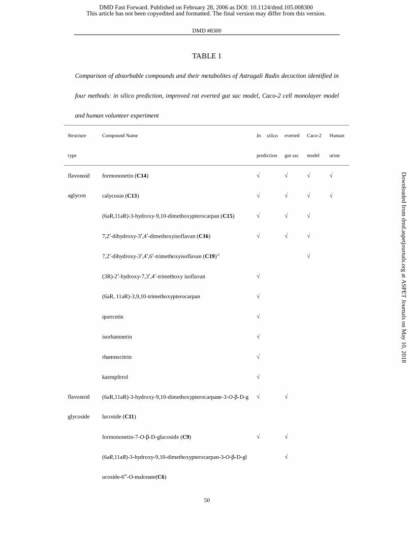
DMD #8300
50
TABLE 1
Comparison of absorbable compounds and their metabolites of Astragali Radix decoction identified in
four methods: in silico prediction, improved rat everted gut sac model, Caco-2 cell monolayer model
and human volunteer experiment
Structure
type
Compound Name In silico
prediction
everted
gut sac
Caco-2
model
Human
urine
formononetin (C14) √ √ √ √
calycosin (C13) √ √ √ √
(6aR,11aR)-3-hydroxy-9,10-dimethoxypterocarpan (C15) √ √ √
7,2′-dihydroxy-3′,4′-dimethoxyisoflavan (C16) √ √ √
7,2′-dihydroxy-3′,4′,6′-trimethoxyisoflavan (C19) a √
(3R)-2′-hydroxy-7,3′,4′-trimethoxy isoflavan √
(6aR, 11aR)-3,9,10-trimethoxypterocarpan √
quercetin √
isorhamnetin √
rhamnocitrin √
flavonoid
aglycon
kaempferol √
(6aR,11aR)-3-hydroxy-9,10-dimethoxypterocarpane-3-O-β-D-g
lucoside (C11)
√ √
formononetin-7-O-β-D-glucoside (C9) √ √
flavonoid
glycoside
(6aR,11aR)-3-hydroxy-9,10-dimethoxypterocarpan-3-O-β-D-gl
ucoside-6″-O-malonate(C6)
√
This article has not been copyedited and formatted. The final version may differ from this version.DMD Fast Forward. Published on February 28, 2006 as DOI: 10.1124/dmd.105.008300
at ASPE
T Journals on M
ay 10, 2018dm
d.aspetjournals.orgD
ownloaded from

DMD #8300
51
7,2′-dihydroxy-3′,4′-dimethoxyisoflavan-7-O-β-D-glucoside-6″
-O-malonate (C7)
√
calycosin-7-O-β-D-glucoside (C8) √
(6aR,11aR)-3-hydroxy-9,10-dimethoxypterocarpan-3-O-β-D-sa
mbubioside (C10)a
√
7,3′-dihydroxy-2′,4′-dimethoxyisoflavan-7-O-β-D-glucoside
(C12)
√
calycosin sulphate (C18) √ √ √
calycosin-7-O-β-D-glucuronide (C1) √ √
formononetin-7-O-β-D-glucuronide (C17) √ √
(6aR,11aR)-3-hydroxy-9,10-dimethoxypterocarpan-3-O-β-D-
glucuronide (C2)
√ √
7,2′-dihydroxy-3′,4′-dimethoxyisoflavan-2′-O-β-D-glucuronide
(C3)
√
7,2′-dihydroxy-3′,4′,5′-trimethoxyisoflavan-7-O-β-D-glucuroni
de (C4)
√
7,2′-dihydroxy-3′,4′-dimethoxyisoflavan-7-O-β-D-glucuronide
(C5)
√ √
7,3′-dihydroxy-2′,4′-dimethoxyisoflavan-7-O-β-D-glucosyl-3′-
O-β-D-glucuronide (C21)
√
flavonoid
metaboliteb
daidzein-7-O-β-D-glucuronide (C20) √
Nitrogen γ-aminobutyric acid √
This article has not been copyedited and formatted. The final version may differ from this version.DMD Fast Forward. Published on February 28, 2006 as DOI: 10.1124/dmd.105.008300
at ASPE
T Journals on M
ay 10, 2018dm
d.aspetjournals.orgD
ownloaded from

DMD #8300
52
3-hydroxy-2-methylpyridine √
nicotinic acid √
HDTICc-1 √
contaiting
compound
HDTICc-2 √
bifendate √
(+)-lariciresinol √
lignanoid
(-)-syringaresinol √
vanillic acid √
isoferulic acid √
caffeic acid √
ferulic acid √
phenolic
acid
p-hydroxycinnamic acid √
coumarin coumarin √
a The compound was detected in Astragali Radix for the first time, and its oral availability was not predicted.
b Drug-like properties of flavonoid metabolites were not calculated, and their oral availabilities were not predicted.
c HDTIC: 4-Hydroxy-5-hydroxymethyl-[1,3]dioxolan-2,6′-spirane-5′,6′,7′,8′-tetrahydro-indolizine-3′-carbaldehyde
This article has not been copyedited and formatted. The final version may differ from this version.DMD Fast Forward. Published on February 28, 2006 as DOI: 10.1124/dmd.105.008300
at ASPE
T Journals on M
ay 10, 2018dm
d.aspetjournals.orgD
ownloaded from

This article has not been copyedited and formatted. The final version may differ from this version.DMD Fast Forward. Published on February 28, 2006 as DOI: 10.1124/dmd.105.008300
at ASPE
T Journals on M
ay 10, 2018dm
d.aspetjournals.orgD
ownloaded from
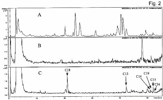
This article has not been copyedited and formatted. The final version may differ from this version.DMD Fast Forward. Published on February 28, 2006 as DOI: 10.1124/dmd.105.008300
at ASPE
T Journals on M
ay 10, 2018dm
d.aspetjournals.orgD
ownloaded from

This article has not been copyedited and formatted. The final version may differ from this version.DMD Fast Forward. Published on February 28, 2006 as DOI: 10.1124/dmd.105.008300
at ASPE
T Journals on M
ay 10, 2018dm
d.aspetjournals.orgD
ownloaded from

This article has not been copyedited and formatted. The final version may differ from this version.DMD Fast Forward. Published on February 28, 2006 as DOI: 10.1124/dmd.105.008300
at ASPE
T Journals on M
ay 10, 2018dm
d.aspetjournals.orgD
ownloaded from