Structural and functional characterization of two alpha-synuclein ...
DOI 10.1007/s00702-005-0378-1
J Neural Transm (2005) 112: 1613–1624
Absence of a-synuclein mRNA expression in normal
and multiple system atrophy oligodendroglia
D. W. Miller1, J. M. Johnson2, S. M. Solano2,Z. R. Hollingsworth2, D. G. Standaert2, and A. B. Young2
1 Cell Biology and Gene Expression Section, Laboratory of Neurogenetics,NIA, Bethesda, MD, and
2 Mass General Institute for Neurodegenerative Disease, Charlestown, MA, USA
Received April 19, 2005; accepted September 13, 2005
Summary. a-Synuclein is a major constituent of glial cytoplasmic inclusions(GCIs), which are pathognomic for multiple system atrophy (MSA). We havepreviously demonstrated that in normal human brain, a-synuclein mRNA has arestricted pattern of neuronal expression and no apparent glial expression. Thecurrent study used double-label in situ hybridization to determine if a-synucleinmRNA is expressed by oligodendroglia of MSA cases. Analysis of MSA braintissue revealed depletion of regional signal for this transcript in many brainareas due to extensive neurodegeneration. Cellular analysis of oligodendrogliain crus cerebri, a GCI-rich region ventral to substantia nigra, revealed an ab-sence of a-synuclein mRNA signal in control and MSA cases. However, anabundance of this transcript was detected in melanin-containing neurons ofsubstantia nigra. Therefore, oligodendroglia do not express a-synuclein mRNAin control and MSA cases suggesting that involvement of a-synuclein in GCIpathology of MSA is due to its ectopic presence in oligodendroglia.
Keywords: Glial cytoplasmic inclusion, in situ hybridization, synucleinopathy.
Introduction
Multiple system atrophy (MSA) is a neurodegenerative disease with variableclinical presentations, including parkinsonism, cerebellar ataxia and autonomicdysfunction (Wenning et al., 1997). Compared to Parkinson’s disease (PD), MSAis not as responsive to dopamine replacement therapy, which suggests thatthe parkinsonism in MSA is due to more widespread degeneration than merelydopaminergic neurons of substantia nigra and is associated with significantpathology in the basal ganglia. MSA is associated with neuronal loss in cau-date, putamen, substantia nigra, pontine nuclei, inferior olives, and cerebellum.The distribution of pathology in brain is variable and is reflected by the pre-dominant clinical phenotype, with the major clinical forms being associated

with predominant pathology in the nigrostriatal system (MSA-P) or the olivo-pontocerebellar system (MSA-C) (Gilman et al., 1999).
The unifying histopathologic hallmark of MSA is the glial cytoplasmicinclusion (GCI), which is a round or crescent-shaped inclusion in the cytoplasmof oligodendroglia in white matter of affected brain regions (Papp et al., 1989;Lantos, 1998). The most sensitive and specific method for detecting GCIsis immunostaining for a-synuclein (Arima et al., 1998; Spillantini et al.,1998; Wakabayashi et al., 1998), a small soluble protein that is more abundantin neurons than glia (Irizarry et al., 1996; Iwai et al., 1995; Maroteaux et al.,1988; Solano et al., 2000). While a-synuclein is an abundant component ofGCIs, it is unknown whether this protein actually originates in oligodendroglia.
Overexpression of a-synuclein is a common theory for disease pathogenesisin synucleinopathies. Genetic studies have indicated that SNCA gene multipli-cation (a-synuclein overexpression) can cause PD (Singleton et al., 2003; Farreret al., 2004), but such a direct relationship in MSA has not been established.Multiplication of the SNCA gene results in an overexpression of a-synucleintranscript in brain and a-synuclein protein in blood, while in brain the over-abundance of a-synuclein protein drives its aggregation (Miller et al., 2004b).GCI-like pathology in addition to Lewy body pathology has been reported inSNCA-triplication cases (Gwinn-Hardy et al., 2000; Miller et al., 2004a). More-over, glial inclusions can be caused by a-synuclein overexpression via oligo-dendroglial-specific promoters in transgenic mice (Kahle et al., 2002; Yazawaet al., 2005). Thus, an overabundance of a-synuclein in oligodendroglia cancause GCI formation. Therefore, we examined the possibility that overexpres-sion of a-synuclein mRNA may occur in oligodendroglia of MSA cases.
Methods
Human brain tissue
Fresh-frozen blocks of human brain tissue were obtained from Harvard Brain Tissue ResourceCenter, the Massachusetts General Hospital Alzheimer’s Disease Resource Center (MGH-ADRC),and the Michigan-ADRC. MSA cases were characterized as either MSA-P (n¼ 5, age range62–72) or MSA-C (n¼ 5, age range 46–77) based on clinical symptoms, the pathologic profile ofneuronal cell loss, and the distribution of GCIs, which was confirmed by immunohistochemicalstaining for a-synuclein-containing GCIs (see below). Control cases (n¼ 10, age range 60–82)had no history of neurological disease and all tissue had a post-mortem interval less than 24hours. Frozen sections (12mm) from multiple regions were cut at �20�C, mounted on slides, andstored at �70�C.
Immunohistochemistry
Frozen, slide-mounted sections were thawed at room temperature and fixed in 4% paraformalde-hyde for 10 min. After rinsing in 0.1 M phosphate-buffered saline (PBS), sections were treatedfor 60 min with blocking solution (3% normal goat serum, 0.3% Triton X-100, and 3% H2O2 inPBS). After rinsing with PBS (3�5 min), the sections were incubated with primary antibody(H3C, 1:5000) overnight at 4�C. H3C is a mouse monoclonal antibody that recognizes theC-terminus of human a-synuclein (generously provided by D. Clayton and J. George, Univ. ofIllinois). Excess primary antibody was rinsed from the sections with PBS (3�5 min) after whichthey were incubated in biotinylated goat-anti-mouse secondary antibody (1:500; Jackson Immu-noResearch Inc.) for 60 min. Slides were rinsed with PBS (3�5 min) and an amplification stepwas conducted by incubating slides in ABC mixture (ABC Vectastain Elite, Vector Laboratories
1614 D. W. Miller et al.
Inc., Burlingame, CA) for 60 min followed by thorough rinsing with PBS (3�10 min). Immu-noreactivity was visualized by exposing slides to PBS containing 0.5 mg=ml diaminobenzideneand 0.03% H2O2 for 4–8 min. Slides were rinsed, counterstained with hematoxylin, and thenexamined via light microscopy to assess a-synuclein immunoreactive GCIs. Negative controlsshowed a negligible level of background stain. Photographs were taken at 40� magnification.
In situ hybridization
a-Synuclein mRNA expression was analyzed by using in situ hybridization with a radiolabeledcRNA probe for human a-synuclein as previously described (Solano et al., 2000). The transcriptfor proteolipid protein (PLP), an abundant protein in myelin, was also examined using a radio-labeled cRNA probe generated against the open reading frame of PLP. In situ hybridization wascarried out using a standard protocol of this laboratory (Kerner et al., 1998). Double-label in situhybridization (DL-ISH) was also conducted using digoxigenin-labeled cRNA probe for PLP(dig-PLP) along with radiolabeled riboprobe for a-synuclein. cRNA hybridization to tissue sectionswas conducted at 50�C for 4 hours. Sections were processed through stringent post-hybridizationwashes that included 0.1 M sodium saline citrate (SSC, 70�C) and RNAse A (37�C). The sectionswere then dehydrated in ethanol and air-dried prior to film exposure.
Film autoradiograms were prepared by apposing the radiolabeled slides to Hyperfilm b-Max(Amersham Co., Sweden) for 3 days (PLP) or 18 days (a-synuclein). Following film exposure,slides were dipped in Ilford K5 autoradiographic emulsion (Polysciences Inc., Warrington, PA;diluted 1:1 with distilled-deionized water), dried overnight, stored at 4�C, and developed after 1week (PLP) and 6 weeks (a-synuclein).
Previous DL-ISH studies in our laboratory have demonstrated that co-hybridization ofdigoxigenin-labeled cRNA probes along with radiolabeled cRNA probes does not interfere withthe signal obtained from the latter (Counihan et al., 1998; Kerner et al., 1998). Such was the casein the current study. Hybridization signal for a-synuclein mRNA was quantified in oligodendrog-lia that were identified by the presence of dig-PLP, which appears as purple cytoplasmic stainfollowing alkaline-phosphatase-mediated detection of digoxigenin-tagged residues (Kerner et al.,1998). A total of 20 dig-PLP-positive cells in the interfascicular tracks of crus cerebri wereanalyzed in each of five cases for each disease condition (control, MSA-P, MSA-C). Additionally,a-synuclein mRNA hybridization signal was quantified in neuromelanin-containing neurons ofthe substantia nigra for each case.
Emulsion autoradiographic analysis was performed under bright and dark field optics.Labeled cells were visualized using bright field optics under a 100� water immersion lens(Leitz). Quantitative microscopic analysis of hybridization intensity and distribution was per-formed using the computer-assisted M1 image analysis system (Imaging Research, St. Catharine’s,Ontario, CA). This system was used to outline the soma of each labeled cell and then quantify theoverlying silver grains. The number of silver grains and the size of the soma (mm2) were recordedin order to calculate the number of grains per 1000mm2. Background signal was evaluated byencircling an equal number of similarly sized area of proximal neuropil and quantifying grainswithin that area as a comparison to cell body grain clusters.
Statistical analysis
Statistical analysis was performed on a commercial statistics software package (SuperANOVA;Abacus Concepts, Inc., Berkeley, CA). Cellular mRNA hybridization signal was quantified fromemulsion autoradiograms and analyzed using an ANOVA with repeated measures design followedby Fisher’s PLSD post-hoc comparisons (significance P<0.05).
Results
GCI distribution in MSA brains
Immunohistochemical staining for a-synuclein in MSA brain tissue revealedGCIs in numerous brain regions including caudate, putamen, midbrain, pons,
Absence of a-synuclein mRNA expression in oligodendroglia 1615

1616 D. W. Miller et al.
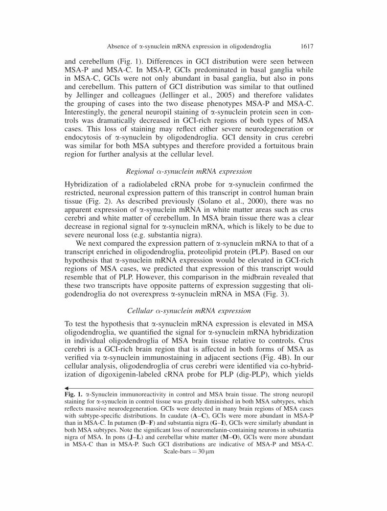
and cerebellum (Fig. 1). Differences in GCI distribution were seen betweenMSA-P and MSA-C. In MSA-P, GCIs predominated in basal ganglia whilein MSA-C, GCIs were not only abundant in basal ganglia, but also in ponsand cerebellum. This pattern of GCI distribution was similar to that outlinedby Jellinger and colleagues (Jellinger et al., 2005) and therefore validatesthe grouping of cases into the two disease phenotypes MSA-P and MSA-C.Interestingly, the general neuropil staining of a-synuclein protein seen in con-trols was dramatically decreased in GCI-rich regions of both types of MSAcases. This loss of staining may reflect either severe neurodegeneration orendocytosis of a-synuclein by oligodendroglia. GCI density in crus cerebriwas similar for both MSA subtypes and therefore provided a fortuitous brainregion for further analysis at the cellular level.
Regional �-synuclein mRNA expression
Hybridization of a radiolabeled cRNA probe for a-synuclein confirmed therestricted, neuronal expression pattern of this transcript in control human braintissue (Fig. 2). As described previously (Solano et al., 2000), there was noapparent expression of a-synuclein mRNA in white matter areas such as cruscerebri and white matter of cerebellum. In MSA brain tissue there was a cleardecrease in regional signal for a-synuclein mRNA, which is likely to be due tosevere neuronal loss (e.g. substantia nigra).
We next compared the expression pattern of a-synuclein mRNA to that of atranscript enriched in oligodendroglia, proteolipid protein (PLP). Based on ourhypothesis that a-synuclein mRNA expression would be elevated in GCI-richregions of MSA cases, we predicted that expression of this transcript wouldresemble that of PLP. However, this comparison in the midbrain revealed thatthese two transcripts have opposite patterns of expression suggesting that oli-godendroglia do not overexpress a-synuclein mRNA in MSA (Fig. 3).
Cellular �-synuclein mRNA expression
To test the hypothesis that a-synuclein mRNA expression is elevated in MSAoligodendroglia, we quantified the signal for a-synuclein mRNA hybridizationin individual oligodendroglia of MSA brain tissue relative to controls. Cruscerebri is a GCI-rich brain region that is affected in both forms of MSA asverified via a-synuclein immunostaining in adjacent sections (Fig. 4B). In ourcellular analysis, oligodendroglia of crus cerebri were identified via co-hybrid-ization of digoxigenin-labeled cRNA probe for PLP (dig-PLP), which yields
1Fig. 1. a-Synuclein immunoreactivity in control and MSA brain tissue. The strong neuropilstaining for a-synuclein in control tissue was greatly diminished in both MSA subtypes, whichreflects massive neurodegeneration. GCIs were detected in many brain regions of MSA caseswith subtype-specific distributions. In caudate (A–C), GCIs were more abundant in MSA-Pthan in MSA-C. In putamen (D–F) and substantia nigra (G–I), GCIs were similarly abundant inboth MSA subtypes. Note the significant loss of neuromelanin-containing neurons in substantianigra of MSA. In pons (J–L) and cerebellar white matter (M–O), GCIs were more abundantin MSA-C than in MSA-P. Such GCI distributions are indicative of MSA-P and MSA-C.
Scale-bars¼ 30 mm
Absence of a-synuclein mRNA expression in oligodendroglia 1617

1618 D. W. Miller et al.

Fig. 3. Relative mRNA expression for a-synuclein (A–C) and proteolipid protein (PLP; D–F)in human midbrain. Oligodendroglia contain an abundance of PLP and therefore the transcriptfor this protein serves as a useful marker of these cells. Opposite patterns of a-synuclein andPLP mRNA expression are apparent. Note the robust signal for PLP mRNA and the negligible
signal for a-synuclein mRNA throughout white matter, particularly in crus cerebri (cc)
1Fig. 2. Film autoradiograms of a-synuclein mRNA distribution in control and MSA braintissue. In sections of caudate the specificity of the antisense cRNA probe (A) is revealed bycomparison with the negligible signal for the sense version of this probe (B). The hybridizationsignal for a-synuclein mRNA occurs in a restricted, neuronal pattern in control cases. However,much of this signal is lost in both subtypes of MSA due to extensive neurodegeneration in areassuch as putamen (G, H) and substantia nigra (K, L). Cell loss in basis pontis also underliesdecreased a-synuclein mRNA signal in MSA-C (P), but not in MSA-P (O). The expression ofa-synuclein mRNA is relatively unaltered in the caudate of either subtype of MSA compared tocontrol (C, D). Abbreviations: basis pontis (bp), caudate (C), crus cerebri (cc), cerebellar white
matter (cwm), external globus pallidus (GPe), putamen (P), substantia nigra (SN)
Absence of a-synuclein mRNA expression in oligodendroglia 1619

a purple chromagen (Fig. 4A). The pattern of a-synuclein hybridization signalobserved via single-label in situ hybridization (Solano et al., 2000) was un-altered by co-hybridization of the dig-PLP riboprobe. The cellular signal fora-synuclein mRNA hybridization in dig-PLP-stained oligodendroglia was not
Fig. 4. Quantitative analysis of a-synuclein mRNA hybridization signal in oligodendroglia ofMSA and control cases. A Oligodendroglia of crus cerebri were identified via dig-PLP labeling(purple chromagen; inset, 100�). Neuropil was sampled from proximal areas vacant of cells.Scale-bar¼ 100 mm. B Crus cerebri is a GCI-rich brain region in MSA cases as revealed bya-synuclein immunostaining in an adjacent section. Scale-bar¼ 50 mm. C Quantification ofoverlying silver grains, reflecting hybridization of isotopic cRNA probe for a-synuclein, re-vealed no significant differences between a-synuclein mRNA expression in dig-PLP-labeledoligodendroglia and neuropil of MSA-P, MSA-C, and control cases. This demonstrates thatoligodendroglia do not express a-synuclein mRNA in either MSA or normal adult humanbrain. The robust expression of a-synuclein mRNA in nigral dopamine neurons serves ascomparative reference. Bars represent the mean density of autoradiographic grains overlaying
each cell � SEM
1620 D. W. Miller et al.

significantly different from background signal quantified in proximal neuropil,which suggests that oligodendroglia express a negligible, if any, level ofa-synuclein mRNA. This result was the same in controls and both subtypesof MSA (Fig. 4C). Moreover, the level of a-synuclein mRNA signal was nearly10-fold greater in neuromelanin-containing dopamine neurons of substantianigra than in oligodendroglia and neuropil.
Discussion
Our analysis of a-synuclein mRNA in MSA brain tissue revealed that regionalsignal for this transcript was always depleted in GCI-rich brain areas. Similarly,a-synuclein protein staining in neuropil of these brain areas was greatlydecreased. These two observations are likely to be attributed to massive neu-rodegeneration. Immunostaining for a-synuclein revealed GCIs distributedthroughout the brain in a pattern typical of MSA pathology in which basalganglia regions were primarily affected in MSA-P, and cerebellum and ponswere primarily affected in MSA-C. Each MSA case also had an abundance ofGCIs present in crus cerebri, regardless of disease subtype. Indeed, a gradingscale for MSA pathology indicates that crus cerebri is typically affected in bothMSA-P and MSA-C (Jellinger et al., 2005). Cellular analysis of a-synucleinmRNA expression in oligodendroglia of crus cerebri, a GCI-rich brain region,revealed that these cells do not express a-synuclein mRNA in either control orMSA brain tissue.
Our study in human brain indicates an absence of a-synuclein mRNAexpression in oligodendroglia. This result concurs with the lack of a-synucleinmRNA expression found in oligodendroglia of normal mice (Yazawa et al.,2005). However, a-synuclein has been reported to be transiently expressedby cultured rat oligodendrocytes (Richter-Landsberg et al., 2000) and low levelsof the protein have been detected in glia of proteinase K-treated sections ofnormal human brain (Mori et al., 2002). It is possible that a very low level ofa-synuclein mRNA expression may be beyond the detection limits of in situhybridization. Moreover, subsets of oligodendroglia may differentially expressa-synuclein in MSA, even if the overall average expression is undetectable.Nonetheless, the contrast between high levels of expression in nigral dopamineneurons relative to an absence of expression in oligodendroglia demonstratesthat an overexpression of a-synuclein mRNA does not occur in this disease.
The lack of elevated a-synuclein mRNA in MSA reported here and byothers (Ozawa et al., 2001) suggests that MSA pathogenesis is not mediatedby a-synuclein overexpression. An alternative mechanism may involve alteredinterplay between neurons and oligodendroglia in which neuronal a-synucleinmay be translocated to oligodendroglia. Such a phenomenon may not be uniqueto a-synuclein since other predominantly neuronal proteins have also beenfound in GCIs (Honjyo et al., 2001; Nakamura et al., 1998). a-Synuclein ispresent in human cerebrospinal fluid suggesting that it may be released fromneurons (Borghi et al., 2000; El-Agnaf et al., 2003). Indeed, some cell lines arecapable of Rab5-dependent endocytosis of a-synuclein from media (Sung et al.,2001). Whether mature oligodendroglia have this capability in vivo is unknown,
Absence of a-synuclein mRNA expression in oligodendroglia 1621

but pathologic oligodendroglia of MSA do aberrantly express the endocytosisregulatory proteins Rab5 and Rabaptin-5 (Nakamura et al., 2000). Once inoligodendroglia, a-synuclein may be improperly catabolized due to an absenceof cellular machinery found in neurons. Hyperphosphorylated a-synuclein ispresent in GCIs (Piao et al., 2001; Fujiwara et al., 2002) and in GCI-likeinclusions of transgenic mice that overexpress this protein specifically in oli-godendroglia (Kahle et al., 2002). Therefore, protein kinase hyperactivity maycontribute to the pathogenesis of MSA.
In summary, our results indicate that oligodendroglia do not expressa-synuclein mRNA in either control or MSA cases. This suggests that MSApathogenesis is not mediated by an overexpression of a-synuclein mRNA inoligodendroglia. The involvement of a-synuclein in MSA is likely to involvemisprocessing of a-synuclein protein rather than aberrant mRNA overexpress-sion in oligodendrolglia. a-Synuclein found ectopically in oligodendroglia ofMSA may arise from neurons. Future studies will examine the likely role ofneuronal a-synuclein in MSA pathogenesis.
Acknowledgements
Support for this work was received from NINDS (USPHS grant NS038372, ABY & DGS) andfrom the National Parkinson Foundation (DWM). This work could not be possible withoutgenerous donations of brain tissue from the following brain banks: Harvard Brain Tissue ResourceCenter (supported in part by PHS grant MH068855), MGH-ADRC, Michigan-ADRC. We thankJ. P. Vonsattel and M. Frosch for their expertise in examination of MSA pathology. We alsoacknowledge M. Schlossmacher and M. Cookson for their helpful discussions about the project.
References
Arima K, Ueda K, Sunohara N, Arakawa K, Hirai S, Nakamura M, Tonozuka-Uehara H, Kawai M(1998) NACP=alpha-synuclein immunoreactivity in fibrillary components of neuronal andoligodendroglial cytoplasmic inclusions in the pontine nuclei in multiple system atrophy.Acta Neuropathol (Berl) 96: 439–444
Borghi R, Marchese R, Negro A, Marinelli L, Forloni G, Zaccheo D, Abbruzzese G, Tabaton M(2000) Full length alpha-synuclein is present in cerebrospinal fluid from Parkinson’s diseaseand normal subjects. Neurosci Lett 287: 65–67
Counihan TJ, Landwehrmeyer B, Standaert DG, Kosinski CM, Scherzer CR, Daggett LP,Velicelebi G, Young AB, Penney JB (1998) Expression of N-methyl-D-aspartate receptorsubunit mRNA in the human brain: mesencephalic dopaminergic neurons. J Comp Neurol390: 91–101
El-Agnaf OM, Salem SA, Paleologou KE, Cooper LJ, Fullwood NJ, Gibson MJ, Curran MD,Court JA, Mann DM, Ikeda S, Cookson MR, Hardy J, Allsop D (2003) Alpha-synucleinimplicated in Parkinson’s disease is present in extracellular biological fluids, includinghuman plasma. Faseb J 17: 1945–1947
Farrer M, Kachergus J, Forno L, Lincoln S, Wang DS, Hulihan M, Maraganore D, Gwinn-HardyK, Wszolek Z, Dickson D, Langston JW (2004) Comparison of kindreds with parkinsonismand alpha-synuclein genomic multiplications. Ann Neurol 55: 174–179
Fujiwara H, Hasegawa M, Dohmae N, Kawashima A, Masliah E, Goldberg MS, Shen J, Takio K,Iwatsubo T (2002) alpha-Synuclein is phosphorylated in synucleinopathy lesions. Nat CellBiol 4: 160–164
Gilman S, Low PA, Quinn N, Albanese A, Ben-Shlomo Y, Fowler CJ, Kaufmann H, Klockgether T,Lang AE, Lantos PL, Litvan I, Mathias CJ, Oliver E, Robertson D, Schatz I, Wenning GK (1999)Consensus statement on the diagnosis of multiple system atrophy. J Neurol Sci 163: 94–98
1622 D. W. Miller et al.

Gwinn-Hardy K, Mehta ND, Farrer M, Maraganore D, Muenter M, Yen SH, Hardy J, DicksonDW (2000) Distinctive neuropathology revealed by alpha-synuclein antibodies in hereditaryparkinsonism and dementia linked to chromosome 4p. Acta Neuropathol (Berl) 99: 663–672
Honjyo Y, Kawamoto Y, Nakamura S, Nakano S, Akiguchi I (2001) P39 immunoreactivity inglial cytoplasmic inclusions in brains with multiple system atrophy. Acta Neuropathol (Berl)101: 190–194
Irizarry MC, Kim TW, McNamara M, Tanzi RE, George JM, Clayton DF, Hyman BT (1996)Characterization of the precursor protein of the non-A beta component of senile plaques(NACP) in the human central nervous system. J Neuropathol Exp Neurol 55: 889–895
Iwai A, Masliah E, Yoshimoto M, Ge N, Flanagan L, de Silva HA, Kittel A, Saitoh T (1995) Theprecursor protein of non-A beta component of Alzheimer’s disease amyloid is a presynapticprotein of the central nervous system. Neuron 14: 467–475
Jellinger KA, Seppi K, Wenning GK (2005) Grading of neuropathology in multiple systematrophy: proposal for a novel scale. Mov Disord 20 [Suppl 12]: S29–S36
Kahle PJ, Neumann M, Ozmen L, Muller V, Jacobsen H, Spooren W, Fuss B, Mallon B, MacklinWB, Fujiwara H, Hasegawa M, Iwatsubo T, Kretzschmar HA, Haass C (2002) Hyper-phosphorylation and insolubility of alpha-synuclein in transgenic mouse oligodendrocytes.EMBO Rep 3: 583–588
Kerner JA, Standaert DG, Penney JB, Young AB, Landwehrmeyer GB (1998) Simultaneousisotopic and nonisotopic in situ hybridization histochemistry with cRNA probes. Brain ResProtocols 3: 22–32
Lantos PL (1998) The definition of multiple system atrophy: a review of recent developments.J Neuropathol Exp Neurol 57: 1099–1111
Maroteaux L, Campanelli JT, Scheller RH (1988) Synuclein: a neuron-specific protein localizedto the nucleus and presynaptic nerve terminal. J Neurosci 8: 2804–2815
Miller DW, Cookson MR, Dickson DW (2004a) Glial cell inclusions and the pathogenesis ofneurodegenerative diseases. Neuron Glia Biol 1: 13–21
Miller DW, Hague SM, Clarimon J, Baptista M, Gwinn-Hardy K, Cookson MR, Singleton AB(2004b) Alpha-synuclein in blood and brain from familial Parkinson disease with SNCAlocus triplication. Neurology 62: 1835–1838
Mori F, Tanji K, Yoshimoto M, Takahashi H, Wakabayashi K (2002) Demonstration of alpha-synuclein immunoreactivity in neuronal and glial cytoplasm in normal human brain tissueusing proteinase K and formic acid pretreatment. Exp Neurol 176: 98–104
Nakamura S, Kawamoto Y, Nakano S, Akiguchi I, Kimura J (1998) Cyclin-dependent kinase 5and mitogen-activated protein kinase in glial cytoplasmic inclusions in multiple systematrophy. J Neuropathol Exp Neurol 57: 690–698
Nakamura S, Kawamoto Y, Nakano S, Akiguchi I (2000) Expression of the endocytosisregulatory proteins Rab5 and Rabaptin-5 in glial cytoplasmic inclusions from brains withmultiple system atrophy. Clin Neuropathol 19: 51–56
Ozawa T, Okuizumi K, Ikeuchi T, Wakabayashi K, Takahashi H, Tsuji S (2001) Analysis of theexpression level of alpha-synuclein mRNA using postmortem brain samples from patholog-ically confirmed cases of multiple system atrophy. Acta Neuropathol (Berl) 102: 188–190
Papp MI, Kahn JE, Lantos PL (1989) Glial cytoplasmic inclusions in the CNS of patients withmultiple system atrophy (striatonigral degeneration, olivopontocerebellar atrophy andShy-Drager syndrome). J Neurol Sci 94: 79–100
Piao YS, Hayashi S, Hasegawa M, Wakabayashi K, Yamada M, Yoshimoto M, Ishikawa A,Iwatsubo T, Takahashi H (2001) Co-localization of alpha-synuclein and phosphorylated tauin neuronal and glial cytoplasmic inclusions in a patient with multiple system atrophy of longduration. Acta Neuropathol (Berl) 101: 285–293
Richter-Landsberg C, Gorath M, Trojanowski JQ, Lee VM (2000) alpha-synuclein is devel-opmentally expressed in cultured rat brain oligodendrocytes. J Neurosci Res 62: 9–14
Singleton AB, Farrer M, Johnson J, Singleton A, Hague S, Kachergus J, Hulihan M, Peuralinna T,Dutra A, Nussbaum R, Lincoln S, Crawley A, Hanson M, Maraganore D, Adler C, CooksonMR, Muenter M, Baptista M, Miller D, Blancato J, Hardy J, Gwinn-Hardy K (2003) alpha-Synuclein locus triplication causes Parkinson’s disease. Science 302: 841
Absence of a-synuclein mRNA expression in oligodendroglia 1623
Solano SM, Miller DW, Augood SJ, Young AB, Penney JB Jr (2000) Expression of alpha-synuclein, parkin, and ubiquitin carboxy-terminal hydrolase L1 mRNA in human brain:genes associated with familial Parkinson’s disease. Ann Neurol 47: 201–210
Spillantini MG, Crowther RA, Jakes R, Cairns NJ, Lantos PL, Goedert M (1998) Filamentousalpha-synuclein inclusions link multiple system atrophy with Parkinson’s disease anddementia with Lewy bodies. Neurosci Lett 251: 205–208
Sung JY, Kim J, Paik SR, Park JH, Ahn YS, Chung KC (2001) Induction of neuronal cell death byRab5A-dependent endocytosis of alpha-synuclein. J Biol Chem 276: 27441–27448
Wakabayashi K, Yoshimoto M, Tsuji S, Takahashi H (1998) Alpha-synuclein immunoreactivityin glial cytoplasmic inclusions in multiple system atrophy. Neurosci Lett 249: 180–182
Wenning GK, Tison F, Ben Shlomo Y, Daniel SE, Quinn NP (1997) Multiple system atrophy: areview of 203 pathologically proven cases. Mov Disord 12: 133–147
Yazawa I, Giasson BI, Sasaki R, Zhang B, Joyce S, Uryu K, Trojanowski JQ, Lee VM (2005)Mouse model of multiple system atrophy alpha-synuclein expression in oligodendrocytescauses glial and neuronal degeneration. Neuron 45: 847–859
Authors’ address: D. W. Miller, PhD, Cell Biology and Gene Expression Section, Laboratoryof Neurogenetics, National Institute on Aging, NIH, Bldg 35, Rm 1A-1002, MSC 3707, 35Convent Drive, Bethesda, MD 20892-3707, USA, e-mail: [email protected]
D. W. Miller et al.: Absence of a-synuclein mRNA expression in oligodendroglia1624


![Preclinical development of a vaccine against oligomeric alpha-synuclein … · 2017. 11. 15. · gated alpha-synuclein [6–9]. Alpha-synuclein (a-syn) is an abundant protein in the](https://static.fdocuments.in/doc/165x107/5fc07f533588d914ed7a20f9/preclinical-development-of-a-vaccine-against-oligomeric-alpha-synuclein-2017-11.jpg)