ABOS Self-Assessment Examination Library of Questions · ABOS Self-Assessment Examination Library...
104
COA 1 California Orthopaedic Association SAE ABOS Self-Assessment Examination Library of Questions Contents ADULT RECONSTRUCTION 2 FOOT & ANKLE 22 HAND , WRIST AND ELBOW 33 ONCOLOGY 52 PEDIATRICS 57 SPINE 64 SPORTS MEDICINE - LOWER EXTREMITY 68 TRAUMA 90 UPPER EXTREMITY 104
Transcript of ABOS Self-Assessment Examination Library of Questions · ABOS Self-Assessment Examination Library...

COA 1
California Orthopaedic Association SAE
ABOS Self-Assessment Examination Library of Questions
Contents
ADULT RECONSTRUCTION 2
FOOT & ANKLE 22
HAND , WRIST AND ELBOW 33
ONCOLOGY 52
PEDIATRICS 57
SPINE 64
SPORTS MEDICINE - LOWER EXTREMITY 68
TRAUMA 90
UPPER EXTREMITY 104

COA 2
California Orthopaedic Association SAE
Adult Reconstruction
1. Answer _C_ After further consultation with the above patient she asks you what the consequence of component retention and polyethylene liner exchange is to her long term success of infection resolution?
A) Minimal long term consequence. You expect that open debridement, polyethylene liner exchange, and IV antibiotics will be 90% successful.
B) No consequence, if she fails the open debridement, polyethylene liner exchange, and iv antibiotics she can then proceed with a two stage revision with a 90% chance of success
C) Moderate consequence, if she fails the open debridement, polyethylene liner exchange, and iv antibiotics she can then proceed with a two stage revision with a 70% chance of success
D) Significant consequence, if she fails the open debridement, polyethylene liner exchange, and iv antibiotics she can then proceed with a two stage revision with a 50% chance of success
Discussion: The patient in question #62 has a staph aureus infection for an indeterminate amount of time two years after total knee replacement. The most appropriate treatment option is two stage revision. Although open debridement with liner exchange and IV antibiotics may be appropriate in some infections, such as S epidermidis and acute hematogenous infections, it has been less successful in treating S aureus infection (less than 10%). Therefore, two stage revision is most appropriate in this patient with a staph aureus infection for an indeterminate amount of time. Additionally, it has been shown that results of two stage revision after a failed debridement with liner exchange are worse (30 % failure rate) than with immediate two stage revision (10% failure rate). References: Orthopaedic Knowledge Update 4: Hip and knee reconstruction, page 207/8. Glassman, Lachiewicz, Tanzer.
2. Answer __B_ When planning a THA for a patient with DDH and a significant leg length discrepancy how much length can be added to the shortened limb safely?
A) 2 cm
B) 4 cm
C) 6 cm
D) 8 cm
Discussion: When performing THA in patients with significant DDH and concomitant shortening of the limb, the incidence of sciatic nerve palsy is increased with lengthening greater than 4cm
Reference: Orthopaedic Knowledge Update 4: Hip and knee reconstruction, page 233. Glassman, Lachiewicz, Tanzer.
3. Answer _C_ Patella Clunk Syndrome is most frequently associated with:
A) First generation cruciate retaining knee designs
B) Entrapment of hyperplastic tissue along the anterior femoral flange
C) Occurs with active knee extension from 60 degrees to 30 degrees of extension
D) Occurs with active knee flexion from 30 degrees to 60 degrees

COA 3
California Orthopaedic Association SAE
Discussion: Patella clunk syndrome is associated with first generation PS knee designs with entrapment of hyperplastic scar tissue about the superior aspect of the patella in the intercondylar notch during active knee extension. Reference: References: Orthopaedic Knowledge Update 4: Hip and knee reconstruction, page 152. Glassman, Lachiewicz, Tanzer.
4. Answer _D_ The number of total knee replacements in the US:
A) Has stayed the same from 1993-2010
B) Has decreased slightly from 1993-2010
C) Is expected to increase by 25% through 2020
D) Is expected to increase by over 100% by 2020
E) Will not change through 2020 Discussion: The demand for primary TKAs is expected to grow by 110% by 2020. The number of TKAs in the US tripled from 1993-2010. Reference: Kurtz, S. M., Ong, K. L., Lau, E., & Bozic, K. J. (2014). Impact of the Economic Downturn on Total Joint Replacement Demand in the United States. The Journal of Bone & Joint Surgery, 96(8), 624-630
5. Answer _A_ Analysis of TKA patients in a Medicare sample comparing various hospital length of stays found:
A) That the outpatient group had less overall costs than the groups with longer length of stays
B) Shorter hospital stays may be associated with less favorable outcomes
C) Not all care centers may be able to provide the appropriate support with outpatient discharges
D) Proper screen and surgical timing may affect outcomes for outpatient procedures Discussion: Costs at 2 years were reduced by over $8,000 for the outpatient group and nearly $2000 less compared to the 1-2 day group, and $1100 compared to the 5+ day group. Shorter hospital stays require appropriate services and education to improve outcomes with outpatient discharges. Reference: 1.Lovald, S. T., Ong, K. L., Malkani, A. L., Lau, E. C., Schmier, J. K., Kurtz, S. M., & Manley, M. T. (2013). Complications, mortality, and costs for outpatient and short-stay total knee arthroplasty patients in comparison to standard-stay patients. The Journal of arthroplasty, 29(3), 510-515.
6. Answer _B_ The demographics of primary and revision THA in Medicare patients between 1991-2008:
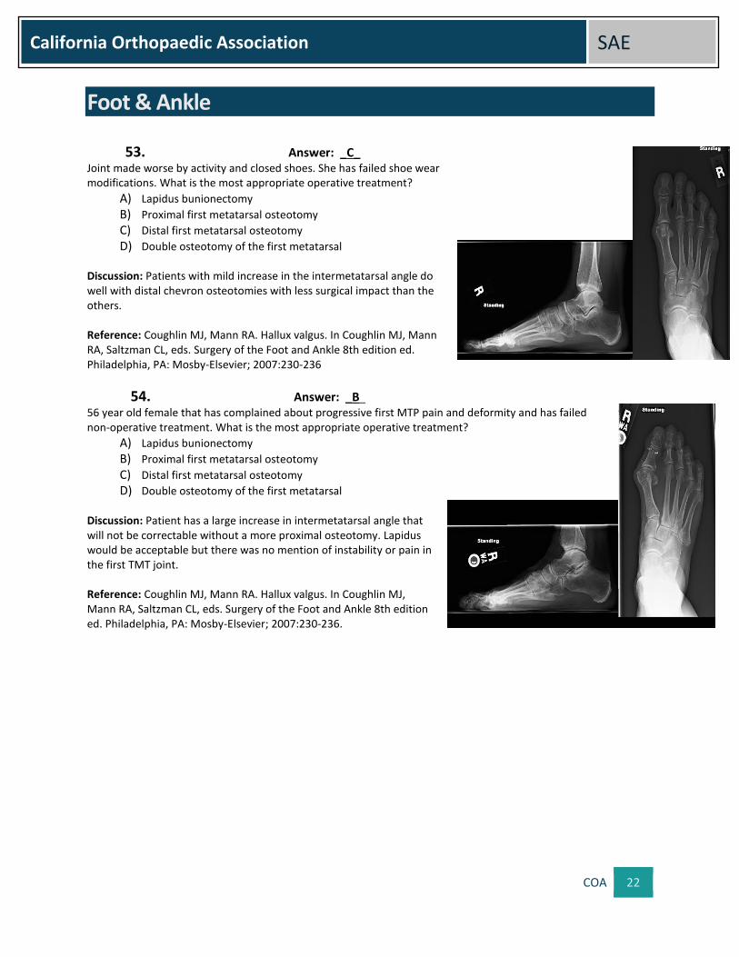
A) The mean length of stay for primary THA increased from 3.7 to 9.1 days
B) The mean length of stay for primary THA decreased from 9.1 to 3.7 days
C) Decreased hospital stay led to fewer readmissions
D) Decreased hospital stay led to fewer discharges to rehabilitation centers Discussion: From 2001-2008, the mean length of stay for primary THA decreased from 9.1 to 3.7 days. The decreased stay corresponded with an increase in the number of patients discharged to rehab centers and to an increase in patient readmissions. Reference: 1.Cram, P., Lu, X., Kaboli, P. J., Vaughan-Sarrazin, M. S., Cai, X., Wolf, B. R., & Li, Y. (2011). Clinical characteristics and outcomes of Medicare patients undergoing total hip arthroplasty, 1991-2008. Jama, 305(15), 1560-1567.

COA 4
California Orthopaedic Association SAE
7. Answer _A_ Proposed benefits of multimodal pain management include
A) Reducing individual doses of analgesics
B) Fewer analgesic gaps
C) Minimize side effects of any one medication
Discussion: Multimodal pain management uses multiple analgesics to achieve a synergistic or additive effect. Consequently, it reduces dosing of any individual analgesic, thereby minimizing side effect. Dosing of additional medications also allows fewer analgesic caps with possibly improved functional outcomes and patient satisfaction Reference: Kehlel, et al. Anesth Analg. 1993. White, Curr Opin Investig Drugs 2008. Skinner Am J Orthop 2004
8. Answer _D_ A patient presents with a painful total hip arthroplasty, as shown below. An infection work-up demonstrates an ESR of 20 and a C-reactive protein of 0.8 mg/L. Metal ions demonstrate a serum cobalt level of 21 ppb and a serum chromium level of 6 ppb. What is the source of the patient’s pain?
A) Metal-on-Metal Articulation B) Polyethylene-induced Osteolysis C) Calcar Fracture D) Modular Junction Corrosion E) Stress Shielding
Discussion: Modular junction corrosion has a differential elevation in the serum cobalt ions when compared to the serum chromium ions. References:Cooper, H. J., et al. (2012). "Corrosion at the head-neck taper as a cause for adverse local tissue reactions after total hip arthroplasty." J Bone Joint Surg Am 94(18): 1655-1661. Huber, M., et al. (2009). "Presence of corrosion products and hypersensitivity-associated reactions in periprosthetic tissue after aseptic loosening of total hip replacements with metal bearing surfaces." Acta Biomater 5(1): 172-180. Korovessis, P., et al. (2006). "Metallosis after contemporary metal-on-metal total hip arthroplasty. Five to nine-year follow-up." J Bone Joint Surg Am 88(6): 1183-1191. Park, Y. S., et al. (2005). "Early osteolysis following second-generation metal-on-metal hip replacement." J Bone Joint Surg Am 87(7): 1515-1521.
9. Answer _E_ A patient presents with a painful metal-on-metal total hip arthroplasty as shown below. Exam findings are all normal except for pain localized to the groin with resisted hip flexion. All infectious workup was normal and metal ion levels were within normal limits. CT scan demonstrated a retroverted component. What would be the best diagnostic modality to delineate the reason for failure?

COA 5
California Orthopaedic Association SAE
A) MRI without contrast B) Ultrasound C) Lumbar spine roentgenograms D) MRI with contrast E) Iliopsoas injection
Discussion: Important exam findings include pain with resisted hip flexion which indicates the diagnosis of iliopsoas impingement, especially in the setting of a retroverted acetabular cup or high-profile cup and articulation. Iliopsoas impingment can be confirmed with an interventional radiology-guided iliopsoas injection. Reference: Fabi DW, Levine BR, Paprosky W, Sporer S, DellaValle CV, Klein G, Levine H, Hartzband M. Metal-on-Metal Total Hip Revisions: A Review of Causes, Clinical Outcomes and a High Incidence of Early Failure. Orthopaedics 2012.
10. Answer _B_ What is the incidence of the finding below in current generation ceramic bearings?
A) .5% B) .004% C) .05% D) .4% E) .02%
Discussion: A distinct issue with COC bearings is the incidence of liner and/or femoral head fracture. Earlier generation COC bearings and ceramic heads alone had a high incidence of fracture with bearing produced before 1990 demonstrating a rate of 13.4% as states preciously. Newer generation Ceramic heads and liners have fortunately improved upon this complication and now have an extremely low incidence of fracture with a reported incidence of 0.004%. Diagnosis of head and liner fracture is relatively straightforward as this is often noticeable on plain roentgenograms. Reference: Willmann G. Ceramic femoral head retrieval data. Clin Orthop Relat Res. 2000 Oct; (379):22-8.
11. Answer _A_ Unique to ceramic bearings is the incidence of squeaking. Current causes of this phenomenon are not entirely known. However, proposed explanations include:
A) Edge-loading and stripe wear B) Corrosion and edge-loading C) Stripe-wear and corrosion D) Anteverted femoral component and corrosion E) Stripe wear and elevated anteverted femoral component
Discussion: Unique to COC THA is the incidence of clinically audible “squeaking”. This phenomenon has a reported incidence range of 0.7% to 20.9%. Causes of this occurrence is currently unknown however, proposed etiologies include edge-loading, stripe-wear, component malposition and altered fluid mechanics of the bearing surface. References:

COA 6
California Orthopaedic Association SAE
Chevillotte C, Trousdale RT, Chen Q, Guyen O, An KN. The 2009 Frank Stinchfield Award: "Hip squeaking": a biomechanical study of ceramic-on-ceramic bearing surfaces. Clin Orthop Relat Res. 2010 Feb;468(2):345-50. doi: 10.1007/s11999-009-0911-x. Epub 2009 Jun 19. Restrepo C, Parvizi J, Kurtz SM, Sharkey PF, Hozack WJ, Rothman RH. The noisy ceramic hip: is component
malpositioning the cause?J Arthroplasty. 2008 Aug;23(5):643-9. doi: 10.1016/j.arth.2008.04.001.
Sexton SA, Yeung E, Jackson MP, Rajaratnam S, Martell JM, Walter WL, Zicat BA, Walter WK. The role of patient
factors and implant position in squeaking of ceramic-on-ceramic total hip replacements. J Bone Joint Surg Br.
2011 Apr;93(4):439-42. doi: 10.1302/0301-620X.93B4.25707
12. Answer _D_ A 70 year-old female with a history of multiple dislocations in her left hip was revised to a dual-mobility construct. Following her surgery, she initially did very well but then went on to dislocate her hip again while getting out of a car. She went to an outside emergency department where she was treated via a closed reduction and discharged home. She later presented to the office with a new onset of painless crepitus with ambulation in her hip as well as a golf ball sized mobile mass in her buttock. New Xrays from your office visit as well as the advanced imaging you order are shown below. What is the diagnosis?
A) Liner fracture
B) Ceramic femoral head fracture
C) A complete dislocation of the femoral head from the acetabular component
D) An intraprosthetic disassociation Discussion: The left femoral head is eccentrically located within the acetabulum. The MRI demonstrates the dual mobility liner in the soft tissue. When the hip was relocated, the articulating polyethylene liner became disassociated from the femoral head. This is a rare but described complication of this device. It is important to recognize that the eccentrically located femoral head within the cup calls for further work-up with advanced imaging. References: Banka, T. R., Ast, M. P., & Parks, M. L. (2014). Early intraprosthetic dislocation in a revision dual-mobility hip prosthesis. Orthopedics, 37(4), e395–7. doi:10.3928/01477447-20140401-63 Langlois, J., Hage, El, S., & Hamadouche, M. (2014). Intraprosthetic dislocation: a potentially serious complication of dual mobility acetabular cups. Skeletal Radiology, 43(7), 1013–1016. doi:10.1007/s00256-014-1824-7 Philippot, R., Boyer, B., & Farizon, F. (2013). Intraprosthetic dislocation: a specific complication of the dual-mobility system. Clinical Orthopaedics and Related Research, 471(3), 965–970. doi:10.1007/s11999-012-2639-2 (Banka, Ast, & Parks, 2014; Langlois, Hage, & Hamadouche, 2014)
13. Answer _C_
http://www.ncbi.nlm.nih.gov/pubmed?term=Trousdale%20RT%5BAuthor%5D&cauthor=true&cauthor_uid=19543782

COA 7
California Orthopaedic Association SAE
A 68 year-old female comes in following a revision total hip arthroplasty for a history of multiple dislocations. She was revised to a modular dual mobility cup. The patient initially did well, however 1 month after her revision she felt a pop in her left hip and presented to the ED where an X-ray was taken. The emergency room physician tried to relocate the hip multiple times under conscious sedation but was not successful. What is the most accurate diagnosis and what is the next best step in treating the patient?
A) Hip dislocation - Attempt closed reduction under general anesthesia in the operating room B) Femoral head and articulating liner dissociation – Take the patient to
the operating room and re-engagement of the polyethylene liner to the femoral head followed by reduction of the joint
C) Acetabular liner and cup disassociation- Take the patient to the operating room and attempt to reengage the acetabular liner and cup followed by hip joint reduction
D) Intraprosthetic disassociation – Take the patient back to the OR for a planned both component revision
Discussion: Though choice D may inevitably happen, this constructed likely failed due to lack of properly engaging the CoCr liner within the cup. As a both component revision poses significant risk to the patient, it is worthwhile taking the patient back to the OR to inspect the locking mechanism in the cup, assuring that the proper size liner was used, and reimpacting the liner. If the hip is found to be stable, it is reasonable to use the current components. Reference: Philippot, R., Boyer, B., & Farizon, F. (2013). Intraprosthetic dislocation: a specific complication of the dual-mobility system. Clinical Orthopaedics and Related Research, 471(3), 965–970. doi:10.1007/s11999-012-2639-2
14. Answer _A_ When used in the setting of recurrent instability, what is the failure rate from instability from registry data that supports the use of dual mobility devices?
A) 1-5% re-dislocation rate B) 5-10% re-dislocation rate C) 15-25% re-dislocation rate D) 25-50% re-dislocation rate E) >50% re-dislocation rate
Discussion: From the Swedish registry, Hailer et.al. looked at the failure rate of 228 patients when they were revised for instability. They found an overall failure rate after revision to be 8%. However, their re-dislocation rate was only 2% (4 patients). Reference: Hailer, N. P., Weiss, R. J., Stark, A., & Kärrholm, J. (2012). Dual-mobility cups for revision due to instability are associated with a low rate of re-revisions due to dislocation: 228 patients from the Swedish Hip Arthroplasty Register. Acta Orthopaedica, 83(6), 566–571. doi:10.3109/17453674.2012.742395
15. Answer: _A_ A 77-year-old male is in your office to discuss definitive treatment for his left knee. He had staged bilateral TKAs nine years ago. Five years ago he developed an infection in his right knee that was treated with a two-stage resection protocol. After reimplantation, he developed another chronic periprosthetic infection which was treated with a right above knee amputation. Two years ago he developed an infection in his left TKA. A debridement with modular bearing exchange failed to cure the infection. He underwent a two-stage resection-reimplantation protocol 1 year ago. At reimplantation, a medial gastrocnemius flap was placed over the anterior knee and all cultures were negative. He presents now to your office with an open wound with an exposed prosthesis. A photo
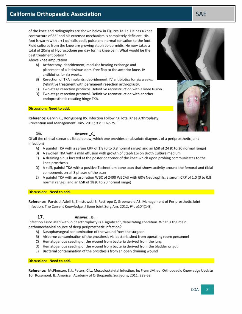
COA 8
California Orthopaedic Association SAE
of the knee and radiographs are shown below in Figures 1a-1c. He has a knee contracture of 85° and his extensor mechanism is completely deficient. His foot is warm with a +1 dorsalis pedis pulse and normal sensation to the foot. Fluid cultures from the knee are growing staph epidermidis. He now takes a total of 20mg of Hydrocodone per day for his knee pain. What would be the best treatment option? Above knee amputation
A) Arthrotomy, debridement, modular bearing exchange and placement of a latissimus dorsi free flap to the anterior knee. IV antibiotics for six weeks.
B) Resection of TKA implants, debridement, IV antibiotics for six weeks. Definitive treatment with permanent resection arthroplasty.
C) Two-stage resection protocol. Definitive reconstruction with a knee fusion. D) Two-stage resection protocol. Definitive reconstruction with another
endoprosthetic rotating hinge TKA. Discussion: Need to add. Reference: Garvin KL, Konigsberg BS. Infection Following Total Knee Arthroplasty: Prevention and Management. JBJS. 2011; 93: 1167-75.
16. Answer: _C_ Of all the clinical scenarios listed below, which one provides an absolute diagnosis of a periprosthetic joint infection?
A) A painful TKA with a serum CRP of 1.8 (0 to 0.8 normal range) and an ESR of 24 (0 to 20 normal range) B) A swollen TKA with a mild effusion with growth of Staph Epi on Broth Culture medium C) A draining sinus located at the posterior corner of the knee which upon probing communicates to the
knee prosthesis D) A stiff, painful TKA with a positive Technetium bone scan that shows activity around the femoral and tibial
components on all 3 phases of the scan E) A painful TKA with an aspiration WBC of 2400 WBC/dl with 60% Neutrophils, a serum CRP of 1.0 (0 to 0.8
normal range), and an ESR of 18 (0 to 20 normal range)
Discussion: Need to add. Reference: Parvisi J, Adeli B, Zmistowski B, Restrepo C, Greenwald AS. Management of Periprosthetic Joint Infection: The Current Knowledge. J Bone Joint Surg Am. 2012; 94: e104(1-9).
17. Answer: _B_ Infection associated with joint arthroplasty is a significant, debilitating condition. What is the main pathomechanical source of deep periprosthetic infection?
A) Nasopharyngeal contamination of the wound from the surgeon B) Airborne contamination of the prosthesis via bacteria shed from operating room personnel C) Hematogenous seeding of the wound from bacteria derived from the lung D) Hematogenous seeding of the wound from bacteria derived from the bladder or gut E) Bacterial contamination of the prosthesis from an open draining wound
Discussion: Need to add. Reference: McPherson, E.J., Peters, C.L., Musculoskeletal Infection, In: Flynn JM, ed. Orthopaedic Knowledge Update 10. Rosemont, IL: American Academy of Orthopaedic Surgeons; 2011: 239-58.

COA 9
California Orthopaedic Association SAE
18. Answer: _E_ Bacteria that have transformed into a biofilm state become significantly more resistant to antibiotics compared to their planktonized state. How much more resistant can they become?
A) Up to 15x more resistant B) Up to 50x more resistant C) Up to 100x more resistant D) Up to 1,000x more resistant E) Up to 15,000x more resistant
Discussion: Need to add. Reference: Howlin RP, Brayford MJ, Webb JS, Cooper JJ, Aiken SS, Stoodley P. Antibiotic-Loaded Synthetic Calcium Sulfate Beads for Prevention of Bacterial Colonization and Biofilm Formation in Periprosthetic Infections. Antimicrobial Agents and Chemotherapy. 2015; 59(1): 111-20.
19. Answer: _B_ Of the treatment regimens listed below, which modality is least likely to reduce the risk of intraoperative colonization of a total joint wound?
A) Vertical laminar airflow system with surrounding plexi-glass shields to the level of the surgeon’s shoulder
B) Personal hooded body exhaust system with inflow above head and exhaust coming out of the bottom of the surgical gown
C) Antibiotic-loaded cement, not exceeding 1 gram per 40 gram bag of PMMA powder
D) Intraoperative ultraviolet light with all personnel appropriately protected with UV-protective gear
E) Perioperative intravenous antibiotics started 1 hour before surgery and continued for 24 hours after surgery
Discussion: Need to add. Reference: McPherson, E.J., Peters, C.L., Musculoskeletal Infection, In: Flynn JM, ed. Orthopaedic Knowledge Update 10. Rosemont, IL: American Academy of Orthopaedic Surgeons; 2011: 239-58.
20. Answer: _A_ A 56-year-old man undergoes a primary TKA. He has no medical problems other than hypertension. The patient is anticoagulated perioperatively with enoxaparin sodium. The surgeon did not use a post-operative drain. The patient has had persistent sero-bloody drainage from the inferior aspect of the knee incision, despite compressive wraps. It is now post-op day 5. What is the appropriate next treatment step?
A) Open exploration, lavage, and change anticoagulation regimen B) Open exploration, lavage, and continue anticoagulation regimen C) Open exploration, lavage, and discontinue anticoagulation regimen D) Discontinue anticoagulation regiment, continue with compressive wraps, start IV antibiotics, and
once drainage has stopped, restart anticoagulation with a different regimen E) Discontinue anticoagulation regimen, continue with compressive wraps, and once drainage has
stopped, restart anticoagulation with a different regimen Discussion: Need to add. Reference: Patel VP, Walsh M, Sehgal B, Preston C, DeWal H, Di Cesare PE. Factors associated with prolonged wound drainage after primary total hip and knee arthroplasty. J Bone Joint Surg Am. 2007; 89(1): 33-8.

COA 10
California Orthopaedic Association SAE
21. Answer: _E_ The responsibility of minimizing infection risk for a patient undergoing a total joint arthroplasty procedure primarily rests with:
A) JACHO B) The hospital administration C) The infection control team within the hospital D) The OR director E) The surgeon
Discussion: Need to add. Reference: McPherson, E.J., Peters, C.L., Musculoskeletal Infection, In: Flynn JM, ed. Orthopaedic Knowledge Update 10. Rosemont, IL: American Academy of Orthopaedic Surgeons; 2011: 239-58.
22. Answer: _B_ You are performing a total shoulder replacement. With an understanding of the intra-operative pathomechanics of bacterial contamination, which habit (intentional or unintentional) listed below would most likely increase the chance of bacterial contamination of the operative wound?
A) Placement of an ioban dressing over the operative extremity B) The continual use of a yankaur suction tip placed into the depths of the surgical wound C) Preventing the opening and closing of the OR door into the main hallway once the total joint
procedure has started D) Pre-positioning of all anticipated implant parts within the operative theatre before the surgical
procedure commences E) The use of body exhaust system by the OR team
Discussion: Need to add. Reference: Givissis P, Karataglis D, Antonarakos P, Symeonidis PD, Christodoulou A. Suction during orthopaedic surgery. How safe is the suction tip? Acta Orthop Belg. 2008; 74: 531-33.
23. Answer: _A_ What main determinate separates acute periprosthetic infection from a chronic periprosthetic infection?
A) The elaboration of a peribacterial biofilm that envelopes the prosthesis, devitalized bone, and soft tissue
B) Gram stain (i.e., gram stain positive vs. gram stain negative organism) C) A bone scan that is positive on all 3 phases D) Wound drainage in a post-operative total joint wound E) The absolute value of the quantitative C-reactive protein
Discussion: Need to add. Reference: McPherson EJ. Adult Reconstruction. In: Miller MD, Thompson SR, Hart JA, eds. Review of Orthopaedics. 6th ed. Philadelphia, PA: Elsevier Saunders; 2012: 353-427.
24. Answer: _D_ Of the below listed options, which would be the best treatment for an established chronic PJI of the hip?
A) A 3-month course of IV antibiotics via a PICC line

COA 11
California Orthopaedic Association SAE
B) Arthrotomy, radical debridement surgery, modular bearing exchange, and placement of dissolvable antibiotic-loaded calcium sulphate beads
C) Implant removal, radical periarticular debridement, placement of dissolvable antibiotic-loaded calcium sulphate beads, and IV antibiotics for 6 weeks
D) Implant removal, radical periarticular debridement, lavage, and placement of a high-dose antibiotic-loaded cement spacer and dissolvable antibiotic-loaded calcium sulphate beads
E) Surgical ablation of the limb, IV antibiotics for 6 weeks, and application of a wound vac to operative site for 7-10 days
Discussion: Need to add. Reference: McPherson EJ. Adult Reconstruction. In: Miller MD, Thompson SR, Hart JA, eds. Review of Orthopaedics. 6th ed. Philadelphia, PA: Elsevier Saunders; 2012: 353-427.
25. Answer: _E_ Based upon the International Consensus Group on Periprosthetic Joint Infection, which statement below is not a recommended technique in the OR?
A) Reduce traffic flow in the OR to an absolute minimum B) Cover all surgical equipment trays with sterile towels (not large drapes) until the procedure
commences C) Change suction tips every 90 minutes during the procedure D) Change gloves every 90 minutes during the procedure E) The use of cloth head caps
Discussion: Need to add. Reference: Jefferson University Hospitals. Consensus document from the International Consensus Group on Periprosthetic Joint Infection. Available at: http://hospitals.jefferson.edu/departments-and-services/orthopedic-surgery/periprosthetic-joint-infection/. Accessibility verified February 10, 2015.
26. Answer: _E_ The advantages of metal-on-metal hip prostheses when compared to metal-on-polyethylene include all of the following EXCEPT:
A) Reduced total wear
B) METAL ON METAL bearings self-polish
C) Large heads increase range of motion
D) MOM bearing’s wear rate is 2% that of METAL ON POLYETHLENE
E) Large heads increase impingement and dislocation rates
Discussion: Need to add.
Reference: OKU-10 Chapter 7 - Pages 76-77
27. Answer: _B_ Compared to METAL ON POLYETHLENE bearing surfaces, METAL ON METAL bearings exhibit a steady state wear rate per year of:
A) 1-2 microns
B) 3-7 microns
C) 8-10 microns
D) 0.25 – 1 micron

COA 12
California Orthopaedic Association SAE
E) 10-12 microns
Discussion: Need to add.
Reference: OKU-10 Chapter 7 - Pages 76-77
28. Answer: _A_ METAL ON METAL bearings exhibit a run in wear rate in the first one million cycles of about:
A) 25 microns
B) 10 angstroms
C) 12 - 17 microns
D) 50 microns
E) 25 angstroms
Discussion: Need to add.
Reference: OKU-10 Chapter 7 - Pages 76-77
29. Answer: _D_ The histology characteristics of an ALVAL lesion are all of the following except:
A) Vasculitis
B) Lymphocytic response
C) Like a type IV delayed hypersensitivity reaction
D) A macrophage response
E) Perivascular infiltrate
Discussion: Need to add.
Reference: OKU-10 Chapter 7 - Page 77
30. Answer: _C_ What is not an identifiable characteristic of a “Pseudotumor?”
A) Joint effusion
B) Local soft tissue reaction
C) Requiring loosening of components
D) METAL ON METAL bearing surfaces
E) Independent of failure or loosening of components
Discussion: Pseudo tumors are thought to occur with greater frequency when there is higher wear concentrations
of metal ions. Component malposition and edge loading are felt to increase wear particles and the possibility of
pseudo tumors. ALVAL pseudo tumor from metal-on-metal wear.
Reference: OKU-10 Chapter 7 - Page 77
31. Answer: _C_ How much greater is the systemic distribution of metal ikons in the body in patients with METAL ON METAL bearing articulations:
A) 10-15 fold
B) 1 to 3 fold

COA 13
California Orthopaedic Association SAE
C) 5 to 10 fold
D) 4 fold
E) Greater than 15 fold
Discussion: Need to add.
Reference: OKU-10 Chapter 7- Page 77
32. Answer: _C_ Has there been any proof the increased systemic metal ions from METAL ON METAL implants increases the risk of malignancy?
A) Some proof
B) Yes, in both sexes
C) No
D) Yes, for young females
E) No for females but yes for males Discussion: Current studies have not proven a direct relationship between metal ion levels and primary
malignancies
Reference: OKU-10 Chapter 7- Page 77
33. Answer: _D_ A patient underwent a metal on metal total hip arthroplasty. A few years later, the patient began experiencing pain and an antalgic limp. The patient ultimately was revised and intraoperative images are shown below. What is a major risk factor associated with metal-on-metal total hip arthroplasty that may have contributed to the findings below:
A) Male gender B) Anteverted acetabular cup position C) Third body wear D) Vertical acetabular cup position E) Age
Discussion: Causes of these local soft tissue ramifications have been thought to be attributed to component malposition (particularly vertical and retroverted), female gender and femoral head size.
References: 1. Grammatopoulos G, Pandit H, Glyn-Jones S, McLardy-Smith P, Gundle R, Whitwell D, Gill HS, Murray DW.
Optimal acetabular orientation for hip resurfacing. J Bone Joint Surg Br. 2010 Aug;92(8):1072-8. 2. Onda K, Nagoya S, Kaya M, Yamashita T. Cup-neck impingement due to the malposition of the implant as a
possible mechanism for metallosis in metal-on-metal total hip arthroplasty. Orthopedics. 2008 Apr;31(4):396.
34. Answer _C_ Work-up of a painful metal-on-metal total hip arthroplasty demonstrated a large effusion on MRI. ESR and CRP were within normal limits. Metal ion levels were found to be highly elevated. Preoperative cell count demonstrated 917 WBCs and 51% PMNs and cultures were negative. Intra-op pathologic specimens demonstrated

COA 14
California Orthopaedic Association SAE
all areas <5 WBCs/HPF. Imaging demonstrated well fixed components with a vertically oriented acetabular cup and appropriately oriented femoral component. What is the likely best course of treatment?
A) Total hip arthroplasty explant and placement of antibiotic spacer B) Femoral head and liner exchange to a ceramic on polyethylene
bearing with retention of acetabular and femoral components C) Acetabular cup and femoral component revision D) Femoral component revision and conversion to a ceramic on
polyethylene bearing with retention of acetabular cup E) Acetabular cup revision and conversion to a ceramic on
polyethylene bearing with retention of femoral component Discussion: Infection needs to be investigated, as a reason for MOM THA failure. However, one needs to be aware that distinction between septic failure and MOM-related failures can, at times, be difficult to differentiate. MOM reactions can mimic infection with elevated inflammatory markers (ESR and CRP), elevated synovial cell counts (need a manual count ordered) and effusions that grossly resemble purulent material. With a stable ingrown cup that is malpositioned and with associated elevated metal ion levels, acetabular cup revision should be considered particularly with a vertical component as this can predict an increase in the risk of polyethylene liner edge-loading and ultimately early failure either via liner fracture or early liner wear from edge-loading. Reference: Hsu AR, Kim JD, Fabi D, Levine BR. Adverse reactions in metal-on-metal total hip arthroplasty: two cases presenting as pseudoseptic acetabular component loosening. Am J Orthop (Belle Mead NJ). 2011 Oct;40 (10):509-513.
35. Answer _E_ Which of the following is considered an “off-label” use for the only modular dual mobility device currently approved by the FDA?
A) Using an “on-growth” cup that accepts the CoCr liner B) Using an “in-growth” cup that accepts the CoCr liner C) Using ceramic head from the same manufacturer D) Using a CoCr head from the same manufacturer E) Using the modular dual mobility bearing with a stem from a different manufacturer.
Discussion: It is considered to be an off-label use of the Stryker Dual Mobility System to use a head from a different manufacture. The reason being is that in the smaller bearing, the 22mm bearing, the head size is actually 22.2mm. Therefore, using this device when a different companies stem is retained, is not recommended. The Trident cup is an HA coated on-growth cup and the Tritanium cup is a porous in-growth cup that both accept the CoCr liner that. You can use either ceramic or CoCr bearings as long as they are from Stryker. References: stryker.com

COA 15
California Orthopaedic Association SAE
36. Answer: _B_ A patient underwent a total hip arthroplasty. A few years later, the patient began experiencing pain and an antalgic limp. The patient ultimately was revised. An intra-operative image is shown below. What process led to the failure of the total hip arthroplasty?
A) Osteolysis B) Corrosion C) Infection D) Malposition E) Implant Fracture
Discussion: Corrosion at the trunnion of modular total hip implants can lead to an adverse local soft tissue reaction like that seen in metal-on-metal articulations. The liberated chromium ions interact with organic phosphate ions forming a chromium (III)
phosphate precipitate on the interface surface. References: Cooper, H. J., et al. (2012). "Corrosion at the head-neck taper as a cause for adverse local tissue reactions after total hip arthroplasty." J Bone Joint Surg Am 94(18): 1655-1661. Kop, A. M. and E. Swarts (2009). "Corrosion of a hip stem with a modular neck taper junction: a retrieval study of 16 cases." J Arthroplasty 24(7): 1019-1023.
37. Answer _D_ A painful dual modular total hip arthroplasty was worked-up and found to have an ESR and C-reactive protein within normal limits, elevated serum metal ion levels, and the intra-operative pathologic specimen that demonstrates:
A) Infection B) Osteolysis C) Fracture D) Aseptic Lymphocyte-dominated Vasculitis-
Associated Lesion E) Non-Hodgkins Lymphoma
Discussion: Corrosion at modular junctions can lead to aseptic lymphocyte-
dominated vasculitis-associated lesion like that seen in metal-on-metal articulations. References: Cooper, H. J., et al. (2012). "Corrosion at the head-neck taper as a cause for adverse local tissue reactions after total hip arthroplasty." J Bone Joint Surg Am 94(18): 1655-1661. De Smet, K. A. (2005). "Belgium experience with metal-on-metal surface arthroplasty." Orthop Clin North Am 36(2): 203-213.
38. Answer _E_ What serum metal ion level indicates a failing total hip arthroplasty?
A) 1 ppb B) 3 ppb

COA 16
California Orthopaedic Association SAE
C) 5 ppb D) 7 ppb E) There is no consensus serum metal ion level for a failing total hip arthroplasty
Discussion: Serum ion concentrations of both cobalt and chromium are used for screening and diagnosis, though research has indicated mixed results regarding formal cutoff levels. Additionally, serum metal ion levels are poor predictors of soft tissue damage and the need for revision surgery. References: Griffin, J. W., et al. (2012). "Management of failed metal-on-metal total hip arthroplasty." World Journal of Orthopedics 3(6): 70-74. Kwon, Y. M., et al. (2014). "Risk stratification algorithm for management of patients with dual modular taper total hip arthroplasty: consensus statement of the American Association of Hip and Knee Surgeons, the American Academy of Orthopaedic Surgeons and the Hip Society." J Arthroplasty 29(11): 2060-2064.
39. Answer _A_ Off-axis loading of the femoral head on the stem trunnion results in _____
A) Double the amount of micomotion with a given amount of force applied to the femoral head B) Cold welding of the head to the trunnion C) Increased head-neck dissociation D) Notching of the stem trunnion leading to trunnion breakage E) Increases the load required to initiate micromotion at the head-neck interface
Discussion: Off-axis loading of the femoral head on the stem trunnion causes the female components to tip with respect to the trunnion and double the amount of micomotion with a given force applied to the femoral head. References: Shareef, N. and D. Levine (1996). "Effect of manufacturing tolerances on the micromotion at the Morse taper interface in modular hip implants using the finite element technique." Biomaterials 17(6): 623-630.
40. Answer: _D___ Polyethylene wear is decreased by all of the following except:
A) Medializing the hip center of rotation B) Increased off-set of the femoral stem C) Intentional cross-linking of the polyethylene D) Thicker polyethylene components E) Ceramic femoral heads
Discussion: Several studies have shown that the “Charnley principle” of reducing the joint reaction force have a
favorable effect on polyethylene wear. Contrary to popular belief, in a wear simulator study of modular
polyethylene components (supporting metal back), the wear of 3mm thick components was less than that of 6mm
thick components.
References: Schmalzried TP. et al: Wear is a function of use, not time. Clin. Orthop. 381:36-46, 2000. Shen FW, Lu Z, McKellop HA. Wear versus thickness and other features of 5-Mrad crosslinked UHMWPE acetabular liners. Clin Orthop Relat Res. 2011 Feb;469(2):395-404.
41. Answer: _E___

COA 17
California Orthopaedic Association SAE
In a study of >1,000 total hips implanted by 11 surgeons, the single factor that most influenced polyethylene wear
was:
A) The type of polyethylene
B) The femoral head material
C) The size of the bearing
D) Cementless fixation of both components
E) The implanting surgeon
Discussion: The surgeon not only determines the position of the components, the biomechanics of the
reconstruction, and the quality of fixation, he also plays a role in patient selection and post-operative activity.
Reference: Schmalzried TP, Dorey FJ, McClung CD, Scott, DL, Zahiri CA, Sanford WM, Kem L, Humphrey, W:
Factors contributing to the variability of short-term radiographic wear rates in total hip replacement. Orthop.
Trans. 22:81-82, and 737-738, 1998.
42. Answer: _E___ In a survey study of surgeon-recommendations for activity following total joint replacement, the majority (>50%)
of surgeons agree that patients can to all of the following except:
A) Walking B) Cycling up inclines C) Doubles tennis D) Golf E) Skiing groomed slopes
Discussion: Surgeon recommendations for activity following total joint replacement are highly variable. In a
survey of the membership of the AAHKS, only 44% recommended unlimited skiing of groomed slopes.
Reference: Swanson EA, Schmalzried TP, Dorey FJ. Activity recommendations after total hip and knee arthroplasty: A survey of the American Association for Hip and Knee Surgeons. Arthroplasty. J Arthroplasty 2009 Sep;24 (6 Suppl):120-6.
43. Answer: __B__ Following joint replacement, the average patient takes about how many gait cycles per year?
A) 1 million
B) 2 million
C) 4 million
D) 5 million
E) Unknown
Discussion: Early pedometer studies indicated an average of about 1 million gait cycles per year. Studies with a
microprocessor worn on the ankle (greater accuracy) have shown that patients average about 2 million gait cycles
per year, although the range is broad.
Reference: Silva M et al.: Average patient walking activity approaches 2 million cycles per year: Pedometers
under-record walking activity. J. Arthroplasty 17:693-697, 2002.
44. Answer: _C___

COA 18
California Orthopaedic Association SAE
All of the following are true except:
A) Walking speed decreases with aging
B) Steps per day decreases with aging
C) The highest wear rates are in the first five years post arthroplasty
D) Polyethylene wear rate decreases with patient aging
E) Ten years post arthroplasty with crosslinked poly, osteolysis is not an issue.
Discussion: The highest wear rates are in the first five years post-arthroplasty for most patients. As patients age,
both walking speed and steps per day decrease. Consequently, polyethylene wear decreases with patient aging,
and osteolysis is rare with 10 years follow-up.
Reference: Battenberg AK, Hopkins JS, Kupiec AD, Schmalzried TP. The 2012 Frank Stinchfield Award: Decreasing
Patient Activity With Aging: Implications for Crosslinked Polyethylene Wear. Clin Orthop Relat Res. 2013
Feb;471(2):386-92.
45. Answer: _D___ What is the most likely cause of massive osteolysis seen in this 47 year old patient?
A) Use of ceramic liner
B) Use of large femoral head
C) Backside wear of the acetabular component
D) Infection
E) Component malpositioning Discussion: This patient has extensive osteolysis which is most likely due to the use of a large femoral head resulting in high volumetric wear. The patient has a cemented all polyethylene acetabular liner and a monolithic femoral component which despite loosening of the acetabular component appear to be well positioned. There is no ceramic liner or any possibility of backside wear as this patient has a one piece polyethylene component. References: Lachiewicz PF, Heckman DS, Soileau ES, Mangla J, Martell JM. Femoral head size and wear of highly cross-linked polyethylene at 5 to 8 years. Clin Orthop Relat Res. 2009 Dec;467(12):3290-6. McKellop HA. Bearing surfaces in total hip replacements: state of the art and future developments. Instr Course Lect. 2001;50:165-79.
46. Answer: __E__ A 67 year old woman is presenting with severe hip pain 2 years following total hip arthroplasty. She has active underlying inflammatory bowel disease and her ESR and CRP is always elevated. Aspiration of the hip was performed revealing neutrophil count of 6000 per ul and neutrophil differential of 82%. What is the most likely cause of her symptoms:
A) Fracture of ceramic liner B) Loosening of acetabular component C) Bone on bone impingement D) Periprosthetic fracture E) Chronic periprosthetic infection
Discussion: The femoral stem is subsided and is loose. The cause of stem loosing is likely to be infection in this patient as the neutrophil count and differential are both very high. In recent years multiple studies, using receiver operating characteristics (ROC) analysis have determined the threshold for neutrophil count and differential for

COA 19
California Orthopaedic Association SAE
chronic and acute periprosthetic joint infections. The threshold for neutrophil count and differential for chronic hip infection is 3200 cells/ul (when the serology is abnormal) and neutrophil differential of 80%. References: Schinsky MF, Della Valle CJ, Sporer SM, Paprosky WG. Perioperative testing for joint infection in patients undergoing revision total hip arthroplasty. J Bone Joint Surg Am. 2008 Sep;90(9):1869-75. Erratum in: J Bone Joint Surg Am. 2010 Mar;92(3):707 Ghanem E, Parvizi J, Burnett S, Sharkey PF, Keshavarzi N, Aggarwal A, Barrack RA. Cell count and differential of aspirated fluid in the diagnosis of infected total knee arthroplasty. J Bone and Joint Surg, 90(8): 1637-43, 2008 Bedair H, Ting N, Jacovides C, Saxena A, Moric M, Parvizi J, Della Valle CJ. The Mark Coventry Award: Diagnosis of Early Postoperative TKA Infection Using Synvoial Fluid Analysis. Clin Orthop Relat Res May 2010 (E-Pub).
47. Answer: _D___ Which of the following is not a predictor of discharge to an extended care facility following primary, elective total knee arthroplasty?
A) Older age (>80 years old) B) Female gender C) Higher ASA score D) Primary vs. Revision TKA E) Medicare Insurance status
Discussion: Looking at nearly 8,000 patient discharges, Bozic et al performed a stepwise linear regression analysis of patients treated at 3 high volume total joint centers and found that 29% of patients were discharged to an ECF. Medicare insurance, older age, female gender, higher ASA score were all associated with a higher risk of discharge to an ECF. Other studies have identified older age, poor pre-operative mobility, the use of gait aids, and the lack of a care giver as predictors of rehabilitation risk. Reference: Predictors of Discharge to an Inpatient Extended Care Facility After Total Hip or Knee Arthroplasty. K Bozic, AWagie, J Naessens, D Berry, H Rubash. J Arthroplasty 21,6: 151, 2006 Predicting Risk of Extended Inpatient Rehabilitation After Hip or Knee Arthroplasty. LB Oldmeadow, H McBurney, V Robertson. J Arthroplasty 18,6: 775, 2003
48. Answer: _D___ Of the options given below, which is most common cause for readmission following primary total knee arthroplasty?
A) Disorders of the urinary tract B) Complications associated with venous thromboembolism C) Length of stay D) Cardiac events E) Pneumonia
Discussion: Several papers have looked at causes for readmission following primary total joint arthroplasty. Vorhies et al found that cardiac events (including congestive heart failure, myocardial infarction and dysrhythmias) made up nearly 50% of all readmissions. Pneumonia was approximately 2.6%, urinary tract disorders (2.1%) and VTE issues were less than 2%. Along with Vorhies et al, Bini et al in another recent paper showed no correlation with a shorter LOS and readmissions looking at large patient population. Reference: Readmission and Length of Stay After Total Hip Arthroplasty in a National Medicare Sample. JS Vorhies, Y Wang, J Herndon, WJ Maloney, JI Huddleston. J Arthroplasty, 26:6, 119 2011

COA 20
California Orthopaedic Association SAE
Does Discharge Disposition after primary total joint arthroplasty affect readmission rates? Bini SA, Fithian DC, Paxton LW. J Arthroplasty 2010; 25:1
49. Answer: _D___ Of the options below, which is the closest to the current average length of stay (LOS) following primary total joint
replacement in the United States?
A) 1 day
B) 2 days
C) 3 days
D) 4 days
E) 5 days
Discussion: 3.7 to 4.0 days is the average LOS in the United States based on recent literature. Length of stay of one
day or less has been reported in some centers.
Reference: The Influence of Procedure Volumes and Standardization of Care on Quality and Efficiency in Total
Joint Replacement Surgery. Bozic KJ, Maselli J, Pekow PS, Lindenauer PK, Vail TP, Auerbach AD. JBJS Am 2010 Nov
17;92(16):2643-52
50. Answer: _D___ Of the options below, which average length of stay (LOS) following primary total joint replacement in the
United States is associated with the highest rate of complications and readmissions?
A) 2 days
B) 3 days
C) 4 days
D) 5 days
Discussion: Higher volume centers tend to have shorter LOS fewer complications, while lower volume centers
tend to have higher LOS and higher complications.
Reference: The Influence of Procedure Volumes and Standardization of Care on Quality and Efficiency in Total
Joint Replacement Surgery. Bozic KJ, Maselli J, Pekow PS, Lindenauer PK, Vail TP, Auerbach AD. JBJS Am 2010 Nov
17;92(16):2643-52
51. Answer: _B___ Of the options below, which is the closest to the current average readmission rate following primary total
joint replacement in the United States?
A) 1-3%
B) 3-5%
C) 5-7%
D) 7-9%
E) >10%
Discussion: The current reported readmission rates for all comers is approximately 4% in multiple papers. The rate
can be much higher or lower in various cohorts and multiple factors can affect readmission, however, the average
rate remains approximately 4%.

COA 21
California Orthopaedic Association SAE
Reference: The Influence of Procedure Volumes and Standardization of Care on Quality and Efficiency in Total
Joint Replacement Surgery. Bozic KJ, Maselli J, Pekow PS, Lindenauer PK, Vail TP, Auerbach AD. JBJS Am 2010 Nov
17;92(16):2643-52
52. Answer: _E___ A 69-year-old patient presents to the emergency room with wound healing problems three weeks following total knee arthroplasty. Blood tests are done with C-reactive protein= 12.6 mg/L and Erythrocyte sedimentation rate=56 mm/hr. Figure 1 shows the appearance of the wound. What is the next step in management?
A) Administration of oral antibiotic and recheck of wound in two weeks
B) Admission for observation and intravenous antibiotic administration C) Irrigation and debridement
D) One or two stage exchange
E) Aspiration of the joint
Discussion: According to the Guidelines issued by the American Academy of Orthopedic Surgeons (AAOS) for diagnosis of periprosthetic joint infection, patients with abnormal serology should undergo aspiration of the joint. The aspirate needs to be sent for cell count, neutrophil percentage, and culture. Recent studies have determined the appropriate threshold for cell count and neutrophil percentage both in the acute setting (as is the case here) and later time points for patients with suspected chronic periprosthetic joint infection. References: Della Valle C, Parvizi J, Bauer TW, Dicesare PE, Evans RP, Segreti J, Spnaghel M, Watters WC, Keith M, Turkleson CM, Wies JL, Hitchcock K. American Academy of Orthopaedic Surgeons Clinical Guideline on: The Diagnosis of periprosthetic joint infections. J Bone Joint Surg 20:93(14): 1355-7, 2011 Bedair H, Ting N, Jacovides C, Saxena A, Moric M, Parvizi J, Della Valle CJ. The Mark Coventry Award: Diagnosis of Early Postoperative TKA Infection Using Synvoial Fluid Analysis. ClinOrthopRelat Res 469(1): 34-40, 2011. Ghanem E, Parvizi J, Burnett S, Sharkey PF, Keshavarzi N, Aggarwal A, Barrack RA. Cell count and differential of aspirated fluid in the diagnosis of infected total knee arthroplasty. J Bone and Joint Surg, 90(8): 1637-43, 2008
COA 22
California Orthopaedic Association SAE
Foot & Ankle
53. Answer: _C_ Joint made worse by activity and closed shoes. She has failed shoe wear modifications. What is the most appropriate operative treatment?
A) Lapidus bunionectomy
B) Proximal first metatarsal osteotomy
C) Distal first metatarsal osteotomy
D) Double osteotomy of the first metatarsal
Discussion: Patients with mild increase in the intermetatarsal angle do well with distal chevron osteotomies with less surgical impact than the others. Reference: Coughlin MJ, Mann RA. Hallux valgus. In Coughlin MJ, Mann RA, Saltzman CL, eds. Surgery of the Foot and Ankle 8th edition ed. Philadelphia, PA: Mosby-Elsevier; 2007:230-236
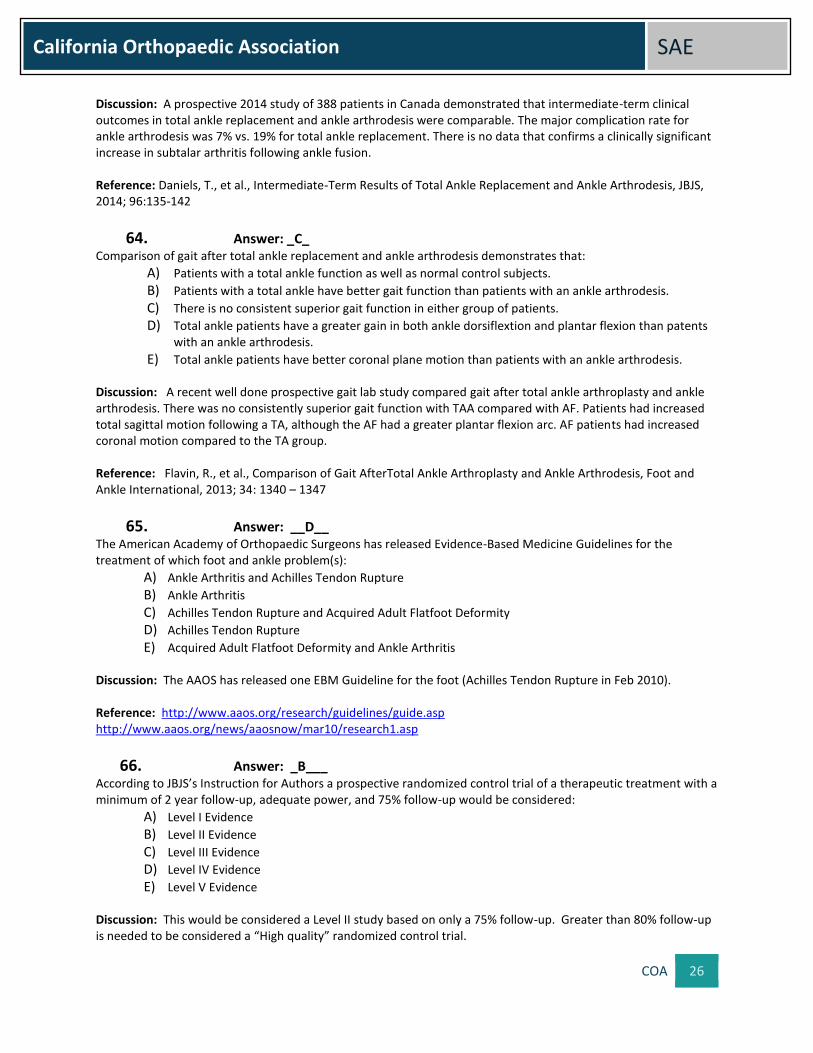
54. Answer: _B_
56 year old female that has complained about progressive first MTP pain and deformity and has failed non-operative treatment. What is the most appropriate operative treatment?
A) Lapidus bunionectomy
B) Proximal first metatarsal osteotomy
C) Distal first metatarsal osteotomy
D) Double osteotomy of the first metatarsal Discussion: Patient has a large increase in intermetatarsal angle that will not be correctable without a more proximal osteotomy. Lapidus would be acceptable but there was no mention of instability or pain in the first TMT joint. Reference: Coughlin MJ, Mann RA. Hallux valgus. In Coughlin MJ, Mann RA, Saltzman CL, eds. Surgery of the Foot and Ankle 8th edition ed. Philadelphia, PA: Mosby-Elsevier; 2007:230-236.

COA 23
California Orthopaedic Association SAE
55. Answer: _A_ 64 year old female with complaints of increasing pain and deformity of her foot with activity and has failed non-operative treatment. On examination she has increased pain at the first tarsal-metatarsal joint with dorsal and plantar motion. What is the most appropriate operative treatment?
A) Lapidus bunionectomy
B) Proximal first metatarsal osteotomy
C) Distal first metatarsal osteotomy
D) Double osteotomy of the first metatarsal Discussion: Patient with a large deformity but also symptomatic first TMT arthrosis. Reference: Coughlin MJ, Mann RA. Hallux valgus. In Coughlin MJ, Mann RA, Saltzman CL, eds. Surgery of the Foot and Ankle 8th edition ed. Philadelphia, PA: Mosby-Elsevier; 2007:230-236
56. Answer: _C_ The majority of hallux valgus deformities in middle aged patients is related to what type of pathologic process?
A) Rheumatologic
B) Traumatic
C) Degenerative
D) Neoplastic Discussion: Hallux valgus is a degenerative instability of the first ray in these patients with an acquired deformity. The medial collateral ligament is elongated and the pathology shows degeneration often including cystic changes. Reference: Coughlin MJ, Mann RA. Hallux valgus. In Coughlin MJ, Mann RA, Saltzman CL, eds. Surgery of the Foot and Ankle 8th edition ed. Philadelphia, PA: Mosby-Elsevier; 2007:230-236
57. Answer: _E__ A 55-year-old man with a history of unknown trauma to his ankle 20 years ago complains of ankle pain. His ankle range of motion is limited by pain from 5 degrees dorsiflexion to 25 degrees plantar flexion with normal hindfoot motion. He has a plantigrade foot. Radiographs demonstrate end-stage ankle arthritis. If he undergoes an ankle fusion for this condition:
A) it will normalize stresses across the adjacent hindfoot joints B) it will decrease the energy of walking compared to a normal ankle C) it will increase the stride length on the affected side after surgery D) it will increase the stride length on the affected side after surgery E) approximately 1/3 of normal sagittal plane motion of the foot will remain
Discusssion: Need to add.
References: Mazur J, Schwartz E, Simon R. - Ankle arthrodesis. Long-term follow-up with gait analysis. J Bone Joint Surg Am. 1979; 61(7):964-975 Thomas R, Daniels T, Parker K. - Gait analysis and functional outcomes following ankle arthrodesis for isolated ankle arthritis J Bone Joint Am Surg 2006; 88(3):526-535
58. Answer: __C__ Which of the following statements about the biomechanics of the ankle joint is NOT correct?

COA 24
California Orthopaedic Association SAE
A) The bony anatomy, ligaments, and joint capsule guide and restrain movement between the talus and the mortise
B) Talus has a continuously changing axis of rotation as it moves from maximum dorsiflexion to maximum plantar flexion relative to the mortise
C) The ankle joint has half the surface contact area of the knee joint yet is exposed to the same maximal joint forces
D) The talus and mortise widen slightly from posterior to anterior E) When the talus is plantar flexed, its narrowest portion sits in the ankle mortise and allows rotatory
movement between the talus and mortise Discussion: The ankle joint has one third the contact surface area compared to the knee joint. The joint is exposed to maximal forces that are 5-7 times body weight compared to the knee joint which experiences up to 3-4 times body weight. References: Kimizula, M., Kurosawa, H., Fukubayashi. 1980. Load bearing pattern of the ankle joint: Contact area and pressure distribution. Archives of Orthopaedic and Trauma Surgery 96(1), pp. 45-9.
Stauffer, R.N., Chao, E.Y.S., Brewster, R.C. 1977. Force and motion analysis of the normal, diseased, and prosthetic ankle joint. Clinical Orthopaedics 127, pp. 189-196. M. Kamran Shahid. A review of current total ankle replacements with reference to the stress distribution in the ankle joint. J.Orthopaedics 2011;8(4)e7
59. Answer: _B___ Which of the following ligaments does NOT contribute to the stability of the syndesmosis?
A) Anterior inferior tibiofibular ligament B) Anterior talofibular ligament C) Posterior inferior tibiofibular ligament D) Interosseous ligament E) Deltoid ligament
Discussion: The syndesmosis is stabilized laterally by the anterior inferior tibiofibular ligament (AIFTL), posterior inferior tibiofibular ligament (PITFL), Interosseous ligament (IOL), interosseousmemberane, and inferior transverse ligament (TL). The syndesmosis is stabilized medially by the Deltoid ligament complex. The anterior talofibular ligament (ATFL) prevents anterior subluxation of the tibiotalar joint and does not impact the syndesmosis. Reference: Hermans JJ, Beumer A, de Jong TA, Kleinrensink GJ. Anatomy of the distal tibiofibularsyndesmosis in adults: a pictorial essay with a multimodality approach. J Anat. 2010 Dec;217(6):633-45. Zalavras C, Thordarson D., Ankle syndesmotic injury, J Am AcadOrthop Surg. 2007 Jun;15(6):330-9
60. Answer: _B__ According to recent literature, approximately what percentage of all ankle sprains in competitive football are high ankle sprains (syndesmosis injuries)?
A) 5% B) 25% C) 50% D) 75% E) 90%
Discussion: According to a recent review of the NCAA injury surveillance system, about 24% of all ankle sprains in college football are syndesmosis injuries (i.e., high ankle sprains). In addition, it is estimated that about 15% of players participating in the NFL combine have a history of syndesmosis injuries. Boytim (5) reported 18 of 98 (18%) acute syndesmotic injuries when looking at members of the

COA 25
California Orthopaedic Association SAE
Minnesota Vikings football team. Reference: Hunt KJ, George E, Harris AH, Dragoo JL., Epidemiology of Syndesmosis Injuries in Intercollegiate Football: Incidence and Risk Factors From National Collegiate Athletic Association Injury Surveillance System Data from 2004-2005 to 2008-2009., Clin J Sport Med. 2013 Jan 21 Boytim MJ, Fischer DA, Neumann L. Syndesmotic ankle sprains.Am. J. Sports Med. 1991; 19:294-8.
61. Answer: __E__ A 62-year-old tennis player ruptured his Achilles tendon 12 months ago. He initially chose non-operative treatment, but continued to have weakness and difficulty ambulating. During surgery extensive debridement there is a 6cm gap between viable tissue ends. Which of the following surgical techniques most likely will provide the best clinical outcome?
A) Primary repair with the foot in maximal plantar flexion followed by a gradual stretching program B) Reconstruction with hamstring autograft C) Achilles repair augmented with transfer of the posterior tibial tendon D) Achilles repair augmented with transfer of the extensor digitorum longus
E) Achilles repair augmented with transfer of the flexor hallucis longus Discussion: The gap is not likely to be repairable primarily. The Flexor HallucisLongus tendon transfer is adjacent to the Achilles, works in phase and has acceptable strength. References: Will RE, Galey SM Outcome of single incision flexor hallucislongus transfer for chronic Achilles tendinopathy. Foot Ankle Int. 2009 Apr;30(4):315-7
62. Answer: _C_ What is the most common location of a talar dome osteochondral lesion?
A) Antero-lateral
B) Postero-medial
C) Superior medial boarder
D) Superior lateral boarder Discussion: Traditional teaching is that the majority of osteochondral lesions of the talus occur either anterolaterally or posteromedially. Several recent studies, however, demonstrate that the majority of talar lesions occur at the superior central medial surface, followed by the superior central lateral. Medial lesions tend to be lager and deeper than lateral lesions. References: Raikin, S; Elias, I, et al: Osteochondral Lesions of the Talus: Localization and Morphologic Data from 424 Patients Using a Novel Anatomical Grid Scheme. Foot Ankle Int. 28(2): 154-161, 2007 Hembree, W; Wittstein, J, et al: Magnetic Resonance Imaging Features of Osteochondral Lesions of the Talus. Foot Ankle Int. 33 (7): 591 – 597, 2012
63. Answer: _C_ Intermediate-Term (5 year) results of total ankle replacement and ankle arthrodesis demonstrate:
A) A superior clinical outcome for total ankle patients
B) A clinically significant increase in subtalar arthritis in ankle fusion patients
C) Comparable clinical outcomes for both groups
D) A higher rate of major complications with ankle fusion
E) None of the above
COA 26
California Orthopaedic Association SAE
Discussion: A prospective 2014 study of 388 patients in Canada demonstrated that intermediate-term clinical outcomes in total ankle replacement and ankle arthrodesis were comparable. The major complication rate for ankle arthrodesis was 7% vs. 19% for total ankle replacement. There is no data that confirms a clinically significant increase in subtalar arthritis following ankle fusion. Reference: Daniels, T., et al., Intermediate-Term Results of Total Ankle Replacement and Ankle Arthrodesis, JBJS, 2014; 96:135-142
64. Answer: _C_ Comparison of gait after total ankle replacement and ankle arthrodesis demonstrates that:
A) Patients with a total ankle function as well as normal control subjects.
B) Patients with a total ankle have better gait function than patients with an ankle arthrodesis.
C) There is no consistent superior gait function in either group of patients.
D) Total ankle patients have a greater gain in both ankle dorsiflextion and plantar flexion than patents with an ankle arthrodesis.
E) Total ankle patients have better coronal plane motion than patients with an ankle arthrodesis. Discussion: A recent well done prospective gait lab study compared gait after total ankle arthroplasty and ankle arthrodesis. There was no consistently superior gait function with TAA compared with AF. Patients had increased total sagittal motion following a TA, although the AF had a greater plantar flexion arc. AF patients had increased coronal motion compared to the TA group. Reference: Flavin, R., et al., Comparison of Gait AfterTotal Ankle Arthroplasty and Ankle Arthrodesis, Foot and Ankle International, 2013; 34: 1340 – 1347
65. Answer: __D__ The American Academy of Orthopaedic Surgeons has released Evidence-Based Medicine Guidelines for the treatment of which foot and ankle problem(s):
A) Ankle Arthritis and Achilles Tendon Rupture
B) Ankle Arthritis
C) Achilles Tendon Rupture and Acquired Adult Flatfoot Deformity
D) Achilles Tendon Rupture
E) Acquired Adult Flatfoot Deformity and Ankle Arthritis Discussion: The AAOS has released one EBM Guideline for the foot (Achilles Tendon Rupture in Feb 2010). Reference: http://www.aaos.org/research/guidelines/guide.asp http://www.aaos.org/news/aaosnow/mar10/research1.asp
66. Answer: _B___ According to JBJS’s Instruction for Authors a prospective randomized control trial of a therapeutic treatment with a minimum of 2 year follow-up, adequate power, and 75% follow-up would be considered:
A) Level I Evidence
B) Level II Evidence
C) Level III Evidence
D) Level IV Evidence
E) Level V Evidence Discussion: This would be considered a Level II study based on only a 75% follow-up. Greater than 80% follow-up is needed to be considered a “High quality” randomized control trial.

COA 27
California Orthopaedic Association SAE
Level I: “High-quality randomized controlled trial with statistically significant difference or no statistically significant difference but narrow confidence intervals.” Level II: Lesser-quality randomized controlled trial (e.g., <80% follow-up, no blinding, or improper randomization) Reference: http://jbjs.org/public/instructionsauthors.aspx
67. Answer: _C___ Which is NOT presently an AAOS “Recommendation Grade?”
A) Strong
B) Moderate
C) Weak
D) Inconclusive
E) Consensus Discussion: The AAOS recently changed their designated Treatment Recommendation Grade from “Weak” to “Limited.” This was done so as not to imply that the treatment in question is “weak”, but rather that there is evidence to support the treatment in question (ex. multiple Level IV studies with consistent findings) albeit evidence that is not robust enough to support a high grade of recommendation. The language associated with a “Limited” Treatment Recommendation Grade is: “Treatment X is an OPTION.” Reference: http://www.aaos.org/research/guidelines/guide.asp
68. Answer: _C___ A 55-year-old man complains of pain in the right ankle. It has been getting worse for more than 10 years. He has 10 degrees dorsiflexion and 40 degrees plantar flexion with pain throughout the range of motion. The patient has failed conservative treatment and wishes to undergo a total ankle arthroplasty instead of an arthrodesis. With regard to total ankle arthroplasty vs fusion, the arthroplasty results in:
A) A lower rate of reoperation B) A higher rate of adjacent joint arthritic changes C) A more normal gait pattern D) Ability to correct large coronal plane deformities (greater than 15 degrees) E) An increase in hindfoot motion
Discussion: Need to add.
References: Coester L., Saltzman C., et. al - Long Term Results following ankle arthrodesis for post traumatic arthritis - J Bone Joint Surg, 2001; 83:219-228 SooHoo N., Zingmond D, Ko, C. - Comparison of Re-Operation rates following ankle arthrodesis and ankle fusion - JBJS AM, 2007; 89:2143-2149 Piriou P, Culpan MP, Mullins M, et al.- Ankle replacement versus Ankle Arthrodesis: A Comparative Gait Analysis Study - Foot and Ankle International 2008; 29(1):3-9. Mazur J, Schwartz E, Simon R. - Ankle arthrodesis. Long-term follow-up with gait analysis. J Bone Joint Surg Am. 1979; 61(7):964-975 Thomas R, Daniels T, Parker K. - Gait analysis and functional outcomes following ankle arthrodesis for isolated ankle arthritis - J Bone Joint Am Surg 2006 88(3):526-535 Guyer A, Richardson G. - Current Concepts Review: Total Ankle Arthroplasty Foot and Ankle International 2008. 29(2): 256-264
COA 28
California Orthopaedic Association SAE
69. Answer: _D___ In this same patient, relative indications for performing a total ankle replacement over an ankle arthrodesis include:
A) Avascular necrosis of the talus B) History of infection C) Greater than 15 degrees varus D) Pre-existing ipsilateral hindfoot fusion E) Significantly increased range of motion postoperatively compared to preoperatively
Discussion: Need to add.
References: Coester L., Saltzman C., et. Al - Long Term Results following ankle arthrodesis for post traumatic arthritis - J Bone Joint Surg, 2001; 83:219-228 Mazur J, Schwartz E, Simon R. - Ankle arthrodesis. Long-term follow-up with gait analysis. J Bone Joint Surg Am. 1979; 61(7):964-975 Thomas R, Daniels T, Parker K. - Gait analysis in function outcomes following ankle arthrodesis for isolated ankle arthritis - J Bone Joint Am Surg 2006; 88(3):526-535 Guyer A, Richardson G. - Current Concepts Review: Total Ankle Arthroplasty Foot and Ankle Int; February 2008; vol. 29, 2: pp. 256-264.
70. Answer: _E__ A 21 year-old collegiate football player suffers an external rotation of his ankle. He has difficulty walking afterward. He has no fractures. Which of the following ankle ligaments is most likely to be the initial structure injured?
A) Calcaneofibular ligament B) Anterior talofibular ligament C) Deep deltoid ligament D) Superficial deltoid ligament E) Anterior inferior tibiofibular ligament
Discussion: High ankle sprains are external rotation injuries of the ankle and syndesmosis. They often occur in skiers, hockey players, and running and cutting athletes, particularly in collision sports. The anterior inferior tibifibular ligament is the initial ligament injured. External rotation of the foot on the leg causes the talus to press against the lateral malleolus. This rotational movement first affects the anterior inferior tibiofibular ligament of the syndesmosis. If external rotation continues, the interosseous membrane and then the posterior tibiofibular ligament will be injured. Clanton’s study supports that the anterior inferior tibiofibular ligament is the most commonly injured ligament in ankle sprains where the mechanism is of injury is external rotation. This occurs regardless of the position of the foot at the time of injury. Pure dorsiflexion causes the interosseus ligaments to tighten and abduction on a neutral ankle can cause interosseus injury when preceded by deltoid injury or medial malleolus fracture. References: Clanton TO, Paul P. Syndesmosis injuries in athletes. Foot Ankle Clin. 2002 Sep;7(3):529-49. Hopkinson WJ, St Pierre P, Ryan JB, Wheeler JH.Syndesmosis sprains of the ankle. Foot Ankle. 1990 Jun; 10(6):325-30.
71. Answer: _D__ Which of the following is true for “item response theory” (i.e., computer adaptive testing) in test administration?

COA 29
California Orthopaedic Association SAE
A) All questions in the instrument question bank are administered to all patients and a summative score
is calculated
B) A small portion of the questions in the overall instrument are administered, selected at random
C) Item response theory typically includes more questions that classical test theory.
D) Each question is selected from the question bank based on the patient’s response to the previous
question
E) Computer adaptive tests are typically scored on a scale of 100 possible points
Discussion: Need to add.
References: Assessment. 2011 Sep;18(3):291-307. The value of item response theory in clinical assessment: a
review. Thomas ML
72. Answer: _C__ In selecting an instrument for patient-reported outcome measurement (PROM), which of the following is NOT an important feature of the instrument?
A) Inclusion of a validated PROM instrument
B) Measurement of the domain(s) of interest to the patient population and disorder
C) Include objective findings like degrees of motion and radiographs
D) Allow meaningful comparison to other series and studies of similar populations
E) Be responsive to detect a change in the condition with time or intervention
Discussion: Need to add.
References: Validation of PROMIS ® Physical Function computerized adaptive tests for orthopaedic foot and ankle outcome research. Hung M, Baumhauer JF, Latt LD, Saltzman CL, SooHoo NF, Hunt KJ; National Orthopaedic Foot & Ankle Outcomes Research Network. Clin Orthop Relat Res. 2013 Nov;471(11):3466-74
73. Answer: __D__ Which clinical test for syndesmosis injury has the fewest false-positive results and smallest inter-observer variance?
A) Squeeze test B) Fibular translation C) Cotton test D) External rotation stress test E) Anterior drawer test
Discussion: The external rotation stress test helps to diagnose high ankle sprains and syndesmotic injuries. The athlete's knee is flexed 90 degrees and the ankle is in neutral. Stabilizing the tibia and fibula with one hand, the examiner externally rotates the ankle with the other. Pain over the syndesmosis indicates a positive test. Beumer et al tested the squeeze, fibula translation, Cotton, and external rotation tests. None of the syndesmotic tests was uniformly positive in chronic syndesmotic injury. The external rotation test had the fewest false-positive results, the fibula translation test the most. The external rotation test had the smallest inter-observer variance. References:: Beumer A, Swierstra BA, Mulder PG: Clinical diagnosis of syndesmotic ankle instability: Evaluation of stress tests behind the curtains. ActaOrthopScand 2002;73:667-669. PMID:12553515 Boytim MJ, Fischer DA, Neumann L: Syndesmotic ankle sprains. Am J Sports Med 1991;19:294-298. PMID:1907807

COA 30
California Orthopaedic Association SAE
74. Answer: __B__ All of the following statements about syndesmotic ankle injuries are true EXCEPT:
A) They are usually caused by external rotation or eversion of the ankle B) The injuries result in tearing of the anterior talofibular ligament C) Purely ligamentous injuries (high ankle sprains) are common in contact sports, and rarely require
surgery D) Syndesmotic injuries are more common on artificial surfaces E) High ankle sprains result in 2-3x more time to return to play compared to inversion ankle injuries
Discussion: The anterior talofibular ligament is injured during an inversion ankle sprain. The syndesmotic ligaments include: the anterior-inferior tibiofibular ligament; the interosseous ligament; the inferior transverse ligament; and the posterior-inferior tibiofibular ligament. Reference: Zalavras C, Thordarson D. Ankle syndesmotic injury. The Journal of the American Academy of Orthopaedic Surgeons. Jun 2007;15(6):330-339.
75. Answer: _B___ An active 35-year-old woman suffers an acute Achilles tendon rupture. Which of the following statements applies to patients undergoing NONOPERATIVE treatment compared to operative treatment?
A) They have a higher risk of skin problems B) They have a higher risk for rerupture C) They are less likely to return to sport D) They have lower patient satisfaction scores E) Their ultimate strength is decreased
Discussion: A higher rerupture rate has been reported in Achilles tendon ruptures treated non-operatively compared to operatively. Skin problems are less common in patients treated no operatively. Return to sports; patient satisfaction; and ultimate strength has been reported as equivalent when compared to operative treatment. References: Weber M, Niemann M, Lanz R, Müller T. Nonoperative treatment of acute rupture of the achilles tendon: results of a new protocol and comparison with operative treatment. Am J Sports Med. 2003 Sep-Oct;31 (5):685-91. Willits K, Amendola A, Bryant D, Mohtadi NG, Giffin JR, Fowler P, Kean CO, Kirkley A. Operative versus nonoperative treatment of acute Achilles tendon ruptures: a multicenter randomized trial using accelerated functional rehabilitation. J Bone Joint Surg Am. 2010 Dec 1;92(17):2767-75.
76. Answer: __E__ What is the biggest advantage of surgical repair of an acute Achilles tendon rupture with early range of motion compared to non-operative treatment with immobilization in a short-leg cast for 6 weeks?
A) Lower rate of infection B) Lower rate of nerve injury C) Better skin cosmesis D) Lower rate of DVT/ VTE E) Lower rate of re-rupture
Discussion: Need to add.

COA 31
California Orthopaedic Association SAE
References: Bhandari M, Guyatt GH, Siddiqui F, et al: Treatment of acute Achilles tendon ruptures: A systematic overview and metaanalysis. ClinOrthopRelat Res 2002;400:190-200 Khan RJ, Fick D, Keogh A, et al. Treatment of acute Achilles tendon ruptures: A meta-analysis of randomized, controlled trials. J Bone Joint Surg Am 2005;87:2202-2210 Willits K, Amendola A, Bryant D, Mohtadi NG, Giffin JR, Fowler P, Kean CO, Kirkley A. Operative versus nonoperative treatment of acute Achilles tendon ruptures: a multicenter randomized trial using accelerated functional rehabilitation. J Bone Joint Surg Am. 2010 Dec 1;92(17):2767-75. Patel A, Ogawa B, Charlton T, Thordarson D Incidence of deep vein thrombosis and pulmonary embolism after Achilles tendon rupture. ClinOrthopRelat Res. 2012 Jan;470(1):270-4.
77. Answer: __E__ Which factor increases the chance of wound complications after Achilles tendon repair?
A) Increased body mass index B) Immediate surgery C) Male gender D) Age over 40 years old E) Tobacco use
Discussion: Need to add. References: Bruggeman NB, Turner NS, Dahm DL, Voll AE, Hoskin TL, Jacofsky DJ, Haidukewych GJ. Wound complications after open Achilles tendon repair: an analysis of risk factors. ClinOrthopRelat Res. 2004 Oct;(427):63-6.
78. Answer: _B___ What type of talar osteochondral lesion is associated with the poorest functional outcome following primary debridement and drilling?
A) Lesions > 0.7cm sq. B) Uncontained lesions C) Medial lesions D) Lateral lesions
Discussion: Debridement and drilling is the primary treatment of choice for almost all osteochondral lesions of the talus that have failed conservative care. Uncontained lesions along the shoulder of the talus, lesions > 1.5cm sq, and large cystic lesions have the poorest functional outcome. Reference: Cuttica, D; Smith, Bret, et al: Osteochondral Lesions of the Talus: Predictors of Clinical Outcome. Foot Ankle Int. 33 (11): 1045 – 1051, 2011
79. Answer: _D___ There are several classification systems of talar osteochondral lesions. Which one is based on plain radiographs?
A) Mintz B) Anderson C) Ferkel D) Berndt and Harty
Discussion: In 1959 Berndt and Harty proposed a classification system for osteochondral lesions based on plain x-ray films. This system continues to be useful for acute injuries. It is inappropriate to use the Bernt and Harty

COA 32
California Orthopaedic Association SAE
classification for chronic osteochonral lesions, 50% of which may not be visualized on plain films. Other systems, based on MRI (Mintz), arthroscopy (Anderson), or CT (Ferkel), are more appropriate. Reference: Berndt, A; Harty, M: Tanschondral Fractures of the Talus. J Bone Joint Surg , 41-A (6), 988 – 1019, 1959
80. Answer: _C___ A 55-year-old man complains of pain in the right ankle. It has been getting worse for more than 10 years. He has 10 degrees dorsiflexion and 40 degrees plantar flexion with pain throughout the range of motion. The patient has failed conservative treatment and wishes to undergo a total ankle arthroplasty instead of an arthrodesis. With regard to total ankle arthroplasty vs fusion, the arthroplasty results in:
A) a lower rate of reoperation B) a higher rate of adjacent joint arthritic changes C) a more normal gait pattern D) ability to correct large coronal plane deformities (greater than 15 degrees)
E) an increase in hindfoot motion
Discussion: Need to add. References: Coester L., Saltzman C., et. al - Long Term Results following ankle arthrodesis for post traumatic arthritis - J Bone Joint Surg, 2001; 83:219-228 SooHoo N., Zingmond D, Ko, C. - Comparison of Re-Operation rates following ankle arthrodesis and ankle fusion - JBJS AM, 2007; 89:2143-2149 Piriou P, Culpan MP, Mullins M, et al.- Ankle replacement versus Ankle Arthrodesis: A Comparative Gait Analysis Study - Foot and Ankle International 2008; 29(1):3-9. Mazur J, Schwartz E, Simon R. - Ankle arthrodesis. Long-term follow-up with gait analysis. J Bone Joint Surg Am. 1979; 61(7):964-975 Thomas R, Daniels T, Parker K. - Gait analysis and functional outcomes following ankle arthrodesis for isolated ankle arthritis - J Bone Joint Am Surg 2006 88(3):526-535 Guyer A, Richardson G. - Current Concepts Review: Total Ankle Arthroplasty Foot and Ankle International 2008. 29(2): 256-264
81. Answer: _D___ There are several classification systems of talar osteochondral lesions. Which one is based on plain radiographs?
A) Mintz B) Anderson C) Ferkel D) Berndt and Harty
Discussion: In 1959 Berndt and Harty proposed a classification system for osteochondral lesions based on plain x-ray films. This system continues to be useful for acute injuries. It is inappropriate to use the Bernt and Harty classification for chronic osteochonral lesions, 50% of which may not be visualized on plain films. Other systems, based on MRI (Mintz), arthroscopy (Anderson), or CT (Ferkel), are more appropriate. Reference: Berndt, A; Harty, M: Tanschondral Fractures of the Talus. J Bone Joint Surg , 41-A (6), 988 – 1019, 1959

COA 33
California Orthopaedic Association SAE
Hand , Wrist and Elbow
82. Answer _D_ The following option would be most appropriate for an 18-year old athlete with a fragmented, unsalvageable proximal pole scaphoid nonunion:
A) Vascularized bone graft
B) Scaphoid excision, 4-corner fusion
C) Proximal row carpectomy
D) Osteochondral rib autograft
E) Silicone prosthesis
Discussion: In the rare case of an unsalvageable, fragmented proximal pole scaphoid nonunion, reconstructive options such as bone grafting procedures would not be possible as the fragmented proximal pole is not a contained defect and not salvageable. Silicone scaphoid prostheses have uniformly failed. Salvage procedures such as a scaphoid excision and 4-corner fusion or proximal row carpectomy would not be ideal in this young patient. Using the osteochondral rib autograft to reconstruct the proximal pole of the unsalvageable scaphoid has been met with some good mid- to long- term results. References: Sandow MJ. Proximal scaphoid costo-osteochondral replacement arthroplasty. J Hand Surg Br. 1998 Apr;23(2):201-8. Yao J, Read B, Hentz VR. “The Fragmented Proximal Pole Scaphoid Nonunion Treated With Rib Autograft, A Case Series and Review of the Literature,” J Hand Surg [Am]. 2013 Nov;38(11):2188-92.
83. Answer _C_ Following the scaphoid, this carpal bone is the most commonly fractured:
A) Trapezium B) Lunate C) Triquetrum D) Capitate E) Pisiform
Discussion: Triquetral fractures are the second most common fractures of the carpus, second only to scaphoid fractures. Triquetral ridge fractures, typically avulsions of either the dorsal intercarpal or dorsal radiocarpal ligaments, are the most common type of triquetral fractures, accounting for up to 93% of these fractures. References: Höcker K, Menschik A. Chip fractures of the triquetrum. Mechanism, classification and results. J Hand Surg Br 1994; 19:584. Marchessault J, Conti M, Baratz ME. Carpal fractures in athletes excluding the scaphoid. Hand Clin 2009; 25:371.
84. Answer _E_ Which of the following methods of fixation for scaphoid waist hypertrophic nonunions in conjunction with the addition of bone graft would be considered LEAST effective?
A) Headless screw fixation B) Plate fixation C) Nitinol staple fixation D) Intramedullary radial strut grafts
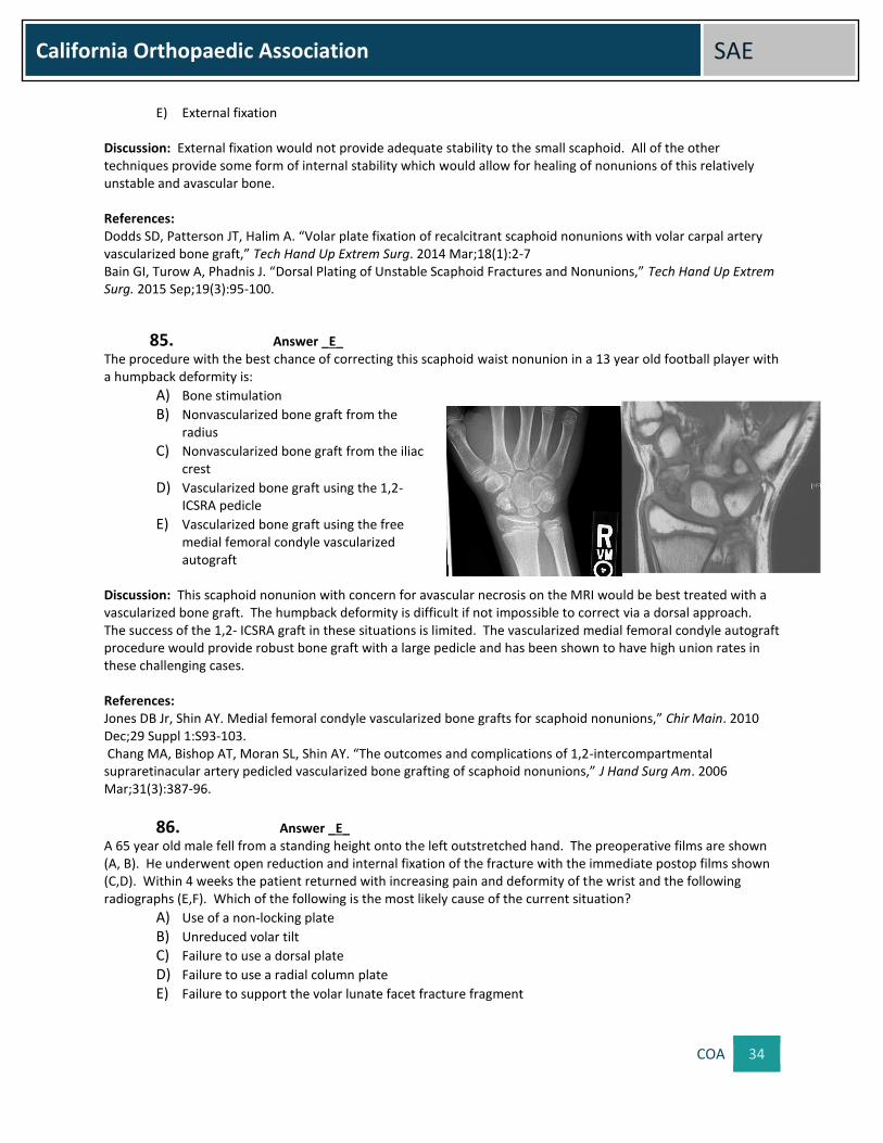
COA 34
California Orthopaedic Association SAE
E) External fixation Discussion: External fixation would not provide adequate stability to the small scaphoid. All of the other techniques provide some form of internal stability which would allow for healing of nonunions of this relatively unstable and avascular bone. References: Dodds SD, Patterson JT, Halim A. “Volar plate fixation of recalcitrant scaphoid nonunions with volar carpal artery vascularized bone graft,” Tech Hand Up Extrem Surg. 2014 Mar;18(1):2-7 Bain GI, Turow A, Phadnis J. “Dorsal Plating of Unstable Scaphoid Fractures and Nonunions,” Tech Hand Up Extrem Surg. 2015 Sep;19(3):95-100.
85. Answer _E_ The procedure with the best chance of correcting this scaphoid waist nonunion in a 13 year old football player with a humpback deformity is:
A) Bone stimulation
B) Nonvascularized bone graft from the radius
C) Nonvascularized bone graft from the iliac crest
D) Vascularized bone graft using the 1,2-ICSRA pedicle
E) Vascularized bone graft using the free medial femoral condyle vascularized autograft
Discussion: This scaphoid nonunion with concern for avascular necrosis on the MRI would be best treated with a vascularized bone graft. The humpback deformity is difficult if not impossible to correct via a dorsal approach. The success of the 1,2- ICSRA graft in these situations is limited. The vascularized medial femoral condyle autograft procedure would provide robust bone graft with a large pedicle and has been shown to have high union rates in these challenging cases.
References: Jones DB Jr, Shin AY. Medial femoral condyle vascularized bone grafts for scaphoid nonunions,” Chir Main. 2010 Dec;29 Suppl 1:S93-103. Chang MA, Bishop AT, Moran SL, Shin AY. “The outcomes and complications of 1,2-intercompartmental supraretinacular artery pedicled vascularized bone grafting of scaphoid nonunions,” J Hand Surg Am. 2006 Mar;31(3):387-96.
86. Answer _E_ A 65 year old male fell from a standing height onto the left outstretched hand. The preoperative films are shown (A, B). He underwent open reduction and internal fixation of the fracture with the immediate postop films shown (C,D). Within 4 weeks the patient returned with increasing pain and deformity of the wrist and the following radiographs (E,F). Which of the following is the most likely cause of the current situation?
A) Use of a non-locking plate
B) Unreduced volar tilt
C) Failure to use a dorsal plate
D) Failure to use a radial column plate
E) Failure to support the volar lunate facet fracture fragment
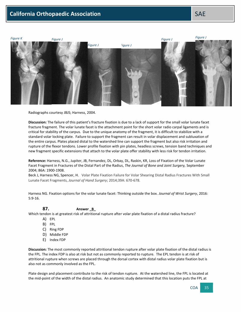
COA 35
California Orthopaedic Association SAE
Radiographs courtesy JBJS; Harness, 2004. Discussion: The failure of this patient’s fracture fixation is due to a lack of support for the small volar lunate facet fracture fragment. The volar lunate facet is the attachment point for the short volar radio carpal ligaments and is critical for stability of the carpus. Due to the unique anatomy of the fragment, it is difficult to stabilize with a standard volar locking plate. Failure to support the fragment can result in volar displacement and subluxation of the entire carpus. Plates placed distal to the watershed line can support the fragment but also risk irritation and rupture of the flexor tendons. Lower profile fixation with pin plates, headless screws, tension band techniques and new fragment specific extensions that attach to the volar plate offer stability with less risk for tendon irritation. Reference: Harness, N.G., Jupiter, JB, Fernandez, DL, Orbay, DL, Raskin, KR, Loss of Fixation of the Volar Lunate Facet Fragment in Fractures of the Distal Part of the Radius, The Journal of Bone and Joint Surgery, September 2004; 86A: 1900-1908. Beck J, Harness NG, Spencer, H. Volar Plate Fixation Failure for Volar Shearing Distal Radius Fractures With Small
Lunate Facet Fragments, Journal of Hand Surgery; 2014;39A: 670-678.
Harness NG. Fixation options for the volar lunate facet: Thinking outside the box. Journal of Wrist Surgery, 2016: 5:9-16.
87. Answer _B_ Which tendon is at greatest risk of attritional rupture after volar plate fixation of a distal radius fracture?
A) EPL
B) FPL
C) Ring FDP
D) Middle FDP
E) Index FDP Discussion: The most commonly reported attritional tendon rupture after volar plate fixation of the distal radius is the FPL. The index FDP is also at risk but not as commonly reported to rupture. The EPL tendon is at risk of attritional rupture when screws are placed through the dorsal cortex with distal radius volar plate fixation but is also not as commonly involved as the FPL. Plate design and placement contribute to the risk of tendon rupture. At the watershed line, the FPL is located at the mid-point of the width of the distal radius. An anatomic study determined that this location puts the FPL at
Figure K Figure J
Figure J Figure J
Figure J Figure J

COA 36
California Orthopaedic Association SAE
risk of rupture. Soong, et al found that placement of a volar plate distal to the watershed line increases the risk of tendon rupture. The most prominent portion of the watershed line is located at the volar ulnar corner. The FPL sulcus is located just radial to the prominence of the volar lunate facet. When attempting fixation of the volar ulnar corner of the distal radius, one must be cautious in placing implants distal to the watershed line to avoid FPL irritation. References: Soong M, Earp B, Bishop G, Leung A, Blazar P, Volar Locking Plate Implant Prominence and Flexor Tendon Rupture, J Bone Joint Surg Am, 2011; 93(4): 328 -335 . Limthongthan R, Bachourd S, Jacoby A, Osterman L. Distal Radius Volar Locking Plate Design and Associated
Vulnerability of the Flexor Pollicis Longus. Journal of Hand Surgery, 2014; 39(5): 852–
88. Answer _E_ A 70-year-old female fell and sustained a tibial plateau fracture and ipsilateral fracture of the distal radius (A, B).
The distal radius fracture was reduced and splinted in the ER and the tibial plateau fracture was treated with ORIF.
Two weeks later, the distal radius fracture displaced due to severe osteoporosis (C, D). The decision is made to
treat the distal radius fracture operatively, but the patient requires full weight bearing through the forearm and
wrist for ambulation with a walker. What is the best option for operative stabilization of the wrist fracture?
A) Volar plate B) Dorsal plate C) External fixator D) Fragment specific fixation E) Dorsal spanning plate
Discussion: One of the advantages and relative indications for dorsal spanning plate fixation of distal radius
fractures is in the poly trauma patient who might require forceful use of the injured upper extremity for
ambulation. In this situation, a spanning plate will allow for immediate weight bearing with crutches. In addition,
the spanning plate provides the advantages of an external fixator without exposed hardware.
Highly comminuted, intra articular fractures in elderly osteoporotic patients can be difficult to manage with
conventional plates and screws. Bridge plating spans the osteoporotic segment and utilizes ligamentotaxis to
maintain reduction.
Figure A Figure b
Figure C
Figure D

COA 37
California Orthopaedic Association SAE
Lauder et al, found that the use of spanning plates for distal radius fractures was safe and led to few complications.
Outcomes were similar to other treatment methods. Wrist motion returned to a normal level except for wrist
flexion, which remained diminished, compared to the uninjured wrist at long-term follow up in their series.
References: Lauder A, Agnew S, Bakri K, Allan C, Hanel D, Huang J; Functional outcomes following bridge plate
fixation for distal radius fractures, Journal of hand surgery, 2015, (40) 8: 1554-1562.
Brogan D, Richard M, Ruch D, Kakar S, Management of severely comminuted distal radius fractures. Journal of
hand surgery; 2015 (40) 9: 1905-1914.
89. Answer _C_ Patients with post-traumatic lack of elbow flexion (less than 100 degrees) can benefit from:
A) Anterior band of MCL
B) Posterior band of MCL
C) Posterior band of MCL and Ulnar nerve transposition
D) Anterior band of MCL and Ulnar nerve transfusion Discussion: The posterior band of MCL gets tight in flexion. If patients have a capsular contracture and cannot flex the elbow beyond 100 degrees, they can have significant improvement in elbow flexion without instability by releasing the posterior band of the MCL Reference: Lindenhovius AL, Jupiter JB. The posttraumatic stiff elbow: a review of the literature. Journal of Hand Surgery Dec 2007
90. Answer _E_ The “column” procedure:
A) Refers to the Medial column of the elbow joint
B) Refers to the Lateral approach to the elbow joint
C) Involves preservation of collateral ligaments
D) Described by Morrey
E) B , C, and D Discussion: Morrey in 1998 with a series of 38 elbows with restricted elbow motion who underwent the release of the elbow contracture using this approach. There were low rates of complication and motion improved in flexion and extension in all patients. The lateral UCL must be preserved in order to avoid instability and all the releases were done via a lateral approach. Reference: Mansat P, Morrey B. The Column Procedure: A Limited Lateral Approach for Extrinsic Contracture of the Elbow. Journal of Bone & Joint Surgery, Nov 1998.
91. Answer _D_ Elbow arthroplasty is considered in stiff elbow in patients
A) Greater than 50% articular wear
B) Young active patients
C) Low demand patients with more advanced ager
D) A and C Discussion: Although there are no specific guidelines for elbow replacement in elbow stiffness, most agree that it should be reserved for low demand patients with greater than 50% articular cartilage loss. Elbow arthroplasty in the setting of elbow fusion or Heterotopic bone formation remains very controversial and is not commonly performed.
COA 38
California Orthopaedic Association SAE
Reference: Reference; Evans P, Nandi S, Maschke S, Hoyen H, Lawton J. Prevention and Treatment of Elbow Stiffness. Journal of Hand Surgery, April 2009
92. Answer _B_ Botulinum Toxin A
A) Has no role in elbow surgery
B) Has been shown effective in ORIF of elbow fractures
C) Should only be used for cosmetic purposes
D) Can help reduce the incidence of HO in the elbow Discussion: Although, the use of Botox is not a standard mode of preventing elbow stiffness, there is some evidence to show its effectiveness in preventive elbow stiffness in patients that undergo ORIF of distal humerus fractures. Reference: Evans P, Nandi S, Maschke S, Hoyen H, Lawton J. Prevention and Treatment of Elbow Stiffness. Journal of Hand Surgery, April 2009
93. Answer: _A_ Which statement is true regarding the use of total elbow arthroplasty (TEA) for the treatment of distal humerus fractures?
A) In osteoporotic, low-demand patients, functional outcomes following TEA are similar or better than ORIF
B) TEA is associated with a lower complication rate as compared to ORIF in treatment of fractures of the distal humerus
C) Osteoporosis is a contraindication to TEA D) Olecranon osteotomy is the preferred approach for implantation of a TEA E) The most common complication following TEA for distal humerus fracture is infection
Discussion: In elderly or low-demand patients with osteoporosis and severe fracture comminution, or in patients with pre-existing inflammatory arthritis, some studies have shown that a total elbow arthroplasty (TEA) may give better functional outcomes than ORIF of distal humeral fractures. In these studies, there was not a significant difference in complication rates between TEA and ORIF; however, the TEA prosthesis does have some risk of loosening with long-term overuse, and a permanent restriction of weight bearing less than 5 pounds is recommended. In cases where TEA is being considered, the surgeon cannot use an olecranon osteotomy approach; the ulnar component of the arthroplasty uses this bone for fixation, and osteotomy would compromise this area. A triceps-sparing approach is preferable, since it provides adequate space for implant insertion while allowing for immediate postoperative mobilization with no restrictions for triceps healing. References: Egol K.A., Tsai P., Vazques O., et al: Comparison of functional outcomes of total elbow arthroplasty vs plate fixation for distal humerus fractures in osteoporotic elbows. Am J Orthop (Belle Mead NJ) 40. (2): 67-71.2011
94. Answer: _A_ The palmar neurovascular advancement flap (Moberg) is most appropriate for reconstruction of which of the following defects?
A) Thumb pulp B) Index finger pulp C) Thumb nail bed D) Thumb dorsum E) Index dorsal middle phalanx

COA 39
California Orthopaedic Association SAE
Discussion: Use of the Moberg flap requires an independent dorsal blood supply for digit viability as seen in the thumb but not predictably in the fingers. The Moberg flap therefore is best used for volar pulp defects in the thumb of up to 1 cm (see Figures 1, 2, and 3). Additional advancement can be achieved by exposing and mobilizing the neurovascular structures. Flexion contractures, a frequent outcome of this mobilization, are better tolerated in the thumb than in the fingers. Index finger pulp loss can be treated with the cross-finger flap. A flap from the first dorsal metacarpal artery can be used to cover dorsal thumb defects, and defects over the index dorsal middle phalanx can be covered using skin grafts or with the extended dorsal metacarpal artery flaps. The Moberg flap does not have sufficient length to cover nailbed defects. References: BaumeisterS,MenkeH,WittemannM,GermannG. Functional outcome after the Moberg advancement
flap in the thumb.J Hand Surg 2002;27(1):105-114. Friedrich JB, Katolik LI, Vedder NB. Soft tissue reconstruction of the hand. J Hand Surg 2009;34(6):1148-1155
95. Answer: _D__ Which of the following has been associated with lower rates of return to play following UCL reconstruction?
A) Ulnar neuritis B) Use of a muscle splitting approach C) Increased internal rotation of the ipsilateral shoulder D) Posteromedial chondromalacia E) Younger age at time of injury
Discussion: The authors retrospectively reviewed 29 of 161 (18%) baseball players who were treated for the combined posteromedial chondromalacia (PMC) and UCL injury. Return to play was Level 1 in 22 patients (76%), Level 2 in four patients (14%), Level 3 in two patients (7%), and Level 4 in one (3%) patient. The data suggest that baseball players with concomitant PMC, may have lower rates of return to of play compared with historical controls.
References: Ulnar collateral ligament reconstruction in throwing athletes: a review of current concepts. AAOS exhibit selection. Jones KJ, Osbahr DC, Schrumpf MA, Dines JS, Altchek DW. J Bone Joint Surg Am. 2012 Apr 18; 94(8):e49 Does posteromedial chondromalacia reduce rate of return to play after ulnar collateral ligament reconstruction? Osbahr DC, Dines JS, Rosenbaum AJ, Nguyen JT, Altchek DW.ClinOrthopRelat Res. 2012 Jun; 470(6):1558-64.
96. Answer: _C_ Which of the following statements is true regarding the treatment of lateral epicondylitis?
A) Corticosteroid injections for lateral epicondylitis are associated with improved long-term patient outcomes
B) Plasma rich platelet (PRP) has a potent anti-inflammatory effect on local tissue C) Non-operative management of lateral epicondylitis will result in successful resolution of symptoms in
90% of patients D) Outcomes following arthroscopic treatment of lateral epicondylitis are superior to open approaches E) Worker’s compensation status has no correlation with outcomes following treatment for lateral
epicondylitis Discussion: Medial and lateral epicondylitis are overuse injuries that respond well to non-operative management. Their etiology is degenerative and related to repetitive overuse and underlying tendinopathy. Nonsteroidal anti-inflammatory drugs and localized corticosteroid injections yield moderate symptomatic relief in short term but do not demonstrate benefit on long-term follow-up. Platelet-rich plasma injections may be advantageous in cases of chronic lateral epicondylitis. If 6 to 12 months of non-operative treatment fails, then surgical intervention can be undertaken.

COA 40
California Orthopaedic Association SAE
Reference: Taylor SA, Hannafin JA. Evaluation and management of elbow tendinopathy. Sports Health. 2012 Sep;4(5):384-93.
97. Answer: _B___ What is the most common complication after total elbow arthroplasty in rheumatoid patients?
A) Aseptic Loosening B) Infection C) Wound related D) Polyethyene and bushing failure E) Periprosthetic fracture
Discussion: Need to add. References: The Elbow and Its Disorders: 4th Edition by Bernard F. Morrey Pp10-439
98. Answer: _B___ Total elbow arthroplasty has the longest survivorship in which cohort of patients?
A) Post-traumatic B) Rheumatoid C) Osteoarthritic D) Congenital deformity
Discussion: Need to add. References: The Elbow and Its Disorders: 4th Edition by Bernard F. Morrey Pp10-439
99. Answer: _C___ What structure is most commonly affected after total elbow arthroplasty?
A) Median nerve B) Radial nerve C) Ulnar nerve D) Biceps tendon E) Radial head
Discussion: Need to add. References: The Elbow and Its Disorders: 4th Edition by Bernard F. Morrey Pp10-439
100. Answer: _A___ What is the most common complication after unlinked total elbow arthroplasty?
A) Instability B) Stiffness C) Polyethylene wear D) Ulnar neuropathy E) Periprosthetic fracture
Discussion: Need to add.

COA 41
California Orthopaedic Association SAE
References: The Elbow and Its Disorders: 4th Edition by Bernard F. Morrey Pp10-439
101. Answer: _D___ What is the most common long-term concern regarding total elbow arthroplasty for post-traumatic arthritis?
A) Instability B) Stiffness C) Periprosthetic fracture D) Mechanical complication E) Ulnar neuropathy
Discussion: Need to add.
References: The Elbow and Its Disorders: 4th Edition by Bernard F. Morrey Pp10-439
102. Answer: _A___ The palmar neurovascular advancement flap (Moberg) is most appropriate for reconstruction of which of the following defects?
A) Thumb pulp B) Index finger pulp C) Thumb nail bed D) Thumb dorsum E) Index dorsal middle phalanx
Discussion: Use of the Moberg flap requires an independent dorsal blood supply for digit viability as seen in the thumb but not predictably in the fingers. The Moberg flap therefore is best used for volar pulp defects in the thumb of up to 1 cm (see Figures 1, 2, and 3). Additional advancement can be achieved by exposing and mobilizing the neurovascular structures. Flexion contractures, a frequent outcome of this mobilization, are better tolerated in the thumb than in the fingers. Index finger pulp loss can be treated with the cross-finger flap. A flap from the first dorsal metacarpal artery can be used to cover dorsal thumb defects, and defects over the index dorsal middle phalanx can be covered using skin grafts or with the extended dorsal metacarpal artery flaps. The Moberg flap does not have sufficient length to cover nailbed defects. References: BaumeisterS,MenkeH,WittemannM,GermannG. Functional outcome after the Moberg advancement
flap in the thumb.J Hand Surg 2002;27(1):105-114. Friedrich JB, Katolik LI, Vedder NB. Soft tissue reconstruction of the hand. J Hand Surg 2009;34(6):1148-1155.
103. Answer: _D___ A patient presents with spontaneous inability to flex his thumb IP joint and index finger DIP joint for four weeks. The figure demonstrates his clinical posture when asked to flex these digits. What is the recommended treatment at this time?
A) Tapering steroid dose pack B) Release of the deep transverse carpal ligament C) Forearm exploration and release of the offending structures D) At least six months of non-operative treatment E) Referral to neurologist for possible demyelinating disorder
Discussion: Anterior interosseous nerve palsy, sometimes referred to as “honeymoon palsy” is secondary to nerve dysfunction in the anterior compartment of the forearm. One of the causes can be secondary to compressions of the nerve by the wife sleeping on the husbands’ outstretched forearm. The anterior interosseous nerve supplies

COA 42
California Orthopaedic Association SAE
the pronator quadratus, the flexor pollicis longus and the FDP of the index and long fingers. Weakness of the long flexor to the thumb and index finger affect pinch strength and cause difficulty in grasp. There are no sensory changes. AIN palsy is thought to be a type of neuritis and anatomic factors are rarely a cause, so surgical exploration is not indicated. Though, steroids are often prescribed for peripheral nerve problems, they are not indicated in this instance. Recovery usually occurs in less than six months and rarely are these disorders associated with a systematic demyelinating disorder. The differential diagnosis includes Parsonage Turner syndrome, flexor tendon rupture, or facticious disorder.
References: Kiloh LG, Nevin S. Isolated neuritis of the anterior interosseous nerve. Br J Med 1952;1:850-1 Miller-Breslow AM, Terrono A, Millerender LH. Nonoperative treatment of anterior interosseous nerve paralysis. J Hand Surg 1990; 15A:493-96.
104. Answer: _E___ Extracorporeal shock wave therapy (ESWT) for lateral elbow pain:
A) Dramatically improves symptoms for a long period of time B) Dramatically improves symptoms for a short period of time C) Moderately improves symptoms for a long period of time D) Moderately improves symptoms for a short period of time E) Provides little or no benefit
Discussion: Shock wave therapy for lateral epicondylitis is a novel modality for lateral epicondylitis. Pettrone and McCall have shown encouraging results with this treatment modality. However, the study published by his group was not Level I or II evidence. When Level I-II evidence studies have been completed and other studies reviewed regarding shock wave therapy for lateral epicondylitis, it failed to show benefit over placebo. Therefore, its use cannot be recommended as definitive treatment for lateral epicondylitis. References: Pettrone FA, McCall BR. Extracorporeal shock wave therapy without local anesthesia for chronic lateral epicondylitis. J Bone Joint Surg Am. 2005 Jun; 87(6):1297-304. Chung B, Wiley JP. Effectiveness of extracorporeal shock wave therapy in the treatment of previously untreated lateral epicondylitis: a randomized controlled trial. Am J Sports Med. 2004 Oct-Nov; 32(7):1660-7. Speed CA, Nichols D, Richards C, Humphreys H, Wies JT, Burnet S, Hazleman BL. Extracorporeal shock wave therapy for lateral epicondylitis–a double blind randomized controlled trial. J Ortho Res. 2002 Sep; 20(5):895-8. Buchbinder R, Green SE, Youd JM, Assendelft WJ, BarnsleyL, Smidt N. Systematic review of the efficacy and safety of shock wave therapy for lateral elbow pain. J Rheumatol. 2006Jul;33(7):1351-63
105. Answer: _B___ When considering outcomes of reverse shoulder arthroplasty in younger (less than 60 year old) patients, which of the following is true?
A) ASES scores were significantly lower in younger patients than older patients B) Complication rates were higher in younger patients than older patients C) Humeral component loosening is more common in younger patients D) Notching was more common in younger patients E) Range of motion was lower in younger patients than older patients
Discussion: Ek et al examined 41 patients under the age of 65 following reverse shoulder arthroplasty. They found that motion and outcome scores were equivalent compared to older cohorts, but complication rates were significantly higher with a higher revision rate.

COA 43
California Orthopaedic Association SAE
Reference: Ek ET, Neukom L, Catanzaro S, Gerber S. Reverse total shoulder arthroplasty for massive irreparable rotator cuff tears in patients younger than 65 years old: results after five to fifteen years. JSES 2013 Feb 2
106. Answer: _D___ A 54-year-old male presents with complaint of wrist stiffness and pain for the last several years. He vaguely remembers history of prior trauma. Examination reveals swelling over the dorso radial wrist. Radiographs are shown in Figures 1 and 2. He desires some long-term pain relief while maintaining strength and some degree of motion. The best treatment option is:
A) Splinting B) Steroid injection C) Proximal row carpectomy D) Scaphoid excision partial wrist fusion E) Complete wrist fusion
Discussion: Scapholunate advanced collapse progresses through four stages, first involving the distal radioscaphoid joint, then the proximal radioscaphoid joint, then mid carpal joint, and finally pan-carpal arthritis including the radiolunate joint. Surgical treatments include proximal row carpectomy and partial wrist fusion after scaphoid excision. In this case, there is clear midcarpal involvement, which is a contra-indication to proximal row carpectomy because of the irregular capitate head. Splinting and steroid injections are unlikely to provide long term relief, and complete wrist arthrodesis is reserved for stage 4 pan-carpal arthritis. References: Strauch RJ. Scapholunate advanced collapse and scaphoid nonunion advanced collapse arthritis – update on evaluation and treatment. J Hand Surg Am 2011; 36(4):729-735. Review.PubMed PMID: 21463735. Mulford JS, Ceulemans LJ, Nam D, Axelrod TS. Proximal row carpectomy vs four corner fusion for scapholunate(Slac) or scaphoid nonunion advanced collapse (Snac)wrists: a systematic review of outcomes. J Hand Surg Eur2009; 34(2):256-63. Review. PubMed PMID: 19369301 Dacho A, Grundel J, Holle G, Germann G, Sauerbier M. Long-term results of midcarpal arthrodesis in the treatment of scaphoid nonunion advanced collapse (SNAC-Wrist) and scapholunate advanced collapse (SLAC-Wrist). Ann Plast Surg 2006; 56(2):139-44. PubMed PMID: 16432320.
107. Answer: _D___ Flexor carpi radialis tendinitis is most frequently related to:
A) DeQuervains tenosynovitis B) Scapholunate advanced collapse C) Scaphoid fracture D) Scaphotrapezial arthrosis E) Carpal tunnel syndrome
Discussion: Women in their 50s are most commonly affected. The FCR tendon is nearly encircled by the trapezial ridge and the tendon occupies approximately 90% of the area of the tunnel. Fitton, et al demonstrated that in 29/30 patients, arthrosis at the scaphotrapezial joint was associated with the tendonitis. References: Fitton, Shae, and Goldie. JBJS, Br 50:359-363, 1968. Bishop AT, Gabel G, Carmichael SW. Flexor carpi radialis tendinitis. Part I: Operative anatomy. J Bone Joint Surg

COA 44
California Orthopaedic Association SAE
Am.1994 Jul; 76(7):1009-14.
108. Answer: _D___ Six months after undergoing ORIF for a distal radius fracture, a patient presents complaining
about persistent wrist pain. On physical exam, there is a slight click appreciated in the wrist through midrotation of her forearm. Pronation/supination: 50/50. The DRUJ feels stable to stress exam at neutral rotation. Based on this information and the x-ray below (Figure 1), which of the following is recommended?
A) Static progressive splinting B) Wrist arthroscopy and TFCC debridement C) Reassurance and follow-up in three months D) Removal of hardware E) DRUJ stabilization
Discussion: An untoward outcome from a distal radius fracture can occur and may arise from a number of sources. Specific to the injury, this may include residual articularmalalignment at the DRUJ and/or radiocarpal level, angularmalunion, TFCC tear ± DRUJ instability, intercarpal ligament tear, and capsular contracture to name a few. Complications related to open distal radius treatment with volar-locked plating can occur with malpositioned plates or screws and may also result in tendon irritation or rupture. In this case, the patient had continued pain. The x-raysdemonstrate an errant screw placed into the DRUJ, causing the ascribed symptoms. Reassurance is not indicated because of a mechanical problem. Static progressive splinting can be a useful adjunct for improving contracture after distal radius fracture, but is indicated when there is a satisfactory DRUJ articulation. This would likely only lead to increased pain and lack of improvement. Wrist arthroscopy and TFCC debridement or DRUJ stabilization are other treatments performed after distal radius fracture care, but the information provided should not lead to either of these options as a logical answer.
References: Arora R, Lutz M, Hennerbichler A, Krappinger D, Espen D, Gabl M. Complications following internal fixation of unstable distal radius fracture with a palmar locking-plate. J Orthop Trauma 2007;21:316-22.
Orbay J. Volar plate fixation of distal radius fractures. Hand Clin21 2005:347-354. Berglund LM and Messer TM. Complications of volar plate fixation for managing distal radius fractures. J Am Acad OrthoSurg 2009;17:369-377.
109. Answer: _B___ Late complications of volar plating of distal radius fractures can be ecreased by:
A) Open reduction and internal fixation of all associated ulnar styloid fractures B) Intraoperative use of fluoroscopy C) Limiting the number of diaphyseal screws D) Keeping the plate permanently E) Bicortical purchase of distal screws
Discussion: Reliance on standard PA and lateral x-rays has been shown to miss screw penetration of the dorsal cortex, where extensor tendons are vulnerable, as well as penetration of the radial articular surface. Repair of associated ulnar styloid fractures is often unnecessary in the absence of DRUJ instability. The traditional principle of screw purchase of the distal cortex does not apply to volar locking plates, as the plate and subchondral bone provide stability, and screw penetration is to be avoided. Placement of the plate at the volar lip of the radius places the plate near flexor tendons and may lead to attenuation and rupture. Volar locking plates are not intended to be removed in a routine manner.
COA 45
California Orthopaedic Association SAE
References: Sammer D, Shah H, Shauver M, Chung K. The effect of ulnar styloid fractures on patient outcomes after volar locking plating of distal radius fractures. J Hand Surg 2009;34A:1595-1602. Joseph S, Harvey J. The dorsal horizon view: detecting screw protrusion at the distal radius. J Hand Surg 2011;36A:1691-1693. Tweet M, Calfee R, Stern P. Rotational fluoroscopy assists in detection of intra-articular screw penetration during volar plating of the distal radius. J Hand Surg 2010;35A:619-627. Klug R, Press C, Gonzalez M. Rupture of the flexor pollicis longus tendon after volar fixed-angle plating of a distal radius fracture: A case report. J Hand Surg 2007;32A:984-988.
110. Answer: _D___ A 54-year-old male presents with complaint of wrist stiffness and pain for the last several years. He vaguely remembers history of prior trauma. Examination reveals swelling over the dorsoradial wrist. Radiographs are shown in Figures 1 and 2. He desires some long-term pain relief while maintaining strength and some degree of motion. The best treatment option is:
A) Splinting B) Steroid injection C) Proximal row carpectomy D) Scaphoid excision partial wrist fusion E) Complete wrist fusion
Discussion: Scapholunate advanced collapse progresses through four stages, first involving the distal radioscaphoid joint, then the proximal radioscaphoid joint, then mid carpal joint, and finally pan-carpal arthritis including the radiolunate joint. Surgical treatments include proximal row carpectomy and partial wrist fusion after scaphoid excision. In this case, there is clear midcarpal involvement, which is a contra-indication to proximal row carpectomy because of the irregular capitate head. Splinting and steroid injections are unlikely to provide long term relief, and complete wrist arthrodesis is reserved for stage 4 pan-carpal arthritis. References: Strauch RJ. Scapholunate advanced collapse and scaphoid nonunion advanced collapse arthritis – update on evaluation and treatment. J Hand Surg Am 2011;36(4):729-735. Review.PubMed PMID: 21463735. Mulford JS, Ceulemans LJ, Nam D, Axelrod TS. Proximal row carpectomy vs four corner fusion for scapholunate(Slac) or scaphoid nonunion advanced collapse (Snac)wrists: a systematic review of outcomes. J Hand Surg Eur2009;34(2):256-63. Review. PubMed PMID: 19369301 Dacho A, Grundel J, Holle G, Germann G, Sauerbier M. Long-term results of midcarpal arthrodesis in the treatment of scaphoid nonunion advanced collapse (SNAC-Wrist) and scapholunate advanced collapse (SLAC-Wrist). Ann Plast Surg 2006;56(2):139-44. PubMed PMID: 16432320.
111. Answer: _E___ In scapholunate advanced collapse (SLAC), which joint is spared arthritic changes?
A) Scapho-trapezial-trapezoid B) Scaphocapitate

COA 46
California Orthopaedic Association SAE
C) Lunocapitate D) Radioscaphoid E) Radiolunate
Discussion: Scapholunate dissociation can lead to irreducible malalignment and cartilage degeneration in a scapholunate advanced collapse (SLAC) wrist sequence. Arthritic changes begin at the radial styloid and distal scaphoid. The entire radioscaphoid joint becomes involved next. Later, the midcarpal joint is involved. Finally a pancarpal arthritis spares only the radiolunate joint. References: Garcia-Elias M. Carpal instability. In Wolfe SW, Hotchkiss RN, Pederson WC, Kozin SH (eds). Green’s Operative Hand Surgery 6th edition. Elsevier: Philadelphia 2010;465-522. Watson HK, Weinzweig J, Zeppieri J. The natural progression of scaphoid instability. Hand Clin 1997;13:39-49.
112. Answer: _D___
Which of the following is an independent risk factor for community-acquired MRSA infections of the hand? A) Diabetes mellitus B) Immunocompromised states C) Incarceration D) Intravenous drug use E) Participation in team sports
Discussion: In both of the provided references, community acquired MRSA (CA-MRSA) has only been linked to the use of intravenous drugs, as an independent risk factor. Some studies have suggested a link to the other options above, however, none have proven to be useful as a predictor of CA-MRSA infection of the hand. References: Imahara SD, Friedrich JB. Community-Acquired Methicillin-Resistant Staphylococcus aureus in Surgically Treated Hand Infections. J Hand Surg 2009;35A:97-103. Nourbakhsh A, Papafragkou S, Dever LL, Capo J, Tan V. Stratification of the Risk Factors of Community-Acquired Methicillin-Resistant Staphylococcus Aureus Hand Infection. J Hand Surg 2010;35A:1135–1141.
113. Answer: _D___ The agent of choice used preoperatively for decolonizing MRSA carriers to reduce nosocomial infection is:
A) Sulfamylon B) Chlorhexidine C) Neosporin D) Mupirocin E) Keflex
Discussion: Use of mupirocin applied to the nares for 5 days preoperatively for the purposes of MRSA decolonization has been shown to effectively reduce the rate of surgical site infection, and is more effective than chlorhexidine. However, decolonization of unselected patients with mupirocin is not currently recommended, as mupirocin resistance appears to be developing. References: Simor AE. Staphylococcus aureus decolonization as a prevention strategy. Infect Dis Clin of North Am 2009;23(1):133-51. Anderson DJ. Staphylococcal surgical site infections. Infect Dis Clin North Am 2009;23(1):53-72.

COA 47
California Orthopaedic Association SAE
Kirby JP. Prevention of surgical site infection. Surg Clin North Am 2009;89(2):365-389.
114. Answer: _D___ Which of the following types of collagen is least affected by the collagenase compounds utilized in collagenase clostridium histolyticum injection for the treatment of Dupuytren's contracture?
A) Type I B) Type II C) Type III D) Type IV E) Type V
Discussion: The compounds that comprise the collagenase clostridium histolyticum injection are a derived from the bacterium Clostridium histolyticum. Type IV collagen is least affected by these collagenase compounds. This is felt to be significant as type IV collagen makes up the basement membranes of the digital arteries and nerves that are closely opposed to the contracted collagen cord.
References: Desai SS, Hentz VR. The Treatment of Dupuytren’s Disease. J Hand Surg 2011;36,5:936-942. Gilpin D, Coleman S, Hall S, Houston A, Karrasch J, Jones N. Injectable Collagenase Clostridium histolyticum: A New Nonsurgical Treatment for Dupuytren’s Disease. J Hand Surg 2010;35A: 2027-2038.
115. Answer: _D___ Which of the following genes is up regulated within Dupuytren's disease tissue as compared with the normal fascia?
A) Frizzled-related protein B) Glutathione peroxidase C) TIMP metalloproteinase D) Tenascin C (TNC) E) Phosphatidic acid phosphatase type 2B
Discussion: Glutathione peroxidase 3, aldehyde dehydrogenase 1, A1, lysyl oxidase-like 2, Phosphatidic acid phosphatase type 2B, tenascin XB, angiotensin II receptor, type1 frizzled-related protein and TIMP metallopeptidase inhibitor 3 are down regulated in Dupuytren disease. In tissue biopsies, significant fold changes were observed for ADAM12, POSTN, and TNC in the cord and/or nodule when compared with that of normal fascia. ADAM12 and POSTN are associated with accelerated or abnormal cell growth,whereas TNC has been associated with fibrotic diseases and cell migration.
References: Rehman S, Salway F, Stanley J, Ollier WER, Day P, Bayat P. Molecular Phenotypic Descriptors of Dupuytren’s Disease Defined Using Informatics Analysis of the Transcriptome. J Hand Surg; 33A:3592008. Shih B, D Wijeratne, Armstrong DJ, Lindau T, Day P, Bayat A. Identification of Biomarkers in Dupuytren’s Disease by Comparative Analysis of Fibroblasts Versus Tissue Biopsies in Disease-Specific Phenotypes. J Hand Surg;34A:124:2009.
116. Answer: _E___ Which of the following epitenon suture techniques has the lowest tensile strength?
A) Interlocking horizontal mattress B) Cross-stitch C) Interlocking cross stitch D) Silverskiold

COA 48
California Orthopaedic Association SAE
E) Simple Running
Discussion: The simple running suture has the lowest tensile strength of the listed techniques. It is interesting to note that the epitenon suture, usually performed with a 6-0 Prolene suture, is a balance between increasing tensile strength and increasing work of flexion. Tensile strength does not directly correlate with resistance to gap formation and therefore is a separate variable to be considered.
References: Kubota H, Aoki M, Pruitt DL, Manske PR. Mechanical Properties of Various Circumferential Tendon Suture Techniques. J Hand Surg Br 1996;21B:4:474-480. Dona E, Turner AWL, Gianoutsos MP, Walsh WR. Biomechanical Properties of Four Circumferential Flexor Tendon Suture Techniques. J Hand Surg 2003;28A:824-831.
117. Answer: __E__ In a Zone II flexor digitorum superficialis laceration the intact vincula tendinum can be responsible for proximal interphalangeal joint flexion of up to what percent of normal?
A) 10% B) 33% C) 50% D) 74% E) 93%
Discussion: The vincula have classically been associated with providing a vascular supply to the flexor tendons; however, it has recently been shown via cadaver studies that the intact vincula may provide 93% of proximalinterphalangeal joint motion, normally created by the action of the flexor digitorum superficialis. 69% of normal distal interphalangeal motion was recreated in the face of a complete flexor digitorum profundus laceration. The authors recommend testing function against resistance to elucidate this phenomenon in the setting of likely flexor tendon laceration with intact function.
References: Shao F, Tonosaki A, Watanabe Y. Vincula tendinum of human hands with special reference to vascular patency. Kaibogaku Zasshi 1995;70(6):569-76. Stewart DA, Smitham PJ, Gianoutsos MP, Walsh WR. Biomechanical Influence of the Vincula Tendinum on Digital Motion After Isolated Flexor Tendon Injury: A Cadaveric Study. J Hand Surg 2007;32A:1190–1194.
118. Answer: _D___ Patients with the congenital deformity depicted in Figure 1 often have:
A) Limited elbow range of motion B) Carpal tunnel syndrome C) Subluxation of the extensor carpi ulnaris tendon D) Short stature E) Mucupolysaccharidase deficiency
Discussion: Most patients with Madelungs have adyschondrosteosis, or a combination of the deformity of the distal radius, short forearms, and short stature. Elbow range of motion is rarely affected and carpal tunnel syndrome and extensor tendon abnormalities are uncommon as well. Reference: Green’s Operative Hand Surgery section on Congenital Hand.Page 528, volume 1 in 4th Edition (Ezaki).
119. Answer: _B___

COA 49
California Orthopaedic Association SAE
The period of time following fertilization during which limb bud development is most rapid and when most congenital anomalies occur is:
A) 1-4 weeks B) 4-8 weeks C) 8-12 weeks D) 12-14 weeks E) 14-18 weeks
Discussion: The most rapid period of limb development is between 4 and 8 weeks after fertilization. The majority of congenital anomalies occur during this period of time. At 8 weeks gestation all limb structures are present and further development is characterized by maturation and differentiation of existing structures. References: Bamshad M, Watkins WS, Dixon ME, et al: Reconstructing the history of human limb development: Lessons from birth defects. Pediatr Res 45:291-299, 1999. Kozin SH. Embryology. p 1375 in Green’s Operative Hand Surgery ed Green DP, Hotchkiss RN, Pederson WC, Wolfe SW Churchill Livingstone, Philadelphia PA 2005.
120. Answer: _A___ The arterial supply of the flap shown in Figures 1 and 2 is:
A) First dorsal metacarpal artery B) Dorsal antebrachial superficialis artery C) Second common digital artery D) Deep to the aponeurosis E) Supplying terminal skin on the dorsum of the index finger at the level of
the middle phalanx
Discussion: This patient underwent treatment with a kite flap. The first dorsal metacarpal artery provides the blood supply for this flap. It is a very predictable and constant artery that arises from the radial artery. Foucher and Braun noted only 2 of 30 dissections demonstrated that it arose from the dorsal superficial antebrachial artery. It lies primarily on the dorsal radial side of the index finger. Its terminal skin supply is the dorsal aspect of the proximal phalanx of the index finger. While some aponeurotic fibers may cross over the artery, it lies on (superficial to) the aponeurosis. References: FoucherG,BraunJB.Anewisl and flap transfer from the dorsum of the index to the thumb. Plast Reconstr Surg 1979;63:344-9. Shi SM, Lu YP. Island skin flap with neurovascular pedicle from the dorsum of the index finger for reconstruction of the thumb. Microsurgery 1994;15:145-8.
121. Answer: _D___ A 52 year-old male presented with a history of increasing pain in his right forearm over the previous 6 months. A
radiograph is shown in Figure 1. The median nerve was explored above the elbow andan aponeurotic band (held by the probe in Figure 2) was noted crossing the nerve and brachial artery. The structure is the:
A) Arcade of Frohse
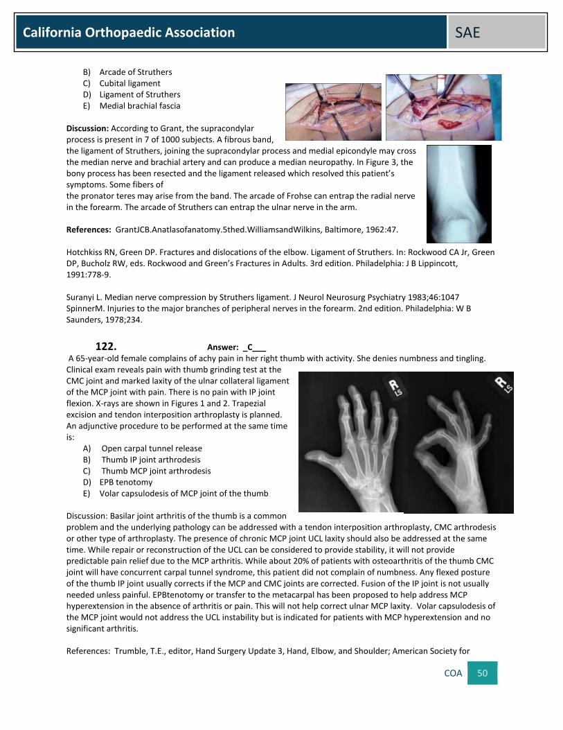
COA 50
California Orthopaedic Association SAE
B) Arcade of Struthers C) Cubital ligament D) Ligament of Struthers E) Medial brachial fascia
Discussion: According to Grant, the supracondylar process is present in 7 of 1000 subjects. A fibrous band, the ligament of Struthers, joining the supracondylar process and medial epicondyle may cross the median nerve and brachial artery and can produce a median neuropathy. In Figure 3, the bony process has been resected and the ligament released which resolved this patient’s symptoms. Some fibers of the pronator teres may arise from the band. The arcade of Frohse can entrap the radial nerve in the forearm. The arcade of Struthers can entrap the ulnar nerve in the arm. References: GrantJCB.Anatlasofanatomy.5thed.WilliamsandWilkins, Baltimore, 1962:47. Hotchkiss RN, Green DP. Fractures and dislocations of the elbow. Ligament of Struthers. In: Rockwood CA Jr, Green DP, Bucholz RW, eds. Rockwood and Green’s Fractures in Adults. 3rd edition. Philadelphia: J B Lippincott, 1991:778-9. Suranyi L. Median nerve compression by Struthers ligament. J Neurol Neurosurg Psychiatry 1983;46:1047 SpinnerM. Injuries to the major branches of peripheral nerves in the forearm. 2nd edition. Philadelphia: W B Saunders, 1978;234.
122. Answer: _C___ A 65-year-old female complains of achy pain in her right thumb with activity. She denies numbness and tingling. Clinical exam reveals pain with thumb grinding test at the CMC joint and marked laxity of the ulnar collateral ligament of the MCP joint with pain. There is no pain with IP joint flexion. X-rays are shown in Figures 1 and 2. Trapezial excision and tendon interposition arthroplasty is planned. An adjunctive procedure to be performed at the same time is:
A) Open carpal tunnel release B) Thumb IP joint arthrodesis C) Thumb MCP joint arthrodesis D) EPB tenotomy E) Volar capsulodesis of MCP joint of the thumb
Discussion: Basilar joint arthritis of the thumb is a common problem and the underlying pathology can be addressed with a tendon interposition arthroplasty, CMC arthrodesis or other type of arthroplasty. The presence of chronic MCP joint UCL laxity should also be addressed at the same time. While repair or reconstruction of the UCL can be considered to provide stability, it will not provide predictable pain relief due to the MCP arthritis. While about 20% of patients with osteoarthritis of the thumb CMC joint will have concurrent carpal tunnel syndrome, this patient did not complain of numbness. Any flexed posture of the thumb IP joint usually corrects if the MCP and CMC joints are corrected. Fusion of the IP joint is not usually needed unless painful. EPBtenotomy or transfer to the metacarpal has been proposed to help address MCP hyperextension in the absence of arthritis or pain. This will not help correct ulnar MCP laxity. Volar capsulodesis of the MCP joint would not address the UCL instability but is indicated for patients with MCP hyperextension and no significant arthritis. References: Trumble, T.E., editor, Hand Surgery Update 3, Hand, Elbow, and Shoulder; American Society for

COA 51
California Orthopaedic Association SAE
Surgery of the Hand,Rosemont, Ill., c. 2003, p. 510. Tomaino, M. M., Pellegrini, V.D., Burton, R. I., Arthroplasty of the basal joint of the thumb, Long-term follow-up after ligament reconstruction with tendon interposition, JBJS 1995;77(3); 346-355.
123. Answer: _E___ A 45-year-old female presents with a painful arthritic PIP joint of the dominant index finger. She has 20 degrees of painful motion and an apex-radial angular deformity of 20 degrees and has failed conservative care. The patient prefers to have only one procedure. Which treatment will provide durable pain relief while maintaining pinch strength?
A) Joint debridement with radial collateral ligament reconstruction B) Surface replacement arthroplasty C) Silastic implant arthroplasty D) Radial closing wedge osteotomy proximal phalanx E) PIP fusion
Discussion: This patient requires strong, pain-free pinch and grip to pursue her potting. While loss of motion in the PIP joint is a concern for the patient, her primary impairment is her pain and angular deformity involving the joint. Neither joint debridement with ligament reconstruction nor proximal phalange neck osteotomy will improve her pain or prevent progression of the joint degeneration. While surface replacement and Silastic implant arthroplasties may initially resolve pain and improve motion, both are prone to gradually deteriorating motion and eventual failure. The Silastic implant offers minimal stability to the stress of lateral pinch. Only a joint fusion offers pain free stability for pinch and grip. While this comes at the expense of motion, a successful fusion will require no future revision. References: Sweets T, Stern PJ. Proximal Interphalangeal Joint Prosthetic Arthoplasty. J Hand Surg 2010;35A(7):1190-93. Branam BR, Tuttle HG, Stern PJ, et al. Resurfacing arthroplasty versus silicone arthroplasty for proximal interphalangeal joint osteoarthritis. J Hand Surg 2007;32A:775-88. Minamikawa Y, Imaeda T, Amadio PC, et al. Lateral stability of proximal interphalangeal joint replacement. J Hand Surg1994;19A:1050-54.
124. Answer: _B___ Late complications of volar plating of distal radius fractures can be decreased by:
A) Open reduction and internal fixation of all associated ulnar styloid fractures B) Intraoperative use of fluoroscopy C) Limiting the number of diaphyseal screws D) Keeping the plate permanently E) Bicortical purchase of distal screws
Discussion: Reliance on standard PA and lateral x-rays has been shown to miss screw penetration of the dorsal cortex, where extensor tendons are vulnerable, as well as penetration of the radial articular surface. Repair of associated ulnar styloid fractures is often unnecessary in the absence of DRUJ instability. The traditional principle of screw purchase of the distal cortex does not apply to volar locking plates, as the plate and subchondral bone provide stability, and screw penetration is to be avoided. Placement of the plate at the volar lip of the radius places the plate near flexor tendons and may lead to attenuation and rupture. Volar locking plates are not intended to be removed in a routine manner.

COA 52
California Orthopaedic Association SAE
References: Sammer D, Shah H, Shauver M, Chung K. The effect of ulnar styloid fractures on patient outcomes after volar locking plating of distal radius fractures. J Hand Surg 2009;34A:1595-1602. Joseph S, Harvey J. The dorsal horizon view: detecting screw protrusion at the distal radius. J Hand Surg 2011;36A:1691-1693. Tweet M, Calfee R, Stern P. Rotational fluoroscopy assists in detection of intra-articular screw penetration during volar plating of the distal radius. J Hand Surg 2010;35A:619-627. Klug R, Press C, Gonzalez M. Rupture of the flexor pollicis longus tendon after volar fixed-angle plating of a distal radius fracture: A case report. J Hand Surg 2007;32A:984-988.
125. Answer: _A___ Which of the following complications is most common following UCL reconstruction?
A) Ulnar neuropathy B) Medial epicondyle fracture C) Postoperative stiffness D) Hematoma E) Ligament retear
Discussion: A recent systematic review of published studies evaluating reconstruction of the UCL in overhead athletes was performed using the Ovid Medline database. Overall, 83% of patients in all studies had an excellent result. There was an overall 10% complication rate, with the most common complication being postoperative ulnar neuropathy, which occurred in 6% of patients. Transition to the muscle-splitting approach was associated with better outcomes than detachment of the flexor-pronator mass, as there was only a 70% rate of excellent results and a 20% rate of postoperative ulnar neuropathy in patients treated with detachment of the flexor-pronator mass compared with 87% excellent results and a 6% rate of postoperative ulnar neuropathy in patients treated with a muscle-splitting approach. Reference: Vitale MA, Ahmad CS. The outcome of elbow ulnar collateral ligament reconstruction in overhead athletes: a systematic review. Am J Sports Med. 2008 Jun;36(6):1193-205.
Oncology
126. Answer __B_ Aldoxorubicin is a drug in phase III clinical trials for what tumor:
A) Chordoma B) Soft tissue sarcoma C) Giant cell tumor D) Chondrosarcoma
Discussion: Doxorubicin is a primary drug used in the treatment of soft tissue sarcomas. Aldoxorubicin has a linker that attracts and links protein while circulating in the blood and has preferential absorption to tumor cells reducing the cardiac toxicity seen with doxorubicin. Reference: First Line Aldoxorubicin vs Doxorubicin in Soft Tissue Sarcoma; Chawla; JAMA Oncol. 2015 Dec1 1(9): 1272-80.

COA 53
California Orthopaedic Association SAE
127. Answer _C_ Chordoma can be difficult to distinguish histologically from the following tumors:
A) giant notochordal hamartoma, renal cancer and clear cell sarcoma B) chondrosarcoma, Clear cell sarcoma and renal cancer C) giant notochordal hamartoma, renal cancer, chondrosarcoma and clear cell sarcoma D) renal cancer and giant notochordal hamartoma
Discussion: All of these lesions have clear cells and may be difficult to differentiate from chordoma. Giant notochordal hamartoma has the most similar histologic findings as chordoma but this benign tumor should never break out into the soft tissue. Reference: Mirra, J and Brien, E; Skeletal Radiology, Dec 2001 vol 12 pp 698-709.
128. Answer _A_ Which aggressive benign tumor can be managed with an osteoporosis drug?
A) Giant cell tumor B) Unicameral bone cyst C) Aneurysmal bone cyst D) Non ossifying fibroma
Discussion: Understanding the role of the receptor activator of nuclear factor-kB ligand (RANKL) in giant cell tumor has led to the use of denosumab when a surgical approach would be morbid. Reference: Giant cell tumor of bone: Singh et al. Biologics. 2015 Jul 14; 9: 69-74
129. Answer _C_ Tumor implants addressing leg length discrepancy in a growing child can be done without reopening the incision through what mechanism:
A) Screw extension mechanism B) Replacement of modular midsections C) Lengthening through electromagnetic field D) Lewis expandable adjustable prosthesis (LEAP)
Discussion: The distal femur and proximal tibia contribute 35% and 30% respectively of growth in the lower extremity. Children under the age of 10 can have significant leg length discrepancy after tumor resection in these areas. Lengthening through an electromagnetic field no longer requires an incision to lengthen the limb. Reference: Clin. Orthop. Relat. Res. Wilkins et al. 2001 Jan; (382): 51-8.
130. Answer _D_ Surgical management of metastatic bone disease has not changed in which cancer:
A) Lung cancer B) Renal cancer C) Breast cancer D) Pancreatic cancer E) Thyroid cancer
Discussion: Pancreatic cancer rarely spreads to bone and chemotherapy has had less impact in pancreatic cancers than the more common malignancies that spread to bone.

COA 54
California Orthopaedic Association SAE
Reference: Skeletal Metastases in Pancreatic Cancer; Borad, M et al. yale J boil med. 2009 Mar; 82 (1): 1-6.
131. Answer _B_ Which soft tissue tumor have we altered our surgical management by do a more limited surgical approach:
A) Synovial sarcoma B) Desmoid tumor C) Liposarcoma D) Undifferentiated sarcoma E) Pigmented villonodular synovitis
Discussion: Desmoid tumor or aggressive fibromatosis has an exceptionally high recurrence rate and surgical can lead to significant morbidity. Treatment includes low dose methotrexate and vinblastine with local surgery. Recurrence is common even with an attempt at wide excision and a less aggressive approach should be considered. Reference: Ann Surg Oncol. 2015; 22 (9): 2817-23
132. Answer _A_ Management of a non-union after pathologic femur fracture in a patient who has received high dose radiation therapy should include:
A) Endoprosthetic reconstruction B) Allograft reconstruction C) Repeat nailing D) Plate fixation
Discussion: Patients who have received radiation treatment have a high incidence of non-union after pathologic fracture. Proximal femoral replacement and a constrained liner should be considered to avoid multiple surgeries and immediate weight bearing. Reference: Proximal Femoral Reconstruction…Jawad et al. Orthopedics;feb 2014 vol 37; 187-193.
133. Answer _D_ Treatment for high grade sarcoma greater than 5 cm may include the following:
A) Chemotherapy and surgery
B) Chemotherapy and radiation
C) Radiation and surgery
D) Chemotherapy, radiation and surgery
Discussion: Large, high grade soft tissue sarcomas are commonly treated with neoadjuvant chemotherapy, radiation therapy followed by surgery. Variation in chemotherapy for specific sarcomas such as leiomyosarcoma may be indicated. Reference: Wiki-Based Clinical Practice Guidelines for Management of Adult Onset Sarcoma: Neuhaus, s. et al. 2015; Feb 16
134. Answer _B_ A new technique in improving spatial orientation in pelvic and sacral tumor surgery is:
A) Intraoperative fluoroscopy B) Intraoperative O arm C) Intraoperative MRI
COA 55
California Orthopaedic Association SAE
D) Intraoperative radiographs
Discussion: Intraoperative O arm is a portable CT scan that can be used to navigate surgery in the pelvis. It accesses real time 3D images using navigation systems to confirm tumor location and resection margins potentially reducing local recurrence and improving function. Reference: Computer Assisted Navigation During an A-P En Bloc Resection of a Sacral Tumor; Cureus: Elissa et al. 2015 Nov 4; 7(11) e373
135. Answer _A_ Which bone tumor is treated with chemotherapy?
A) Low grade chondrosarcoma B) Chordoma C) Adamantinoma D) Parosteal osteosarcoma
Discussion: All of these malignant tumors are low grade. Low grade sarcomas are not treated with chemotherapy. The primary treatment is surgery alone. Chordoma and other low grade sarcomas can be treated with radiation therapy (ie. Proton/carbon ion therapy) in an attempt to reduce the risk of local recurrence in very high risk patients. Reference: Randomized phase II trial of hypofractionated proton versus carbon ion radiation for chordoma; Uhl, M et al. radiat oncol. 2014 9;100
136. Answer _A_ Which primary solitary round cell tumor has the best prognosis?
A) Lymphoma B) Ewing’s sarcoma C) Myeloma D) Rhabdomyosarcoma
Discussion: The disease free survival with a solitary bone lymphoma is between 80-90% in patients under the age of 60. This is significantly better than the other round cell tumors discussed above. Reference: Malignant lymphoma of bone; Demircay et al. clin orthop relat res; 2013 aug; 471 (8) 2684-2690.
137. Answer _D_ The best way to differentiate well differentiated liposarcoma (atypical lipoma) and lipoma on MRI is the following:
A) Size B) Location C) Shape D) Signal alteration
Discussion: Both lipoma and well differentiated liposarcoma have bright signal on T-1 and low signal on T-2. If there is a dark linear striations in the mass, one should be suspicious of the well differentiated liposarcoma. Local recurrence is not uncommon with this tumor. Reference: MRI Web Clinic Jan 2014; Dr. Awh; Lipoma and liposarcoma;

COA 56
California Orthopaedic Association SAE
Osteoporosis
138. Answer: _C__ How does PTH peptide (Teriparatide) effect bone metabolism and how is it used in the treatment of osteoporosis?
A) Continual use for less than 3 years B) Continual use for 3-5 years C) Continual use for more than 5 years D) Intermittent use only
Discussion: Anabolic effect on bone and stimulates bone formation. Continual use leads to net bone loss. Reference: OKU-10 Chapter 16 Page 188
139. Answer: _A__ What is the definition of osteoporosis?
A) Patient with T score of less than -2.5 B) Patient with T score of more than -2.5 C) Patient with T score of less than -1.5 D) Patient with T score of less than 1
Discussion: Criteria for Osteoporosis Normal BMD within 1 SD of the young adult reference mean Osteopenia BMD between -1.0 to -2.5 SD of the young
adult reference mean Osteoporosis BMD2.5 SD below the young adult reference mean BMD = bone mineral density; SD = standard deviation(Data from the World Health Organization, Reprinted with permission from Flynn, JM (ed), Orthopaedic Knowledge Update 10, Rosemont, IL American Academy of Orthopaedic Surgeons, 2011, Page 188 Reference: OKU-10 Chapter 16 Page 188
140. Answer: _ C__ What is the definition of osteopenia?
A) Patient with T score between -.5 and -2.5 B) Patient with T score between -.5 and -3.5 C) Patient with T score between -1.0 and -2.5 D) Patient with T score under -1.0
Discussion: Criteria for Osteoporosis Normal BMD within 1 SD of the young adult reference means Osteopenia BMD between -1.0 to -2.5 SD of the young adult reference mean Osteoporosis BMD 2.5 SD below the young adult reference mean BMD = bone mineral density; SD = standard deviation. References: Data from the World Health Organization Reprinted with permission from Flynn, JM (ed), Orthopaedic Knowledge Update 10, Rosemont, IL American Academy of Orthopaedic Surgeons, 2011, Page 188 OKU-10 Chapter 16 Page 188
141. Answer: _ A__ What is a main category of treatment for osteoporosis?
A) Calcium
B) Iron

COA 57
California Orthopaedic Association SAE
C) Magnesium
D) Vitamin C Discussion: Calcium is important in the treatment of osteoporosis, however, large dosages of calcium can build up in the arteries and lead to heart problems. Reference: OKU-10 Chapter 16 Page 188
Pediatrics
142. Answer _B_ Figures 1 is the radiograph of a 22-month-old boy who has infantile idiopathic scoliosis. What is the preferred initial treatment to minimize curve progression?
A) Observation
B) Serial casting
C) Rib to spine growing rods
D) Spine to spine growing rods
E) Posterior spinal fusion with instrumentation Discussion: Infantile idiopathic scoliosis includes scoliosis seen in patients from 0 to 3 years of age without other structural or congenital causes. Many patients with infantile idiopathic scoliosis with less than a 20-degree rib vertebral angle difference (RVAD) and phase 1 ribs will have resolution of their curvature with no treatment. However, in this child the RVAD is clearly greater than 20 degrees at the apex with significant overlap of the rib head over the vertebral body at the apex (phase 2 ribs). Thus the best initial treatment for this child is serial body casting with de-rotational molding (i.e. Cotrel-Mehta casting). Serial casting is most effective on children less than 2 years of age, and effectiveness falls off as a child gets older. Even if the casting does not completely resolve the scoliosis, it can often be used to delay future surgery. Growing rod instrumentation would be indicated in a child less than 10 years of age who fails treatment with serial body casting. Posterior spinal fusion would be indicated after 10 years of age in a child who has progressive deformity or has graduated from growing rod treatment. References: Sanders JO, D'Astous J, Fitzgerald M, Khoury JG, Kishan S, Sturm PF. Derotational casting for progressive infantile scoliosis. J Pediatr Orthop. 2009 Sep;29(6):581-7. doi: 10.1097/BPO.0b013e3181b2f8df. PubMed PMID: 19700987. Tis JE, Karlin LI, Akbarnia BA, Blakemore LC, Thompson GH, McCarthy RE, Tello CA, Mendelow MJ, Southern EP; Growing Spine Committee of the Scoliosis Research Society. Early onset scoliosis: modern treatment and results. J Pediatr Orthop. 2012 Oct-Nov;32(7):647-57. Review. PubMed PMID: 22955526. Waldron SR, Poe-Kochert C, Son-Hing JP, Thompson GH. Early onset scoliosis: the value of serial risser casts. J Pediatr Orthop. 2013 Dec;33(8):775-80. doi: 10.1097/BPO.0000000000000072. PubMed PMID: 23965912.
143. Answer _C_

COA 58
California Orthopaedic Association SAE
The approximate increase in risk of complications with each surgical procedure for spinal growing rod lengthening is:
A) 5%
B) 10%
C) 25%
D) 50%
E) 90% Discussion: Bess et al. show that growing rods carry a significant risk of complications with each lengthening, even though the actual surgical procedure itself is relatively minor. Complications are quite common and should be expected and patients should be counseled regarding this risk. Their reported increase in risk of complications with each surgical procedure was 24%, with a total of 58% of all patients receiving at least one complication. Complications include skin dehiscence, implant failure, anchor failure, superficial and/or deep infection. Submuscular implantation of growing rods has a lower risk of complications overall than subcuticular implantation. References: Bess S, Akbarnia BA, Thompson GH, Sponseller PD, Shah SA, El Sebaie H, Boachie-Adjei O, Karlin LI, Canale S, Poe-Kochert C, Skaggs DL. Complications of growing-rod treatment for early-onset scoliosis: analysis of one hundred and forty patients. J Bone Joint Surg Am. 2010 Nov 3;92(15):2533-43.
144. Answer _E_ A Ponte osteotomy of T6-T7 involves the excision of which anatomic structures?
A) Ligamentum flavum, inferior articular process of T6
B) Ligamentum flavum, superior articular process of T6, inferior articular process of T7
C) Ligamentum flavum, superior articular process of T7, inferior articular process of T6
D) Ligamentum flavum, superior articular process of T6, inferior articular process of T7, transverse processes of T6 and T7
E) Ligamentum flavum, superior articular process of T7, inferior articular process of T6, transverse processes of T6 and T7
Discussion: The Ponte osteotomy is a very powerful procedure that is used in the correction of many different types of spinal deformity. The procedure was initially described to increase spinal flexibility in correcting Scheuermann’s kyphosis, but this has been adapted to increase spinal flexibility in other diagnoses, including adolescent idiopathic scoliosis, early onset scoliosis, congenital scoliosis, and others. The osteotomy involves the excision of all midline tissues posterior to the thecal sac and epidural fat. The ligamentum flavum, the inferior articular process of the level above, and the superior articular process of the level below are all excised to increase spinal flexibility, utilizing the disk as the hinge anteriorly. Excision of the transverse processes are not involved in a Ponte osteotomy, although they are often removed or debulked to increase bony surface area for fusion and increase area for pedicle screws to be placed. References: Geck MJ, Macagno A, Ponte A, Shufflebarger HL. The Ponte procedure: posterior only treatment of Scheuermann's kyphosis using segmental posterior shortening and pedicle screw instrumentation. J Spinal Disord Tech. 2007 Dec;20(8):586-93. PubMed PMID: 18046172. Ponte A. Posterior column shortening for Scheuermann’s kyphosis. In: Haher TR, Merola AA, eds. Surgical Techniques for the Spine. 1st ed. New York: Thieme Verlag; 2003:107–113.
145. Answer _B_

COA 59
California Orthopaedic Association SAE
The use of a thoracolumbar orthosis for treatment of adolescent idiopathic scoliosis has been shown to significantly decrease the rate of progression of the curvature. The threshold for achieving a 90% success rate of treatment was shown to be approximately:
A) 6 hours
B) 12 hours
C) 18 hours
D) 20 hours
E) 24 hours Discussion: The recent BRAIST study showed that thoracolumbar orthosis treatment for adolescent idiopathic scoliosis was quite effective in preventing patients from reaching a surgical threshold of 50 degrees, with a reported treatment success rate of 72% for bracing versus 48% for observation alone. Temperature monitors in the TLSO braces were used to determine compliance and hours worn. Patients who wore the brace 12.9 hours or more had over a 90% rate of treatment success. As patients wore the brace longer than this, the rate of treatment success improved up to 93% at greater than 17.7 hours of wear, indicating diminishing returns for brace wear beyond 12.9 hours daily. References: Weinstein SL, Dolan LA, Wright JG, Dobbs MB. Effects of bracing in adolescents with idiopathic scoliosis. N Engl J Med. 2013 Oct 17;369(16):1512-21
146. Answer _D_ Which are the following is not a pediatric manifestations of a proximal femoral cam deformity?
A) SCFE
B) Legg-Calve-Perthes
C) Multiple epiphyseal dysplasia
D) Coxa Valga
E) Coxa Vara
F) Repetitive microtrauma to the lateral physis of the proximal femur Discussion: Slipped capital femoral epiphysis results in a posteriorly displaced femoral head and a femoral shaft that moves anteriorly creating a cam deformity. Legg-Calve-Perthes and multiple epiphyseal dysplasia both result in femoral head deformity and shortening of the femoral head-neck junction. Coxa vara results in a shortened head-neck offset causing a cam deformity. Repetitive microtrauma to the lateral physis is believed to be cause the formation of a cam deformity in an athletic population. Coxa valga generally does not result in cam impingement. References: Siebenrock KA, Wahab KH, Werlen S, Kalhor M, Leunig M, Ganz R. Abnormal extension of the femoral head epiphysis as a cause of cam impingement. Clin Orthop Relat Res. 2004 Jan;(418):54-60. Ganz R, Parvizi J, Beck M, Leunig M, Nötzli H, Siebenrock KA. Femoroacetabular impingement: a cause for osteoarthritis of the hip Clin Orthop Relat Res. 2003 Dec;(417):112-20.
147. Answer _B_ When does the proximal femoral physis typically close in males and females?
A) Around age 10 in males and age 12 in females
B) Around age 16 in males and age 14 in females
C) Around age 8 in both males and females
D) Around age 18 in both males and females

COA 60
California Orthopaedic Association SAE
Discussion: Males typically fuse later than females. The timing of this closure and remaining growth left may influence the resection of the femoral head-neck junction. Reference: Dvonch VM, Bunch WH Pattern of closure of the proximal femoral and tibial epiphyses in man. .J Pediatr Orthop. 1983 Sep;3(4):498-501.
148. Answer _B_ Treatment of a young athletic patient with a labral tear and concomitant combined femoroacetabular impingement is best done by:
A) Open surgical dislocation approach with resection of the labrum
B) Arthroscopic treatment of the femoroacetabular impingement with repair of the labrum
C) Peri-acetabular osteotomy Discussion: While there is no established superior surgical approach to address FAI, multiple studies have demonstrated superiority of labral repair compared to labral debridement. References: Espinosa N, Beck M, Rothenfluh DA, Ganz R, Leunig M. J Treatment of femoro-acetabular impingement: preliminary results of labral refixation. Surgical technique. Bone Joint Surg Am. 2007 Mar;89 Suppl 2 Pt.1:36-53. Philippon MJ, Briggs KK, Yen YM, Kuppersmith DA. Outcomes following hip arthroscopy for femoroacetabular impingement with associated chondrolabral dysfunction: minimum two-year follow-up. J Bone Joint Surg Br. 2009 Jan;91(1):16-23.
149. Answer _B_ What is the prevalence of radiographic femoroacetabular impingement in asymptomatic adolescents compared with an adult population?
A) Similar in incidence to adults regardless of physeal status
B) Similar in incidence to adults as the physis closes
C) Significantly higher than adults
D) Non-existent Discussion: Multiple studies based upon CT scans of asymptomatic adolescents have demonstrated an incidence of radiographic FAI similar to adults as the physis nears closing. References: Bixby SD, Kienle KP, Nasreddine A, Zurakowski D, Kim YJ, Yen YM. Reference values for proximal femoral anatomy in adolescents based on sex, physis, and imaging plane. Am J Sports Med. 2013 Sep;41(9):2074-82. Carsen S, Moroz PJ, Rakhra K, Ward LM, Dunlap H, Hay JA, Willis RB, Beaulé PE. The Otto Aufranc Award. On the etiology of the cam deformity: a cross-sectional pediatric MRI study. Clin Orthop Relat Res. 2014 Feb;472(2):430-6 Monazzam S, Bomar JD, Dwek JR, Hosalkar HS, Pennock AT. Development and prevalence of femoroacetabular impingement-associated morphology in a paediatric and adolescent population: a CT study of 225 patients. Bone Joint J. 2013 May;95-B(5):598-604
150. Answer _D_ When x-rays suggest a diagnosis of osteochondroma, the next study most appropriate to clarify the diagnosis is:
A) Ultrasound
B) MRI
C) CAT Scan

COA 61
California Orthopaedic Association SAE
D) Additional x-rays including a tangential view
E) Bone Scan Discussion: Plain x-rays taken with the appropriate orientation will almost always show the pathognomonic findings of osteochondroma without the need for more costly studies. Reference: OKU Musculoskeletal Tumors 2 p.104
151. Answer _B_ The short leg gait pattern is characterized by all of the following factors EXCEPT:
A. Stance time reduced on the short side B. Increased pelvic rotation on the short side C. Stride length reduced on the short side D. Cadence (steps/minute) increased E. Walking velocity marginally reduced
Discussion: Due to the difference in limb lengths, an individual will spend less time with a shorter stride length, increased cadence, and a slight decrease in velocity on the short side. Pelvic rotation is not significantly affected by a limb length inequality. Reference: Principles of Deformity Correction. Dror Paley. Springer. 2005.
152. Answer _A_ Patients with limb length discrepancy use all of these compensatory mechanisms EXCEPT:
A. Hip flexion on the long side B. Equinus of the ankle on the short side C. Pelvic tilt with lowering of the pelvis on the short side D. Knee flexion on the long side E. Reduced stride length on the short side
Discussion: In order to compensate for a short lower limb, the patient will tilt his/her pelvis forward and plantaflex the ankle on the short side to gain more length. It follows that the short limb will have a decreased stride length compared to the contralateral side. To balance both sides, the individual tends to flex their knee rather than their hip on the long side. Reference: Principles of Deformity Correction. Dror Paley. Springer. 2005.
153. Answer __C_ When x-rays suggest a diagnosis of osteoid osteoma, the next study most appropriate to clarify the diagnosis is:
A) Ultrasound
B) MRI
C) CAT Scan
D) Bone Scan
E) Incision Biopsy Discussion: CAT Scan provides best imaging for the “nidus.” Ultrasound is unhelpful. MRI and bone scan findings are non-specific and are often “over-read” as suggesting a more serious lesion. Reference: OKU Musculoskeletal Tumors 2 p. 94
154. Answer_B_
COA 62
California Orthopaedic Association SAE
Fibrous cortical defect and non-ossifying fibroma:
A) Often are present with a pathologic fracture
B) Often are “incidental findings” on x-rays taken after trauma
C) Have persistent pain and tenderness
D) Have a significant risk of malignant transformation
E) Tend to persist long after skeletal maturity Discussion: These conditions are most often identified as “incidental findings.” They seldom present with a fracture, are rarely painful, have minimal risk of malignant transformation, and often regress in late adolescence. Reference: OKU Musculoskeletal Tumors 2 p. 121
155. Answer_C_ The Precise2 nail can lengthen to a maximum of:
A) 10 cm B) 6 cm C) 8 cm D) 9 cm E) 12 cm
Discussion: Limb lengthening places significant stress on the surrounding soft tissue envelope (muscle/tendons, nerves, vessels) as well as the proximal and distal joints. In order to avoid complications such as neuropraxia and joint contractures or dislocations, it is recommended that gradual lengthening not exceed 8cm in any limb segment. The Precise2 nail settings do not allow lengthening greater than 8cm. Reference: Principles of Deformity Correction. Dror Paley. Springer. 2005.
156. Answer_E_ All of these factors characterize the Precise2 nail EXCEPT:
A) It is currently the only FDA-approved lengthening device on the market B) It has a reverse mechanism C) It uses an internal magnet to lengthen D) It can be inserted either antegrade or retrograde into the femur E) It is designed to lengthen only once a day
Discussion: The Precise2 nail is the only FDA-approved lengthening device on the market. Internal settings are activated and allow the nail to lengthen using an external magnet applied over a skin marking. If there is a lack of regenerate bone in the gap, the device settings can be temporarily changed or reversed to allow compression across the gap to stimulate and regenerate bone formation. The technique allows for insertion into the femur in either an antegrade or retrograde manner depending on the circumstance and also offers options in terms of nail length. The technique follows previously published Ilizarov bone lengthening principles of 1mm lengthening/day divided in three separate sessions or 0.33mm three times per day. The daily frequency and amount of lengthening can be adjusted as necessary. Reference: Principles of Deformity Correction. Dror Paley. Springer. 2005.
157. Answer_D All of these are potential complications of lengthening EXCEPT:
A) Premature consolidation of osteotomy B) Hip or knee dislocation C) Loosening of interlocking screw

COA 63
California Orthopaedic Association SAE
D) Decreased soft tissue tension E) Delayed regenerate bone formation
Discussion: Complications are not all that uncommon especially with large amounts of limb length discrepancy correction. If the frequency of the lengthening is not sufficient, the external magnet is not properly applied or the initial osteotomy is incomplete, and premature consolidation of the regenerate can occur. If pre-operative work-up has not identified an unstable or mal-aligned joint, or if the lengthening proceeds too aggressively without proper therapy or splinting as necessary, joint subluxation or dislocation can occur due to the increased soft tissue tension in the limb. Due to the fact that only the distal aspect of the interlocking screw is threaded and the shank is smooth, screw back-out has been reported. Placement of a fully threaded titanium screw can help avoid screw back-out. If the osteotomy is performed in diaphyseal bone, or the lengthening proceeds too quickly, delay in regenerate bone formation in the gap may occur requiring either an increase in weight-bearing especially indoors, temporary reversal of the lengthening and/or placement of either synthetic or autogenous graft material in the regenerate gap. Reference: Principles of Deformity Correction. Dror Paley. Springer. 2005.
158. Answer_C_ How do physicians determine the realistic functional goals of interdisciplinary management?
A) Routine physical therapy evaluation B) Patient examination without review of previous examinations C) Complete interdisciplinary evaluation with team meeting to establish functional status based on
established criteria D) Ignore fluctuation in neurologic pathology E) Do not allow patients to participate in community activities
Discussion: Need to add. References: Principles of Orthopaedic Practice, copyright 1997, McGraw-Hill, Chapter 34: Orthopaedic Management in Myelomeningocele. Mercado, E. et al Does Spinal Fusion influence quality of life in neuromuscular scoliosis? Spine2007; 32(19, Suppl): S 120-S125
159. Answer_E_ What are the significant physical impairments leading to the inability to maintain ambulatory status?
A) Define objective criteria for ambulation B) Define muscle requirements for ambulation C) Define range of motion requirements for ambulation D) Evaluate physical impairments E) All of the above
Discussion: Need to add. References: Principles of Orthopaedic Practice, copyright 1997, McGraw-Hill, Chapter 34: Orthopaedic Management in Myelomeningocele. Mercado, E. et al Does Spinal Fusion influence quality of life in neuromuscular scoliosis? Spine2007; 32(19, Suppl): S 120-S125
160. Answer_B_ What are the significant physical impairments leading to the inability to maintain independent sitting activities?

COA 64
California Orthopaedic Association SAE
A) Ignore physical impairments for wheelchair patients B) Define criteria needed for independent wheelchair activities C) Lumbar kyphosis and pelvic obliquity do not interfere with wheelchair activities D) Hip contractures do not matter in wheelchair patients E) Spinal orthotic management not necessary
Discussion: Need to add. References: Principles of Orthopaedic Practice, copyright 1997, McGraw-Hill, Chapter 34: Orthopaedic Management in Myelomeningocele. Mercado, E. et al Does Spinal Fusion influence quality of life in neuromuscular scoliosis? Spine2007; 32(19, Suppl): S 120-S125
161. Answer_E_ What are the orthotic requirements to prevent deformity and loss of functional skills?
A) Orthotic management coincidental with initiation of ambulatory skills
B) Foot surgery only to facilitate orthotic management
C) Ankle orthotic management needs rigid hindfoot control in neutral
D) Orthotic management must maintain hip and knee extension for weight-bearing
E) All of the above Discussion: Need to add. References: Principles of Orthopaedic Practice, copyright 1997, McGraw-Hill, Chapter 34: Orthopaedic Management in Myelomeningocele. Mercado, E. et al Does Spinal Fusion influence quality of life in neuromuscular scoliosis? Spine2007; 32(19, Suppl): S 120-S125
Spine
162. Answer _B_ When compared to open surgery, minimally invasive surgery has the following effect on the spinal muscles:
A) Greater rate of muscular atrophy after surgery
B) Lesser rate of muscular atrophy after surgery
C) Equal rate of muscular atrophy after surgery Discussion: Animal and human studies have looked at muscle injury in open and minimally invasive surgery. MRI studies have evaluated the muscles and the results have shown that there is less muscle atrophy in minimally invasive spine surgeries. In particular, the multifidus muscle is affected the most. There have also been further studies that show that muscle strength may be better with the preserved muscle and this may be a reason why patients who undergo minimally invasive surgery recover faster. Reference: Fan S, Hu Z, Zhao F, et al. Multifidus muscle changes and clinical effects of one-level posterior lumbar interbody fusion: minimally invasive procedure versus conventional open approach. Eur Spine J. 2010 Feb;19(2):316-24
163. Answer _C__ The primary concern of lateral interbody fusions at this time are:
COA 65
California Orthopaedic Association SAE
A) Greater rate of blood loss
B) Greater post-operative times
C) Post-operative neurologic palsy
D) Vascular injury Discussion: Lateral interbody fusion is a transpsoas procedure where the psoas muscle is split and a tubular
retractor is placed to create a working window. This tends to result in less blood loss and shorted operative time
compared to an anterior lumbar inter body fusion. Also, there is less risk to the vena cava and aorta. However,
the biggest risk is the potential for a post-operative femoral nerve palsy and this has been noted even in cases
where the neuromonitoring remains normal.
Reference: Rodgers WB, Gerber EJ, Patterson JR. Intraoperative and early postoperative complications in extreme lateral interbody fusion (XLIF): An analysis of 600 cases. Spine (Phila Pa 1976). 2011 Jan 1;36(1):26-32.
164. Answer _B_ When using lateral interbody fusion to correct degenerative scoliosis, which of the following is least likely to be corrected?
A) Cobb angle
B) Sagittal imbalance
C) Foraminal stenosis
D) Rotatory subluxation Discussion: The biggest benefit of the lateral inter body fusion is that it is able to correct coronal deformity (Cobb angle) and increase foraminal height. It is also able to correct rotatory subluxations. However, it has been increasingly noted that it cannot correct sagittal imbalance and a pedicle subtraction osteotomy or Smith-Peterson osteotomy is necessary to correct sagittal imbalance. Reference: Alimi M, Hofstetter CP, Cong GT. et al. Radiological and clinical outcomes following extreme lateral interbody fusion. J Neurosurg Spine. 2014 Jun;20(6):623-35.
165. Answer _D_ What is the only FDA approved use of BMP-2?
A) Lateral interbody fusion
B) Posterolateral instrumented fusion
C) TLIF
D) Anterior lumbar interbody fusion in a LT cage Discussion: At this time, the only approved indication of BMP-2 is for anterior lumbar inter body fusion in a LT cage. All the other procedures are considered off-label use of BMP-2. Reference: Burkus JK, Gornet MF, Dickman CA, et al. Anterior lumbar interbody fusion using rhBMP-2 with tapered interbody cages. J Spinal Disord Tech. 2002 Oct;15(5):337-49.
166. Answer __D_ The primary benefit of minimally invasive surgery at this time is:
A) It is less costly
B) Surgical time is less
C) Better outcomes when compared to open surgery
D) Fewer risks of complications such as wound infections

COA 66
California Orthopaedic Association SAE
Discussion: At this time, the best support for minimally invasive surgery is that there are fewer complications such as would infections. Reference: Parker SL, Adogwa O, Witham TF, et al. Post-Operative Infection after Minimally Invasive versus Open Transforaminal Lumbar Interbody Fusion (TLIF): Literature Review and Cost Analysis. Minim Invasive Neurosurg. 2011 Feb;54(1):33-7.
167. Answer: _C_ Compared with standing radiographs of the spine, measurement of the magnitude of scoliosis on non-weight-bearing radiographs typically:
A) Estimates accurately B) Underestimates by about 5 degrees C) Underestimates by about 10 degrees D) Underestimates by about 15 degrees E) Underestimates by about 20 degrees
Discussion: Supine MRI images tend to underestimate plain radiographs by 10° on average. However, radiographical and MRI measures showed a strong positive correlation (r= 0.90-0.94) for all curves, structural or nonstructural, and this correlation was not influenced by patient age or body mass index. The relationship allowed the development of a simple linear equation for converting MR image measures to radiograph measures with an acceptable absolute error of ±5°. References: Lee MC, Solomito M, Patel A. Supine magnetic resonance imaging Cobb measurements for idiopathic scoliosis are linearly related to measurements from standing plain radiographs. Spine (Phila Pa 1976). 2013; 38(11):E656-661.
168. Answer: _B_ In patients with adult scoliosis, the most reliable radiographic predictor of poor functional outcome is:
A) Coronal imbalance B) Sagittal imbalance C) Curve location (thoracic, thoracolumbar, lumbar) D) Apical rotation E) Rotatory subluxation
Discussion: Positive sagittal balance was the most reliable predictor of clinical symptoms in both operative and non-operative patient groups. Thoracolumbar and lumbar curves generated less favorable scores than thoracic curves in both patient groups. Significant coronal imbalance of greater than 4 cm was associated with deterioration in pain and function scores for unoperated patients but not in patients with previous surgery. Reference: Glassman SD, Berven S, Bridwell K, Horton W, Dimar JR. Correlation of radiographic parameters and clinical symptoms in adult scoliosis. Spine (Phila Pa 1976). 2005; 30(6): 682-688.
169. Answer: _E_ In adult patients with spinal deformity, posterior pelvic rotation (retroversion) can help to compensate for sagittal imbalance while secondary coronal curves may help compensate for coronal imbalance. Following successful surgical realignment, the greatest improvement in functional outcome was observed:
A) In those with pre-operative uncompensated coronal imbalance B) In those with pre-operative compensated coronal imbalance C) In those with pre-operative uncompensated sagittal imbalance D) In those with pre-operative compensated sagittal imbalance E) Equally in those with pre-operative uncompensated and compensated sagittal imbalance

COA 67
California Orthopaedic Association SAE
Discussion: A study of 125 patients with flatback deformity compared surgical outcomes in 27 patients with compensated flatback (pelvic incidence-lumbar lordosis mismatch [PI-LL] > 10° and increased sagittal vertical axis [SVA] < 5 cm) and 98 patients with decompensated flatback (SVA > 5 cm). Surgical correction of flatback deformity demonstrated similar functional outcome score improvements for patients in both groups at one-year postoperatively. The authors conclude that evaluation of flatback deformity should extend beyond measuring SVA. Among patients with concordant pain and disability, PI-LL mismatch must be evaluated for flatback deformity patients and can be considered a primary indication for surgery. References: Smith JS, Singh M, Klineberg E, Shaffrey CI, Lafage V, Schwab FJ, Protopsaltis T, Ibrahimi D, Scheer JK, Mundis G Jr, Gupta MC, Hostin R, Deviren V, Kebaish K, Hart R, Burton DC, Bess S, Ames CP; International Spine Study Group. Surgical treatment of pathological loss of lumbar lordosis (flatback) in patients with normal sagittal vertical axis achieves similar clinical improvement as surgical treatment of elevated sagittal vertical axis: clinical article. J Neurosurg Spine. 2014;21(2):160-70.
170. Answer: _E_ Perioperative complication rates in scoliosis surgery among elderly patients between the ages of 65 to 85 are reported to be approximately:
A) 30% B) 40% C) 50% D) 60% E) 70%
Discussion: A multicenter spinal deformity database study of 206 patients distributed patients among age groups as follows: 25 to 44 (n = 47), 45 to 64 (n = 121), and 65 to 85 (n = 38) years of age. Perioperative complication rates of 17%, 42%, and 71% were observed, respectively (P < 0.001). Within each age group, at 2-year follow-up there were significant improvements in ODI (P ≤ 0.004), SRS-22 (P ≤ 0.001), back pain (P < 0.001), and leg pain (P ≤ 0.04). Improvement in ODI and leg pain NRS were significantly greater among elderly patients (P = 0.003, P = 0.02, respectively), when compared with younger patients. Collectively, these data demonstrate the potential benefits of surgical treatment for adult scoliosis and suggest that the elderly, despite facing the greatest risk of complications, may stand to gain a disproportionately greater improvement in disability and pain with surgery. References: Smith JS, Shaffrey CI, Glassman SD, Berven SH, Schwab FJ, Hamill CL, Horton WC, Ondra SL, Sansur CA, Bridwell KH; Spinal Deformity Study Group. Risk-benefit assessment of surgery for adult scoliosis: an analysis based on patient age. Spine (Phila Pa 1976). 2011; 36(10):817-24.
171. Answer: _C_ In adults over 50 years of age with degenerative (de novo) lumbar scoliosis, the majority of patients would experience a 10-year curve progression of approximately: A) 10 degrees
B) 20 degrees C) 30 degrees D) 40 degrees E) 50 degrees
Discussion: The authors studied 200 patients older than age 50 years with back pain and recent onset of scoliosis with curves from T12 to L5 and the apex at L2 or L3 and not exceeding 60 degrees. The curves progressed an average of 3 degrees per year over a 5-year period in 73% of patients. Grade 3 apical rotation, a Cobb angle of 30 or more, lateral vertebral translation of 6 mm or more, and the prominence of L5 in relation to the intercrest line were important factors in predicting curve progression. Reference: Pritchett JW, Bortel DT. Degenerative symptomatic lumbar scoliosis. Spine (Phila Pa 1976).

COA 68
California Orthopaedic Association SAE
1993;18(6):700-3.
Sports Medicine - Lower Extremity
172. Answer _B_ Which autologous biologic has been shown to be most effective at treating the symptoms of osteoarthritis?
A) Leukocyte-rich PRP
B) Leukocyte-poor PRP
C) Platelet poor plasma (PPP)
D) Whole Blood Reference: Riboh et al. Effect of leukocyte concentration on the efficacy of PRP in the treatment of knee osteoarthritis. Discussion: Only non-neutrophil containing PRP has shown efficacy greater than hyaluronic acid for the treatment of symptoms of OA.
173. Answer _A_ Platelet Rich Plasma has been shown by randomized controlled trials to successfully treat all of the following conditions EXCEPT:
A) Rotator cuff tears
B) Lateral epicondylitis
C) Patellar tendinopathy
D) Symptoms of OA Reference: Saltzman et al. Does the use of PRP at the time of surgery improve clinical outcomes in arthroscopic rotator cuff repair. Arthroscopy, 2015. Discussion: Multiple randomized controlled trials (RCT) show no effect of PRP use with surgical repair of the rotator cuff. RTC’s show an effect of PRP treatment that is greater than other standard treatments (different for each study) for the other conditions listed.
174. Answer _A_ The maximum volume of bone marrow that should aspirated at each iliac crest harvest site in order to maximize stem cell recovery is:
A) 1-4cc
B) 10-12cc
C) 25-30cc
D) 50-60cc Reference: .Muschler GF et al. Aspiration to obtain osteoblast progenitor cells from human bone marrow: The influence of aspiration volume. J Bone Joint Surg Am. 1997 Nov; 79(11): 1699-709. Discussion: Maximum recovery of bone marrow mesechmal cells occurs with the first 1cc of marrow aspiration. Additional aspiration dilutes the cells with peripheral blood, so no more than a 4cc aspiration is recommended at each harvest site.
175. Answer _C_
COA 69
California Orthopaedic Association SAE
The FDA allows all of the following procedures during autologous stem cell harvest and treatment EXCEPT:
A) Concentrating cells using a centrifuge
B) Mincing and grinding of tissue to maximize cell recovery
C) Culturing cells to increase cell volume
D) Sorting cells in the clinic/OR to improve purity Reference: Docket No. FDA-2014-D-1696-0001: Minimal Manipulation of Human Cells, Tissues, and Cellular and Tissue-Based Products: Draft Guidance for Industry and Food and Drug Administration Staff (December 2014) Discussion: Harvested autologous cells are not allowed to leave the operating or clinic room where they were harvested per FDA regulations. If cells are taken outside the harvest room, the procedure do not meet criteria for minimal manipulation and is subject to regulation by the FDA.
176. Answer __D_ Which of the following is not a known factor in rotator cuff healing after rotator cuff repair?
A) Muscle quality
B) Osteoporosis
C) Age of the patient
D) Quality of the rotator cuff tissue Discussion: Muscle quality has been shown to be an important factor in rotator cuff repair and outcomes (Gladstone, AJSM 2006; Chung AJSM 2011). Age of the patient is also an important factor with increasing age being a key factor in increased re-tear rates. Cheung et al found that osteoporosis, as well as fatty infiltration, and tear retraction, were independent factors in re-tear rates (Chung AJSM 2011). Although quality of the rotator cuff is likely also a factor, it is unable to be quantified. References: Fatty infiltration and atrophy of the rotator cuff do not improve after rotator cuff repair and correlate with poor functional outcome. - http://www.ncbi.nlm.nih.gov/pubmed/17337727 Gladstone JN, Bishop JY, Lo IK, Flatow EL. Am J Sports Med. 2007 May;35(5):719-28 Factors affecting rotator cuff healing after arthroscopic repair: osteoporosis as one of the independent risk factors. - http://www.ncbi.nlm.nih.gov/pubmed/21813440 Chung SW, Oh JH, Gong HS, Kim JY, Kim SH. Am J Sports Med. 2011 Oct;39(10):2099-107. doi: 10.1177/0363546511415659. Epub 2011 Aug 3. PMID: 21813440
177. Answer __C_ Which of the following regarding microfracture of the footprint is true?
A) Improved outcomes at 2 years
B) Improved healing rates at 2 years
C) No change in outcomes or function at 2 years
D) Increased PRP delivery postoperatively Discussion: Osti et al (Int Orthop 2013) evaluated microfracture of the footprint in a randomized study of rotator cuff tears. They found that patients that had a microfracture of the footprint had early decreases in pain, but no differences in function, healing at 2 years. References: Microfractures at the rotator cuff footprint: a randomised controlled study. http://www.ncbi.nlm.nih.gov/pubmed/23760681 Osti L, Del Buono A, Maffulli N.
COA 70
California Orthopaedic Association SAE
Int Orthop. 2013 Nov;37(11):2165-71. doi: 10.1007/s00264-013-1952-z. Epub 2013 Jun 13. PMID: 23760681
178. Answer _C__ In a patient with a degenerative rotator cuff tear who opts for physical therapy, which is the most likely outcome?
A) Surgery within 3 months due to increased pain and loss of function
B) Injections at 6 weeks to improve function despite a decrease in pain
C) Significant improvement at 6 weeks and 3 months in ASES scores
D) Failure of physical therapy Discussion: Based on the Neer Award paper from 2012, physical therapy was shown to improve ASES scores at 6 weeks and 3 months in a majority of patients. The patients overall had excellent outcomes with non-operative management of their atraumatic rotator cuff tears. References: Effectiveness of physical therapy in treating atraumatic full-thickness rotator cuff tears: a multicenter prospective cohort study. - http://www.ncbi.nlm.nih.gov/pubmed/23540577 Kuhn JE, Dunn WR, Sanders R, An Q, Baumgarten KM, Bishop JY, Brophy RH, Carey JL, Holloway BG, Jones GL, Ma CB, Marx RG, McCarty EC, Poddar SK, Smith MV, Spencer EE, Vidal AF, Wolf BR, Wright RW; MOON Shoulder Group. J Shoulder Elbow Surg. 2013 Oct;22(10):1371-9. doi: 10.1016/j.jse.2013.01.026. Epub 2013 Mar 27. PMID: 23540577
179. Answer _A__ What is the likely outcome of a tear that disrupts the anterior aspect of the supraspinatus tendon compared to one in the central aspect of the supraspinatus (crescent)?
A) Increased likelihood of greater tear size and more muscle degeneration
B) Increased pain and worse patient reported outcomes preoperatively
C) Worse outcomes post operatively
D) Decreased likelihood of tear progression Discussion: From Namdari et al (JSES 2014) “In the setting of painful small and medium-sized rotator cuff tears, disruption of the anterior supraspinatus tendon was associated with greater tear size and more advanced supraspinatus muscle degeneration. However, anterior supraspinatus tendon integrity had no influence on the clinical presentation or the functional and structural results of cuff repair surgery.” References: Characteristics of small to medium-sized rotator cuff tears with and without disruption of the anterior supraspinatus tendon. - http://www.ncbi.nlm.nih.gov/pubmed/23937927 Namdari S, Donegan RP, Dahiya N, Galatz LM, Yamaguchi K, Keener JD. J Shoulder Elbow Surg. 2014 Jan;23(1):20-7. doi: 10.1016/j.jse.2013.05.015. Epub 2013 Aug 9. PMID: 23937927
180. Answer _C_ Patients with horizontal cleavage tears of the meniscus:
A) Cannot be improved with a repair
B) Who undergo repair have a success rate that is significantly lower than repair of other types of meniscus tears
C) Can undergo repair with a success rate of 78%
D) Usually do not have knee pain
E) Can be treated as an incidental finding during arthroscopy

COA 71
California Orthopaedic Association SAE
Discussion: Although horizontal cleavage tears often have a degenerative component, they can be repaired with a success rate comparable to other tear configurations. Reference: Kurzweil PR, Lynch NM, Coleman S, and Kearney B. Repair of Horizontal Meniscus Tears: A Systematic Review. Arthroscopy: The Journal of Arthroscopic and Related Surgery, Vol 30, No 11 (November), 2014: pp 1513-1519
181. Answer _C_ Root Tears of the Meniscus:
A) Are most commonly seen in males in their 40s
B) Typically occur in teenage girls
C) Are usually found in females in their 50s
D) Can be ignored as an incidental finding in arthroscopy
E) Repair is typically unsuccessful
Discussion: 83% of patients undergoing repair for root tears were female, with a mean age of 55 years. Complete or near-complete healing was seen in 96% with significant improvement in outcome scores. Reference: Feucht MJ Kuhle J, Bode G, Mehl J, Schal, Sudkamp NP, Niemeyer P. Arthroscopic Transtibial Pullout for Repair of Posterior MM Root Tears: A Systematic Review of Clinical, Radiographic, and Second-Look Arthroscopic Results. Arthroscopy: The Journal of Arthroscopic and Related Surgery, Vol 31, No 9 (September), 2015: pp 1808-1816
182. Answer: _D___ Typical intra-articular pathology seen in patients with pincer type of impingement includes which of the following:
A) Labral-chondral separation B) Chondral Flap Located in the Anterolateral Femoral Head C) Focal Acetabular Anterolateral Chondral Damage D) Posterior femoral head chondral damage E) LigamentumTeres Tear
Discussion: Pincer impingement results in crushing of the labrum, chondral damage which is just a few millimeters from the acetabular rim, but is global, and contra-coup damaged – damage of the posterior femoral head and posterior acetabulum. Labralchondral separation, with anterolateral acetabularchondral flaps is classically seen with cam impingement. Anterior Femoral head lesions are not commonly seen as part of the pathophysiology of pincer impingement. Reference: Beck M, Kalhor M, Leunig M, Ganz R: Hip Morphology Influences The Pattern of Damage to the Acetabular Cartilage: Femoroacetabular Impingement As A Cause of Early Arthritis of the Hip. J Bone Joint Surg 87(B): 1012 – 1018, 2005
183. Answer: _A__ Typical Intra-articular pathology seen in patients with cam type impingement includes:
A) Labral-chondral separation B) Chondral Flap Located in the Anterolateral Femoral Head C) Global Acetabular Anterolateral Chondral Damage D) Posterior femoral head chondral damage E) LigamentumTeres Tear

COA 72
California Orthopaedic Association SAE
Discussion: Cam impingement results in Labralchondral separation, with deep anterolateral acetabularchondral flaps also commonly seen. Crushing of the labrum, global chondral damage which is just a few millimeters from the acetabular rim, and contra-coup damage – damage of the posterior femoral head and posterior acetabulum are hallmarks of pincer impingement. Femoral head lesions are not commonly seen as part of the pathophysiology of cam impingement. Reference: Beck M, Kalhor M, Leunig M, Ganz R: Hip Morphology Influences The Pattern of Damage to the Acetabular Cartilage: Femoroacetabular Impingement As A Cause of Early Arthritis of the Hip. J Bone Joint Surg 87(B): 1012 – 1018, 2005
184. Answer: _C__ The most common type of femoroacetabular impingement is:
A) Cam B) Pincer C) Combined D) Crossing Sign E) SCFE
Discussion: Combined type femoroacetabular impingement – where patients have radiographic features of both cam and pincer type of impingement, is the most common. Cam impingement is more common in young males, and occurs more commonly than isolated pincer impingement. The crossing sign is a radiographic feature of cranial retroversion, a type of pincer impingement. SCFE – slipped capital femoral epiphysis is a cause of cam impingement. Reference: Beck M, Kalhor M, Leunig M, Ganz R: Hip Morphology Influences The Pattern of Damage to the Acetabular Cartilage: Femoroacetabular Impingement As A Cause of Early Arthritis of the Hip. J Bone Joint Surg 87(B): 1012 – 1018, 2005
185. Answer: _E_ Literature has shown the best approach for management of combined type of femoroacetabular impingement in a young athletic male with Tonnis 0 radiographic changes is:
A) Open surgical dislocation B) Mini-open approach combined with arthroscopy C) Arthroscopy D) Peri-acetabular osteotomy E) No single approach has been shown to be superior
Discussion: Current meta-analyses have not demonstrated superiority of one approach (open surgical dislocation, mini-open and arthroscopy) over another at this time. Tonnis 0 is a rating for no arthritis. Peri-acetabular osteotomy is performed for hip dysplasia. Reference: Matsuda DK, Carlisle JC, Arthurs SC, Wierks CH, Philippon MJ: Comparative Systematic Review of the Open Dislocation, Mini-Open and Arthroscopic Surgeries for Femoroacetabular Impingement. Arthroscopy 27(2): 252-269, 2011 Botser IB, Smith TW Jr, Nasser R, Domb BG: Open Surgical Dislocation versus Arthroscopy for Femoral Acetabular Impingement. A Comparison of Clinical Outcomes. Arthroscopy 27(2): 270 – 278, 2011 Ng VY, Arora N, Best TM, Pan X, Ellis TJ: Efficacy of Surgery for Femoroacetabular Impingement. American Journal of Sports Medicine, 38(11): 2337 – 2345, 2010
186. Answer: _B_

COA 73
California Orthopaedic Association SAE
Many hip arthroscopists perform hip arthroscopy with just 2 portals – the Anterior and the Anterolateral portals. To allow for maneuverability, most surgeons also connect the two portals. Connecting these two portals results in cutting what structures:
A) The Zona Orbicularis B) The Iliofemoral Ligament C) The Ischiofemoral Ligament D) The Pubofemoral Ligament E) The LigamentumTeres
Discussion: The Iliofemoral Ligament is the capsuloligamentous structure that is cut when connecting the anterior and anterolateral portals. Reference: Telleria JM, Lindsey DP, GioriNJ, Safran MR: An Anatomic Arthroscopic Description of the Hip Capsular Ligaments for the Hip Arthroscopist. Journal of Arthroscopy and Related Research 27(5): 628-636, 2011
187. Answer _A_ The Figures show the radiographs of a 75-year-old man who underwent a revision total knee arthroplasty with a long-stemmed tibial component. In rehabilitation, he reports fullness and tenderness in the proximal medial leg (at the knee). What strategy would best limit this postoperative problem?
A) A base plate with an offset tibial stem attachment
B) A bone ingrowth surface on the augment
C) A non-stemmed tibial base plate
D) Allograft bone instead of metal augments
E) Bone cement to smooth the outline of the proximal medial tibia Discussion: The problem with this reconstruction is the medial protrusion of the base plate. The use of a base plate with an offset stem can prevent the protrusion and thus the impingement and pain. Allograft bone or smoothing the outline with cement would be just as prominent and likely to cause pain. An ingrowth surface may improve soft-tissue attachment but would still leave the implant protruding medially and likely to cause pain. A nonstemmed tibial base plate would lead to less medial protrusion but at the expense of a smaller area for load carriage on the proximal tibia. Reference: Gustke K: Cemented tibial stems are not
requisite in revision. Orthopedics 2004;27:991-992
188. Answer _B_

COA 74
California Orthopaedic Association SAE
The Figure shows the radiograph of a patient who underwent a total knee revision with a posterior stabilized mobile-bearing prosthesis and now has recurrent knee dislocations. What is the most likely cause?
A) Loose extension gap B) Loose flexion gap C) Malrotation of the tibial component D) Malrotation of the femoral component E) Poor prosthetic design
Discussion: The patient has a posterior stabilized total knee revision, and the femoral component has dislocated over the tibial polyethylene cam/post. This usually indicates a loose flexion gap, or “flexion instability.” A loose flexion gap can occur due to undersizing of the femoral component, anteriorization of the femoral component, excessive distal augmentation of the distal femur, or collateral ligament insufficiency, especially if combined with posterior capsular insufficiency. Isolated laxity of the extension gap (with a well-balanced flexion gap) causes varus/valgus instability, but it rarely causes the femoral component to “jump” the tibial cam of a posterior stabilized tibial insert. Malrotation of the components may cause patellar instability or a rotational instability of the tibiofemoral joint but should not cause a frank posterior dislocation of the tibia, unless combined with other errors of balancing. Although a mobile-bearing total knee arthroplasty may be more sensitive to errors in balancing than a fixed-bearing total knee arthroplasty, this complication does not reflect a faulty prosthetic design. References: Pellicci PM, Tria AJ Jr, Garvin KL (eds): Orthopaedic Knowledge Update: Hip and Knee Reconstruction 2. Rosemont, IL, American Academy of Orthopaedic Surgeons, 2000, pp 339-365. Lotke PA, Garino JP: Revision Total Knee Arthroplasty. New York, NY, Lippincott-Raven, 1999, pp 173-186, 227-249. Clarke HD, Scuderi GR: Flexion instability
189. Answer _B_ The Figure shows the radiograph of an active 60-year-old woman. Which of the following variables is considered the strongest contraindication to a unicompartmental knee arthroplasty in this patient?
A) Obesity B) Fixed varus deformity of more than 15 degrees C) Five degree flexion contracture D) Contralateral knee osteoarthritis E) Joint subluxation of 5 mm
Discussion: Unicompartmental arthroplasty of the knee for single compartment arthrosis has recently become more popular. Contraindications to unicompartmental knee arthroplasty include fixed varus or valgus deformity of more than 5 degrees, restricted range of motion, fixed flexion contracture, joint subluxation of 5 mm or greater, and arthrosis of the opposite and/or patellofemoral compartment. Reference: Cossey AJ, Spriggins AJ: The use of computer-assisted surgical navigation to prevent malalignment in unicompartmental knee arthroplasty. J Arthroplasty 2005; 20:29-34. Iorio R, Healy WL: Unicompartmental arthritis of the knee. J Bone Joint Surg Am 2003; 85:1351-1364. Argenson JN, Chevrol-Benkeddache Y, Aubaniac JM: Modern unicompartmental knee arthroplasty with cement: A three to ten-year follow-up study. J Bone Joint Surg Am 2002; 84:2235-2239.
190. Answer _C_

COA 75
California Orthopaedic Association SAE
Which of the following is the strongest contraindication to unicompartmental knee arthroplasty (UKA)? A) Patient age of younger than 60 years B) Patient age of older than 80 years C) Anterior cruciate ligament (ACL) deficiency D) Varus deformity of 5 degrees E) Outerbridge grade II chondromalacia of the patella
Discussion: UKA prostheses cannot substitute for an absent ACL, and if arthroplasty is indicated, these patients should receive a total knee arthroplasty rather than a UKA. Age is not an absolute contraindication, and the procedure has been advocated for young patients as well as older patients if they meet the appropriate indications for an arthroplasty. Varus deformities of the mechanical axis of up to 10 degrees generally are not a contraindication to unicompartmental arthroplasty, as long as the knee can be properly balanced at the time of surgery. Modest chondromalacia of the patellofemoral joint, especially if asymptomatic, is not a contraindication to UKA. References: Lotke PA (ed): Knee Arthroplasty: Master Techniques in Orthopaedic Surgery. New York, NY, Raven Press, 1995, pp 275-293. Insall JN, Windsor RE, Scott WN, et al (eds): Surgery of the Knee, ed 2. New York, NY, Churchill Livingstone, 1993, pp 805-814. Tabor OB Jr, Tabor OB: Unicompartmental arthroplasty: A long-term follow-up study. J Arthroplasty 1998;13:373-379.
191. Answer _D_ A 38-year-old man who is an avid tennis player has had persistent pain over the medial aspect of his knee for the past 6 years. He notes that the pain occurs on a daily basis with any significant activity. Nonsteroidal anti-inflammatory drugs have failed to provide relief. What is the best course of action?
A) Total knee arthroplasty B) Unicompartmental arthroplasty C) Insertion of a unispacer D) Tibial osteotomy E) Knee arthroscopy
Discussion: In a relatively young patient who is an avid tennis player, the treatment of choice is a joint preserving procedure. The radiographs reveal varus alignment with loading of the medial compartment. After all nonsurgical management options have been used, the best treatment option is a medial opening wedge osteotomy. A lateral closing wedge osteotomy of the proximal tibia is also a reasonable option, but it is not one of the choices. A unicompartmental arthroplasty or a total knee arthroplasty would place significant restrictions in this patient. A unispacer may be a temporizing procedure but is controversial and without substantial data in the literature. The knee arthroscopy will not address the medial compartment osteoarthritis. References: Nagel A, Insall JN, Scuderi GR: Proximal tibial osteotomy: A subjective outcome study. J Bone Joint Surg Am 1996;78:1353-1358. Rinonapoli E, Mancini GB, Corvaglia A, et al: Tibial osteotomy for varus gonarthrosis: A 10- to 21-year followup study. Clin Orthop 1998;353:185-193. Manifold SG, Kelly MA, Richardson L, et al: Osteotomies about the knee, in Fitzgerald RH, Kaufer H, Malkani AL
(eds): Orthopaedics. St Louis, MO, Mosby, 2002, pp 947-961.

COA 76
California Orthopaedic Association SAE
192. Answer _B_ Which of the following statements best describes the outcome of the routine use of continuous passive motion (CPM) machines after total knee arthroplasty (TKA)?
A) CPM is likely to improve early range of motion and final range of motion B) CPM may improve early range of motion but is unlikely to improve final range of motion C) CPM is likely to decrease postoperative pain D) CPM is likely to improve extension but not flexion E) CPM is likely to restore quicker ambulatory ability
Discussion: Although CPM machines are used widely in the United States for patients undergoing TKA, the benefit seems to be marginal, if any. Numerous randomized trials have shown that final outcomes after total knee arthroplasty are unaffected by the use of CPM machines postoperatively. Some studies have suggested that use of CPM may improve flexion in the first few weeks, but any short-term benefit from the machine was lost by intermediate-term follow-up. Aside from potential improvement in flexion within the first few postoperative weeks, there does not appear to be any benefit from the machines. There is no improvement in pain, ambulation, or extension. The cost-effectiveness of these machines has been questioned by many authors. References: Pellicci PM, Tria AJ, Garvin KL (eds): Orthopaedic Knowledge Update: Hip and Knee Reconstruction 2. Rosemont, IL, American Academy of Orthopaedic Surgeons, 2000, pp 287-293. McInnes J, Larson MG, Daltroy LH, et al: A controlled evaluation of continuous passive motion in patients undergoing total knee arthroplasty. JAMA 1992;268:1423-1428. Kumar PJ, McPherson EJ, Dorr LD, et al: Rehabilitation after total knee arthroplasty: A comparison of 2 rehabilitation techniques. Clin Orthop 1996;331:93-101.
193. Answer _A_ Figure 25 shows the radiograph of an 84-year-old woman who has pain and is unable to extend her knee. History reveals that she underwent total knee arthroplasty 8 years ago. Aspiration and studies for infection are negative. During revision surgery, management of the tibial bone loss should consist of:
A) Reconstruction with a metal augmented revision tibial implant
B) Reconstruction with a hinged prosthesis C) Reconstruction with a structural allograft D) Reconstruction with iliac crest bone graft E) Filling the defect with cement
Discussion: Massive bone loss encountered in revision total knee arthroplasty remains a significant challenge. Recent reports have shown high success rates using structural allograft to reconstruct large structural bone defects. A hinged prosthesis is not required in this setting. In this patient, a large amount of posterior cortex has been lost, making the area too large to fill with cement or iliac crest bone graft. Because of her age, the treatment of choice is a revision tibial implant and metal augments. Structural allograft would be suitable in a younger patient. References: Mow CS, Wiedel JD: Structural allografting in revision total knee arthroplasty. J Arthroplasty 1996;11:235-241. Engh GA, Herzwurm PJ, Parks NL: Treatment of major defects of bone with bulk allografts and stemmed components during total knee arthroplasty. J Bone Joint Surg Am 1997;79:1030-1039. Clatworthy MG, Ballance J, Brick GW, et al: The use of structural allograft for uncontained defects in revision total knee arthroplasty: A minimum five-year review. J Bone Joint Surg Am 2001;83:404-411.

COA 77
California Orthopaedic Association SAE
194. Answer _D_ A 78-year-old patient undergoing revision total knee arthroplasty has bone loss throughout the knee at the time of revision. A distal femoral augment is used to restore the joint line. One month after surgery, the patient reports pain and is unable to ambulate. A lateral radiograph is shown in the Figure. What is the most likely etiology of this problem?
A) Inadequate restoration of the joint line B) Patellar tendon rupture C) Excessive internal rotation of the tibial component D) Flexion gap instability E) Hyperextension of the femoral component
Discussion: Instability is a leading cause of failure following total knee arthroplasty. Instability can present as global instability, extension gap (varus/valgus) instability, or flexion gap (anterior/posterior) instability. Treatment options are numerous based on the exact pathology. The radiograph reveals anterior/posterior instability with dislocation consistent with flexion gap instability. A loose flexion gap can allow the femoral component to ride above the tibial cam post mechanism, resulting in dislocation. Distal femoral augments treat extension gap instability, whereas tibial augments can treat both flexion and extension gap instability. Posterior condyle augments at the distal femur can also be used to treat flexion gap instability. Flexion gap instability is further aggravated by extension mechanism incompetence. Note the excessively thin patella on the lateral radiograph. References: Pagnano MW, Hanssen AD, Lewallen DG, et al: Flexion instability after primary cruciate retaining total knee arthroplasty. Clin Orthop 1998;356:39-46. McAuley J, Engh GA, Ammeen DJ: Treatment of the unstable total knee arthroplasty. Inst Course Lect 2004;53:237-241. Naudie DD, Rorabeck CH: Managing instability in total knee arthroplasty with constrained and linked implants. Instr Course Lect 2004;53:207-215.
195. Answer _D_ One Figure shows the AP radiograph of a 70-year-old patient who is scheduled to undergo unicompartmental knee arthroplasty. The second Figure shows the immediate postoperative radiograph, and the radiograph obtained 6 months after surgery, shows a medial tibial plateau fracture. The etiology of the fracture is best related to:
A) Marked osteoporosis B) Reduced contact area of a unicompartmental knee
arthroplasty for load transmission C) Excessive medial placement of the tibial component of the
unicompartmental knee arthroplasty D) Multiple drill holes that violate the medial cortex E) Osteonecrosis of the medial tibial plateau
Discussion: While all of the above may contribute to the etiology of a tibial plateau fracture following unicompartmental knee arthroplasty, the recent literature has clearly noted that pin placement for fixation of tibial resection guides is the most critical factor associated with a tibial plateau fracture following unicompartmental knee arthroplasty. Vince and Cyran suggest that fractures associated with unicompartmental knee arthroplasty might be avoidable by limiting the number and paying attention to the location of the pin holes that are created to secure the tibial resection guides. Brumby and associates suggest avoiding multiple guide pin holes in the proximal tibia for unicompartmental knee arthroplasty. They currently recommend the use of one centrally placed pin and an ankle clamp to stabilize the resection guide.

COA 78
California Orthopaedic Association SAE
Yang and associates note that a medial tibial plateau fracture in association with minimally invasive unicompartmental knee arthroplasty can be eliminated by avoiding fixation pins close to the medial tibial cortex. References: Brumby SA, Carrington R, Zayontz S, et al: Tibial plateau stress fracture: A complication of unicompartmental knee arthroplasty using 4 guide pinholes. J Arthroplasty 2003;18:809-812. Yang KY, Yeo SJ, Lo NN: Stress fracture of the medial tibial plateau after minimally invasive unicompartmental knee arthroplasty: A report of 2 cases. J Arthroplasty 2003;18:801-803. Vince KG, Cyran LT: Unicompartmental knee arthroplasty: New indications, more complications? J Arthroplasty 2004;19:9-16.
196. Answer _D_ What of the following are viable options for managing bone loss in revision TKA?
A) Cement augmentation for small defects
B) Modular metal augments and/or allograft for larger defects
C) Tibial and/or femoral porous cones/sleeves
D) All of the above Discussion: Managing defects in Revision TKA can be challenging. Pre-operative planning is required and the defect size, location, and containment may require one or all of the above augmentation options during reconstruction to provide both length and rotational stability to the final implant construct. References: OKU 10, Chapter 36 - Page 474 Barnett, Mayer, Gondusky, Patel, Gorab. The Use of Stepped Porous Metaphyseal Sleeves for Tibial Defects in Revision TKA. Journal of Arthroplasty 29 (2014) 1219-1224
197. Answer: _E__ A 69-year-old patient presents to the emergency room with wound healing problems three weeks following total knee arthroplasty. Blood tests are done with C-reactive protein= 12.6 mg/L and Erythrocyte sedimentation rate=56 mm/hr. What is the next step in management?
A) Administration of oral antibiotic and recheck of wound in two weeks B) Admission for observation and intravenous antibiotic administration C) Irrigation and debridement D) One or two stage exchange E) Aspiration of the joint
Discussion: According to the Guidelines issued by the American Academy of Orthopedic Surgeons (AAOS) for diagnosis of periprosthetic joint infection, patients with abnormal serology should undergo aspiration of the joint. The aspirate needs to be sent for cell count, neutrophil percentage, and culture. Recent studies have determined the appropriate threshold for cell count and neutrophil percentage both in the acute setting (as is the case here) and later time points for patients with suspected chronic periprosthetic joint infection. References: Della Valle C, Parvizi J, Bauer TW, Dicesare PE, Evans RP, Segreti J, Spnaghel M, Watters WC, Keith M, Turkleson CM, Wies JL, Hitchcock K. American Academy of Orthopaedic Surgeons Clinical Guideline on: The Diagnosis of periprosthetic joint infections. J Bone Joint Surg 20:93(14): 1355-7, 2011
COA 79
California Orthopaedic Association SAE
Bedair H, Ting N, Jacovides C, Saxena A, Moric M, Parvizi J, Della Valle CJ. The Mark Coventry Award: Diagnosis of Early Postoperative TKA Infection Using Synvoial Fluid Analysis. ClinOrthopRelat Res 469(1): 34-40, 2011. Ghanem E, Parvizi J, Burnett S, Sharkey PF, Keshavarzi N, Aggarwal A, Barrack RA. Cell count and differential of aspirated fluid in the diagnosis of infected total knee arthroplasty. J Bone and Joint Surg, 90(8): 1637-43, 2008
198. Answer: _D___ A 72 year-old-patient has undergone a prior two-stage exchange arthroplasty of the knee for infection. Recent recurrence of infection lead to resection arthroplasty and antibiotic cement spacer insertion of the same knee. The infecting organism is Candida Albicans. Which of the following anti-fungal agents is known to have activity against Candida species and be thermostable with good elution profile when added to polymethylmethacrylate?
A) Amphotericin B) Anidulafungin C) Itraconazole D) Voriconazole E) Flucytosine
Discussion: Treatment of periprosthetic joint infection caused by fungal agents is challenging. Excision arthroplasty and prolonged intravenous antifungal agents appear to be the mainstay of therapy. Delivery of local antifungal agents by addition to PMMA is also preferred. Although amphotericin can be added to PMMA and appears to be thermostable, its elution from cement is very poor. An agent with excellent elution profile is fluconazole. Unfortunately the latter is not available in sterile powder form and hence cannot be added to PMMA spacer. Voriconazole is available in powdered form and is thermostable which makes it suitable for incorporation in polymethylmethacrylate spacer. Using this method, very high local concentrations of voriconazole are achieved which would be expected to sterilize the operative site, whilst avoiding any potential systemic side effects of voriconazole. Although all of the agents listed have activity against C. albicans, only voriconazole is thermostable with good elution profile from PMMA. References: Grimsrud C, Raven R, Fothergill AW, Kim HT. The in vitro elution characteristics of antifungal-loaded PMMA bone cement and calcium sulfate bone substitute.Orthopedics.8;34(8), 2011.
Azzam K, Parvizi J, Jungkind D, Hanssen A, Fehring T, Springer B, Bozic K, Della Valle C, Pulido L, Barrack R. Microbiological, clinical, and surgical features of fungal prosthetic joint infections: a multi-institutional experience. J Bone Joint Surg Am. 2009 Nov;91Suppl 6:142-9.
199. Answer: __D__ A 56 year old woman undergoes irrigation and debridement of an infected total knee arthroplasty and is placed on an antibiotic. She develops redness of skin involving her entire body. The antibiotic is discontinued. Which of the following antibiotics is likely to have been used:
A) Clindamycin B) Tobramycin C) Erthromycin D) Vancomycin E) Gentamycin
Discussion: The culprit here is likely to be vancomycin. This is the typical “red man syndrome’ that has been associated with vancomycin. Vancomycin can cause two types of hypersensitivity reactions, the red man syndrome and anaphylaxis. Red man syndrome has often been associated with rapid infusion of the first dose of the drug and was initially attributed to impurities found in vancomycin preparations. Even after improvement in vancomycin's purity, however, reports of the syndrome persist. The syndrome is a result of histamine release by mast cells. Although other anti-infective agents such as ciprofloxacin, amphotericin B, rifampicin and teicoplanin can also cause this syndrome, this problem is commonly associated with vancomycin. Discontinuation of the vancomycin infusion and administration of diphenhydramine can abort most of the reactions.

COA 80
California Orthopaedic Association SAE
References: Davis RL, Smith AL, Koup JR. The 'red man's syndrome' and slow infusion of vancomycin [letter]. Ann Intern Med. 1986;104:285–286
Williams PD, Laska DA, Shetler TJ, McGrath JP, White SL, Hoover DM. Vancomycin-induced release of histamine from rat peritoneal mast cells and a rat basophil cell line (RBL-1). Agents Actions. 1991;32:217–223
200. Answer: _C___ Patients with ankylosing spondylitis undergoing total knee arthroplasty are likely to experience which of the following complications:
A) Infection B) Instability C) Heterotypic ossification D) Periprosthetic fracture E) Patellar loosening
Discussion: Patients with ankylosing spondylitis are likely to have achieved significant pain relief and improvement in function following total knee arthroplasty. These patients, however, are also likely to experience a higher incidence of complications particularly stiffness and heterotropic ossification. In one series the incidence of HO following TKA in patients with AS was 20 percent. References: Parvizi J, Duffy GP, Trousdale RT: Total Knee Arthroplasty in Patients with Ankylosing Spondylitis. J. Bone Joint Surg. 83 (A): 1312-1316, 2001 Fintersbush, A.; Amir, D.; Vatashki, E.; and Husseini, N.: Joint surgery in severe ankylosing spondylitis. Acta Orthop Scand 59: 491-496, 198
201. Answer: _A___ Hip arthroscopy mobility is limited due to a deep soft tissue envelope and dense capsule, as well as constrained bony anatomy. The areas hardest to see is:
A) The PosteroInferior Femoral Head B) The Lateral Femoral Head C) The Posterior Acetabulum D) The Anterior Acetabulum E) The Ligamentum Teres
Discussion: The posteroinferior femoral head is the most difficult area to see arthroscopically Reference: Bond JL, Knutson ZA, Ebert A, Guanche CA: The 23-point Arthroscopic Examination of the Hip: Basic Setup, Portal Placement, and Surgical Technique. Arthroscopy 25(4): 416 – 429, 2009
202. Answer: _D___ The structure that serves as an arthroscopic reference landmark in the peripheral compartment of the hip is the:
A) Transverse Ligament B) Lesser Trochanter C) Intertrochanteric Line D) Medial Synovial Fold E) Ischiofemoral Ligament
Discussion: The medial synovial fold serves the reference landmark for the peripheral compartment of the hip.

COA 81
California Orthopaedic Association SAE
Reference: Dienst M, Godde S, Seil R, Hammer D, Kohn D. Hip arthroscopy without traction: In vivo anatomy of the peripheral hip joint cavity. Arthroscopy 17: 924-931, 2001.
203. Answer: _E___ The best way to access the peripheral compartment of the hip for hip arthroscopy is:
A) Hip Abduction B) Traction of 50 lbs C) Hip Adduction D) Hip External Rotation E) Hip Flexion
Discussion: Hip flexion relaxes the anterior capsule to allow access to the peripheral compartment. This is done with traction off. References: Dienst M, Gödde S, Seil R, Hammer D, Kohn D. Hip arthroscopy without traction: In vivo anatomy of the peripheral hip joint cavity. Arthroscopy 17:924-931, 2001. Dienst M. Hip arthroscopy: Technique and anatomy. OperTech Sports Med13:13-23, 2005
204. Answer: _E___ Causes of Pincer Impingement include all of the following except:
A) CoxaProtrusio B) Relative Cranial Retroversion C) Retroversion of the Acetabulum D) CoxaProfunda E) Loss of Femoral Head Neck Offset
Discussion: Pincer impingement is acetabular based impingement. Causes include over coverage of the femoral head by the acetabulum – either globally, such as protrusion or profunda, or focally, such as cranial retroversion. Retroversion also causes anterior over coverage. Loss of femoral head neck offset is cam impingement Reference: Beck M, Kalhor M, Leunig M, Ganz R: Hip Morphology Influences The Pattern of Damage to the Acetabular Cartilage: Femoroacetabular Impingement As A Cause of Early Arthritis of the Hip. J Bone Joint Surg 87(B): 1012 – 1018, 2005
205. Answer: _C___ Causes of Cam impingement include all of the following except:
A) Loss of femoral head-neck offset B) Residuals of slipped capital femoral epiphysis (SCFE) C) Valgus femoral neck D) Short femoral neck E) Varus Femoral neck F) Residuals of Legg Calve Perthes
Discussion: Cam impingement is the result of loss of femoral head neck offset, that may be the result of slipped capital femoral epiphysis, or coxa magna, varus femoral neck or short femoral neck. Valgus femoral neck does not result in cam impingement. Reference: Beck M, Kalhor M, Leunig M, Ganz R: Hip Morphology Influences The Pattern of Damage to the Acetabular Cartilage: Femoroacetabular Impingement As A Cause of Early Arthritis of the Hip. J Bone Joint Surg 87(B): 1012 – 1018, 2005

COA 82
California Orthopaedic Association SAE
206. Answer: _C___ In a 17 year old high school football player, which of the following grafts has the highest rate of early re-rupture?
A) Hamstring autograft B) Patella tendon autograft C) Tibialis allograft D) Quadriceps tendon autograft E) All have an equal chance of re-rupture
Discussion: In one study the authors concluded that young, active individuals having undergone an allograft ACL reconstruction were significantly more likely to experience clinical failure requiring revision reconstruction compared with those who underwent autologous graft reconstruction. Another study found that a high activity allograft group was 2.6 to 4.2 times more likely to fail when compared with low-activity allografts and low- and high-activity autografts. Thus, allografts have a higher failure rate when used in younger patients. References: Pallis M, Svoboda SJ, Cameron KL, Owens BD: Survival comparison of allograft and autograft anterior cruciate ligament reconstruction at the United StatesMilitaryAcademy.Am J Sports Med. 2012 Jun;40(6):1242-6. PMID: 22534281 Barrett GR, Luber K, Replogle WH, Manley JL: Allograft anterior cruciate ligament reconstruction in the young, active patient: Tegner activity level and failure rate. Arthroscopy. 2010 Dec;26(12):1593-601. PMID:20952145
207. Answer: _B___ Double bundle ACL reconstruction results in slightly less anterior knee laxity near full knee extension but also results in:
A) Higher graft forces with knee flexion B) Higher graft forces with knee extension C) Lower graft forces with knee flexion D) Lower graft forces with knee extension E) No change in graft in graft forces
Discussion: The mean laxities for single-bundle reconstructions were within 1.1 mm of those of the intact knee between 0 degrees and 90 degrees. The single-bundle reconstruction produced graft forces, knee laxities, and coupled tibial rotations that were closest to normal. Double bundle ACL reconstruction tended to reduce laxities, but the reductions were accompanied by markedly higher forces in the posterolateral graft near 0 degrees. Reference: Markolf KL, Park S, Jackson SR, McAllister DR: Anterior-posterior and rotatory stability of single and double-bundle anterior cruciate ligament reconstructions. J Bone Joint Surg Am. 2009 Jan;91(1):107-18.PMID: 19122085
208.
209. Answer: _E___ PCL Injuries in athletes occur most frequently with which of the following injury mechanisms?
A) Fall on flexed knee with foot plantarflexed B) Hyperflexion C) Hyperextension D) Posteriorly directed force applied to proximal tibia with the knee flexed (“dashboard injury”) E) Varus stress
COA 83
California Orthopaedic Association SAE
Discussion: The most common mechanism of PCL injury in athletes is a posterior-directed blow to the proximal tibia with the knee in flexion and the foot plantar flexed. However, hyperflexion or hyperextension in the presence or absence of a posterior-directed tibial force have also been implicated as injury mechanisms. Reference: F. Petrigliano, D. McAllister: Isolated posterior cruciate ligament injuries of the knee. Sports Med Arthrosc. 2006 Dec;14(4):206-12. PMID: 17135970
210. Answer: _B___ What is the best physical examination maneuver to make the diagnosis of an isolated PCL injury?
A) Anterior drawer test B) Posterior drawer test C) Pivot shift test D) Jerk test E) McMurray’s test
Discussion: The most accurate test for assessing PCL integrity is the posterior drawer test. The posterior drawer test is performed with the patient supine with the hip flexed to 45 degrees, the knee flexed to 90 degrees, and the foot in neutral position. Both hands are placed behind the subject’s proximal tibia and a posterior-directed force is applied to the tibia, assessing the position of the medial tibial plateau relative to the medial femoral condyle. It is important to realize that the tibial plateau will sublux posteriorly in this position with a PCL injury. Thus, the examiner must first reduce the tibia by pulling anteriorly. This is followed by applying a posteriorly directed force. The magnitude of posterior translation is assessed and this measurement is utilized to grade the degree of laxity. Posterior displacement of 0-5 mm is designated a Grade I injury, 5-10 mm a Grade II injury, and greater than 10 mm a Grade III injury. References: Petrigliano, D. McAllister: Isolated posterior cruciate ligament injuries of the knee. Sports Med Arthrosc. 2006 Dec;14(4):206-12. PMID: 17135970 D. McAllister, F. Petrigliano: Diagnosis and treatment of posterior cruciate ligament injuries. Curr Sports Med Rep. 2007 Oct;6(5):293-9. PMID: 17883964
211. Answer: _E___ What is the best treatment option for an 18 year old soccer player with an acute isolated grade 1 PCL injury?
A) Open reduction, internal fixation B) Single bundle PCL reconstruction C) ACL reconstruction D) Double bundle PCL reconstruction E) Physical therapy
Discussion: Currently, there is no clinical data to suggest that contemporary reconstruction techniques can improve knee stability in low-grade (I/II) PCL injury or improve outcomes in this cohort of patients. As such, isolated, low-grade PCL injuries are often treated non-operatively with a regimen that includes protected weight-bearing in the acute stage followed by structured rehabilitation focused on improving quadriceps strength. Collectively, the current literature suggests that most of these patients will return to sport. References: F. Petrigliano, D. McAllister: Isolated posterior cruciate ligament injuries of the knee. Sports Med Arthrosc. 2006 Dec;14(4):206-12. PMID: 17135970 D. McAllister , F. Petrigliano: Diagnosis and treatment of posterior cruciate ligament injuries. Curr Sports Med Rep. 2007 Oct;6(5):293-9. PMID: 17883964

COA 84
California Orthopaedic Association SAE
212. Answer: _C___ In comparison to patients with normal patellar height ratio, patients with patella alta exhibit which of the following findings on MR imaging in knee extension?
A) The patella is located in a more medial position relative to the center of the trochlear groove. B) (Lateral) patellar tilt is not significantly different C) Contact area between patellar and trochlear cartilage surfaces is reduced D) The patella is more flexed with respect to the femur
Discussion: Need to add. Reference: (Ward, Terk et al. 2007)
213. Answer: _D___ Which of the following statements regarding infrapatellar contracture syndrome is false?
A) It is associated with patella infera
B) It is associated with loss of knee extension torque
C) It is associated with loss of knee flexion
D) Its mechanical effects can be replicated simply by surgical distalization of the tibial tuberosity
Discussion: Infrapatellar contracture syndrome is a complex deformity of the infrapatellar and retropatellar tissues. Laboratory models involving simple shortening or lowering of the patellar tendon do not re-create its mechanical effects. Reference: (Paulos, Wnorowski et al. 1994; Meyer, Brown et al. 1997; Ahmad, Kwak et al. 1998; Upadhyay, Vollans et al. 2005)
214. Answer: _E___ A 15 year old girl presents with a patellar dislocation after twisting her knee in a soccer game. In counseling her and her family about management, which of the following has the greatest impact on future risk of patellar dislocation:
A) Examination reveals a tight lateral retinaculum B) Lateral xrays show Type B trochlear dysplasia C) Lateral xrays show patella alta D) There is a positive family history of patellar problems E) The patient reports a prior history of patellar dislocation
Discussion: Prior history of patellar dislocation is associated with a 50% risk of subsequent patellar instability, more than twice the risk for patients with no prior history of patellar instability. Reference: Fithian, Paxton et al. 2004)
215. Answer: _A___ A 17 year old male basketball player is considering patellar stabilization for recurrent disabling patellar instability including multiple dislocations. Which of the following surgical interventions is NOT indicated for treatment of patellar instability?
A) Isolated lateral release

COA 85
California Orthopaedic Association SAE
B) MPFL reconstruction
C) Elmslie-Trillat distal realignment
D) MPFL repair and distal realignment
Discussion: Isolated lateral release is rarely indicated and is not recommended by experts in a published survey (Fithian, Paxton et al. 2004). Outcomes of lateral release for instability have been disappointing(Fabbriciani, Panni et al. 1992; Panni, Tartarone et al. 2005).
References: Ahmad, C. S., S. D. Kwak, et al. (1998). "Effects of patellar tendon adhesion to the anterior tibia on knee mechanics." Am J Sports Med26(5): 715-724. The effects of patellar tendon adhesion on the knee extensor mechanism and on the kinematics and contact areas for both the patellofemoral and tibiofemoral joints were determined for five cadaveric knees in an open kinetic chain testing configuration. Patellar tendon adhesion decreased the distance from the inferior patellar pole to the tibial tuberosity, effectively creating patella infera. When compared with the controls, knees with adhesion had medial and distal translation of the patella, as well as increased patellar flexion. Although the patellar articular contact location shifted distally, the overall contact area did not change significantly. For the tibia, adhesion resulted in significant medial, proximal, and anterior translation, and internal rotation. Adhesion also resulted in a posterior shift of the tibial contact location. For the extensor mechanism, adhesion decreased the knee extension force created by the quadriceps muscle on the tibia, indicating a decrease in the effective moment arm of the extensor mechanism. Furthermore, as a result of patellar tendon adhesion, the angle formed by the quadriceps and patellar tendons decreased, suggesting an increase in patellofemoral joint reaction force with adhesion. The increased patellofemoral joint reaction force and the altered contact location may be related to anterior knee pain after knee trauma and knee surgery. Therefore, patients should be observed for subtle patella infera, which may indicate patellar tendon adhesion. Fabbriciani, C., A. S. Panni, et al. (1992). "Role of arthroscopic lateral release in the treatment of patellofemoral disorders." Arthroscopy : the journal of arthroscopic & related surgery : official publication of the Arthroscopy Association of North America and the International Arthroscopy Association8(4): 531-536. The results of 50 arthroscopic lateral releases are reported. The average follow-up period was 36 months with a range of 18-52 months. Satisfactory results were achieved in 71% of 21 patients with patellar pain alone and in 76% of those (Betz RR, Lonergan R, Patterson R. The percutaneous lateral retinacular release Orthopaedics 1982;5:57-62) with instability. Patients with patellofemoral osteoarthritis or patellar dislocation were excluded from the series. Postoperative hemarthrosis occurred in 10%. Unsatisfactory results could be related to incomplete release, severe chondromalacia, or insufficient rehabilitation. Lateral release is capable of producing high rates of success with a low incidence of complication when used to treat patellar pain with tight retinaculum, patellar instability, and subluxation. Fithian, D. C., E. W. Paxton, et al. (2004). "Lateral retinacular release: a survey of the International Patellofemoral Study Group." Arthroscopy : the journal of arthroscopic & related surgery : official publication of the Arthroscopy Association of North America and the International Arthroscopy Association20(5): 463-468. PURPOSE: The purpose of this investigation was to determine current views regarding lateral release among experienced knee surgeons with a specific interest in the patellofemoral joint. TYPE OF STUDY: Scientific survey. METHODS: A questionnaire was developed and mailed to all members of an international group with a specific interest in disorders of the patellofemoral joint. Frequencies and percentages of responses were calculated for each question to determine surgeon consensus. We measured agreement among responses using the kappa statistic. This provided an indication of consistency for each question as well as correlation among the responses to different questions. RESULTS: The survey response rate was 60%. Isolated lateral release was estimated to account for only 1 to 5 surgical cases per respondent per year, or 2% of cases performed annually. In the setting of arthroscopy or exploration, 74% of respondents believed that lateral release calls for specific informed consent. Strong consensus was found that objective evidence is needed to justify lateral release, but agreement was poor as to what clinical evidence provides the most appropriate indication for the procedure. CONCLUSIONS: Even among experienced knee surgeons with a special interest in diseases of the patellofemoral articulation, isolated lateral release is rarely
COA 86
California Orthopaedic Association SAE
performed. Strong consensus was found that isolated lateral release should not be undertaken without prior planning in the form of objective clinical indications and preoperative informed consent. LEVEL OF EVIDENCE: Level V. Fithian, D. C., E. W. Paxton, et al. (2004). "Epidemiology and natural history of acute patellar dislocation." Am J Sports Med32(5): 1114-1121. BACKGROUND: The goals of this study were to (1) define the epidemiology of acute patellar dislocation, (2) determine the risk of subsequent patellar instability episodes (subluxation and/or redislocation) during the study period, and (3) identify risk factors for subsequent instability episodes. STUDY DESIGN: Prospective cohort study. METHODS: The authors prospectively followed 189 patients for a period of 2 to 5 years. Historical data, injury mechanisms, and physical and radiographic measurements were recorded to identify potential risk factors for poor outcomes. RESULTS: Risk was highest among females 10 to 17 years old. Patients presenting with a prior history of instability were more likely to be female (P < .05) and were older than first-time dislocation patients (P < .05). Fewer first-time dislocators (17%) had episodes of instability during follow-up than patients with a previous history of instability (49%) (P < .01). After adjusting for demographics, patients with a prior history had 7 times higher odds of subsequent instability episodes during follow-up than first time dislocators (adjusted odds ratio = 6.6, P < .001). CONCLUSIONS: Patellar dislocators who present with a history of patellofemoral instability are more likely to be female, are older, and have greater risk of subsequent patellar instability episodes than first-time patellar dislocators. Risk of recurrent patellar instability episodes in either knee is much higher in this group than in first-time dislocators. Meyer, S. A., T. D. Brown, et al. (1997). "Retropatellar contact stress in simulated patella infera." Am J Knee Surg10(3): 129-138. Six fresh-frozen cadaver knee joints were used to study changes in retropatellar contact mechanics accompanying patella infera. The knees were tested on a servohydraulic testing machine under conditions simulating stair descent at 10 degrees, 30 degrees, 60 degrees, and 90 degrees of knee flexion. A slotted metallic block mechanism embedded in the region of the tibial tubercle allowed selective distal offset of the patellar tendon insertion so as to model conditions of 0, 6, 13, 19, and 25 mm of patella infera. Patellofemoral and quadriceps tendofemoral contact areas and contact stresses were recorded using Pressensor contact film and quantitated using digital image analysis. Patella infera significantly altered retropatellar contact mechanics. Contact areas migrated proximally on the patella and decreased in size with progressive severity of patella infera. However, the peak and spatial mean retropatellar contact stresses were not elevated correspondingly. Apparently, quadriceps tendofemoral contact was initiated at progressively lower angles of knee flexion as the patella infera progressed. Under conditions of extreme infera at high flexion angles, the magnitude of tendofemoral contact force approached that of retropatellar contact force. These data indicate that in patella infera, patellofemoral contact stresses are not elevated appreciably. Therefore, the disabling symptoms associated with patella infera may be due to factors other than local mechanical overload. Panni, A. S., M. Tartarone, et al. (2005). "Long-term results of lateral retinacular release." Arthroscopy : the journal of arthroscopic & related surgery : official publication of the Arthroscopy Association of North America and the International Arthroscopy Association21(5): 526-531. PURPOSE: We evaluated the outcomes of lateral retinacular release (LRR) after a long-term follow-up period of 5 to 12 years. TYPE OF STUDY: Long-term retrospective clinical follow-up study. PATIENTS AND METHODS: Between 1986 and 1994, 120 LRRs were performed in the Orthopaedic Department of the Catholic University of Rome. A total of 100 patients were evaluated. We divided the patients into 2 groups: group I contained 50 patients with patellar pain and no signs of instability; the remaining 50 patients, with clear signs of patellar instability, made up group II. Standard weight-bearing radiographs, axial views of the knee at 45 degrees , and dynamic computed tomography scans were performed in all patients preoperatively and at follow-up evaluation. Chondral damage was classified at the time of lateral release according to the criteria of Outerbridge and Dunlop. We used the Lysholm II score, which was modified for patellofemoral pathology and a clinical grading system of Busch and de Haven, to evaluate clinical outcomes at follow-up evaluation. RESULTS: In group I (pain), 70% reported satisfactory outcomes at follow-up evaluation compared with 50% in group II (P < .05) (instability). Compared with a previously published analysis of 3-year outcomes in this same patient population, there was very little change in group I patients, whereas group II showed a significant decrease in good outcomes over time. The worst results were obtained in cases with serious cartilage damage and

COA 87
California Orthopaedic Association SAE
exposure of the subchondral bone at the time of lateral release. CONCLUSIONS: LRR is a procedure offering a good percentage of success in the management of a stable patella with excessive lateral pressure and elective location of pain on the lateral retinaculum. In patellar instability the results are less favorable in long-term follow-up evaluation. The presence of high-grade joint surface injury is a poor prognostic indicator for lateral release. LEVEL OF EVIDENCE: Level IV. Paulos, L. E., D. C. Wnorowski, et al. (1994). "Infrapatellar contracture syndrome. Diagnosis, treatment, and long-term followup." Am J Sports Med22(4): 440-449. Infrapatellar contracture syndrome is an uncommon but recalcitrant cause of reduced range of motion after knee surgery or injury. The results and conclusions presented here are based on a retrospective clinical study evaluating the long-term outcome in 75 patients who developed infrapatellar contracture syndrome. These 75 patients (76 knees) were evaluated at an average followup of 53 months after the index (inciting) procedure or injury. Comparing subgroups within the study population, factors that correlated with poorer results or more severe infrapatellar contracture syndrome were found to be acute anterior cruciate ligament repair or reconstruction, the use of patellar tendon autograft for anterior cruciate ligament reconstruction, nonisometric graft placement, multiple surgical procedures, use of closed manipulation, and the development of patella infera. We concluded that appropriate procedures can substantially increase the range of motion in patients with infrapatellar contracture syndrome. However, residual functional morbidity persists in many patients, and the outcome, as determined by subjective knee function scores, is only fair. The natural history of an anterior cruciate ligament-deficient knee appears to be more benign than the natural history of a knee that develops infrapatellar contracture syndrome. Upadhyay, N., S. R. Vollans, et al. (2005). "Effect of patellar tendon shortening on tracking of the patella." The American journal of sports medicine33(10): 1565-1574. BACKGROUND: Although 10% postoperative patellar tendon shortening after bone-patellar tendon-bone autograft reconstruction of the anterior cruciate ligament has been reported, there are no published studies assessing the effect of shortening on patellofemoral joint biomechanics under physiological loading conditions. PURPOSE: To investigate the influence of patellar tendon shortening on patellofemoral joint biomechanics. STUDY DESIGN: Controlled laboratory study. METHODS: The authors evaluated the patellofemoral contact area, the location of contact, and the patellofemoral joint reaction force and contact stresses in 7 cadaveric knees before and after 10% patellar tendon shortening. Shortening was achieved using a specially designed device. Experimental conditions simulating those occurring during level walking were employed: physiological quadriceps loads and corresponding angles of tibial rotation were applied at 15 degrees , 30 degrees , and 60 degrees flexion of the knee. Patellofemoral joint contact areas were measured before and after shortening using the silicone oil-carbon black powder suspension squeeze technique. RESULTS: After patellar tendon shortening, patellofemoral joint contact areas were displaced proximally on the patellar surface and distally on the femoral surface. Although the contact area increased by 18% at 15 degrees of knee flexion (P = .04), no significant change occurred at 30 degrees or 60 degrees of knee flexion (P > .05). Patellofemoral contact stress remained unchanged after patellar tendon shortening (P > .05) at each flexion angle. CONCLUSION: Our results suggest that a 10% shortening of the patellar tendon does not alter patellar contact stresses during locomotion. It is not clear whether apparent changes in contact location in all positions and contact area at 15 degrees would have clinical consequences. Ward, S. R., M. R. Terk, et al. (2007). "Patella alta: association with patellofemoral alignment and changes in contact area during weight-bearing." J Bone Joint Surg Am89(8): 1749-1755. BACKGROUND: Patella alta is a condition which may predispose individuals to patellofemoral joint dysfunction. We compared patellofemoral joint alignment and contact area in subjects who had patella alta with subjects who had normal patellar position, to determine the effect of high vertical patellar positions on knee extensor mechanics. METHODS: Twelve subjects with patella alta and thirteen control subjects participated in the study. Lateral patellar displacement (subluxation), lateral tilt, and patellofemoral joint contact area were quantified from axial magnetic resonance images of the patellofemoral joint acquired at 0 degrees , 20 degrees , 40 degrees , and 60 degrees of knee flexion with the quadriceps contracted. RESULTS: With the knee at 0 degrees of flexion, the subjects with patella alta demonstrated significant differences compared with the control group, with greater lateral displacement (mean [and standard error], 85.4% +/- 3.6% and 71.3% +/- 3.0%, respectively, of patellar width lateral to the deepest

COA 88
California Orthopaedic Association SAE
point in the trochlear groove; p = 0.007), greater lateral tilt (mean, 21.6 degrees +/- 1.9 degrees and 15.5 degrees +/- 1.8 degrees ; p = 0.028), and less contact area (157.6 +/- 13.7 mm(2) and 198.8 +/- 14.3 mm(2); p = 0.040). Differences in displacement and tilt were not observed at greater knee flexion angles; however, contact area differences were observed at all angles evaluated. When data from both groups were combined, the vertical position of the patella was positively associated with lateral displacement and lateral tilt at 0 degrees of flexion and was negatively associated with contact area at all knee flexion angles. CONCLUSIONS: These data indicate that the vertical position of the patella is an important structural variable that is associated with patellofemoral malalignment and reduced contact area in patients with patella alta.
216. Answer: _B___ Which of the following indications has the best overall outcome for RTSA?
A) 4-part proximal humerus fracture B) Cuff tear arthropathy C) Revision for failed TSA D) Primary OA E) Chronic shoulder instability
Discussion: When comparing the outcomes of reverse total shoulder arthroplasty, patients with a primary diagnosis of cuff tear arthropathy have the best overall outcomes when evaluating pain scores, range of motion, and functional outcome scores. References: Kelly JD 2nd, Zhao JX, Hobgood ER, Norris TR. Clinical results of revision shoulder arthroplasty using the reverse prosthesis. J Shoulder Elbow Surg. 2012 Nov;21(11):1516-25. Boyle MJ, Youn SM, Frampton CM, Ball CM. Functional outcomes of RTSA vs hemiarthroplasty for acute proxiaml humeral fractures. JSES 2013. Jan; 22(1)32-.7.
217. Answer: _A___ When comparing knotless constructs to suture anchors for arthroscopic shoulder labral stabilization, which is the following is true?
A) The knotless devices have a lower single load to displace 2 mm
B) There is no biomechanical difference between the knotless construct and the suture anchors
C) Clinical studies show improved redislocation rates with knotless devices
D) The knotless device has a higher load to failure than the standard suture anchor with cyclic loading
E) Operative time is significantly less with knotless anchors
Discussion: Provencher et al, evaluated the stability of knotless devices compared to standard suture anchors in a biomechanical study. They found that the knotless devices had a lower single load to 2 mm displacement compared to standard suture anchors. However, no clinical differences have been noted at this time. Reference: A biomechanical analysis of anterior Bankart repair using suture anchors. Nho SJ, Frank RM, Van Thiel GS, Wang FC, Wang VM, Provencher MT, Verma NN. Am J Sports Med. 2010 Jul;38(7):1405-12. doi: 10.1177/0363546509359069. Epub 2010 Apr 1.
218. Answer: _B___ Which of the following regarding rotator cuff tears is true?
A) Double row and transosseous equivalent repairs have a lower re-tear rate for all tears B) Transosseous repairs have an improved footprint restoration when compared to other repair
constructs C) Muscle atrophy and fatty infiltration are reversible after repair of massive rotator cuff tears
COA 89
California Orthopaedic Association SAE
D) MRI has a high sensitivity and specificity for subscapularis tears E) A high acromio-humeral index (>7 mm) suggests a chronic retracted rotator cuff tear
Discussion: Nassos et al demonstrated that the transosseous repairs had improved footprint restoration with a watertight construct compared to traditional single row and double row repairs. MRI is relatively low sensitivity and specificity for subscapularis tears, especially compared to suprraspinatus tears. Many studies have shown that atrophy and fatty infiltration are not reversible after massive cuff repair. A low (not how) AHI suggests a chronic retracted cuff tear. Reference: Nassos JT, Elattrache NS, Angel MJ, Tibone JE< Limpisvasiti O, Lee TQ. A watertight construct in arthroscopic rotator cuff repair. JSES 2012.
219. Answer: _B___ When considering outcomes of reverse shoulder arthroplasty in younger (less than 60) patients, which of the following is true?
A) ASES scores were significantly lower in younger patients than older patients B) Complication rates were higher in younger patients than older patients C) Humeral component loosening is more common in younger patients D) Notching was more common in younger patients E) Range of motion was lower in younger patients than older patients
Discussion: Ek et al examined 41 patients under the age of 65 following reverse shoulder arthroplasty. They found that motion and outcome scores were equivalent compared to older cohorts, but complication rates were significantly higher with a higher revision rate. Reference: Ek ET, Neukom L, Catanzaro S, Gerber S. Reverse total shoulder arthroplasty for massive irreparable rotator cuff tears in patients younger than 65 years old: results after five to fifteen years. JSES 2013 Feb 2
220. Answer: _C___ The structure most at risk with an anterior portal for hip arthroscopy is:
A) The Femoral Artery B) The Femoral Nerve C) The Lateral Femoral Cutaneous Nerve D) The Lateral Femoral Circumflex Artery E) The Medial Femoral Circumflex Artery
Discussion: The lateral femoral cutaneous nerve Reference: Byrd JW, Pappas JN, Pedley MJ. Hip arthroscopy: An anatomic study of portal placement and relationship to the extraarticular structures. Arthroscopy 11:418-423, 1995.
221. Answer: _E___ Literature has shown the best approach for management of combined type of femoroacetabular impingement in a young athletic male with Tonnis 0 radiographic changes is
A) Open surgical dislocation B) Mini-open approach combined with arthroscopy C) Arthroscopy D) Peri-acetabular osteotomy E) No single approach has been shown to be superior

COA 90
California Orthopaedic Association SAE
Discussion: Current meta-analyses have not demonstrated superiority of one approach (open surgical dislocation, mini-open and arthroscopy) over another at this time. Tonnis 0 is a rating for no arthritis. Peri-acetabular osteotomy is performed for hip dysplasia. Reference: Matsuda DK, Carlisle JC, Arthurs SC, Wierks CH, Philippon MJ: Comparative Systematic Review of the Open Dislocation, Mini-Open and Arthroscopic Surgeries for Femoroacetabular Impingement. Arthroscopy 27(2): 252-269, 2011 Botser IB, Smith TW Jr, Nasser R, Domb BG: Open Surgical Dislocation versus Arthroscopy for Femoral Acetabular Impingement. A Comparison of Clinical Outcomes. Arthroscopy 27(2): 270 – 278, 2011 Ng VY, Arora N, Best TM, Pan X, Ellis TJ: Efficacy of Surgery for Femoroacetabular Impingement. American Journal of Sports Medicine, 38(11): 2337 – 2345, 2010
222. Answer: _E___ The most common cause of graft failure following ACL reconstruction is:
A) Re-injury B) Loss of fixation C) Tunnels place improperly D) Graft fails to incorporate E) Combination of factors
Discussion: Mode of failure as deemed by the revising surgeon was traumatic (32%), technical (24%), biologic (7%), combination (37%), and infection (<1%). Thus, traumatic reinjury was deemed by surgeons to be the most common single mode of failure, but a combination of factors represents the most common mode of failure. Reference: Wright RW, et al: Descriptive epidemiology of the Multicenter ACL Revision Study (MARS) cohort. Am J Sports Med. 2010 Oct;38(10):1979-86.PMID: 20889962
Trauma
223. Answer __D_ A 66-year-old female teacher slips and falls running after a 6 year old student. A radiograph of her right hip is shown below. She is an avid bird watcher and cares for her elderly parents. Her past medical history includes a history of breast cancer 11 years ago and migraine headaches. Which treatment would give her the best outcome and the lowest probability of reoperation?
A) Closed reduction and cannulated screw fixation
B) Open reduction and cannulated screw fixation
C) Cemented hip hemi-arthroplasty
D) Total hip arthroplasty
E) Uncemented hip hemi-arthroplasty
Discussion: Radiograph shows a displaced right femoral neck fracture. The patient is healthy woman who is high functioning professionally and personally. A total hip arthroplasty will give her the best outcome and the lowest probability of re-operation rates compared to both internal fixation and hemi-arthroplasty. Femoral head AVN are between 30-50% with displaced femoral neck fractures (Garden III-IV). Large studies support treatment of these fractures with an arthroplasty rather than with internal fixation. Type of arthroplasty has also been studied: total hip arthroplasty have better functional outcome especially in the mid to long term, and a lower

COA 91
California Orthopaedic Association SAE
reoperation rate. Dislocation rate is however higher with a total hip arthroplasty. A hemi-arthroplasty remains a valuable option in older dependent patients with significant medical comorbidities where the goal of the surgery is pain relief as much as mobility. Hedbeck et al. performed a randomized controlled trial involving 120 elderly patients with acutely displaced femoral neck fractures that were treated with either bipolar hemiarthroplasty or THA. They showed Harris hip scores and EQ-5D scores in favour of THA. They suggested treatment with THA in elderly, lucid patients with displaced femoral neck fractures. Hopley et al performed a metanalysis of 15 papers (four randomised controlled trials, three quasirandomised trials, and eight retrospective cohort studies, totaling 1890 patients). They showed a lower risk of reoperation (relative risk 0.57, 95% confidence interval 0.34 to 0.96, risk difference 4.4%, 95% confidence interval 0.2% to 8.5%). Total hip arthroplasty consistently showed better ratings in the Harris hip score (three studies, 246 patients, weighted mean difference 5.4, 95% confidence interval 2.7 to 8.2) after follow-up periods of 12 to 48 months. The standardised mean difference of different scores from five studies was 0.42 (95% confidence interval 0.24 to 0.61), indicating a medium functional advantage of total hip arthroplasty over hemiarthroplasty. Total hip arthroplasty was associated with a slightly higher risk of dislocation (relative risk 1.48, 95% confidence interval 0.89 to 2.46) and general complications (1.14, 0.87 to 1.48). References:-Comparison of bipolar hemiarthroplasty with total hip arthroplasty for displaced femoral neck fractures: a concise four-year follow-up of a randomized trial. Hedbeck CJ, Enocson A, Lapidus G, Blomfeldt R, Törnkvist H, Ponzer S, Tidermark J Bone Joint Surg Am. 2011 Mar 2;93(5):445-50 Primary total hip arthroplasty versus hemiarthroplasty for displaced intracapsular hip fractures in older patients:
systematic review. Hopley C, Stengel D,Ekkernkamp A,Wich M BMJ 2010;340:c2332
224. Answer __C_ A 76 year old retired healthy neurologist falls on concrete (6 feet) putting on holiday lights on his porch. He sustains a completely displaced femoral neck fracture. A lively discussion takes place between you and the patient regarding his treatment (hemi versus total hip arthroplasty). Which of the following statement is true?
A) Risk of deep venous thrombosis is lower with a hemi-arthroplasty
B) Higher risk of reoperation with a total hip arthroplasty
C) Lower risk of dislocation with a hemi-arthroplasty
D) Higher risk of peri-prosthetic fracture with a hemi-arthroplasty
E) Lower blood loss with a total hip arthroplasty
Discussion: The patient is a 76 year old healthy active male who has an average life expectancy of 10.34 years (2016 Social Security actuarial table). His life expectancy is likely higher than 10 years because of his level of education and his socioeconomic circumstances. His risk of reoperation after a hemiarthroplasty would be significant and his hip function would deteriorate fast. A drawback of a total hip arthroplasty compared to a hemi arthroplasty is the higher risk of the dislocation with the former.
References:-Comparison of bipolar hemiarthroplasty with total hip arthroplasty for displaced femoral neck fractures: a concise four-year follow-up of a randomized trial. Hedbeck CJ, Enocson A, Lapidus G, Blomfeldt R, Törnkvist H, Ponzer S, Tidermark J Bone Joint Surg Am. 2011 Mar 2;93(5):445-50 Primary total hip arthroplasty versus hemiarthroplasty for displaced intracapsular hip fractures in older patients:
systematic review. Hopley C, Stengel D,Ekkernkamp A,Wich M BMJ 2010;340:c2332
225. Answer __D_

COA 92
California Orthopaedic Association SAE
Your patient would like to delay the surgery by 7 days until he is evaluated by his personal internist.Which one of the following statements is true regarding the timing of his surgery?
A) Presence of acute medical comorbidities does not affect peri-operative mortality
B) Timing of surgery does not affect peri-operative mortality
C) 30 day mortality is not affected by delaying up to 10 days
D) 1 year mortality is increased if surgery is delayed by more than 4 days
E) 90 day mortality rate is decreased if it is to wait for an evaluation by a physician familiar with the patient Discussion: Moran et al studied 2660 elderly hip fracture patients to determine whether a delay in surgery affects postoperative mortality. The 30-day mortality for patients for whom the surgery had been delayed for more than 4 days was 10.7%, compared to 7.3% in those delayed 1-4 days. The group delayed >4 days also had significantly increased mortality at 90 days and 1 year. Patients who had been admitted with an acute medical comorbidity that required treatment prior to the surgery had a 30-day mortality of 17%, which was nearly 2.5 times greater than that for patients without an acute comorbidity. The study concluded that patients with medical comorbidities that delayed surgery had 2.5 times the risk of death within 30 days after the surgery compared with patients without comorbidities. Mortality was not increased when the surgery was delayed up to four days for patients who were otherwise fit for hip fracture surgery, however, a delay of more than four days significantly increased mortality. Reference: Early mortality after hip fracture: is delay before surgery important? Moran CG, Wenn RT, Sikand M, Taylor AM. J Bone Joint Surg Am. 2005 Mar;87(3):483-9.
226. Answer __C_ The patient decides to go ahead with a total hip arthroplasty the day after hearing your cogent arguments. His family, however, wants to discuss the pros and cons of cementing or not cementing the femoral stem. Which one of the following statements is true regarding this controversial topic?
A) Intraoperative mortality is higher with uncemented femoral implants
B) Bone shape and quality do not affect femoral stem fixation
C) Intraoperative proximal femur fractures are more common with uncemented femoral implant
D) Cemented implants should be avoided in patients with latex allergy
E) Antibiotic cement does not lower infection rate Discussion: Intraoperative proximal femur fractures are a known complication of uncemented femoral stem in older patients because of osteoporosis (density and brittleness) (Inngul et al). The anatomy of the proximal femur changes (so called stove pipe appearance); most uncemented femoral stems are not designed for those femurs (Langslet et al ). The short and longer term functional outcomes for cemented stems seem to be superior ( Viberg et al ). References: Cemented versus uncemented arthroplasty in patients with a displaced fracture of the femoral neck. A randomized clinical trial. C. Inngul, R. Blomfeldt, S. Ponzer, A. Enocson The Bone & Joint Journal. 2015 Nov: 97B (11):1475-80. Cemented versus uncemented hemiarthroplasty for displaced femoral neck fractures: 5-year followup of a
randomized trial. Langslet E1, Frihagen F, Opland V, Madsen JE, Nordsletten L, Figved W. Clin Orthop Relat Res.
2014 Apr;472(4):1291-9.
Lower reoperation rate for cemented hemiarthroplasty than for uncemented hemiarthroplasty and internal fixation following femoral neck fracture 12- to 19-year follow-up of patients aged 75 years or more Bjarke Viberg, Søren Overgaard, Jens Lauritsen, and Ole Ovesen Acta Orthopaedica 2013; 84 (3): 254–259
227. Answer __B_ The mechanism of action bisphosphonate drugs (Fosamax, Boniva, Actonel. etc.) Is:
A) Inhibit osteoblast function
B) Inhibit osteoclast

COA 93
California Orthopaedic Association SAE
C) Block the Renal excretion of calcium
D) Increase phosphate absorption in the out Discussion: The Mechanism of Action of bisphosphonate therapy is to inhibit osteoclast function by interfering with protein prenylation & inhibiting the mevalonate pathway of cholesterol synthesis. Reference: Balach T, Baldwin PC, Intravia J, Atypical Femur Fractures Associated With Diphosphonate Use. J Am Acad Orthop Surg 2015; 23: 550-557
228. Answer __D_ Randomized controlled trial of bisphosphonate medication and placebo of calcium alone have shown:
A) No statistically significant differences between the treatment groups
B) Alteration in liver function tests when used for more than 3 Years
C) A 22.5% incidence of atypical femur fractures
D) Them to Be effective in increasing BMD & reducing the incidence of a second fracture
Discussion: The Fracture Intervention Trial (FIT), the Fracture Intervention Trial Long Term Extension (FLEX), and the
Health Outcome Reduced Incidence Zoledronic Acid Pivotal Fracture Trial (HORIZON) demonstrated the benefits of
bisphosphonate therapy
References: Saleh A, Hegde VV, Potty AP, & Lane JM. Bisphosphonate Therapy and Atypical Fractures. Orthop Clin N Am
44 2013: 137-151
229. Answer _D__ A 76 Year Old Asian Female Has Been On Fosamax Therapy for 7 Years. She Presents To Your Office With A 3 Month History Of Hip & Thigh Pain, & Difficulty Bearing Weight. Radiographs Of Her Femur Reveal Thickening Of The Lateral Cortex With Beaking, & A Small Uni-Cortical Lucent Line in The Proximal Third Of Her Femur. Your Treatment Recommendation Would Be:
A) Increase The Dose Of Her Fosamax And Add Supplemental Calcium
B) Decrease The Dose Of Her Fosamax And Add Vitamin D
C) Prescribe Crutches Or A Walker With A 12 Week Period Of Protected Weight Bearing
D) Schedule A Prophylactic Femoral Nailing Discussion: This patient has a Stage 2 Bisphosphonate Lesion with an incomplete stress fracture. She is at high risk for a sudden atraumatic fracture & is best treated by Prophylatic Nailing. Reference: Einhorn TA, Bogdan Y, Tornetta P. Bisphosphonate-Associated Fractures of the Femur: Pathophysiology and Treatment. J Orthop Trauma 2013: 433-438
230. Answer _A__ Following internal fixation of an atypical femur fracture you should advise your patient that:
A) Delayed union & nonunion occur in 10%-25% of cases
B) plate Fixation leads to better rates of healing than IM nailing
C) Outcomes are closely related to BMI & smoking status
D) The contralateral femur should be prophylactically nailed Discussion: Tornetta & others have shown in a multi-center study that there is a high incidence of delayed union & nonunion of atypical fractures despite good initial treatment.

COA 94
California Orthopaedic Association SAE
Reference: Teo BJX, Koh JSB, Goh AK, et al. Post-Operative Outcomes of Atypical Femoral Subtrochanteric Fracture In Patients On Bisphosphonate Therapy. Bone Joint J. 2014: 658-564
231. Answer _D_ 28 year old involved in soccer match injury and has leg pain and deformity. His skin is intact and you choose intramedullary nailing as your treatment of choice. Your management of this patient now should now include:
A) MRI knee to r/o internal derangement
B) MRI ankle to r/o internal derangement
C) CT knee to rule out proximal fractures
D) CT ankle to rule out distal fractures
E) Full length contralateral radiograph for templating
Discussion: This patient has a torsional mechanism of injury. There is a high rate of occult posterior malleolus fractures with this injury. Proximal ligament injuries are possible but atypical. Preliminary or definitive stabilization of that injury should occur prior to nail placement. Postoperative weight bearing might also be restricted due to this injury. Reference: Boraiah, Sreevathsa et al. “High Association of Posterior Malleolus Fractures with Spiral Distal Tibial Fractures.” Clinical Orthopaedics and Related Research466.7 (2008): 1692–1698. PMC. Web. 7 Mar. 2016.
232. Answer _C_ 22 year old injures knee during motorcycle crash. He has moderate swelling of his knee but lateral abrasions. What techniques can help avoid deformity if an intramedullary nail is the selected treatment?
A) Placement of screws that block a posteriorly directed nail path and doing nail with leg in near full extension
B) Doing nail with leg in near full extension and percutaneous pointed clamp application
C) Placement of screws that block a posteriorly directed nail path, doing nail with leg in near full extension, and percutaneous pointed clamp application
Discussion: Proximal third tibial shaft fractures are challenging to manage with intramedullary fixation with classic apex anterior and valgus deformations. Adjuncts other than just nail insertion are usually required to prevent deformity. Reference: Nork SE1, Barei DP, Schildhauer TA, Agel J, Holt SK, Schrick JL, Sangeorzan BJ. Intramedullary nailing of proximal quarter tibial fractures. J Orthop Trauma. 2006 Sep;20(8):523-8.
233. Answer _D_
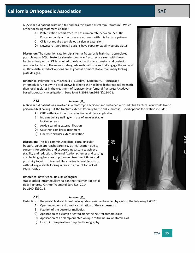
COA 95
California Orthopaedic Association SAE
A 95 year old patient sustains a fall and has this closed distal femur fracture. Which of the following statements is true?
A) Plate fixation of this fracture has a union rate between 95-100%
B) Posterior condylar fractures are not seen with this fracture pattern
C) CT is not required to rule out articular extension
D) Newest retrograde nail designs have superior stability versus plates Discussion: The nonunion rate for distal femur fractures is high than appreciated, possible up to 30%. Posterior shearing condylar fractures are seen with these fractures frequently. CT is required to rule out articular extension and posterior condylar fractures. The newest retrograde nails with screws that engage the rod and multiple distal interlock options are as good as or more stable than many locking plate designs. Reference: Pekmezci M1, McDonald E, Buckley J, Kandemir U. Retrograde intramedullary nails with distal screws locked to the nail have higher fatigue strength than locking plates in the treatment of supracondylar femoral fractures: A cadaver-based laboratory investigation. Bone Joint J. 2014 Jan;96-B(1):114-21.
234. Answer _B_ A 26 year old patient was involved in a motorcycle accident and sustained a closed tibia fracture. You would like to perform tibial nailing but the fracture extends laterally to the ankle mortise. Good options for fixation include:
A) ORIF with direct fracture reduction and plate application
B) Intramedullary nailing with use of angular stable locking screws
C) Ankle spanning external fixation
D) Cast then cast brace treatment
E) Fine wire circular external fixation Discussion: This is a comminuted distal extra-articular fracture. Open approaches are risky at this location due to concerns for stripping and exposure necessary to achieve stability and reduction. External fixation schemes and casting are challenging because of prolonged treatment times and proximity to joint. Intramedullary nailing is feasible with or without angle stable locking screws to account for lack of lateral cortex Reference: Boyer et al. Results of angular-stable locked intramedullary nails in the treatment of distal tibia fractures. Orthop Traumatol Surg Res. 2014 Dec;100(8):901-5.
235. Answer _D_ Reduction of the unstable distal tibio-fibular syndesmosis can be aided by each of the following EXCEPT:
A) Open reduction and direct visualization of the syndesmosis
B) Fixation of the posterior malleolus
C) Application of a clamp oriented along the neutral anatomic axis
D) Application of an clamp oriented oblique to the neural anatomic axis
E) Use of intra-operative computed tomography

COA 96
California Orthopaedic Association SAE
Discussion: Clamp induced malreduction of the syndesmosis has been shown to occur with instrument placement oblique to the anatomic axis of the syndesmosis. Each of the other interventions has been shown to improve or aid in reduction. Reference: Phisitkul P, Ebinger T, Goetz J, Vaseenon T, Marsh JL. Forceps reduction of the syndesmosis in rotational ankle fractures: a cadaveric study. J Bone Joint Surg Am. 2012 Dec 19;94(24):2256-61.
236. Answer _A_ Malreduction of the syndesmosis:
A) Has been shown to occur in up to 15% of cases even when a direct open reduction is performed
B) Is not affected by the morphology of the distal tibial incisura
C) Has little effect on patient reported functional outcomes
D) Is evaluated by the modified Cotton or external rotation stress test
E) Is unaffected by fibular reduction Discussion: The study by Miller et al. identified syndesmotic mal-reduction (as determined by postoperative computed tomography) in 16% of cases where a formal open reduction was performed. Reference: Miller AN, Carroll EA, Parker RJ, Boraiah S, Helfet DL, Lorich DG. Direct visualization for syndesmotic stabilization of ankle fractures. Foot Ankle Int. 2009 May;30(5):419-26.
237. Answer _E_ Screw stabilization of the distal tibio-fibular syndesmosis is ideally performed:
A) With a single 3.5mm fully threaded tricortical screw
B) With a single 4.5mm fully threaded tricortical screw
C) With a single 3.5mm fully threaded quadricortical screw
D) With two 3.5mm fully threaded tricoritcal screws
E) Current literature has not demonstrated superiority of one fixation strategy over another Discussion: Multiple biomechanical and clinical studies have evaluated fixation strategies for the distal tibio-fibular syndesmosis and one strategy (screw size and number of cortices) has not been identified as superior to another. Reference: Van Heest TJ, Lafferty PM. Injuries to the ankle syndesmosis. J Bone Joint Surg Am. 2014 Apr 2;96(7):603-13.
238. Answer _C_ A recent randomized controlled trial comparing static versus dynamic fixation of the ankle syndesmosis showed:
A) Improved syndesmotic reduction with the dynamic fixation device
B) Statistically significant improved functional outcome scores at all recorded time points with the dynamic fixation device
C) Lower rates of reoperation with the dynamic fixation device
D) Statistically significant rate in return to work and sports with the dynamic fixation device
E) Statistically significant increase in range of ankle dorsiflexion and plantarflexion with the dynamic fixation device
Discussion: Rates of reoperation for all reasons (implant failure, bother, loss of reduction) was greater in the group with syndesmotic screw stabilization. Reduction, functional outcome scores (at all time points), return to work/school and range of motion (both dorsi- and plantarflexion) were not found to be statistically different.

COA 97
California Orthopaedic Association SAE
Reference: Laflamme M, Belzile EL, Bédard L, van den Bekerom MP, Glazebrook M, Pelet S. A prospective randomized multicenter trial comparing clinical outcomes of patients treated surgically with a static or dynamic implant for acute ankle syndesmosis rupture. J Orthop Trauma. 2015 May;29(5):216-23.
239. Answer: __E__ A 77-year-old lady presents after falling and sustaining an injury to her knee. His medical history is significant only for osteoporosis and diabetes. Radiographs and representative CT scan images are shown in Figures A and B. What is the most appropriate treatment method for this patient's injury?
A) Traction and splinting B) Lateral on-locking buttress plate application C) Retrograde supracondylar nail fixation D) Knee spanning External fixation E) Lateral locking plate application
Discussion: The injury shown in Figures A and B represents a low comminuted metaphyseal extraarticular distal femur fracture in osteoporotic bone. Because of the poor bone quality and distal extent of this injury, the best fixation construct for treatment of this fracture in this patient is lateral locking plate application. Intramedullary nailing may be possible but the low extent of the fracture makes it difficult to maintain stability with most current implants. Knee spanning is typically temporary treatment and does not allow for early mobilization. References: Egol KA, Kubiak EN, Fulkerson E, Kummer FJ, Koval KJ. Biomechanics of locked plates and screws. J Orthop Trauma. 2004 Sep;18(8):488-93. Weight M, Collinge C. Early results of the less invasive stabilization system for mechanically unstable fractures of the distal femur (AO/OTA types A2, A3, C2, and C3).J Orthop Trauma. 2004 Sep;18(8):503-8.
240. Answer: __D__ A 25-year-old man sustains a femur fracture in a motorcycle accident. AP radiograph is shown below. Prior to surgery, a CT scan of the knee is ordered for preoperative planning. Which of the following additional findings is most likely to be discovered?
A) Tibial spine fracture B) Impaction fracture of medial femoral condyle C) Femoral neck fracture D) Coronal plane fracture (Hoffa Fracture) of lateral condyle E) Avulsion fracture of lateral tibial plateau (Segond Fracture)
Discussion: The "Hoffa fracture" is a coronal plane fracture of the femoral condyle that is often missed on plain radiographs of supracondylar and intercondylar femur fractures. It involves the lateral condyle more frequently than the medial. Identification is important as it may impact operative planning and likely require screw fixation in the anteroposterior plane. Nork et al. reviewed 202 supracondylar-intercondylar distal femoral fractures and found a 38% prevalence of associated coronal plane fractures. Impaction fractures and femoral neck fractures have no well defined association with distal femur fractures. Segond fractures suggest ACL disruption.

COA 98
California Orthopaedic Association SAE
Reference: Nork SE, Segina DN, Aflatoon K, Barei DP, Henley MB, Holt S, Benirschke SK.The association between supracondylar-intercondylar distal femoral fractures and corona
241. Answer: _C___ Nonunions and delayed unions of the distal femur have been attributed to all of the following EXCEPT:
A) Infection B) Bone loss C) Locking internal fixation with too little flexibility D) Locking internal fixation with too much flexibility E) Use of nonlocking plates with highly comminuted metaphyseal fractures
Discussion: Infection and bone loss are too common etiologies for delayed or nonunion after high energy trauma. Locked plates rely on the plate/screw interface, and each provides not only axial stability but also angular stability. Nonlocking plates cannot provide adequate stability in the setting of comminuted metaphyseal fractures. New literatures suggest that there may be a higher than appreciated rate of nonunion in distal femur fractures fixed with locking plates, and that this may be related to locking constructs that create too stiff of a biomechanical environment. This stiffness may inhibit normal patterns of fracture healing. References: Gardner MJ, Helfet DL, Lorich DG. Has locked plating completely replaced conventional plating? Am J Orthop (Belle Mead NJ). 2004 Sep;33(9):439-46. PMID:15509108 (Link to Abstract) Lujan TJ, Henderson CE, Madey SM, Fitzpatrick DC, Marsh JL, Bottlang M. Locked plating of distal femur fractures leads to inconsistent and asymmetric callus formation. J Orthop Trauma. 2010 Mar;24(3):156-62. doi: 10.1097/BOT.0b013e3181be6720. Henderson CE, Lujan TJ, Kuhl LL, Bottlang M, Fitzpatrick DC, Marsh JL. 2010 mid-America Orthopaedic Association Physician in Training Award: healing complications are common after locked plating for distal femur fractures. Clin Orthop Relat Res. 2011 Jun;469(6):1757-65. doi: 10.1007/s11999-011-1870-6. Epub 2011 Mar 22.
242. Answer: _E___ Which of the following substances is consistently osteoinductive?
A) Freeze dried allograft B) Demineralized bone matrix C) Calcium Phosphate D) Fresh cancellous allograft E) Autogenous iliac crest cancellous graft
Discussion: Prepared cancellous allografts provide scaffolding for osteoconduction but do not provide progenitor cells and growth factors for osteoinduction. Demineralized bone matrix may have active inductive proteins, but there is significant variability based on donor profile and preparation technique. Calcium phosphate provides structural support but has no inherent biologic activity. Autogenous bone graft contains osteoblasts, endosteal osteoprogenitor cells capable of forming new bone, and a structural matrix that acts as a scaffold, making it the gold standard for bone grafting. References: Bone-graft substitutes: facts, fictions, and applications. Greenwald AS, Boden SD, Goldberg VM, Khan Y, Laurencin CT, Rosier RN; American Academy of Orthopaedic Surgeons. The Committee on Biological Implants. J Bone Joint Surg Am. 2001; 83-A Suppl 2 Pt 2:98-103. PMID: 11712842
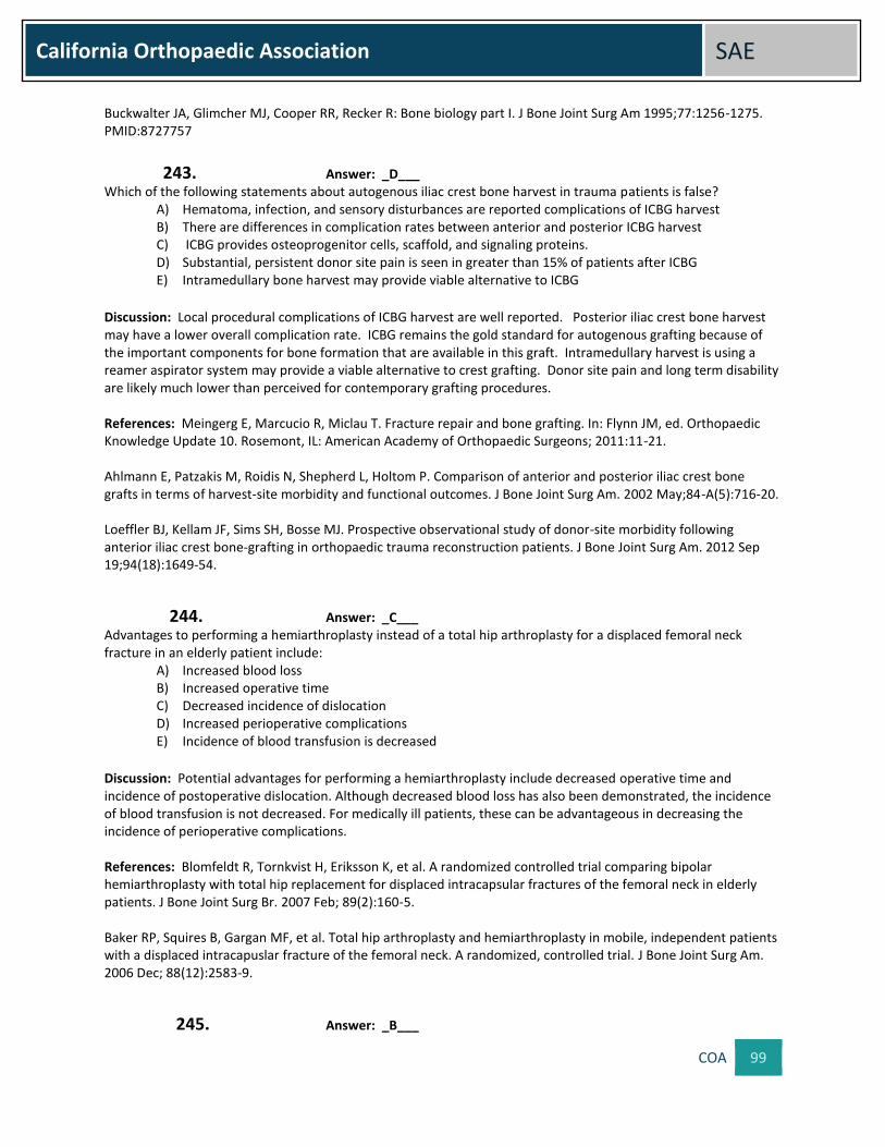
COA 99
California Orthopaedic Association SAE
Buckwalter JA, Glimcher MJ, Cooper RR, Recker R: Bone biology part I. J Bone Joint Surg Am 1995;77:1256-1275. PMID:8727757
243. Answer: _D___ Which of the following statements about autogenous iliac crest bone harvest in trauma patients is false?
A) Hematoma, infection, and sensory disturbances are reported complications of ICBG harvest B) There are differences in complication rates between anterior and posterior ICBG harvest C) ICBG provides osteoprogenitor cells, scaffold, and signaling proteins. D) Substantial, persistent donor site pain is seen in greater than 15% of patients after ICBG E) Intramedullary bone harvest may provide viable alternative to ICBG
Discussion: Local procedural complications of ICBG harvest are well reported. Posterior iliac crest bone harvest may have a lower overall complication rate. ICBG remains the gold standard for autogenous grafting because of the important components for bone formation that are available in this graft. Intramedullary harvest is using a reamer aspirator system may provide a viable alternative to crest grafting. Donor site pain and long term disability are likely much lower than perceived for contemporary grafting procedures. References: Meingerg E, Marcucio R, Miclau T. Fracture repair and bone grafting. In: Flynn JM, ed. Orthopaedic Knowledge Update 10. Rosemont, IL: American Academy of Orthopaedic Surgeons; 2011:11-21. Ahlmann E, Patzakis M, Roidis N, Shepherd L, Holtom P. Comparison of anterior and posterior iliac crest bone grafts in terms of harvest-site morbidity and functional outcomes. J Bone Joint Surg Am. 2002 May;84-A(5):716-20. Loeffler BJ, Kellam JF, Sims SH, Bosse MJ. Prospective observational study of donor-site morbidity following anterior iliac crest bone-grafting in orthopaedic trauma reconstruction patients. J Bone Joint Surg Am. 2012 Sep 19;94(18):1649-54.
244. Answer: _C___ Advantages to performing a hemiarthroplasty instead of a total hip arthroplasty for a displaced femoral neck fracture in an elderly patient include:
A) Increased blood loss B) Increased operative time C) Decreased incidence of dislocation D) Increased perioperative complications E) Incidence of blood transfusion is decreased
Discussion: Potential advantages for performing a hemiarthroplasty include decreased operative time and incidence of postoperative dislocation. Although decreased blood loss has also been demonstrated, the incidence of blood transfusion is not decreased. For medically ill patients, these can be advantageous in decreasing the incidence of perioperative complications. References: Blomfeldt R, Tornkvist H, Eriksson K, et al. A randomized controlled trial comparing bipolar hemiarthroplasty with total hip replacement for displaced intracapsular fractures of the femoral neck in elderly patients. J Bone Joint Surg Br. 2007 Feb; 89(2):160-5. Baker RP, Squires B, Gargan MF, et al. Total hip arthroplasty and hemiarthroplasty in mobile, independent patients with a displaced intracapuslar fracture of the femoral neck. A randomized, controlled trial. J Bone Joint Surg Am. 2006 Dec; 88(12):2583-9.
245. Answer: _B___

COA 100
California Orthopaedic Association SAE
In regards to surgical timing for an elderly patient with a displaced femoral neck fracture, which of the following is true?
A) Mortality at 3 months is increased if surgery is delayed greater than 12 hours B) 1 year mortality is increased if surgery is delayed greater than 4 days C) Timing of operative intervention does not affect postoperative mortality D) Mortality at 6 months is decreased if surgery is delayed for 3 days E) Delay of operative intervention up to 72 hours due to associated medical comorbidities does not
affect postoperative mortality
Discussion: Numerous studies have reported the correlation between time to surgery and mortality rates associated with femoral neck and hip fractures. It is recommended that patients are treated as expediently as possible with appropriate optimization of their medical comoribities. References: Moran CG, Wenn RT, Sikand M, Taylor AM. Early mortality after hip fracture: is delay before surgery important? J Bone Joint Surg Am. 2005 Mar;87 (3):483-9 Radcliff TA, Henderson WG, Stoner TJ, et al. Patient risk factors, operative care, and outcomes among older community-dwelling male veterans with hip fracture. J Bone Joint Surg Am. 2008 Jan; 90(1):34-42.
246. Answer: _D___ A 70-year-old male sustains a displaced transcervical femoral neck fracture while riding his bicycle. The optimal treatment for this patient is:
A) Open reduction internal fixation with cannulated screws B) Closed reduction and traction C) Hemiarthroplasty D) Total hip arthroplasty E) Open reduction internal fixation with sliding hip screw
Discussion: In a chronologically old, but physiologically young and cognitively intact patient, the optimal treatment is a total hip arthroplasty. Prior studies have demonstrated early failure rates nearing 50% for attempted internal fixation with nonunion and osteonecrosis as the most common complications. Reoperation and complication rates for primary total hip arthroplasty in this same cohort have been consistently less than 10%. Patient reported pain control as well as hip function scores are improved with a total hip arthroplasty as well. References: Callaghan JJ, Liu SS, Haidukewych GJ. Subcapital fractures. A Changing Paradigm. J Bone and Joint Surg Br. 2012 Nov; 94(11 Suppl A):19-21 Chammout GK, Mukka SS, Carlsson T, et al. Total hip replacement versus open reduction and internal fixation of displaced femoral neck fractures: a randomized long-term follow up study. J Bone Joint Surgery Am. 2012 Nov 7; 94(21):1921-8.
247. Answer: _A___ A 75-year-old female with a Garden I femoral neck fracture is most appropriately treated with:
A) Operative fixation with cannulated screws B) Closed reduction and traction C) Bipolar hemiarthroplasty D) Total hip arthroplasty E) Unipolar hemiarthroplasty

COA 101
California Orthopaedic Association SAE
Discussion: Nonoperative treatment of Garden 1 femoral neck fractures is associated with prolonged immobilization and rates of displacement nearing 50%. In situ fixation with multiple screws can be performed safely and is recommended to prevent further displacement and improve mobility. The role of releasing an intracapsular hematoma in these patients is controversial. References: Rodriguez-Merchan EC. In situ fixation of nondisplaced intracapsular fractures of the proximal femur. Clin Orthop Relat Res. 2002 Jun;(399):42-51 Verheyen CC, Smulders TC, van Walsum AD. High secondary displacement rate in the conservative treatment of impacted femoral neck fractures in 105 patients. Arch Orthop Trauma Surg. 2005 Apr;125(3):166-8 Shuqiang M, Kunzheng W, Zhichao T, et al. Outcome of non-operative management in Garden 1 femoral neck fractures. Injury. 2006; 37(10):974-8
248. Answer: _C___ Which of the following statements regarding optimal treatment modalities for depressed tibial plateau fractures is correct?
A) The standard surgical approach for tibial plateau fracture is a medial arthrotomy with submeniscal window
B) High-energy bicondylar fracture-dislocations are best managed by early open reduction with internal fixation on the day of injury
C) Fractures with articular depression of <2mm, in presence of a stable ligamentous knee exam, are amenable to non-operative management
D) Fractures with articular depression >2mm are best managed by indirect reduction through ligamentotaxis
Discussion: Most articular fractures of the tibial plateau are due to low-energy trauma mechanisms which exert valgus stress on the lateral tibial plateau, leading to articular depression or split/depression fractures. Articular depression of <2mm in presence of a stable knee exam represents the rare condition which is amenable to non-operative management. Unstable ligamentous knee injuries associated with tibial plateau fractures are exclusively managed by surgical fixation, in terms of ORIF. The “standard” surgical approach is represented by an anterolateral arthrotomy and submeniscal window for direct visualization of the depressed articular surface. Anatomic fracture reduction cannot be achieved by ligamentotaxis alone, because the depressed articular fractures frequently incarcerated within the metaphyseal bone. The safe strategy for managing high-energy bicondylar fracture dislocations is represented by a staged protocol of initial spanning external fixation, and delayed conversion to ORIF, due to the high risk of soft tissue-injury related complications, including fracture blisters and degloving injuries which may lead to wound breakdown and postoperative infection. References: Papagelopoulos PJ, Partsinevelos AA, Themistocleous GS, Mavrogenis AF, Korres DS, Soucacos PN: Complications after tibial plateau fracture surgery. Injury 2006, 37(6):475-484. Manidakis N, Dosani A, Dimitriou R, Stengel D, Matthews S, Giannoudis P: Tibial plateau fractures: functional outcome and incidence of osteoarthritis in 125 cases. International orthopaedics 2010, 34(4):565-570. Luo CF, Sun H, Zhang B, Zeng BF: Three-column fixation for complex tibial plateau fractures. Journal of orthopaedic trauma 2010, 24(11):683-692. Giannoudis PV, Tzioupis C, Papathanassopoulos A, Obakponovwe O, Roberts C: Articular step-off and risk of post-traumatic osteoarthritis. Evidence today. Injury 2010, 41(10):986-995. Broome B, Mauffrey C, Statton J, Seligson D: How to avoid the ‘trap door’ effect in reducing tibial plateau fractures. Orthopaedics Today 2011, 6:8-9.

COA 102
California Orthopaedic Association SAE
249. Answer: _B___ Which of the following statements regarding fracture classification systems for tibial plateau fractures is correct?
A) The Schatzker classification is based on the AO/OTA classification and characterizes all extra- and intra-articular tibial plateau fractures (A-,B-,C-types)
B) The Schatzker classification is based on the AO/OTA classification and characterizes all intra-articular tibial plateau fractures (B- and C-types)
C) The Schatzker and AO/OTA classifications are distinct entities which characterize different fracture patterns without any overlap in classification
D) The Moore classification was designed to characterize low-energy postero-medial fracture fragments
Discussion: The AO/OTA classification is designed to characterize any meta- or diaphyseal fracture in any anatomic location. Applied to tibial plateau fractures, the AO/OTA system is used to classify either extra- (A-type) or intra-articular (B- or C-type) fractures. In contrast, the Schatzker classification is exclusively applied to intra-articular fracture, either partial articular (equivalent to AO/OTA B-types) or bicondylar / complete articular fractures (equivalent to AO/OTA C-types). The Moore classification is exclusively applied to high-energy uni- or bicondylar tibial plateau fracture-dislocations, which are associated with a high risk for associated significant ligamentous and vascular injuries. References: Moore TM: Fracture-dislocation of the knee. Clin. Orthop. Relat. Res 1981, 156:128-40. Fakler JK, Ryzewicz M, Hartshorn C, Morgan SJ, Stahel PF, Smith WR: Optimizing the management of Moore type I postero-medial split fracture dislocations of the tibial head: description of the Lobenhoffer approach.J. Orthop. Trauma 2007, 21(5):330-6. Marsh JL, Slongo TF, Agel J, Broderick JS, Creevey W, DeCoster TA, Prokuski L, Sirkin MS, Ziran B, Henley B, Audige L: AO/OTA fracture and dislocation classification compendium - 2007. J. Orthop. Trauma2007, 21, Suppl. 10. Stahel PF, Smith WR, Morgan SJ: Posteromedial fracture fragments of the tibial plateau: an unsolved problem? J. Orthop. Trauma2008, 22(7):504. Zeltser DW, Leopold SS: Classifications in brief: Schatzker classification of tibial plateau fractures. Clin. Orthop. Relat. Res 2013, 471(2):371-4.
250. Answer: _D___ Which of the following statements regarding novel treatment options for depressed tibial plateau fractures is incorrect?
A) Articular fracture reduction may be achieved by indirect visualization using arthroscopy- or fluoroscopy-guided techniques
B) Recent studies postulate the use of inflatable bone tamps to achieve anatomic reduction of depressed articular tibial plateau fragments
C) Any of the above-mentioned reduction techniques may be safely combined with minimal-invasive fracture fixation modalities, either with plates (MIPO) or percutaneous lag screws
D) The use of inflatable bone tamps represents a safe technique associated with low risk for technical intraoperative complications
Discussion: Most articular fractures of the tibial plateau are due to low-energy trauma mechanisms which exert valgus stress on the lateral tibial plateau, leading to articular depression or split/depression fractures. Articular depression of <2mm in presence of a stable knee exam represents the rare condition which is amenable to non-operative management. Unstable ligamentous knee injuries associated with tibial plateau fractures are exclusively managed by surgical fixation, in terms of ORIF. The “standard” surgical approach is represented by an anterolateral arthrotomy and submeniscal window for direct visualization of the depressed articular surface. Anatomic fracture reduction cannot be achieved by ligamentotaxis alone, because the depressed articular fractures frequently

COA 103
California Orthopaedic Association SAE
incarcerated within the metaphyseal bone. The safe strategy for managing high-energy bicondylar fracture dislocations is represented by a staged protocol of initial spanning external fixation, and delayed conversion to ORIF, due to the high risk of soft tissue-injury related complications, including fracture blisters and degloving injuries which may lead to wound breakdown and postoperative infections. References: Musahl V, Tarkin I, Kobbe P, Tzioupis C, Siska PA, Pape HC: New trends and techniques in open reduction and internal fixation of fractures of the tibial plateau. J. Bone Joint Surg. (Br) 2009, 91(4):426-433. Biggi F, Di Fabio S, D'Antimo C, Trevisani S: Tibial plateau fractures: internal fixation with locking plates and the MIPO technique. Injury 2010, 41(11):1178-1182. Siegler J, Galissier B, Marcheix PS, Charissoux JL, Mabit C, Arnaud JP: Percutaneous fixation of tibial plateau fractures under arthroscopy: a medium term perspective. Orthop. Traumatol. Surg. Res. 2011, 97(1):44-50. Hahnhaussen J, Hak DJ, Weckbach S, Heiney JP, Stahel PF: Percutaneous inflation osteoplasty for indirect reduction of depressed tibial plateau fractures. Orthopedics 2012, 35(9):768-772. Mauffrey C, Fader R, Hammerberg EM, Hak DJ, Stahel PF: Incidence and pattern of technical complications in balloon-guided osteoplasty for depressed tibial plateau fractures: A pilot study in 20 consecutive patients. Patient Saf. Surg. 2013, 7(1):8.
251. Answer: _A___ Which of the following statements regarding locked plating technique for tibial plateau fractures is correct?
A) Locked plating is the preferred method of tibial plateau fracture fixation in patients with osteoporosis B) Locked plating technology has completely replaced the need for dual plating in bicondylar tibial
plateau fractures C) Locked plating represents the fixation of choice for lateral tibial plateau fractures
D) Locked plating represents the new “gold standard” for any tibial plateau fracture
Discussion: The introduction of locked plating technology for periarticular fractures in the past decade has induced
a paradigm-shift, which led to an over-utilization of locking plates for unjustified indications. Locked plating
unquestionably provides superior biomechanical properties in osteoporotic fractures and for selected bicondylar
tibial plateau fractures. However, “dual plating” remains a valid concept for complex bicondylar fractures, in terms
of providing increased medial support and improved reduction of medial condyle fracture by adjunctive medial
buttress plating for selected indications. Isolated lateral plateau fractures (Schatzker types I-III) represents another
entity where conventional plating may provide superior biomechanical properties by allowing compression to the
fracture with the use of non-locking screws. In summary, locked plating technology has improved the quality of
fracture fixation and retention of fixation in a variety of indications related to complex periarticular fractures,
however, the indication for locking plates in tibial plateau fractures should be scrutinized and applied judiciously.
References: Smith WR, Ziran BH, Anglen JO, Stahel PF: Locking plates: tips and tricks. Instr. Course Lect. 2008,
57:25-36.
Zhang Y, Fan DG, Ma BA, Sun SG: Treatment of complicated tibial plateau fractures with dual plating via a 2-
incision technique. Orthopedics 2012, 35(3):e359-64.
Biggi F, Di Fabio S, D’Antimo C, Trevisani S: Tibial plateau fracture: internal fixation with locking plates and the
MIPO technique. Injury 2010, 41(11):1178-82.
Yoo BJ, Beingessner DM, Barei DP: Stabilization of the posteromedial fragment in bicondylar tibial plateau
fractures: a mechanical comparison of locking and nonlocking single and dual plating methods. J. Trauma 2010,
69(1):148-55.

COA 104
California Orthopaedic Association SAE
Anglen J, Kyle RF, Marsh JL, Virkus WW, Watters WC 3rd, Keith MW, Turkelson CM, Wies JL, Boyer KM: Locking
plates for extremity fractures. J. Am. Acad. Orthop. Surg. 2009, 17(7):465-72.
Upper Extremity
252. Answer: _A___ Which of the following statements is true regarding the use of PRP in upper extremity injuries?
A) The use of PRP have no improvement in structural integrity, clinical outcomes and perioperative morbidity following rotator cuff repairs
B) The use of PRP can improve structural healing but not clinical outcomes following rotator cuff repairs C) The use of PRP can significantly improve healing of lateral epicondylitis D) PRP can be helpful to decrease symptoms from shoulder impingement E) PRP can improve clinical outcomes following rotator cuff repairs
Discussion: PRP has been studied in a randomized double-blinded study to show that there’s no difference in post-operative narcotic use, clinical outcomes scores and function up to a year from surgery Reference: Weber SC, Kauffman JI, Parise C, Weber SJ, Katz SD: Platelet – rich fibrin matrix in the management of arthroscopic repair of the rotator cuff: a prospective randomized double-blinded study. Am J Sports Med 2013 Feb 1 (20: 263-70