Abnormalities horizontal Clinical, oculographic and...
8
Journal of Neurology, Neurosurgery, and Psychiatry 1990;53:200-207 Abnormalities of horizontal gaze. Clinical, oculographic and magnetic resonance imaging findings. II Gaze palsy and internuclear ophthalmoplegia A M Bronstein, P Rudge, M A Gresty, G Du Boulay, Joan Morris Abstract The site of lesions responsible for horizontal gaze palsy and various types of internuclear ophthalmoplegia (INO) was established by identifying the common areas where the abnormal MRI signals from patients with a given ocular-motor disorder overlapped. Patients with uni- lateral gaze palsy had lesions in the paramedian area of the pons, including the abducens nucleus, the lateral part of the nucleus reticularis pontis caudalis and the nucleus reticularis pontis oralis. Patients with abducens nucleus lesions showed additional clinical signs of lateral rectus weakness. Lesions responsible for bilateral gaze palsy involved the pontine tegmental raphe. Since this region con- tains the saccadic omnipause neurons, this finding suggests that damage to omnipause cells produces slowing of sac- cades rather than opsoclonus, as pre- viously proposed. All INOs, regardless of the presence of impaired abduction or convergence, had similar MRI appearan- ces. Frequently the lesions in patients with INO, were not confined to the medial longitudinal fasciculus (MLF) but also involved neighbouring structures at the pontine and mid-brain levels. There was a statistically significant association bet- ween the clinical severity of the INO and the presence of abnormal abduction or convergence. The findings suggest that the lesions outside the MLF, which may affect abducens, gaze or convergence pathways, are responsible for the presence of features additional to INO, depending on the magnitude of functional disruption they produce.. The difficulty in identifying the site of lesions responsible for specific ocular-motor defects has been a major obstacle to a better clinical understanding of many neuro-ophthalmo- logical syndromes. In the preceding paper' we presented magnetic resonance imaging (MRI) and electro-oculographic data from a group of patients with lateral rectus weakness of central origin showing that, if the images from the scans are overlapped and processed statisti- cally, it is possible to obtain good clinical/ topographic correlations. It was found that the preferred site of lesions was the posterior half of the intra-brainstem abducens fasciculus, which agrees with the present view that lesions of the abducens nucleus itself produce conjugate gaze palsy rather than isolated lateral rectus weak- ness.2" In this paper we present the findings in patients with horizontal gaze palsy and with internuclear ophthalmoplegia (INO). An outstanding problem in human gaze palsies has been to locate in vivo the para- median pontine reticular formation (PPRF), which is the immediate pre-motor centre for saccade generation. Although initially defined by animal workers in functional terms as the area of the pontine tegmentum whose damage leads to a gaze palsy, recent single unit record- ing studies have shown that the PPRF is a conglomerate of various groups of cells with different functions and properties.5 Accordin- gly, the possibility arises that selective lesions within the PPRF might affect horizontal gaze in different ways. In this paper we will present some evidence supporting this view. With respect to INO, one of our interests was to determine the cause of slow abduction found in some patients. Conceivably, the slow abduc- tion might be due to an extension of the lesion outside the medial longitudinal fasciculus (MLF) into either the PPRF or the abducens nucleus or fasciculus.6 Alternatively it has been postulated that disordered inhibition of the antagonist medial rectus, also produced by the MLF lesion, slows the abducting movement.7 This view, however, assumed that the lesions were confined to the MLF. We have re- examined this question by using the MRI to define the size and/or location of lesions in these two types of INO. Material and methods The methodology has been fully described in the companion paper' so that a brief comment will suffice here. The identification of patients with INO and horizontal gaze palsy was made on a clinical basis. Separate eyes DC electro- oculography was used to confirm the presence of INO and to distinguish between INOs with normal and slowed abduction, according to the lower limit of abducting velocity (mean-2SD) in our normal population.6 More information on the patients will be found in the results section of each group. Three separate analyses of the MRI scans were made; (1) identification of the brainstem area where the areas of abnormal MRI signal Medical Research Council Neuro- Otology Unit, Institute of Neurology A M Bronstein M A Gresty P Rudge Institute of Neurology G du Boulay Computing and Statistics Unit, Institute of Neurology, National Hospital, Queen Square, London J Morris Correspondence to: Dr A M Bronstein, MRC Neuro-Otology Unit, Institute of Neurology, National Hospital, Queen Square, London, WCIN 3GB, United Kingdom. Received 24 May 1989 and in revised form 4 September 1989. Accepted 18 September 1989 200 on 27 May 2018 by guest. Protected by copyright. http://jnnp.bmj.com/ J Neurol Neurosurg Psychiatry: first published as 10.1136/jnnp.53.3.200 on 1 March 1990. Downloaded from
-
Upload
trinhxuyen -
Category
Documents
-
view
226 -
download
3
Transcript of Abnormalities horizontal Clinical, oculographic and...
Journal of Neurology, Neurosurgery, and Psychiatry 1990;53:200-207
Abnormalities of horizontal gaze. Clinical,oculographic and magnetic resonance imagingfindings. II Gaze palsy and internuclearophthalmoplegia
AM Bronstein, P Rudge, M A Gresty, G Du Boulay, Joan Morris
AbstractThe site of lesions responsible forhorizontal gaze palsy and various types ofinternuclear ophthalmoplegia (INO) wasestablished by identifying the commonareas where the abnormal MRI signalsfrom patients with a given ocular-motordisorder overlapped. Patients with uni-lateral gaze palsy had lesions in theparamedian area of the pons, includingthe abducens nucleus, the lateral part ofthe nucleus reticularis pontis caudalisand the nucleus reticularis pontis oralis.Patients with abducens nucleus lesionsshowed additional clinical signs of lateralrectus weakness. Lesions responsible forbilateral gaze palsy involved the pontinetegmental raphe. Since this region con-tains the saccadic omnipause neurons,this finding suggests that damage toomnipause cells produces slowing of sac-cades rather than opsoclonus, as pre-viously proposed. All INOs, regardless ofthe presence of impaired abduction orconvergence, had similar MRI appearan-ces. Frequently the lesions in patientswith INO, were not confined to the mediallongitudinal fasciculus (MLF) but alsoinvolved neighbouring structures at thepontine and mid-brain levels. There was astatistically significant association bet-ween the clinical severity of the INO andthe presence of abnormal abduction orconvergence. The findings suggest thatthe lesions outside the MLF, which mayaffect abducens, gaze or convergencepathways, are responsible for thepresence of features additional to INO,depending on the magnitude offunctionaldisruption they produce..
The difficulty in identifying the site of lesionsresponsible for specific ocular-motor defectshas been a major obstacle to a better clinicalunderstanding of many neuro-ophthalmo-logical syndromes. In the preceding paper' wepresented magnetic resonance imaging (MRI)and electro-oculographic data from a group ofpatients with lateral rectus weakness of centralorigin showing that, if the images from thescans are overlapped and processed statisti-cally, it is possible to obtain good clinical/topographic correlations. It was found that thepreferred site oflesions was the posterior halfof
the intra-brainstem abducens fasciculus, whichagrees with the present view that lesions of theabducens nucleus itself produce conjugate gazepalsy rather than isolated lateral rectus weak-ness.2" In this paper we present the findings inpatients with horizontal gaze palsy and withinternuclear ophthalmoplegia (INO).An outstanding problem in human gaze
palsies has been to locate in vivo the para-median pontine reticular formation (PPRF),which is the immediate pre-motor centre forsaccade generation. Although initially definedby animal workers in functional terms as thearea of the pontine tegmentum whose damageleads to a gaze palsy, recent single unit record-ing studies have shown that the PPRF is aconglomerate of various groups of cells withdifferent functions and properties.5 Accordin-gly, the possibility arises that selective lesionswithin the PPRF might affect horizontal gazein different ways. In this paper we will presentsome evidence supporting this view.With respect to INO, one ofour interests was
to determine the cause of slow abduction foundin some patients. Conceivably, the slow abduc-tion might be due to an extension of the lesionoutside the medial longitudinal fasciculus(MLF) into either the PPRF or the abducensnucleus or fasciculus.6 Alternatively it has beenpostulated that disordered inhibition of theantagonist medial rectus, also produced by theMLF lesion, slows the abducting movement.7This view, however, assumed that the lesionswere confined to the MLF. We have re-examined this question by using the MRI todefine the size and/or location of lesions inthese two types of INO.
Material and methodsThe methodology has been fully described inthe companion paper' so that a brief commentwill suffice here. The identification of patientswith INO and horizontal gaze palsy was madeon a clinical basis. Separate eyes DC electro-oculography was used to confirm the presenceof INO and to distinguish between INOs withnormal and slowed abduction, according to thelower limit of abducting velocity (mean-2SD)in our normal population.6 More informationon the patients will be found in the resultssection of each group.Three separate analyses of the MRI scans
were made; (1) identification of the brainstemarea where the areas of abnormal MRI signal
Medical ResearchCouncil Neuro-Otology Unit, InstituteofNeurologyA M BronsteinM A GrestyP RudgeInstitute of NeurologyG du BoulayComputing andStatistics Unit,Institute of Neurology,National Hospital,Queen Square,LondonJ MorrisCorrespondence to:Dr A M Bronstein, MRCNeuro-Otology Unit,Institute of Neurology,National Hospital, QueenSquare, London, WCIN3GB, United Kingdom.Received 24 May 1989 and inrevised form4 September 1989.Accepted 18 September 1989
200
on 27 May 2018 by guest. P
rotected by copyright.http://jnnp.bm
j.com/
J Neurol N
eurosurg Psychiatry: first published as 10.1136/jnnp.53.3.200 on 1 M
arch 1990. Dow
nloaded from
Abnormalities of horizontal gaze. II Gaze palsy and INO
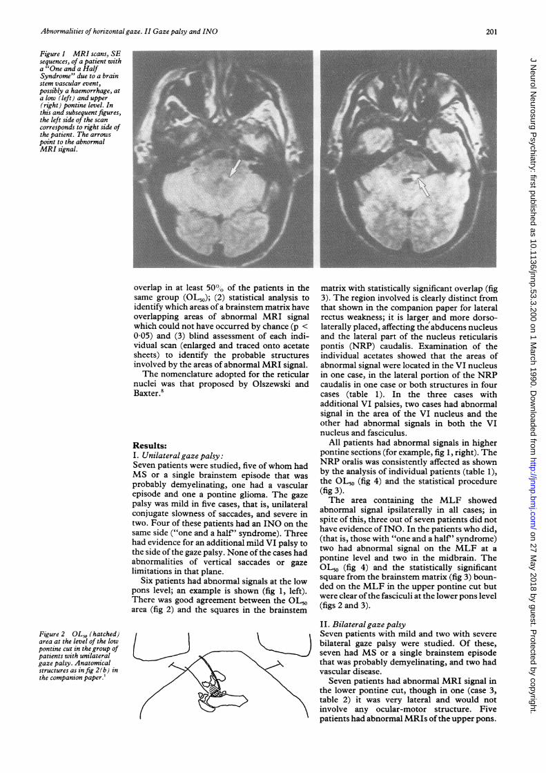
Figure I MRI scans, SEsequences, of a patient witha "One and a HalfSyndrome" due to a brainstem vascular event,possibly a haemorrhage, ata low (left) and upper(right) pontine level. Inthis and subsequentfigures,the left side of the scancorresponds to right side ofthe patient. The arrowspoint to the abnormalMRI signal.
overlap in at least 500° of the patients in thesame group (OL50); (2) statistical analysis toidentify which areas of a brainstem matrix haveoverlapping areas of abnormal MRI signalwhich could not have occurred by chance (p <0 05) and (3) blind assessment of each indi-vidual scan (enlarged and traced onto acetatesheets) to identify the probable structuresinvolved by the areas of abnormal MRI signal.The nomenclature adopted for the reticular
nuclei was that proposed by Olszewski andBaxter.8
Results:I. Unilateralgazepalsy:Seven patients were studied, five of whom hadMS or a single brainstem episode that wasprobably demyelinating, one had a vascularepisode and one a pontine glioma. The gazepalsy was mild in five cases, that is, unilateralconjugate slowness of saccades, and severe intwo. Four of these patients had an INO on thesame side ("one and a half' syndrome). Threehad evidence for an additional mild VI palsy tothe side of the gaze palsy. None of the cases hadabnormalities of vertical saccades or gazelimitations in that plane.
Six patients had abnormal signals at the lowpons level; an example is shown (fig 1, left).There was good agreement between the OL50area (fig 2) and the squares in the brainstem
Figure 2 OL50 (hatched)area at the level of the lowpontine cut in the group ofpatients with unilateralgaze palsy. Anatomicalstructures as infig 2(b) inthe companion paper.'
matrix with statistically significant overlap (fig3). The region involved is clearly distinct fromthat shown in the companion paper for lateralrectus weakness; it is larger and more dorso-laterally placed, affecting the abducens nucleusand the lateral part of the nucleus reticularispontis (NRP) caudalis. Examination of theindividual acetates showed that the areas ofabnormal signal were located in the VI nucleusin one case, in the lateral portion of the NRPcaudalis in one case or both structures in fourcases (table 1). In the three cases withadditional VI palsies, two cases had abnormalsignal in the area of the VI nucleus and theother had abnormal signals in both the VInucleus and fasciculus.
All patients had abnormal signals in higherpontine sections (for example, fig 1, right). TheNRP oralis was consistently affected as shownby the analysis of individual patients (table 1),the OL50 (fig 4) and the statistical procedure(fig 3).The area containing the MLF showed
abnormal signal ipsilaterally in all cases; inspite of this, three out of seven patients did nothave evidence of INO. In the patients who did,(that is, those with "one and a half' syndrome)two had abnormal signal on the MLF at apontine level and two in the midbrain. TheOL50 (fig 4) and the statistically significantsquare from the brainstem matrix (fig 3) boun-ded on the MLF in the upper pontine cut butwere clear ofthe fasciculi at the lower pons level(figs 2 and 3).
II. Bilateral gaze palsySeven patients with mild and two with severebilateral gaze palsy were studied. Of these,seven had MS or a single brainstem episodethat was probably demyelinating, and two hadvascular disease.
Seven patients had abnormal MRI signal inthe lower pontine cut, though in one (case 3,table 2) it was very lateral and would notinvolve any ocular-motor structure. Fivepatients had abnormal MRIs ofthe upper pons.
201
on 27 May 2018 by guest. P
rotected by copyright.http://jnnp.bm
j.com/
J Neurol N
eurosurg Psychiatry: first published as 10.1136/jnnp.53.3.200 on 1 M
arch 1990. Dow
nloaded from
Bronstein, Rudge, Gresty, Du Boulay, Morris
Gaze palsy
Unilateral
1
11
0DBilateral
/f-1Upper pons
Lower pons
INONormal abd. Slow abd.
9 _
1
14
111111 IlIlI l I llllll II lil l1 16 1 16
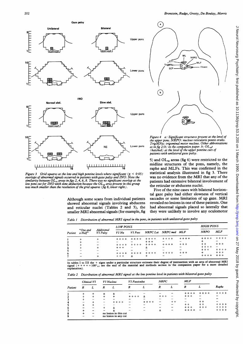
Figure 3 Grid squares at the low and high pontine levels where significant (joverlaps of abnormal signals occurred in patients with gaze palsy and INO. A>similarity between OL50 areas in figs 2, 4, 6, 8. There was no significant overllow pons cutfor INO with slow abduction because the OL,o area present in thiwas much smaller than the resolution of the grid squares (fig 8, lower right).
Although some scans from individuashowed abnormal signals involvingand reticular nuclei (Tables 2 anmsmaller MRI abnormal signals (for ex.
Upper pons
Figure 4 a: Significant structures present at the level ofthe upper pons. NRPO: nucleus reticularis pontis oralis.TrigMNu: trigeminal motor nucleus. Other abbreviations
Lower pons as in fig 2(b) in the companion paper. b: OL,0s(hatched) at the level of the upper pontine cuts ofpatients with unilateral gaze palsy.
5) and OL50 areas (fig 6) were restricted to themidline structures of the pons, namely, theraphe and MLFs. This was confirmed in the
D < 0.05) statistical analysis illustrated in fig 3. ThereVote the was no evidence from the MRI that any of theap at the patients had extensive bilateral involvement of
tS group the reticular or abducens nuclei.
Five of the nine cases with bilateral horizon-tal gaze palsy had either slowness of vertical
1 patients saccades or some limitation of up gaze. MRIabducens revealed no lesions in one of these patients. Onei 3), the had abnormal signals placed so laterally thatample, fig they were unlikely to involve any oculomotor
Table 1 Distribution of abnormal MRI signal in the pons, in patients with unilateralgaze palsy
LOWPONS HIGH PONS"One and Additional
Patient a Half' VI Palsy VI Nu VI Fasc NRPC Lat NRPC med MLF NRPO MLF
1 + + ++++ ++++ ++++ ++++ ++++ ++++ ++++
2 + - - - + + _3 + - ++++ ++++ ++++ ++++ +++ ++ +++
4 + + + - -_ _ +++ -
5 - - - - - - - +++ ++++6 - - ++++ ++++ ++++ ++++ +++ + +++7 - +++ + - - ++++ +++
In tables I to III the + signs under a particular structure estimate their degree of intersection with an area of abnormal MRIsignal (++++ =100,,; see the end of the material and methods section in the companion paper for a more detailedexplanation).
Table 2 Distribution of abnormal MRI signal at the low pontine level in patients with bilateralgaze palsy
Clinical VI VI Nucleus VI Fasciculus NRPC MLF
Patient R L R L R L R L R L Raphe
1 + + i i - - - - ++++ ++++ ++++2 + + + + ++++ ± +++ _ ++3 + - - - - - _ _ _ _ _4 - + - ++++ - ++++ - ++ - ++ _5 + + - - - - - - ++ ++6 - - + ± - - - - +++ +++ ++++7 + - - - - - - +++8 + - no lesion in this cut9 - - no lesion in any cut
202
on 27 May 2018 by guest. P
rotected by copyright.http://jnnp.bm
j.com/
J Neurol N
eurosurg Psychiatry: first published as 10.1136/jnnp.53.3.200 on 1 M
arch 1990. Dow
nloaded from
Abnormalities of horizontal gaze. II Gaze palsy and INO
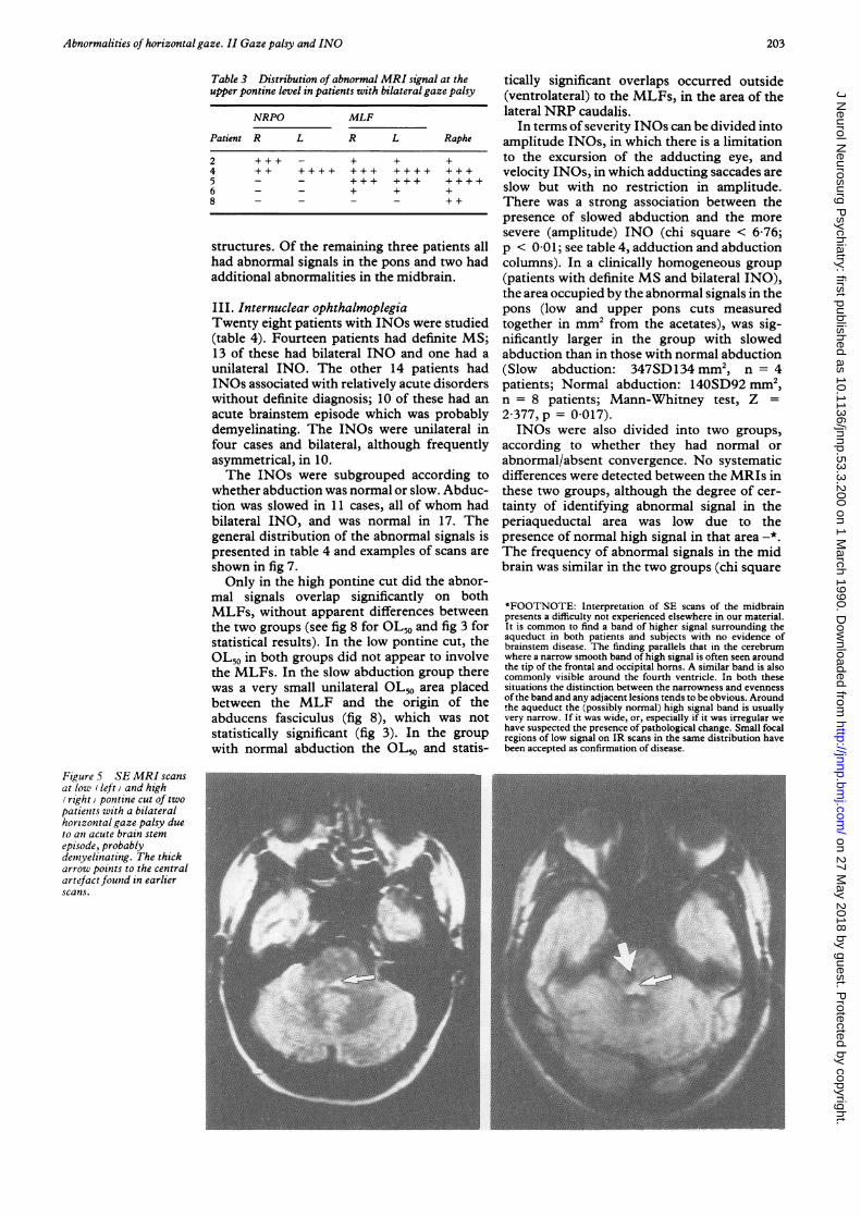
Table 3 Distribution of abnormal MRI signal at theupper pontine level in patients with bilateral gaze palsy
NRPO MLF
Patient R L R L Raphe
2 +++ - + + +4 ++ ++++ +++ ++++ +++5 - - +++ +++ ++++6 - - + + +8 - - - - ++
structures. Of the remaining three patients allhad abnormal signals in the pons and two hadadditional abnormalities in the midbrain.
III. Internuclear ophthalmoplegiaTwenty eight patients with INOs were studied(table 4). Fourteen patients had definite MS;13 of these had bilateral INO and one had aunilateral INO. The other 14 patients hadINOs associated with relatively acute disorderswithout definite diagnosis; 10 of these had anacute brainstem episode which was probablydemyelinating. The INOs were unilateral infour cases and bilateral, although frequentlyasymmetrical, in 10.The INOs were subgrouped according to
whether abduction was normal or slow. Abduc-tion was slowed in 11 cases, all of whom hadbilateral INO, and was normal in 17. Thegeneral distribution of the abnormal signals ispresented in table 4 and examples of scans areshown in fig 7.Only in the high pontine cut did the abnor-
mal signals overlap significantly on bothMLFs, without apparent differences betweenthe two groups (see fig 8 for OL50 and fig 3 forstatistical results). In the low pontine cut, theOL50 in both groups did not appear to involvethe MLFs. In the slow abduction group therewas a very small unilateral OL50 area placedbetween the MLF and the origin of theabducens fasciculus (fig 8), which was notstatistically significant (fig 3). In the groupwith normal abduction the OL50 and statis-
tically significant overlaps occurred outside(ventrolateral) to the MLFs, in the area of thelateral NRP caudalis.
In terms of severity INOs can be divided intoamplitude INOs, in which there is a limitationto the excursion of the adducting eye, andvelocity INOs, in which adducting saccades areslow but with no restriction in amplitude.There was a strong association between thepresence of slowed abduction and the moresevere (amplitude) INO (chi square < 6 76;p < 0-01; see table 4, adduction and abductioncolumns). In a clinically homogeneous group(patients with definite MS and bilateral INO),the area occupied by the abnormal signals in thepons (low and upper pons cuts measuredtogether in mm2 from the acetates), was sig-nificantly larger in the group with slowedabduction than in those with normal abduction(Slow abduction: 347SD134 mm2, n = 4patients; Normal abduction: 140SD92 mm2,n = 8 patients; Mann-Whitney test, Z2377,p = 0017).INOs were also divided into two groups,
according to whether they had normal orabnormal/absent convergence. No systematicdifferences were detected between the MRIs inthese two groups, although the degree of cer-tainty of identifying abnormal signal in theperiaqueductal area was low due to thepresence of normal high signal in that area -*.The frequency of abnormal signals in the midbrain was similar in the two groups (chi square
*FOOTNOTE: Interpretation of SE scans of the midbrainpresents a difficulty not experienced elsewhere in our material.It is common to find a band of higher signal surrounding theaqueduct in both patients and subjects with no evidence ofbrainstem disease. The finding parallels that in the cerebrumwhere a narrow smooth band of high signal is often seen aroundthe tip of the frontal and occipital homs. A similar band is alsocommonly visible around the fourth ventricle. In both thesesituations the distinction between the narrowness and evennessof the band and any adjacent lesions tends to be obvious. Aroundthe aqueduct the (possibly normal) high signal band is usuallyvery narrow. If it was wide, or, especially if it was irregular wehave suspected the presence of pathological change. Small focalregions of low signal on IR scans in the same distribution havebeen accepted as confirmation of disease.
Figure 5 SE MRI scansat low (left) and high(right v pontine cut of twopatients with a bilateralhorizontal gaze palsy dueto an acute brain stemepisode, probablydemyelinating. The thickarrow points to the centralartefact found in earlierscans.
203
on 27 May 2018 by guest. P
rotected by copyright.http://jnnp.bm
j.com/
J Neurol N
eurosurg Psychiatry: first published as 10.1136/jnnp.53.3.200 on 1 M
arch 1990. Dow
nloaded from
Bronstein, Rudge, Gresty, Du Boulay, Morris
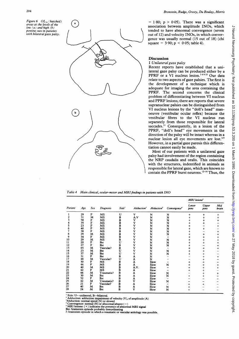
Figure 6 OL,O (hatched)areas at the levels of thelow (a) and high (b)pontine cuts in patientswith bilateral gaze palsy.
= 1 80; p > 0 05). There was a significantassociation between amplitude INOs, whichtended to have abnormal convergence (sevenout of 12) and velocity INOs, in which conver-
gence was usually normal (15 out of 18) (chisquare = 3 90; p < 0-05; table 4).
DiscussionI Unilateralgaze palsyRecent reports have established that a uni-lateral gaze palsy can be produced either by aPPRF or a VI nucleus lesion.34910 Our datarelate to two aspects of gaze palsies. The first isthe development of a technique which isadequate for imaging the area containing thePPRF. The second concerns the clinicalproblem of differentiating between VI nucleusand PPRF lesions; there are reports that severe
supranuclear palsies can be distinguished fromVI nucleus lesions by the "doll's head" man-oeuvre (vestibular ocular reflex) because thevestibular fibres to the VI nucleus run
separately from those responsible for lateralsaccades." Consequently, in a lesion of thePPRF, "doll's head" eye movements in thedirection of the palsy will be intact whereas in anuclear lesion all eye movements are lost.49However, in a partial gaze paresis this differen-tiation cannot easily be made.Most of our patients with a unilateral gaze
palsy had involvement of the region containingthe NRP caudalis and oralis. This coincideswith the structures, indentified in animals as
responsible for lateral gaze, which are known tocontain the PPRF burst neurons.'2'4 Thus, the
Table 4 Main clinical, ocular-motor and MRIfindings in patients with INO
MRI lesions?
Lower Upper MidPatient Age Sex Diagnosis Side' Adduction' Abduction' Convergence' pons pons brain
1 29 F MS U V N N + + +2 31 M MS B A.V N N + + +3 58 F MS B V N N + + +4 35 F MS B V N N + + -5 23 F MS B V N N + - +6 40 F MS B V N N + + -7 38 F MS B V N N + - +8 29 M MS B V N N + + +9 34 F MS B V N - + + +10 29 M MS B V N N + + +11 20 F Bst U V N N + + +12 27 F Bst B V N N + - +13 65 M Vascular? B V N - + + +14 23 M Bst U V N N + + -15 37 M Bst U V N N + + -16 31 F Bst B A N - - + +17 48 M Vascular? U A N - - +18 30 F MS B A Slow - + + -19 44 F MS B A Slow N + + +20 40 M MS B A.V. Slow - + + +21 42 F MS B A Slow - + + -22 66 M Traumatic? B A Slow N - + +23 30 M Bst B A Slow N + + -24 52 F Bst B A Slow - - - -25 42 M Traumatic? B V Slow N26 21 F Vascular? B A Slow - + +27 40 M Bst B A Slow N + - -28 24 M Bst B V Slow N + - -
' Side: U-unilateral, B-bilateral.2Adduction: adduction impairment of velocity (V), of amplitude (A)'Abduction: normal speed (N) or slowed4Convergence: normal (N) or abnormal/absent (-)5 MRI lesions: (+) indicates the presence of abnormal MRI signalBst: brainstem episode probably demyelinating?: brainstem episode in which a traumatic or vascular aetiology was possible.
204
C:-=Z:>
on 27 May 2018 by guest. P
rotected by copyright.http://jnnp.bm
j.com/
J Neurol N
eurosurg Psychiatry: first published as 10.1136/jnnp.53.3.200 on 1 M
arch 1990. Dow
nloaded from

Abnormalities of horizontal gaze. II Gaze palsy and INO
Figure 7 SE MRI scansat a high pontine cut intwo patients with bilateralINO. The one on the leftbelongs to a patient withnormal abduction velocitiesand the one on the the rightto a patient with slowabduction. The low pontinecut appearances (notshown) were npormal in thetwo cases.
significance ofour findings is that the OL50 areadrawn for our patients with unilateral gazepalsy is a template which identifies the locationof the PPRF on anMR image of the pons. Also,of clinical relevance, the patients with gazepalsy with VI nucleus involvement had signs ofadditional lateral rectus weakness. Thus, thepresence of this sign may be useful in identify-ing the site of the lesion in the mild gaze palsieswhich are more typical of demyelinating dis-ease.Four of the cases with unilateral gaze palsy
had an ipsilateral INO, the so called "one and ahalf' syndrome. Since ipsilateral abnormalsignal in the area of the MLF was seen in allpatients, regardless of whether they had anINO, it appears that high signal areas in theMRI do not always show a good clinicalcorrelation. The MLF abnormalities some-times occurred at different levels from thoseproducing the gaze palsy whereas in mostprevious descriptions of "one and a half' in
Slow abd.
\ ~~/\
CUp
Upper pons
Lower pons
Figure 8 OL50s (hatched) in patients with INO, with normal and slow abduction, atthe levels of the low and high pontine cuts.
vascular and neoplastic disease, it is probablethat a single lesion was responsible for the gazepalsy and the INO.915
II Bilateral gaze palsyAn unexpected finding in our patients withbilateral gaze palsy was the small areas ofabnormal signal confined to midline structuresof the pons, probably involving both MLFsand the raphe area ventral to them. The struc-tures involved probably included the nucleicentralis superioris and the medial part of theNRP caudalis. The latter has been recentlyfound to constitute a special subdivision ofNRP caudalis, the nucleus raphe interpositus,which contains a specialised type ofneuron, the"omnipause cells".'6 17 There were no sig-nificant lesions in the area of the abducensnucleus, the NRP oralis or the lateral part oftheNRP caudalis.Omnipause neurons normally inhibit
excitatory burst neurons, discharging tonicallyin between saccades and stopping abruptlyduring saccades." 18 It was first proposed thatomnipause neurons are the gate through whichdescending inputs, for example from thesuperior colliculus, trigger saccades. Because ofthis function it was postulated that opsoclonus,a condition in which saccades are incessantlyand chaotically generated, resulted from mal-functioning of the omnipause neurons.'9 Morerecently an alternative model of the saccadegenerator has been put forward in which directstimulation from long-lead burst neurons caninitiate the discharge in excitatory burstneurons, with omnipause neurons retaininga less crucial role of modifying saccadedynamics.20 To test this hypothesis, Kanekoand Fuchs2' produced discrete experimentallesions with ibotenic acid, an axon sparingreversible neurotoxin, in the omnipause area(nucleus raphe interpositus) in two monkeys.The animals developed slowness of saccades,more in the horizontal than in the verticalplane, but no opsoclonus was noted. Our data
Normal abd.\205
on 27 May 2018 by guest. P
rotected by copyright.http://jnnp.bm
j.com/
J Neurol N
eurosurg Psychiatry: first published as 10.1136/jnnp.53.3.200 on 1 M
arch 1990. Dow
nloaded from

Bronstein, Rudge, Gresty, Du Boulay, Morris
are consistent with this hypothesis and withother observations in humans. For example,pathological examination of the nucleus rapheinterpositus in patients with opsoclonus hasfailed to reveal abnormalities22 and the area ofthe nucleus raphe interpositus has been foundto be damaged in isolated pathological reportsof patients with slow saccades.2324The possibility that involvement of either
abducens or burst neurons or the descendingaxons of excitatory burst neurons as theycourse towards the VI nucleus25 could havecontributed to the findings in our patients,however, cannot be ruled out entirely. None ofour patients had extensive bilateral tegmentallesions which would also cause bilateral gazepalsy. Such patients with large reticular lesionswould be more severely disabled than the oneswe report here who were alert and ambulatory.We conclude that a mild bilateral gaze palsy in apatient who is otherwise not seriously ill ismore likely to be due to a central raphe lesionrather than to extensive bilateral tegmentallesions.
III. Internuclear ophthalmoplegiaIn our sample of patients with INO the areaswith significant overlap coincided with the siteof the MLF in the higher pontine cuts. Therewas no significant overlap in the region of theMLF in the low cuts. From this evidence itwould seem that in demyelinating disease theprevalent lesions producing INO are in theMLF rostral to the VI nucleus above the levelat which axons from the abducens neuronsdecussate and enter the fasciculi.Although it is accepted that INO is due to
lesions of the MLF,26 there is doubt about theorigin of impaired abduction and convergencewhich are features frequently accompanyingINOs. The MRIs of patients with both pureINOs and INOs with slow abduction showedareas of abnormal signal including the MLFbut with considerable extension to adjacentstructures including the abducens nucleus andfasciculus, NRP caudalis-oralis and the raphearea, all of which could impair abduction. Inour study slow abduction was associated withbilateral, more severe (amplitude) INOs andwith more extensive pontine lesions. Thiswould suggest that slow abduction depends onthe degree of functional impairment of theseadjacent structures. Unfortunately, with thecurrent limitation of MRI technique, thedegree of functional impairment of a structureby a lesion cannot be determined from theappearance on the scan.The presence of slow abduction in INO has
been attributed to a single MLF lesioninterrupting inhibitory fibres in the MLFwhich would normally silence the medial rectusmotor neuron during abducting saccades.7Evidence for this theory was based on reportsshowing disinhibited EMG activity in themedial rectus in INO.27 However, against thisexplanation is firstly, that the authors assumedthat no other lesions involving horizontal gazecentres were present7 which our present workindicates is unlikely, and secondly, repeatedexperimental studies have failed to find in-
hibitory fibres in the MLF.2"The MRI revealed abnormalities in the
midbrain as frequently in patients with normalas in those with abnormal convergence and nospecific location was systematically associatedwith impaired convergence. As with abductionfailure, deranged convergence was associatedwith more severe INOs.Thus, this study agrees with Cogan's im-
pression3" that the presence of abnormal con-vergence or abduction in patients with INO(what he called anterior or posterior INO,respectively) is related to concomitant damageto mid brain or pontine structures responsiblefor those associated signs. Our data show thatsuch structures, as indicated by an abnormalMRI signal, are more frequently involved bythe disease process than was previously suspec-ted. However, the presence of an abnormalsignal does not necessarily indicate impair-ment. It is the magnitude of the functionaldisruption produced by the lesion that deter-mines whether a defect will become clinicallymanifest.
We are grateful to the Multiple Sclerosis Society of GreatBritain and Northern Ireland for generous financial support andto Drs I Ormerod, D Miller, A Kermode and Mr D MacManusof the NMR Research Group at the National Hospital, QueenSquare for their patient work in the collection ofthe MR images.
1 Bronstein AM, Morris J, Du Boulay G, Gresty MA, RudgeP. Abnormalities of horizontal gaze. Clinical, oculo-graphic and magnetic resonance imaging findings. IAbducens palsy. J Neurol Neurosurg Psychiatry 1990;53:194-99.
2 Carpenter MB, McMasters RE, Hanna GR. Disturbances ofconjugate horizontal eye movements in the monkey. ArchNeurol 1963;8:231-47.
3 Meienberg 0, Buttner-Ennever JA, Kraus-Ruppert R.Lesion of the abducens nucleus as the cause of a conjugategaze paralysis. G Lennerstrand, DS Zee, EL Keller, eds.Functional Basis of Ocular Motility Disorders. Oxford:Pergamon Press, 1982:527-9.
4 Pierrot-Deseilligny Ch, Goasguen J. Isolated abducensnucleus damage due to histocytosis X. Brain 1984;107:1019-32.
5 Fuchs AF, Kaneko CRS, Scudder CA. Brainstem control ofsaccadic eye movements. Ann Rev Neurosci 1985;8:307-37.
6 Bird AC, Leech J. Internuclear ophthalmoplegia. Br JOphthal 1976;60:645-51.
7 Feldon SE, Hoyt WF, Stark L. Disordered inhibition ininternuclear ophthalmoplegia. Brain 1980;103:113-7.
8 Olszewski J, Baxter D. Cytoarchitecture of the human brainstem. Basel: Reinhardt 1954.
9 Pierrot-Deseilligny CH, Chain F, Serdaru M, Gray F,L'Hermitte F. The "One-and-a-half' Syndrome. Elec-tro-oculographic analyses of five cases with deductionsabout the physiological mechanisms of lateral gaze. Brain1981;104:665-99.
10 Kommerell G, Henn V, Bach M, Lucking CH. Unilaterallesion of the paramedian pontine reticular formation.Neuro-ophthal 1987;7:93-8.
11 Henn V, Lang W, Hepp K, Reisine H. Experimental gazepalsies in monkeys and their relation to human pathology.Brain 1984;107:619-36.
12 Buttner-Ennever JA, Henn V. An autoradiographic study ofthe pathways from the pontine reticular formationinvolved in horozontal eye movements. Brain Res 1976;108:155-6.
13 Cohen B, Henn V. Unit activity in the pontine reticularformation associated with eye movements. Brain Res1972;46:403-10.
14 Highstein SM, Maekawa K, Steinacker A, Cohen B. Synap-tic input from the pontine reticular nuclei to abducensmotoneurons and internuclear neurons in the cat. BrainRes 1976;112:162-7.
15 Miller Fisher C. Some neuro-opthalmological observations.J Neurol Neurosurg Psychiatry 1967;30.383-9.
16 Buttner-Ennever JA, Cohen B, Pause M, Fries W. RapheNucleus of the Pons containing Omnipause Neurons ofthe oculomotor system in the monkey, and its homologuein man. J Comparative Neurology 1988;267:307-2 1.
17 Strassman A, Evinger C, McCrea RA, Baker RG, HighsteinSM. Anatomy and physiology of intracellularly labelledomnipause neurons in the cat and squirrel monkey. ExperBrain Res 1987;67:436-40.
18 Keller EL. Participation of medial pontine reticular forma-
206
on 27 May 2018 by guest. P
rotected by copyright.http://jnnp.bm
j.com/
J Neurol N
eurosurg Psychiatry: first published as 10.1136/jnnp.53.3.200 on 1 M
arch 1990. Dow
nloaded from
Abnormalities of horizontal gaze. II Gaze palsy and INO
tion in eye movement generation in monkey. J Neuro-physiol 1974;37:316-32.
19 Zee DS, Robinson DA. A hypothetical explanation ofsaccadic oscillations. Ann Neurol 1979;5:405-14.
20 Scudder CA. A new local feedback model of the saccadicburst generator. J Neurophysiol 1988;59:1455-75.
21 Kaneko CRS, Fuchs AF. The effect of ibotenic acid lesionsof the omnipause neurons on saccadic eye movements inthe monkey. Neuroscience Abstracts 1987;13:392.
22 Ridley A, Kennard C, Scholtz CL, Buttner-Ennever JA,Summers B, Turnbull A. Omnipause neurons in two cases
of opsoclonus associated with oat cell carcinoma of thelung. Brain 1987;110:1699-709.
23 Hanson MR, Hamid MA, Tomsak RL, Chou SS, Leigh RJ.Selective saccadic palsy caused by pontine lesions: Clini-cal, Psychological and Pathological correlations. AnnNeurology 1986;20:209-17.
24 Buttner-Ennever JA, Mehraein P, Uemura T, Tateishi J,Kanaseki T, Arai Y. A neuroanatomical analysis of theoculomotor deficits seen in a case of Gaucher disease. ProcNeuropathol Verona 1988.
25 Strassman A, Highstein SM, McCrea RA. Anatomy and
Physiology of Saccadic Burst Neurons in the AlertSquirrel Monkey. I. Excitatory Burst Neurons. JournalComp Neurol 1986;249:337-57.
26 Atlas SW, Grossman RI, Savino PJ, et al.. InternuclearOphthalmoplegia: MR-Anatomic Correlation. AmericanJournal of Neuroradiology 1987;8:243-7.
27 Loeffler JD, Hoyt WF, Slatt B. Motor excitation andinhibition in internuclear palsy. Arch Neurol 1966;15:664-71.
28 Pola J, Robinson DA. An explanation ofeye movements seenin internuclear ophthalmoplegia. Arch Neurol 1976;33:447-52.
29 Pola J, Robinson DA. Oculomotor signals in medial longi-tudinal fasciculus of the monkey. J Neurophysiol 1978;41:245-59.
30 King WM, Lisberger SG, Fuchs AF. Responses of fibers inmedial longitudinal fasciculus (MLF) of alert monkeysduring horizontal and vertical conjugate eye movementsevoked by vestibular or visual stimuli. J Neurophysiol1976;39:1 135-49.
31 Cogan DG. Internuclear ophthalmoplegia, typical andatypical. Arch of Opthalmol 1970;84:583-9.
207
on 27 May 2018 by guest. P
rotected by copyright.http://jnnp.bm
j.com/
J Neurol N
eurosurg Psychiatry: first published as 10.1136/jnnp.53.3.200 on 1 M
arch 1990. Dow
nloaded from


![Vegetative postencephalitic syndromes - JNNP’s ambition ...jnnp.bmj.com/content/jnnp/s1-7/27/248.full.pdf · SENSORIMOTOR NEUROLOGY. [143] Tumours ofthe frontal lobe ... scrupulosity,](https://static.fdocuments.in/doc/165x107/5ada19d77f8b9a137f8cff6b/vegetative-postencephalitic-syndromes-jnnps-ambition-jnnpbmjcomcontentjnnps1-727248fullpdfsensorimotor.jpg)