ABGs & Electrolyte Disorders - atcomdce.orgatcomdce.org/files/Presentations/Acid Base and...
91
ABGs & Electrolyte Disorders Paul R Hinchey MD, MBA, EMT-P Medical Director WakeMed Mobile Critical Care
Transcript of ABGs & Electrolyte Disorders - atcomdce.orgatcomdce.org/files/Presentations/Acid Base and...

ABGs & Electrolyte Disorders
Paul R Hinchey MD, MBA, EMT-P
Medical Director
WakeMed Mobile Critical Care

Objectives
� Review acid / base imbalance
� Review anion gap and non-anion gap metabolic acidosis
� Review electrolyte disorders� Na
� K
� Ca
� HCO3
� BUN
� Cr

pH
� pH is the inverse log of the Hydrogen ion concentration
� It is a logarithmic function so small changes in the number represent large changes in H concentration
� Normal physiologic pH is 7.35-7.45� Normal enzymatic function occurs only within a
very narrow pH� Rapid derangement or more gradual but more
severe derangement causes a loss of enzymatic function and inability to maintain cellular processes resulting in death


ABGs
� pH/pO2/pCO2/HCO3/BE/Sat
� HCO3 in ABG is calculated NOT measured
� HCO3 in a Chem 7 is measured
� Base deficit/excess is the amount of bicarbonate that would have to be added to achieve a normal pH

Acid / Base Disorders
� Respiratory � Acidosis
� Alkalosis
� Metabolic� Acidosis
� Anion gap
� Non-anion gap
� Alkalosis
� Compensation mechanisms

Compensatory Mechanism
� Intravascular buffers� HGB / plasma proteins
� Phosphate
� Bicarbonate buffer system (1/2 the buffer effect)� H2O + CO2 = H2CO3 = H+ + HCO3
-
� Respiratory � Respiratory control of CO2 concentration
� Renal compensation� retention/excretion of HCO3
-

Fundamentals
� Look at the pH
� Primary respiratory disorder
� Look at CO2
� Primary metabolic disorder
� Look at HCO3
� Any compensation that occurs will not exceed the magnitude of the primary disorder

ABG Interpretation
� Key to success is systematic approach
� Remember the principal rule….
� The body does not compensate past the primary disturbance
� Identify your abnormalities in pH (7.35-7.45), pCO2 (35-45) and HCO3 (22-26)
� Lets look at isolated disorders to start
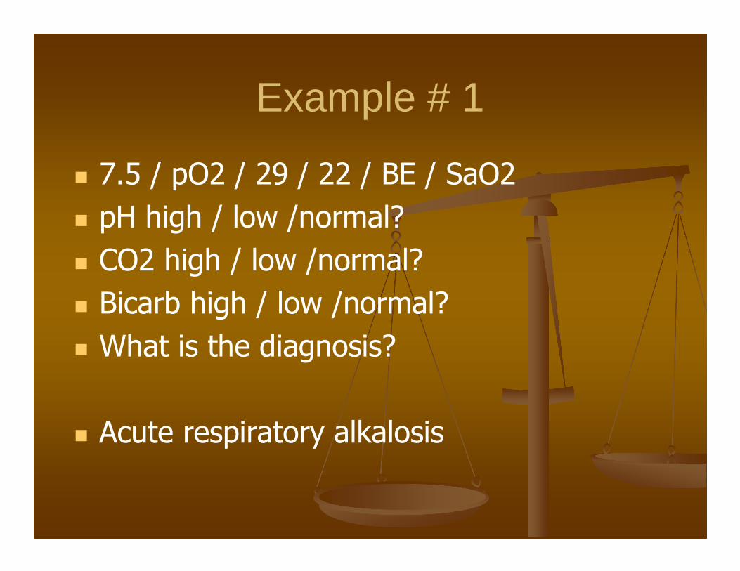
Example # 1
� 7.5 / pO2 / 29 / 22 / BE / SaO2
� pH high / low /normal?
� CO2 high / low /normal?
� Bicarb high / low /normal?
� What is the diagnosis?
� Acute respiratory alkalosis

Respiratory Alkalosis
� Primary derangement is caused by excessive off-gassing of CO2
� Long term compensation is HCO3-
excretion by the kidney

Respiratory Alkalosis
� Anxiety
� Hypoxia
� Lung ds with or without hypoxia
� Central nervous system ds
� Drugs – salicylates, catecholamines
� Pregnancy
� Sepsis
� Hepatic encephalopathy
� Mechanical ventilation

Example # 2
� 7.25 / pO2 / 60 / 26 / BE / SaO2
� pH high / low /normal?
� CO2 high / low /normal?
� Bicarb high / low /normal?
� What is the diagnosis?
� Acute respiratory acidosis
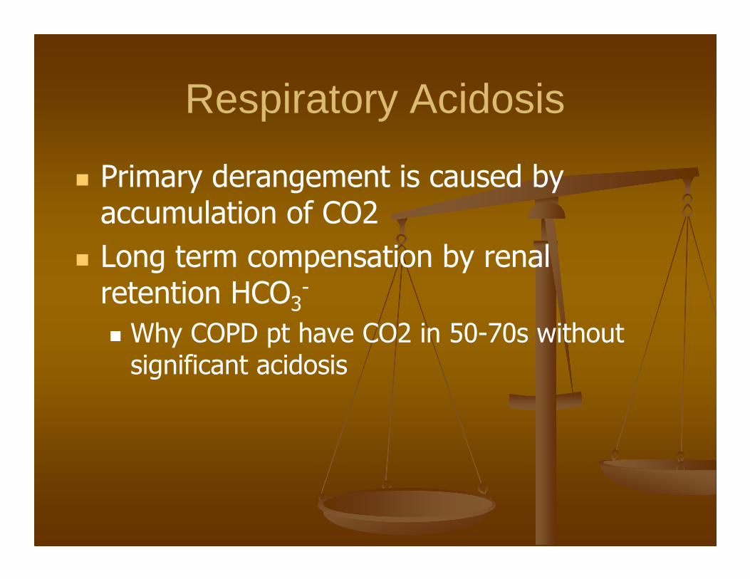
Respiratory Acidosis
� Primary derangement is caused by accumulation of CO2
� Long term compensation by renal retention HCO3
-
� Why COPD pt have CO2 in 50-70s without significant acidosis

Causes of Respiratory Acidosis
� CNS depression
� Drugs, cns event, cns masses
� Neuromuscular disorders
� Myopathies, neuropathies
� Acute airway obstruction
� Upper airway, laryngospasm, bronchospasm
� Impaired lung motion
� Hemothorax, pneumothorax, flail chest
� Ventilator dysfunction

Example # 3
� 7.34 / pO2 / 60 / 31 / BE / SaO2
� pH high / low /normal?
� CO2 high / low /normal?
� Bicarb high / low /normal?
� What is the diagnosis?
� Chronic respiratory acidosis with metabolic compensation (alkalosis)

Example # 4
� 7.50 / pO2 / 48 / 36 / BE / SaO2
� pH high / low /normal?
� CO2 high / low /normal?
� Bicarb high / low /normal?
� What is the diagnosis?
� Metabolic alkalosis with respiratory compensation (acidosis)

Metabolic Alkalosis
� Caused by loss of endogenous acids or introduction of exogenous base
� Primary compensatory mechanism is CO2
retention (hypoventilation)
� Causes are subdivided by chloride response

Metabolic Alkalosis
� Urinary chloride low
� Vomiting, nasogastric suctioning
� Diuretic use
� Posthypercapnia
� Urinary chloride high/normal
� Excess mineralocorticoids –Conn/Cushing syndrome, exogenous steroid, licorice
� Diuretics
� Excess alkali administration
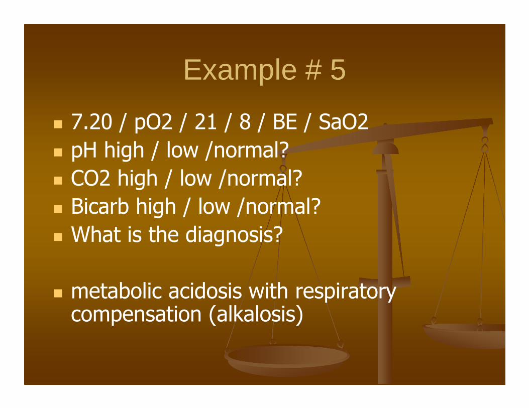
Example # 5
� 7.20 / pO2 / 21 / 8 / BE / SaO2
� pH high / low /normal?
� CO2 high / low /normal?
� Bicarb high / low /normal?
� What is the diagnosis?
� metabolic acidosis with respiratory compensation (alkalosis)

Metabolic Acidosis
� Causes are divided by anion gap vs non-anion gap metabolic acidosis
� Anion Gap is the normal gap between positive ions and negative ions

Metabolic Acidosis
� May be anion gap or non-anion gap
� Calculate the anion gap
� Most labs provide this for you
� Positive ions – negative ions
� Na – (HCO3 + Cl)
� Potassium is excluded because of its very small contribution to extracellular cations

Anion Gap Metabolic Acidosis
M - methanol
U - uremia
D - DKA
P – Phenol, paraldehyde
I – INH, Iron
L - lactate
E – ethanol, ethylene glycol
S - salicylates

Non-Anion Gap Metabolic Acidosis
� Usually the result of derangement of the bicarbonate system� Gi Bicarbonate loss
� Diarrhea
� Renal Bicarbonate loss� RTA� Early renal failure� Carbonic anhydrase inhibitors� Aldosterone inhibitors
� Not usually a concern in the acute setting� Requires more extensive work-up including urine
studies

Non-Anion Gap Metabolic Acidosis
� Hypercholoremic Metabolic Acidosis
� May be induced by excessive volume resuscitation with saline
� NaCl + H2O = HCl + NaOH
� Strong acid and base should balance?
� Normal ratio of Na : Cl is 140 :100
� Now provide Na and Cl in 1:1 ratio
� Excessive Cl leads to acidosis

Ok now it gets tricky
� Mixed acid base disorders
� Respiratory acidosis w/(AG or NAG) metabolic acidosis and metabolic alkalosis
� OR Respiratory alkalosis w/(AG or NAG) metabolic acidosis and metabolic alkalosis
� OR (AG or NAG) metabolic acidosis and metabolic alkalosis
� Luckily you can’t have 4 disorders at the same time because you can’t hyper and hypo ventilate
at the same time!

ABG Interpretation
� Step 1 � Look at pH if <7.4 is acidosis if >7.4 is alkalosis as
primary disorder
� Step 2 : Calculate the anion gap� Na – (HCO3 + Cl)� If gap is > or = 20 there is a primary metabolic
acidosis regardless of pH or bicarbonate
� Step 3 : Calculate the excess anion gap� Calc AG – Norm AG (12)� Add this to the measured bicarb
� If > normal bicarb (>30) have metabolic alkalosis� If < normal bicarb (<23) there is a non-AG metabolic
acidosis

Example # 6
� 7.50 / pO2 / 20 / 15 / BE / SaO2
� pH high / low /normal?
� CO2 high / low /normal?
� Bicarb high / low /normal?
� What is the diagnosis?
� This looks like respiratory alkalosis…and it is. But there is more to it.
� Look at the bicarb…

Example # 6
� 7.50 / pO2 / 20 / 15 / BE / SaO2
� Na 140; Cl 103; HCO3 15� AG = 140-(103+15) = 22
� Rule # 2 if AG >/= to 20 there is a primary metabolic acidosis regardless of pH or HCO3
� Now rule #3� Excess gap = 22-12 =10 Add to bicarb
� 10+15=25
� >30 or <23? No

Example #6
� Where does this stuff come from?
� This patient had a centrally mediated respiratory alkalosis (salicylates directly stimulate
the medulla) but….
� They also ingested a large quantity of ASA and had a primary metabolic acidosis as well.
� No additional disorder is present.

Example # 7
� 7.40 / pO2 / 40 / 24 / BE / SaO2
� pH high / low /normal?
� CO2 high / low /normal?
� Bicarb high / low /normal?
� What is the diagnosis?
� Nothing wrong here….right?
� Calculate the anion gap

Example # 7
� 7.40 / pO2 / 40 / 24 / BE / SaO2
� Na 145; Cl 100; HCO3 24� AG = 145-(100+24) = 21
� Rule # 2 if AG >/= to 20 there is a primary metabolic acidosis regardless of pH or HCO3
� Now rule #3� Excess gap = 21-12 =9 Add to bicarb
� 9+24=33
� >30 or <23? Yes!
� There is a metabolic alkalosis as well!
Example # 7
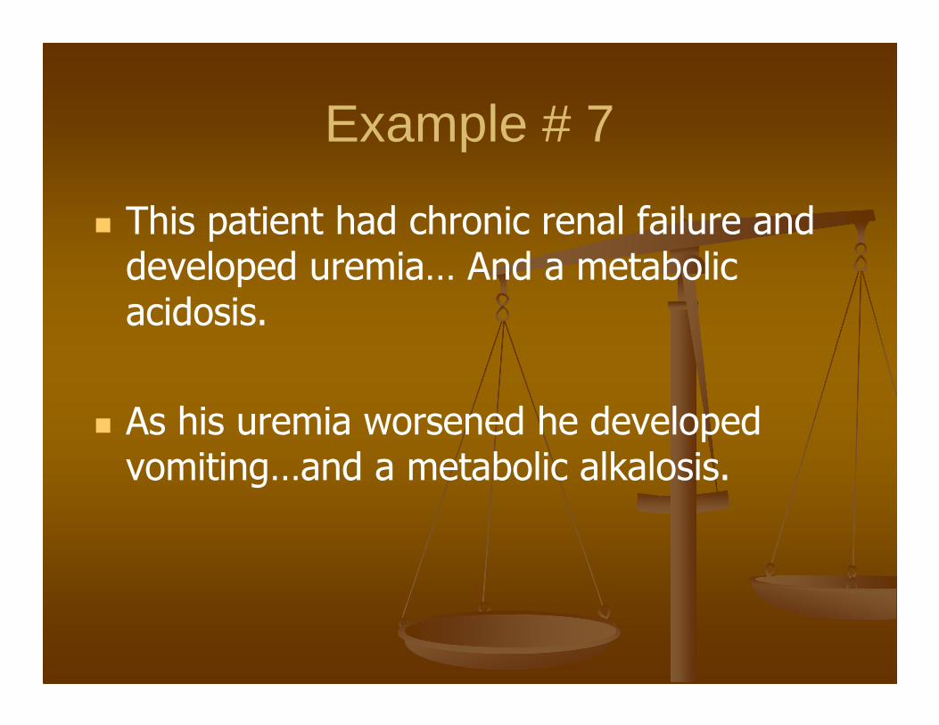
� This patient had chronic renal failure and developed uremia… And a metabolic acidosis.
� As his uremia worsened he developed vomiting…and a metabolic alkalosis.

Example # 8
� 7.50 / pO2 / 20 / 15 / BE / SaO2
� pH high / low /normal?
� CO2 high / low /normal?
� Bicarb high / low /normal?
� What is the diagnosis?
� There is an alkalosis but the bicarb is low and the CO2 is low….this ain’t right?
� Calculate the anion gap
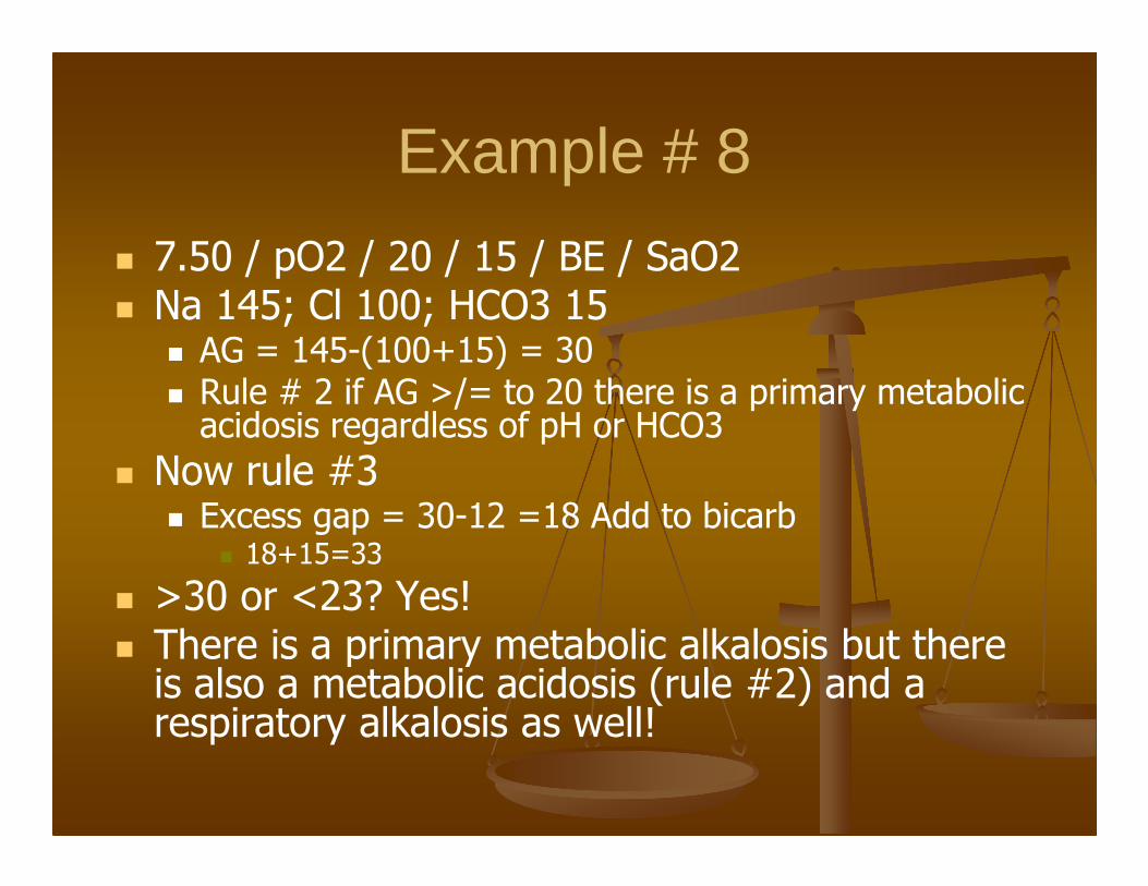
Example # 8
� 7.50 / pO2 / 20 / 15 / BE / SaO2� Na 145; Cl 100; HCO3 15
� AG = 145-(100+15) = 30� Rule # 2 if AG >/= to 20 there is a primary metabolic
acidosis regardless of pH or HCO3
� Now rule #3� Excess gap = 30-12 =18 Add to bicarb
� 18+15=33
� >30 or <23? Yes!� There is a primary metabolic alkalosis but there
is also a metabolic acidosis (rule #2) and a respiratory alkalosis as well!

OK how does that happen?
� This patient had pneumonia which resulted in the respiratory alkalosis
� They also vomiting which caused the metabolic alkalosis and…
� They had alcoholic ketoacidosis which caused the metabolic acidosis.

Example # 9
� 7.10 / pO2 / 50 / 15 / BE / SaO2
� pH high / low /normal?
� CO2 high / low /normal?
� Bicarb high / low /normal?
� What is the diagnosis?
� There is an acidosis with both a high CO2 and a low bicarb.
� Calculate the anion gap

Example # 9
� 7.10 / pO2 / 50 / 15 / BE / SaO2� Na 145; Cl 100; HCO3 15
� AG = 145-(100+15) = 30� Rule # 2 if AG >/= to 20 there is a primary metabolic
acidosis regardless of pH or HCO3
� Now rule #3� Excess gap = 30-12 =18 Add to bicarb
� 18+15=33
� >30 or <23? Yes!� There is a primary respiratory acidosis but there
is also a metabolic acidosis (rule #2) and a metabolic alkalosis as well!

OK how does that happen?
� This patient was obtunded and hypoventilating hence the respiratory acidosis; had vomiting leading to the metabolic alkalosis and had diabetic ketoacidosis which caused their anion gap metabolic acidosis.

Example # 10
� 7.15 / pO2 / 15 / 5 / BE / SaO2
� pH high / low /normal?
� CO2 high / low /normal?
� Bicarb high / low /normal?
� What is the diagnosis?
� Clearly there is a metabolic acidosis and a respiratory alkalosis. By now you know there must be something else…

Example # 10
� 7.15 / pO2 / 15 / 5 / BE / SaO2� Na 145; Cl 100; HCO3 15
� AG = 140-(110+5) = 25� Rule # 2 if AG >/= to 20 there is a primary metabolic acidosis
regardless of pH or HCO3
� Now rule #3� Excess gap = 25-12 =13 Add to bicarb
� 13+5=18
� >30 or <23? Yes!� This looks like a AG metabolic acidosis with respiratory
compensation. However the change in bicarb does not correspond to the AG acidosis alone. There is a NAG acidosis as well!

OK how does that happen?
� This patient had DKA with a respiratory alkalosis. However the patient also had a NAG acidosis caused by hypercholoremic metabolic acidosis.

Had enough?

Osmosis
� Concentrations across a semipermeable membrane want to become equal
� If solute can not pass through the membrane, water will move into or out of the cell to make the concentrations equal.
� Osmotic pressure describes the driving force of this mechanism

RBCs in solution
Hypotonic Hypertonic

Sodium Regulation (135-145)
� Sodium is regulated by:
� Renin-Angiotensin system
� Renin triggers secretion of Aldosterone
� Aldosterone
� Increases sodium reabsorption
� Increases potassium excretion

Water regulation
� Water homeostasis is maintained by the hypothalamus
� Senses rise in osmolarity and triggers
� Thirst
� Release of antidiuretic hormone
� ADH increases water reabsorption in the distal tubule
� Usually Na and H2O changes are proportionate and do not cause changes in sodium concentration
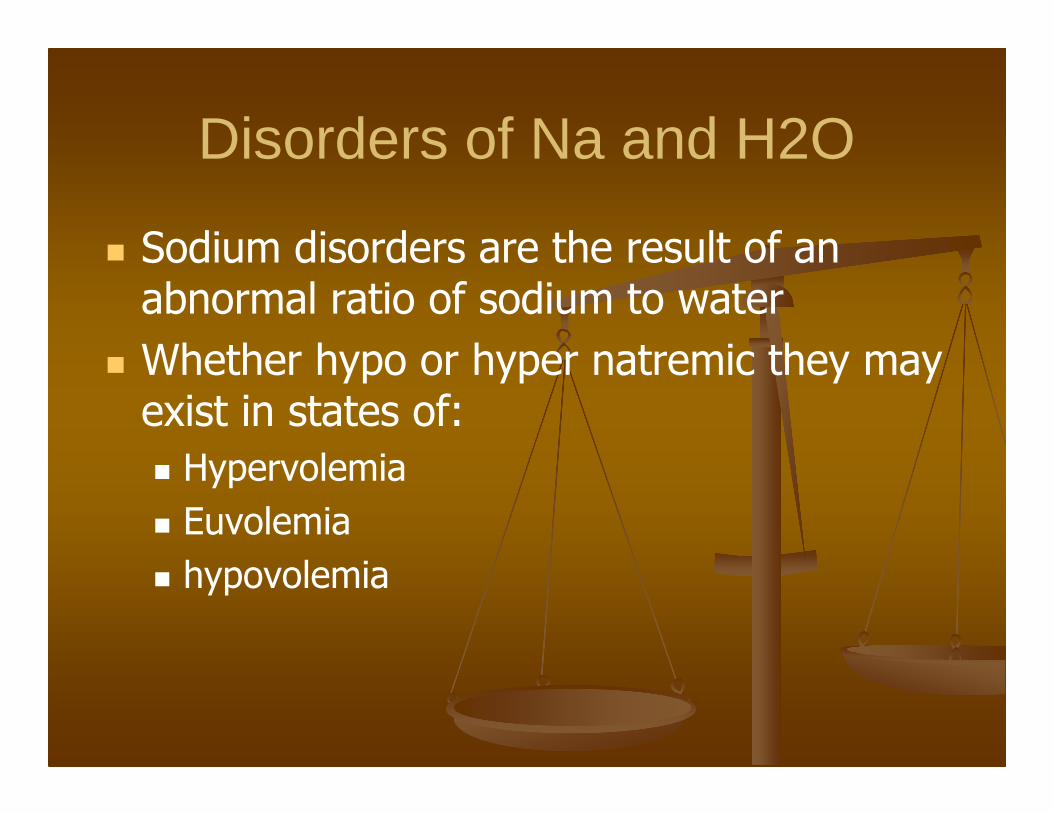
Disorders of Na and H2O
� Sodium disorders are the result of an abnormal ratio of sodium to water
� Whether hypo or hyper natremic they may exist in states of:
� Hypervolemia
� Euvolemia
� hypovolemia


Hyponatremia
� Hyponatremia is Na <130
� Is the most common electrolyte abnormality
� Most patients are stable
� Profound hyponatremia or rapid change can cause cerebral edema and increased ICP due to influx of water into the cell.

Factitious Hyponatremia
� Caused by how sodium is measured
� Pseudohyponatremia (normal osmolarity)
� Hyperlipidemia or hyperproteinemia
� Redistributive hyponatremia (osm >295)
� Osmotic particles in ECF cause fluid shift from cells in to ECF giving appearance of low Na
� Hyperglycemia (na falls 1.5 for every 100 glu)
� Mannitol administration

True hyponatremia (Osm < 280)
� May occur with hypovolemic, hypervolemic or euvolemic states depending on extracellular volume.
� These are differentiated by urine na excretion studies
� We don’t collect 24 hr urine studies so…..don’t sweat the details

Hypovolemic
Hypervolemic
Euvolemic
Addisons is mineralcorticoiddefficiency

Signs and Symptoms� Anorexia � Nausea and vomiting � Difficulty concentrating � Confusion � Lethargy � Agitation � Headache � Seizures
� Variable degrees of cognitive impairment (eg, difficulty with short-term recall; loss of orientation to person, place, or time; frank confusion or depression)
� Acute severe hyponatremia : coma; fixed, unilateral, dilated pupil; decorticate or decerebrate posturing; and respiratory arrest
� Volume depletion : Dry mucous membranes, tachycardia, diminished skin turgor, orthostatics
� Volume overload : Pulmonary rales, S3 gallop, jugular venous distention, peripheral edema, or ascites

Treatment of Hyponatremia
� Treatment of hyponatremia is dependent on the symptoms
� Overriding theme is SLOW correction
� More aggressive therapy if na < 120 and significant change in mental status or coma, or seizures. (“hot salts”)
� Rapid correction can cause central pontine myelinolysis (destruction of cells of pons)

Signs and Symptoms of CPM
� Dysarthria
� Dysphagia
� Seizures
� Altered mental status
� Quadriparesis
� Hypotension
� typically begin 1-3 days after correction of serum sodium level.

Treatment of Hyponatremia
� Should not be corrected faster than 0.5meq/Hr to target > 120
� Na to be infused =TBW x (desired –actual)/ na concentraion of solution
� Example : 50kg pt with Na 105
� (.6x50kg)(120-105)/513mEq/L(if 3%saline)
� =0.877L or 877mL
� 15meq/0.5meq/hr = 30 hrs or 29 mL/hr

Treatment of Hyponatremia
� If NS is used contains 154meq Na
� Na to be infused =TBW x (desired –actual)/ na concentraion of solution
� Example : 50kg pt with Na 105
� (.6x50kg)(120-105)/154mEq/L
� =2.9L or 2900 mL
� 15meq/0.5meq/hr = 30 hrs
� or 97 mL/hr

Hypernatremia
� Na > 145
� Is rare in those able to control own fluid intake (seen in infants and elderly)
� Because pt is usually debilitated these have worst prognosis of electrolyte d/o.
� Causes cerebral cellular degeneration causes brain shrinkage and puts mechanical traction on vessels.


Signs and Symptoms
� Anorexia
� Restlessness
� Nausea and vomiting
� Altered mental status
� Lethargy or irritability
� Stupor or coma.
� Twitching, hyperreflexia, ataxia, tremor, seizure
� Focal deficits such as hemiparesis have been reported.

Causes of Hypernatremia
� Poor PO intake
� Excess excretion
� Diuretics
� Lithium
� Diabetes insipidus
� Intake of hypertonic solution
� Mineral/glucocorticoid excess
� Conn or Cushings Syndrome
� (hyperaldosteronism hypercortisolism)

Treatment of Hypernatremia
� Treatment caveats are the same for hyponatremia
� Correct imbalance slowly (<1meq/hr) this is often complicated as the patient is frequently volume depleted and may require rapid volume resuscitation for hypovolemic shock.

� Free Water Deficit =
� (kg) X 0.6 (TBW) X ([Serum Na/140] - 1)
� Example 60 kg male with na of 155
� =60 x 0.6 x [(155/140)-1] = 3.9L
� Must add to maintenance fluids and correct over 48hrs
� Priority is volume replacement. Once volume replacement is achieved patients may be switched to free water.

Potassium (3.5-5)
� Is the major intracellular ion
� Only 2% of potassium is extracellular
� The large ratio of intracellular to extracellular K is responsible for the membrane charge in excitable tissue
� 90% of daily potassium load is secreted by kidneys
� Aldosterone increases K secretion by the kidney (retains sodium and indirectly water)

Hypokalemia
� 2 most common causes are malnutrition due to alcohol abuse and diuretics
� Also affected by acid base status with alkalosis causing movement into cells and acidosis out of cells

Hypokalemia Treatment
� K 3.0 - 3.5meq/L is rarely symptomatic
� Underlying cardiovascular disease and digitalis toxicity predispose patients to more severe dysrhythmias and should be more aggressively corrected.
� Each 0.3meq deficit represents 100meq of total body deficit.
� Max IV replacement should be 0.3-1meq/kg/hr

Hyperkalemia
� Is most deadly of electrolyte disorders
� Lab error is the #1 cause of hyperkalemia
� May be divided into external and internal balance disorders
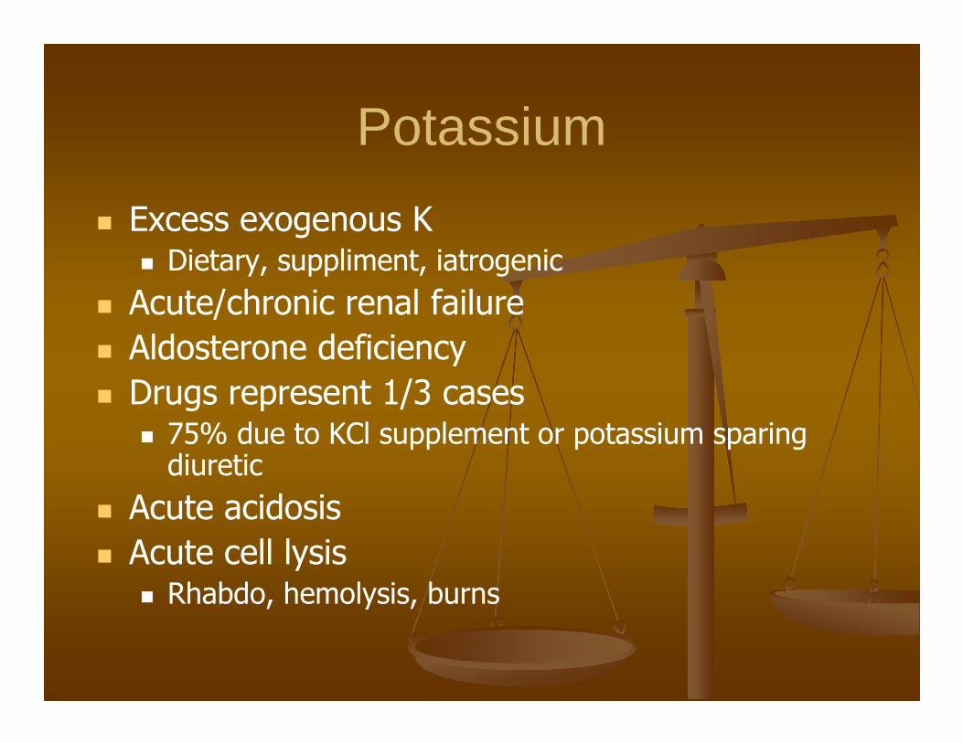
Potassium
� Excess exogenous K� Dietary, suppliment, iatrogenic
� Acute/chronic renal failure
� Aldosterone deficiency
� Drugs represent 1/3 cases� 75% due to KCl supplement or potassium sparing
diuretic
� Acute acidosis
� Acute cell lysis � Rhabdo, hemolysis, burns

Hyperkalemia
� Neuromuscular
� Weakness, paresthesias, tetany
� EKG changes
� Peak T (5.5-6)
� PR prolongation/loss of p (6.0-6.5)
� Gradual widening of QRS
� AV nodal and BB blocks

Hyperkalemia
� Management
� Temporizing measures
� CaCl
� HCO3
� Albuterol
� Insulin
� Increased excretion
� Furosemide
� Kayexylate

BUN
� Blood urea nitrogen
� Urea is formed from protein catabolism and is produced by the liver
� Ingested proteins
� Body proteins (including blood in GI tract)
� Elevated nitrogen levels is called azotemia

Cr
� Creatinine is a breakdown product of creatinine phosphate from muscle
� Breakdown and renal filtration are usually at a fairly fixed rate
� Cr is not reabsorbed by the kidney
� Creatinine is used as a proxy for evaluating renal function

Low Cr
� Low levels of Cr can be seen in
� Malnutrition due to decreased mm mass
� Late stage muscular dystrophy
� Myasthenia gravis
� Not common
� Not useful

Elevated Cr
� Elevated Cr levels may be seen in renal failure� pre renal
� Failure to perfuse the kidney
� Renal� Injury to kidney, renal tubules
� Post renal� Failure to excrete urine
� Causes back pressure in system
� Stops glomerular filtration

� Pre-renal
� Failure to perfuse the kidney adequately
� Shock states
� Dehydration
� ACEI / diuretic use

� Renal
� Infection (sepsis, pyelonephritis)
� Toxins/drugs (NSAIDS, aminoglycosides, iodinated contrast, lithium)
� Rhabdomyolysis
� Hemolysis (hemoglobin damages the tubules)
� Nephropathies (nephrotic & nephritic syndromes)

� Post renal
� Atonic bladder (dm, drugs, spinal cord injury)
� Bladder outlet obstruction
� BPH
� Catheter obstruction
� Kidney stones (not really)
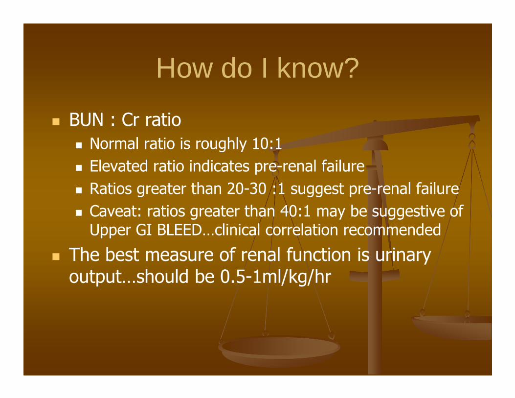
How do I know?
� BUN : Cr ratio
� Normal ratio is roughly 10:1
� Elevated ratio indicates pre-renal failure
� Ratios greater than 20-30 :1 suggest pre-renal failure
� Caveat: ratios greater than 40:1 may be suggestive of Upper GI BLEED…clinical correlation recommended
� The best measure of renal function is urinary output…should be 0.5-1ml/kg/hr

Calcium (8.5-10.5; 2.2-2.5)
� Calcium homeostastis is regulated by parathyroid hormone and calcitonin.
� PTH causes release from body stores, increases reabsorption from the kidneys and raises blood calcium
� Vitamin D increases absorption from the GI tract
� Calcitonin raises calcium excretion from the kidney, increases trapping in the bones and lowers serum levels

Role of Calcium
� Bone formation and maintenance
� Muscle contraction
� All processes that involve exocytosis including synaptic transmission and hormone release.
� 40% of calcium is bound to albumin

Hypercalcemia
� Stones� Polyuria� Nocturia� Dehydration� Renal stones� Renal failure
� Bones� Osteopenia/Ca deposits
� Abdominal moans� Constipation� Nausea� Anorexia� Pancreatitis� Gastric ulcer
� Psychiatric groans� Lethargy � Weakness� Confusion� Coma� Hypotonia� Hyporeflexia� Paresis
� Cardiac effects� Syncope� Arrhythmia� Hypotension� Shortened QT

� 90% of hypercalcemia is caused by malignancy or hyperparathyroidism
� Cancer
� 80% causes by bony metastisis
� 20% caused by PTH produced by cancer cells

Emergent Treatment
� Acute management is directed at elimination of excess calcium
� Volume replacement
� Furosemide
Hypocalcemia
� Numbness and tingling in the perioral area, fingers, toes� Muscle cramps may progress to carpopedal spasm and tetany
� Chvostek sign – tapping the facial nerve causes facial mm spasm� Trousseau sign - carpal spasm after 3 minutes of inflation of a pressure
cuff 20 mm Hg above the patient's systolic pressure
� Irritability, impaired intellectual capacity, depression, and personality changes
� Parkinsonism, Choreoathetosis� Grand mal, petit mal, focal seizures� Laryngospasm and bronchospasm� Signs of mental retardation may be present in children, adults may
have dementia.� prolongation of the QTc interval, arrhythmias, hypotension, and
heart failure.
Treatment of Hypocalcemia
� Calcium

Adrenal Insufficiency
� Is the loss of gluco and mineralocorticoid production from the adrenals
� Primary adrenal insufficiency is related to failure of the organ (50 per 1,000,000)
� Secondary adrenal insufficiency is related to disorders of the hypothalamic pituitary system. Commonly exogenous steroid.

Adrenal Crisis� Results from acute exacerbation of chronic adrenal insufficiency� Usually caused by some stressor
� Surgery� Anesthesia (eg, etomidate)� Volume loss� Trauma� Asthma� Hypothermia� Alcohol� Myocardial infarction� Fever / infection / sepsis� Hypoglycemia� Pain� Psychoses or depression� Exogenous steroid withdrawal
Signs and symptoms
� Weakness (99%)
� Pigmentation of skin (98%)
� Weight loss (97%)
� Abdominal pain (34%)
� Salt craving (22%)
� Diarrhea (20%)
� Constipation (19%)
� Syncope (16%)
� Lab Abnormalities� Hypoglycemia (67%)
� Hyponatremia (88%)
� Hyperkalemia (64%)
� Hypercalcemia (6-33%)
� Findings are non-specific and usually symptoms relate to illness that precipitated event
� Look for shock unresponsive to fluids or pressors

Treatment
� Volume replacement
� Correct lab abnormalities as needed
� Administer
� Hydrocortisone 100mg IV or
� Dexamethasone 4 mg IV
� Treat the underlying disorder