Abdominal Imaging Manifestations of Systemic Autoimmune Diseasesc.ymcdn.com/sites/€¦ · ·...
30
Abdominal Imaging Manifestations of Systemic Autoimmune Diseases Kristen Olinger, MD 1 , Maitraya Patel, MD 2 , Cecilia Jude, MD 2 , Shaden Mohammad, MD 2 , Daniel Margolis, MD 3 , Anokh Pahwa, MD 2 1 Department of Radiological Sciences, David Geffen School of Medicine at UCLA 2 Department of Radiology, Olive View-UCLA Medical Center 3 Department of Radiology, Weill-Cornell Medicine
Transcript of Abdominal Imaging Manifestations of Systemic Autoimmune Diseasesc.ymcdn.com/sites/€¦ · ·...

Abdominal Imaging Manifestations of Systemic Autoimmune Diseases
Kristen Olinger, MD1, Maitraya Patel, MD2, Cecilia Jude, MD2, Shaden Mohammad, MD2,
Daniel Margolis, MD3, Anokh Pahwa, MD2 1Department of Radiological Sciences, David Geffen School of
Medicine at UCLA
2Department of Radiology, Olive View-UCLA Medical Center
3Department of Radiology, Weill-Cornell Medicine

Financial Disclosures • None
Overview • Introduction
• Epidemiology, Pathophysiology and Abdominal Radiologic Features of: • Systemic Lupus Erythematosus (SLE)
• Rheumatoid Arthritis
• Systemic Vasculitis Syndromes (Polyarteritis nodosa, Takayasu arteritis)
• Systemic Sclerosis Syndrome
• Behcet’s Disease
• Autoimmune Hepatitis
• IgG4-related Disease
• Conclusion
Target Audience • General and abdominal radiologists, residents and fellows

Introduction
• Systemic autoimmune diseases are complex entities
• Abdominal involvement is seen in a large proportion of cases
• Radiologists should be familiar with clinical presentations of these disease processes and understand the abdominal radiologic findings that help guide clinical management
• After review of the presentation, participants will be able to: • Discuss the epidemiology and pathophysiology of abdominal
manifestations of systemic autoimmune diseases
• Identify CT, MRI, and fluoroscopic patterns associated with these disease entities
• Substantiate an integrated radiological and clinical approach to managing systemic autoimmune diseases

Systemic Lupus Erythematosus
• Epidemiology: • Peak onset in 20-40 years old • Female to male predominance of 9-13:1 (however, lower female predominance
in the juvenile/elderly) • Higher rates in African-American, African-Caribbean descent population
• Pathophysiology: • Autoimmune process characterized by inflammation, immune complex
deposition, vasculitis and vasculopathy • Activation of the complement system leads to formation of immune complexes
in tissues • Immune complexes recruit B lymphocytes, which lead to autoantibody creation • Dysfunction of the normal suppressive control of the immune system leads to
an uncontrolled, widespread autoimmune response • Fibrinoid necrosis of the small vessels leads to end-organ ischemia,
hemorrhage • Can also see increase in lymphoreticular malignancy, infection • Hypothesized environmental triggers include UV light, smoking, infection • Diagnosis made if 4 out of the 11 WHO clinical criteria are met
•Malar rash
•Discoid lesions
•Photosensitivity
•Oral ulcers
•Nonerosive arthritis
•Serositis
•Renal involvement
•Seizures/psychosis
•Hematologic abnormalities
•Immunologic abnormalities
•Positive antinuclear
antibody test

Systemic Lupus Erythematosus • Radiologic Features:
• Vascular findings: • Arterial and veno-occlusive disease, particularly when
antiphospholipid syndrome is present • End-organ ischemia (e.g. ischemic enteritis, splenic
infarction) • Gastrointestinal findings:
• Reflux esophagitis (due to esophageal dysmotility) • Gastritis • Pancreatitis (from the autoimmune process or as a side
effect of medications) – acute and chronic • Ischemic enteritis – particularly SMA territory • Budd-Chiari syndrome • Acalculous cholecystitis
• Renal findings: • Chronic renal failure, including renal atrophy • Renal vein thrombosis, ischemic infarcts
A. Contrast enhanced axial CT image in
a 48-year-old female with known lupus,
abdominal pain and elevated lactate
demonstrates small bowel wall
thickening. B. On the follow-up CT with
oral contrast 1 day later, there is
progression of small bowel wall
thickening. Findings likely represent
ischemic bowel secondary to lupus,
which affects smaller mesenteric arteries
and veins, not well seen by CT.

A. Coronal reconstruction from contrast-
enhanced CT in a 41-year-old male with
lupus and abdominal pain. Findings
including diffuse small bowel wall
thickening likely representing ischemic
enteritis and ascites.
B. Coronal reconstruction from
contrast-enhanced CT in a 40-year-old
male with lupus and epigastric pain.
Peripheral wedge-shaped areas of
hypo-enhancement are seen in the left
kidney and spleen, consistent with
infarcts of varying age.
Systemic Lupus Erythematosus
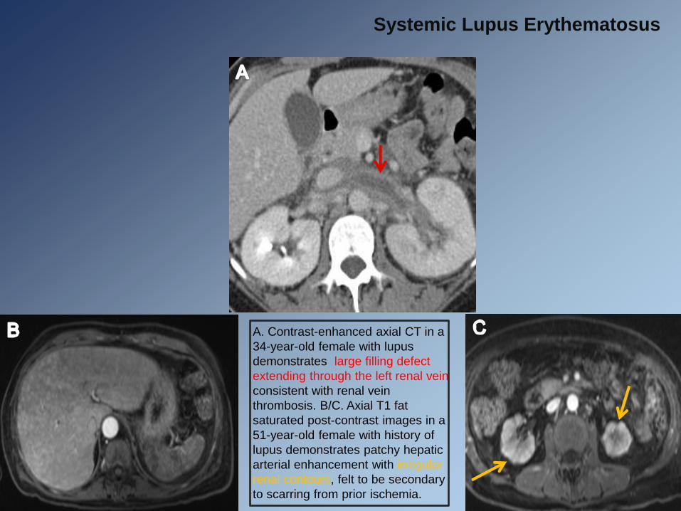
A. Contrast-enhanced axial CT in a
34-year-old female with lupus
demonstrates large filling defect
extending through the left renal vein
consistent with renal vein
thrombosis. B/C. Axial T1 fat
saturated post-contrast images in a
51-year-old female with history of
lupus demonstrates patchy hepatic
arterial enhancement with irregular
renal contours, felt to be secondary
to scarring from prior ischemia.
Systemic Lupus Erythematosus

Rheumatoid Arthritis
• Epidemiology: • Peak incidence at 40-60 years • Female to male predominance of 2-3:1 • Up to 70% of patients express HLA-DR4 (in particular the HLA-DR B1 allele) • Extra-articular manifestations occur in up to 40% of patients
• Pathophysiology: • Activation of CD4 T cells leads to a systemic inflammatory response
• Antigens are presented by macrophages • Leads to production of T lymphocytes • Cytokine and antibody production occurs • Antibody production leading to immune complex deposition • Ultimately results in pannus formation
• Exact cause is unknown • Hypothesized environmental triggers include infection (EBV), smoking • Per the American College of Rheumatology criteria, diagnosis is made based
upon a combination of radiographic findings of location-specific arthritis (peripheral joints) and presence of rheumatoid factor
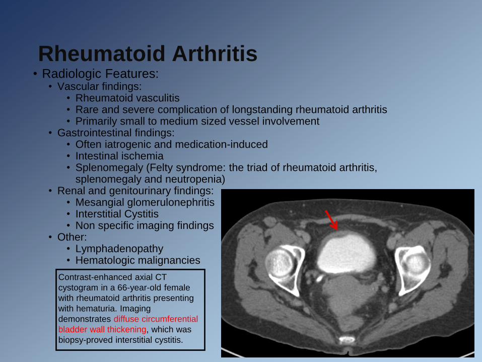
Rheumatoid Arthritis • Radiologic Features:
• Vascular findings: • Rheumatoid vasculitis • Rare and severe complication of longstanding rheumatoid arthritis • Primarily small to medium sized vessel involvement
• Gastrointestinal findings: • Often iatrogenic and medication-induced • Intestinal ischemia • Splenomegaly (Felty syndrome: the triad of rheumatoid arthritis,
splenomegaly and neutropenia) • Renal and genitourinary findings:
• Mesangial glomerulonephritis • Interstitial Cystitis • Non specific imaging findings
• Other: • Lymphadenopathy • Hematologic malignancies
Contrast-enhanced axial CT
cystogram in a 66-year-old female
with rheumatoid arthritis presenting
with hematuria. Imaging
demonstrates diffuse circumferential
bladder wall thickening, which was
biopsy-proved interstitial cystitis.

Systemic Vasculitis Syndromes
• Polyarteritis Nodosa (PAN):
• Epidemiology: • Peak incidence in 40-60 years old
• Female to male predominance of 1.6-2:1
• Hepatitis B infection has been strongly associated with PAN
• Pathophysiology: • Fibrinoid necrotizing vasculitis affecting the small to medium-sized arteries
• Preferential involvement at the vascular bifurcations
• Transmural inflammation of the vessel wall leads to fibrinoid necrosis, thereby weakening the vessel wall and resulting in microaneurysms and hemorrhage
• Fibrous thickening leading to thrombosis and vessel occlusion can be seen in the chronic stage
• Exact cause is unknown

Polyarteritis Nodosa • Radiologic Features:
• Vascular findings: • Microaneurysms, luminal irregularity, stenoses and arterial occlusion
• Gastrointestinal findings: • Involvement in 50-70% of cases, with hepatic involvement in 50-60% and
pancreatic involvement in 25-35% • Bowel ischemia, infarction, hemorrhage, perforation
• Renal findings: • Most frequently involved organ (80-90%) • Arterial aneurysms, infarction
Coronal MIP reconstruction from
contrast-enhanced CT angiography in
a 63-year-old male with polyarteritis
nodosa. Characteristic findings
include beaded intrahepatic arteries,
intraparenchymal microaneurysms
and a resulting subcapsular
hematoma. Similar findings in the
pancreas are partially seen. Additional
splenic and renal involvement was
present (not included).
Systemic Vasculitis Syndromes
• Takayasu Arteritis:
• Epidemiology: • Peak incidence in younger patients (<50 years old)
• Female to male predominance of 8-9:1
• Increased prevalence in Asian and Indian populations
• Pathophysiology: • Granulomatous large vessel vasculitis leading to destruction of the arterial
media
• Adventitial mononuclear infiltrate with perivascular cuffing of the vasa vasorum early in the disease
• Predominately affects the aorta and its major branches
• Results in aneurysms, stenosis and occlusions
• Exact cause is unknown
• Clinical presentation includes classic “pulseless” disease in the late-phase
Takayasu Arteritis • Radiologic Features:
• Acute phase: wall thickening and wall enhancement of the aorta and/or branches
• Chronic phase: stenosis, occlusion, aneurysmal dilation and pseudo-aneurysms of the aorta and/or branches
• Type I-V classification system based upon location
• I: aortic arch branches, classic type
• IIa: ascending thoracic aorta, aortic arch and its branches
• IIb: type IIa plus involvement of the descending thoracic aorta
• III: thoracic descending aorta, abdominal aorta, renal arteries or a combination
• IV: abdominal aorta and/or renal arteries
• V: generalized involvement of all aortic segments and its branches
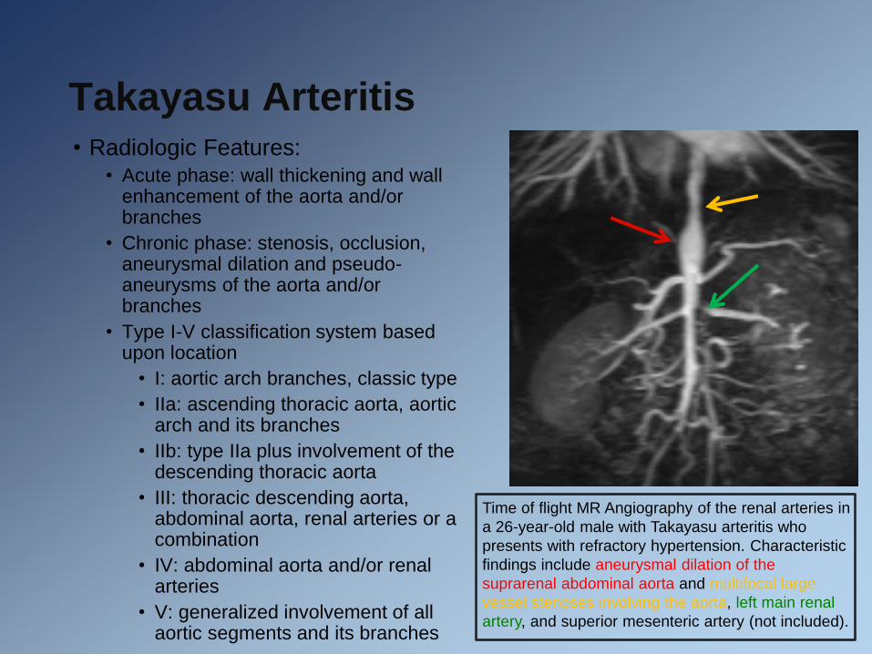
Time of flight MR Angiography of the renal arteries in
a 26-year-old male with Takayasu arteritis who
presents with refractory hypertension. Characteristic
findings include aneurysmal dilation of the
suprarenal abdominal aorta and multifocal large
vessel stenoses involving the aorta, left main renal
artery, and superior mesenteric artery (not included).
Systemic Sclerosis Syndrome
• Also known as scleroderma • Epidemiology:
• Peak incidence in 30-50 years old • Female predilection, with female to male predominance of 3:1 (up to 10:1 in
reproductive years)
• Pathophysiology: • Collagen and extracellular matrix protein deposition disorder which results in
abnormal immune response and tissue fibrosis • Microvascular disease
• Early involvement of small vessels • Therefore organs with dense capillary networks are preferentially affected
• Skin ivolvement can be limited (primarily hand and distal forearm involvement and associated with CREST syndrome: calcinosis, Reynaud’s phenomenon, esophageal dysmotility, sclerodactyly and telangiectasia) or diffuse (involves upper arm, shoulder and trunk and associated with more visceral involvement)
• Exact cause is unknown, hypothesized exposures include silica, solvents, radiation, viral (CMV, HHV5, parvovirus B19)

Systemic Sclerosis Syndrome • Radiologic Features:
• Vascular findings:
• Small vessel stenoses
• Gastrointestinal findings: seen in up to 90% of patients
• Esophageal involvement (dilation, hypomobility, incompetency of the lower esophageal sphincter) in up to 90% of cases
• Up to 50% of cases demonstrate small bowel involvement
• Smooth muscle atrophy, hypomotility in small bowel leads to dilation, air-fluid level
• “Hidebound” bowel sign: narrow separation of the valvulae conniventes despite small bowel dilation
• Renal findings: in up to 25% of patients
• Scleroderma renal crisis: abrupt onset hypertension, acute renal failure, spotted nephrogram due to focal ischemia in the setting of severe arteriolar narrowing
A. Coronal reconstruction from CT
enterography in a patient with
systemic sclerosis demonstrates
narrowing of the spaces between
the valvulae conniventes despite
bowel dilation, resulting in a
characteristic “hidebound bowel”
appearance. Multiple cystic
lesions in the pancreatic head are
incidentally noted. B. Barium
upper GI series demonstrates the
“stack of coins” appearance in
hidebound bowel.

A. Axial contrast-enhanced CT in a 60-year-
old female with known systemic sclerosis and
abdominal pain demonstrates diffuse small
bowel dilation and the characteristic
“hidebound bowel” appearance. Perforation
was present (not well seen). B. Coronal
reconstruction in contrast-enhanced CT in a
56-year-old female with mixed connective
tissue disorder (SLE/scleroderma)
demonstrates a partially obstructing fecalith in
the descending colon. C. Coronal T2 MRI
image from a 31-year-old female with history
of systemic sclerosis and recurrent pseudo-
obstruction demonstrates the “hidebound
bowel” appearance.
Systemic Sclerosis Syndrome

A/B. Axial CT images with oral contrast in a
58-year-old female with history of
scleroderma and unexplained weight loss
demonstrates esophageal distension and
dilation of the jejunum and proximal ileum
with the characteristic “hidebound bowel”
appearance of the small bowel. C. Non-
contrast axial CT image in a 75-year-old
female with history of scleroderma and
abdominal pain demonstrates markedly
distended loops of small bowel without
discrete transition point.
Systemic Sclerosis Syndrome

Behcet’s Disease
• Epidemiology: • Peak incidence in 20-30 years old • Males more commonly affected • More prevalent along the Silk Road (Eastern Asia, Mediterranean and
Middle East) • Increased risk associated with HLA-B51 allele
• Pathophysiology: • Abnormal autoimmune response and hyperfunctioning of neutrophils • Leads to vasculitis and perivascular inflammatory infiltrates • Chronic, recurring symptoms • Exact cause is unknown • Hypothesized triggers include viral infection, environmental factors
• Diagnostic criteria:
• Minor or major apthous ulcers occurring at least 3 times in a 12-month period
• Recurrent genital apthous ulcers
• Eye lesions (uveitis, retinal vasculitis)
• Skin lesions (erythema nodosum, pseudofolliculosis, acneform nodules)
• Positive pathergy test

Behcet’s Disease • Radiologic Features:
• Vascular findings: • In up to 30% of cases • Mural thickening of the aorta/SVC • Vascular thrombosis • Aneurysmal dilatation
• Gastrointestinal findings: • Separate from oral ulcerations • In up to 50-60% of cases • Most commonly due to small vessel
vasculitis in the bowel wall • Bowel wall thickening • Localized versus diffuse intestinal
ulcerations, particularly ileocecal region
• Complications: bowel perforation, fistulas, hemorrhage
• Pancreatitis
A-C. Axial and coronal reformats from time of flight
MRA in a 39-year-old male with family history of
Behcet’s and splenic artery aneurysm status post
coiling. Numerous splenic collateral vessels and
fusiform aneurysm of the celiac artery and left internal
iliac artery are seen.

Autoimmune Hepatitis
• Epidemiology: • All ages can be affected, however most commonly 40-60 years old
• Female to male predominance of 3.6:1
• Pathophysiology: • Hypothesis includes environmental agent triggering an inflammatory cascade
directed at liver antigens
• Results in necroinflammatory and fibrotic changes in the liver
• Type 1: “classic”
• Characterized by elevated circulating antinuclear antibodies (ANA), smooth muscle antibodies, antiactin antibodies, atypical perinuclear antineutrophilic cytoplasmic antibodies (pANCA) and autoantibodies against soluble liver antigen (SLA) and liver-pancreas antigen (LP)
• Type 2: rare
• Antibodies to liver-kidney microsomes (ALKM-1) and liver cytosol antigen (ALC-1)

Autoimmune Hepatitis • Radiologic Features:
• Widely variable, ranging from normal to manifestations of end-stage liver disease • Imaging is particularly useful to identify complications of cirrhosis and to evaluate for
autoimmune involvement of additional abdominal organs
• Hepatomegaly • Macronodular cirrhosis (more commonly seen in disease processes where hepatocellular
regeneration plays a significant role) • Liver surface nodularity • Hypertrophy of the caudate lobe with atrophy of the right hepatic lobe • Confluent hepatic fibrosis from chronic liver injury (appears as wedge-shaped areas of
hypoattenuation on CT, T2 hyperintensity on MR) • Sequelae of portal hypertension (varices, ascites, splenomegaly) • Periportal edema
A/B. Axial T1 fat
saturated post
contrast MRI images
in a 48-year-old
female with
autoimmune hepatitis
demonstrates a
macronodular liver
with confluent hepatic
fibrosis.

A. Contrast enhanced CT scan in a
47-year-old female with autoimmune
hepatitis demonstrates geographic
hypoattenuation and a macronodular
pattern of cirrhosis.
B. Coronal reformat from a contrast
enhanced CT scan in a 49-year-old
female with autoimmune hepatitis
demonstrates a macronodular
pattern of cirrhosis.
Autoimmune Hepatitis

IgG4-Related Disease: • Epidemiology:
• Peak incidence in middle-age/elderly (mean age 50-60s) • Male to female predominance of 3-7:1
• Pathophysiology: • Extensive infiltration of IgG4-positive plasma cells and T
lymphyocytes into organs • Leads to characteristic “storiform” (whorled) and obliterative
fibrosis, tumefactive lesions • Pathogenesis is poorly understood
• Radiologic Features: • Pancreas is the most commonly involved organ
• Sclerosing pancreatitis is a form of autoimmune pancreatitis • Diffuse versus less commonly focal enlargement of the
pancreas (focal can mimic tumor) • Hypoechoic rim of pancreatic parenchyma • Less commonly associated with severe peripancreatic
stranding, changes of chronic pancreatitis (such as calcifications, pseudocyst formation)
• Diffuse or segmental narrowing of the main pancreatic duct • Hepatobiliary tract: seen in 60-80% of patients with autoimmune
pancreatitis • Sclerosing cholangitis • Intrapancreatic common bile duct most commonly affected
2
A. Axial contrast-enhanced CT images in
a 63-year-old male with epigastric pain
demonstrates diffuse enlargement of the
pancreas with a subtle hypointense rim of
pancreatic parenchyma, consistent with
autoimmune pancreatitis. B. Follow-up
CT in same patient 6 months following
initiation of steroid therapy demonstrates
resolution of findings.

IgG4-Related Disease:
• Radiologic Features: • Renal: seen in up to 35% of patients with
autoimmune pancreatitis • 5 patterns of involvement:
• Bilateral cortical lesions (round or wedge-shaped peripheral lesions): most common
• Diffuse patchy involvement • Soft tissue rim around the kidneys • Bilateral renal sinus nodules • Diffuse wall thickening of the renal
pelvis • Other:
• Lymphadenopathy • Retroperitoneal fibrosis: in 20% of
patients with autoimmune pancreatitis, periaortic soft tissue mass that can surround or encase the ureters
• Periaortitis • Sclerosing mesenteritis • Rare: gastritis/ulceration, prostatitis
A/B. Contrast enhanced CT scan in a 42-year-old male
presenting with malabsorption and weight loss. Findings
include diffuse pancreatic enlargement with a rim of
hypoenhancing pancreatic parenchyma and peripheral
wedge-shaped areas of hypoenhancement within the
bilateral kidneys, consistent with autoimmune pancreatitis
and IgG4-related renal disease.

A-C. Axial and coronal reformated
contrast-enhanced CT in a 70-year-old
male with history of autoimmune
pancreatitis with acutely elevated
alkaline phosphatase/bilirubin.
Findings include a rim of soft tissue
thickening surrounding the bilateral
kidneys and insinuating into the left
renal hilum. Of note, IgG4 levels were
above the reportable range (above 300
mg/dl).
IgG4-Related Disease

A. Coronal MRCP image from a 76-year-old male with history of biliary
stricture demonstrates a smooth distal common bile duct stricture with
proximal intrahepatic biliary dilatation. IgG4 levels were elevated at 117. B.
Coronal MRCP image from a 46-year-old male with history of primary
sclerosing cholangitis demonstrates mild intrahepatic biliary duct beading,
including areas of stenoses and aneurysmal dilatation. IgG 4 levels were
elevated at 227 and findings were consistent with IgG4 cholangiopathy.
IgG4-Related Disease
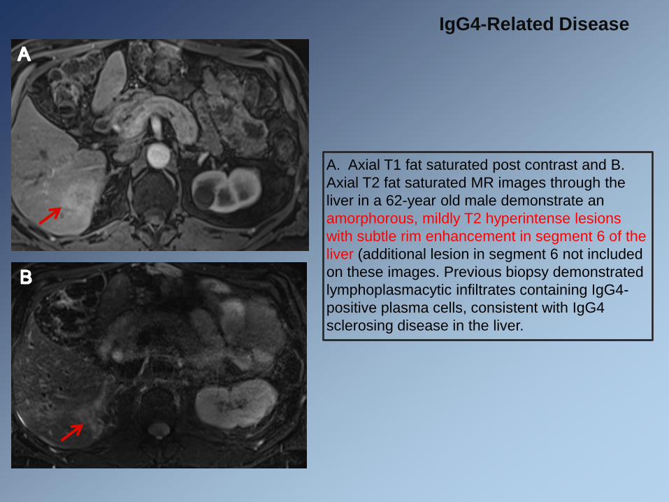
A. Axial T1 fat saturated post contrast and B.
Axial T2 fat saturated MR images through the
liver in a 62-year old male demonstrate an
amorphorous, mildly T2 hyperintense lesions
with subtle rim enhancement in segment 6 of the
liver (additional lesion in segment 6 not included
on these images. Previous biopsy demonstrated
lymphoplasmacytic infiltrates containing IgG4-
positive plasma cells, consistent with IgG4
sclerosing disease in the liver.
IgG4-Related Disease

A-C. Axial contrast-enhanced CT images in
a 50-year-old male with history of
mesenteric mass demonstrate an ill-
defined mesenteric mass which encases
the SMA and mesenteric branches,
surrounds the pancreas and extends into
the porta hepatis. Percutaneous biopsy
demonstrated “dense storiform fibrosis with
lymphoplasmacytic infiltrate” which in the
setting of elevated IgG4 (285) most likely
represented IgG4-related disease.
IgG4-Related Disease

Summary
• Systemic autoimmune diseases can be complex
• Many cases demonstrate abdominal involvement
• Predilection for specific organs and vascular involvement can be helpful in narrowing the differential diagnosis
• An improved understanding of the epidemiology and pathophysiology, in addition to the radiologic presentation, can help guide diagnosis and management

References • Lisnevskaia L, Murphy G and Isenberg D. Systemic Lupus Erythematosus. Lancet. 2014; 384(9957): 1878-
1888.
• Lalani TA, Kanne JP, Hafield GA, Chen P. Imaging Findings in Systemic Lupus Erythematosus. RadioGraphics. 2004; 24: 1069-1086.
• Sommer OJ, Kladosek A, Weiler V, et al. Rheumatoid Arthritis: A Practical Guide to State-of-the-Art Imaging, Image Interpretation, and Clinical Implications. RadioGraphics. 2005; 25: 381-398.
• Cojocaru M, Mihaela I, Silosi I, Vrabie CD, Tanasescu R. Extra-articular Manifestations in Rheumatoid Arthritis. Maedica. 2010; 5(4): 286-291.
• Ha HK, Lee SH, Rha SE, et al. Radiologic Features of Vasculitis Involving the Gastrointestinal Tract. RadioGraphics. 2000; 20: 779-794.
• Gotway MB, Araoz PH, Macedo TA, et al. Imaging Findings in Takayasu’s Arteritis. AJR. 2005; 184(6): 1945-1950.
• Gabrielli A, Avvedimentoa EV and Krieg T. Scleroderma. N Engl J Med. 2009; 360: 1989-2003.
• Pickhardt PJ. The “Hide-bound” Bowel Sign. Radiology. 1999; 213(3): 837-838.
• Sakane T, Takeno M, Suzuki N and Inaba G. Behcet’s Disease. N Engl J Med. 1999; 341: 1284-1291.
• Krawitt EL. Autoimmune Hepatitis. N Engl J Med. 2006; 354: 54-66.
• Faria SC, Ganesan K, Mwangi I, et al. MR Imaging of Liver Fibrosis: Current State of the Art. RadioGraphics. 2009; 29(6): 1615-1635.
• Chung SY, Ha HK, Kim JH et al. Radiologic Findings of Behcet Syndrome Involving the Gastrointestinal Tract. RadioGraphics. 2001; 21: 911-926.
• Martinez-de-Alegria A, Baleato-Gonzalez S, Garcia-Figueiras R, et al. IgG4-related Disease from Head to Toe. RadioGraphics. 2015; 35: 2007-2025.
Contact • Kristen Olinger, MD David Geffen School of Medicine at UCLA