Bacterial Infections in Cirrhotic Patients in a Tertiary ...

7/07/2012
1
Vincent Vandecaveye, Frederik De Keyzer
Department of Radiology
University Hospitals Leuven, Leuven, Belgium
CLINICAL APPLICATIONS OF DIFFUSION-
WEIGHTED IMAGING IN THE ABDOMEN
ABDOMINAL DIFFUSION-WEIGHTED
IMAGING
Post-treatment imaging
•Abdominopelvic tumors
post-surgical
post-chemoradiation
Fibrosis versus tumoral recurrence
Hepatic imaging
•Non-cirrhotic liver
lesion characterization
detection of metastases
•Cirrhotic liver
Tumor versus pseudo-tumor
Prediction of lesion progression
Transplantation?
Extrahepatic imaging
•Primary tumor detection
Pancreaticobiliary
Endometrial carcinoma
Uterine cervical carcinoma
Intestinal tumors
Prostate cancer
Renal cancer
• Staging
Lymph nodes
Distant metastatic spread
Second primary tumors

7/07/2012
2
differences in microstructure
*Diffusion-weighted image contrast -Mathematical quantification Apparent diffusion coefficient - No need for exogenic contrast agent - No irradiation magnetic
-Worldwide -> not routine - High expertise per centre
-Research - Clinical routine
Facilitated diffusion Restricted diffusion
Changes in H20 mobility
Native images: b-value (increasing the sensitivity for impeded diffusion) Calculated ADC map

7/07/2012
3
Correct shim position: volume shim > auto shim
• High gradient strengths
• Field-strength? 1,5T vs 3T
* Minimal TE
* Maximum Bandwidth
* Realistic spatial resolution: tumor detection
* Volume shim (anteroposterior)
* Volume of interest + edge reserve
Edge of scan volume increased artefacts
3T
DWI optimum Minimal
slices 31-36 31-36
Distance factor 0% 0%
Scan position isocenter isocenter
Fase encodering anterior-posterior anterior-posterior
FoV read 380 mm 380 mm
FoV phase 100% 81%
Slice thickness 5 mm 5 mm
TR 6600 ms 5100
TE 67 ms 84 ms
Averages 3 4
Concatenations 1 1
Fat Suppr SPAIR Fat Sat
Fat sat mode strong strong
Base resolution 192 128
Phase resolution 80% 100%
Partial Fourier 7/8 6/8
Acceleration factor PE (SENSE-
GRAPPA) 2 0
Bandwidth 1736Hz/Px 1502
EPI factor 154 104
b-value 0,50,100,300,600,1000 0,50,100,300,600,1000
6 good reasons to use a high b-value (b600 b1000)

7/07/2012
4
Similar image quality high vs low b-value
Higher accuracy high vs low b-value
CHOICE OF B-VALUE
• Current criteria in our center b0-b100-b600-b1000 (free breathing DWI):
• Lymph nodes: ADC malignant < 0,001mm2/sec < benign
• Sens: 76-83%; spec 94%
• Colorectal, gastro-intestinal and ovarian
• Non-cirrhotic liver : malignant (b1000+) <0,0011<benign (b1000-/+)
T2 anatomical correlate
• Cirrhotic liver : b600 SI + anatomical imaging – contrast-enhanced
• Skeletal/non-hepatic soft tissue metastasis: b1000+ / anatomical correlate
• Primary/tumor: b1000+/ anatomical correlate
• CAVE: pancreatic applications: anatomical correlate and ADC
BASIC IMAGE INTERPRETATION: PRIMARY STAGING
Major cause for false positives: abscess, hematoma, granuloma – low ADC

7/07/2012
5
BASIC IMAGE INTERPRETATION: TUMOR RECURRENCE
End treatment TN2
Primary location: b1000 + (ADC) + anatomical correlate
Nodal disease: b1000 + ADC (TH 0,0014)
T2 SHINE THROUGH
False positive b1000

7/07/2012
6
LOW T2
INSUFFICIENT SIGNAL @ B0
False positive ADC
NO CONTRAST LESION
TO BACKGROUND
B1000 indeterminate
detection
Characterization
7/07/2012
7
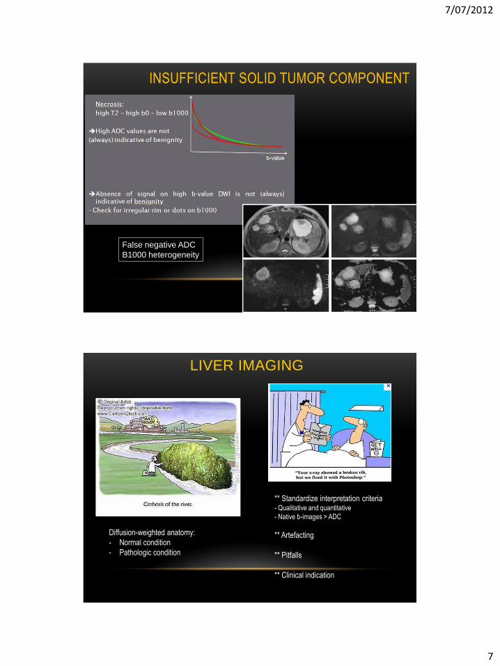
INSUFFICIENT SOLID TUMOR COMPONENT
False negative ADC
B1000 heterogeneity
LIVER IMAGING
** Standardize interpretation criteria - Qualitative and quantitative
- Native b-images > ADC
** Artefacting
** Pitfalls
** Clinical indication
Diffusion-weighted anatomy:
- Normal condition
- Pathologic condition

7/07/2012
8
LIVER IMAGING
- Both on clinical indication and methodology a distinct difference should be made between
the non-cirrhotic and cirrhotic liver
- Capture all morphologic features on native DWI images for characterization
- ADC is highly dependent of signal variations on b0-b1000
- Main indication in “non-cirrhotic” liver is staging (liver metastases)
- Main indication in “cirrhotic liver” is detection of HCC
- Consider benign lesions as “avoid a false positive”
- Should we really make a difference between lesion detection and characterization
- If you detect a liver metastasis you are very likely to call it a metastasis
b0 b100
b600 b1000
+
*
*
* Bile viscous fluid:
Prolonged SI on native DWI
NON-CIRRHOTIC LIVER: ANATOMY
b0 b100
b600 b1000
b0 b100
b600 b1000
b0 b100
b600 b1000
* Segment 5 and 6:
adjacent hepatic flexure
of colon
-Signal loss
- distortion artefact
-Due to air tissue interface
Left lobe:
Cardiac pulse
Base of lung
Signal loss at high b

7/07/2012
9
NON-CIRRHOTIC : DETECTION OF METASTASES
ADCb0-1000
56 patients with primary tumor
Sens = 96,6%
Spec = 93%
Combination of
b1000 signal intensity(SI)
ADC
Metastases: Native DWI b50-b1000
B1000-SI
b1000 - => NPV > 90%
STOP
b1000 + => ADC
=> T2(Te63/Te363)
Metastases – hemangioma – FNH/adenoma
Cave necrotic metastases
Native images signal heterogeneity !!!
b0 b1000 b600 b50
FNH
b0 b1000 b600 b50
Metastases
Hemangioma
b0 b1000 b600 b50
b0 b1000 b600 b50 Adenoma
T2-TE 63 T2-TE 263 SI
b
SI
b
SI
b
SI
b
ADC
ADC
ADC
ADC

7/07/2012
10
Benign lesions: cyst/hemangioma
Absent signal on b1000 or T2 – shine through - always ADC>0.00120
Cyst
hemangioma
Cave cavernous hemangioma: may be heterogeneous, may show areas of low ADC
THUS: diagnosis should preferentially be made by early-late T2
Value for DWI: avoid false positive – exclude M+ in subcentimetric lesions seen on CT
Benign lesions:
cyst/hemangioma
b1000
M+

7/07/2012
11
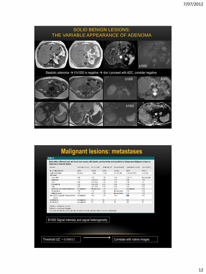
Solid benign lesions FNH and adenoma have a highly variable appearance on DWI/ADC
Exploit every possible morpholgic feature
ADC-threshold: dependent on base signal on native images
Overlap with metastases of adenocarcinoma, but less signal on native DWI
Variable ADC and SI of lesions due to heteroheneity in histologic background:
Steatotic adenoma - FNH
Teleangiectatic / inflammatory adenoma….
b0 b1000 b600 b50
b0 b1000 b600 b50
SOLID BENIGN LESIONS: FNH
b0 b1000 b600 b50 ADC
If no signal on native b-values consider benign – don t calculate ADC
ADC range: 0.0011 – 0.0015
Also check for morphologic clues
M+
FNH
FNH
7/07/2012
12
SOLID BENIGN LESIONS:
THE VARIABLE APPEARANCE OF ADENOMA
Steatotic adenoma if b1000 is negative don t proceed with ADC, consider negative
b1000
b1000
b1000
ADC
ADC
Malignant lesions: metastases
Threshold UZ: ~ 0.00011 Correlate with native images
B1000 Signal intensity and signal heterogeneity

7/07/2012
13
MALIGNANT LESIONS: METASTASES
M+ breast cancer
CAVE necrotic and mucinous metastases: lesion heterogeneity – false negative ADC
M+ colorectal major additional value = detection of millimetric metastases = better staging
IMAGING OF CIRRHOSIS
Screening: early detection of HCC curability
Negative: follow-up Positive
Staging
Transplantation Surgery - RFA
TACE
SIRS
Chem-lip
Systemisch
chemotherapie
supportief Anatomy
Cirrhosis
Complications

7/07/2012
14
Cirrhosis: diffusion-weighted anatomy
b0 b100
b600 b1000
Fibrotic septa
b0 b100
b600 b1000
Iron overload
of liver
b0 b100
b600 b1000
Liver background in cirrhosis is most important pitfall
PATHOPHYSIOLOGY: CELLULAR CHANGES
Tumorperfusion
Window of opportunity: early cellular
changes
Exclusion of microcirculatory pollution
B-value > 300
Hypovascular HCC
Hypervascular pseudotumor
Cellular
7/07/2012
15
b0 b300 b1000
Microperfusion
True diffusion
Regenerative nodule
Cirrhotic liver
b600
True diffusion
b0 b300 b1000
True diffusion
b600
High grade dysplastic nodule/early HCC
Cirrhotic liver
Cirrhosis image generation Microperfusion
True diffusion
ADC ADC
> 2cm < 2cm
b600-
SIratio
T2-CE
MRI b600-SIratio
T2-CE
MRI
True positive 28 27 31 23
False positive 2 2 7 16
True negative 9 9 34 25
False
negative 0 1 3 11
Sensitivity 100% 96.4% 91.2% 67.6%
Specificity 81.8% 81.8% 82.9% 61.0%
Accuracy 94.9% 92.3% 86.7% 64.0%
PPV 93.3% 93.1% 81.6% 59.0%
NPV 100% 90.0% 91.9% 69.4%
1. No ADC quantification :
Noise versus diffusion restriction
2.b600-SI optimal threshold
Cellular changes => malignant transformation
Exclude perfusion effects => pseudotumor
3. Improved conspicuity of small lesions Early detection of HCC
Pretransplant staging?
4. Prediction of lesion progression Timing of follow-up/intervention
Eligibility for transplantation
V. Vandecaveye et al, Eur radiol 2009
Cirrhosis: signal generation
7/07/2012
16
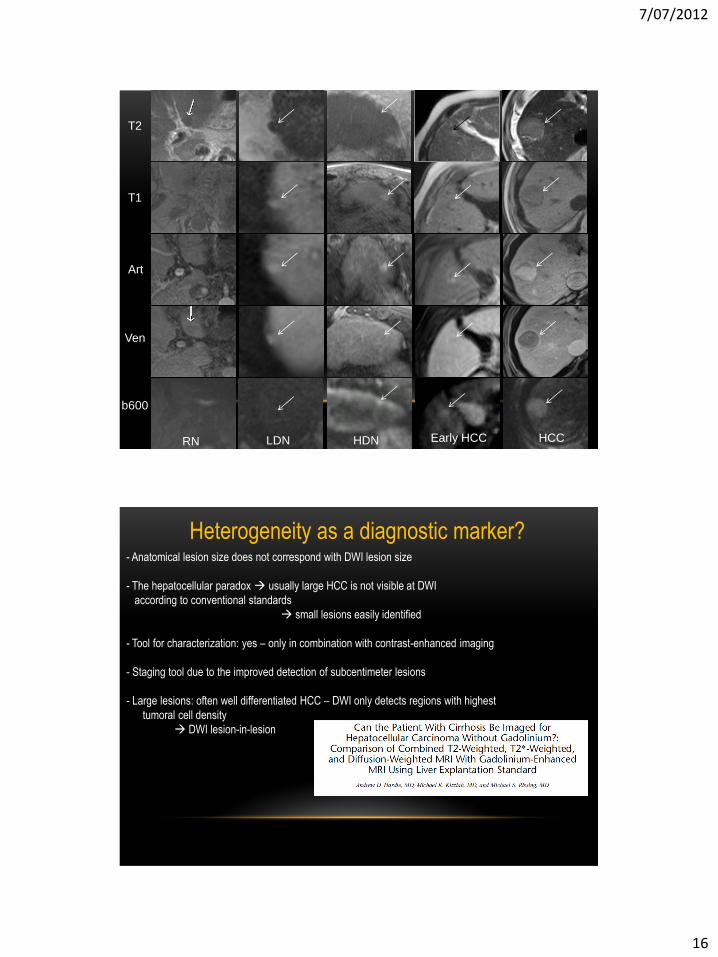
RN LDN HDN Early HCC
T2
Art
T1
b600
Ven
HCC
Heterogeneity as a diagnostic marker? - Anatomical lesion size does not correspond with DWI lesion size
- The hepatocellular paradox usually large HCC is not visible at DWI
according to conventional standards
small lesions easily identified
- Tool for characterization: yes – only in combination with contrast-enhanced imaging
- Staging tool due to the improved detection of subcentimeter lesions
- Large lesions: often well differentiated HCC – DWI only detects regions with highest
tumoral cell density
DWI lesion-in-lesion

7/07/2012
17
Well differentiated HCC
Heterogeneity as a diagnostic marker
Detection and characterization HCC
HCC

7/07/2012
18
Staging
Pitfalls
b50 B600 Ratio = 1,3
Low grade dysplastic nodule
HCC – fibrotic variant Fatty degeneration
Always correlate DW-MRI to conventional MRI characterization

7/07/2012
19
Pitfalls
Nodular regeneration after portal vein thrombosis
Confluent fibrosis
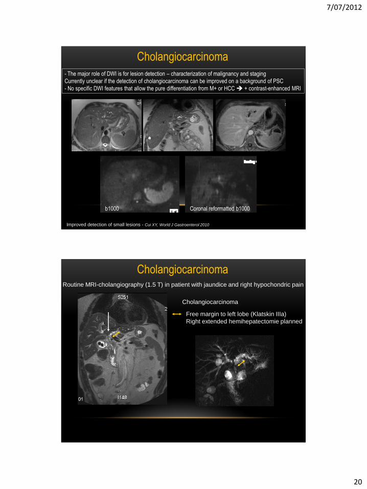
Cholangiocarcinoma
7/07/2012
20
Cholangiocarcinoma
- The major role of DWI is for lesion detection – characterization of malignancy and staging
Currently unclear if the detection of cholangiocarcinoma can be improved on a background of PSC
- No specific DWI features that allow the pure differentiation from M+ or HCC + contrast-enhanced MRI
Improved detection of small lesions - Cui XY, World J Gastroenterol 2010
b1000 Coronal reformatted b1000
Routine MRI-cholangiography (1.5 T) in patient with jaundice and right hypochondric pain
Cholangiocarcinoma
Free margin to left lobe (Klatskin IIIa)
Right extended hemihepatectomie planned
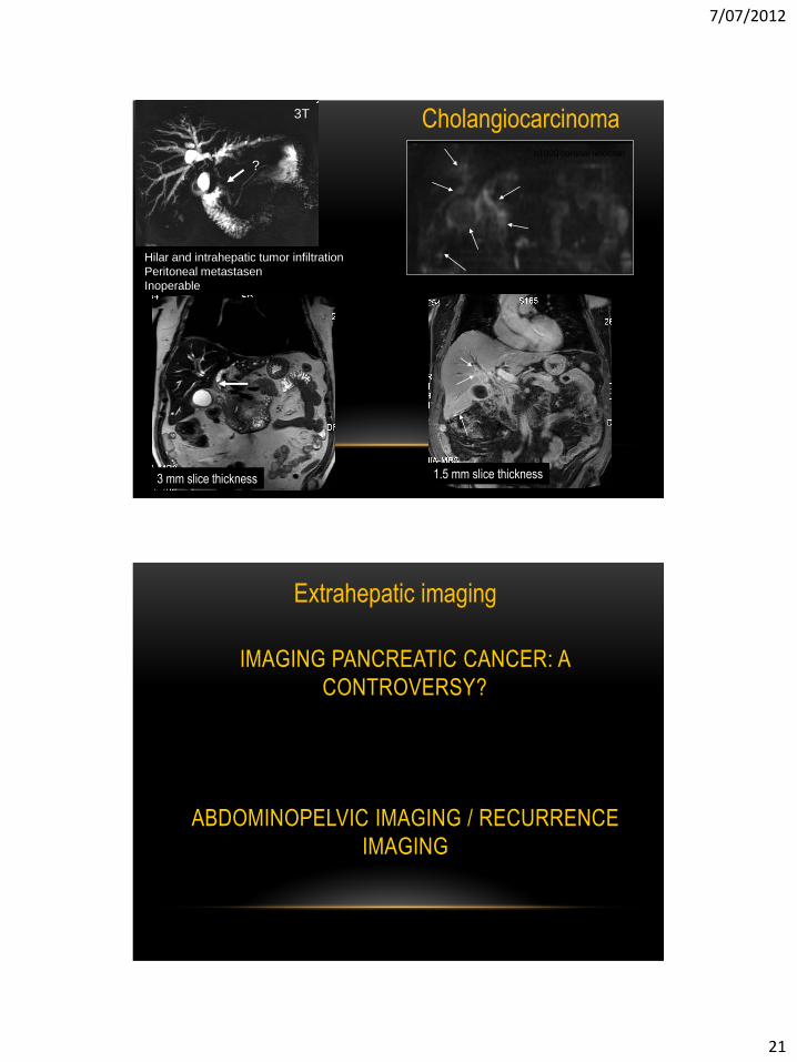
Cholangiocarcinoma
7/07/2012
21
3 mm slice thickness 1.5 mm slice thickness
?
3T
b1000 coronal reformat
1.5 mm
Hilar and intrahepatic tumor infiltration
Peritoneal metastasen
Inoperable
3 mm slice thickness 1.5 mm slice thickness
Cholangiocarcinoma
Extrahepatic imaging
IMAGING PANCREATIC CANCER: A
CONTROVERSY?
ABDOMINOPELVIC IMAGING / RECURRENCE
IMAGING

7/07/2012
22
PANCREATIC DWI: ANATOMY
- Highly variable depending on the presence of chronic pancreatitis, lipomatosis, etc….
b0
b1000 b600
b100
IMAGING PANCREATIC CANCER: A
CONTROVERSY?
• High b-value DWI/ADC shows high accuracy for pancreatic adenocarcinoma
• Ichikawa T, Abdominal Imaging 2007
• Kartalis N, Eur Radiol 2009
• Pancreatic adenocarcinoma shows hyperintense signal on high b-value due to diffusion restriction
• CAVE variable contrast with pancreatic carcinoma
• Anatomical imaging
Exocrine pancreas
Pancreatic adenocarcinoma
Less restriction
More restriction

7/07/2012
23
PANCREATIC DWI: INDICATIONS AND
INTERPRETATION
** Currently no clear agreement
UZ Leuven: indications and interpretation
** Detection of neuro-endocrine tumors
b1000 + anatomy
** Detection of carcinoma in chronic pancreatitis
b1000 + ADC + anatomy
** Facilitate detection of small adenocarcinoma in dilated Wirsung duct
small sized lesions: b1000+ anatomy
** Malignant transformation of IPMN
solid component in cyst?: b1000+anatomy
Diagnosis of pancreatic neuro-endocrine tumor
NEURO-ENDOCRINE TUMOR

7/07/2012
24
NEURO-ENDOCRINE TUMOR
T2
Arterial phase
? ?
b50 b1000 Chromogranine staining
T1
Patient with repetitive hypoglycemic attacks
Improved lesions conspicuity -> improved lesion detection
CARCINOMA IN CHRONIC
PANCREATITIS
7/07/2012
25
Facilitation of diffusion in inflammatory fibrotic tissue
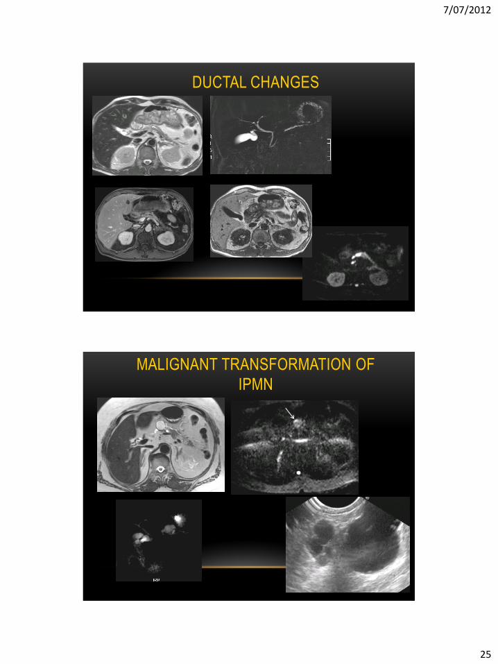
DUCTAL CHANGES
MALIGNANT TRANSFORMATION OF
IPMN

7/07/2012
26
RESTAGING POST-TREATMENT
Koh D, Collins D; AJR 2007
B1000 +
B1000 -
Surrounding tissue:
Inflammation-fibrosis
•b1000 –
•ADC often increased
•Variable in late phase
Tumoral recurrence:
Solid hypercellular
•b1000 +
•ADC often decreased
TUMOR HETEROGENEITY
Viable rim
Necrotic center

7/07/2012
27
TSE-MRI
ADC=0,00105
Pelvic recurrence confirmed by surgical biopsy
Patient with elevation of CEA
after total mesorectal excision and
neo-adjuvant (chemo)radiotherapy
Fibrosis/inflammation versus tumour recurrence?
POST-TREATMENT IMAGING
RESTAGING POST-TREATMENT Prior neo-adjuvant chemoradiation and surgery for rectal cancer
CT shows intrapelvic mass – normal tumormarker CEA
b1000
Pelvic excenteration tumor recurrence
DWI as an additional imaging technique
Screen for restrictive lesions
Signal suppresion in inflammation
Application chemoradiation>surgery

7/07/2012
28
b1000
b1000
ADC=0,00142
ADC= 0,00098
Patient with prior low anterior resection after neo-adjuvant chemoradiation for rectal cancer. Clinically increasing tumor marker, Increased FDG-uptake at the site of resection.
Pitfall: abscess !!!!! Major cause of false positive
Correlate to conventional imaging
Area of liquefaction -> restricted diffusion -> likely abcess
-> facilitated diffusion -> likely (tumoral) necrosis
b1000 ADC= 0,00098

7/07/2012
29
TAKE HOME MESSAGES
Investing into knowledge and training Investing into communication