A z-bones-of-the-skull
97
-
Upload
samuel-clover -
Category
Documents
-
view
6.114 -
download
11
Transcript of A z-bones-of-the-skull
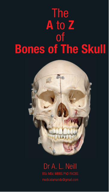
TheA to Z
ofBones of The Skull
© D
r Am
anda
Nei
ll
The A to Z of the Bones of the Skull
© A. L. Neill1
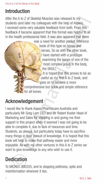
IntroductionAfter the A to Z of Skeletal Muscles was released to mystudents (and later my colleagues with the help of Aspen),I received some very valuable feedback from both. From thisfeedback it became apparent that this format was helpful to allin the health professional field. It was also apparent that there
was a need for another pocket referencebook of this type on bones andnerves. So as with the other book,I have started with a prototypeexamining the bones of one of themost complex group in the body,the SKULL.It is hoped that this proves to be as
useful as my first A to Z book, andgoes on to become a more
comprehensive but quick and simple referencefor all bones.
AcknowledgementI would like to thank Aspen Pharmacare Australia andparticularly Mr Greg Lam CEO and Mr Robert Koster Head ofMarketing and Sales for stepping in and giving me theirsupport in this project when it seemed I was not going to beable to complete it, due to lack of resources and time.Students, as always, but particularly today have to sacrificemany things in their pursuit of knowledge. It is hoped that thisbook will help to make that pathway easier and moreenjoyable. As with my other ventures in this A to Z series wewant to give knowledge to any who wish to use it.
DedicationTo MICKEY 390335, and to stopping pettiness, spite andmisinformation wherever it lies.©
Dr A
man
da N
eill
2
The A to Z of the Bones of the Skull
© A. L. Neill
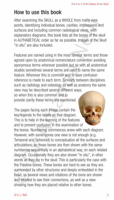
How to use this bookAfter examining the SKULL as a WHOLE from many viewpoints, identifying individual bones, cavities, impressions andsurfaces and including common radiological views, withexplanatory diagrams, this book lists all the bones of the skullin ALPHABETICAL order as far as possible. Images of the Skull“in situ” are also included.
Features are named using in the most familiar terms and thoseagreed upon by anatomical nomenclature convention avoidingeponymous terms wherever possible but as with all anatomicalstudies sometimes several terms are used to name the samefeature. Wherever this is common and to save confusionreference is made to each term. Similarly between disciplinessuch as radiology and osteology as well as anatomy the sameview may be described several different ways,so when this is also common and toprovide clarity these terms are mentioned.
The pages facing each image contain thekey/legends to the labels on that diagram.This is to help in the learning of the featuresand to prevent confusion in the examination ofthe bones. Numbering commences anew with each diagram.However, with some bones one view is not enough (e.g.Temporal and Sphenoid) to conceptualize all the surfaces andarticulations, so these bones are then shown with the samenumbering sequentially in an alphabetical way, on each relateddiagram. Occasionally they are also shown “in situ”, in otherwords as they lay in the skull. This is particularly the case withthe Palatine bones. These bones are hard to see as they aresurrounded by other structures and deeply embedded in thehead, so several views and rotations of the bone are shownand labelled to see their connections, as well as a viewshowing how they are placed relative to other bones.©
Dr A
man
da N
eill
The A to Z of the Bones of the Skull
© A. L. Neill3
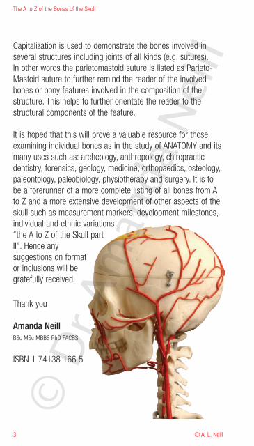
Capitalization is used to demonstrate the bones involved inseveral structures including joints of all kinds (e.g. sutures).In other words the parietomastoid suture is listed as Parieto-Mastoid suture to further remind the reader of the involvedbones or bony features involved in the composition of thestructure. This helps to further orientate the reader to thestructural components of the feature.
It is hoped that this will prove a valuable resource for thoseexamining individual bones as in the study of ANATOMY and itsmany uses such as: archeology, anthropology, chiropracticdentistry, forensics, geology, medicine, orthopaedics, osteology,paleontology, paleobiology, physiotherapy and surgery. It is tobe a forerunner of a more complete listing of all bones from Ato Z and a more extensive development of other aspects of theskull such as measurement markers, development milestones,individual and ethnic variations -“the A to Z of the Skull partII”. Hence anysuggestions on formator inclusions will begratefully received.
Thank you
Amanda NeillBSc MSc MBBS PhD FACBS
ISBN 1 74138 166 5
© D
r Am
anda
Nei
ll
4
The A to Z of the Bones of the Skull
© A. L. Neill
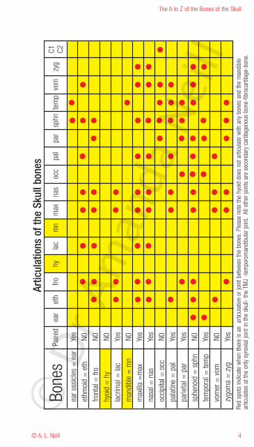
Red
spot
sin
dica
tewh
enth
ere
isan
artic
ulat
ion
orjo
intb
etwe
enth
ebo
nes.
Plea
seno
teth
ehy
oid
does
nota
rticu
late
with
any
bone
san
dth
em
andi
ble
artic
ulat
esat
the
only
syno
vialj
oint
inth
esk
ull-
the
TMJ
-tem
poro
man
dibu
larj
oint
.Al
loth
erjo
ints
are
seco
ndar
yca
rtila
geno
usbo
ne-fi
broc
artil
age-
bone
.
© D
r Am
anda
Nei
ll
The A to Z of the Bones of the Skull
© A. L. Neill5
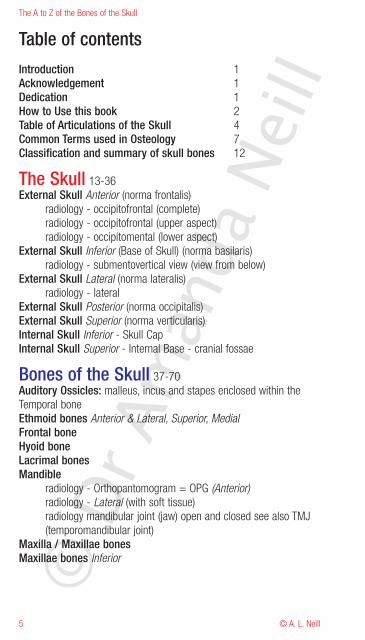
Table of contentsIntroduction 1Acknowledgement 1Dedication 1How to Use this book 2Table of Articulations of the Skull 4Common Terms used in Osteology 7Classification and summary of skull bones 12
The Skull 13-36External Skull Anterior (norma frontalis)
radiology - occipitofrontal (complete)radiology - occipitofrontal (upper aspect)radiology - occipitomental (lower aspect)
External Skull Inferior (Base of Skull) (norma basilaris)radiology - submentovertical view (view from below)
External Skull Lateral (norma lateralis)radiology - lateral
External Skull Posterior (norma occipitalis)External Skull Superior (norma verticularis)Internal Skull Inferior - Skull CapInternal Skull Superior - Internal Base - cranial fossae
Bones of the Skull 37-70Auditory Ossicles: malleus, incus and stapes enclosed within theTemporal boneEthmoid bones Anterior & Lateral, Superior, MedialFrontal boneHyoid boneLacrimal bonesMandible
radiology - Orthopantomogram = OPG (Anterior)radiology - Lateral (with soft tissue)radiology mandibular joint (jaw) open and closed see also TMJ(temporomandibular joint)
Maxilla / Maxillae bonesMaxillae bones Inferior©
Dr A
man
da N
eill
6
The A to Z of the Bones of the Skull
© A. L. Neill
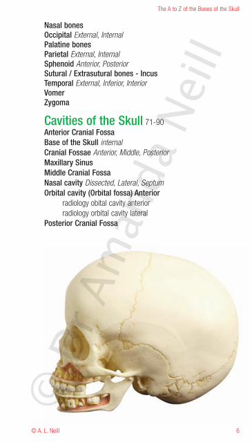
Nasal bonesOccipital External, InternalPalatine bonesParietal External, InternalSphenoid Anterior, PosteriorSutural / Extrasutural bones - IncusTemporal External, Inferior, InteriorVomerZygoma
Cavities of the Skull 71-90Anterior Cranial FossaBase of the Skull internalCranial Fossae Anterior, Middle, PosteriorMaxillary SinusMiddle Cranial FossaNasal cavity Dissected, Lateral, SeptumOrbital cavity (Orbital fossa) Anterior
radiology obital cavity anteriorradiology orbital cavity lateral
Posterior Cranial Fossa
© D
r Am
anda
Nei
ll
The A to Z of the Bones of the Skull
© A. L. Neill7
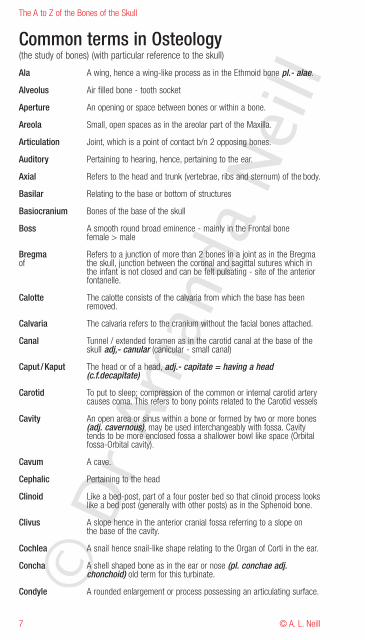
Common terms in Osteology(the study of bones) (with particular reference to the skull)
Ala A wing, hence a wing-like process as in the Ethmoid bone pl.- alae.
Alveolus Air filled bone - tooth socket
Aperture An opening or space between bones or within a bone.
Areola Small, open spaces as in the areolar part of the Maxilla.
Articulation Joint, which is a point of contact b/n 2 opposing bones.
Auditory Pertaining to hearing, hence, pertaining to the ear.
Axial Refers to the head and trunk (vertebrae, ribs and sternum) of the body.
Basilar Relating to the base or bottom of structures
Basiocranium Bones of the base of the skull
Boss A smooth round broad eminence - mainly in the Frontal bonefemale > male
Bregma Refers to a junction of more than 2 bones in a joint as in the Bregmaof the skull, junction between the coronal and sagittal sutures which in
the infant is not closed and can be felt pulsating - site of the anteriorfontanelle.
Calotte The calotte consists of the calvaria from which the base has beenremoved.
Calvaria The calvaria refers to the cranium without the facial bones attached.
Canal Tunnel / extended foramen as in the carotid canal at the base of theskull adj,- canular (canicular - small canal)
Caput /Kaput The head or of a head, adj.- capitate = having a head(c.f.decapitate)
Carotid To put to sleep; compression of the common or internal carotid arterycauses coma. This refers to bony points related to the Carotid vessels
Cavity An open area or sinus within a bone or formed by two or more bones(adj. cavernous), may be used interchangeably with fossa. Cavitytends to be more enclosed fossa a shallower bowl like space (Orbitalfossa-Orbital cavity).
Cavum A cave.
Cephalic Pertaining to the head
Clinoid Like a bed-post, part of a four poster bed so that clinoid process lookslike a bed post (generally with other posts) as in the Sphenoid bone.
Clivus A slope hence in the anterior cranial fossa referring to a slope onthe base of the cavity.
Cochlea A snail hence snail-like shape relating to the Organ of Corti in the ear.
Concha A shell shaped bone as in the ear or nose (pl. conchae adj.chonchoid) old term for this turbinate.
Condyle A rounded enlargement or process possessing an articulating surface.© D
r Am
anda
Nei
ll
8
The A to Z of the Bones of the Skull
© A. L. Neill
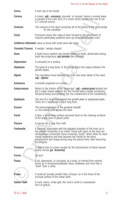
Cornu A horn (as in the Hyoid)
Corona A crown. adj.- coronary, coronoid or coronal; hence a coronal planeis parallel to the main arch of a crown which passes from ear to ear(c.f. coronal suture).
Cranium The cranium of the skull comprises all of the bones of the skull exceptfor the mandible.
Crest Prominent sharp thin ridge of bone formed by the attachment ofmuscles particularly powerful ones eg Temporalis/Sagittal crest
Cribiform/EthmoidA sieve or bone with small sieve-like holes.
Cuneate /Cuneus A wedge / wedge-shaped
Dens A tooth hence dentine and dental relating to teeth, denticulate havingtooth-like projections adj dentate See odontoid
Depression A concavity on a surface
Diaphysis The body of a long bone. In the young this is the region between thegrowth plates.
Diploë The cancellous bone between the inner and outer tables of the skull,adj.- diploic.
Eminence A smooth projection on a bone.
Endocranium Refers to the interior of the “braincase” adj.- endocranial divided intothe 3 major fossae anterior (for the Frontal lobes) middle (containingTemporal lobes) and posterior (for the containment of the Cerebellum).
Epiphysis The end of a long bone beyond the growth plate or epiphyseal plate.There are 2 epiphyses to each long bone.
Euryon The ectocranial point of the greatest breadthi.e. the widest point across the skull.
Facet A face, a small bony surface (occlusal facet on the chewing surfacesof the teeth) seen in planar joints.
Fissure A narrow slit or gap from cleft.
Fontanelle A fountain, associated with the palpable pulsation of the brain as inthe anterior fontanelle of an infant. These soft spots on the skull arecartilagenous connective tissue coverings “joints” which allow for skullcranial expansion and then become the mould for the bonedevelopment and shape joining long the sutural lines, later becomingthe Bregma.
Foramen A natural hole in a bone usually for the transmission of blood vesselsand/or nerves.(pl. foramina).
Fornix An arch
Fossa A pit, depression, or concavity, on a bone, or formed from severalbones as in temporomandibular fossa. Shallower and more like a“bowl” than a cavity
Fovea A small pit (usually smaller than a fossa)- as in the fovea of theocclusal surface of the molar tooth.
Gallus /Galli A cock, hence, crista galli, the cock's comb (i e possessiveform of gallus).©
Dr A
man
da N
eill
The A to Z of the Bones of the Skull
© A. L. Neill9
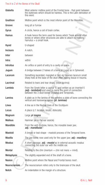
Glabella Most anterior midline point of the Frontal bone - that point betweenthe eyebrows which should be hairless. This is the Latin derivation ofthis word.
Gnathion Midline point which is the most inferior point of the Mandible.
Groove long pit or furrow
Gyrus A circle, hence a coil of brain cortex.
Hamus A hook hence the term used for bones which “hook around otherbones or where other structures are able to attach by hooking -hamulus = a small hook.
Hyoid U-shaped
Incisura A notch.
Inter between
Intra within
Introitus An orifice or point of entry to a cavity or space.
Jugum A bridge between 2 halves of a bone pl.( juga) as in Sphenoid .
Lacerum Something lacerated, mangled or torn eg foramen lacerum smallsharp hole at the base of the skull often ripping tissue in trauma.
Lacrimal Related to tears and tear drops. (noun lacrima)
Lambda From the Greek letter a capital 'L' and written as an inverted V.(adj.- lambdoid) and used to name the point of connectionbetween the 3 skull bones Occipital and Temporals.
Lamina A plate as in the lamina of the vertebra a plate of bone connecting thevertical and transverse spines (pl. laminae)
Linea A line as in the Nuchal lines of the Occitipum
Locus A place (c.f. location, locate, dislocate).
Magnum Large pl magna
Malleus Hammer (as in the ear ossicle)
Mandible From the verb to chew, hence, the movable lower jaw;adj.- mandibular.
Mastoid A breast or teat shape - mastoid process of the Temporal bone.
Maxilla The jaw-bone; now used only for the upper jaw; adj.- maxillary.
Meatus A short passage; adj.- meatal as in external acoustic meatusconnecting the outer ear with the middle ear.
Mental Relating to the chin (mentum = chin not mens = mind).
Metaphysis The slightly expanded end of the shaft of a bone.
Nasion Midline point where the Nasal and Frontal bones meet .
Neurocranium The neurocranium refers only to the braincase of the skull.
Notch An indentation in the margin of a structure.© D
r Am
anda
Nei
ll
10
The A to Z of the Bones of the Skull
© A. L. Neill
BO
NE
SO
FT
HE
SK
UL
L
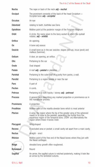
Nucha The nape or back of the neck adj.- nuchal.
Occiput The prominent convexity of the back of the head Occipitum =Occipital bone adj.- occipital
Occulus An eye
Odontoid relating to teeth, toothlike see Dens
Opisthion Midline point at the posterior margin of the Foramen Magnum
Orbit A circle; the name given to the bony socket in which the eyeballrotates; adj - orbital.
Orifice An opening.
Os A bone adj osseus
Ossicle A small bone as in the ear ossicles: stapes (stirrup), incus (anvil) andmalleus (hammer).
Ostium A door, an opening, an orifice.
Otic Pertaining to the ear
Ovale Oval shaped
Palate A roof adj.- palatal or platatine.
Parietal Pertaining to the outer wall of a cavity from paries, a wall.
Parotid Pertaining to a region beside or near the ear
Pars A part of
Pecten A comb.
Petrous Pertaining to a rock / rocky / stoney adj.- petrosal
Process A general term describing any marked projection or prominence as inthe mandibular process.
Prominens A projection
Prosthion Midline point of the maxilla alveolar bone which is most anterior
Pterion A wing; the region where the tip of the greater wing of the sphenoidmeets or is close to the parietal, separating the frontal from thesquamous region of the temporal bone. (TERY- on) Alternatively theregion where these 4 bones meet.
Pterygoid Wing shaped
Recess A secluded area or pocket; a small cavity set apart from a main cavity.
Rectus straight - erect
Rhinion Midline point of the free end of the Nasal bones where they join withthe Nasal cartilages
Ridge elevated bony growth often roughened.
Rotundum Round
Sagittal An arrow, the sagittal suture is notched posteriorly, making it look likean arrow by the lambdoid sutures.©
Dr A
man
da N
eill
The A to Z of the Bones of the Skull
© A. L. Neill11
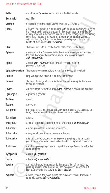
Sella A saddle; adj.- sellar, sella turcica = Turkish saddle.
Sesamoid grainlike
Sigmoid S-shaped, from the letter Sigma which is S in Greek.
Sinus A space usually within a bone lined with mucous membrane, such asthe frontal and maxillary sinuses in the head, (also, a modified BVusually vein with an enlarged lumen for blood storage and containingno or little muscle in its wall). Sinuses may contain air, venous orarterial blood, lymph or serous fluid depending upon location andhealth of the subject adj.- sinusoid.
Skull The skull refers to all of the bones that comprise the head.
Spheno A wedge i.e. the Sphenoid is the bone which wedges in the base ofthe skull between the unpaired frontal and occipital bonesadj.- sphenoid .
Spine A thorn adj.- spinous descriptive of a sharp, slenderprocess/protrusion.
Splanchocranium The splanchocranium refers to the facial bones of the skull.
Sulcus long wide groove often due to a BV indentation
Suture The saw-like edge of a cranial bone that serves as joint betweenbones of the skull.
Stylos An instrument for writing hence adj.- styloid a pencil-like structure.
Symphysis A joint or a growth
Tectum A roof.
Tegmen A covering.
Temporal Refers to time and the fact that grey hair (marking the passage oftime) often appears first at the site of the temporal bone.
Tentorium A tent.
Trabecula A “little” beam i.e. supporting structure or strut pl. trabeculae
Tubercle A small process or bump, an eminence..
Tuberculum A very small prominence, process or bump.
Tuberosity A large rounded process or eminence, a swelling or large roughprominence often associated with a tendon or ligament attachment.
Turbinate A child’s spinning top, hence shaped like a top. An old term for thenasal conchae.
Tympanum A drum pl. tympani
Uncus A hook adj.- uncinate.
Vagina A sheath; hence, invagination is the acquisition of a sheath bypushing inwards into a structure, and evagination is similar butproduced by pushing outwards adj.- vaginal.
Zygoma A yoke , hence, the bone joining the maxillary, frontal, temporal &sphenoid bones adj.- zygomatic.© D
r Am
anda
Nei
ll
12
The A to Z of the Bones of the Skull
© A. L. Neill
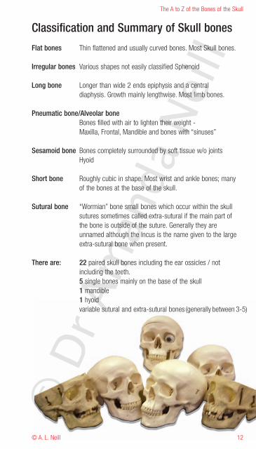
Classification and Summary of Skull bones
Flat bones Thin flattened and usually curved bones. Most Skull bones.
Irregular bones Various shapes not easily classified Sphenoid
Long bone Longer than wide 2 ends epiphysis and a centraldiaphysis. Growth mainly lengthwise. Most limb bones.
Pneumatic bone/Alveolar boneBones filled with air to lighten their weight -Maxilla, Frontal, Mandible and bones with “sinuses”
Sesamoid bone Bones completely surrounded by soft tissue w/o jointsHyoid
Short bone Roughly cubic in shape. Most wrist and ankle bones; manyof the bones at the base of the skull.
Sutural bone “Wormian” bone small bones which occur within the skullsutures sometimes called extra-sutural if the main part ofthe bone is outside of the suture. Generally they areunnamed although the Incus is the name given to the largeextra-sutural bone when present.
There are: 22 paired skull bones including the ear ossicles / notincluding the teeth.5 single bones mainly on the base of the skull1 mandible1 hyoidvariable sutural and extra-sutural bones (generally between 3-5)
© D
r Am
anda
Nei
ll
The A to Z of the Bones of the Skull
© A. L. Neill13
TH
ES
KU
LL
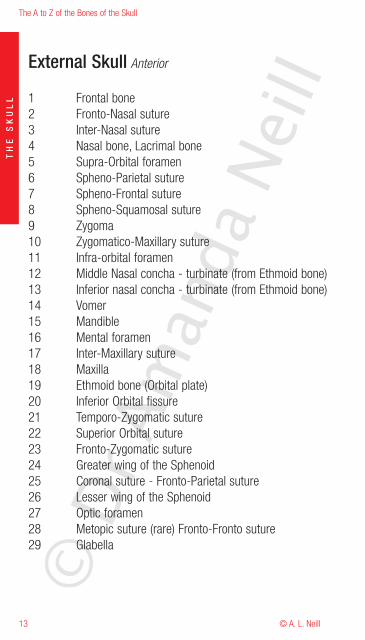
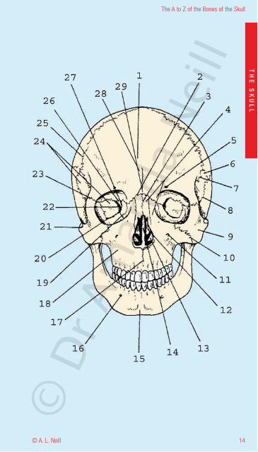
External Skull Anterior
1 Frontal bone2 Fronto-Nasal suture3 Inter-Nasal suture4 Nasal bone, Lacrimal bone5 Supra-Orbital foramen6 Spheno-Parietal suture7 Spheno-Frontal suture8 Spheno-Squamosal suture9 Zygoma10 Zygomatico-Maxillary suture11 Infra-orbital foramen12 Middle Nasal concha - turbinate (from Ethmoid bone)13 Inferior nasal concha - turbinate (from Ethmoid bone)14 Vomer15 Mandible16 Mental foramen17 Inter-Maxillary suture18 Maxilla19 Ethmoid bone (Orbital plate)20 Inferior Orbital fissure21 Temporo-Zygomatic suture22 Superior Orbital suture23 Fronto-Zygomatic suture24 Greater wing of the Sphenoid25 Coronal suture - Fronto-Parietal suture26 Lesser wing of the Sphenoid27 Optic foramen28 Metopic suture (rare) Fronto-Fronto suture29 Glabella
© D
r Am
anda
Nei
ll
14
The A to Z of the Bones of the Skull
© A. L. Neill
TH
ES
KU
LL
© D
r Am
anda
Nei
ll
The A to Z of the Bones of the Skull
© A. L. Neill15
TH
ES
KU
LL
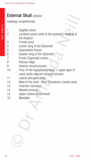
External Skull Anterior
radiology occipitofrontal
1 Sagittal suture2 Lamboid suture (view to the posterior) meeting at
the Bregma3 Frontal sinus4 Lesser wing of the Sphenoid5 Supraorbital fissure6 Greater wing of the Sphenoid7 Fronto-Zygomatic suture8 Petrous ridge9 Anterior clinoid process10 Floor of the hypophyseal fossa + upper apex of
nasal cavity adjacent to nasal sinuses11 Lateral pterygoid plate12 Base of the skull - floor of posterior cranial cavity13 Foramen rotundum14 Mastoid process15 Upper central incisor tooth16 Mandible
© D
r Am
anda
Nei
ll
16
The A to Z of the Bones of the Skull
© A. L. Neill
TH
ES
KU
LL
© D
r Am
anda
Nei
ll

The A to Z of the Bones of the Skull
© A. L. Neill17
TH
ES
KU
LL
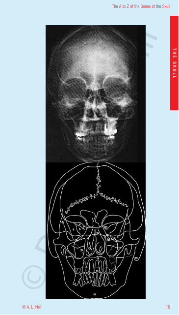
External Skull Anteriorupper and lower views in detail
radiology occipitofrontal (upper)
1 Frontal sinus2 Ethmoid sinus3 Maxillary sinus4 Foramen rotundum5 Supraorbital fissure6 Anterior clinoid process7 Posterior clinoid process8 Petrous ridge9 Floor of the hypophyseal fossa + upper apex of
nasal cavity adjacent to nasal sinuses10 Crista galli11 Frontal process of zygoma12 Middle concha - turbinate13 Inferior concha - turbinate14 Lateral border of Greater wing of sphenoid15 Greater wing of sphenoid16 Lesser wing of sphenoid17 Hard palate18 Infraorbital foramen19 Zygomaticofacial foramen20 Coronoid process of the mandible21 Soft tissue of lower lid22 Pterygoid plates of the sphenoid
© D
r Am
anda
Nei
ll
18
The A to Z of the Bones of the Skull
© A. L. Neill
TH
ES
KU
LL
© D
r Am
anda
Nei
ll
The A to Z of the Bones of the Skull
© A. L. Neill19
TH
ES
KU
LL
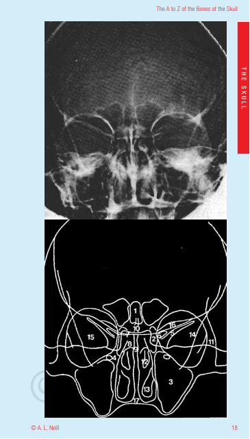
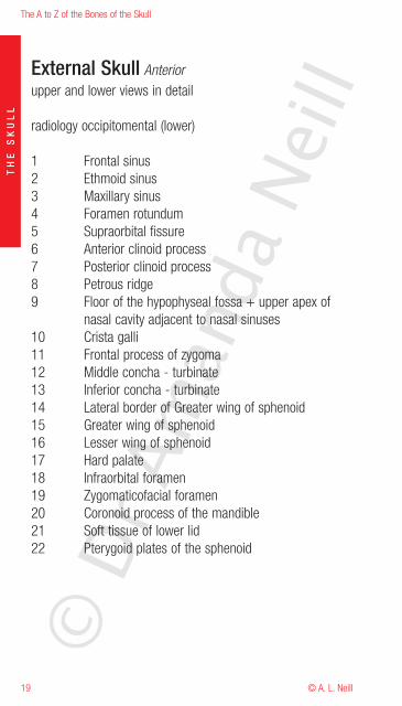
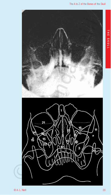
External Skull Anteriorupper and lower views in detail
radiology occipitomental (lower)
1 Frontal sinus2 Ethmoid sinus3 Maxillary sinus4 Foramen rotundum5 Supraorbital fissure6 Anterior clinoid process7 Posterior clinoid process8 Petrous ridge9 Floor of the hypophyseal fossa + upper apex of
nasal cavity adjacent to nasal sinuses10 Crista galli11 Frontal process of zygoma12 Middle concha - turbinate13 Inferior concha - turbinate14 Lateral border of Greater wing of sphenoid15 Greater wing of sphenoid16 Lesser wing of sphenoid17 Hard palate18 Infraorbital foramen19 Zygomaticofacial foramen20 Coronoid process of the mandible21 Soft tissue of lower lid22 Pterygoid plates of the sphenoid
© D
r Am
anda
Nei
ll
20
The A to Z of the Bones of the Skull
© A. L. Neill
TH
ES
KU
LL
© D
r Am
anda
Nei
ll
The A to Z of the Bones of the Skull
© A. L. Neill21
TH
ES
KU
LL
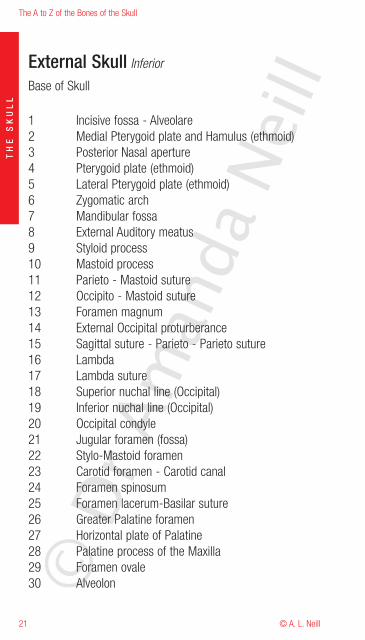
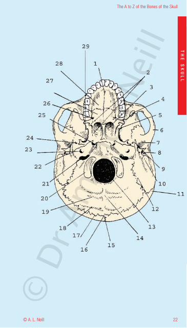
External Skull Inferior
Base of Skull
1 Incisive fossa - Alveolare2 Medial Pterygoid plate and Hamulus (ethmoid)3 Posterior Nasal aperture4 Pterygoid plate (ethmoid)5 Lateral Pterygoid plate (ethmoid)6 Zygomatic arch7 Mandibular fossa8 External Auditory meatus9 Styloid process10 Mastoid process11 Parieto - Mastoid suture12 Occipito - Mastoid suture13 Foramen magnum14 External Occipital proturberance15 Sagittal suture - Parieto - Parieto suture16 Lambda17 Lambda suture18 Superior nuchal line (Occipital)19 Inferior nuchal line (Occipital)20 Occipital condyle21 Jugular foramen (fossa)22 Stylo-Mastoid foramen23 Carotid foramen - Carotid canal24 Foramen spinosum25 Foramen lacerum-Basilar suture26 Greater Palatine foramen27 Horizontal plate of Palatine28 Palatine process of the Maxilla29 Foramen ovale30 Alveolon©
Dr A
man
da N
eill
22
The A to Z of the Bones of the Skull
© A. L. Neill
TH
ES
KU
LL
© D
r Am
anda
Nei
ll
The A to Z of the Bones of the Skull
© A. L. Neill23
TH
ES
KU
LL
External Skull Inferior
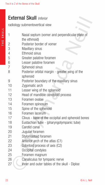
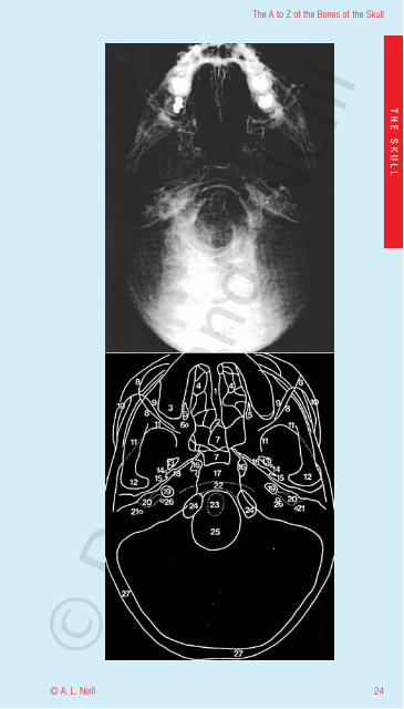
radiology submentovertical view
1 Nasal septum (vomer and perpendicular plate ofthe ethmoid)
2 Posterior border of vomer3 Maxillary sinus4 Ethmoid sinus5 Greater palatine foramen6 Lesser palatine foramen7 Sphenoid sinus8 Posterior orbital margin - greater wing of the
sphenoid9 Posterior boundary of the maxillary sinus10 Zygomatic arch11 Lesser wing of the sphenoid12 Head of mandible condyloid process13 Foramen ovalae14 Foramen spinosum15 Spine of the sphenoid16 Foramen lacerum17 Clivus - base of the occipital and sphenoid bones18 Eustachian tube - (pharyngotympanic tube)19 Carotid canal20 Jugular foramen21 Stylomastoid foramen22 Anterior arch of the atlas (C1)23 Odontoid process of axis (C2)24 Occipital condyles25 Foramen magnum26 Canaliculus for tympanic nerve27 Inner and outer tables of the skull - Diploe©
Dr A
man
da N
eill
24
The A to Z of the Bones of the Skull
© A. L. Neill
TH
ES
KU
LL
© D
r Am
anda
Nei
ll
The A to Z of the Bones of the Skull
© A. L. Neill25
TH
ES
KU
LL
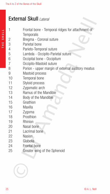
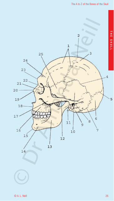
External Skull Lateral
1 Frontal bone - Temporal ridges for attachment ofTemporalis
2 Bregma - Coronal suture3 Parietal bone4 Parieto-Temporal suture5 Lambda - Occipito-Parietal suture6 Occipital bone - Occipitum7 Occipito-Mastoid suture8 Porion - upper margin of external auditory meatus9 Mastoid process10 Temporal bone11 Styloid process12 Zygomatic arch13 Ramus of the Mandible14 Body of the Mandible15 Gnathion16 Maxilla17 Zygoma18 Prosthion19 Rhinion20 Nasal bone21 Lacrimal bone22 Nasion23 Glabella24 Frontal bone25 Greater wing of the Sphenoid
© D
r Am
anda
Nei
ll
26
The A to Z of the Bones of the Skull
© A. L. Neill
TH
ES
KU
LL
© D
r Am
anda
Nei
ll
The A to Z of the Bones of the Skull
© A. L. Neill27
TH
ES
KU
LL
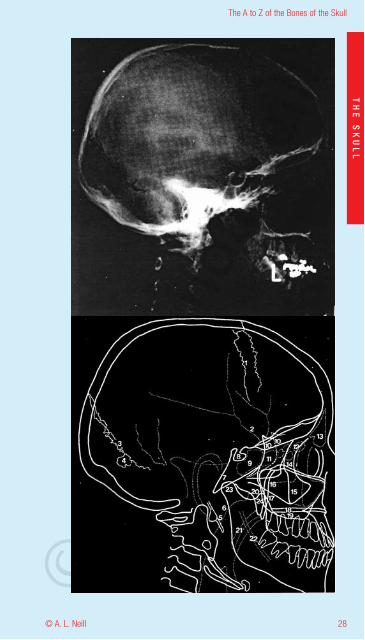
External Skull Lateral / obliqueradiology
1 Coronal suture2 Impression for Middle meningeal artery3 Lambdoid suture4 Wormian bone - Extrasutural bone5 Styloid process6 Posterior wall of Nasopharynx7 Clivus - (base of Sphenoid and Occiptal bones)8 Hypophyseal fossa9 Sphenoid sinus10 Greater wing of the Sphenoid11 Posterior Air cells in the Ethmoid - Ethmoid sinus12 Anterior Air cells in the Ethmoid - Ethmoid sinus13 Frontal sinus14 Zygoma - frontal process15 Maxilla - malar process16 Zygoma - Arch17 Posterior border of the Maxillary sinus18 Hard palate - Palatine bone19 Alveolar bone in Maxilla20 Pterygoid plates21 Soft tissue of Soft Palate and Uvula22 Mandibular canal23 Head of Mandible24 Coronoid process of Mandible
© D
r Am
anda
Nei
ll
28
The A to Z of the Bones of the Skull
© A. L. Neill
TH
ES
KU
LL
© D
r Am
anda
Nei
ll
The A to Z of the Bones of the Skull
© A. L. Neill29
TH
ES
KU
LL
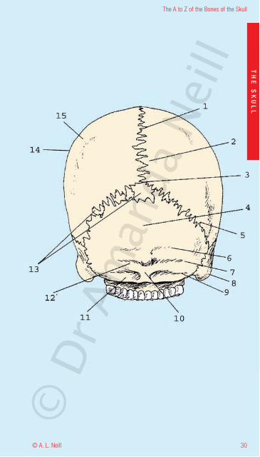
External Skull Posterior
1 Sagittal sinus2 Parietal foramen3 Lambda Pareito - Occipital suture4 Occipital bone5 Lambdoid suture6 Superior nuchal line7 Highest nuchal line8 Superior nuchal line9 Mastoid process10 External Occitipal protruberance11 Inferior nuchal groove12 Superior nuchal groove13 Sutural bones - Inca14 Euyron15 Parietal bone
© D
r Am
anda
Nei
ll
30
The A to Z of the Bones of the Skull
© A. L. Neill
TH
ES
KU
LL
© D
r Am
anda
Nei
ll
The A to Z of the Bones of the Skull
© A. L. Neill31
TH
ES
KU
LL
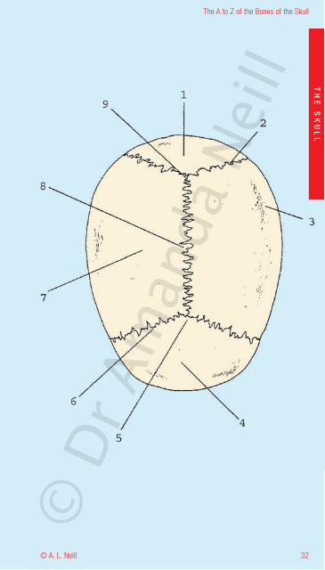
External Skull Superior
1 Occipital bone2 Lambdoid suture - Occipito-Parieto suture3 Parietal eminence - Euryon4 Frontal bone5 Bregma6 Coronal suture7 Parietal bone8 Sagittal suture9 Lambda
© D
r Am
anda
Nei
ll
32
The A to Z of the Bones of the Skull
© A. L. Neill
TH
ES
KU
LL
© D
r Am
anda
Nei
ll
The A to Z of the Bones of the Skull
© A. L. Neill33
TH
ES
KU
LL
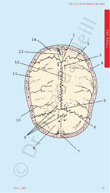
Internal Skull Inferior
Skull cap
1 Lambda2 Lambdoid suture3 Parietal foramen4 Diploe5 Bregma6 Coronal suture7 Frontal crest and Diploe - inner and outer tables of
the skull bone8 Frontal bone9 Depressions for arachnoid granulations10 Grooves for middle meningeal vessels11 Parietal bone12 Sagittal suture13 Groove for superior sagittal sinus14 Occipital bone
© D
r Am
anda
Nei
ll
34
The A to Z of the Bones of the Skull
© A. L. Neill
TH
ES
KU
LL
© D
r Am
anda
Nei
ll
The A to Z of the Bones of the Skull
© A. L. Neill35
TH
ES
KU
LL
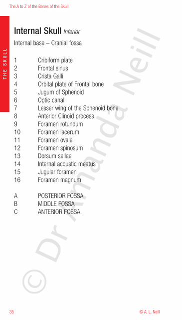
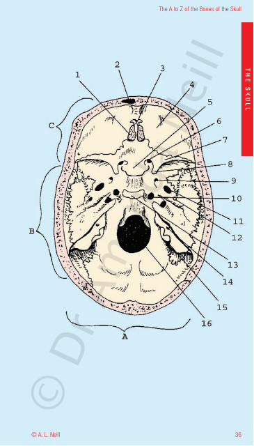
Internal Skull Inferior
Internal base – Cranial fossa
1 Cribiform plate2 Frontal sinus3 Crista Galli4 Orbital plate of Frontal bone5 Jugum of Sphenoid6 Optic canal7 Lesser wing of the Sphenoid bone8 Anterior Clinoid process9 Foramen rotundum10 Foramen lacerum11 Foramen ovale12 Foramen spinosum13 Dorsum sellae14 Internal acoustic meatus15 Jugular foramen16 Foramen magnum
A POSTERIOR FOSSAB MIDDLE FOSSAC ANTERIOR FOSSA
© D
r Am
anda
Nei
ll
36
The A to Z of the Bones of the Skull
© A. L. Neill
TH
ES
KU
LL
© D
r Am
anda
Nei
ll
The A to Z of the Bones of the Skull
© A. L. Neill37
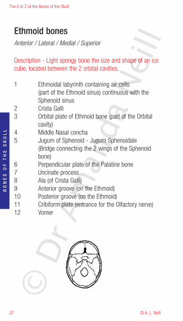
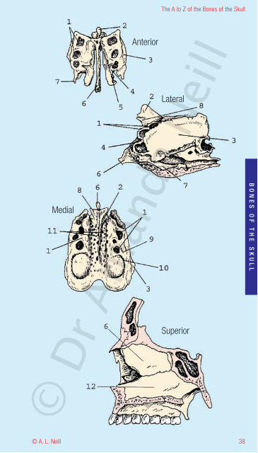
Ethmoid bonesAnterior / Lateral / Medial / Superior
Description - Light spongy bone the size and shape of an icecube, located between the 2 orbital cavities.
1 Ethmoidal labyrinth containing air cells(part of the Ethmoid sinus) continuous with theSphenoid sinus
2 Crista Galli3 Orbital plate of Ethmoid bone (part of the Orbital
cavity)4 Middle Nasal concha5 Jugum of Sphenoid - Jugum Sphenoidale
(Bridge connecting the 2 wings of the Sphenoidbone)
6 Perpendicular plate of the Palatine bone7 Uncinate process8 Ala (of Crista Galli)9 Anterior groove (on the Ethmoid)10 Posterior groove (on the Ethmoid)11 Cribiform plate (entrance for the Olfactory nerve)12 Vomer
BO
NE
SO
FT
HE
SK
UL
L
© D
r Am
anda
Nei
ll
38
The A to Z of the Bones of the Skull
© A. L. Neill
Anterior
Lateral
Medial
Superior
BO
NE
SO
FT
HE
SK
UL
L
© D
r Am
anda
Nei
ll
The A to Z of the Bones of the Skull
© A. L. Neill39
BO
NE
SO
FT
HE
SK
UL
L
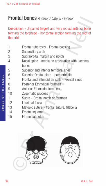
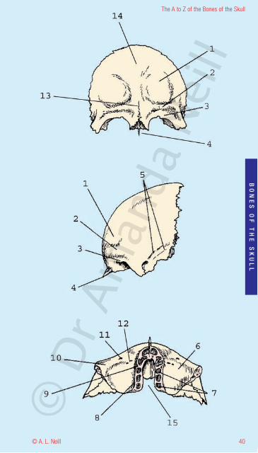
Frontal bones Anterior / Lateral / Inferior
Description - Unpaired largest and very robust anterior boneforming the forehead - horizontal section forming the roof ofthe orbit.
1 Frontal tuberosity - Frontal bossing2 Superciliary arch3 Supraorbital margin and notch4 Nasal spine - medial to articulation with Lacrimal
bones5 Superior and inferior temporal lines6 Superior Orbital plate - pars orbitalis7 Frontal and Ethmoid air cells - Frontal sinus8 Posterior Ethmoidal foramen9 Anterior Ethmoidal foramen10 Zygomatic process11 Supra - Orbital notch or foramen12 Lacrimal fossa13 Metopic suture - frontal suture, Glabella14 Frontal squama15 Ethmoidal notch
© D
r Am
anda
Nei
ll
40
The A to Z of the Bones of the Skull
© A. L. Neill
BO
NE
SO
FT
HE
SK
UL
L
© D
r Am
anda
Nei
ll
The A to Z of the Bones of the Skull
© A. L. Neill41
BO
NE
SO
FT
HE
SK
UL
L
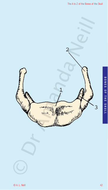
Hyoid bone
Description - Small U-shaped bone. Attached to the styloidprocesses via ligaments, this bone has no articulations and isnot normally broken in normal trauma. It may be broken inhanging and strangulation.
1 Body of hyoid2 Greater horn (cornu)3 Lesser horn (cornu)
© D
r Am
anda
Nei
ll
42
The A to Z of the Bones of the Skull
© A. L. Neill
BO
NE
SO
FT
HE
SK
UL
L
© D
r Am
anda
Nei
ll
The A to Z of the Bones of the Skull
© A. L. Neill43
BO
NE
SO
FT
HE
SK
UL
L
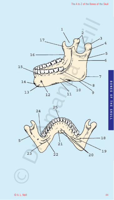
Mandible Lateral / Posterior
Description - The lower jaw bone joins the skull via a singlepaired joint the temporomandibular joint (TMJ) via thecondyles and a cartilaginous plate in the Temporal fossa.
Primary function - mastication.
1 Mandibular notch2 Pterygoid fovea3 Head of Mandible - Condyle - Condylar process4 Neck of Mandible5 Posterior border of ramus of mandible6 Ramus - Vertical ramus7 Angle of mandible8 Oblique line9 Inferior border10 Body - Horizontal ramus11 Base12 Mental foramen13 Mental tubercle - Gnathion14 Mental protuberance15 Alveolar bone surrounding teeth16 Anterior border of ramus17 Coronoid process - Endocoronial ridge18 Mandibular foramen19 Lingula20 Superior and inferior mental spines21 Digastric fossa22 Mylohyoid line23 Mylohyoid groove24 Extramolar sulcus25 Infradentale©
Dr A
man
da N
eill
44
The A to Z of the Bones of the Skull
© A. L. Neill
BO
NE
SO
FT
HE
SK
UL
L
© D
r Am
anda
Nei
ll
The A to Z of the Bones of the Skull
© A. L. Neill45
BO
NE
SO
FT
HE
SK
UL
L
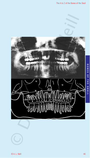
Mandible Anteriorradiologyorthopantomogram = OPG
Used to show mandibular fractures and an overview ofsinuses and complete dentition
1 Central incisor2 Lateral incisor3 Canine4 First premolar5 Second premolar6 First molar7 Second molar8 Pulp chamber9 Coronoid process10 Head of mandible11 Zygoma12 Maxillary sinus13 Anterior nasal spine of maxilla14 Vomer (nasal septum)15 Sites for the third molar16 Hard palate
© D
r Am
anda
Nei
ll
46
The A to Z of the Bones of the Skull
© A. L. Neill
BO
NE
SO
FT
HE
SK
UL
L
© D
r Am
anda
Nei
ll
The A to Z of the Bones of the Skull
© A. L. Neill47
BO
NE
SO
FT
HE
SK
UL
L
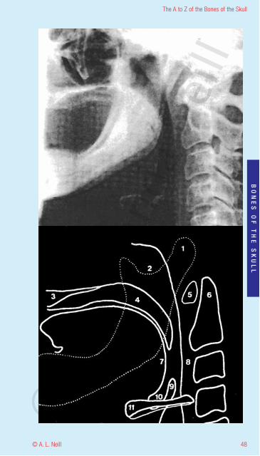
Mandible LateralradiologyShowing relationship to surrounding soft tissue
1 Head of mandible – Condylar process2 Neck of the Mandible3 Hard Palate4 Soft Palate5 Anterior notch of the Atlas (C1)6 Odontoid process of the Axis (C2) – Dens7 Posterior aspect of the tongue8 Retropharyngeal sac9 Epiglottis10 Vallecula - fold anterior to epiglottis11 Hyoid bone
© D
r Am
anda
Nei
ll
48
The A to Z of the Bones of the Skull
© A. L. Neill
BO
NE
SO
FT
HE
SK
UL
L
© D
r Am
anda
Nei
ll
The A to Z of the Bones of the Skull
© A. L. Neill49
BO
NE
SO
FT
HE
SK
UL
L
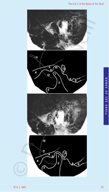
Mandibular jointTemporo-Mandibular joint (TMJ) LateralradiologyOpen - upperShut - lower
1 Head of mandible – Condylar process2 Neck of the Mandible3 Coronoid process4 Zygomatic arch5 External auditory meatus + Handle of the malleus6 Articular cubicle7 Tympanic plate8 Mastoid process9 Groove for posterior belly of Digastric muscle10 Mandibular fossa11 Greater wing of the sphenoid (basal surface)12 Lesser wing of the sphenoid
© D
r Am
anda
Nei
ll
50
The A to Z of the Bones of the Skull
© A. L. Neill
BO
NE
SO
FT
HE
SK
UL
L
© D
r Am
anda
Nei
ll
The A to Z of the Bones of the Skull
© A. L. Neill51
BO
NE
SO
FT
HE
SK
UL
L
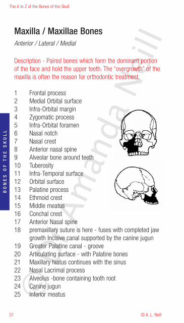
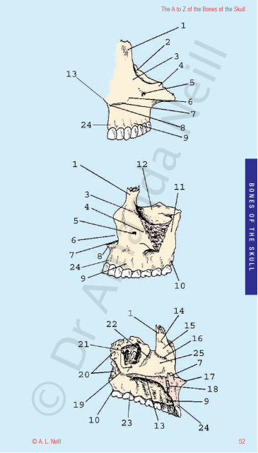
Maxilla / Maxillae BonesAnterior / Lateral / Medial
Description - Paired bones which form the dominant portionof the face and hold the upper teeth. The “overgrowth” of themaxilla is often the reason for orthodontic treatment.
1 Frontal process2 Medial Orbital surface3 Infra-Orbital margin4 Zygomatic process5 Infra-Orbital foramen6 Nasal notch7 Nasal crest8 Anterior nasal spine9 Alveolar bone around teeth10 Tuberosity11 Infra-Temporal surface12 Orbital surface13 Palatine process14 Ethmoid crest15 Middle meatus16 Conchal crest17 Anterior Nasal spine18 premaxillary suture is here - fuses with completed jaw
growth Incisive canal supported by the canine jugun19 Greater Palatine canal - groove20 Articulating surface - with Palatine bones21 Maxillary hiatus continues with the sinus22 Nasal Lacrimal process23 Alveolus -bone containing tooth root24 Canine jugun25 Inferior meatus©
Dr A
man
da N
eill
52
The A to Z of the Bones of the Skull
© A. L. Neill
BO
NE
SO
FT
HE
SK
UL
L
© D
r Am
anda
Nei
ll
The A to Z of the Bones of the Skull
© A. L. Neill53
BO
NE
SO
FT
HE
SK
UL
L
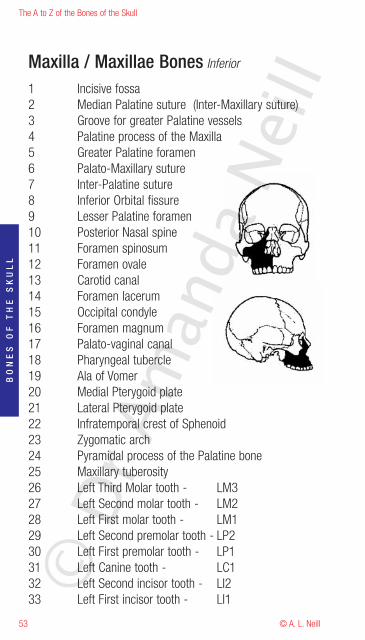
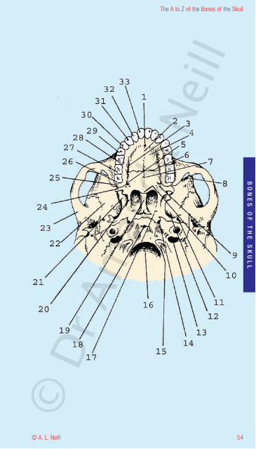
Maxilla / Maxillae Bones Inferior
1 Incisive fossa2 Median Palatine suture (Inter-Maxillary suture)3 Groove for greater Palatine vessels4 Palatine process of the Maxilla5 Greater Palatine foramen6 Palato-Maxillary suture7 Inter-Palatine suture8 Inferior Orbital fissure9 Lesser Palatine foramen10 Posterior Nasal spine11 Foramen spinosum12 Foramen ovale13 Carotid canal14 Foramen lacerum15 Occipital condyle16 Foramen magnum17 Palato-vaginal canal18 Pharyngeal tubercle19 Ala of Vomer20 Medial Pterygoid plate21 Lateral Pterygoid plate22 Infratemporal crest of Sphenoid23 Zygomatic arch24 Pyramidal process of the Palatine bone25 Maxillary tuberosity26 Left Third Molar tooth - LM327 Left Second molar tooth - LM228 Left First molar tooth - LM129 Left Second premolar tooth - LP230 Left First premolar tooth - LP131 Left Canine tooth - LC132 Left Second incisor tooth - LI233 Left First incisor tooth - LI1©
Dr A
man
da N
eill
54
The A to Z of the Bones of the Skull
© A. L. Neill
BO
NE
SO
FT
HE
SK
UL
L
© D
r Am
anda
Nei
ll
The A to Z of the Bones of the Skull
© A. L. Neill55
BO
NE
SO
FT
HE
SK
UL
L
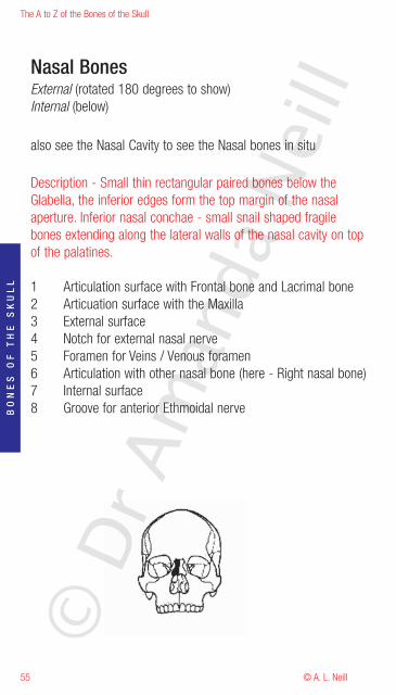
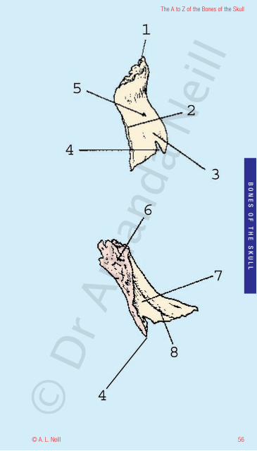
Nasal BonesExternal (rotated 180 degrees to show)Internal (below)
also see the Nasal Cavity to see the Nasal bones in situ
Description - Small thin rectangular paired bones below theGlabella, the inferior edges form the top margin of the nasalaperture. Inferior nasal conchae - small snail shaped fragilebones extending along the lateral walls of the nasal cavity on topof the palatines.
1 Articulation surface with Frontal bone and Lacrimal bone2 Articuation surface with the Maxilla3 External surface4 Notch for external nasal nerve5 Foramen for Veins / Venous foramen6 Articulation with other nasal bone (here - Right nasal bone)7 Internal surface8 Groove for anterior Ethmoidal nerve
© D
r Am
anda
Nei
ll
56
The A to Z of the Bones of the Skull
© A. L. Neill
BO
NE
SO
FT
HE
SK
UL
L
© D
r Am
anda
Nei
ll
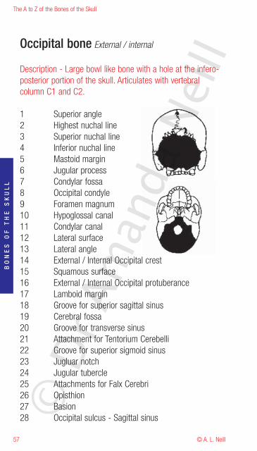
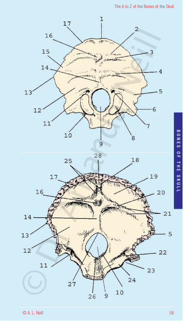
Occipital bone External / internal
Description - Large bowl like bone with a hole at the infero-posterior portion of the skull. Articulates with vertebralcolumn C1 and C2.
1 Superior angle2 Highest nuchal line3 Superior nuchal line4 Inferior nuchal line5 Mastoid margin6 Jugular process7 Condylar fossa8 Occipital condyle9 Foramen magnum10 Hypoglossal canal11 Condylar canal12 Lateral surface13 Lateral angle14 External / Internal Occipital crest15 Squamous surface16 External / Internal Occipital protuberance17 Lamboid margin18 Groove for superior sagittal sinus19 Cerebral fossa20 Groove for transverse sinus21 Attachment for Tentorium Cerebelli22 Groove for superior sigmoid sinus23 Jugluar notch24 Jugular tubercle25 Attachments for Falx Cerebri26 Opisthion27 Basion28 Occipital sulcus - Sagittal sinus
The A to Z of the Bones of the Skull
© A. L. Neill57
BO
NE
SO
FT
HE
SK
UL
L
© D
r Am
anda
Nei
ll
58
The A to Z of the Bones of the Skull
© A. L. Neill
BO
NE
SO
FT
HE
SK
UL
L
© D
r Am
anda
Nei
ll
The A to Z of the Bones of the Skull
© A. L. Neill59
BO
NE
SO
FT
HE
SK
UL
L
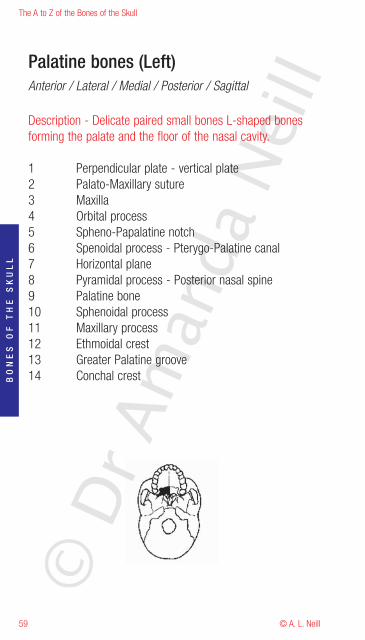
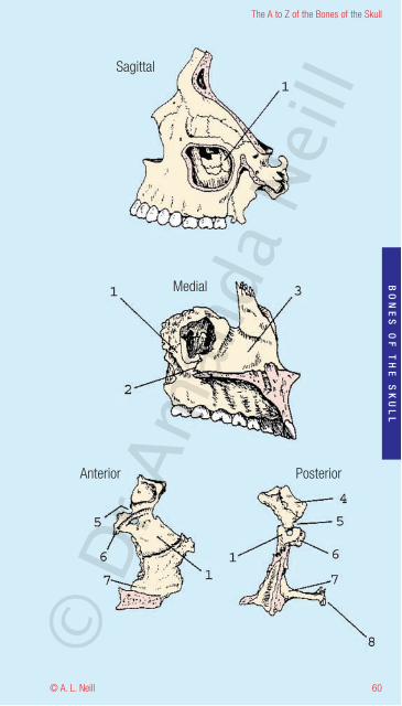
Palatine bones (Left)Anterior / Lateral / Medial / Posterior / Sagittal
Description - Delicate paired small bones L-shaped bonesforming the palate and the floor of the nasal cavity.
1 Perpendicular plate - vertical plate2 Palato-Maxillary suture3 Maxilla4 Orbital process5 Spheno-Papalatine notch6 Spenoidal process - Pterygo-Palatine canal7 Horizontal plane8 Pyramidal process - Posterior nasal spine9 Palatine bone10 Sphenoidal process11 Maxillary process12 Ethmoidal crest13 Greater Palatine groove14 Conchal crest
© D
r Am
anda
Nei
ll
60
The A to Z of the Bones of the Skull
© A. L. Neill
BO
NE
SO
FT
HE
SK
UL
L
Sagittal
Medial
Anterior Posterior
© D
r Am
anda
Nei
ll
The A to Z of the Bones of the Skull
© A. L. Neill61
BO
NE
SO
FT
HE
SK
UL
L
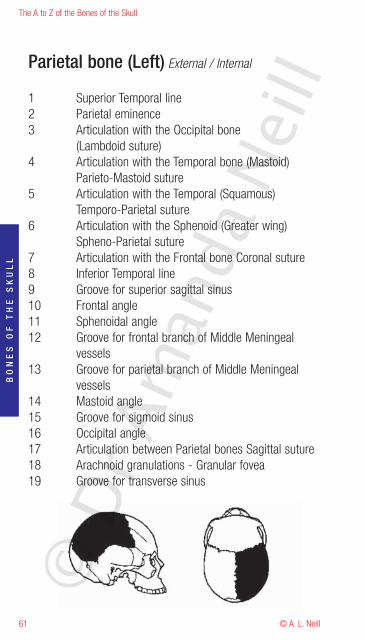
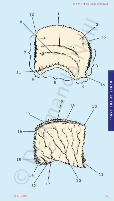
Parietal bone (Left) External / Internal
1 Superior Temporal line2 Parietal eminence3 Articulation with the Occipital bone
(Lambdoid suture)4 Articulation with the Temporal bone (Mastoid)
Parieto-Mastoid suture5 Articulation with the Temporal (Squamous)
Temporo-Parietal suture6 Articulation with the Sphenoid (Greater wing)
Spheno-Parietal suture7 Articulation with the Frontal bone Coronal suture8 Inferior Temporal line9 Groove for superior sagittal sinus10 Frontal angle11 Sphenoidal angle12 Groove for frontal branch of Middle Meningeal
vessels13 Groove for parietal branch of Middle Meningeal
vessels14 Mastoid angle15 Groove for sigmoid sinus16 Occipital angle17 Articulation between Parietal bones Sagittal suture18 Arachnoid granulations - Granular fovea19 Groove for transverse sinus
© D
r Am
anda
Nei
ll
62
The A to Z of the Bones of the Skull
© A. L. Neill
BO
NE
SO
FT
HE
SK
UL
L
© D
r Am
anda
Nei
ll
The A to Z of the Bones of the Skull
© A. L. Neill63
BO
NE
SO
FT
HE
SK
UL
L
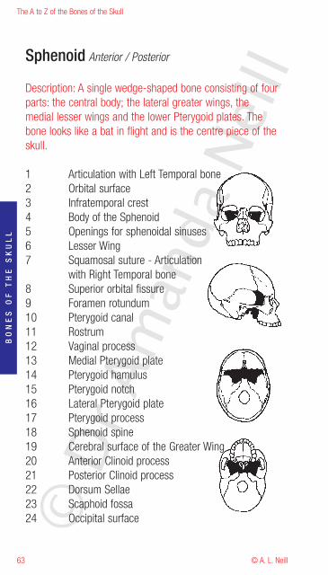
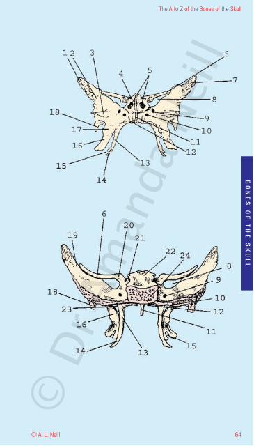
Sphenoid Anterior / Posterior
Description: A single wedge-shaped bone consisting of fourparts: the central body; the lateral greater wings, themedial lesser wings and the lower Pterygoid plates. Thebone looks like a bat in flight and is the centre piece of theskull.
1 Articulation with Left Temporal bone2 Orbital surface3 Infratemporal crest4 Body of the Sphenoid5 Openings for sphenoidal sinuses6 Lesser Wing7 Squamosal suture - Articulation
with Right Temporal bone8 Superior orbital fissure9 Foramen rotundum10 Pterygoid canal11 Rostrum12 Vaginal process13 Medial Pterygoid plate14 Pterygoid hamulus15 Pterygoid notch16 Lateral Pterygoid plate17 Pterygoid process18 Sphenoid spine19 Cerebral surface of the Greater Wing20 Anterior Clinoid process21 Posterior Clinoid process22 Dorsum Sellae23 Scaphoid fossa24 Occipital surface©
Dr A
man
da N
eill
64
The A to Z of the Bones of the Skull
© A. L. Neill
BO
NE
SO
FT
HE
SK
UL
L
© D
r Am
anda
Nei
ll
The A to Z of the Bones of the Skull
© A. L. Neill65
BO
NE
SO
FT
HE
SK
UL
L
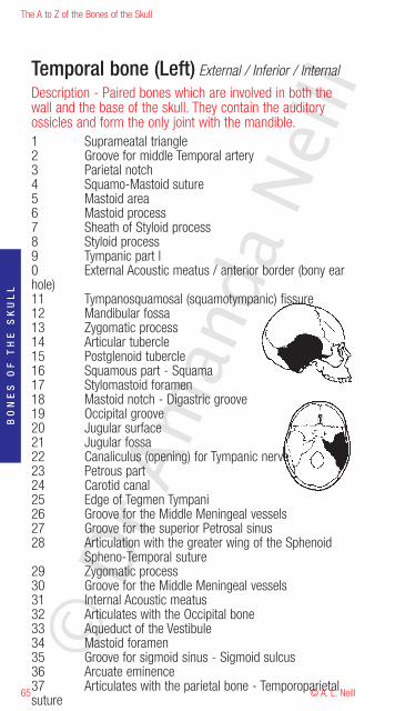
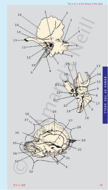
Temporal bone (Left) External / Inferior / Internal
Description - Paired bones which are involved in both thewall and the base of the skull. They contain the auditoryossicles and form the only joint with the mandible.1 Suprameatal triangle2 Groove for middle Temporal artery3 Parietal notch4 Squamo-Mastoid suture5 Mastoid area6 Mastoid process7 Sheath of Styloid process8 Styloid process9 Tympanic part l0 External Acoustic meatus / anterior border (bony earhole)11 Tympanosquamosal (squamotympanic) fissure12 Mandibular fossa13 Zygomatic process14 Articular tubercle15 Postglenoid tubercle16 Squamous part - Squama17 Stylomastoid foramen18 Mastoid notch - Digastric groove19 Occipital groove20 Jugular surface21 Jugular fossa22 Canaliculus (opening) for Tympanic nerve23 Petrous part24 Carotid canal25 Edge of Tegmen Tympani26 Groove for the Middle Meningeal vessels27 Groove for the superior Petrosal sinus28 Articulation with the greater wing of the Sphenoid
Spheno-Temporal suture29 Zygomatic process30 Groove for the Middle Meningeal vessels31 Internal Acoustic meatus32 Articulates with the Occipital bone33 Aqueduct of the Vestibule34 Mastoid foramen35 Groove for sigmoid sinus - Sigmoid sulcus36 Arcuate eminence37 Articulates with the parietal bone - Temporoparietalsuture
© D
r Am
anda
Nei
ll
66
The A to Z of the Bones of the Skull
© A. L. Neill
BO
NE
SO
FT
HE
SK
UL
L
© D
r Am
anda
Nei
ll
The A to Z of the Bones of the Skull
© A. L. Neill67
BO
NE
SO
FT
HE
SK
UL
L
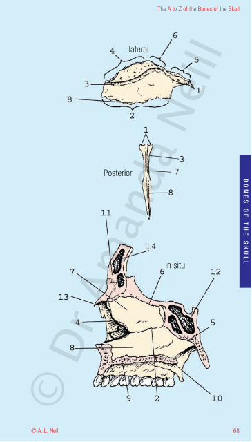
Vomer Lateral / Posterior / in situ
Description - A single small narrow frail plough-shapedmidline bone. Deviation of this bone may obstruct the nasalairways.
1 Ala (Alae)2 Articulation with Maxillae and Palatine
Maxillovomer suture / palatinovomer suture3 Groove for the nasopalantine nerves and vessels4 Articulation with nasal cartilages5 Articulation with Sphenoid bone6 Articulation with the Ethmoid plate7 Perpendicular plate of the Ethmoid8 Body of Vomer9 Maxillae areolar bone10 Medial pterygoid plate11 Frontal bone12 Sphenoid sinus13 Anterior of nasal bones14 Frontal sinus
© D
r Am
anda
Nei
ll
68
The A to Z of the Bones of the Skull
© A. L. Neill
BO
NE
SO
FT
HE
SK
UL
L
in situ
lateral
Posterior
© D
r Am
anda
Nei
ll
The A to Z of the Bones of the Skull
© A. L. Neill69
BO
NE
SO
FT
HE
SK
UL
L
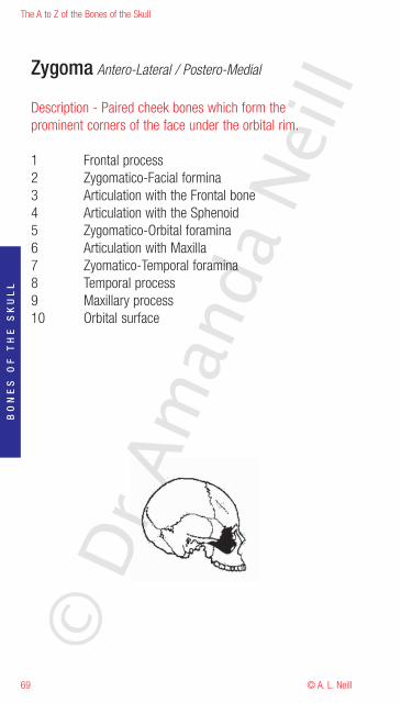
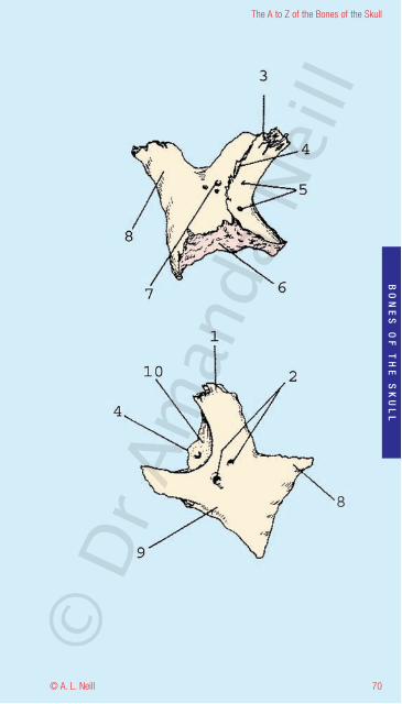
Zygoma Antero-Lateral / Postero-Medial
Description - Paired cheek bones which form theprominent corners of the face under the orbital rim.
1 Frontal process2 Zygomatico-Facial formina3 Articulation with the Frontal bone4 Articulation with the Sphenoid5 Zygomatico-Orbital foramina6 Articulation with Maxilla7 Zyomatico-Temporal foramina8 Temporal process9 Maxillary process10 Orbital surface
© D
r Am
anda
Nei
ll
70
The A to Z of the Bones of the Skull
© A. L. Neill
BO
NE
SO
FT
HE
SK
UL
L
© D
r Am
anda
Nei
ll
The A to Z of the Bones of the Skull
© A. L. Neill71
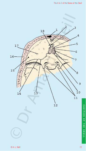
Anterior Cranial FossaSuperior view from the top
1 Frontal sinus2 Frontal crest3 Foramen Caecum4 Cranionasal exit of the anterior ethmoidal nerve5 Slit for the process of the dura mater6 Anterior Ethmoidal groove7 Concealed entry for the anterior Ethmoidal nerve8 Posterior Ethmoidal groove9 Concealed entry for the posterior Ethmoidal nerve10 Jugam Sphenoidale
Optic canals12 Anterior Clinoid process13 Fronto-Sphenoid suture14 Lesser wing of the Sphenoid15 Vascular foramen16 Diploe17 Orbital plate of the Frontal bone18 Crista Galli
CA
VIT
IES
OF
TH
ES
KU
LL
© D
r Am
anda
Nei
ll
72
The A to Z of the Bones of the Skull
© A. L. Neill
CA
VIT
IES
OF
TH
ES
KU
LL©
Dr A
man
da N
eill
The A to Z of the Bones of the Skull
© A. L. Neill73
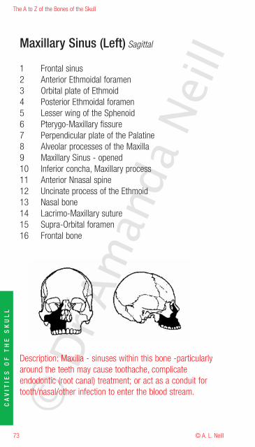
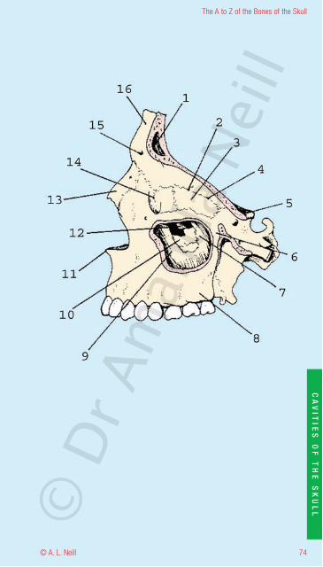
Maxillary Sinus (Left) Sagittal
1 Frontal sinus2 Anterior Ethmoidal foramen3 Orbital plate of Ethmoid4 Posterior Ethmoidal foramen5 Lesser wing of the Sphenoid6 Pterygo-Maxillary fissure7 Perpendicular plate of the Palatine8 Alveolar processes of the Maxilla9 Maxillary Sinus - opened10 Inferior concha, Maxillary process11 Anterior Nnasal spine12 Uncinate process of the Ethmoid13 Nasal bone14 Lacrimo-Maxillary suture15 Supra-Orbital foramen16 Frontal bone
Description: Maxilla - sinuses within this bone -particularlyaround the teeth may cause toothache, complicateendodontic (root canal) treatment; or act as a conduit fortooth/nasal/other infection to enter the blood stream.
CA
VIT
IES
OF
TH
ES
KU
LL
© D
r Am
anda
Nei
ll
74
The A to Z of the Bones of the Skull
© A. L. Neill
CA
VIT
IES
OF
TH
ES
KU
LL©
Dr A
man
da N
eill

The A to Z of the Bones of the Skull
© A. L. Neill75
Middle Cranial Fossa (Left)Superior view from the top
1 Greater wing of the Sphenoid2 Superior Orbital fissure3 Middle Clinoid process4 Optic canal5 Anterior Clinoid process6 Hypophysial fossa7 Posterior Clinoid process8 Dorsum Sellae9 Foramen lacerum10 Trigeminal impression Foramen rotundum12 Groove for superior petrosal sinus13 Groove for greater petrosal nerve14 Diploe15 Arcuate eminence16 Tegmen typani17 Temporal bone18 Groove for the Parietal branch of the middle
Meningeal vessels
CA
VIT
IES
OF
TH
ES
KU
LL
© D
r Am
anda
Nei
ll
76
The A to Z of the Bones of the Skull
© A. L. Neill
CA
VIT
IES
OF
TH
ES
KU
LL©
Dr A
man
da N
eill
The A to Z of the Bones of the Skull
© A. L. Neill77
CA
VIT
IES
OF
TH
ES
KU
LL
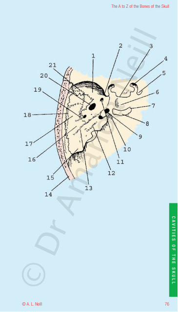
Nasal Cavity - dissected to show Right nasal cavity andinferior nasal conchaePara - Sagittal section of Right Nasal cavitylooking in through the Right side of the nose to show sinusesaround the area
1 Semilunar hiatus2 Ethmoid bulla3 Air cells of Ethmoid sinus4 Sphenoidal sinuses5 Spheno-Palatine foramen6 Perpendicular plate of the palatine7 Inferior nasal concha and meatus8 Opening of the maxillary sinus9 Uncinate process of Ethmoid bone10 Right Frontal sinus
Note middle and superior nasal conchae have been dissectedand removed.
Description: inferior nasal conchae - small paired frail bonesextending along the lateral walls of the nasal cavity. Involvedin olfaction, humidification. Highly vascularized and when thesoft tissue surrounding this is swollen, it may cause nasalobstruction and sinuses. Please note when "picking one'snose" the finger never reaches the nasal conchae, stoppingjust short of this area.©
Dr A
man
da N
eill
78
The A to Z of the Bones of the Skull
© A. L. Neill
CA
VIT
IES
OF
TH
ES
KU
LL©
Dr A
man
da N
eill
The A to Z of the Bones of the Skull
© A. L. Neill79
CA
VIT
IES
OF
TH
ES
KU
LL
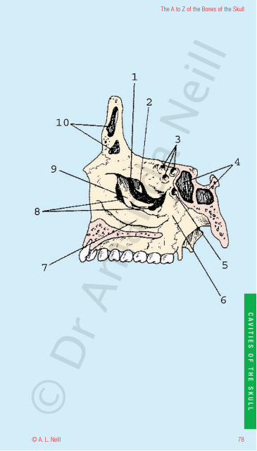
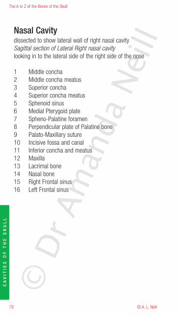
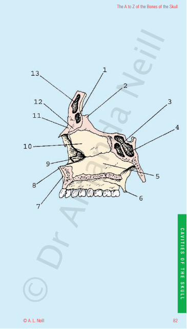
Nasal Cavitydissected to show lateral wall of right nasal cavitySagittal section of Lateral Right nasal cavitylooking in to the lateral side of the right side of the nose
1 Middle concha2 Middle concha meatus3 Superior concha4 Superior concha meatus5 Sphenoid sinus6 Medial Pterygoid plate7 Spheno-Palatine foramen8 Perpendicular plate of Palatine bone9 Palato-Maxillary suture10 Incisive fossa and canal11 Inferior concha and meatus12 Maxilla13 Lacrimal bone14 Nasal bone15 Right Frontal sinus16 Left Frontal sinus
© D
r Am
anda
Nei
ll
80
The A to Z of the Bones of the Skull
© A. L. Neill
CA
VIT
IES
OF
TH
ES
KU
LL©
Dr A
man
da N
eill
The A to Z of the Bones of the Skull
© A. L. Neill81
CA
VIT
IES
OF
TH
ES
KU
LL
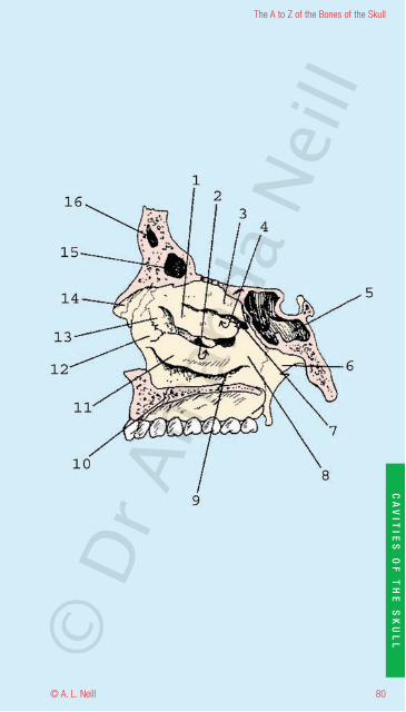
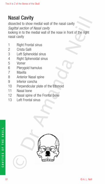
Nasal Cavitydissected to show medial wall of the nasal cavitySagittal section of Nasal cavitylooking in to the medial wall of the nose in front of the rightnasal cavity
1 Right Frontal sinus2 Crista Galli3 Left Sphenoidal sinus4 Right Sphenoidal sinus5 Vomer6 Pterygoid hamulus7 Maxilla8 Anterior Nasal spine9 Inferior concha10 Perpendicular plate of the Ethmoid11 Nasal bone12 Nasal spine of the Frontal bone13 Left Frontal sinus
© D
r Am
anda
Nei
ll
82
The A to Z of the Bones of the Skull
© A. L. Neill
CA
VIT
IES
OF
TH
ES
KU
LL©
Dr A
man
da N
eill
The A to Z of the Bones of the Skull
© A. L. Neill83
CA
VIT
IES
OF
TH
ES
KU
LL
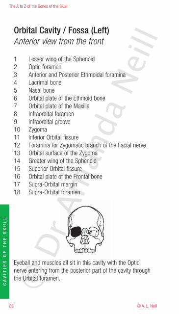
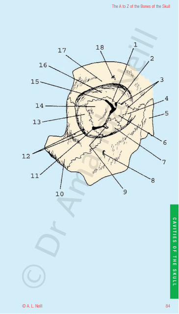
Orbital Cavity / Fossa (Left)Anterior view from the front
1 Lesser wing of the Sphenoid2 Optic foramen3 Anterior and Posterior Ethmoidal foramina4 Lacrimal bone5 Nasal bone6 Orbital plate of the Ethmoid bone7 Orbital plate of the Maxilla8 Infraorbital foramen9 Infraorbital groove10 Zygoma11 Inferior Orbital fissure12 Foramina for Zygomatic branch of the Facial nerve13 Orbital surface of the Zygoma14 Greater wing of the Sphenoid15 Superior Orbital fissure16 Orbital plate of the Frontal bone17 Supra-Orbital margin18 Supra-Orbital foramen
Eyeball and muscles all sit in this cavity with the Opticnerve entering from the posterior part of the cavity throughthe Orbital foramen.©
Dr A
man
da N
eill
84
The A to Z of the Bones of the Skull
© A. L. Neill
CA
VIT
IES
OF
TH
ES
KU
LL©
Dr A
man
da N
eill
The A to Z of the Bones of the Skull
© A. L. Neill85
CA
VIT
IES
OF
TH
ES
KU
LL
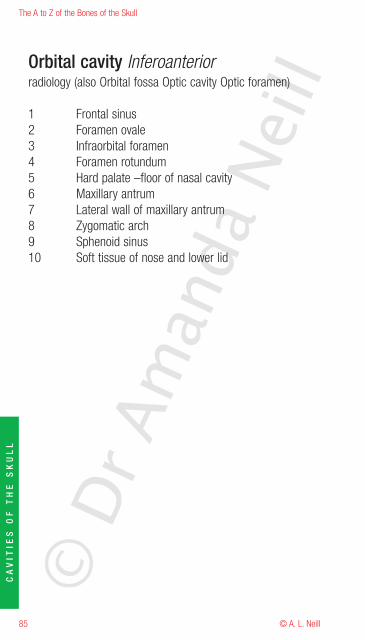
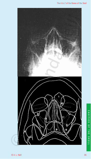
Orbital cavity Inferoanteriorradiology (also Orbital fossa Optic cavity Optic foramen)
1 Frontal sinus2 Foramen ovale3 Infraorbital foramen4 Foramen rotundum5 Hard palate –floor of nasal cavity6 Maxillary antrum7 Lateral wall of maxillary antrum8 Zygomatic arch9 Sphenoid sinus10 Soft tissue of nose and lower lid
© D
r Am
anda
Nei
ll
86
The A to Z of the Bones of the Skull
© A. L. Neill
CA
VIT
IES
OF
TH
ES
KU
LL©
Dr A
man
da N
eill
The A to Z of the Bones of the Skull
© A. L. Neill87
CA
VIT
IES
OF
TH
ES
KU
LL
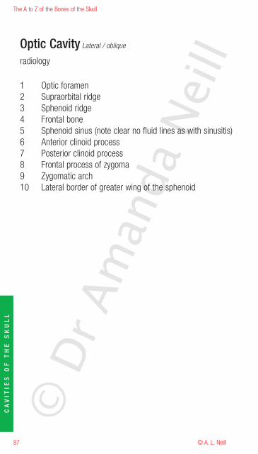
Optic Cavity Lateral / oblique
radiology
1 Optic foramen2 Supraorbital ridge3 Sphenoid ridge4 Frontal bone5 Sphenoid sinus (note clear no fluid lines as with sinusitis)6 Anterior clinoid process7 Posterior clinoid process8 Frontal process of zygoma9 Zygomatic arch10 Lateral border of greater wing of the sphenoid
© D
r Am
anda
Nei
ll
88
The A to Z of the Bones of the Skull
© A. L. Neill
CA
VIT
IES
OF
TH
ES
KU
LL©
Dr A
man
da N
eill
The A to Z of the Bones of the Skull
© A. L. Neill89
CA
VIT
IES
OF
TH
ES
KU
LL
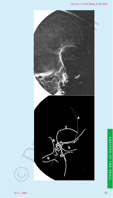
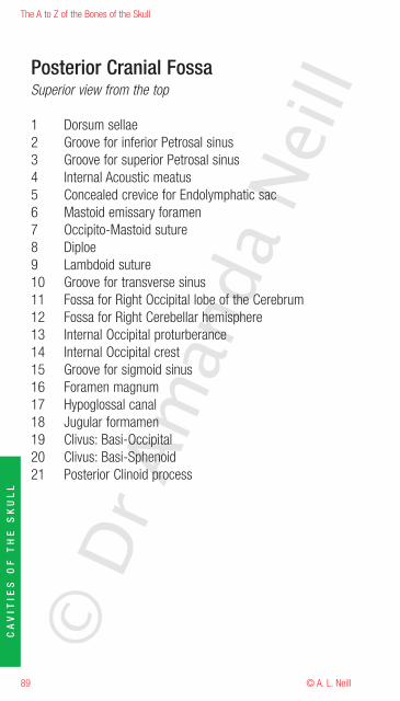
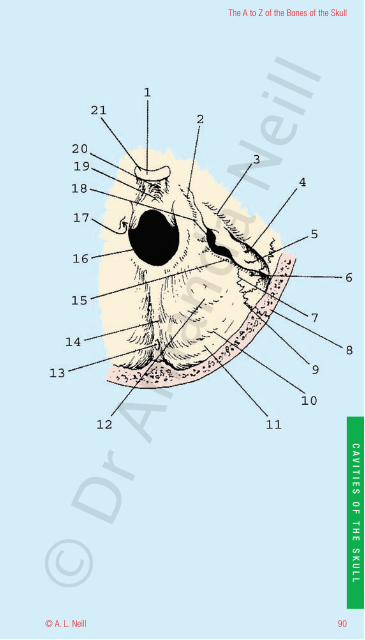
Posterior Cranial FossaSuperior view from the top
1 Dorsum sellae2 Groove for inferior Petrosal sinus3 Groove for superior Petrosal sinus4 Internal Acoustic meatus5 Concealed crevice for Endolymphatic sac6 Mastoid emissary foramen7 Occipito-Mastoid suture8 Diploe9 Lambdoid suture10 Groove for transverse sinus11 Fossa for Right Occipital lobe of the Cerebrum12 Fossa for Right Cerebellar hemisphere13 Internal Occipital proturberance14 Internal Occipital crest15 Groove for sigmoid sinus16 Foramen magnum17 Hypoglossal canal18 Jugular formamen19 Clivus: Basi-Occipital20 Clivus: Basi-Sphenoid21 Posterior Clinoid process
© D
r Am
anda
Nei
ll
90
The A to Z of the Bones of the Skull
© A. L. Neill
CA
VIT
IES
OF
TH
ES
KU
LL©
Dr A
man
da N
eill
The A to Z of the Bones of the Skull
© A. L. Neill91
Biography and Aim of the A to Z and Dr Amanda Neill
Dr Amanda Neill has a medical degree with specializations and research inthe areas of Histology, Pathology, and Anatomy and Forensic medicine, witha separate specialization (MSc in renal glomerular disease). After teachingfor many years at university of Sydney and completing her PhD on “theimmunopathology of cerebral malaria” Amanda developed the onlyaccredited RCAGP continuing education course on anatomy. Developingcourses on the anatomy of the Back (Back to the Back) and the Head andNeck the PG program for Dental graduates and writing a number ofmanuals, booklets and programs for medical, dental, nursing and otherhealth students. Qualifying as a GMP (Graduate medical program) facilitatorshe has seen and been involved in the transition form the classic medicalcourse to the GMP and the integration and amalgamation of the classicpreclinical and clinical medical subjects to the total self directed computercontent based course. Moving to Macquarie University, she brought anddeveloped her anatomy and histology program from scratch, conceptualizingand developing the virtual anatomy laboratory using her Flagship grant. Thismassive project is still in development.
Despite modern computer developments and because of her diverseteaching, research and medical background Amanda knows the value oflearning the fundamental building blocks the A to Z of health and medicinein order to write and know the whole medical book. She is passionate aboutdeveloping accessible and wide reaching medical and educational programsfor all levels: the student, the postgraduate, the health and medicalprofessional. Particularly in anatomy and its branches after all we are allANATOMY!!
Looking for collaboration in her projects Amanda developed links with theNSW Department of Forensic medicine, the University of Sydney, theCoroner’s court, the Royal colleges of Anaethetists and Biomedical scientists(of which she is currently the secretary) and commercial sponsors such asAspen. So anatomy@mac has spread and involved students from all theuniversity divisions and the all walks of life.
Always looking to improve accessibility and application of knowledge andskills (such as Anatomy in Action and morphing@mac Art Anatomyexhibitions) and schools science projects Amanda and Aspen arecollaborating on a series of A to Z pocket references to be used as handyguides and aids for all those interested in health and medicine, particularlythe busy medical practitioner.
We want these to be a guide and a help for you and want your help andfeedback in order to make the manuals and the accompanying websites abenefit for you. You can be a part of this project too. Write to usAndOf course if you want to get a HEAD do Amanda’s A to Z.
© D
r Am
anda
Nei
ll
92
The A to Z of the Bones of the Skull
© A. L. Neill
© D
r Am
anda
Nei
ll
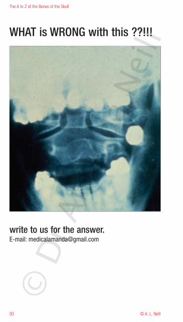
WHAT is WRONG with this ??!!!
write to us for the answer.E-mail: [email protected]
The A to Z of the Bones of the Skull
© A. L. Neill93
© D
r Am
anda
Nei
ll
94
The A to Z of the Bones of the Skull
© A. L. Neill
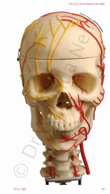
Graphic Design Left Hemisphere
© D
r Am
anda
Nei
ll
Aspen Pharmacare Australia Pty Ltd34-36 Chandos Street, St Leonards NSW 2065
ABN 51 096 236 985
Dr. A. L. NEILLBSc MSc MBBS PhD FACBS
0414248747
Contact www.aspenpharma.com.au forlogin and passwords for the complete
A to Z and the AspenAtlas online.