A unique amino acid substitution, T126I, in human - Ziheng Yang
9
A unique amino acid substitution, T126I, in human genotype C of hepatitis B virus S gene and its possible influence on antigenic structural change Fengrong Ren a, ⁎ , Asahito Tsubota b,d , Takatsugu Hirokawa a,c , Hiromitsu Kumada d , Ziheng Yang e , Hiroshi Tanaka a a Center for Information Medicine, Tokyo Medical and Dental University, 1-5-45 Yushima, Bunkyo, Tokyo 113-8510, Japan b Institute of Clinical Medicine and Research, Jikei University School of Medicine, 163-1 Kashiwa-shita, Kashiwa, Chiba 277-8567, Japan c Computational Biology Research Center, National Institute of Advanced Industrial Science and Technology, 2-41-6 Aomi, Koutou-ku, Tokyo 135-0064, Japan d Department of Gastroenterology, Toranomon Hospital, 2-2-2, Toranomon, Minato, Tokyo 105-8410, Japan e Department of Biology, University College London, Darwin Building, Gower Street, London WC1E 6BT, England, United Kingdom Received 10 May 2006; received in revised form 21 June 2006; accepted 5 July 2006 Available online 29 July 2006 Received by Takashi Gojobori Abstract Amino acid substitutions in the S gene of hepatitis B virus (HBV), especially in the ‘a’ determinant region, have been suggested to affect the antigenicity of the virus and the clinical outcome of the infected patient. However, no convincing evidence has been presented for this hypothesis, partly because the 3D structure of the S protein has not been determined. Comparative analysis of viral genes offers an approach to testing this hypothesis, as it may reveal signals of natural selection and provide insights into the functional significance of the observed amino acid substitutions. In this study, we analyze HBV S gene sequences obtained from 24 patients infected with HBV genotypes B or C, together with 16 representative viral strains of HBV genotypes A–F retrieved from GenBank. We use phylogenetic methods to infer evolutionary changes among HBV genotypes and to identify amino acid residues potentially under positive selective pressure. Furthermore, we employ the fragment assembly method to predict structural changes in the S protein. The results showed that an amino acid substitution within the ‘a’ determinant, T126I, was unique to genotype C, may affect the antigenicity of the HBsAg, and may result in poorer clinical outcomes of patients infected with genotype C viral strains. We suggest that an integrated approach of evolutionary comparison and structural prediction is useful in generating hypotheses for further laboratory validation. © 2006 Elsevier B.V. All rights reserved. Keywords: Chronic HBV infection; Clinical outcome; ‘a’ determinant; Ancestral viral sequence; Positive selection; 3D structure 1. Introduction The hepatitis B virus (HBV) has been well studied since the early 1960s (Blumberg et al., 1965; Dane et al., 1970; Galibert et al., 1979; Okochi and Murakami, 1968). HBV infection, however, is still a significant worldwide public health problem. Chronic HBV infection can lead to liver cirrhosis (LC), which severely damages liver function. Chronic HBV infection is also associated with an increased risk of developing hepatocellular carcinoma (HCC), which is one of the major causes of human death. HBV is a double-stranded DNA virus with a very compact genome of only about 3200 bp. It encodes four proteins: S, P, C and X. Some regions of the genome encode two proteins using different reading frames. The HBV has been divided into eight genotypes, A to H, based on an intergroup divergence of 8% or greater of the complete nucleotide sequence, and these genotypes apparently have different geographic distributions (Norder et al., 1992, 1993, 2004; Okamoto et al., 1988). Recent studies have revealed that there may be significant differences in clinical course and outcome among patients infected with different HBV genotypes (Mayerat et al., 1999; Grandjacques et al., 2000; Ding et al., 2001; Chu et al., 2002). For example, patients infected with Gene 383 (2006) 43 – 51 www.elsevier.com/locate/gene Abbreviations: HBV, hepatitis B virus; 3D structure, three-dimensional structure. ⁎ Corresponding author. Tel.: +81 3 58034762; fax: +81 3 58030247. E-mail address: [email protected] (F. Ren). 0378-1119/$ - see front matter © 2006 Elsevier B.V. All rights reserved. doi:10.1016/j.gene.2006.07.018
Transcript of A unique amino acid substitution, T126I, in human - Ziheng Yang

) 43–51www.elsevier.com/locate/gene
Gene 383 (2006
A unique amino acid substitution, T126I, in human genotype C of hepatitis Bvirus S gene and its possible influence on antigenic structural change
Fengrong Ren a,⁎, Asahito Tsubota b,d, Takatsugu Hirokawa a,c, Hiromitsu Kumada d,Ziheng Yang e, Hiroshi Tanaka a
a Center for Information Medicine, Tokyo Medical and Dental University, 1-5-45 Yushima, Bunkyo, Tokyo 113-8510, Japanb Institute of Clinical Medicine and Research, Jikei University School of Medicine, 163-1 Kashiwa-shita, Kashiwa, Chiba 277-8567, Japan
c Computational Biology Research Center, National Institute of Advanced Industrial Science and Technology, 2-41-6 Aomi, Koutou-ku, Tokyo 135-0064, Japand Department of Gastroenterology, Toranomon Hospital, 2-2-2, Toranomon, Minato, Tokyo 105-8410, Japan
e Department of Biology, University College London, Darwin Building, Gower Street, London WC1E 6BT, England, United Kingdom
Received 10 May 2006; received in revised form 21 June 2006; accepted 5 July 2006Available online 29 July 2006
Received by Takashi Gojobori
Abstract
Amino acid substitutions in the S gene of hepatitis B virus (HBV), especially in the ‘a’ determinant region, have been suggested to affect theantigenicity of the virus and the clinical outcome of the infected patient. However, no convincing evidence has been presented for this hypothesis,partly because the 3D structure of the S protein has not been determined. Comparative analysis of viral genes offers an approach to testing thishypothesis, as it may reveal signals of natural selection and provide insights into the functional significance of the observed amino acidsubstitutions. In this study, we analyze HBV S gene sequences obtained from 24 patients infected with HBV genotypes B or C, together with 16representative viral strains of HBV genotypes A–F retrieved from GenBank. We use phylogenetic methods to infer evolutionary changes amongHBV genotypes and to identify amino acid residues potentially under positive selective pressure. Furthermore, we employ the fragment assemblymethod to predict structural changes in the S protein. The results showed that an amino acid substitution within the ‘a’ determinant, T126I, wasunique to genotype C, may affect the antigenicity of the HBsAg, and may result in poorer clinical outcomes of patients infected with genotype Cviral strains. We suggest that an integrated approach of evolutionary comparison and structural prediction is useful in generating hypotheses forfurther laboratory validation.© 2006 Elsevier B.V. All rights reserved.
Keywords: Chronic HBV infection; Clinical outcome; ‘a’ determinant; Ancestral viral sequence; Positive selection; 3D structure
1. Introduction
The hepatitis B virus (HBV) has been well studied since theearly 1960s (Blumberg et al., 1965; Dane et al., 1970; Galibert etal., 1979;Okochi andMurakami, 1968).HBVinfection, however,is still a significant worldwide public health problem. ChronicHBV infection can lead to liver cirrhosis (LC), which severelydamages liver function. Chronic HBV infection is also associated
Abbreviations: HBV, hepatitis B virus; 3D structure, three-dimensionalstructure.⁎ Corresponding author. Tel.: +81 3 58034762; fax: +81 3 58030247.E-mail address: [email protected] (F. Ren).
0378-1119/$ - see front matter © 2006 Elsevier B.V. All rights reserved.doi:10.1016/j.gene.2006.07.018
with an increased risk of developing hepatocellular carcinoma(HCC), which is one of the major causes of human death.
HBV is a double-stranded DNA virus with a very compactgenome of only about 3200 bp. It encodes four proteins: S, P, Cand X. Some regions of the genome encode two proteins usingdifferent reading frames. The HBV has been divided into eightgenotypes, A to H, based on an intergroup divergence of 8% orgreater of the complete nucleotide sequence, and these genotypesapparently have different geographic distributions (Norder et al.,1992, 1993, 2004; Okamoto et al., 1988). Recent studies haverevealed that there may be significant differences in clinicalcourse and outcome among patients infected with different HBVgenotypes (Mayerat et al., 1999; Grandjacques et al., 2000; Dinget al., 2001; Chu et al., 2002). For example, patients infected with

Fig. 1. Model of two-loop structure of the ‘a’ determinant in the envelope gene of HBV. Small circles represent amino acids and bold lines represent the disulphidebridges. Two small solid circles represent the two substituted sites found in this study.
44 F. Ren et al. / Gene 383 (2006) 43–51
the genotypeC viruswere found to showpoorer clinical outcomesthan those infected with the genotype B virus, although bothgenotypes are predominant in East Asia (Kao et al., 2000; Orito etal., 2001a,b). However, it is unclear which genetic differencesbetween the genotypes are responsible for the clinical differences.One difficulty is the lack of sequential viral samples forlongitudinal studies, which may be necessary for revealingevolutionary changes in the viral gene. Another difficulty is thelack of 3D structures of some HBV proteins, making it difficult toassess the structural changes caused by amino acid substitutionsand their functional significance.
In this study, 24 HBV small surface antigen (HBsAg) se-quences sampled from 24 patients showing quite different cli-nical outcomes were analyzed. The HBsAg is the majorcomponent of the envelope of the hepatitis virion. It is 226amino acid residues long, completely embedded in the P generegion (Norder et al., 1994). A key region for HBVantigenicity
Table 1Sequence number and basic clinical information of the 24 patients
Sequence Gender Date (age) of diagnosis of cirrhosis Genotype Subtype De
B1 M 1988 (24) B adw NoB2 M 1994 (28) B adw NoB3 M 1999 (28) B adw NoB4 F 1988 (33) B adw NoB5 M 1992 (33) B adw NoB6 a M 1986 (38) B adw NoB7 M 1990 (41) B adw NoB8 F 1991 (47) B adw NoB9 M 1994 (40) B adw NoB10 M 1974 (45) B adw NoB11 M 1991 (51) B adw YeB12 M 1977 (50) B adw NoB13 M 1991 (55) B adw YeB14 M 1995 (57) B adw NoB15 M 1982 (58) B adw YeB16 M 1988 (66) B adw YeB17 M 1983 (69) B adw NoC1 M 1984 (27) C adw NoC2 b M 1989 (35) C adw NoC3 M 1987 (49) C adr YeC4 F 1990 (56) C adr NoC5 M 1986 (58) C adr YeC6 b M 1992 (66) C adw YeC7 M 1983 (68) C adw Ye
HCC: Hepatocellular carcinoma.SAE: Severe acute exacerbation of chronic hepatitis accompanied by jaundice.a Sequence in which two stop codons were found.b Sequences in which one stop codon was found.
(Howard, 1995), called the ‘a’ determinant, is located in thecentral region (residues 124–147). The ‘a’ determinant has beenpredicted to be a double-loop structure projecting from thesurface of the HBV particle (Stirk et al., 1992; Tiollais et al.,1981, 1985) (see Fig. 1), and it has been suggested that theamino acid changes in this region could affect immune res-ponses (Howard, 1995). However, no convincing evidence atthe 3D level has been presented for this hypothesis because ofthe lack of information about the structure of the S protein.
We employed bioinformatics approaches to infer amino acidsubstitutions that probably have influenced the S protein structureand so affected the HBsAg function. First, we performedphylogenetic analysis using 24 sequences from patients as wellas representative strains of six HBV human genotypes, A to F,obtained from GenBank. We inferred the ancestral sequences ofthese viral strains with the reconstructed phylogenetic tree toestimate what kind of amino acid substitutions occurred in each
velopment of HCC Development of SAE Clinical course
No AliveNo Alive (progressive)Yes AliveNo AliveNo AliveNo Alive (progressive)No Alive (progressive)No AliveNo AliveNo Alive
s No AliveNo Died of renal failure, GI bleeding
s No AliveYes Died of hepatic failure
s No Died of pneumonias No Died of HCC
No Died of renal failureYes AliveNo Alive (progressive)
s No Died of HCCYes Died of hepatic failure
s No Died of HCCs No Died of HCCs No Died of HCC

Fig. 2. The reconstructed phylogenetic tree of the 40 S gene sequences by the neighbor-joining method. Amino acid substitutions within the ‘a’ determinant region ofthe S gene are shown along the branches, based on reconstruction of ancestral sequences using CODEML.
45F. Ren et al. / Gene 383 (2006) 43–51
viral strain. We also detected sites that probably have undergonepositive selection using both S and P gene reading frames toinvestigate the selection pressures acting on different genes.Second, the possible structural changes of the S protein caused byamino acid substitutions found in this studywere computationallypredicted at the 3D level. Finally, the possible relationship bet-ween amino acid substitutions and clinical outcomes was dis-cussed based on the results obtained in this analysis.
2. Materials and methods
2.1. Sequence data
Twenty-four HBV S gene sequences were isolated from 24Japanese patients with chronic HBV infection, whose clinicalcharacteristics are shown in Table 1. Serum samples analyzed for
nucleotide sequencing were obtained at the time when cirrhosiswas confirmed by liver biopsy specimens, ultrasonography, and/or computed tomography. DNA extraction, polymerase chainreaction-based amplification, nucleotide sequencing and deter-mination of genotypes or subtypes were described previously(Tsubota et al., 1998, 2001). All sequences determined were678 bp in length, without insertions or deletions in the alignment.
Sixteen S gene sequences of genotypes A to F were retrievedfrom GenBank and analyzed together with the 24 sequencesdetermined in this study from Japanese patients.We selected thesesequences based on an evolutionary study of HBV in which eachof these viral strains was estimated to be representative of anHBVhuman genotype (Fares and Holmes, 2002). They areHBVADW4A, HHVBF and HHVBFFOU for genotype F;HHVBBAS and HHVBE4 for genotype E; HBVGEN1,HPBHBVAA and HBVAYWE for genotype D; HHVCCHA,

Fig. 3. Detection of positively selected sites. (a)–(b) show the results obtained by using the S gene reading frame, and (c)–(d) show the results by using the P genereading frame. The ordinate indicates the estimated probability for positively selected sites, whereas the abscissa indicates the amino acid site. The amino acid sites ofthe P gene are numbered from the starting position of the S gene, but two nucleotides are shifted.
46 F. Ren et al. / Gene 383 (2006) 43–51
HPBADR1CG and HPBCGADR for genotype C; HPBADW1,HPBA3HMS2 and AB014366 for genotype B; and HBVGEN2and HVHEPB for genotype A. These HBV strains are included inorder to confirm the distribution of the 24 patient samples amongthe HBV phylogenies and also to infer the ancestral sequence ofeach viral genotype. Sequences from genotypes G and Hwere notused, as they are highly similar to those of genotypes A and F,respectively (Norder et al., 2004). A total of 40 S gene sequenceswere used in this study.
2.2. Phylogenetic analysis
2.2.1. Reconstructing the phylogenetic treeThe phylogenetic relationships among the 40 strains were
inferred using programs in the PHYLIP package (Felsenstein,
1995). Kimura's two-parameter model was used to calculatedistances among the viral sequences, which are analyzedusing the neighbor-joining method (Saitou and Nei, 1987) toreconstruct the tree. To confirm whether or not the treetopology depends on tree-making methods, we also usedmaximum likelihood (Felsenstein, 1981) to reconstruct thetree.
2.2.2. Inferring ancestral sequencesThe amino acid sequences at the ancestral nodes of the
reconstructed tree were inferred by using maximum likelihoodunder the JTT substitution model (Jones et al., 1992), im-plemented in the CODEML program in the PAML package(Yang et al., 1995; Yang, 1997). Amino acid substitutions alongeach branch were then examined.
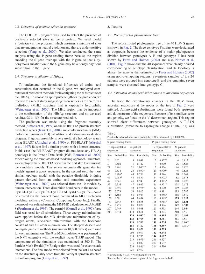
Table 2Positively selected sites with probability N0.5 estimated by CODEML
S gene reading frame P gene reading frame
16 representativesequences
24 patientsequences
16 representativesequences
24 patientsequences
Site Probability Site Probability Site Probability Site Probability
2 0.62 3 0.941 2 0.957⁎ 4 0.8123 0.502 5 0.742 4 0.900 26 0.52524 0.861 21 1.000⁎⁎ 7 0.534 44 0.92644 0.634 24 0.959⁎ 29 0.990⁎ 46 0.52445 0.986⁎ 40 0.758 32 0.544 70 0.64747 0.985⁎ 44 0.839 44 0.972⁎ 82 0.67349 0.641 45 0.94 45 0.973⁎ 100 0.72659 0.516 47 0.987⁎ 46 0.971⁎ 108 0.672110 0.695 49 0.976⁎ 82 0.570 109 0.723122 0.679 53 0.915 100 0.88 115 0.785127 0.657 56 0.943 109 0.956⁎ 118 0.649159 0.58 64 0.634 113 0.989⁎ 125 0.912161 0.645 67 0.658 115 0.968⁎ 126 0.531204 0.775 85 0.877 117 0.894 142 0.532207 0.949 88 0.808 120 0.593 144 0.864213 0.674 110 0.611 122 0.723 210 0.53
126 0.982⁎ 125 0.898 212 0.693143 0.709 128 0.551 213 0.54159 0.747 130 0.734 214 0.960⁎
184 0.722 136 0.897 215 0.959⁎
189 0.671 139 0.723200 0.917 142 0.648210 0.935 144 0.827213 0.973⁎ 206 0.827215 0.807 212 0.637216 0.986⁎ 214 0.596221 0.912
⁎: probability N0.95; ⁎⁎: probability N0.99.Sites in the ‘a’ determinant region of the S gene are in bold.
47F. Ren et al. / Gene 383 (2006) 43–51
2.3. Detection of positive selection pressure
The CODEML program was used to detect the presence ofpositively selected sites in the S protein. We used model8 (beta&ω) in the program, which assumes a mixture of sitesthat are undergoing neutral evolution and that are under positiveselection (Yang et al., 2000). We also conducted the sameanalysis using the P gene reading frame because the regionencoding the S gene overlaps with the P gene so that a sy-nonymous substitution in the S gene may be a nonsynonymoussubstitution in the P gene.
2.4. Structure prediction of HBsAg
To understand the functional influences of amino acidsubstitutions that occurred in the S gene, we employed com-putational predictionmethods for investigating the 3D structure oftheHBsAg. To choose an appropriate length for the prediction,wereferred to a recent study suggesting that residues 98 to 156 form amulti-loop (MHL) structure that is especially hydrophilic(Weinberger et al., 2000). The MHL region may be importantto the conformation of the HBsAg structure, and so we usedresidues 98 to 156 for the structure prediction.
The prediction was made using the fragment assemblymethod (Simons et al., 1997) on the ROBETTA protein structureprediction server (Kim et al., 2004), molecular mechanics (MM)/molecular dynamics (MD) calculation and a structural evaluationprogram. Fragment assembly is very useful if a homology searchusing BLAST (Altschul et al., 1990) or PSI-BLAST (Altschulet al., 1997) fails to find a similar protein with a known structure.In this case, the PSI-BLAST program did not detect any HBsAghomologs in the Protein Data Bank (PDB, Berman et al., 2000)for exploiting the template-based modeling approach. Therefore,we employed the ROBETTA server in the first step to enumeratethe candidate models. This server automatically generates tenmodels against a query sequence. In the second step, the mostsimilar topology model with the putative disulphide bridgingpattern derived from an amino acid mutation experiment(Weinberger et al., 2000) was selected from the 10 models byhuman intervention. Three disulphide bond pairs in the model—Cys124–Cys137, Cys107–Cys138 andCys147–Cys139—couldbe created via the connect bond command in MOE molecularmodeling software (Chemical Computing Group Inc.). Finally,themodel was refined using theMM/MD calculation onAMBER8 (Pearlman et al., 1995). The parm96 (Cornell et al., 1995) forcefield was used for all simulations. Three energy minimizationswere applied before the MD simulation: minimization of hy-drogen atoms, side-chain minimization with the backboneconstraint and full atom minimization. The steepest descent andconjugate gradient methods (maximum 10,000 cycles) were usedfor eachminimization. The 8 nsMD simulation was performed inthe NVT ensemble with the explicit water TIP3P model. Thetemperature of the simulation was maintained at 300 K. TheParticle Mesh Ewald (PME) algorithm was used for electrostaticinteractions. The final model was selected from the last 4 ns basedon the structure quality score from the Verify3D protein structureevaluation program (Luthy et al., 1992).
3. Results
3.1. Reconstructed phylogenetic tree
The reconstructed phylogenetic tree of the 40 HBV S genesis shown in Fig. 2. The three genotype F strains were designatedas outgroups because the evidence of a major phylogeneticdivision between genotypes A–E and genotype F has beenshown by Fares and Holmes (2002) and also Norder et al.(2004). Fig. 2 shows that the 40 sequences were clearly dividedcorresponding to genotype classification, and its topology isalmost the same as that estimated by Fares and Holmes (2002)using non-overlapping regions. Seventeen samples of the 24patients were grouped into genotype B, and the remaining sevensamples were clustered into genotype C.
3.2. Estimated amino acid substitutions in ancestral sequences
To trace the evolutionary changes in the HBV virus,ancestral sequences at the nodes of the tree in Fig. 2 wereinferred. Amino acid substitutions were found both upstreamand downstream of the sequences. Because of its significance toantigenicity, we focus on the ‘a’ determinant region. This regionshowed clear differences between genotypes. A T131Nsubstitution (threonine to asparagine change at site 131) was

48 F. Ren et al. / Gene 383 (2006) 43–51
found on the branch leading to genotype A, a T126I substitutionto genotype C and a F134Y substitution to genotype D. Also, aT143S substitution was found on the branch that dividedgenotypes A and B from genotypes C–F. Since clinicalinformation is available for the 24 strains from Japanesepatients, which are from genotypes B and C only, we areespecially interested in the branch connecting these twogenotypes. Two amino acid changes were identified on thatbranch. The first is T126I, which is the third residue on the firstloop of the ‘a’ determinant. The second is T143S, in the middleof the second loop of the ‘a’ determinant. Thus, except for C2,all genotype C sequences of the patients have an isoleucine atsite 126 and, except for C1, all have a serine at site 143. In thesetwo cases, a secondary substitution I126T occurred to C2 andS143W to C1, respectively. In the genotype B group of thepatients, two of the 17 sequences were found to have amino acidchanges at site 126 as well. The first is a substitution T126I toB2, and the second is T126A to B7. Moreover, three sequenceswere found to have stop codons in the 24 sequences from theJapanese patients: B6 with one, C2 with two, and C6 with one.
3.3. Estimated positively selected sites of HBV S gene
Fig. 3 and Table 2 show the results of the codon-basedanalysis to identify positively selected sites. When the S genereading frame was used (Fig. 3a–b), the central region seemedto be conserved compared to the upstream and downstream of
Fig. 4. (a) Cartoon representations of predicted 3D structure model of HBsAg (view fkey substitution residues are shown in space filling representations. (b) A diagram ostructure model of HBsAg.
the sequence. Within the ‘a’ determinant region, only site 127was predicted to be a possible site for positive selection (withprobability P=0.66) for the 16 representative HBV strains,whereas two sites were detected in the samples of the 24patients: site 126 (with P=0.98) and site 143 (with P=0.71).On the other hand, the results obtained by using the P genereading frame showed a different pattern (Fig. 3c–d). Sevensites were found to be under positive selection within the ‘a’determinant for 16 representative strains, and four sites weredetected in the samples of 24 sequences from the Japanesepatients, none of which had PN0.95. Interestingly, thedownstream region of the P gene seemed quite conservedexcept for the last 20 residues for both the representative strainsand the patient samples.
3.4. Predicted 3D structures of HBsAg
Fig. 4 shows the 3D structure of HBsAg predicted by usingthe fragment assembly method based on the B1 sequence,which is closest to the ancestor of genotype B. Ten models withdifferent fold topologies were initially obtained from theROBETTA prediction server based on fragment assemblymethod. Only one model had the putative three disulphide bondpairs consistent with the result that has been reported so far(Weinberger et al., 2000). This model then was refined usingMM/MD calculation and the feasibility of the model wasevaluated by Verify3D that calculates the structural quality
acing the C-terminal region at left, rotated 180° at right about the y-axis). Severalf relative accessible surface area (RASA) of each amino acid position in the 3D

49F. Ren et al. / Gene 383 (2006) 43–51
score. The best model from the last 4 ns MD trajectories had anacceptable structural quality score (25.44), which is fairly closeto the score expected for a correct structure (26.58) having thisprotein size. Fig. 4a shows the best model with several keysubstitution residues, which was prepared with MolScript(Kraulis, 1991) and Raster3D (Merritt and Bacon, 1997). Wealso analyzed relative accessible surface area (RASA) of theresidues in this model (Fig. 4b) using InsightII/Homologyprogram (Accelrys Inc.). It was estimated that four sites–T126,Q129, T140 and T143–were exposed on the surface of HBsAg.
4. Discussion
Amino acid substitutions in the HBV S gene, especially inthe ‘a’ determinant region, have been described in vaccinatedchildren and patients treated with hepatitis B immunoglobulin.Many studies have pointed out that replacement at some aminoacid sites could affect the antigenicity of the HBsAg, resultingin the loss of recognition by antibodies and leading to evasion ofthe virus from the neutralizing antibody response (Fujii et al.,1992; Ghany et al., 1998; Ni et al., 1995; Protzer-Knolle et al.,1998). Since most reported amino acid substitutions have beenfound within the second of the two loops of the ‘a’ determinant,the second loop has been considered important; in particular,sites 141 to 145 are thought to be essential for antibody binding(Howard, 1995).
We focus on the 24 sequences from the Japanese patients, asclinical information is available for them only. Out of the 10amino acid substitutions inferred to have occurred in the sample,7 are found in the ‘a’ determinant region (Fig. 2). Since patientsinfected with the genotype C virus had poorer clinical outcomesthan those infected with the genotype B virus, we focus on theancestral branch that separates genotypes B and C. Two aminoacid substitutions were observed on these branches: T126I andT143S.
T126I—unique substitution to genotype C. We suggest thatthe T126I substitution in the first loop may be more importantthan the T143S substitution in the second loop. First, the T126Isubstitution was found on the ancestral branch that directlyleads to the genotype C group on the reconstructed tree (Fig. 2).The results of a study on the genetic diversity of HBV byNorder et al. (2004) support this finding. They analyzed 630HBV S gene sequences, including all human genotypes as wellas nonhuman primate HBV strains (chimpanzee, gorilla, gibbonand orangutan) and found that most human genotype C strainshave 126I, whereas almost all human nongenotype C strainshave 126T. On the other hand, T143S occurred on the branchthat separated genotype B and all other genotypes, and thus itwas not unique to genotype C.
Second, the large difference in chemical properties betweenthreonine and isoleucine means that the T126I substitution mayhave a major impact on the antigenicity of the HBsAg.According to Kyte and Doolittle's (1982) method for displayingthe hydropathy parameter of a protein, isoleucine has thehighest value (+4.5) among 20 amino acids, while threonineshows a negative value (−0.7). This is the largest differenceamong amino acid replacements observed in the samples of 24
sequences from the Japanese patients. At site 143, in contrast,two amino acid substitutions, T143S and S143W, caused onlyminor changes in the hydropathy parameter (threonine: −0.7;serine: −0.8; tryptophan: −0.9).
Third, patients whose S gene possessed an isoleucine at site126 exhibited poorer clinical conditions irrespective of geno-type classifications (see Table 1). Therefore, it seemed rea-sonable to suppose that the T126I substitution has a majorimpact on the antigenicity of HBV.
Predicted 3D structure. Our hypothesis is supported by theresults of the structure prediction of HBsAg. As mentionedabove, the B1 sequence is the closest to the ancestral branch ofgenotype B in the phylogenetic tree, and no amino acidsubstitution occurred in the ‘a’ determinant to this sequence.Thus it is appropriate to discuss the structural differencebetween genotypes B and C based on the predicted result of thissequence. Four amino acid sites–126, 129, 140 and 143–werepredicted to be exposed to the surface of HBsAg (Fig. 4). As theT126I substitution involves the largest change in chemicalproperties, it is most likely to cause structural changes in theHBsAg.
Stop codons. Stop codons are usually not allowed in protein-coding genes, as they cause the unexpected termination ofprotein translation. However, stop codons were found in thePreC/C encoding region, and they resulted in immunologicalescape of the virus and led to fulminant hepatitis (Kosaka et al.,1991). Stop codons observed in the S gene in the samples of 24Japanese patients may have a similar role, as all patients withstop codons in the viral S gene showed worse clinical outcomesirrespective of the genotype.
Positively selected sites. The amino acid sequence in the ‘a’determinant is thought to be highly conserved due to its bio-logical function, with substitutions being restricted to similaramino acids (Howard, 1995). In our analysis, the ‘a’ deter-minant region was conserved on the whole, but site 126 wasinferred to be under positive selection with high probability(0.98). Thus, amino acid substitutions at this site might lead toadaptive evolution of the viral gene, resulting in altered anti-genicity and increased virulence. In contrast, a conserved regionwas detected to be located in the downstream rather than in the‘a’ determinant when the P gene reading frame was used, whichobviously corresponds to the distribution of the functionaldomains of the P gene.
The P gene has been divided into several functional regions. TheDNA polymerase region overlaps with the S gene (Norder et al.,1994). Within the polymerase domain, so-called A–E regions canbe identified on the basis of homology with corresponding regionsof other polymerases. It has been reported that the C region(residues 547–559) in which a tyrosine–methionine–aspartate–aspartate (YMDD) motif (residues 551–554) exists is especiallyimportant in function and thus is strictly conserved (Gunther et al.,1999). Apparently, the coding regions for important functions ofthese two genes do not overlap. In addition, a familiarsubstitution, YMDD to YIDD, which can affect the outcome ofchronic hepatitis, was not found in any of the 16 representativestrains or 24 patient sequences (Fig. 5). Thus the effect from thissubstitution can be excluded in this study.

Fig. 5. Aligned partial sequences of the P gene at amino acid level by ClustalW. (a) is the result of 16 representative HBV strains from the database, whereas (b) is thatof the 24 patient sequences. The YMDD motif is indicated by a light grey shadow.
50 F. Ren et al. / Gene 383 (2006) 43–51
Overlapping reading frames in the viral genome. The over-lapping reading frames in the viral genome may cause difficultyin codon-based analysis, because a nonsynonymous substitu-tion in one gene might be a synonymous substitution in another.However, the functionally important regions of the S and Pgenes appear to be separate in the HBV genome. The portion ofthe P gene that overlaps with the S gene does not appear to befunctionally important except for the conserved YMDD motif,so the evolutionary process of this region of the genome isdominated by the selective pressure on the S gene. Our resultsdetecting positively selected sites in the S gene are also highlyconsistent with the predicted structural changes in the S protein.
The results of this study suggest that evolutionary changes inthe S gene may be important in determining the clinical outcomeof a hepatitis B patient. Important changes include substitutionsthat drastically change the properties of amino acids in key regionsof the protein, such as the ‘a’ determinant, and substitutions to stopcodons. The unique amino acid change, T126I, observed ingenotype C, is strongly suspected to be one of the factors thataffect the antigenicity of the HBsAg and to result in poorer clinicaloutcomes. It should be noted that factors not considered in this
study may also influence clinical outcomes, including the viralsubtype and the patient's age (Tsubota et al., 2001).
Finally, we would like to emphasize that combining molecularevolutionary analysis with protein structure prediction appears tobe a powerful approach to the study of viral evolution. Theevolutionary analysis can pinpoint when and where importantamino acid substitutions occurred, and structural prediction helpsto assess their functional significance. The combined approachappears to be effective in generating biological hypotheses, whichmay be verified through further experimental tests.
Acknowledgment
This study is supported by a Grant-in-Aid for ScientificResearch from the Ministry of Education, Culture, Sports,Science and Technology of Japan to F.R. and H.T.
References
Altschul, S.F., Gish, W., Miller, W., Myers, E.W., Lipman, D.J., 1990. Basiclocal alignment search tool. J. Mol. Biol. 215, 403–410.

51F. Ren et al. / Gene 383 (2006) 43–51
Altschul, S.F., et al., 1997. Gapped BLAST and PSI-BLAST: a new generationof protein database search programs. Nucleic Acids Res. 25, 3389–3402.
Berman,H.M., et al., 2000. The protein data bank.NucleicAcids Res. 28, 235–242.Blumberg, B.S., Alter, H.J., Visnich, S., 1965. A new antigen in leukemia sear.
JAMA 191, 541–546.Chu, C.J., Hussain, M., Lok, A.S., 2002. Hepatitis B virus genotype B is
associated with earlier HBeAg seroconversion compared with hepatitis Bvirus genotype C. Gastroenterology 122, 1756–1762.
Cornell, W.D., et al., 1995. A second generation force field for the simulation ofproteins, nucleic acids, and organic molecules. J. Am. Chem. Soc. 117,5179–5197.
Dane, D.S., Cameron, C.H., Briggs, M., 1970. Virus-like particles in serum ofpatients with Australia-antigen-associated hepatitis. Lancet i, 695–698.
Ding, X., et al., 2001. Hepatitis B virus genotype distribution among chronichepatitis B virus carriers in Shanghai, China. Intervirology 44, 43–47.
Fares, M., Holmes, E., 2002. A revised evolutionary history of hepatitis B virus(HBV). J. Mol. Evol. 54, 807–814.
Felsenstein, J., 1981. Evolutionary trees from DNA sequences: a maximumlikelihood approach. J. Mol. Evol. 17, 368–376.
Felsenstein, J., 1995. PHYLIP: Phylogeny Inference Package, Ver. 3.572.University of Washington, Seattle, WA.
Fujii, H., et al., 1992. Gly 145 to Arg substitution in HBsAg antigen of immuneescape mutant of hepatitis B virus. Biochem. Biophys. Res. Commun. 184,1152–1157.
Galibert, F., Madart, E., Fitoussi, F., Tiollais, P., Charnay, P., 1979. Nucleotidesequences of the hepatitis B virus genome (subtype ayw) cloned in E. coli.Nature 281, 646–650.
Ghany, M.G., et al., 1998. Hepatitis B virus S mutants in liver transplantrecipients who were reinfected despite hepatitis B immune globulinprophylaxis. Hepatology 27 (1), 213–222.
Grandjacques, C., et al., 2000. Rapid detection of genotypes and mutations in thepre-core promoter and the pre-core region of hepatitis B virus genome:correlationwith viral persistence and disease severity. J. Hepatol. 33, 430–439.
Gunther, S., et al., 1999. Absence of mutations in the YMDD motif/B region ofthe hepatitis B virus polymerase in famciclovir therapy failure. J. Hepatol.30, 749–754.
Howard, C.R., 1995. The structure of hepatitis B envelope and molecularvariants of hepatitis virus. J. Viral Hepatitis 2, 165–170.
Jones, D.T., Taylor, W.R., Thornton, J.M., 1992. The rapid generation of mutationdata matrices from protein sequences. Comput. Appl. Biosci. 8, 275–282.
Kao, J., Chen, P., Lai, M., Chen, D., 2000. Hepatitis B genotype correlates withclinical outcomes in patients with chronic hepatitis B. Gastroenterology 118,554–559.
Kim, D.E., Chivian, D., Baker, D., 2004. Protein structure prediction andanalysis using the Robetta server. Nucleic Acids Res. 32, 526–531.
Kosaka, Y., et al., 1991. Fulminant hepatitis B: induction by hepatitis B virusmutants defective in the precore region and incapable of encoding e antigen.Gastroenterology 100, 1087–1094.
Kraulis, P.J., 1991. MOLSCRIPT: a program to produce both detailed andschematic plots of protein structures. J. Appl. Crystallogr. 24, 946–950.
Kyte, J., Doolittle, R.F., 1982. A simple method for displaying the hydropathiccharacter of a protein. J. Mol. Biol. 157 (1), 105–132.
Luthy, R., Bowie, J.U., Eisenberg, D., 1992. Assessment of protein models withthree-dimensional profiles. Nature 356, 83–85.
Mayerat, C., Mantegani, A., Frei, P.C., 1999. Does hepatitis B virus (HBV)genotype influence the clinical outcome of HBV infection? J. Viral Hepatitis6, 299–304.
Merritt, E.A., Bacon, D.J., 1997. Raster3D: photorealistic molecular graphics.Methods Enzymol. 277, 505–524.
Ni, F., Fag, D., Gan, R.B., Li, Z.P., 1995. A new escape mutant of hepatitis Bvirus with an Asp to Ala substitution in aa 144 of the envelope major protein.Res. Virol. 146 (6), 205–210.
Norder, H., Hammas, B., Lofdahl, S., Courouce, A.M., Magnius, L.O., 1992.Comparison of the amino acid sequences of nine different serotypes ofhepatitis B surface antigen and genomic classification of the correspondinghepatitis B virus strains. J. Gen. Virol. 73, 1201–1208.
Norder, H., et al., 1993. Genetic relatedness of hepatitis B viral strains of diversegeographical origin and natural variations in the primary structure of thesurface antigen. J. Gen. Virol. 74, 1627–1632.
Norder, H., Courouce, A.M., Magnius, L.O., 1994. Complete genomes,phylogenetic relatedness, and structure proteins of six strains of the hepatitisB virus, four of which represent two new genotypes. Virology 198, 489–503.
Norder, H., et al., 2004. Genetic diversity of hepatitis B virus strains derivedworldwide: genotypes, subgenotypes, and HBsAg subtypes. Intervirology47, 289–309.
Okamoto, H., et al., 1988. Typing hepatitis B virus by homology in nucleotidesequence: comparison of surface antigen subtypes. J. Gen.Virol. 69, 2575–2583.
Okochi, K., Murakami, S., 1968. Observation on Australia antigen in Japan. VoxSang. 15, 374–385.
Orito, E., et al., 2001a. A case-control study for clinical and molecular biologicaldifferences between hepatitis B viruses of genotypes B and C. Japan HBVGenotype Research Group. Hepatology 33, 218–223.
Orito, E., et al., 2001b. Geographic distribution of hepatitis B virus (HBV)genotype in patients with chronic HBV infection in Japan. J. Viral Hepatitis34, 590–594.
Pearlman, D.A., et al., 1995. AMBER, a package of computer programs forapplying molecular mechanics, normal mode analysis, molecular dynamicsand free energy calculations to simulate the structural and energeticproperties of molecules. Comput. Phys. Commun. 91, 1–41.
Protzer-Knolle, U., et al., 1998. Hepatitis B virus with antigenically alteredhepatitis B surface antigen is selected by high-dose hepatitis B immuneglobulin after liver transplantation. Hepatology 27, 254–263.
Saitou, N., Nei, M., 1987. The neighbor-joining method: a new method forreconstructing phylogenetic trees. Mol. Biol. Evol. 4, 406–425.
Simons, K.T., Kooperberg, C., Huang, E., Baker, D., 1997. Assembly of proteintertiary structures from fragments with similar local sequences using simulateannealing and Bayesian scoring functions. J. Mol. Biol. 268, 209–225.
Stirk, H.J., Thornton, J.M., Howard, C.R., 1992. A topological model forhepatitis B surface antigen. Intervirology 33, 148–158.
Tiollais, P., Charnay, P., Vyas, G.N., 1981. Biology of hepatitis. Science 213,406–411.
Tiollais, P., Pourcel, C., Dejean, A., 1985. The hepatitis B virus. Nature 317,489–495.
Tsubota, A., et al., 1998. Deletions in the hepatitis B virus core gene mayinfluence the clinical outcome in hepatitis B e antigen-positive asymptom-atic healthy carriers. J. Med. Virol. 56 (4), 287–293.
Tsubota, A., Arase, Y., Ren, F., Tanaka, H., Ikeda, K., Kumada, H., 2001.Genotype may correlate with liver carcinogenesis and tumor characteristicsin cirrhotic patients infected with hepatitis B virus subtype adw. J. Med.Virol. 65, 257–265.
Weinberger, K.M., Bauer, T., Bohm, S., Jilg, W., 2000. High genetic variability ofthe group-specific a-determinant of hepatitis B virus surface antigen (HBsAg)and the corresponding fragment of the viral polymerase in chronic virus carrierslacking detectable HBsAg in serum. J. Gen. Virol. 81, 1165–1174.
Yang, Z., 1997. PAML: a program package for phylogenetic analysis bymaximum likelihood. Comput. Appl. Biosci. 13, 555–556 (http://abacus.gene.ucl.ac.uk/software/paml.html).
Yang, Z., Kumar, S., Nei, M., 1995. A new method of inference of ancestralnucleotide and amino acid sequences. Genetics 141, 1641–1650.
Yang, Z., Nielsen, R., Goldman, N., Pedersen, A.K., 2000. Codon-substitutionmodels for heterogeneous selection pressure at amino acid sites. Genetics155, 431–449.