A technique for pediatric chest wall reconstruction using custom ...
7
TECHNICAL NOTE A technique for pediatric chest wall reconstruction using custom-designed titanium implants: description of technique and report of two cases Colin J. Anderson 1 • Murray D. Spruiell 1 • Erin F. Wylie 2 • Caitlin M. McGowan 2 • Frederic W. -B. Deleyiannis 3 • Nathan J. Donaldson 2 • Travis C. Heare 2 Received: 11 July 2015 / Accepted: 26 December 2015 / Published online: 18 January 2016 Ó The Author(s) 2016. This article is published with open access at Springerlink.com Abstract Purpose We present a surgical technique for chest wall reconstruction using custom-designed titanium implants developed for two female patients to provide both chest wall symmetry and adequate stability for staged breast reconstruction. Methods A retrospective review was performed for two adolescent female patients with large chest wall defects who underwent the described technique. The etiology of the chest wall deficiency was secondary to Poland’s syn- drome in one patient, and secondary to surgical resection of osteosarcoma in the other patient. For each patient, a fine- cut computed tomography scan was obtained to assist with implant design. After fabrication of the prosthesis, recon- struction was performed though a curvilinear thoracotomy approach with attachment of the implant to the adjacent ribs and sternum. Wound closure was obtained with use of synthetic graft material, local soft tissue procedures, and flap procedures as necessary. Results The two patients were followed post-operatively for 35 and 38 months, respectively. No intra-operative or post-operative complications were identified. Mild scoliosis that had developed in the patient following chest wall resection for osteosarcoma did not demonstrate any further progression following reconstruction. Conclusions We conclude that this technique was suc- cessful at providing a stable chest wall reconstruction with satisfactory cosmetic results in our patients. Keywords Chest wall reconstruction Á Chest wall resection Á Chest wall deficiency Á Poland’s syndrome Á Pediatric orthopedics Introduction The chest wall serves the vital structural and functional purposes of protecting the intra-thoracic organs, promoting respiration, and supporting the actions of the upper extremities [1]. Given its significant physiologic impor- tance, chest wall deficiency has the potential to cause devastating morbidity and even life-threatening conse- quences. In cases of chest wall deficiency, surgical recon- struction is indicated to improve structural integrity, respiratory function, and unsatisfactory cosmetic appear- ance [2–4]. A variety of etiologies may result in chest wall defi- ciency ranging from acquired conditions to congenital deformities [1, 4]. While only 1.8 % of solid tumors in children occur in the chest wall, a majority of the tumors are malignant [5]. Due to the necessity for wide-surgical excision, chest wall resection for sarcoma often results in defects of significant size. Acquired defects may also result from debridement of necrotic or fibrotic tissue secondary to the sequelae of radiation therapy, traumatic injuries, or infection. In addition to acquired conditions, there are a variety of congenital disorders that also cause chest wall & Travis C. Heare [email protected] 1 Department of Orthopaedic Surgery, University of Colorado, Anschutz Medical Campus, 12631 E. 17th Avenue, Mail Stop B202, Aurora, CO 80045, USA 2 Department of Orthopaedic Surgery, Musculoskeletal Research Center, Children’s Hospital Colorado, Anschutz Medical Campus, 13123 East 16th Avenue, B060, Aurora, CO 80045, USA 3 Department of Plastic and Reconstructive Surgery, Children’s Hospital Colorado, Anschutz Medical Campus, 13123 East 16th Avenue, B467, Aurora, CO 80045, USA 123 J Child Orthop (2016) 10:49–55 DOI 10.1007/s11832-015-0709-1
Transcript of A technique for pediatric chest wall reconstruction using custom ...
TECHNICAL NOTE
A technique for pediatric chest wall reconstruction usingcustom-designed titanium implants: description of techniqueand report of two cases
Colin J. Anderson1 • Murray D. Spruiell1 • Erin F. Wylie2 • Caitlin M. McGowan2 •
Frederic W. -B. Deleyiannis3 • Nathan J. Donaldson2 • Travis C. Heare2
Received: 11 July 2015 / Accepted: 26 December 2015 / Published online: 18 January 2016
� The Author(s) 2016. This article is published with open access at Springerlink.com
Abstract
Purpose We present a surgical technique for chest wall
reconstruction using custom-designed titanium implants
developed for two female patients to provide both chest
wall symmetry and adequate stability for staged breast
reconstruction.
Methods A retrospective review was performed for two
adolescent female patients with large chest wall defects
who underwent the described technique. The etiology of
the chest wall deficiency was secondary to Poland’s syn-
drome in one patient, and secondary to surgical resection of
osteosarcoma in the other patient. For each patient, a fine-
cut computed tomography scan was obtained to assist with
implant design. After fabrication of the prosthesis, recon-
struction was performed though a curvilinear thoracotomy
approach with attachment of the implant to the adjacent
ribs and sternum. Wound closure was obtained with use of
synthetic graft material, local soft tissue procedures, and
flap procedures as necessary.
Results The two patients were followed post-operatively
for 35 and 38 months, respectively. No intra-operative or
post-operative complications were identified. Mild
scoliosis that had developed in the patient following chest
wall resection for osteosarcoma did not demonstrate any
further progression following reconstruction.
Conclusions We conclude that this technique was suc-
cessful at providing a stable chest wall reconstruction with
satisfactory cosmetic results in our patients.
Keywords Chest wall reconstruction � Chest wallresection � Chest wall deficiency � Poland’s syndrome �Pediatric orthopedics
Introduction
The chest wall serves the vital structural and functional
purposes of protecting the intra-thoracic organs, promoting
respiration, and supporting the actions of the upper
extremities [1]. Given its significant physiologic impor-
tance, chest wall deficiency has the potential to cause
devastating morbidity and even life-threatening conse-
quences. In cases of chest wall deficiency, surgical recon-
struction is indicated to improve structural integrity,
respiratory function, and unsatisfactory cosmetic appear-
ance [2–4].
A variety of etiologies may result in chest wall defi-
ciency ranging from acquired conditions to congenital
deformities [1, 4]. While only 1.8 % of solid tumors in
children occur in the chest wall, a majority of the tumors
are malignant [5]. Due to the necessity for wide-surgical
excision, chest wall resection for sarcoma often results in
defects of significant size. Acquired defects may also result
from debridement of necrotic or fibrotic tissue secondary to
the sequelae of radiation therapy, traumatic injuries, or
infection. In addition to acquired conditions, there are a
variety of congenital disorders that also cause chest wall
& Travis C. Heare
1 Department of Orthopaedic Surgery, University of Colorado,
Anschutz Medical Campus, 12631 E. 17th Avenue, Mail Stop
B202, Aurora, CO 80045, USA
2 Department of Orthopaedic Surgery, Musculoskeletal
Research Center, Children’s Hospital Colorado, Anschutz
Medical Campus, 13123 East 16th Avenue, B060, Aurora,
CO 80045, USA
3 Department of Plastic and Reconstructive Surgery, Children’s
Hospital Colorado, Anschutz Medical Campus, 13123 East
16th Avenue, B467, Aurora, CO 80045, USA
123
J Child Orthop (2016) 10:49–55
DOI 10.1007/s11832-015-0709-1

deformity. Of particular interest with respect to chest wall
reconstruction, Poland’s syndrome is a rare disorder char-
acterized by the absence of multiple ribs, the absence of
pectoralis musculature, and hypoplasia or aplasia of the
breast or nipple, that is capable of producing significant
chest wall deficiencies [4, 6].
Despite the myriad operative procedures currently uti-
lized for chest wall reconstruction, no consensus exists on
an optimal surgical strategy. The purpose of this study is to
present a reconstruction technique using custom-designed
titanium implants that were developed with the goals of
improving chest wall stability as well as cosmetic results
over currently described techniques. We present two cases
of adolescent females with prominent cosmetic concerns
due to chest wall and breast asymmetry for which this
technique was developed.
Surgical technique
A fine-cut thoracic computed tomography scan is obtained
and three-dimensional pre-operative planning is performed
for prosthetic design. The intact contralateral ribs are
mirrored to the side of the defect to provide a template for
shaping thoracic symmetry. A custom prosthesis is
designed to reconstruct the defect with attachments to
adjacent ribs as well as the sternum with care taken to
ensure the implant is capable of completely spanning the
defect anteriorly to posteriorly. Subsequently, the custom
implant is fabricated from titanium (Biomet Inc., Warsaw,
IN, USA). Pre-operatively, when inadequate soft-tissue
coverage is expected following placement of the implant,
consultation with plastic surgery should be obtained to plan
for skin or flap coverage needs.
At the time of surgery, the patient is placed in the lateral
decubitus position, and the surgical field is draped to
include the sternum anteriorly and the spine posteriorly. A
curvilinear thoracotomy incision is made overlying the
level of the defect. Full-thickness fasciocutaneous flaps are
elevated from the chest wall, and exposure is extended
anteriorly to the sternum and posteriorly to the deficient
ribs. Sections of pectoralis, latissimus dorsi, or other peri-
scapular musculature may require division for adequate
exposure. If a sternal osteotomy is planned, a periosteal
elevator is used to circumferentially expose the sternum,
and osteotomy is performed with an oscillating saw pro-
tected with a malleable retractor.
Attention is then turned to fitting of the prosthesis. The
anterior costal cartilages and sternum are trimmed to match
the sternal component. Likewise, the deficient ribs are
exposed circumferentially and trimmed to fit rib docking
sites on the implant. The pleural defect may be covered
with graft or mesh depending on surgeon preference. Prior
to final fixation, a chest tube should be placed. Subse-
quently, the prosthesis is placed and secured. For fixation
to the sternum, 4–5 transosseous #5 Fiberwire� (Arthrex,
Inc., Naples, FL, USA) sutures are placed through pre-
fabricated holes in the implant. The first implant design
(described in Case 1 below) utilized tubes with locking
trap-doors that provided secure fixation to the ribs. How-
ever, due to prominence on the chest wall post-operatively,
the second prosthesis (described in Case 2) was designed
with smaller rib docking sites where two transosseous #5
Fiberwire� sutures are placed through pre-fabricated holes
for fixation to the ribs. For closure, the sectioned posterior
musculature is approximated with reduction of the scapula.
The subcutaneous tissue is closed with interrupted sutures
and the skin is closed with a running subcuticular suture.
The wound is sterilely dressed and the chest tube placed to
vacuum suction. Post-operative analgesia is provided with
an epidural pain catheter.
Case 1
A 15-year-old girl with a history significant for congenital
chest wall deficiency secondary to Poland’s syndrome
presented for evaluation due to cosmetic concerns and
desire for breast reconstruction. Examination of the chest
Fig. 1 Oblique view of the three-dimensional model of the prosthesis
for reconstruction of the chest wall defect secondary to Poland’s
syndrome. Colors represent osteotomized sternum (red), sternal plate
(blue), prosthetic ribs (gray and orange), and repositioned second
through fifth ribs (green)
50 J Child Orthop (2016) 10:49–55
123

demonstrated athelia and amastia, absence of the pectoralis
and serratus musculature on palpation, and anterior defi-
ciency of the second through fifth ribs with associated
paradoxical respiratory movement. Brachysyndactyly of
the ipsilateral small finger was also noted. For recon-
struction, the custom prosthesis was designed with planned
derotational osteotomy of the sternum to allow for ade-
quate thoracic expansion (Fig. 1). Volumetric evaluation
demonstrated a theoretical increase in lung volume of 32 %
(Fig. 2). However, pre- and post-operative pulmonary
function test data were not obtained. The procedure was
performed as described above (Figs. 3, 4). Closure was
performed with a latissimus myocutaneous rotational flap
performed in coordination with plastic surgery.
There were no observed intra-operative or post-opera-
tive complications. Eight months after the procedure,
breast reconstruction was initiated with placement of a
tissue expander between the latissimus flap and the tita-
nium prosthesis, followed 5 months later by definitive
breast implant placement. Fifteen months post-chest wall
reconstruction, the patient underwent right nipple recon-
struction and fat augmentation, with additional fat aug-
mentation performed at 23 months. At 38 months post-
operatively, the patient continues to be subjectively satis-
fied with the results functionally and cosmetically. No
evidence of rib fracture or implant subsidence has yet been
identified in the follow-up period. A subset of the surgical
data for this patient has been previously reported from the
perspective of breast reconstruction [7].
Case 2
A 16-year-old girl with a history of left chest wall
osteosarcoma status post chest wall resection 5 years ear-
lier presented with complaints of poor cosmetic appearance
due to asymmetry. The initial sarcoma resection included
removal of the entire fourth as well as portions of the third
and fifth ribs, with polypropylene mesh (Marlex�; Davol,
Warwick, RI, USA) placement for closure. On examina-
tion, there was superolateral displacement of the left breast
into the defect as well as hypoplasia of the nipple and
areola. Lung herniation and paradoxical respiratory
movement were also present. Radiographs demonstrated a
levoconvex upper thoracic scoliosis measuring 20� that haddeveloped in the 5 years since the resection. Reconstruc-
tion was planned with a custom prosthesis (Fig. 5). At the
time of the reconstruction, the existing polypropylene
mesh, placed during the initial resection, was found to be
densely scarred to both the lung and chest wall. To obtain
exposure, the mesh was elevated in continuum with the
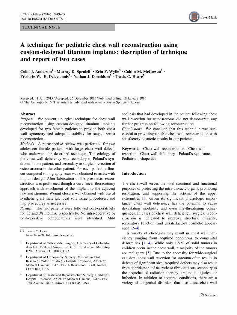
fasciocutaneous flap and breast (Fig. 6a). Next, the pros-
thesis was secured anteriorly to the sternum, superiorly to
the second rib, inferiorly to the sixth rib, and posteriorly to
the remnant third and fifth ribs (Fig. 6b). Lastly, the fas-
ciocutaneous flap with adherent mesh was closed over the
defect.
There were no observed intra-operative or post-opera-
tive complications. Immediate post-operative radiographs
Fig. 2 Superior view of three-
dimensional model
demonstrating the difference
between a the native sternum
(yellow) and rib position, and
b the reconstructed symmetrical
chest wall after sternal
osteotomy and implant
placement
Fig. 3 Intra-operative photograph showing placement of the pros-
thesis with FiberWire� sutures securing the prosthesis to the sternum
anteriorly and to the sixth rib inferiorly. The Gore-tex� graft is visible
underneath the prosthesis, and the chest tube is located inferiorly.
Image oriented with anterior to the right and superior to the top
J Child Orthop (2016) 10:49–55 51
123

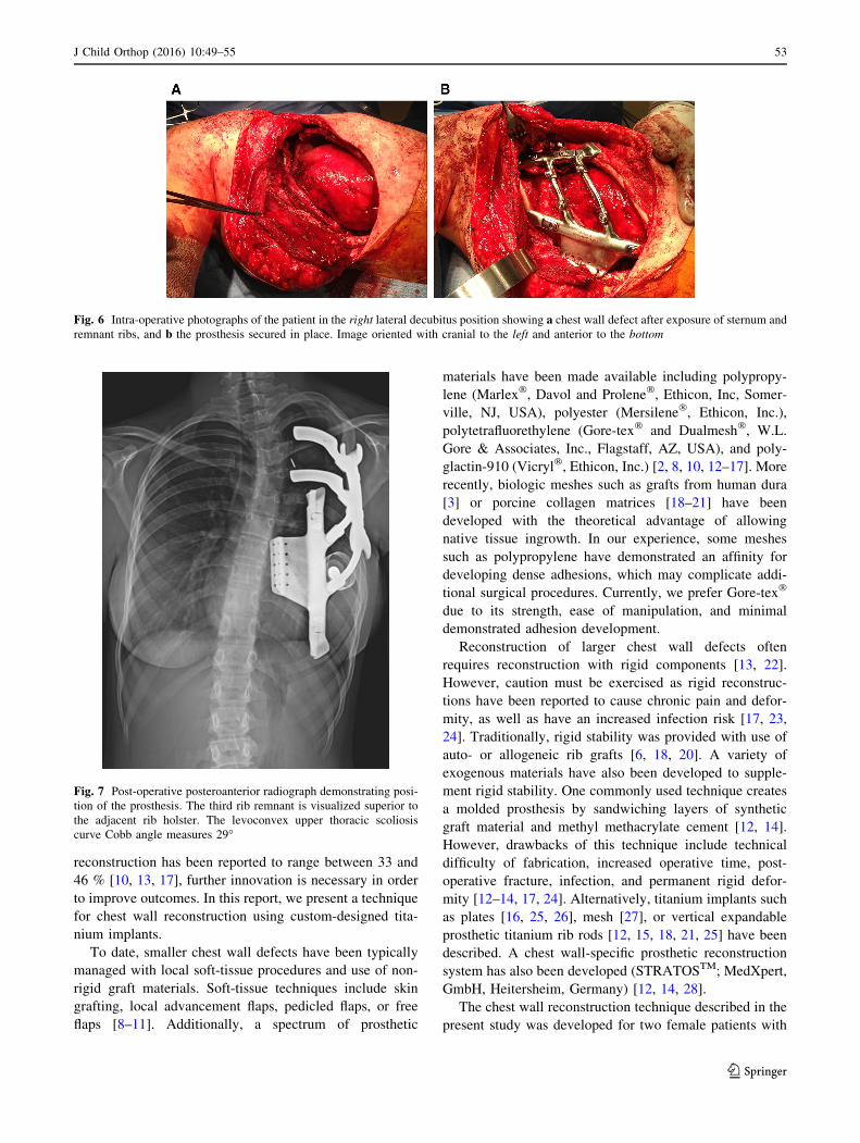
demonstrated post-procedural progression in the upper
thoracic scoliosis to 25�, which is concluded to be a result
of expansion forces on the chest wall defect by the pros-
thesis. No further progression has yet been noted in the
follow-up period (Fig. 7). In the early post-operative per-
iod, it was noted that the posterior remnant third rib dis-
sociated from the prosthesis with superior migration.
However, continued monitoring has not demonstrated fur-
ther change in position. Due to continued breast asymme-
try, at 21 months post-reconstruction, the patient
underwent left breast soft-tissue expander placement with
definitive implant placement at 27 months. At 35 months
post-operatively, the patient continues to be subjectively
satisfied with the cosmetic and functional results of the
chest wall reconstruction.
Discussion
Over the past half-century, remarkable progress has been
made in the treatment of chest wall deficiencies; however,
the numerous operative techniques described are a reflec-
tion that an optimal surgical strategy has not yet been
reached. As the complication rate following chest wall
Fig. 4 Post-operative a anteroposterior and b lateral radiographs demonstrating stable position of the prosthesis
Fig. 5 Frontal (a) and oblique
(b) views of the three-
dimensional model of the
prosthesis for reconstruction of
the chest wall defect secondary
to osteosarcoma resection,
designed to attach to the
sternum anteriorly, to the
second rib superiorly, to the
sixth rib inferiorly, and to the
third and fifth rib remnants
posteriorly. Colors represent
sternum (orange), inferior ribs
and vertebra (yellow), superior
ribs and vertebra after
repositioning with the prosthesis
in place (green), rib clips (blue).
Suture holes shown as small
rods protruding from the
anterior sternal plate
52 J Child Orthop (2016) 10:49–55
123
reconstruction has been reported to range between 33 and
46 % [10, 13, 17], further innovation is necessary in order
to improve outcomes. In this report, we present a technique
for chest wall reconstruction using custom-designed tita-
nium implants.
To date, smaller chest wall defects have been typically
managed with local soft-tissue procedures and use of non-
rigid graft materials. Soft-tissue techniques include skin
grafting, local advancement flaps, pedicled flaps, or free
flaps [8–11]. Additionally, a spectrum of prosthetic
materials have been made available including polypropy-
lene (Marlex�, Davol and Prolene�, Ethicon, Inc, Somer-
ville, NJ, USA), polyester (Mersilene�, Ethicon, Inc.),
polytetrafluorethylene (Gore-tex� and Dualmesh�, W.L.
Gore & Associates, Inc., Flagstaff, AZ, USA), and poly-
glactin-910 (Vicryl�, Ethicon, Inc.) [2, 8, 10, 12–17]. More
recently, biologic meshes such as grafts from human dura
[3] or porcine collagen matrices [18–21] have been
developed with the theoretical advantage of allowing
native tissue ingrowth. In our experience, some meshes
such as polypropylene have demonstrated an affinity for
developing dense adhesions, which may complicate addi-
tional surgical procedures. Currently, we prefer Gore-tex�
due to its strength, ease of manipulation, and minimal
demonstrated adhesion development.
Reconstruction of larger chest wall defects often
requires reconstruction with rigid components [13, 22].
However, caution must be exercised as rigid reconstruc-
tions have been reported to cause chronic pain and defor-
mity, as well as have an increased infection risk [17, 23,
24]. Traditionally, rigid stability was provided with use of
auto- or allogeneic rib grafts [6, 18, 20]. A variety of
exogenous materials have also been developed to supple-
ment rigid stability. One commonly used technique creates
a molded prosthesis by sandwiching layers of synthetic
graft material and methyl methacrylate cement [12, 14].
However, drawbacks of this technique include technical
difficulty of fabrication, increased operative time, post-
operative fracture, infection, and permanent rigid defor-
mity [12–14, 17, 24]. Alternatively, titanium implants such
as plates [16, 25, 26], mesh [27], or vertical expandable
prosthetic titanium rib rods [12, 15, 18, 21, 25] have been
described. A chest wall-specific prosthetic reconstruction
system has also been developed (STRATOSTM; MedXpert,
GmbH, Heitersheim, Germany) [12, 14, 28].
The chest wall reconstruction technique described in the
present study was developed for two female patients with
Fig. 6 Intra-operative photographs of the patient in the right lateral decubitus position showing a chest wall defect after exposure of sternum and
remnant ribs, and b the prosthesis secured in place. Image oriented with cranial to the left and anterior to the bottom
Fig. 7 Post-operative posteroanterior radiograph demonstrating posi-
tion of the prosthesis. The third rib remnant is visualized superior to
the adjacent rib holster. The levoconvex upper thoracic scoliosis
curve Cobb angle measures 29�
J Child Orthop (2016) 10:49–55 53
123

large chest wall defects where it was felt that previous
reconstruction techniques would not be capable of pro-
viding restoration of thoracic symmetry or allowing for
staged breast reconstruction. In the design of this tech-
nique, we believe that the success of this reconstruction is
contingent upon stable restoration of anterior to posterior
thoracic continuity from the sternum to the remnant ribs or
spine. While the aforementioned previously described
reconstruction techniques have been utilized for successful
reconstruction of large chest wall defects in other patients,
none were specifically designed with the provision of
restoring anterior to posterior thoracic continuity. Conse-
quently, careful three-dimensional pre-operative planning
is essential for the size and shape of the prosthesis. With
both implants designed in this fashion, both patients in this
series have demonstrated satisfactory clinical and func-
tional outcomes after approximately 3 years and have
undergone successful staged breast reconstruction proce-
dures without evidence of implant collapse or subsidence.
Additionally, it is also important that pre-operative plan-
ning includes skin or soft-tissue flap coverage needs due to
the resultant increase in the size of the chest wall following
placement of the implant. In the first patient with Poland’s
syndrome, consultation with plastic surgery colleagues was
obtained pre-operatively and latissimus flap closure was
performed at the end of the procedure. However, flap
closure was not necessary in the second case as the change
in thoracic size was not as marked.
Chest wall deficiency has previously been associated
with the development of scoliosis [2, 3, 19, 25, 29–31].
Factors that have been reported to increase the risk of the
development of scoliosis include younger age, resection
during periods of rapid growth, increased number of ribs
resected, and posterior rib resections [30]. It has not been
established if rigid chest wall reconstruction is capable of
modifying the risk of scoliosis. In the patient with chest
wall deficiency secondary to resection of osteosarcoma, the
mildly progressive upper thoracic curve that had developed
during the 5 years following her chest wall resection has
not demonstrated further progression following the
reconstruction.
The main weaknesses in this study are the limited
number of patients and relatively short follow-up period.
Complications are common following chest wall recon-
struction and are reported to range from 33-46 %, with
respiratory complications being the most frequent at
11-24 % [10, 13, 17]. While a majority of these occur in
the early post-operative period, chest wall reconstructions
are known to be susceptible to later failure secondary to the
mobility of the thorax with constant respiratory motion.
While no complications or failures were observed in a
follow-up period of approximately 3 years in our two
patients, this study is not able to make a determination of
long-term safety. Nonetheless, we believe that this tech-
nique may be considered as a surgical option for recon-
struction of large chest wall defects when restoration of
chest wall symmetry and superior stability are desired.
Acknowledgments We would like to thank Jennifer Bruny, MD for
her contributions in the completion of this study.
Compliance with ethical standards
Conflict of interest Colin J. Anderson MD, Murray D. Spruiell
MD, Erin F. Wylie BA, Caitlin M. McGowan BA, Frederic W.-B.
Deleyiannis MD, Nathan J. Donaldson DO, and Travis C. Heare MD
declare that they have no conflict of interest.
Ethical standards All procedures performed in studies involving
human participants were in accordance with the ethical standards of
the institutional and/or national research committee and with the 1964
Helsinki declaration and its later amendments or comparable ethical
standards.
Informed consent Informed consent was obtained from all indi-
vidual participants included in the study.
Open Access This article is distributed under the terms of the
Creative Commons Attribution 4.0 International License (http://crea
tivecommons.org/licenses/by/4.0/), which permits unrestricted use,
distribution, and reproduction in any medium, provided you give
appropriate credit to the original author(s) and the source, provide a
link to the Creative Commons license, and indicate if changes were
made.
References
1. Clemens MW, Evans KK, Mardini S, Arnold PG (2011) Intro-
duction to chest wall reconstruction: anatomy and physiology of
the chest and indications for chest wall reconstruction. Semin
Plast Surg 25:5–15
2. Grosfeld JL, Rescorla FJ, West KW, Vane DW, DeRosa GP,
Provisor AJ, Weetman R (1988) Chest wall resection and
reconstruction for malignant conditions in childhood. J Pediatr
Surg 23:667–673
3. Soyer T, Karnak I, Ciftci AO, Senocak ME, Tanyel FC,
Buyukpamukcu N (2006) The results of surgical treatment of
chest wall tumors in childhood. Pediatr Surg Int 22:135–139
4. van Aalst JA, Phillips JD, Sadove AM (2009) Pediatric chest wall
and breast deformities. Plast Reconstr Surg 124:38e–49e
5. Kumar APM, Green A, Smith JW (1977) Combined therapy for
malignant tumors of the chest wall in children. J Pediatr Surg
12:991–999
6. Fokin AA, Robicsek F (2002) Poland’s syndrome revisited. Ann
Thorac Surg 74:2218–2225
7. Rodriguez IE, Heare T, Bruny J, Deleyiannis FWB (2014) Cus-
tomized titanium implant for chest wall reconstruction in com-
plex Poland syndrome. Plast Reconstr Surg Glob Open 2:1–4
8. Incarbone M, Pastorino U (2001) Surgical treatment of chest wall
tumors. World J Surg 25:218–230
9. Losken A, Thourani VH, Carlson GW, Jones GE, Culbertson JH,
Miller JI, Mansour KA (2004) A reconstructive algorithm for
plastic surgery following extensive chest wall resection. Br J
Plast Surg 57:295–302
54 J Child Orthop (2016) 10:49–55
123
10. Mansour KA, Thourani VH, Losken A, Reeves JG, Miller JI Jr,
Carlson GW, Jones GE (2002) Chest wall resections and recon-
struction: a 25-year experience. Ann Thorac Surg 73:1720–1725
11. Watson WL, James AG (1947) Fascia lata grafts for chest wall
defects. J Thorac Surg 16:399–406
12. Berthet JP, Canaud L, D’Annoville T, Alric P, Marty-Ane CH
(2011) Titanium plates and Dualmesh: a modern combination for
reconstructing very large chest wall defects. Ann Thorac Surg
91(6):1709–1716
13. Deschamps C, Tirnaksiz BM, Darbandi R, Trastek VF, Allen MS,
Miller DL, Arnold PG, Pairolero C (1999) Early and long-term
results of prosthetic chest wall reconstruction. J Thorac Cardio-
vasc Surg 117:588–591
14. Gonfiotti A, Santii PF, Campanacci D, Innocenti M, Ferrarello S,
Caldarella A, Janni A (2010) Malignant primary chest-wall
tumours: techniques of reconstruction and survival. Eur J Car-
diothorac Surg 38:39–45
15. Knott EM, Shah SR, Jones G, Hetherington M, Sharp RJ (2012)
Treatment of chest wall osteosarcoma presenting as second pri-
mary after treatment of neuroblastoma. J Pediatr Surg 47:5e–7e
16. Rocco G (2011) Chest wall resection and reconstruction
according to the principles of biomimesis. Semin Thorac Car-
diovasc Surg 23:307–313
17. Weyant MJ, Bains MS, Venkatraman E, Downey RJ, Park BJ,
Flores RM, Rizk N, Rusch VW (2006) Results of chest wall
resection and reconstruction with and without rigid prosthesis.
Ann Thorac Surg 81:279–285
18. Lieber J, Kirschner HJ, Fuchs J (2012) Chest wall repair in
Poland syndrome: complex single-stage surgery including Ver-
tical Expandable Prosthetic Titanium Rib stabilization–a case
report. J Pediatr Surg 47:1e–5e
19. Lin SR, Kastenberg ZJ, Bruzoni M, Albanese CT, Dutta S (2012)
Chest wall reconstruction using implantable cross-linked porcine
dermal collagen matrix (Permacol). J Pediatr Surg 47:1472–1475
20. Oliveira C, Zamakhshary M, Alfadda T, Alhabshan F, Alshalaan
H, Miller S, Kim PC (2012) An innovative method of pediatric
chest wall reconstruction using Surgisis and swinging rib tech-
nique. J Pediatr Surg 47:867–873
21. Smith MD, Campbell RM (2006) Use of a biodegradable patch
for reconstruction of large thoracic cage defects in growing
children. J Pediatr Surg 41:46–49
22. Netscher DT, Baumholtz MA (2009) Chest reconstruction: I
Anterior and anterolateral chest wall and wounds affecting res-
piratory function. Plast Reconstr Surg 124:240e–252e
23. Hanna WC, Ferri LE, McKendy KM, Turcotte R, Sirois C,
Mulder DS (2011) Reconstruction after major chest wall resec-
tion: can rigid fixation be avoided? Surgery 150:590–597
24. Lardinois D, Muller M, Furrer M, Banic A, Gugger M, Krueger
T, Ris HB (2000) Functional assessment of chest wall integrity
after methylmethacrylate reconstruction. Ann Thorac Surg
69:919–923
25. Stephenson JT, Song K, Avansino JR, Mesher A, Waldhausen JH
(2011) Novel titanium constructs for chest wall reconstruction in
children. J Pediatr Surg 46:1005–1010
26. Tuggle DW, Mantor PC, Foley DS, Markley MM, Puffinbarger N
(2004) Using a bioabsorbable copolymer plate for chest wall
reconstruction. J Pediatr Surg 39:626–628
27. Lasko D, Thompson WR, Buckner DM, Sola JE (2008) Titanium
mesh prosthesis repair of symptomatic Poland syndrome in a
premature infant. J Pediatr Surg 43:234–237
28. Jackson L, Singh M, Parikh D (2011) A technical innovation in
paediatric chest wall reconstruction. Pediatr Surg Int 27:629–633
29. Dingemann C, Linderkamp C, Weidemann J, Bataineh ZA, Ure
B, Nustede R (2012) Thoracic wall reconstruction for primary
malignancies in children: short- and long-term results. Eur J
Pediatr Surg 22:34–39
30. Scalabre A, Parot R, Hameury F, Cunin V, Jouve JL, Chotel F
(2014) Prognostic risk factors for the development of scoliosis
after chest wall resection for malignant tumors in children. J Bone
Joint Surg Am 15 96:e10(1-7)
31. DeRosa GP (1985) Progressive scoliosis following chest wall
resection in children. Spine 10:618–622
J Child Orthop (2016) 10:49–55 55
123