
A survey of zoonotic nematodes of commercial key fish ... · A survey of zoonotic nematodes of...
18
Contents lists available at ScienceDirect Fisheries Research journal homepage: www.elsevier.com/locate/ fishres Full length article A survey of zoonotic nematodes of commercial key fish species from major European fishing grounds—Introducing the FP7 PARASITE exposure assessment study Arne Levsen a, ⁎ , Cecilie S. Svanevik a , Paolo Cipriani b,c , Simonetta Mattiucci b,c , Mélanie Gay d , Lee C. Hastie e , Ivana Bušelić g , Ivona Mladineo g , Horst Karl h , Ute Ostermeyer h , Kurt Buchmann i , Dánjal P. Højgaard k , Ángel F. González m , Santiago Pascual m , Graham J. Pierce e,f,m a National Institute of Nutrition and Seafood Research (NIFES), P.O. Box 2029 Nordnes, 5817 Bergen, Norway b Department of Ecological and Biological Sciences Tuscia University, Largo dell’Università s.n.c., 01100 Viterbo, Italy c Department of Public Health and Infectious Diseases, Section of Parasitology, Sapienza University of Rome, P.le Aldo Moro, 5, 00185 Rome, Italy d French Agency for Food, Environmental and Occupational Health and Safety (ANSES), Laboratory for Food Safety, Bld Bassin Napoléon, 62200 Boulogne-sur-Mer, France e Oceanlab, University of Aberdeen,Main Street, Newburgh, Aberdeenshire, AB41 6AA, UK f CESAM & Departamento de Biologia, Universidade de Aveiro, 3810-193 Aveiro, Portugal g Institute of Oceanography and Fisheries (IZOR), Setaliste I. Mestrovica 63, 21 000 Split, Croatia h Max Rubner Institute, Federal Research Institute of Nutrition and Food, Department of Safety and Quality of Milk and Fish Products, Hermann-Weigmann-Straße 1, 24103 Kiel, Germany i Department of Veterinary and Animal Sciences, Faculty of Health and Medical Sciences, University of Copenhagen, Stigbøjlen 7, DK-1870 Frederiksberg C., Denmark k Faroe Marine Research Institute (FAMRI) – Havstovan, P.O. Box 305, Nóatún 1, 110 Tórshavn, Faroe Islands m Institute of Marine Sciences (IIM-CSIC), Eduardo Cabello 6, 36208 Vigo, Spain ARTICLE INFO Keywords: EU fisheries Zoonotic parasites Anisakis spp. Surveillance Atlantic Mediterranean ABSTRACT Harvesting and exploiting limited fisheries resources in a sustainable manner also implies achieving maximum added value from the raw material. However, the presence of parasites in the products may adversely affect consumer perception and/or pose a direct health hazard. As a major stepping-stone of the PARASITE project, an epidemiological survey was carried out to provide the basis for analysis and prediction of consumer exposure risk due to the presence of anisakid nematodes in fish from European wild-catch fisheries. The project consisted of nine separate workpackages (WP) where the exposure risk assessment survey was organized within WP2. The sur- veillance task also provided the data or samples needed for data management and sample storage (WP3, Biobank), molecular and genetic parasite species identification (WP4), and statistical modelling and inference (WP8). In total 17,760 fish belonging to 16 teleost species were examined for anisakids, with special emphasis on economically and ecologically important species such as Atlantic mackerel, herring, European hake, Atlantic cod and anchovy. The target fish species were sampled at four major European fishing areas including the Barents Sea, North Sea, Baltic Sea, Grand Sole Bank, waters off NW Spain and Portugal, central and western parts of the Mediterranean Sea, and the Adriatic Sea. Thus, the survey represents the largest and most comprehensive epidemiological data compilation of anisakids ever generated in terms of geographical range as well as number of fish host species and sample size. An important requirement of the survey was the use of commonly accepted nematode detection methods, i.e. the UV-press method or artificial digestion, to quantify infection level and spatial distribution of anisakid larvae in the target fish species. The basic layout, set-up and principles of the method, along with a discussion of possible source of errors are described. Additionally, the molecular and genetic markers which were used to identify and characterize different species and populations of anisakids from the targeted fish host species and geographical areas, are reviewed as well. Some basic parasite infection characteristics of each fish host species, and any relationships with the presumably most important infection predictors, i.e. fish host body size and fishing locality, are presented and discussed. Emphasis is put on anisakid occurrence in the flesh of the fish. Based on the findings, a graphical exposure risk profile is introduced, including fish species or products thereof, which due to common processing or preparation practices, are at highest risk to act as source of anisakiasis in Europe. http://dx.doi.org/10.1016/j.fishres.2017.09.009 Received 14 February 2017; Received in revised form 11 September 2017; Accepted 12 September 2017 ⁎ Corresponding author. E-mail address: [email protected] (A. Levsen). Fisheries Research xxx (xxxx) xxx–xxx 0165-7836/ © 2017 Elsevier B.V. All rights reserved. Please cite this article as: Levsen, A., Fisheries Research (2017), http://dx.doi.org/10.1016/j.fishres.2017.09.009
Transcript of A survey of zoonotic nematodes of commercial key fish ... · A survey of zoonotic nematodes of...

Contents lists available at ScienceDirect
Fisheries Research
journal homepage: www.elsevier.com/locate/fishres
Full length article
A survey of zoonotic nematodes of commercial key fish species from majorEuropean fishing grounds—Introducing the FP7 PARASITE exposureassessment study
Arne Levsena,⁎, Cecilie S. Svanevika, Paolo Ciprianib,c, Simonetta Mattiuccib,c, Mélanie Gayd,Lee C. Hastiee, Ivana Bušelićg, Ivona Mladineog, Horst Karlh, Ute Ostermeyerh, Kurt Buchmanni,Dánjal P. Højgaardk, Ángel F. Gonzálezm, Santiago Pascualm, Graham J. Piercee,f,m
a National Institute of Nutrition and Seafood Research (NIFES), P.O. Box 2029 Nordnes, 5817 Bergen, Norwayb Department of Ecological and Biological Sciences Tuscia University, Largo dell’Università s.n.c., 01100 Viterbo, Italyc Department of Public Health and Infectious Diseases, Section of Parasitology, Sapienza University of Rome, P.le Aldo Moro, 5, 00185 Rome, Italyd French Agency for Food, Environmental and Occupational Health and Safety (ANSES), Laboratory for Food Safety, Bld Bassin Napoléon, 62200 Boulogne-sur-Mer,Francee Oceanlab, University of Aberdeen,Main Street, Newburgh, Aberdeenshire, AB41 6AA, UKf CESAM&Departamento de Biologia, Universidade de Aveiro, 3810-193 Aveiro, Portugalg Institute of Oceanography and Fisheries (IZOR), Setaliste I. Mestrovica 63, 21 000 Split, Croatiah Max Rubner Institute, Federal Research Institute of Nutrition and Food, Department of Safety and Quality of Milk and Fish Products, Hermann-Weigmann-Straße 1,24103 Kiel, Germanyi Department of Veterinary and Animal Sciences, Faculty of Health and Medical Sciences, University of Copenhagen, Stigbøjlen 7, DK-1870 Frederiksberg C., Denmarkk Faroe Marine Research Institute (FAMRI) – Havstovan, P.O. Box 305, Nóatún 1, 110 Tórshavn, Faroe Islandsm Institute of Marine Sciences (IIM-CSIC), Eduardo Cabello 6, 36208 Vigo, Spain
A R T I C L E I N F O
Keywords:EU fisheriesZoonotic parasitesAnisakis spp.SurveillanceAtlanticMediterranean
A B S T R A C T
Harvesting and exploiting limited fisheries resources in a sustainable manner also implies achieving maximumadded value from the raw material. However, the presence of parasites in the products may adversely affectconsumer perception and/or pose a direct health hazard. As a major stepping-stone of the PARASITE project, anepidemiological survey was carried out to provide the basis for analysis and prediction of consumer exposure riskdue to the presence of anisakid nematodes in fish from European wild-catch fisheries. The project consisted of nineseparate workpackages (WP) where the exposure risk assessment survey was organized within WP2. The sur-veillance task also provided the data or samples needed for data management and sample storage (WP3, Biobank),molecular and genetic parasite species identification (WP4), and statistical modelling and inference (WP8). In total17,760 fish belonging to 16 teleost species were examined for anisakids, with special emphasis on economicallyand ecologically important species such as Atlantic mackerel, herring, European hake, Atlantic cod and anchovy.The target fish species were sampled at four major European fishing areas including the Barents Sea, North Sea,Baltic Sea, Grand Sole Bank, waters off NW Spain and Portugal, central and western parts of the MediterraneanSea, and the Adriatic Sea. Thus, the survey represents the largest and most comprehensive epidemiological datacompilation of anisakids ever generated in terms of geographical range as well as number of fish host species andsample size. An important requirement of the survey was the use of commonly accepted nematode detectionmethods, i.e. the UV-press method or artificial digestion, to quantify infection level and spatial distribution ofanisakid larvae in the target fish species. The basic layout, set-up and principles of the method, along with adiscussion of possible source of errors are described. Additionally, the molecular and genetic markers which wereused to identify and characterize different species and populations of anisakids from the targeted fish host speciesand geographical areas, are reviewed as well. Some basic parasite infection characteristics of each fish hostspecies, and any relationships with the presumably most important infection predictors, i.e. fish host body size andfishing locality, are presented and discussed. Emphasis is put on anisakid occurrence in the flesh of the fish. Basedon the findings, a graphical exposure risk profile is introduced, including fish species or products thereof, whichdue to common processing or preparation practices, are at highest risk to act as source of anisakiasis in Europe.
http://dx.doi.org/10.1016/j.fishres.2017.09.009Received 14 February 2017; Received in revised form 11 September 2017; Accepted 12 September 2017
⁎ Corresponding author.E-mail address: [email protected] (A. Levsen).
Fisheries Research xxx (xxxx) xxx–xxx
0165-7836/ © 2017 Elsevier B.V. All rights reserved.
Please cite this article as: Levsen, A., Fisheries Research (2017), http://dx.doi.org/10.1016/j.fishres.2017.09.009

1. Introduction
The EU fisheries industry is the fifth largest in the world. In theEuropean Union, close to 5 million tons of wild fish catches are pro-cessed every year. Fishing and fish processing provide jobs for at least275,000 people. Moreover, the EU is among the leading fish markets inthe world with imports accounting for approximately €21 billion in2014, more than 40% of world fish imports in value, with increasingtrend. It should be noted, however, that Norway as an EU third countryaccounts for 23% of the EU seafood imports alone. The average con-sumption of fisheries products in the EU-28 countries was 24.9 kg/person in 2011. The annual per capita consumption rate varies greatly,however, from 5.3 kg in Hungary to 56.8 kg in Portugal (EuropeanCommission, 2016).
Nevertheless, consumers expect safe and healthy fish and fisheryproducts. However, some of the most important fish species caught bythe European fishing industries are at risk of carrying parasites whenput on the market. In Europe, anisakid nematodes are the most relevantgroup of parasites in terms of consumer health risk and product quality,with Anisakis and Pseudoterranova as the genera of greatest concernbecause several species are considered a human health hazard(Mattiucci et al., 2017a). The term anisakiasis refers to the zoonoticdisease provoked through accidental ingestion of viable larvae of cer-tain Anisakis species which infect the edible parts of fish or squid.Among the nine nominal species belonging to the genus Anisakis(Mattiucci and Nascetti, 2008; Mattiucci et al., 2014), A. simplex (sensustricto) and A. pegreffii have been confirmed to cause disease in humans(D’Amelio et al., 1999; Umehara et al., 2007; Mattiucci et al., 2011; ,2013; Lim et al., 2015; Mladineo et al., 2016; Bao et al., 2017). It wasfurther demonstrated that A. pegreffii may provoke gastric (GA), in-testinal (IA) and gastro-allergic anisakiasis (GAA) (Mattiucci et al.,2011, 2013; Lim et al., 2015; Mladineo et al., 2016), while both A.simplex (s. s.) and A. pegreffii larvae may cause allergic reactions inhumans (Daschner et al., 2000). Although international regulations,e.g. EU No. 1276/2011, demand deep-freezing for at least 24 h of anyfishery product to be consumed raw or semi-raw, this so-called freezingrequirement is not necessarily practiced by private households or localguesthouses and restaurants. Thus, consumption of local or privatelyprepared dishes based on fresh, only lightly processed fish such as bo-querones in Spain and marinated anchovies in Spain and Italy, probablyrepresents a major source of anisakiasis in Europe.
Self-control programs such as HACCP (hazard analysis and criticalcontrol points) procedures in the fish industry are hampered by the factthat the epidemiology of anisakid parasites in fish caught and marketedin Europe is not well understood. The collection of data on the completelife cycle, geographical and seasonal distribution, prevalence, intensity,and infection site of parasites of public health importance in wild fishstocks and fishery products has so far been based mainly on non-sys-tematic and opportunistic sampling, lacking appropriate monitoringprograms coordinated on a pan-European scale. Therefore, a systematicepidemiological survey of the economically most important fish speciesand stocks from European fishing grounds could provide the basis foranalyzing and modelling parasite prevalence and abundance.
In fish, the majority of Anisakis larvae are typically seen as whitishto greyish, flat and tight coils, measuring a few mm across. Larvae thatreside in the fish flesh are very hard to detect by the naked eye sincethey are often transparent and may have penetrated deeply into thefillets. Moreover, the larval occurrence in terms of their abundance andspatial distribution seems largely to depend on fish host species andtheir respective feeding behavior. Thus, piscivorous species such asadult hake and cod are usually more heavily infected with anisakidlarvae compared to strict plankton feeders such as sardine, anchovy andcapelin (for reviews of the literature, see Mladineo and Poljak, 2014;Šimat et al., 2015; Cipriani et al., 2016; Levsen et al., 2016; Zoricaet al., 2016). However, we know only little about the spatial distribu-tion of anisakid larvae in various economically important fish species,
i.e. where in the fish the larvae primarily reside. This is especiallyimportant whenever anisakid larvae occur in the flesh (fillets and bellyflaps) of fish.
The main objective of the anisakid exposure assessment work-package (WP2) of the PARASITE project was to provide comprehensiveand comparable epidemiological data with respect to zoonotic parasitesin the economically most important fish species or stocks originatingfrom major European fishing grounds. The study focused on Anisakisspecies (mainly A. simplex and A. pegreffii), extending to other speciessuch as Pseudoterranova decipiens (s. l.), Contraceacum osculatum (s. l.)and Hysterothylacium aduncum (non-zoonotic species but may haveaesthetical quality reducing effect if present abundantly), where ade-quate data were available. Thus, the current report provides a basicoverview of the methods which were commonly applied to detect ani-sakid nematodes in the actual fish samples, and to identify them mo-lecularly to species level. The report further summarizes some basicepidemiological results with emphasis on larval occurrence in the fishflesh, and analyses through GAM modelling the relationships betweenlarval occurrence and fish host body size which is already known to actas important driver of anisakid infection patterns in many fish speciesand fishing areas. Finally, we introduce a graphical exposure risk pro-file based on prevalence data of Anisakis spp. in the flesh of several fishspecies which are commonly prepared and consumed in a raw or onlylightly processed state.
2. Material and methods
2.1. Target fish species
The primary decision criteria for the target fish species of the surveyconcerned their importance in terms of: 1) annual consumption vo-lume/sales value, 2) significance for the fresh fish market, 3) basis forraw or semi-raw products such as sushi and sashimi, and 4) parasitehistory (e.g., former RASFF –Rapid Alert System for Food and Feed –notifications). Thus, the pelagic fish species included in the survey wereherring (Clupea harengus), sardine (Sardinus pilchardus), anchovy(Engraulis encrasicolus), Atlantic mackerel (Scomber scombrus), chubmackerel (S. colias) and blue whiting (Micromesistius poutassou).European hake (Merluccius merluccius), haddock (Melanogrammus ae-glefinus), Atlantic cod (Gadus morhua) and monkfish (Lophius piscatoriusand L. budegassa), in addition of two flatfish species – plaice(Pleuronectes platessa) and four-spotted megrim (Lepidorhombus boscii) –represented species preferring demersal habitats. On a smaller scale, orwhenever available, we also investigated whiting (Merlangius mer-langus), European sea bass (Dicentrarchus labrax) and silver scabbardfish(Lepidopus caudatus) since these, too, are commercially utilized on anindustrial scale and are of importance in a number of major Europeanseafood markets including Spain, UK, Italy and France. Some of the fishspecies to be included in the survey, e.g. Atlantic mackerel, cod andhake, occur and are commercially utilized in several of the present NEAtlantic fishing areas. Thus, the epidemiological data obtained fromthese species and areas have been particularly analyzed for the effect ofspecific habitat characteristics, geographical location and fish hostmigration patterns on the diversity and distribution of anisakid species(see also Levsen et al., 2017; Gay et al., 2017; Pascual et al., 2017).
2.2. Sample size and fish host biometric data
Fish host sample size varied among host species, sampling localitiesand sampling date/year. In general, smaller or medium sized species,e.g. anchovy, herring, mackerel or blue whiting were sampled and ex-amined in greater quantities compared to larger species such as Atlanticcod, haddock or monkfish (Table 1). This was partly due to the fact thatprocessing and UV-inspection of smaller fish is less labor- and time-intensive than examining larger fish. Additionally, samples of someother fish species, e.g. European sea bass, were more costly to obtain
A. Levsen et al. Fisheries Research xxx (xxxx) xxx–xxx
2

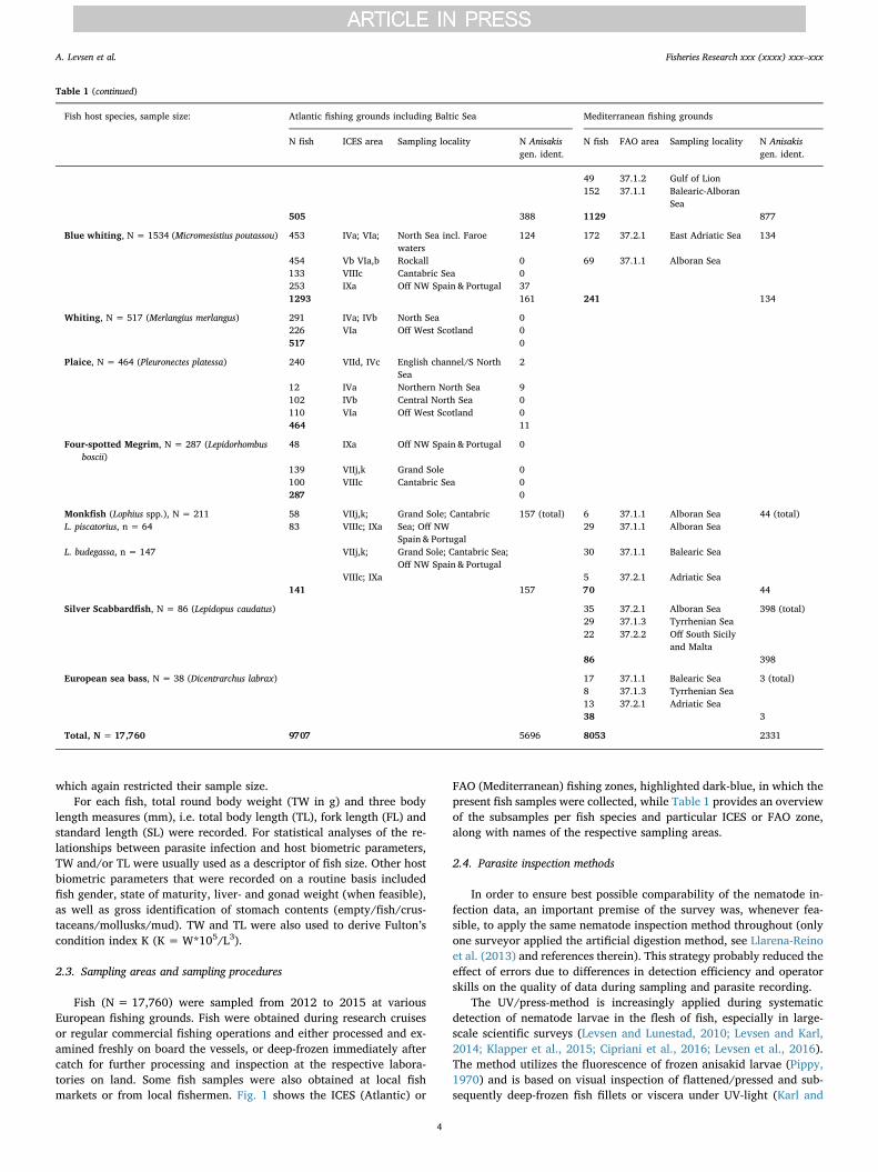
Table 1Fish host species and sample size, along with number of Anisakis spp. subsamples for genetic species analysis, by fishing ground given as ICES/FAO fishing zones and name of samplinglocality.
Fish host species, sample size: Atlantic fishing grounds including Baltic Sea Mediterranean fishing grounds
N fish ICES area Sampling locality N Anisakisgen. ident.
N fish FAO area Sampling locality N Anisakisgen. ident.
Atlantic mackerel, N = 1801 (Scomber scombrus) 526 IVa,b North Sea 472 168 37.2.1 North Adriatic Sea 92 (total)231 VIId English Channel 693 30 37.1.3 Tyrrhenian Sea300 Vb1 Faroe Islands waters 56 19 37.1.1 Alboran Sea300 IIa Southeastern Norwegian
Sea272 70 37.1.2 Gulf of Lion
157 VIIIc; Cantabric Sea; Off NWSpain & Portugal
102
IXa1514 1595 287 92
Chub mackerel, N = 507 (Scomber colias) 21 IXa Off NW Spain & Portugal 0 344 37.2.1 Adriatic Sea 175 (total)100 37.1.1 Balearic Sea42 37.1.3 Tyrrhenian Sea
21 0 486 175
Herring, N = 2673 (Clupea harengus), four stocks:North Sea (n = 1010) English Channel(n = 242) Norw. spring spawning (n = 726)Baltic Sea (N = 695)
1010 IVa,b North Sea 1395 (total)
242 VIId English Channel276 IIa Norwegian Sea150 IVa North Sea300 Vb1 Faroe Islands waters600 BAL24 Southwestern Baltic Sea95 BAL25 Central Baltic Sea2673 1395
Sardine, N = 1704 (Sardina pilchardus) 140 IXa Off NW Spain & Portugal 0 908 37.2.1 East Adriatic Sea 61 (total)200 37.2.2 Off South Sicily356 37.1.1 Off West Sardinia100 37.1.3 North Tyrrhenian
Sea140 0 1564 61
Anchovy, N = 5108 (Engraulis encrasicolus) 956 VIIIc Cantabric Sea 0 645 37.2.1 North Adriatic Sea 547 (total)528 37.2.1 Central Adriatic
Sea518 37.2.1 East Adriatic Sea280 37.2.2 South Adriatic-
Ionian Sea108 37.3.1 Aegean Sea200 37.2.2 Off South Sicily
Tyrrhenian-Ligurian
1323 37.1.3 Sea Sardinian-Balearic-
550 37.1.1 Alboran Sea956 0 4152 547
Atlantic cod (Gadus morhua), N = 755 (incl. 234cod of which only the flesh was examined)
146 (46) I Southern Barents Sea 0
386 (188) BAL24, 25 Baltic Sea 76130 IVa Northern North Sea 191393 IVb Central North Sea 0755 (234) 1989
Haddock, N = 441 (Melanogrammus aeglefinus) 150 I Southern Barents Sea 0291 IVa,b; North Sea; Off West
Scotland0
441 VIa 0
European hake, N = 1634 (Merluccius merluccius) 75 IVb; VIa North Sea; Off WestScotland
388 (total) 72 37.2.1 North Adriatic Sea 877 (total)
188 VII Grand Sole 137 37.2.1 West Adriatic Sea242 VIIIc; IXa Cantabric Sea; Off NW
Spain & Portugal257 37.2.1 East Adriatic Sea
96 37.2.2 South Adriatic-Ionian Sea
27 37.3.1 Aegean Sea64 37.2.2 Off South Sicily241 37.1.3 Tyrrhenian-
Ligurian Sea34 37.1.1 Off West Sardinia
(continued on next page)
A. Levsen et al. Fisheries Research xxx (xxxx) xxx–xxx
3
which again restricted their sample size.For each fish, total round body weight (TW in g) and three body
length measures (mm), i.e. total body length (TL), fork length (FL) andstandard length (SL) were recorded. For statistical analyses of the re-lationships between parasite infection and host biometric parameters,TW and/or TL were usually used as a descriptor of fish size. Other hostbiometric parameters that were recorded on a routine basis includedfish gender, state of maturity, liver- and gonad weight (when feasible),as well as gross identification of stomach contents (empty/fish/crus-taceans/mollusks/mud). TW and TL were also used to derive Fulton’scondition index K (K =W*105/L3).
2.3. Sampling areas and sampling procedures
Fish (N = 17,760) were sampled from 2012 to 2015 at variousEuropean fishing grounds. Fish were obtained during research cruisesor regular commercial fishing operations and either processed and ex-amined freshly on board the vessels, or deep-frozen immediately aftercatch for further processing and inspection at the respective labora-tories on land. Some fish samples were also obtained at local fishmarkets or from local fishermen. Fig. 1 shows the ICES (Atlantic) or
FAO (Mediterranean) fishing zones, highlighted dark-blue, in which thepresent fish samples were collected, while Table 1 provides an overviewof the subsamples per fish species and particular ICES or FAO zone,along with names of the respective sampling areas.
2.4. Parasite inspection methods
In order to ensure best possible comparability of the nematode in-fection data, an important premise of the survey was, whenever fea-sible, to apply the same nematode inspection method throughout (onlyone surveyor applied the artificial digestion method, see Llarena-Reinoet al. (2013) and references therein). This strategy probably reduced theeffect of errors due to differences in detection efficiency and operatorskills on the quality of data during sampling and parasite recording.
The UV/press-method is increasingly applied during systematicdetection of nematode larvae in the flesh of fish, especially in large-scale scientific surveys (Levsen and Lunestad, 2010; Levsen and Karl,2014; Klapper et al., 2015; Cipriani et al., 2016; Levsen et al., 2016).The method utilizes the fluorescence of frozen anisakid larvae (Pippy,1970) and is based on visual inspection of flattened/pressed and sub-sequently deep-frozen fish fillets or viscera under UV-light (Karl and
Table 1 (continued)
Fish host species, sample size: Atlantic fishing grounds including Baltic Sea Mediterranean fishing grounds
N fish ICES area Sampling locality N Anisakisgen. ident.
N fish FAO area Sampling locality N Anisakisgen. ident.
49 37.1.2 Gulf of Lion152 37.1.1 Balearic-Alboran
Sea505 388 1129 877
Blue whiting, N = 1534 (Micromesistius poutassou) 453 IVa; VIa; North Sea incl. Faroewaters
124 172 37.2.1 East Adriatic Sea 134
454 Vb VIa,b Rockall 0 69 37.1.1 Alboran Sea133 VIIIc Cantabric Sea 0253 IXa Off NW Spain & Portugal 371293 161 241 134
Whiting, N = 517 (Merlangius merlangus) 291 IVa; IVb North Sea 0226 VIa Off West Scotland 0517 0
Plaice, N = 464 (Pleuronectes platessa) 240 VIId, IVc English channel/S NorthSea
2
12 IVa Northern North Sea 9102 IVb Central North Sea 0110 VIa Off West Scotland 0464 11
Four-spotted Megrim, N = 287 (Lepidorhombusboscii)
48 IXa Off NW Spain & Portugal 0
139 VIIj,k Grand Sole 0100 VIIIc Cantabric Sea 0287 0
Monkfish (Lophius spp.), N = 211 58 VIIj,k; Grand Sole; Cantabric 157 (total) 6 37.1.1 Alboran Sea 44 (total)L. piscatorius, n = 64 83 VIIIc; IXa Sea; Off NW
Spain & Portugal29 37.1.1 Alboran Sea
L. budegassa, n = 147 VIIj,k; Grand Sole; Cantabric Sea;Off NW Spain & Portugal
30 37.1.1 Balearic Sea
VIIIc; IXa 5 37.2.1 Adriatic Sea141 157 70 44
Silver Scabbardfish, N = 86 (Lepidopus caudatus) 35 37.2.1 Alboran Sea 398 (total)29 37.1.3 Tyrrhenian Sea22 37.2.2 Off South Sicily
and Malta86 398
European sea bass, N = 38 (Dicentrarchus labrax) 17 37.1.1 Balearic Sea 3 (total)8 37.1.3 Tyrrhenian Sea13 37.2.1 Adriatic Sea38 3
Total, N = 17,760 9707 5696 8053 2331
A. Levsen et al. Fisheries Research xxx (xxxx) xxx–xxx
4

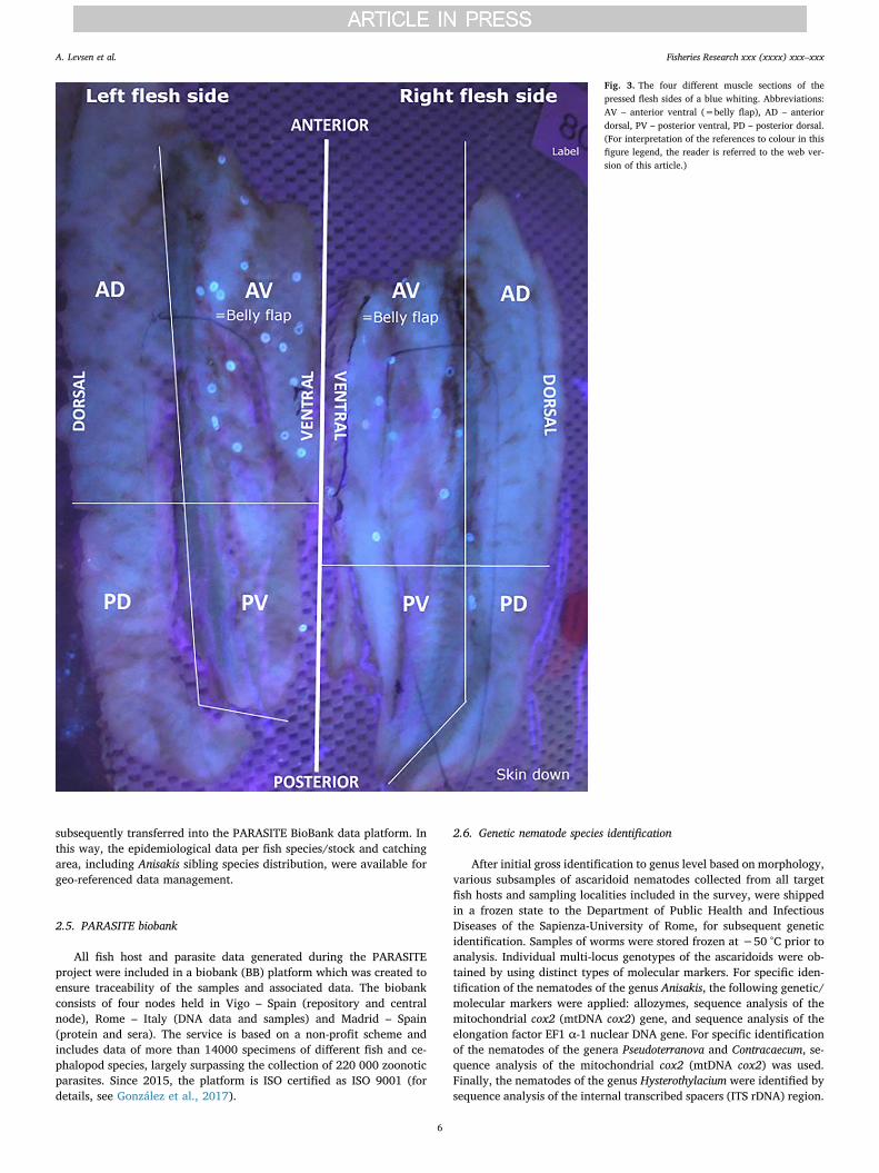
Leinemann, 1993). Like many other eukaryote organisms, anisakidnematodes accumulate lipofuscin, an auto-fluorescent pigment, withintheir cells. When the parasite cells break, the lipofuscin is released andupon excitement of the larvae with UV light, they fluoresce muchbrighter than the surrounding fish flesh. Prior to the pressing process,each fish is gutted and manually filleted before placing the visceralorgans and both left and right-side flesh (fillets incl. belly flaps) intoclear plastic bags. The samples are then pressed to 1–2 mm thick layersin a hydraulic or pneumatic pressing device (holding time approx. 5 s at800–1400 kPa). The bags containing the pressed fillets or viscera arethen deep-frozen prior to visual inspection under a 366 nm UV-lightsource equipped with both up- and down-light. Any anisakid larvaepresent appear as fluorescent spots in the samples; the brightnessprobably depending on various factors such as anisakid species in-volved, their size and age, the extent of encapsulation, and possibly, ifthe freezing-thawing cycle affects the integrity of the larvae.
Another advantage compared to the other widely used nematodeinspection method, i.e. artificial digestion of soft tissue in an aqueouspepsin/HCl-solution, is that the UV-press method allows determinationof the approximate larval infection site in both the flesh and the viscera.Thus, to facilitate the screening of the fish flesh, each flesh side (fillet+ belly flap) is divided into 4 sections in the following manner; ante-rior ventral (AV) which corresponds roughly to belly flap, anteriordorsal (AD), posterior ventral (VP) and posterior dorsal (DP) (Fig. 2).After pressing, the different sections of each flesh side are readily re-cognized since – depending on fish species – the lateral line or redmuscle area, or both, may be used as reference points/axis (Fig. 3).Whenever larger fish such as cod, haddock or monkfish were examined,
each fillet or fish side was cut into smaller parts which were processedseparately.
The nematode detection efficiency of the UV-press method was re-cently evaluated and compared with the artificial digestion method(Pepsin/HCl) in a ring trial (see Gómez-Morales et al., 2017). The re-sults showed that the number of Anisakis spp. larvae recovered by theUV-press method had higher level of agreement (90%) with the numberof spiked larvae compared with the number of larvae recovered byartificial digestion (83%).
The epidemiological data sets were organized as separate MicrosoftExcel-workbooks per fish species examined. Each data set was con-secutively updated according to the results from the genetic identifi-cation whenever new data were available. The data sets were
Fig. 1. ICES and FAO Northeast Atlantic fishing zones; the present fish sampling zones are highlighted dark-blue. I: Southern Barents Sea; IIa: Southeastern Norwegian Sea; IVa,b: NorthSea; Vb: Faroe Islands waters; VIa,b: Rockall; VIIj,k: Grand Sole; VIId: English Channel; VIIIc: Cantabric Sea; IXa: Off NW Spain & Portugal; BAL 24: Southwestern Baltic Sea; BAL 25:Central Baltic Sea; 37.1.1: Western Mediterranean; 37.1.2: Gulf of Lion; 37.1.3: Tyrrhenian Sea; 37.2.1: North Adriatic Sea; 37.2.2: South Adriatic-Ionian Sea; 37.3.1: Aegean Sea.
Fig. 2. Anatomical body sections of fish, exampled by a mackerel, used for parasite in-fection site recording when applying the UV-press nematode inspection method.
A. Levsen et al. Fisheries Research xxx (xxxx) xxx–xxx
5
subsequently transferred into the PARASITE BioBank data platform. Inthis way, the epidemiological data per fish species/stock and catchingarea, including Anisakis sibling species distribution, were available forgeo-referenced data management.
2.5. PARASITE biobank
All fish host and parasite data generated during the PARASITEproject were included in a biobank (BB) platform which was created toensure traceability of the samples and associated data. The biobankconsists of four nodes held in Vigo – Spain (repository and centralnode), Rome – Italy (DNA data and samples) and Madrid – Spain(protein and sera). The service is based on a non-profit scheme andincludes data of more than 14000 specimens of different fish and ce-phalopod species, largely surpassing the collection of 220 000 zoonoticparasites. Since 2015, the platform is ISO certified as ISO 9001 (fordetails, see González et al., 2017).
2.6. Genetic nematode species identification
After initial gross identification to genus level based on morphology,various subsamples of ascaridoid nematodes collected from all targetfish hosts and sampling localities included in the survey, were shippedin a frozen state to the Department of Public Health and InfectiousDiseases of the Sapienza-University of Rome, for subsequent geneticidentification. Samples of worms were stored frozen at −50 °C prior toanalysis. Individual multi-locus genotypes of the ascaridoids were ob-tained by using distinct types of molecular markers. For specific iden-tification of the nematodes of the genus Anisakis, the following genetic/molecular markers were applied: allozymes, sequence analysis of themitochondrial cox2 (mtDNA cox2) gene, and sequence analysis of theelongation factor EF1 α-1 nuclear DNA gene. For specific identificationof the nematodes of the genera Pseudoterranova and Contracaecum, se-quence analysis of the mitochondrial cox2 (mtDNA cox2) was used.Finally, the nematodes of the genus Hysterothylacium were identified bysequence analysis of the internal transcribed spacers (ITS rDNA) region.
Fig. 3. The four different muscle sections of thepressed flesh sides of a blue whiting. Abbreviations:AV – anterior ventral (=belly flap), AD – anteriordorsal, PV – posterior ventral, PD – posterior dorsal.(For interpretation of the references to colour in thisfigure legend, the reader is referred to the web ver-sion of this article.)
A. Levsen et al. Fisheries Research xxx (xxxx) xxx–xxx
6

For allozyme analysis of Anisakis spp. larvae, standard horizontalstarch gel electrophoresis was performed to analyze the variation at fourallozyme loci of diagnostic value in Anisakis species (Mattiucci et al., 1997,2014). The actual loci were, 1) adenylate kinase (Adk-2, EC 2.7.4.3), 2)leucine-alanine peptidase (Pep C-1, Pep C-2, EC 3.4.11), and, 3) superoxidedismutase (Sod-1, EC 1.15.1.1). Genetic analysis of the allozyme data wasperformed using BIOSYS-2 software, while any deviation from the Hardy-Weinberg equilibrium was estimated with a χ2 test. The tissue homo-genates of Anisakis spp. larvae from the starch gel electrophoresis, werepreserved at −20 °C and subsequently used to extract genomic DNA fromeach individual larva examined. Total DNA was extracted using the ce-tyltrithylammonium bromide method (CTAB) (for details, see Mattiucciet al., 2014), or with the DNeasy® Blood& Tissue 120 kit (Qiagen) fol-lowing the manufacturer’s instructions. DNA was subsequently quantifiedby using the Qubit™ dsDNA HS Assay Kit with Qubit 2.0 (Invitrogen™).
The mitochondrial cytochrome c oxidase subunit II (cox2) gene wasamplified using the primers 211F (5′-TTTTCTAGTTATATAGATTGRTTYAT-3′) and 210R (5′-CACCAACTCTTAAAATTA TC-3′), as pre-viously reported by Mattiucci et al. (2014, 2015) and Timi et al. (2014)for the species of the genera Anisakis, Contracaecum and Pseudoterra-nova, respectively. Polymerase chain reaction (PCR) was carried outaccording to the procedures described by Mattiucci et al. (2014) andTimi et al. (2014). The sequences obtained for the mtDNA cox2 gene inthe present study, were analyzed and aligned with the sequences of thesame gene from other previously characterised anisakid species usingGenBank Blast software and ClustalX (Thompson et al., 1997).
For the elongation factor (EF1 α-1 nDNA) nuclear gene which wasstudied in the sibling species of the A. simplex (s. l.) complex, the pri-mers EF-F (5′-TCCTCAAGCGTTGTTATCTGTT-3′) and EF-R (5′-AGTTTTGCCACTAGCGGTTCC-3′) were used (see Mattiucci et al.,2016). PCRs were carried out in a 25 μl volume containing 0.5 μl ofeach primer 10 mM, 2.5 μl of MgCl2 25 mM (Promega), 1.5 μl of 5 xbuffer (Promega), DMSO 0.08 mM, 0.5 μl of dNTPs 10 mM (Promega),5 U of Go-Taq Polymerase (Promega) and 2 μl of total DNA. PCR tem-perature conditions were as follows: 94 °C for 3 min (initial denatura-tion), followed by 35 cycles at 94 °C for 45 s (denaturation), 58 °C for40 s (annealing), 72 °C for 1 min (extension) and followed by post-amplification at 72 °C for 10 min. An initial sample of 50 individuals,belonging to the two species, A. pegreffii and A. simplex (s. s.), previouslyidentified by allozymes, were sequenced at the elongation factor 1alpha 1 gene. The obtained sequences were aligned in order to detectany fixed diagnostic nucleotide positions, which would allow to sepa-rate the two species under examination (see Mattiucci et al., 2016).
Subsamples of Hysterothylacium spp. larvae or adults were identified tospecies level by sequence analysis of the internal transcribed spacers (ITSrDNA) region. PCR amplification was performed using the primers NC5(5′-GTAGGTGAACCTGCGGAAGGATCATT-3′) and NC2 (5′-TTAGTTTCTTTTCCTCCGCT-3′), as reported by Zhu et al. (2000). PCRamplification conditions were as follows: 94 °C for 5 min (initial dena-turation), followed by 30 cycles at 94 °C for 30 s (denaturation), 55 °C for30 s (annealing), 72 °C for 30 s (extension) and a final elongation step at72 °C for 5 min (Zhu et al., 2000). Obtained sequences were analyzed withGenBank Blast software and aligned with previously characterised se-quences by applying ClustalX (Thompson et al., 1997). Phylogeneticanalysis of the sequences obtained from specimens of the genera Anisakis,Pseudoterranova, Contracaecum and Hysterothylacium, was inferred with theBayesian inference method and performed by using MrBayes (Ronquistet al., 2012) while Bayesian analysis was performed with Jmodeltest(Posada, 2008), using the Akaike Information Criterion (AIC) (Posada andBuckley, 2004). Posterior probabilities were estimated and used to assesssupport for each branch in inferred phylogeny with probabilities whereP≥ 95% was indicative of significant support (Reeder, 2003).
2.7. Infection data analyses
In order to account for the effect of fish size on estimates of Anisakis
spp. prevalence and abundance, we fitted generalised additive models(GAM) to prevalence and abundance, for each species and each sam-pling area of the Atlantic and Mediterranean Sea. To avoid overfitting,the complexity of smoothers was restricted by setting a maximum valuefor bases dimension (k = 4). For abundance data, negative binomial orPoisson models were fitted, the latter being appropriate only for area-species combinations with low maximum abundance (< 10); for highermaximum abundance levels, Poisson model diagnostics indicatedoverdispersion of the abundance data. For presence-absence data, bi-nomial models were fitted. In all cases, the default link functions wereapplied, i.e. log for Poisson and negative binomial models, and logit forbinomial models. No models were fitted if sample size was<20, ifprevalence was< 0.05. Additionally, if prevalence was 100%, nomodel was fitted. Regardless of whether the size effect was significant,models were used to predict Anisakis spp. presence or abundance (meanand SE) in fish of (a), mean size sampled for the species and (b), meansize sampled for the modelled species-area combination. All modelswere fitted in R. Note that standard error values are reported to indicateconfidence in the mean value, but in the case of non-normal distribu-tions, confidence intervals must be calculated prior to back-transfor-mation of results onto the response scale.
3. Results and discussion
The results focus mainly on occurrence and distribution of zoonoticanisakid species in the edible parts of fish, which for most species im-plies the flesh. However, some fish species such as sardine and anchovy,are commonly consumed round, i.e. uneviscerated, due to their smallsize, and often only lightly processed as in salting or marinating. Thispractice is common in Mediterranean countries and, although restau-rants are obliged to freeze the fish prior to processing, many householdsprepare the dishes without prior thermal treatment (Cipriani et al.,2016). Therefore, in certain coastal regions in southern Europe, Anisakisspp. infections in the viscera of some fish species may represent aconsumer health risk. However, for general assessment of the consumerexposure risk related to the presence of anisakid larvae in particularlyrelevant fish species or products thereof (see Section 3.2.4. – Anisakisspp. exposure risk profile), three aspects were primarily consideredhere; 1) larval occurrence in the fish flesh and, whenever appropriate,their preferred infection site, 2) fish host body size/length, and, 3)differences in anisakid species composition related to geographic areaor fish stock. For fish or products processed mainly for the fresh fishmarkets, the preferred site of the larvae was recorded to assess thespatial distribution pattern as basis for advising the industry as topossible trimming of fillets in order to reduce the probability of parasitepresence.
3.1. Basic Anisakis spp. infection characteristics by fish host species
3.1.1. Atlantic mackerel (Scomber scombrus)Mackerel from off NW Spain and Portugal showed significantly
higher prevalence, abundance and intensity of Anisakis sp. larvaecompared to their North- and Norwegian Sea and Mediterranean con-geners, both when considering overall infection (viscera and flesh) andinfection in the flesh. The by far lowest infection levels were recordedin mackerel from the Mediterranean fishing grounds, with only around4% larval prevalence in the fish flesh and a maximum intensity of one(1) larva. Regardless of fish host size or catching area, the largestproportions of muscle residing larvae occurred in the ventral sections ofthe fish flesh which for most fish species including Atlantic mackerel,comprises the belly flaps (see Levsen et al., 2017).
GAM-analyses revealed a highly significantly positive relationshipbetween both prevalence and abundance of flesh residing Anisakis spp.larvae, and body size/length of mackerel from the North Sea (Fig. 4a,d). However, the relationship tended to weaken with lower latitudes,e.g. the same variables were only very weakly related in mackerel
A. Levsen et al. Fisheries Research xxx (xxxx) xxx–xxx
7

caught off NW Spain and Portugal (Fig. 4b, e). Moreover, no significanteffect of host body length on larval prevalence and abundance in theflesh was apparently present in fish caught in the southern NorwegianSea (Fig. 4c, f) which represented the northernmost catching locality ofAtlantic mackerel. Due to generally very low larval infection levels inthe fish flesh, GAMs were not fitted for S. scombrus from any of theMediterranean fishing grounds.
Genetic anisakid species identification revealed that A. simplex sensustricto (s. s.) is the dominating species in mackerel from the Atlanticfishing areas. However, we recorded four A. pegreffii larvae in the vis-cera of three mackerel caught in the northernmost fishing areas, i.e. theNorth Sea and southern Norwegian Sea. Similarly, 11 A. pegreffii wereidentified in mackerel caught in the southern North Sea including theEnglish Channel. In the waters off NW Spain and Portugal, A. simplex (s.s.) constitutes still the largest sibling fraction (86%), with A. pegreffiiand A. simplex (s. s.)/A. pegreffii F1 hybrids occurring at much lowerfrequencies (11% and 3%, respectively). In the mackerel caught in theMediterranean, A. pegreffii appears to be the dominating species withonly three A. physeteris larvae detected in three individual mackerel.The findings imply that Anisakis sibling species may be useful supple-mentary biological markers to further elucidate changing migrationpatterns or intermixing between different spawning stocks of Atlanticmackerel (see Levsen et al., 2017).
3.1.2. Chub mackerel (Scomber colias)Overall prevalence of anisakid larvae in chub mackerel collected in
the Atlantic Ocean and Mediterranean Sea was 60.7% while overallmean intensity reached 15.3. Although the infection differed greatlybetween fillets and viscera, both in terms of prevalence (p < 0.001)and mean intensity (p < 0.01), 18.1% prevalence and mean intensityof 2.1 in the flesh/fillets is important from a consumer’s point of view.The anterio-ventral (AV) parts of the fillets were the preferred infectionsite in the flesh of chub mackerel, carrying between 67% (Tyrrhenian
Sea) and 100% (Atlantic waters, ICES IXa) of all muscle residingAnisakis spp. larvae, respectively (Table 2).
Highest overall prevalence (100%) and mean abundance(145.9 ± 84.5) was recorded in chub mackerel originating from theCentral and southern Adriatic Sea. These findings illustrate the im-portance of fish host size as predictor of parasite occurrence in theAdriatic Sea since the chub mackerel were on average more than 12 cmlonger than the other Adriatic samples. This trend was also apparent inthe flesh of these fish where both prevalence (78.9%) and meanabundance (3.8 ± 2.9) were significantly higher compared to theother chub mackerel from the Adriatic Sea (n = 325; prevalence21.2%; abundance 0.33 ± 0.81). However, a highly significantly po-sitive effect of fish host body size/length on larval prevalence andabundance in the fish flesh was still present in pooled samples of chubmackerel from all Adriatic localities (n = 344) (Fig. 5).
3.1.3. Herring (Clupea harengus) (four stocks)The herring samples of the Norwegian spring spawning stock (NSS),
which were obtained in the southern Norwegian Sea and around theFaroe Islands, showed significantly higher prevalence and abundance ofA. simplex (s. s.) larvae compared to herring of the other stocks(Table 3). The differences were most pronounced for larvae residing inthe fish flesh. Both prevalence and mean abundance of muscle residinglarvae in herring of the NSS stock were more than twice as high,reaching 37.1% and 0.6, respectively, compared to herring belonging tothe North Sea- or western Baltic stock. However, the herring of the NSSstock were significantly larger (p < 0.001) than their congeners of theother stocks. Indeed, there seems to be a marked accumulation effect offish size on A. simplex (s. s.) abundance in NE Atlantic herring. Thus, inall stocks considered here, there was a markedly positive relationshipbetween fish host size (TL) and overall larval prevalence and abun-dance. The effect of fish host body length as major predictor of larvaloccurrence in the flesh of herring is illustrated in Fig. 6, exemplified by
Fig. 4. GAM smoothing curves fitted to effect of fish host body length (TL) on prevalence and abundance of Anisakis spp. larvae in the flesh of Atlantic mackerel (Scomber scombrus) fromthe North Sea (a, d), off NW Spain and Portugal (b, e) and southern Norwegian Sea (c, f), respectively. Dashed lines represent 95% conf. intervals around the main effects.
A. Levsen et al. Fisheries Research xxx (xxxx) xxx–xxx
8

the present Norwegian spring spawning- and Baltic stock samples. Thefindings underline the importance of proper freezing, especially of largeNE Atlantic herring, before consumption in a semi-raw state such as inpickled or salted herring. However, herring caught east of BornholmIsland (ICES division BAL25) turned out to be uninfected and werehence not included in the analysis. This finding indicates that the actualfish belong to a separate Baltic herring stock entity with another mi-gration pattern which probably excludes southern or western areas ofthe Baltic Sea (e.g., BAL 24) where Anisakis spp. fish infections com-monly occur. Additionally, there were no marked differences betweenthe herring stocks with respect to relative larval distribution in the fishflesh, i.e., between 87% and 91% of the larvae occurred in the bellyflaps, with almost equal proportions lodging in the left and right fleshside (Table 3).
Genetic anisakid species identification using diagnostic allozymesand sequence analysis of the mtDNA-cox2 gene, revealed that the entiresubsample of Anisakis (n = 1395) collected from all herring stocksconsidered here, belonged to A. simplex (s. s.). However, 481 Anisakisspp. larvae, collected from herring sampled in the Norwegian-, Baltic-and North Sea including the English Channel, were further analyzedwith respect to intraspecific genetic differentiation within the
populations from the four sampling areas by applying mtDNA cox2sequence analysis. Genetic differentiation at population level betweenthe different fishing areas, was estimated and compared based on mo-lecular variance analysis and Fst values. Haplotype network construc-tion showed significant differences in frequencies between samples ofA. simplex (s. s.) from the actual areas. The results indicate a geneticsub-structuring of A. simplex (s. s.) from herring fished in different areasof the NE Atlantic, which again seems largely to correspond to thedifferent herring stocks reported in the literature. Thus, the A. simplex(s. s.) population in herring from the Norwegian Sea appears to be themost differentiated one, whereas lowest level of genetic differentiationwas observed between the North Sea and Baltic Sea populations. Theresults suggest that mtDNA cox2 is a suitable genetic marker for A.simplex (s. s.) population structure analysis, which may also proveuseful in further investigations of the herring stock structure in the NEAtlantic Ocean (see Mattiucci et al., 2017b).
3.1.4. Sardine (Sardina pilchardus)Sardines from the Atlantic fishing ground (ICES IXa) showed com-
paratively high Anisakis spp. prevalence in both viscera (approximately62%) and in the flesh (17%) which appears to be considerably higher
Table 2Sample size and basic fish host biometric data, along with basic Anisakis spp. infection parameters of chub mackerel (Scomber colias) from the Mediterranean Sea and Atlantic Ocean.
Fishing area N fish TL TW Musculature Viscera
P (%) Abund./Intensity Rel. distr.Vtrl: Drsl
Rel. distr.Left: Right
P (%) Abund./Intensity
Adriatic Sea (FAO 37.2.1and 37.2.2)
344 220 ± 46 (105–400) 81 ± 35 (8–397) 24.4 A: 0.5 ± 1.3 (0–9) 85: 15 53: 47 73.8 A: 12.4 ± 39.2 (0–326)
I: 2.2 ± 1.9 (1–9) I: 16.8 ± 44.9 (1–326)Tyrrhenian Sea (FAO
37.1.3)42 333 ± 23 (280–390) 352 ± 78 (200–589) 7.1 A: 0.1 ± 0.3 (0–1) 67: 33 100: 0 54.8 A: 2.7 ± 2.6 (0–10)
I: 1.0 (1) I: 3.7 ± 2.3 (1–10)Balearic Sea (FAO
37.1.1)100 285 ± 20 (250–350) 206 ± 45 (130–357) 0 / / / 9.0 A: 0.1 ± 0.5 (0–3)
I: 1.6 ± 0.7 (1–3)Atlantic Ocean (ICES
IXa)21 286 ± 27 (250–335) 207 ± 55 (141–309) 23.8 A: 0.4 ± 0.8 (0–2) 100: 0 56: 44 61.9 A: 5.8 ± 8.0 (0–26)
I: 1.8 ± 0.5 (1–2) I: 9.4 ± 8.3 (1–26)
Abbreviations;: TL − Total body length (mm); TW − Total body weight (g); P − prevalence, A − Abundance, I − Intensity, both given as mean ± SD (range); Rel. distr. − Relativedistribution (%); Vtrl − ventral portion of fish flesh (corresponds roughly to belly flap); Drsl − dorsal portion of fish flesh.
Fig. 5. GAM smoothing curves fitted to effect of fish host body length (TL) on prevalence (a) and abundance (b) of Anisakis spp. larvae in the flesh of chub mackerel (Scomber colias) fromthe Adriatic Sea. Dashed lines represent 95% conf. intervals around the main effects.
A. Levsen et al. Fisheries Research xxx (xxxx) xxx–xxx
9

Table 3Sample size and basic fish host biometric data, along with basic A. simplex (s. s.) infection parameters of herring (Clupea harengus) of four NE Atlantic stocks.
Herring stock N fish TL TW Musculature Viscera
P (%) Abundance/Intensity Rel. distr. Vtrl:Drsl
Rel. distr.Left:Right
P (%) Abundance/Intensity
North Sea 1252 273 ± 28 (149–358) 171 ± 55 (40–435) 17.4 A: 0.3 ± 0.7 (0–5) 89: 11 50: 50 81.2 A: 11.2 ± 17.0 (0–176)I: 1.6 ± 0.9 (1–5) I: 13.7 ± 17.6 (1–176)
Norw. springspawning
726 334 ± 27 (275–405) 317 ± 70 (153–509) 37.1 A: 0.6 ± 1.0 (0–8) 91: 9 51: 49 92.6 A: 11.2 ± 12.6 (0–112)
I: 1.6 ± 1.1 (1–8) I: 12.1 ± 12.7 (1–112)Baltic 695 261 ± 29 (168–305) 153 ± 50 (31–268) 14.8 A: 0.2 ± 0.5 (0–3) 87: 13 54: 46 65.5 A: 4.2 ± 6.5 (0–45)
0-Western −600 216 ± 10 (189–240) 106 ± 9 (90–126) I: 1.3 ± 0.5 (1–3) Negative Negative 0 I: 6.4 ± 7.1 (1–45)-Central −95 Negative Negative
Abbreviations: TL – Total body length (mm); TW – Total body weight (g); P – Prevalence; A – Abundance, I – Intensity, both given as mean ± SD (range); Rel. distr. – Relativedistribution (%); Vtrl – ventral portion of fish flesh (corresponds roughly to belly flap); Drsl – dorsal portion of fish flesh.
Fig. 6. GAM smoothing curves fitted to effect of fish host body length (TL) on prevalence and abundance of A. simplex (s. s.) larvae in the flesh of herring (Clupea harengus) belonging tothe Norwegian spring spawning (NSS) (a, c) and Baltic (b, d) stocks. Dashed lines represent 95% conf. intervals around the main effects.
A. Levsen et al. Fisheries Research xxx (xxxx) xxx–xxx
10

than previously reported infection levels in sardine from this fishingarea (see Rodriguez et al., 2017).
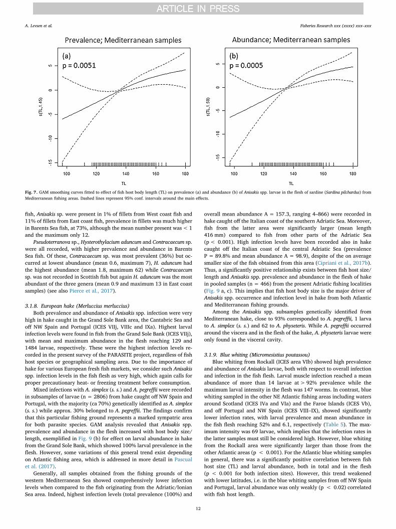
In sardines from the Mediterranean, one of the most importantfindings is the occurrence of A. pegreffii in almost 50% of the fish caughtoff West Sardinia (FAO 37.1.1), exceeding all other Mediterranean lo-cations. This finding is apparently closely related to fish host body sizesince sardines from the latter locality were on average more than20 mm larger than the rest of the sample (Table 4). In contrast, thesmallest of the present sardines, sampled in the northern TyrrhenianSea (FAO 37.1.3), were apparently free from A. pegreffii larvae. Thesignificantly positive effect of host body size/length as main predictorof larval infection in the flesh of sardine from the present Mediterra-nean localities, is illustrated in Fig. 7.
Another important finding is the presence of larvae in sardine filletswhich was observed for the first time in the present study.Consequently, the risk for consumers arising from these findings isobvious since sardines are often subjected to inadequate thermal pro-cessing or evisceration due to small fish size prior to preparation ofmarinated or salted home-made sardines, traditionally eatenthroughout the Mediterranean region. Nonetheless, even in cases ofhigher prevalence, mean intensity of infection in sardines was usuallylow, between 1 and 2, both in fillets and viscera.
3.1.5. Anchovy (Engraulis encrasicolus)There appears to be much higher Anisakis sp. prevalence and
abundance in anchovies from central areas of the Adriatic Sea and offGalicia (Spain) which makes anchovy a food safety-related “hotspot” inparts of Italy and Spain. For the Atlantic samples, Anisakis spp. pre-valence reached 83% and 29% in the viscera and flesh, respectively, insome batches (Rodriguez et al., 2017). Similar high infection levelswere reported from the Central Adriatic Sea, with prevalences reaching69.5% and 14.6% in the viscera and flesh, respectively (Cipriani et al.,2017a). In either catching area, fish host body size was a significantpredictor of larval prevalence and abundance in the fish flesh, althoughthe former relation was only weak for anchovy from the CentralAdriatic Sea (Fig. 8).
All larvae (N = 547) obtained from anchovies caught in theMediterranean Sea, correspond to A. pegreffii. The infection levels of A.pegreffii were significantly different between the present fishing areas ofthe Mediterranean Sea. Thus, fish from the Central and South AdriaticSea showed highest prevalence and intensity, while anchovies from offsouthern Sicily and the Ionian- and Alboran Sea were apparently un-infected. The vast majority (95.8%) of A. pegreffii larvae were located inthe body cavity while only a smaller fraction (4.2%) was present in thefish flesh. According to the present infection data for A. pegreffii in E.encrasicolus, the Central Adriatic Sea appears to be a “hotspot” for thepresence of this parasite, showing infection levels which are by farhigher than in any other area of the Mediterranean Sea. This finding
could thus be related to ecological or oceanographic characteristics ofthis basin, including both abiotic and biotic factors which contribute tomaintain the life cycle of A. pegreffii at high population size (Ciprianiet al., 2017a).
3.1.6. Atlantic cod (Gadus morhua)There were considerable differences in Anisakis spp. infection level
in cod from different sampling areas, e.g. the prevalence varied be-tween 16% in Baltic cod and 100% in cod from the Barents Sea. Ingeneral, Anisakis spp. prevalence and abundance, both in whole fish andin the fish flesh, correlated with fish length, and tended to increase withincreasing fish size. Fishing area appeared to be a significant effector ofAnisakis spp. infection in the fillets, with higher prevalence for fishsampled in the Barents Sea compared to the North Sea and the BalticSea. However, cod from the Baltic Sea were significantly smaller thanthe other fish sampled. In the North Sea area, two ICES subdivisionswere sampled, i.e. the northern North Sea (IVa) and the Central NorthSea (IVb). Even though these samples have been grouped for the sta-tistical analysis (see Gay et al., 2017), the prevalence both in fillets andoverall fish were statistically different for these two sub-areas(p < 0.05). The infection pattern in area IVa was similar to the patternseen in cod from the Barents Sea compared to area IVb. In the flesh ofcod, most larvae resided in the ventral portion of the fillets, which incod corresponds largely to the belly flaps. In pooled samples coveringall sampling areas, the prevalence of Anisakis spp. in the belly flaps was39% while only 12% carried larvae in the dorsal part of the fillets.
Other zoonotic ascaridoid genera were also observed.Pseudoterranova was present in the flesh of 12% of the sampled fish,with a peak of prevalence of 27% for the northern North Sea sample.Hysterothylacium prevalence varied between 0% for the Baltic Sea and84% for the Barents Sea. The distribution of Contracaecum was also veryvariable, with no Contracaecum isolated from the fish from the CentralNorth Sea and a prevalence of 100% for the commercial size codsampled in the Baltic Sea (see Gay et al., 2017).
3.1.7. Haddock (Melanogrammus aeglefinus)Haddock were sampled around Scotland (East and West coasts) and
in the Barents Sea. Average length was highest in the Barents Sea(584 mm) and higher on the East coast than on the West coast ofScotland (365 mm vs 311 mm). Indeed, there was almost no overlap insize between Scottish and Barents Sea fish. Prevalence and abundanceof Anisakis sp. (genetic species identification was not performed) in-fection followed the same trend as average size: 50% prevalence andaverage abundance 3.4 on the West coast of Scotland, compared to 84%and 14.5 on the East coast, and 100% and 50.5 in the Barents Sea.Highest Anisakis sp. intensity was 183 in a haddock from the BarentsSea. Prevalence and abundance of larvae increased significantly withfish size in the present samples (p < 0.001 in both cases). In Scottish
Table 4Sample size and basic fish host biometric data, along with basic Anisakis pegreffii infection parameters of sardine (Sardina pilchardus) from four Mediterranean sampling localities.
Fishing area N fish TL TW Musculature Viscera
P (%) Abund./Intensity Rel. distr.Vtrl:Drsl
Rel. distr.Left:Right
P (%) Abund./Intensity
Overall Adriatic Sea sample(FAO 37.2.1)
908 137 ± 8 (97–180) 20 ± 5 (6–45) 0.8 A: 0.01 ± 0.1 (0–2) 75:25 25:75 2.9 A: 0.05 ± 0.4 (0–10)
I: 1.1 ± 0.4 (1–2) I: 1.8 ± 1.9 (1–10)Northern Tyrrhenian Sea
(FAO 37.1.3)100 130 ± 6 (115–145) 13 ± 3 (10–21) 0 / / / 0 /
South Sicily (FAO 37.2.2) 200 140 ± 8 (120–170) 25 ± 4 (16–40) 0 / / / 1.5 A: 0.03 ± 0.23 (0–3)I: 1.67 ± 1.15 (1–3)
West Sardinia (FAO 37.1.1) 356 173 ± 12 (140–200) 40 ± 8 (18–58) 4.8 A: 0.05 ± 0.3 (0–2) 89: 11 32: 68 42.4 A: 0.7 ± 1.1 (0–7)I: 1.1 ± 0.3 (1–2) I: 1.7 ± 1.1 (1–7)
Abbreviations: TL – Total body length (mm); TW – Total body weight (g); P – prevalence (%), A – Abundance given as mean ± SD (range); I – Intensity given as mean ± SD (range); Rel.distr. – Relative distribution (%); Vtrl – ventral portion of fish flesh (corresponds roughly to belly flap); Drsl – dorsal portion of fish flesh.
A. Levsen et al. Fisheries Research xxx (xxxx) xxx–xxx
11
fish, Anisakis sp. were present in 1% of fillets from West coast fish and11% of fillets from East coast fish, prevalence in fillets was much higherin Barents Sea fish, at 73%, although the mean number present was< 1and the maximum only 12.
Pseudoterranova sp., Hysterothylacium aduncum and Contracaecum sp.were all recorded, with higher prevalence and abundance in BarentsSea fish. Of these, Contracaecum sp. was most prevalent (36%) but oc-curred at lowest abundance (mean 0.6, maximum 7), H. aduncum hadthe highest abundance (mean 1.8, maximum 62) while Contracaecumsp. was not recorded in Scottish fish but again H. aduncum was the mostabundant of the three genera (mean 0.9 and maximum 13 in East coastsamples) (see also Pierce et al., 2017).
3.1.8. European hake (Merluccius merluccius)Both prevalence and abundance of Anisakis spp. infection were very
high in hake caught in the Grand Sole Bank area, the Cantabric Sea andoff NW Spain and Portugal (ICES VIIj, VIIIc and IXa). Highest larvalinfection levels were found in fish from the Grand Sole Bank (ICES VIIj),with mean and maximum abundance in the flesh reaching 129 and1484 larvae, respectively. These were the highest infection levels re-corded in the present survey of the PARASITE project, regardless of fishhost species or geographical sampling area. Due to the importance ofhake for various European fresh fish markets, we consider such Anisakisspp. infection levels in the fish flesh as very high, which again calls forproper precautionary heat- or freezing treatment before consumption.
Mixed infections with A. simplex (s. s.) and A. pegreffii were recordedin subsamples of larvae (n = 2806) from hake caught off NW Spain andPortugal, with the majority (ca 70%) genetically identified as A. simplex(s. s.) while approx. 30% belonged to A. pegreffii. The findings confirmthat this particular fishing ground represents a marked sympatric areafor both parasite species. GAM analysis revealed that Anisakis spp.prevalence and abundance in the flesh increased with host body size/length, exemplified in Fig. 9 (b) for effect on larval abundance in hakefrom the Grand Sole Bank, which showed 100% larval prevalence in theflesh. However, some variations of this general trend exist dependingon Atlantic fishing area, which is addressed in more detail in Pascualet al. (2017).
Generally, all samples obtained from the fishing grounds of thewestern Mediterranean Sea showed comprehensively lower infectionlevels when compared to the fish originating from the Adriatic/IonianSea area. Indeed, highest infection levels (total prevalence (100%) and
overall mean abundance A = 157.3, ranging 4–866) were recorded inhake caught off the Italian coast of the southern Adriatic Sea. Moreover,fish from the latter area were significantly larger (mean length416 mm) compared to fish from other parts of the Adriatic Sea(p< 0.001). High infection levels have been recorded also in hakecaught off the Italian coast of the central Adriatic Sea (prevalenceP = 89.8% and mean abundance A = 98.9), despite of the on averagesmaller size of the fish obtained from this area (Cipriani et al., 2017b).Thus, a significantly positive relationship exists between fish host size/length and Anisakis spp. prevalence and abundance in the flesh of hakein pooled samples (n = 466) from the present Adriatic fishing localities(Fig. 9 a, c). This implies that fish host body size is the major driver ofAnisakis spp. occurrence and infection level in hake from both Atlanticand Mediterranean fishing grounds.
Among the Anisakis spp. subsamples genetically identified fromMediterranean hake, close to 93% corresponded to A. pegreffii, 1 larvato A. simplex (s. s.) and 62 to A. physeteris. While A. pegreffii occurredaround the viscera and in the flesh of the hake, A. physeteris larvae wereonly found in the visceral cavity.
3.1.9. Blue whiting (Micromesistius poutassou)Blue whiting from Rockall (ICES area VIb) showed high prevalence
and abundance of Anisakis larvae, both with respect to overall infectionand infection in the fish flesh. Larval muscle infection reached a meanabundance of more than 14 larvae at> 92% prevalence while themaximum larval intensity in the flesh was 147 worms. In contrast, bluewhiting sampled in the other NE Atlantic fishing areas including watersaround Scotland (ICES IVa and VIa) and the Faroe Islands (ICES Vb),and off Portugal and NW Spain (ICES VIII–IX), showed significantlylower infection rates, with larval prevalence and mean abundance inthe fish flesh reaching 52% and 6.1, respectively (Table 5). The max-imum intensity was 69 larvae, which implies that the infection rates inthe latter samples must still be considered high. However, blue whitingfrom the Rockall area were significantly larger than those from theother Atlantic areas (p < 0.001). For the Atlantic blue whiting samplesin general, there was a significantly positive correlation between fishhost size (TL) and larval abundance, both in total and in the flesh(p < 0.001 for both infection sites). However, this trend weakenedwith lower latitudes, i.e. in the blue whiting samples from off NW Spainand Portugal, larval abundance was only weakly (p < 0.02) correlatedwith fish host length.
Fig. 7. GAM smoothing curves fitted to effect of fish host body length (TL) on prevalence (a) and abundance (b) of Anisakis spp. larvae in the flesh of sardine (Sardina pilchardus) fromMediterranean fishing areas. Dashed lines represent 95% conf. intervals around the main effects.
A. Levsen et al. Fisheries Research xxx (xxxx) xxx–xxx
12

Fig. 8. GAM smoothing curves fitted to effect of fish host body length (TL) on prevalence and abundance of Anisakis spp. larvae in the flesh of anchovy (Engraulis encrasicolus) from theCantabric Sea (a, c) and the Central Adriatic larval infection “hotspot” (b, d). Dashed lines represent 95% conf. intervals around the main effects.
Fig. 9. GAM smoothing curves fitted to effect of fish host body length (TL) on prevalence and abundance of Anisakis spp. larvae in the flesh of European hake (Merluccius merluccius) fromthe Grand Sole Bank (b) and the Adriatic Sea (a, c). Dashed lines represent 95% conf. intervals around the main effects.
A. Levsen et al. Fisheries Research xxx (xxxx) xxx–xxx
13

Blue whiting sampled in the Mediterranean showed generally muchlower Anisakis larval infection levels compared to their congeners fromthe Atlantic fishing areas. However, blue whiting from the Adriatic Seastill reached comparatively high overall infection values, presenting amaximum intensity of 305 and 8 worms in overall and muscle infection,respectively, at 34% prevalence of larvae residing in the flesh. In con-trast, fish from the Alboran Sea showed only low values of Anisakis, e.g.very low prevalence and abundance in the fish flesh (3% and 0.04,respectively), with maximum intensity not exceeding two larvae(Table 5). Although blue whiting from the Adriatic Sea represented byfar the smallest fish examined, they still carried significantly higherAnisakis burden than their congeners from the Alboran Sea(p < 0.001).
Genetic species identification revealed that the subsample of 124Anisakis larvae from blue whiting caught in the Norwegian Sea and offthe Faroe Islands consisted of A. simplex (s. s.). A subsample of 37worms from off the Portuguese coast (ICES IX) showed a mixed infec-tion with A. simplex (s. s.) (30 specimens) and A. pegreffii (7 specimens).The whole subsample of 134 worms from M. poutassou fished in theAdriatic Sea consisted entirely of A. pegreffii.
3.1.10. Whiting (Merlangius merlangus)Whiting were sampled from trawling surveys on the East and West
coasts of Scotland (ICES areas IV and VI, respectively). Fish from theWest coast were larger on average, mainly due to the presence of largernumbers of fish<120 mm in length that were almost absent from Westcoast samples. Prevalence and abundance of Anisakis sp. (genetic spe-cies identification was not performed) were significantly higher inlarger fish (p < 0.001 in both cases). Overall prevalence of Anisakis sp.was slightly (but non-significantly) higher on the West coast (50.4%versus 42.6%, p = 0.08), although mean abundance was slightly (againnon-significantly) higher in East coast fish (7.9 versus 6.4). Abundanceof Anisakis sp. in the fillets was higher in the East coast samples (22.7%versus 8.9%, p < 0.001), with high numbers present in fillets of largerEast coast fish but not in larger West coast fish. Hysterothylacium wasrecorded in 12.0% of West coast fish and 19.9% of East coast fish whilePseudoterranova was present in 1.0% and 2.7% of fish, respectively (seealso Pierce et al., 2017).
3.1.11. Plaice (Pleuronectes platessa) and four-spotted megrim(Lepidorhombus boscii)
For pooled samples, i.e. all geographical areas included, four-spotted megrim displayed higher Anisakis spp. prevalence in overallfish, in fillets and in viscera (60.6; 30 and 51.9%, respectively) thanplaice (23.9; 0.7 and 23.7%, respectively) (Table 6). Mean weight andlength of all batches from both species were quite similar, so that thedifferences were likely not related to fish size. Thus, the differencescould be due to geographical origins since both species were sampled indifferent ICES areas (North Sea, West of Scotland and English Channelfor the plaice, and Bay of Biscay, Portuguese waters and Southwest ofIreland for the four-spotted megrim). In each species, geographical areainduced significant differences. Highest prevalence (42.7%) was ob-served in plaice sampled in the West of Scotland, while highest pre-valence in four-spotted megrim was observed for fish sampled in theSouthwest of Ireland (82.1%). The highest intensities were observed infour-spotted megrim sampled in the southwest of Ireland, reachingmaximum 80 larvae. In plaice, highest abundance was observed offWest Scotland with values up to 28. Larval prevalence in the flesh ofplaice was very low, with only 3 infected fish out of 464 sampled(0.7%), whereas almost one third (30%) of the four-spotted megrimcarried Anisakis spp. larvae in the flesh. A subsample of 11 larvae iso-lated from plaice caught in the English Channel (ICES VIId) were mo-lecularly identified and were all assigned to Anisakis simplex (s. s.).
3.1.12. Monkfish and black-bellied angler (Lophius piscatorius and L.budegassa)
The two Lophius species were sampled in relatively small numbers.Monkfish was obtained from Atlantic waters, ICES areas VII, VIII andIX, as well as from the Alboran Sea of the Mediterranean, while black-bellied angler was also sampled in the Balearic- and Adriatic Seas.Highest overall Anisakis spp. prevalence and abundances were seen inareas VII and VIIIc, respectively, apparently with a slight tendency ofsmall monkfish to show higher larval abundances in the flesh. Anisakisspp. abundance (but not prevalence) generally increased with fishlength and differences were observed between different sampling per-iods. A difference in Anisakis spp. prevalence was found between maleand female L. budegassa, with a higher prevalence of worms in femalefish. It is worth noticing that the samples of monkfish caught in theAlboran Sea showed mixed infections with three Anisakis species, i.e. A.simplex (s. s.), A. pegreffii and A. physeteris, identified by allozymes and
Table 5Sample size and basic fish host biometric data, along with basic Anisakis spp. infection parameters of blue whiting (Micromesistius pouttassou) from six different fishing areas.
Sampling area(ICES zone)
N fishN = 1534
TL TW Musculature Viscera
P (%) Abundance/Intensity Rel. distr.Vtrl: Drsl
Rel. distr.Left: Right
P (%) Abundance/Intensity
North Sea, Off WScotland (ICESIVa, VIa)
153 223 ± 43 (96–333) 68 ± 41 (5–255) 27.6 A: 0.5 ± 1.1 (0–6) 93: 7 55: 45 71.1 A: 7.0 ± 10.9 (0–77)
I: 1.8 ± 1.4 (1–6) I: 9.9 ± 11.7 (1–77)Off Faroe Isles 300 278 ± 30 (182–352) 119 ± 36 (33–285) 52.3 A: 6.1 ± 11.8 (0–69) 89: 11 56: 44 89.0 A: 15.7 ± 23.9 (0–165)(ICES Vb) I: 11.7 ± 14.2 (1–69) I: 16.8 ± 23.3 (1–165)Rockall area (ICES
VIb)454 324 ± 150 (225–455) 171 ± 77 (56–558) 93.0 A: 14.1 ± 18.5 (0–147) 90: 10 46: 54 98.5 A: 36.4 ± 42.4 (0–309)
I: 15.2 ± 18.8 (1–147) I: 37.0 ± 42.5 (1–309)Off Spain & Port.
(ICES VIIIc,IX)
386 255 ± 34 (200–380) 104 ± 47 (38–382) 50.5 A: 2.1 ± 5.6 (0–45) 96: 4 52: 48 96.9 A: 24.1 ± 70.9 (0–649)
I: 4.1 ± 7.3 (1–45) I: 24.9 ± 71.9 (1–649)Mediterranean Sea 241 251 ± 16 (220–295) 118 ± 24 (81–191) 2.9 A: 0.04 ± 0.3 (0–2) / / 1.4 A: 0.01 ± 0.1 (0–1)Alboran Sea 69 198 ± 31 (111–312) 64 ± 31 (8–229) 34.1 I: 1.5 ± 0.7 (1, 2) 96: 4 49: 51 85.0 I: 1 ± 0 (1)Adriatic Sea 172 A: 0.8 ± 1.5 (0–8) A: 36.3 ± 61.8 (0–303)
I: 2.3 ± 1.8 (1–8) I: 42.7 ± 65.0 (1–303)
Abbreviations: TL – Total body length (mm); TW – Total body weight (g); P – Prevalence; A – Abundance, I – Intensity, both given as mean ± SD (range); Rel. distr. – Relativedistribution (%); Vtrl – ventral portion of fish flesh; Drsl – dorsal portion of fish flesh.
A. Levsen et al. Fisheries Research xxx (xxxx) xxx–xxx
14

mtDNA cox2 sequence analysis. The actual larvae occurred in syntopyin the viscera of the same individual fish host.
3.1.13. Silver scabbardfish (Lepidopus caudatus) and European sea bass(Dicentrarchus labrax)
Silver scabbardfish were obtained from four sampling areas of theMediterranean Sea (Table 1). All fish regardless of sampling area wereinfected with Anisakis spp. larvae, i.e. P = 100%. Scabbardfish caughtoff Malta’s coast and from off southern Sicily showed significantlyhigher overall larval abundance levels (MA = 239 ± 141 andMA = 193 ± 118, respectively) compared to the fish from the othersampling areas (MA = 50 ± 59 in pooled samples) (p < 0.001).However, and more importantly, the larval abundance in the fishmuscle did not differ significantly between the different sampling areas,showing 47% prevalence and MA = 2.2 ± 4.0 with intensity ranging1–24 in pooled fish samples. All genetically identified larvae from theformer areas belonged to A. pegreffii. Identification of larvae fromscabbard fish sampled in the Alboran- and Tyrrhenian Seas revealedthat A. pegreffii was still the most abundant sibling species, however, afew A. physeteris larvae (N = 25) were found in both areas, occurring insyntopy with A. pegreffii in the viscera of one individual fish host ineither locality. Interestingly, in the fish batch originating from the Al-boran Sea, a single scabbard fish had a mixed infection with A. simplex(s. s.) (1 larva), A. nascettii (1 larva), and A. ziphidarum (1 larva), alongwith a majority of larvae belonging to A. pegreffii.
Parasite data of European sea bass are scarce. Of 38 fish collectedand examined during the present survey, only two medium-sized spe-cimens weighing 480 and 500 g, and fished in the central TyrrhenianSea, appeared to be infected with 12 and 1 anisakid larvae, respectively,all situated around the organs of the visceral cavity. Sequence analysisof the mtDNA cox2 gene enabled to identify the nematodes asContracaecum rudolphii sp. A, which matures and reproduces in pisci-vorous birds (Mattiucci et al., 2008).
3.2. Anisakis spp. exposure assessment considerations
3.2.1. Effect of fish sampling and storage proceduresThe different post-catch storage modes applied by the surveyors
before inspecting the fish for parasites, may under some circumstanceshave facilitated post-mortem migration of Anisakis spp. larvae fromtheir original site around the visceral organs into the flesh of the hosts.While this is unlikely to occur in fish that is deep-frozen or evisceratedshortly after catch, larval post-mortem migration may take place incases where the fish is cool-stored for several hours during transportand before processing. Such storage conditions may have prevailed in atleast some of the present Mediterranean samples of anchovy and sar-dine obtained from local fishermen. Larval muscle penetrating behaviormay also be facilitated by the very short migration distance between thevisceral organs and the flesh of small fish such as anchovy and sardine.Thus, temperature and storage time appear to be the most importantvariables determining the activation and motility of Anisakis larvae, asobserved in experimental studies (Cipriani et al., 2016; Šimat et al.,2015). Although storage temperatures above 2 °C seem to be requiredto induce any post-mortem migration of A. pegreffii larvae in anchovy(Cipriani et al., 2016), higher storage temperatures over shorter periodsmay have been the case for some of the present anchovy and sardinesamples from the Mediterranean Sea. Thus, the possibility exists that atleast some of the larval findings in the flesh of both anchovy and sar-dine may have been inflicted by post-mortem migration. Nevertheless,cool-storage and sales of freshly caught, i.e. unfrozen, anchovy andsardine illustrates the importance of these fish species as vector of an-isakiasis when prepared raw or only lightly processed in privatehouseholds in actual regions of Italy and Croatia (Moschella et al.,2004; Mattiucci et al., 2011, 2013; Mladineo et al., 2016).Ta
ble6
Samplesize
andba
sicfish
host
biom
etricda
ta,a
long
withba
sicAnisakisinfectionpa
rametersof
plaice
(Pp,
Pleuronectes
platessa)an
dfour-spo
tted
meg
rim
(Lb,
Lepido
rhom
busboscii)
from
eigh
tNorth
East
Atlan
ticsamplinglocalities.
Fishingarea
Nfish
(spe
cies)
TLTW
Muscu
lature
Viscera
P(%
)Abu
nd./Intensity
Rel.d
istr.V
trl:Drsl
Rel.d
istr.Le
ft:R
ight
P(%
)Abu
nd./Intensity
Southe
rnNorth
Sea(ICES
IVc)
56(Pp)
285
±25
(237
–351
)24
4±
76(130
–491
)0.0
//
/5.4
A:0
.09
±0.39
(0–2
)I:1.67
±0.58
(1–2
)En
glishch
anne
l(ICES
VIId)
184(Pp)
302
±52
300
±23
6(98–
1907
)0.5
A:0
.01
±0.07
(0–1
)f.p
.f.p
.12
.5A:0
.15
±0.42
(0–3
)(210
–570
)I:1.00
±0.00
(1-1)
I:1.17
±0.49
(1–3
)NorthernNorth
Sea(ICES
IVa)
12(Pp)
297
±45
(196
–365
)26
2±
105(70–
475)
0.0
//
/33
.3A:3
.25
±6.69
(0–2
3)I:9.75
±8.92
(4–2
3)Cen
tral
North
Sea(ICES
IVb)
102(Pp)
294
±42
(220
–384
)26
5±
116(91–
540)
1.0
A:0
.02
±0.20
(0–2
)f.p
.f.p
.32
.4A:0
.86
±1.66
(0–8
)I:2.00
±0.00
(2-2)
I:2.67
±1.93
(1–8
)Westof
Scotland
(ICES
VIa)
110(Pp)
257
±38
(220
–271
)14
3±
84(91–
209)
0.9
A:0
.01
±0.10
(0–1
)f.p
.f.p
.42
.7A:2
.94
±5.28
(0–2
8)I:1.00
±0.00
(1-1)
I:6.87
±6.20
(1–2
8)Po
rtug
uese
waters(ICES
IX)
48(Lb)
250
±19
(200
–292
)12
5±
30(66–
201)
12.5
A:0
.13
±0.33
(0–1
)f.p
.f.p
.45
.8A:0
.65
±0.86
(0–4
)I:1.00
±0.00
(1-1)
I:1.41
±0.73
(1–4
)So
uthw
estof
Irelan
d–Ea
st(ICES
VIIj)
67(Lb)
286
±31
(230
–350
)18
3±
70(73–
411)
65.7
A:2
.19
±3.04
(0–1
6)99
:156
:44
82.1
A:8
.61
±11
.35(0–7
5)I:3.34
±3.20
(1–1
6)I:10
.49
±11
.73(1–7
5)So
uthw
estof
Irelan
d–West(ICES
VIIk)
72(Lb)
319
±33
(265
–485
)23
2±
88(133
–771
)33
.3A:1
.32
±4.12
(0–2
8)94
:652
:48
36.1
A:1
.50
±3.50
(0–1
9)I:3.96
±6.45
(1–2
8)I:4.15
±4.83
(1–1
9)Ba
yof
Biscay
(ICES
VIII)
100(Lb)
243
±24
(200
–315
)11
2±
37(62–
232)
12.0
A:0
.17
±0.49
(0–2
)88
:12
29:7
146
.0A:1
.36
±3.05
(0–2
1)I:1.42
±0.51
(1–2
)I:2.96
±3.96
(1–2
1)
Abb
reviations:T
L–To
talb
odyleng
th(m
m);TW
–To
talb
odyweigh
t(g);P–prev
alen
ce(%
),A–Abu
ndan
ce,I
–Intensity,
both
give
nas
mean
±SD
(ran
ge);Rel.d
istr.–
Relativedistribu
tion
(%);Vtrl–
ventralp
ortion
offish
flesh(correspon
dsroug
hlyto
belly
flap
);Drsl−
dorsal
portionof
fish
flesh;
f.p.–
toofew
parasitesforap
prop
riatecalculation.
A. Levsen et al. Fisheries Research xxx (xxxx) xxx–xxx
15

3.2.2. Effect of fish hostResults from GAM analyses indicated that fish host body size is a
major predictor of Anisakis spp. occurrence in most fish species pre-sently analyzed. Thus, there was a clear trend towards significantlypositive relationships between fish host body size/length and bothprevalence and abundance of Anisakis spp. larvae in the fish flesh.However, exceptions from this trend became apparent in Atlanticmackerel where the host length effect on larval muscular infection wasonly very weakly positive or even absent in the present southern- ornorthernmost fishing localities, respectively. These findings indicatethat any accumulation over time of Anisakis spp. larvae does not takeplace in Atlantic mackerel. On the contrary, a trend towards the op-posite pattern, i.e. larger mackerel are carrying fewer larvae, at least insome of the present Atlantic localities, could indicate that larger spe-cimens of the actual stocks or entities are able to cope with the infec-tions immunologically. These aspects of the Anisakis spp. infectionpattern in the present Atlantic mackerel samples are addressed anddiscussed in detail by Levsen et al. (2017). Besides mackerel, a lesssignificant effect of host body size on A. pegreffii prevalence was ap-parently present in anchovy from certain Atlantic and Mediterraneanfishing localities. Thus, other biotic or abiotic factors that are char-acteristics of the actual localities, may be important drivers of the ob-served infection patterns, as well. For details on these aspects of Ani-sakis spp. occurrence in anchovy from Atlantic and Mediterraneanfishing grounds, see Rodriguez et al. (2017) and Cipriani et al. (2017a),respectively.
In general, the effect of body size on infection rate seems to bestrongly related to an ontogenetic change in preferred food source byswitching from microplanktonic food to larger prey including macro-plankton such as krill, or to a mainly piscivorous feeding habit. This hasbeen demonstrated for several fish species including herring and
Atlantic cod (Levsen and Lunestad, 2010; Mouritsen et al., 2010;Münster et al., 2015) to greatly increase anisakid infection probability.This again may result in larval accumulation over time since Anisakisspp. larvae may stay alive for extended periods or even over a given fishhost’s lifetime (Smith, 1984). The observed distribution of anisakidlarvae exhibited very high spatial and temporal variability, even withinhost species. Highly skewed distribution patterns are characteristic formany parasite species (e.g., Shaw and Dobson, 1995) and Anisakisspecies appear to be typical in this respect.
In the present herring samples we observed a highly significantincrease in overall Anisakis simplex (s. s.) abundance with increasing fishsize. Similarly, the extremely high Anisakis spp. infection levels in bothflesh and viscera of European hake from the Grand Sole area (ICES VII)strongly suggest that also this case relates most likely to fish host bodysize since hake from this particular fishing ground were by far thelargest fish (both in terms of TL and TW) of all hake examined in thepresent survey (for details, see Pascual et al., 2017).
The possible presence of Anisakis spp. larvae in the fish flesh re-presents the greatest concern with respect to consumer exposure risk,especially in fish intended for consumption in a fresh state, i.e. withoutprior freezing. Thus, the present survey revealed that the vast majorityof the flesh residing larvae are situated in the ventral portion of the fishmuscle, which in most fish species corresponds to the belly flaps. Thistrend was especially pronounced in the Atlantic samples of chubmackerel (Table 2), Norwegian spring spawning herring (Table 3), bluewhiting (Table 5), and Atlantic cod (see Gay et al., 2017). Thus, trim-ming of the fillets by removing most of the belly flaps would sig-nificantly reduce the probability of Anisakis larvae to be present in thefinal product. For example, Levsen and Lunestad (2010) found wheninvestigating the Anisakis simplex (s. l.) occurrence in Norwegian springspawning herring, that the probability of larval presence in trimmed
Fig. 10. Anisakis spp. exposure risk profile based on parasite prevalence in the flesh of actual fish species if to be consumed raw (sushi/sashimi), or only lightly processed (pickled/marinated/cold-smoked). The risk categories reflect the following Anisakis spp. prevalence ranges in the fish flesh: No risk: 0%; Very low: 1–5%; Low: 6–20%; Moderate: 21–50%; High:51–80%; Very high: > 80%.
A. Levsen et al. Fisheries Research xxx (xxxx) xxx–xxx
16

herring fillets (by removing the belly-flaps) was 5–8 times lower com-pared to untrimmed fillets.
It is also apparent that, for all samples analyzed, larval meanabundances observed in the fish flesh were generally correlated withthe number of worms found in the visceral cavity/organs (global ana-lysis of mean abundances for all samples, p < 0.001). However, anexception from this general trend was seen in Atlantic mackerel whereseveral specimens, up to 5.2% of each batch and sampling locality,carried Anisakis spp. larvae in the flesh only, i.e. the visceral cavity wasnot infected (see Levsen et al., 2017).
3.2.3. Effect of fishing ground/oceanographic areaThe most conspicuous difference between the Atlantic- and
Mediterranean fishing grounds with regards to general Anisakis spp.infection characteristics relates to the relative occurrence of Anisakissibling species. Thus, A. simplex (s. s.) is the predominating species inthe Atlantic samples of the present survey, while A. pegreffii dominatesall samples from the Mediterranean Sea. However, there is considerablesympatric overlap between the western Mediterranean (Alboran Sea)and the Bay of Biscay in the Atlantic. Moreover, a few A. pegreffii werealso found in Atlantic mackerel as far north as the Norwegian Sea. SinceAtlantic mackerel, which spawn in deep water between theMediterranean and the Faroe Islands, undertake extensive feeding mi-grations between the southern spawning- and the northern feedinggrounds (Jansen and Gislason, 2013), the finding of A. pegreffii inmackerel collected in the Norwegian Sea strongly indicate that the fishbecame infected as juveniles in areas much further south such as thesouthern Bay of Biscay. Thus, A. pegreffii may prove a useful supple-mentary marker to further explore the northerly migration routes,especially of the southern and western spawning components of theAtlantic mackerel stock (see Levsen et al., 2017).
Additionally, small numbers of another Anisakis sibling species, A.physeteris, were found in a few specimens of Atlantic mackerel, silverscabbard fish, European hake and monkfish caught in theMediterranean Sea. However, these records were very rare, and noworms were found in the flesh of the hosts. Therefore, A. physeteris isunlikely to pose any significant risk to human health in European wa-ters. The same applies also for A. nascettii (1 larva) and A. ziphidarum (1larva) which were recorded in the visceral cavity, along with several A.pegreffii and A. simplex (s. s.), in a single scabbard fish from the AlboranSea.
Especially within the present Mediterranean sampling areas thereseem to exist geographical “hotspots” of A. pegreffii infections in somefish species, which appear to relate more closely to oceanographiccharacteristics of the actual localities rather than specific fish hosttraits. For example, anchovy caught close to the Italian coast of theCentral and South Adriatic Sea showed comprehensively higher A. pe-greffii prevalence and abundance, both in the viscera and the flesh, thananchovy from all other sampling localities in the Mediterranean Sea.Interestingly, however, the anchovy from the Adriatic infection “hot-spot” area were smaller (mean TL < 130 mm) than their congenersfrom the other Mediterranean fishing grounds, which were larger(mean TL > 130 mm) but showed considerably lower A. pegreffii in-fection levels. This may indicate that specific oceanographic or ecolo-gical factors at the actual fishing area have greater effect on the A.pegreffii infection level than specific fish host characteristics such asbody size (see Cipriani et al., 2017a).
Considering that E. encrasicolus has been reported as the mainsource of human cases of anisakiasis in certain regions of Italy(Moschella et al., 2004; Mattiucci et al., 2011, 2013) and Spain(Daschner et al., 2000), mostly after the consumption of raw homemademarinated anchovies, the present epidemiological data of A. pegreffii inthe flesh of anchovy represent important data to evaluate the consumerexposure risk associated with the parasite. Thus, there could be a re-lation between the high A. pegreffii infection levels of anchovy from theAdriatic Sea and some clinical data on cases of anisakiasis in Italy.
Interestingly, most cases of human anisakiasis in Italy were actuallyrecorded in some regions which are in comparatively close vicinity tothe coast of the Central Adriatic Sea (S. Mattiucci, pers. observation).
3.2.4. Anisakis spp. exposure risk profileIn Fig. 10, we introduce a graphical Anisakis spp. exposure risk
profile including seven (7) fish species which we consider at highestrisk to act as source of anisakiasis in Europe. All species covered in theprofile are more or less frequently consumed in a raw or only lightlyprocessed state, without prior freezing, or may be served under-cooked,i.e. if the heating treatment is inadequate, not reaching 60 °Cthroughout the product. The present risk categories (from no risk to veryhigh) are based on prevalence data of Anisakis spp. larvae in the flesh ofthe actual fish species. Additional information with regards to parti-cular fishing locality or processing mode of products (e.g. whole orfilleted; trimmed or untrimmed fillets) is provided whenever appro-priate. It is important to note, however, that for some of the speciesincluded in the profile, e.g. Atl. mackerel, herring and cod, trimming ofthe fillets by removing the ventralmost parts of the belly flaps (seeFig. 3), would reduce the probability of larval presence in the final(fresh) product by more than 90%.
Acknowledgements
The present work was financially supported by the EuropeanUnion’s 7th Framework Programme for Research, TechnologicalDevelopment and Demonstration, project Parasite risk assessment withintegrated tools in EU fish production value chains (PARASITE), GrantAgreement (GA) no. 312068.
References
Bao, M., Pierce, G.J., Pascual, S., González-Muñoz, M.I., Mattiucci, S., Mladineo, I.,Cipriani, P., Bušelić, I., Strachan, N.J.C., 2017. Assessing the risk of an emergingzoonosis of worldwide concern: anisakiasis. Sci. Rep. 7. http://dx.doi.org/10.1038/srep43699.
Cipriani, P., Acerra, V., Bellisario, B., Sbaraglia, G.L., Cheleschi, R., Nascetti, G.,Mattiucci, S., 2016. Larval migration of the zoonotic parasite Anisakis pegreffii(Nematoda Anisakidae) in anchovy, Engraulis encrasicolus: implications to seafoodsafety. Food Contr. 59, 148–157.
Cipriani, P., Sbaraglia, G.L., Palomba, M., Giulietti, L., Bellisario, B., Bušelić, I., Mladineo,I., Cheleschi, R., Nascetti, G., Mattiucci, S., 2017a. Anisakis pegreffii (Nematoda:Anisakidae) in the European anchovy Engraulis encrasicolus, from the MediterraneanSea: fishing ground as a predictor of the parasite distribution. Fish. Res. http://dx.doi.org/10.1016/j.fishres.2017.03.020. (this volume).
Cipriani, P., Sbaraglia, G.L., Paoletti, M., Giulietti, L., Bellisario, B., Palomba, M., Bušelić,I., Mladineo, I., Nascetti, G., Mattiucci, S., 2017b. The Mediterranean European hake,Merluccius merluccius: detecting drivers influencing the Anisakis spp. larvae dis-tribution. Fish. Res. http://dx.doi.org/10.1016/j.fishres.2017.07.010. (this volume).
D’Amelio, S., Mathiopoulos, K.D., Brandonisio, O., Lucarelli, G., Doronzo, F., Paggi, L.,1999. Diagnosis of a case of gastric anisakidosis by PCR-based restrictionfragmentlength polymorphism analysis. Parassitologia 41, 591–593.
Daschner, A., Alonso-Gomez, A., Cabanas, R., Suarez-de-Parga, J., Lopez-Serrano, M.,2000. Gastroallergic anisakiasis: borderline between food allergy and parasitic dis-ease-clinical and allergologic evaluation of 20 patients with confirmed acute para-sitism by Anisakis simplex. J. Allergy Clin. Immunol. 105, 176–181.
European Commission (EC), 2016. Facts and Figures on the Common Fisheries Policy.Basic Statistical Data, 2016 Edition. (https://ec.europa.eu/fisheries/sites/fisheries/files/docs/body/pcp_en.pdf).
Gómez-Morales, M.A., Martinez Castro, C., Lalle, M., Fernandez, R., Pezzotti, P., Abollo,E., Pozio, E., 2017. UV-press method versus artificial digestion method to detectAnisakidae L3 in fish fillets: comparative study and suitability for the industry. Fish.Res. http://dx.doi.org/10.1016/j.fishres.2016.12.011. (this volume).
Gay, M., Bao, M., MacKenzie, K., Pascual, S., Buchmann, K., Bourgau, O., Couvreur, C.,Hastie, L.C., Levsen, A., Pierce, G.J., 2017. Infection levels and species diversity ofascarid nematode in Atlantic cod, Gadus morhua, are correlated with geographic areaand fish size. Fish. Res. http://dx.doi.org/10.1016/j.fishres.2017.06.006. (thisvolume).
González, A.F., Rodríguez, H., Outeiriño, L., Vello, C., Larsson, C., Pascual, S., 2017. Abiobanking platform for fish-borne zoonotic parasites: a traceable system to preservesamples, data and money. Fish. Res. http://dx.doi.org/10.1016/j.fishres.2017.03.014. (this volume).
Jansen, T., Gislason, H., 2013. Population structure of atlantic mackerel (Scomber scom-brus). PLoS One 8 (5), e64744.
Karl, H., Leinemann, M., 1993. A fast and quantitative detection method for nematodes infish fillets and fishery products. Arch. Lebensmittelhyg. 44, 124–125.
A. Levsen et al. Fisheries Research xxx (xxxx) xxx–xxx
17

Klapper, R., Kuhn, T., Münster, J., Levsen, A., Karl, H., Klimpel, S., 2015. Anisakid ne-matodes in beaked redfish (Sebastes mentella) from three fishing grounds in the NorthAtlantic, with special notes on distribution in the fish musculature. Vet. Parasitol.207, 72–80.
Levsen, A., Karl, H., 2014. Anisakis simplex (s.l.) in Grey gurnard (Eutrigla gurnardus)from the North Sea: food safety considerations in relation to fishing ground anddistribution in the flesh. Food Contr. 36, 15–19.
Levsen, A., Lunestad, B.T., 2010. Anisakis simplex Anisakis simplex third stage larvae inNorwegian spring spawning herring (Clupea harengus L.), with emphasis in larvaldistribution in the flesh. Vet. Parasitol. 171, 247–253.
Levsen, A., Paoletti, M., Cipriani, P., Nascetti, G., Mattiucci, S., 2016. Species compositionand infection dynamics of ascaridoid nematodes in Barents Sea capelin (Mallotusvillosus) reflecting trophic position of fish host. Parasitol. Res. 115, 4281–4291.
Levsen, A., Cipriani, P., Mattiucci, S., Gay, M., Hastie, L.C., MacKenzie, K., Pierce, G.J.,Svanevik, C.S., Højgaard, D.P., Nascetti, G., González, A.F., Pascual, S., 2017.Anisakis species composition and infection characteristics in Atlantic mackerel,Scomber scombrus, from major European fishing grounds –reflecting changing fishhost distribution and migration pattern. Fish. Res. http://dx.doi.org/10.1016/j.fishres.2017.07.030. (this volume).
Lim, H., Bong-Kwang, J., Cho, J., Yooyen, T., Shin, E.H., Chai, J.Y., 2015. Moleculardiagnosis of cause of anisakiasis in humans, South Korea. Emerg. Infect. Dis. 21,342–344.
Llarena-Reino, M., Piñeiro, C., Antonio, J., Outeriño, L., Vello, C., González, A.F., Pascual,S., 2013. Optimization of the pepsin digestion method for anisakids inspection in thefishing industry. Vet. Parasitol. 191, 276–283.
Münster, J., Klimpel, S., Fock, H.O., MacKenzie, K., Kuhn, T., 2015. Parasites as biologicaltags to track an ontogenetic shift in the feeding behaviour of Gadus morhua off Westand East Greenland. Parasitol. Res. 114, 2723–2733.
Mattiucci, S., Nascetti, G., 2008. Advances and trends in the molecular systematics ofanisakid nematodes: with implications for their evolutionary ecology and host-parasite coevolutionary processes. Adv. Parasitol. 66, 47–148.
Mattiucci, S., Nascetti, G., Cianchi, R., Paggi, L., Arduino, P., Margolis, L., Brattey, J.,Webb, S.C., D’Amelio, S., Orecchia, P., Bullini, L., 1997. Genetic and ecological dataon the Anisakis simplex complex, with the evidence for a new species (Nematoda,Ascaridoidea, Anisakidae). J. Parasitol. 83, 401–416.
Mattiucci, S., Paoletti, M., Webb, S.C., Sardella, N., Timi, J.T., Berland, B., Nascetti, G.,2008. Genetic relationships among species of Contracaecum Railliet and Henry, 1912and Phocascaris Host, 1932 (Nematoda: Anisakidae) from pinnipeds based on mi-tochondrial cox2 sequences, and congruence with allozyme data. Parasite 15,408–419.
Mattiucci, S., Paoletti, M., Borrini, F., Palumbo, M., Palmieri, R.M., Gomes, V., Casati, A.,Nascetti, G., 2011. First molecular identification of the zoonotic parasite Anisakispegreffii (Nematoda: Anisakidae) in a paraffin-embedded granuloma taken from acase of human intestinal anisakiasis in Italy. BMC Infect. Dis. 11, 82.
Mattiucci, S., Fazii, P., De Rosa, A., Paoletti, M., Salomone Megna, A., Glielmo, A., DeAngelis, M., Costa, A., Meucci, C., Calvaruso, V., Sorrentini, I., Palma, G., Bruschi, F.,Nascetti, G., 2013. Anisakiasis and gastroallergic reactions associated with Anisakispegreffii infection, Italy. Emerg. Infect. Dis. 19, 496–499.
Mattiucci, S., Cipriani, P., Webb, S.C., Paoletti, M., Marcer, F., Bellisario, B., Gibson, D.I.,Nascetti, G., 2014. Genetic and morphological approaches distinguish the three sib-ling species of the Anisakis simplex species complex, with a species designation asAnisakis berlandi n. sp. for A. simplex sp. C (Nematoda Anisakidae). J. Parasitol. 100,199–214.
Mattiucci, S., Cipriani, P., Paoletti, M., Nardi, V., Santoro, M., Bellisario, B., Nascetti, G.,2015. Temporal stability of parasite distribution and genetic variability values ofContracaecum osculatum sp. D and C. osculatum sp. E (Nematoda: Anisakidae) fromfish of the Ross Sea (Antarctica). Int. J. Parasitol. Parasites Wildl. 4, 356–367.
Mattiucci, S., Acerra, V., Paoletti, M., Cipriani, P., Levsen, A., Webb, S.C., Canestrelli, D.,Nascetti, G., 2016. No more time to stay ’single’ in the detection of Anisakis pegreffii,A. simplex (s. s.) and hybridization events between them: a multi-marker nucleargenotyping approach. Parasitology 143, 998–1011.
Mattiucci, S., Cipriani, P., Paoletti, M., Levsen, A., Nascetti, G., 2017a. Reviewing bio-diversity and epidemiological aspects of anisakid nematodes from the North EastAtlantic Ocean. J. Helminthol. http://dx.doi.org/10.1017/S0022149x1700027X.
Mattiucci, S., Giulietti, L., Paoletti, M., Cipriani, P., Gay, M., Levsen, A., Klapper, R., Karl,H., Bao, M., Pierce, G.J., Nascetti, G., 2017b. Population genetic structure of the
parasite Anisakis simplex (s. s.) collected in Clupea harengus L. from North EastAtlantic fishing grounds. Fish. Res. http://dx.doi.org/10.1016/j.fishres.2017.08.002.(this volume).
Mladineo, I., Poljak, V., 2014. Ecology and genetic structure of zoonotic Anisakis spp.from adriatic commercial fish species. Appl. Environ. Microbiol. 80, 1281–1290.
Mladineo, I., Popovic, M., Drmic-Hofman, I., Poljak, V., 2016. A case report of Anisakispegreffii (Nematoda, Anisakidae) identified from archival paraffin sections of aCroatian patient. BMC Infect. Dis. 16, 42.
Moschella, C.M., Mattiucci, S., Mingazzini, P., De Angelis, G., Assenza, M., Lombardo, F.,Monaco, S., Paggi, L., Modini, C., 2004. Intestinal anisakiasis in Italy: case report. J.Helminthol. 78, 271–273.
Mouritsen, K.N., Hedeholm, R., Schack, H.B., Nukaaraq Møller, L., Storr-Paulsen, M.,Dzido, J., Rokicki, J., 2010. Occurrence of anisakid nematodes in Atlantic cod (Gadusmorhua) and Greenland cod (Gadus ogac), West Greenland. Acta Parasitol. 55, 81–89.
Pascual, S., Rodriguez, H., Pierce, G., Hastie, L., Gonzalez, A.F., 2017. The NE Atlantichake: a neglected. high exposure risk for zoonotic parasites in European fish markets.Fish. Res (this volume).
Pierce, G.J., Bao, M., MacKenzie, K., Dunser, A., Giulietti, L., Mattiucci, S., Cipriani, P.,Hastie, L.C., 2017. Anisakid nematode infections in haddock (Melanogrammus aegle-finus) and whiting (Merlangius merlangus) in the Northeast Atlantic. Fish. Res (thisvolume).
Pippy, J.H., 1970. Use of ultraviolet light to find parasitic nematodes in situ. J. Fish. Res.Board Can. 27, 963–965.
Posada, D., Buckley, T.R., 2004. Model selection and model averaging in phylogenetics.Advantage of the AIC and Bayesian approaches over likelihood ratio test. Syst. Biol.53, 793–808.
Posada, D., 2008. JModelTest: phylogenetic model averaging. Mol. Biol. Evol. 25,1253–1256.
Reeder, T.W., 2003. A phylogeny of the Australian Sphenomorphus group (Scincidae:Squamata) and the phylogenetic placement of the crocodile skinks (Tribolonotus):Bayesian approaches to assessing congruence and obtaining confidence in maximumlikelihood inferred relationships. Mol. Phylogen. Evol. 27, 384–397.
Rodriguez, H., González, A.F., Abollo, E., Pascual, S., 2017. Re-evaluation of the im-portance of Atlantic anchovies (Engraulis encrasicolus) and sardines (Sardina pil-chardus) as a main risk factor for Anisakiosis and/or sensitization to Anisakis in Spain.Fish. Res (this volume).
Ronquist, F., Teslenko, M., van der Mark, P., Ayres, D.L., Darling, A., Hohna, S., Larget, B.,Liu, L., Suchard, M.A., Huelsenbeck, J.P., 2012. MrBayes 3.2: Efficient bayesianphylogenetic inference and model choice across a large model space. Syst. Biol. 61,539–542.
Šimat, V., Miletić, J., Bogdanović, T., Poljak, V., Mladineo, I., 2015. Role of biogenicamines in the post-mortem migration of Anisakis pegreffii (Nematoda: AnisakidaeDujardin, 1845) larvae into fish fillets. Int. J. Food Microbiol. 214, 179–186.
Shaw, D.J., Dobson, A.P., 1995. Patterns of macroparasite abundance and aggregation inwildlife populations: a quantitative review. Parasitology 111, S111–S133.
Smith, J.W., 1984. The abundance of Anisakis simplex L3 in the body cavity and flesh ofmarine teleosts. Int. J. Parasitol. 14, 491–495.
Thompson, J.D., Gibson, T.J., Plewniak, F., Jeanmougin, F., Higgins, D.G., 1997. TheCLUSTAL_X windows interface: flexible strategies for multiple sequence alignmentaided by quality analysis tools. Nucl. Acid Res. 25, 4876–4882.
Timi, J.T., Paoletti, M., Cimmaruta, R., Lanfranchi, A.L., Alarcos, A.J., Garbin, L., George-Nascimento, M., Rodríguez, D.H., Giardino, G.V., Mattiucci, S., 2014. Molecularidentification, morphological characterization and new insights into the ecology oflarval Pseudoterranova cattani in fishes from the Argentine Coast with its differ-entiation from the Antarctic species, P. decipiens sp. E (Nematoda Anisakidae). Vet.Parasitol. 199, 59–72.
Umehara, A., Kawakami, Y., Araki, J., Uchida, A., 2007. Molecular identification oftheetiological agent of the human anisakiasis in Japan. Parasitol. Int. 56, 211–215.
Zhu, X.Q., D’Amelio, S., Paggi, L., Gasser, R.B., 2000. Assessing sequence variation in theinternal transcribed spacer of ribosomal DNA within and among members of theContracaecum osculatum complex (Nematoda: Ascaridoidea: Anisakidae). Parasitol.Res. 86, 677–683.
Zorica, B., Čikeš Keč, V., Vidjak, O., Mladineo, I., Ezgeta Balić, D., 2016. Feeding habitsand helminth parasites of sardine (S. pilchardus) and anchovy (E. encrasicolus) in theAdriatic Sea. Mediterran. Mar. Sci. 17, 216–229.
A. Levsen et al. Fisheries Research xxx (xxxx) xxx–xxx
18


