A surface chemistry approach to studying cell...
7
A surface chemistry approach to studying cell adhesion Milan Mrksich Department of Chemistry, The University of Chicago, 5735 South Ellis Avenue, Chicago, IL 60637, USA. E-mail: [email protected] Received 10th February 2000 Published on the Web 5th June 2000 Model substrates that present peptide and carbohydrate ligands are becoming important mechanistic tools in cell biology. This review surveys the development of self- assembled monolayers of alkanethiolates on gold as a model substrate for studies of cell adhesion. The review begins with a background that illustrates the opportunity for using tailored substrates in biology and then addresses the characteristics that make monolayers well-suited for these studies. The review concludes with a discussion of recent work that is developing dynamic substrates wherein the activity of immobilized ligands can be modulated in real time. 1 Background Most mammalian cells are adherent. They must attach to and spread on an underlying matrix in order to carry out normal metabolism, proliferation and differentiation. The biological matrix that serves this role comprises a collection of insoluble proteins and glycoaminoglycans that are collectively referred to as the extracellular matrix (ECM). 1 In addition to maintaining the organization and mechanical properties of tissue, the ECM presents many peptide and carbohydrate ligands that are recognized by cellular receptors. These receptor–ligand inter- actions are critical to maintaining cell function and enabling cells to respond appropriately to their environments. The primary function of ECM is to mediate the adhesion of cells. 2 Without adhesion, most cells initiate a program of apoptosis that results in their death, while the loss of adhesion-related signal transduction pathways leads to the growth and spreading of cancerous tumors. The study of these and many other interactions between a cell and its matrix is an active area of research in cell biology. Studies of cell adhesion commonly use glass or polystyrene substrates that are coated with a layer of adsorbed protein. Ligands from ECM that are involved in cell–substrate inter- actions are often determined by comparing the behaviors of cells on substrates that are coated with distinct domains from a matrix protein, or from mutants of particular domains. The widespread use of these substrates derives in part from the ease with which they can be prepared: the procedure involves applying an aqueous solution of the protein to the culture substrate and allowing the adsorption to proceed over a period of several hours. Protein-coated substrates are also preferred because they retain the range and properties of ligands found in ECM, including the ability to bind other proteins from solution and to be modified by extracellular enzymes. These same substrates, however, have characteristics that limit their utility for mechanistic studies. An understanding of these limitations, which stem from the complex mechanisms by which proteins adsorb to solid surfaces, provides the rationale for employing a surface chemistry approach to developing tailored substrates. Protein adsorption There is a vast literature dealing with the adsorption of proteins at solid–liquid interfaces. 3,4 This work has not yet provided a complete molecular level understanding of the adsorption process, but gives several general observations. First, proteins adsorb to essentially all non-natural surfaces. The rate for adsorption is fast—for large proteins it can be near the diffusion limit—and is irreversible on the time scale of routine experi- ments. Secondly, adsorbed proteins undergo denaturation. The degree of denaturation is difficult to predict since it depends on many variables, including the structures of the protein and surface, and the composition of the solution (pH, ionic strength, temperature). Denaturation also depends on the concentration of protein in solution; on average, lower concentrations are correlated with greater extents of denaturation since the adsorbed protein has a longer time to denature before the surrounding sites are occupied by neighboring proteins. 5 Finally, even under ideal conditions, the layer of protein is typically heterogeneous and the determination of distribution in orientation and structure is essentially impossible. These characteristics impose many practical limitations on the use of protein-coated substrates in mechanistic cell biology. First, it is difficult to know the density of ligands that are available for binding to cellular receptors—even when the density of adsorbed protein is known precisely—because of the distribution in conformation and orientation of adsorbed protein (Fig. 1). Many studies aimed at investigating the role of ligand density in cell adhesion and migration have improperly assumed a linear correlation between the density of adsorbed protein and Milan Mrksich is Associate Professor of Chemistry at The University of Chicago. He received undergraduate training in Chemistry at the University of Illinois and completed graduate studies at Caltech in 1994. Following a postdoctoral fellowship at Harvard University, he established an independent research program at Chicago to address several aspects of organic materials. His group is studying the physical organic chemistry of interfacial reactions, using tailored substrates to study the adhesion and migration of mammalian cells, developing electroactive surfaces that join the functions of cells and mate- rials, and pursuing bio-inspired approaches to new materials. Dr Mrksich serves as a frequent consultant and advisory board member to biotechnology com- panies. This journal is © The Royal Society of Chemistry 2000 DOI: 10.1039/a705397e Chem. Soc. Rev., 2000, 29, 267–273 267
Transcript of A surface chemistry approach to studying cell...

A surface chemistry approach to studying cell adhesion
Milan Mrksich
Department of Chemistry, The University of Chicago, 5735 South Ellis Avenue, Chicago, IL60637, USA. E-mail: [email protected]
Received 10th February 2000Published on the Web 5th June 2000
Model substrates that present peptide and carbohydrateligands are becoming important mechanistic tools in cellbiology. This review surveys the development of self-assembled monolayers of alkanethiolates on gold as a modelsubstrate for studies of cell adhesion. The review begins witha background that illustrates the opportunity for usingtailored substrates in biology and then addresses thecharacteristics that make monolayers well-suited for thesestudies. The review concludes with a discussion of recentwork that is developing dynamic substrates wherein theactivity of immobilized ligands can be modulated in realtime.
1 Background
Most mammalian cells are adherent. They must attach to andspread on an underlying matrix in order to carry out normalmetabolism, proliferation and differentiation. The biologicalmatrix that serves this role comprises a collection of insolubleproteins and glycoaminoglycans that are collectively referred toas the extracellular matrix (ECM).1 In addition to maintainingthe organization and mechanical properties of tissue, the ECMpresents many peptide and carbohydrate ligands that arerecognized by cellular receptors. These receptor–ligand inter-actions are critical to maintaining cell function and enablingcells to respond appropriately to their environments. Theprimary function of ECM is to mediate the adhesion of cells.2Without adhesion, most cells initiate a program of apoptosis thatresults in their death, while the loss of adhesion-related signal
transduction pathways leads to the growth and spreading ofcancerous tumors. The study of these and many otherinteractions between a cell and its matrix is an active area ofresearch in cell biology.
Studies of cell adhesion commonly use glass or polystyrenesubstrates that are coated with a layer of adsorbed protein.Ligands from ECM that are involved in cell–substrate inter-actions are often determined by comparing the behaviors ofcells on substrates that are coated with distinct domains from amatrix protein, or from mutants of particular domains. Thewidespread use of these substrates derives in part from the easewith which they can be prepared: the procedure involvesapplying an aqueous solution of the protein to the culturesubstrate and allowing the adsorption to proceed over a periodof several hours. Protein-coated substrates are also preferredbecause they retain the range and properties of ligands found inECM, including the ability to bind other proteins from solutionand to be modified by extracellular enzymes. These samesubstrates, however, have characteristics that limit their utilityfor mechanistic studies. An understanding of these limitations,which stem from the complex mechanisms by which proteinsadsorb to solid surfaces, provides the rationale for employing asurface chemistry approach to developing tailored substrates.
Protein adsorption
There is a vast literature dealing with the adsorption of proteinsat solid–liquid interfaces.3,4 This work has not yet provided acomplete molecular level understanding of the adsorptionprocess, but gives several general observations. First, proteinsadsorb to essentially all non-natural surfaces. The rate foradsorption is fast—for large proteins it can be near the diffusionlimit—and is irreversible on the time scale of routine experi-ments. Secondly, adsorbed proteins undergo denaturation. Thedegree of denaturation is difficult to predict since it depends onmany variables, including the structures of the protein andsurface, and the composition of the solution (pH, ionic strength,temperature). Denaturation also depends on the concentration ofprotein in solution; on average, lower concentrations arecorrelated with greater extents of denaturation since theadsorbed protein has a longer time to denature before thesurrounding sites are occupied by neighboring proteins.5Finally, even under ideal conditions, the layer of protein istypically heterogeneous and the determination of distribution inorientation and structure is essentially impossible.
These characteristics impose many practical limitations onthe use of protein-coated substrates in mechanistic cell biology.First, it is difficult to know the density of ligands that areavailable for binding to cellular receptors—even when thedensity of adsorbed protein is known precisely—because of thedistribution in conformation and orientation of adsorbed protein(Fig. 1). Many studies aimed at investigating the role of liganddensity in cell adhesion and migration have improperly assumeda linear correlation between the density of adsorbed protein and
Milan Mrksich is Associate Professor of Chemistry at TheUniversity of Chicago. He received undergraduate training inChemistry at the University of Illinois and completed graduatestudies at Caltech in 1994. Following a postdoctoral fellowshipat Harvard University, he established an independent researchprogram at Chicago to address several aspects of organic
materials. His group is studyingthe physical organic chemistryof interfacial reactions, usingtailored substrates to study theadhesion and migration ofmammalian cells, developingelectroactive surfaces that jointhe functions of cells and mate-rials, and pursuing bio-inspiredapproaches to new materials.Dr Mrksich serves as a frequentconsultant and advisory boardmember to biotechnology com-panies.
This journal is © The Royal Society of Chemistry 2000
DOI: 10.1039/a705397e Chem. Soc. Rev., 2000, 29, 267–273 267

the concentration of protein used to coat the substrates.6 Theactivity of protein-coated substrates can show a dramaticdependence on the choice of substrate. Garcia and coworkers,for example, cultured myoblast cells on two different types ofpolystyrene substrates. Even though both were coated withcomparable densities of fibronectin, cells proliferated on onesubstrate but differentiated on the other.7 Adsorbed proteins canalso exchange with other proteins present in a contactingsolution (or excreted by an attached cell) and lead to changes inthe composition of a substrate over time. For this reason it isdifficult to control the ligand–receptor interactions between acell and substrate over longer periods in culture. All of thesefactors place unavoidable limitations on the interpretation ofexperiments that use protein-coated substrates. Appropriatemodel systems that avoid these limitations would be valuable inproviding unambiguous information on the role of receptor–ligand interactions that mediate cell adhesion.
Model substrates
Model substrates should have several characteristics if they areto be important in mechanistic cell biology. The substrateshould present structurally defined ligands (or proteins) in ahomogenous environment at the interface and permit the densityof each ligand to be controlled independently. The substratemust resist the non-specific adsorption of protein, so that theimmobilized ligands are not obstructed when proteins adsorb,and so the composition of ligands does not change during thecourse of an experiment. The model substrate should becompatible with the conditions of attached cell culture and withroutine methods for the characterization of cells. Finally,methods should be available that can pattern the immobilizationof ligands in specific regions or in gradients.
Many classes of substrates have been used as models forECM, including polymeric resins, single crystalline solids,plasma-treated plastics, supported lipids, and self-assembledmonolayers. This short review will focus exclusively on self-assembled monolayers (SAMs) formed by the adsorption ofalkanethiols to gold. These substrates are structurally well-defined and allow wide flexibility in attaching and patterningligands. Many reports over the last five year period havedemonstrated that this class of model surfaces has thecharacteristics required to be broadly useful in mechanistic cellbiology.
Self-assembled monolayers
Self-assembled monolayers of alkanethiolates on gold formspontaneously upon immersion of a gold-coated substrate in asolution of alkanethiols. The structure of these monolayers isnow well established (Fig. 2).8,9 The sulfur atoms coordinate tothe gold(111) surface to give a densely packed and orderedarray of long chain molecules. The properties of the monolayerdepend on the functional groups that are exposed at the surface.The primary advantage with this class of model substrate is thata variety of functional groups and molecules can be in-
troduced—either before or after the monolayer is formed—through straightforward synthetic procedures. Monolayersprepared on glass slides coated with a 10 nm film of gold aretransparent and compatible with optical and fluorescencemicroscopies used to characterize cell behavior. These thinfilms are also electrically conductive and permit the use ofelectrochemical strategies to modulate the activity of im-mobilized ligands.
2 Key features of SAMs
This section describes three characteristics of SAMs that makethem well-suited for mechanistic studies in biology. First,monolayers prepared from alkanethiols terminated in oligo-(ethylene glycol) groups are inert to the adsorption of protein.Second, the association of proteins with monolayers presentingligands can be studied using surface plasmon resonancespectroscopy. Finally, simple methods are available to patternthe formation of monolayers for directing the attachment ofcells to particular regions of the substrate.
Inert monolayers
It is essential that model substrates prevent the non-specificadsorption of protein because the tendency of proteins to adsorbto non-natural materials would limit the utility of SAMs thatpresent discrete ligands. Not only would the adsorbed proteinobstruct the immobilized ligands, but it could also introduceadditional ligands on the substrate. Prime and Whitesides foundthat monolayers prepared from alkanethiols terminated in shortoligomers of the ethylene glycol group (HS(CH2)11(OCH2-CH2)nOH; n = 3–7) were highly effective at resisting theadsorption of protein.10 These monolayers appear to provide ageneral solution to controlling non-specific adsorption; theyprevent the adsorption of proteins having a range of MW and pI,and under a wide range of solution compositions.11,12 Themechanisms underlying this resistance are not completely clear.Recent studies suggest that the structure of the glycol groups—they can assume either helical or trans-extended conforma-tions—and the degree of solvation of the chains are important.Indeed, the characterization of factors that correlate with proteinresistance, and the development of other inert surface chem-istries (particularly since PEG has the limitation that it oxidizesover time) remain important areas of research in surfacechemistry.
Surface plasmon resonance spectroscopy
The characterization of the interactions (or lack thereof) ofproteins with surfaces requires sensitive analytical tools that can
Fig. 1 Substrates that are conditioned for cell culture by allowing protein toadsorb from solution often present proteins in a range of orientations andconformations. Since not all ligands contained within the protein sequence(denoted by the shaded arrow) are available for binding to receptors, it isdifficult to characterize or to control the density of a ligand presented to anattached cell.
Fig. 2 Representation of the structure of a self-assembled monolayer ofalkanethiolates on gold. The sulfur atoms coordinate to the gold and thetrans-extended alkyl chains present the terminal groups (X,Y) at theinterface. The composition of groups presented at the surface, and thedensities of these groups, can be controlled by adjusting the ratio ofalkanethiols in the solution from which the monolayer assembles.
268 Chem. Soc. Rev., 2000, 29, 267–273

measure the rates and amounts of association. Surface plasmonresonance (SPR) spectroscopy is inherently well suited for thesemeasurements, since it is an optical technique that measureschanges in the refractive index of a solution near a thin goldfilm.13 Because SPR is an in situ technique, it provides kineticdata (with a resolution of 1 Hz) and can measure changes in theamount of adsorbed protein as the composition of the contactingfluid is changed. This technique also has excellent sensitivity; itis capable of detecting adsorbed protein at a density of 5pg mm22. A commercial instrument for performing SPR isavailable from Biacore which makes this technique accessibleto most research groups. The instrument can accept substratesother than those sold by the manufacturer and it offers excellentmicrofluidic manipulation under the control of a straightfor-ward software interface. The use of SPR to measure theassociation of proteins with immobilized ligands will bedescribed in a later section.
Patterned substrates
By patterning substrates into regions that alternately promote orprevent the adsorption of protein, the attachment and spreadingof cells can be controlled. Several methods have been used topattern substrates for cell attachment. They differ in the choiceof methods to pattern the surface chemistry (usually on a scaleof tens of microns) and of methods to modify the surfaceproperties to either permit or prevent protein adsorption.14 Astrategy based on contact printing of SAMs is now the methodof choice for patterning cells (Fig. 3).15,16 This method uses anelastomeric stamp to print a pattern of alkanethiolates on a goldfilm. The stamp is often prepared by casting polydimethylsilox-ane against a photolithographic master, and is ‘inked’ bywetting the face with a cotton swab. The method works bestwhen the ink is a methyl-terminated alkanethiol. After theprinting step, the substrate can be immersed in a solution of asecond alkanethiol to define the surface properties in theremaining regions of gold. When the second alkanethiol is anoligo(ethylene glycol) terminated thiol, the resulting substrateswill permit cells to attach only to the printed regions. Cellsattach best to the patterned substrates after they have beenimmersed in a solution of ECM protein to introduce matrix onthe printed regions. Fig. 3 shows a micrograph of capillaryendothelial cells attached to a patterned substrate. This methodhas been instrumental in studies of the relationship between cellsurvival and morphology.17,18 This method is also finding use indrug discovery and diagnostic assays to position cells inpredetermined locations on chip substrates.
3 Biospecific recognition of protein
The attachment of low molecular weight ligands to SAMsterminated in oligo(ethylene glycol) groups gives substrateswith which proteins can associate biospecifically—by way ofreceptor–ligand interactions—but that prevent the nonspecificassociation of other proteins. The following section describesexamples that illustrate the association of proteins withimmobilized ligands, the action of enzymes on immobilizedmolecules and the attachment of cells to substrates presentingpeptides found in ECM.
Biospecific recognition
An early example of biospecific recognition investigated theassociation of the protein carbonic anhydrase with a mixedSAM presenting benzenesulfonamide ligands and glycolgroups.19 SPR was used to show that the protein bound toligands on the monolayer and that the association was reversible
when buffer was flowed over the substrate. The maximumdensity of protein that associated with the monolayer dependedon the density of ligand in the monolayer. The association ofprotein was biospecific: it was inhibited by the addition of asoluble benzenesulfonamide to the solution and the substratesprevented the non-specific adsorption of several other proteins.Because these experiments provided rate constants for bothassociation and dissociation of the protein, equilibrium bindingconstants could be determined and compared with binding ofsoluble ligand. In this example, the association constant forbinding of protein to immobilized ligand was smaller by a factorof five relative to that for binding a soluble ligand. This samestrategy has been used successfully to prepare monolayers forthe recognition of immobilized D-Ala-D-Ala by vancomycin,20
of Ni(II) by His-tagged proteins21 and of biotin by streptavi-din.22,23 In a related example, Lowe and coworkers haveimmobilized protein immunoglobulins to monolayers to createimmunosensors.24 These examples establish a general strategyfor preparing monolayers with which proteins can specificallyassociate, and serve as the basis for preparing model substratesfor cell adhesion discussed below.
Enzymatic modification of substrate
Monolayers can also be used to investigate the processes bywhich cells modify the ECM with which they interact. Oneexample for which we have designed model substrates comesfrom the migration of cells on surfaces coated with the ECMprotein laminin. The migration is facilitated by the interaction ofthe cell surface enzyme galactosyltransferase (GalTase) with
Fig. 3 (A) Microcontact printing is a convenient and simple method forpatterning monolayers. The method begins with a master mold used to castan elastomeric stamp made of polydimethylsiloxane (PDMS, a). The stampis removed from the master (b), inked with a solution of alkanethiol (c) andbrought into contact with a gold-coated substrate (d). The stamp is left inplace for thirty seconds and on removal gives a substrate having a pattern ofmonolayer only in the regions of contact between the stamp and substrate(e). Immersion of the substrate in a solution of a second alkanethiol resultsin the formation of a different monolayer in the remaining regions of gold(f). (B) An optical micrograph of capillary endothelial cells attached to amonolayer that was patterned into regions terminated in methyl groups (towhich cells attached) and hexa(ethylene glycol) groups (which preventedthe attachment of cells). (C) A micrograph at higher resolution shows theconfinement of cell spreading to the pattern of underlying monolayer.
Chem. Soc. Rev., 2000, 29, 267–273 269
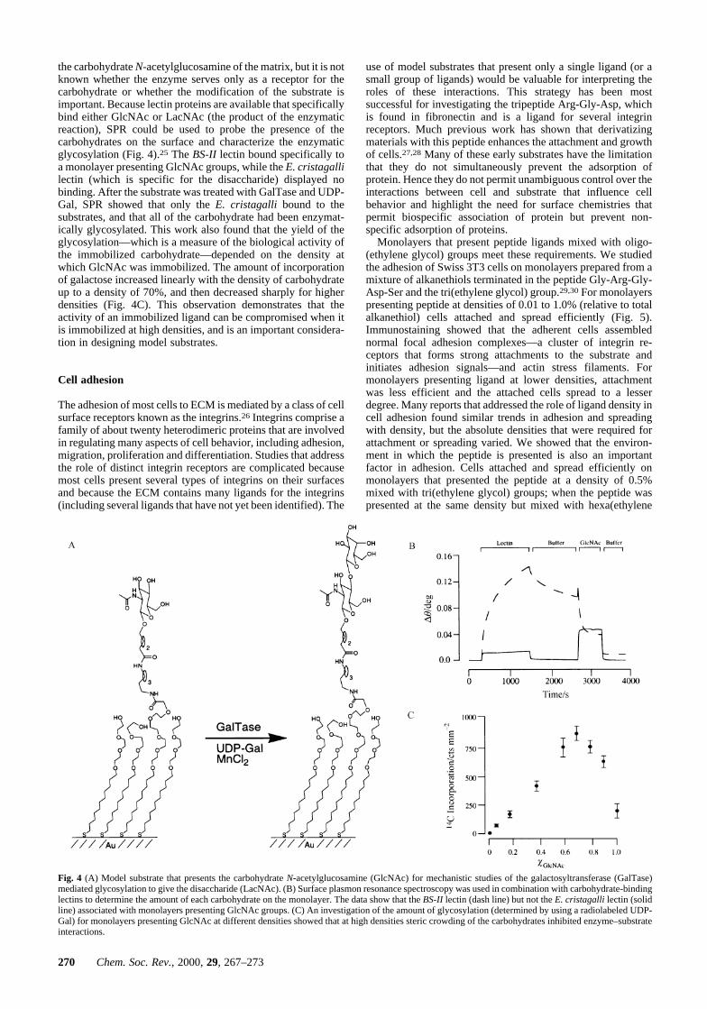
the carbohydrate N-acetylglucosamine of the matrix, but it is notknown whether the enzyme serves only as a receptor for thecarbohydrate or whether the modification of the substrate isimportant. Because lectin proteins are available that specificallybind either GlcNAc or LacNAc (the product of the enzymaticreaction), SPR could be used to probe the presence of thecarbohydrates on the surface and characterize the enzymaticglycosylation (Fig. 4).25 The BS-II lectin bound specifically toa monolayer presenting GlcNAc groups, while the E. cristagallilectin (which is specific for the disaccharide) displayed nobinding. After the substrate was treated with GalTase and UDP-Gal, SPR showed that only the E. cristagalli bound to thesubstrates, and that all of the carbohydrate had been enzymat-ically glycosylated. This work also found that the yield of theglycosylation—which is a measure of the biological activity ofthe immobilized carbohydrate—depended on the density atwhich GlcNAc was immobilized. The amount of incorporationof galactose increased linearly with the density of carbohydrateup to a density of 70%, and then decreased sharply for higherdensities (Fig. 4C). This observation demonstrates that theactivity of an immobilized ligand can be compromised when itis immobilized at high densities, and is an important considera-tion in designing model substrates.
Cell adhesion
The adhesion of most cells to ECM is mediated by a class of cellsurface receptors known as the integrins.26 Integrins comprise afamily of about twenty heterodimeric proteins that are involvedin regulating many aspects of cell behavior, including adhesion,migration, proliferation and differentiation. Studies that addressthe role of distinct integrin receptors are complicated becausemost cells present several types of integrins on their surfacesand because the ECM contains many ligands for the integrins(including several ligands that have not yet been identified). The
use of model substrates that present only a single ligand (or asmall group of ligands) would be valuable for interpreting theroles of these interactions. This strategy has been mostsuccessful for investigating the tripeptide Arg-Gly-Asp, whichis found in fibronectin and is a ligand for several integrinreceptors. Much previous work has shown that derivatizingmaterials with this peptide enhances the attachment and growthof cells.27,28 Many of these early substrates have the limitationthat they do not simultaneously prevent the adsorption ofprotein. Hence they do not permit unambiguous control over theinteractions between cell and substrate that influence cellbehavior and highlight the need for surface chemistries thatpermit biospecific association of protein but prevent non-specific adsorption of proteins.
Monolayers that present peptide ligands mixed with oligo-(ethylene glycol) groups meet these requirements. We studiedthe adhesion of Swiss 3T3 cells on monolayers prepared from amixture of alkanethiols terminated in the peptide Gly-Arg-Gly-Asp-Ser and the tri(ethylene glycol) group.29,30 For monolayerspresenting peptide at densities of 0.01 to 1.0% (relative to totalalkanethiol) cells attached and spread efficiently (Fig. 5).Immunostaining showed that the adherent cells assemblednormal focal adhesion complexes—a cluster of integrin re-ceptors that forms strong attachments to the substrate andinitiates adhesion signals—and actin stress filaments. Formonolayers presenting ligand at lower densities, attachmentwas less efficient and the attached cells spread to a lesserdegree. Many reports that addressed the role of ligand density incell adhesion found similar trends in adhesion and spreadingwith density, but the absolute densities that were required forattachment or spreading varied. We showed that the environ-ment in which the peptide is presented is also an importantfactor in adhesion. Cells attached and spread efficiently onmonolayers that presented the peptide at a density of 0.5%mixed with tri(ethylene glycol) groups; when the peptide waspresented at the same density but mixed with hexa(ethylene
Fig. 4 (A) Model substrate that presents the carbohydrate N-acetylglucosamine (GlcNAc) for mechanistic studies of the galactosyltransferase (GalTase)mediated glycosylation to give the disaccharide (LacNAc). (B) Surface plasmon resonance spectroscopy was used in combination with carbohydrate-bindinglectins to determine the amount of each carbohydrate on the monolayer. The data show that the BS-II lectin (dash line) but not the E. cristagalli lectin (solidline) associated with monolayers presenting GlcNAc groups. (C) An investigation of the amount of glycosylation (determined by using a radiolabeled UDP-Gal) for monolayers presenting GlcNAc at different densities showed that at high densities steric crowding of the carbohydrates inhibited enzyme–substrateinteractions.
270 Chem. Soc. Rev., 2000, 29, 267–273

glycol) groups, fewer cells attached and those that did remainedin a rounded morphology. For all these experiments, cells thatwere cultured on the substrates for 24 hours could be detachedby the addition of a soluble Arg-Gly-Asp peptide, demonstrat-ing that the interactions of cells with the substrates weremediated entirely by the peptide ligand.
These early studies validate the use of SAMs that presentligands for mechanistic studies of adhesion-dependent cellbehavior. With these monolayers, the interactions between asubstrate and cell can be defined completely and maintainedover days in culture. This property will be important fordetermining the influences that other ligands in ECM have onattached cells. A current example comes from studies of thespreading of baby hamster kidney cells on fibronectin.31 Bycomparing adhesion of cells on substrates coated with frag-ments and mutants of fibronectin, it was found that Arg-Gly-Asp alone is insufficient for complete spreading of cells, but thatthe presence of a second peptide, Pro-His-Ser-Arg-Asn, in anadjacent domain permits cells to attach and spread on thesubstrate. The use of protein-coated substrates makes it difficultto make quantitative comparisons of the adhesions on the twosubstrates (for the reasons indicated in Fig. 1). This problemhighlights the opportunity to use model substrates that presentpeptides at controlled densities for mechanistic studies ofadhesion. Indeed, many other peptide ligands have beensuggested to play a role in adhesion, and the monolayersdescribed here would be valuable in determining the properties
of these candidate ligands. In addition to characterizing theproperties of candidate ligands, SAMs may find use in rapidlyidentifying new ligands. This combinatorial screen could beperformed with substrates that are patterned into hundreds ofregions, each of which presents a single peptide ligand at aconstant density.
4 Dynamic substrates
This section discusses the development of dynamic substrates,wherein the activity of immobilized ligands can be modulated inreal time. Such substrates would provide new opportunities formechanistic studies of the pathways by which cells respond tochanges in their environments. Dynamic changes in ECMinfluence cell behavior in many important contexts, includingmigration and differentiation of cells during development andthe metastasis of tumor cells. But studies of these processes aredifficult due to the lack of methods that can unambiguouslychange the properties of ECM underlying a cell.
The development of dynamic substrates begins with thedesign of chemical strategies that can alter the composition ofactive ligands on a substrate. These changes can be caused byisomerization of an immobilized ligand between a structure thatis active and one that is inactive. Alternatively, a ligand can be‘turned on’ by activating the substrate for immobilization of
Fig. 5 (A) Structure of a monolayer that presents the peptide Gly-Arg-Gly-Asp-Ser mixed with tri(ethylene glycol) groups. (B) Optical micrograph of 3T3fibroblasts attached to a monolayer wherein 0.5% of the alkanethiolates present the peptide ligand. (C) Fluorescent micrograph of a cell that was adherenton these monolayers for five hours, fixed and stained with phalloidin–rhodamine to visualize actin stress filaments. The cells assembled stress fibers that werecharacteristic of those found in cells adhered to fibronectin.
Chem. Soc. Rev., 2000, 29, 267–273 271

ligand from solution and, by analogy, ‘turned off’ by releasingit from the substrate. Each of these strategies relies on achemical reaction at the interface. Because the substrates mustbe modulated in real time, the reactions should be initiated bynon-invasive means, using either photochemical or electro-chemical stimuli.
Substrates that immobilize ligands
We developed an electroactive substrate that can turn on theselective immobilization of ligands. The design takes advantageof the ability to carry out electrochemical conversions ofmolecules attached to the monolayers by applying electricalpotentials to the underlying gold. The strategy is based on theDiels–Alder reaction of a substituted cyclopentadiene withbenzoquinone attached to the SAM.32 The reaction of these twomolecules is rapid and selective and gives a covalent attachmentof ligand. The reaction proceeds with well-behaved secondorder kinetics and therefore gives excellent control over thedensity of immobilized ligand, even at low densities wheredirect characterization is not feasible. The reactivity of themonolayer can be turned off by electrochemical reduction thatconverts the quinone to the corresponding hydroquinone, whichis unreactive towards the diene. Because the oxidation ofhydroquinone is reversible over many cycles, it is possible torepeatedly turn the reactivity of the monolayer on and off. Wehave used this chemistry to demonstrate the immobilization ofbiotin for recognition by the protein streptavidin and of thepeptide RGD for the attachment of cells (Fig. 6).32
Substrates that release groups
A similar strategy can be employed to design redox activegroups that can selectively release attached ligands from amonolayer. The alkanethiol requires a molecule that undergoesoxidation (or reduction) to give an intermediate that thenrearranges to release a substituent that tethers the ligand. Wedemonstrated one strategy that used a catechol orthoformategroup as the electroactive linker that tethered the ligand to theSAM.33 Electrochemical oxidation of this group occurred at apotential of 850 mV (versus Ag/AgCl reference) and gave thecorresponding orthoquinone with concomitant hydrolysis, andrelease, of the formate substituent. The conversion wasessentially complete on a single cyclic voltammetric scanshowing that it was rapid and efficient. This reaction, however,
was not suited for the design of culture substrates that couldrelease ligands because application of potentials greater than600 mV (or greater than 2750 mV for reductions) compromisesthe ability of glycol-terminated monolayers to prevent proteinadsorption. Clearly, the development of reactive groups forincorporation into dynamic SAMs must meet several require-ments to be competent for studies involving attached cellculture.
Substrates that isomerize ligands
Willner and coworkers developed a class of photoactivesubstrates that modulate the recognition of a dinitrophenylantigen by an IgG antibody.34 These workers immobilized adinitrophenyl spiropyran on a gold film. When the phenoloxygen was bound in the spiropyran, the aromatic antigen wasnot available to bind antibody. Illumination of the spiropyranwith light at 370 nm resulted in the isomerization of an olefinand ring opening of the pyran to reveal the dinitrophenyl groupwhich was recognized by an antibody. Subsequent illuminationof this substrate at 500 nm returned the phenol to the spiropyranand eliminated binding of the antigen. Because this photo-isomerization is reversible, the activity of the ligand could bealternately turned on and off many times.
These three examples represent the level of control that canbe applied to the preparation of active substrates. Each of thesesubstrates was designed from first principles to exhibit theparticular property. The combination of physical organicprinciples and synthetic chemistry makes possible the molec-ular engineering of substrates that have a range of otherproperties.
5 Conclusions and outlook
This review illustrates the motivation and rationale for usingself-assembled monolayers as model substrates for studies inexperimental cell biology. SAMs have several characteristicsthat make them practical and important tools for a broad rangeof studies. They are easily prepared in ordinary laboratoriesfrom alkanethiols and gold-coated substrates. The peptide-terminated alkanethiols are now accessible by a straightforwardsolid-phase synthesis29 but the gold-coated substrates stillrequire access to metal evaporators. The most importantcharacteristic of these monolayers is the availability of surfaces
Fig. 6 Illustration of a dynamic substrate that can turn on the immobilization of a biotin ligand and permit the protein streptavidin to associate with themonolayer. (A) Reversible electrochemical oxidation of a monolayer that presents hydroquinone mixed with oligo(ethylene glycol) groups affords amonolayer that presents the quinone group. The quinone in turn reacts with a conjugate of cyclopentadiene and biotin (Biotin–Cp) to give a covalent Diels–Alder adduct. (B) SPR showed that streptavidin associated with a monolayer that had been activated in this way (curve for biotin/EG4) but not to monolayersthat presented only oligo(ethylene glycol) groups (EG4) or to monolayers that had not been activated by conversion to the quinone prior to treatment withthe conjugate.
272 Chem. Soc. Rev., 2000, 29, 267–273

that are inert to the adsorption of protein because they providea route to installing biospecific interactions at the interface.These monolayers also have the stability and properties requiredto be used in cell culture. Microcontact printing provides areliable and simple method for patterning substrates to controlthe shapes, sizes and positions of adherent cells. The ability toassemble monolayers from alkanethiols terminated in a widevariety of groups and to modify the groups after the monolayershave assembled makes possible the design of monolayershaving tailored properties, including those that can modulate theactivities of ligands that interact with receptors of an attachedcell.
The examples described in this review illustrate early ways inwhich SAMs are proving useful as model substrates in cellbiology. This approach will be valuable for countless otherstudies that elucidate the interactions of cellular receptors withextracellular matrix that underlie cell proliferation, migrationand differentiation. The use of patterned substrates—includingthose that position several different cell types on a commonsubstrate—will provide new opportunities to study the mecha-nisms by which cells influence one another, including forexample, in the course of development and formation of tissuesand organs. New methods that can immobilize ligands ingradients will be valuable for studies of cell polarization andmigration.35
These characteristics that make SAMs of alkanethiolates ongold excellent substrates for mechanistic cell biology also makethem important in biotechnology. Assays for screening drugcandidates in discovery programs and for analyzing samples inmedical diagnostics are increasingly using engineered cells asthe sensing element.36,37 These efforts have emphasized theneed to control the surface chemistry to ensure the viability ofcells and the reproducibility of data generated in the assays.With the miniaturization of the assay formats there is a need topattern the locations of cells to prevent them from migrating andproliferating on the substrate. The engineering community has agrowing interest in microfabricated devices that join cellularcomponents with sophisticated integrated circuits that are nowavailable. These devices—which are collectively referred to asbio-microelectromechanical systems—will require active sur-face chemistries that can interface the biological functions of acell with electrical processes in the device and may find use asnew sensors and as implantable neural prostheses.
Each of these research areas is at an early stage ofdevelopment and is certain to witness significant growth duringthe next decade. Surface chemists will play an important role inthis emerging field by providing the molecular level engineer-ing to design the substrates, developing synthetic approaches tobuild the substrates and applying a range of analytical andphysical methods to characterize the structures and properties ofthe substrates. The combined efforts of surface chemists, cellbiologists and engineers will surely make exciting contributionsto fundamental biology and to biotechnology.
6 Acknowledgements
I am grateful for the generous support of our work by theNational Institutes of Health, the National Science Foundation,and the Defense Advanced Research Projects Agency.
7 References
1 N. Boudreau and M. Bissell, Curr. Opin. Cell Biol., 1998, 10, 641.2 A. Huttenlocher, R. R. Sandborg and A. F. Horwitz, Curr. Opin. Cell
Biol., 1995, 7, 697.3 V. B. Fainerman, E. Lucassen-Reynders and R. Miller, Colloids Surf. A,
1998, 143, 141.4 J. J. Ramsden, Chem. Soc. Rev., 1995, 24, 73.5 R. R. Seigel, P. Harder, R. Dahint, M. Grunze, F. Josse, M. Mrksich and
G. M. Whitesides, Anal. Chem., 1997, 69, 3321.6 S. P. Palecek, J. C. Loftus, M. H. Ginsberg, D. A. Lauffenburger and
A. F. Horwitz, Nature, 1997, 385, 537.7 A. J. Garcia, M. D. Vega and D. Boettiger, Mol. Biol. Cell, 1999, 10,
785.8 A. Ulman, Chem. Rev., 1996, 96, 1553.9 L. H. Dubois and R. G. Nuzzo, Annu. Rev. Phys. Chem., 1992, 43,
437.10 K. L. Prime and G. M. Whitesides, J. Am. Chem. Soc., 1993, 115,
10 714.11 M. Mrksich, G. S. Sigal and G. M. Whitesides, Langmuir, 1995, 11,
4383.12 G. S. Sigal, M. Mrksich and G. M. Whitesides, J. Am. Chem. Soc., 1998,
120, 3464.13 Z. Salamon, M. F. Brown and G. Tollin, Trends Biochem. Sci., 1999, 24,
213.14 K. Torimitsu, Mater. Sci. Forum, 1997, 250, 69.15 M. Mrksich and G. M. Whitesides, Trends Biotechnol., 1995, 13,
228.16 M. Mrksich, L. E. Dike, J. Y. Tien, D. E. Ingber and G. M. Whitesides,
Exp. Cell Res., 1997, 235, 305.17 C. S. Chen, M. Mrksich, S. Huang, G. M. Whitesides and D. E. Ingber,
Science, 1997, 276, 1345.18 S. Huang, C. S. Chen and D. E. Ingber, Mol. Biol. Cell, 1998, 9,
3179.19 M. Mrksich, J. R. Grunwell and G. M. Whitesides, J. Am. Chem. Soc.,
1995, 117, 12 009.20 J. Rao, L. Yan, B. Xu and G. M. Whitesides, J. Am. Chem. Soc., 1999,
121, 2629.21 G. B. Sigal, C. Bamdada, A. Barberis, J. Strominger and G. M.
Whitesides, Anal. Chem., 1996, 68, 490.22 J. Spinke, M. Liley, H. J. Guder, L. Angermaier and W. Knoll,
Langmuir, 1993, 9, 1821.23 V. H. Perez-Luna, M. J. O’Brien, K. A. Opperman, P. D. Hampton,
G. P. Lopez, L. A. Klumb and P. S. Stayton, J. Am. Chem. Soc., 1999,121, 6469.
24 D. M. Disley, D. C. Cullen, H. X. You and C. R. Lowe, Biosens.Bioelectron., 1998, 13, 1213.
25 B. T. Houseman and M. Mrksich, Angew. Chem., Int. Ed., 1999, 38,782.
26 D. O. Schlaepfer and T. Hunter, Trends Cell Biol., 1998, 8, 151.27 E. Ruoslahti, Annu. Rev. Cell Dev. Biol., 1996, 12, 697.28 S. P. Massia and J. A. Hubbell, J. Cell Biol., 1991, 114, 1089.29 B. T. Houseman and M. Mrksich, J. Org. Chem., 1998, 63, 7552.30 C. Roberts, C. S. Chen, M. Mrksich, V. Martichonok, D. E. Ingber and
G. M. Whitesides, J. Am. Chem. Soc., 1998, 120, 6548.31 S. Aota, M. Nomizu and K. M. Yamada, J. Biol. Chem., 1994, 269,
24 756.32 M. N. Yousaf and M. Mrksich, J. Am. Chem. Soc., 1999, 121, 4286.33 C. D. Hodneland and M. Mrksich, Langmuir, 1997, 13, 6001.34 I. Willner, R. Blonder and A. Dagan, J. Am. Chem. Soc., 1994, 116,
9365.35 C. L. Hypolite, T. L. McLernon, D. N. Adams, K. E. Chapman, C. B.
Herbert, C. C. Huang, M. D. Distefano and W.-S. Hu, Chem. Biol.,1997, 8, 658.
36 G. J. Ding, P. A. Fischer, R. C. Boltz, J. A. Schmidt, J. J. Colaianne, A.Gough, R. A. Rubin and D. K. Miller, J. Biol. Chem., 1998, 273,28897.
37 D. A. Borkholder, J. Bao, N. I. Maluf, E. R. Perl and G. T. A. Kovacs,J. Neurosci. Methods, 1997, 77, 61.
Chem. Soc. Rev., 2000, 29, 267–273 273