Maxillary restoration with complete maxillary prosthesis ...

Case ReportA Suggested Approach of Managing Excessive Maxillary GingivalDisplay in Terminal Dentition
Daniel Saad ,1,2 Celine Moukarzel,1 Naim El Haddad,1 and Anthony Rizk1
1Postgraduate Department, MOI, Faculty of Dentistry, Goethe University, Frankfurt, Germany2Private Practice, Beirut, Lebanon
Correspondence should be addressed to Daniel Saad; [email protected]
Received 15 February 2020; Revised 20 September 2020; Accepted 9 October 2020; Published 20 November 2020
Academic Editor: Anastasios Markopoulos
Copyright © 2020 Daniel Saad et al. This is an open access article distributed under the Creative Commons Attribution License,which permits unrestricted use, distribution, and reproduction in any medium, provided the original work is properly cited.
The aim of this paper is to report a suggested approach for the management of excessive maxillary gingival display with terminaldentition. A segmental osteotomy of the maxillary process was performed, and the latter used as grafting material for lateral sinusaugmentation that was performed simultaneously. Following the graft maturation period, implants were inserted and rehabilitatedwith a fixed dentogingival prosthesis. Consequently, the mandible was prosthetically restored following the new occlusal planedictated by the rehabilitated maxilla. Clinically, the procedure showed a drastic improvement in the patient’s appearance,eliminating the excessive gingival display. Radiologically, it led to a vertical translation of the maxillary process level in an apicaldirection. Nevertheless, the resected process used as grafting material was noticed to have a suboptimal behavior as long as itshowed increased intrasinusal resorption, barely sufficient for a regular implant accommodation. The described therapy conceptseems to be a plausible approach when it comes to manage excessive maxillary gingival displays in edentulous patients or thosepresenting a terminal dentition. However, at the time of sinus augmentation, authors recommend to graft a mixture of resectedmaxillary process and a bone substitute material, in order to get more stable results.
1. Introduction
Nowadays, social media prevalence interferes with everyaspect of one’s life. Many bloggers and social media influen-cers are dictating the way of living by keeping up a stylish life-style. Consequently, a person’s perfect appearance isbecoming a must in society. Hence, dentists are increasinglysolicited to maintain their patients’ glamourous smileswhereas the business of laminate veneers and smile make-overs is flourishing.
One of many stigmas encountered while smiling is whatis commonly known as “gummy smile.” A “gummy smile”refers to an unpleasant excessive maxillary gingival displaywhile smiling. Its clinical correction starts by establishingthe right diagnosis in order to find the appropriate treatment.First, an exhaustive facial examination is necessary to deter-mine the facial and lateral proportions and symmetry, as wellas the smile line, gum exposure, and lip length. Afterward, anintraoral examination is required for the teeth anatomy, thebite plane, and the periodontal state [1].
Determining the etiology of the excessive gingival displayis the next step in the management of such cases. Herein willbe presented several diagnoses implicated in the latter.
The first diagnosis is drug and plaque-induced gingivalhypertrophy. In this case, gingival inflammation due to pla-que or medical treatments—immunosuppressant drugs suchas cyclosporine, antiepileptic drugs such as phenytoin, andcalcium channels blockers such as nifedipine [2]—caninduce a hyperplastic-overgrown gingiva covering the maxil-lary crowns, hence, causing excessive gingival display. Main-taining impeccable oral hygiene may be the treatment ofchoice; however, surgical intervention can be indicated inspecific cases.
Another mechanism for excessive gum display is origi-nated from an altered passive eruption. In fact, teeth eruptionis divided into 2 stages. The first, known as active eruption,consisting of the tooth migration to the occlusal plane,whereas the second stage, known as passive eruption, is dueto the migration of the gingiva apically to uncover the tooth.When the passive eruption is altered, the gingiva will still
HindawiCase Reports in DentistryVolume 2020, Article ID 6975275, 11 pageshttps://doi.org/10.1155/2020/6975275

cover the tooth, thus causing shorter clinical crowns and insome cases excessive gingival display. Colset et al. [3] classi-fied the altered passive eruption into two types each havingtwo subtypes:
(i) Type 1-A: normal distance between cementoenameljunction (CEJ) and alveolar crest with an excessiveamount of keratinized tissue and it can be treatedby gingivectomy
(ii) Type 1-B: CEJ and alveolar crest at the same levelwith excessive amount of keratinized tissue, it canbe treated by gingivectomy and osseous reduction
(iii) Type 2-A: normal distance between CEJ and alveolarcrest with a normal amount of keratinized tissue, itcan be treated by apically positioned flap
(iv) Type 2-B: CEJ and alveolar crest at the same levelwith a normal amount of keratinized tissue, it canbe treated by osseous reduction and apically posi-tioned flap
Furthermore, a third cause would be vertical maxillaryexcess (VME). VME can be defined as a disproportionalgrowth of the maxilla in the vertical direction, which couldlead to a long face syndrome [4]. VME is generally associatedwith an open bite; however, when associated with a normalbite, an excessive gingiva display would hence be coveringthe incisal edge of the canines [5]. Garber and Salama classi-fied VME into 3 degrees:
(i) Degree 1: excessive gingival display of 2-4mm and itcan be treated by orthodontic intrusion, crownlengthening, or botulinum toxin injection
(ii) Degree 2: excessive gingival display of 4–8mm and itcan be treated by [6] lip stabilization technique ororthognathic surgery
(iii) Degree 3: excessive gingival display of more than8mm and can only be treated by orthognathicsurgery
A fourth etiology is a short or hypermobile lip. In fact,short upper lips with less than 15mm of distance betweenthe lower border of the upper lip and the subnasale [7], orhypermobile lips caused by excessive contractions of the lipelevator muscles, can induce an excessive gingival display.This condition may be treated by the lip stabilization tech-nique [6]. When there is a gingival exposure of 1 to 3mm,an injection of botulinum toxin is preferred every 6 monthsto limit the lip elevator activity.
Treatment of excessive gingival display requires a multi-disciplinary approach to achieve successful aesthetic andfunctional results. While the treatment of gummy smile fordentulous patients represents some challenges, albeit moreobstacles are encountered with edentulous patients, in orderto achieve desirable outcomes.
Complete edentulous upper jaws can be rehabilitated byeither conventional removable dentures, or by means of
implant-supported prostheses. In the latter, it consists eitherof a removable implant-supported denture with extendedvestibular flanges or a flange-free cross-arch fixed implant-retained prosthesis with (hybrid design) or without (crowndesign) pink ceramic [8]. Consequently, in order to decideon the optimal therapeutic option regarding the prostheticdesign, a diligent complex and multifactorial treatment plan-ning should be considered, which takes into account the vol-ume of the hard and the soft tissue to be compensated, as wellas the emergence profile of the future prosthetic teeth alongwith their vertical, horizontal, and sagittal positions, and pro-vides at once an adequate lip support and balanced facial har-mony [8, 9]. Within this scope, a benchmark article [8]addressing this matter was published in 2013 where authorstried to establish a comprehensive decision-making tree withregards to implant-supported prosthesis selection based onthe aforementioned patient’s clinical characteristics. Thepublished work documented exhaustively different clinicalsituations of deficient edentulous maxillae. What it did notaddress is the situations where edentulous maxillae areprominent and excessively visible when smiling.
This paper describes an implant-supported prostheticrehabilitation of an excessively prominent maxillary processwith terminal dentition.
2. Case Presentation
In February 2018, one female patient stepped into the privateclinic for a complete oral rehabilitation after years ofneglected oral care. The patient, who is 58 years old, wasseeking for a global dental care and a solution for her exces-sive gingival display. Intraoral examination revealed a termi-nal maxillary dentition. Radiographic examination showedan impacted canine #23. In the mandible, apart the presentnatural anterior teeth, radiologic examination showed thepresence of three restored implants on the right side andfailed bridge over hopeless teeth on the patient’s left side.Extraoral examination revealed a VME reflected by an exces-sive gingival display at smiling, showing unpleasantly thewhole maxillary process all the way up to the tuberosities(Figure 1).
The following treatment plan was agreed on;Maxilla: extraction of the remaining teeth with alveolar
segmental reduction along with a bilateral sinus floor eleva-tion, followed by a maxillary rehabilitation by means of aToronto prosthesis [10] retained by 6 implants also knownas abutment-hybrid overdenture.
Mandible: crowning of the anterior teeth, restoring theleft side by implant-supported bridge, and a new set ofcrowns over the present implants on the right.
Treatment began by rehabilitating the maxilla:First, segmental alveolar osteotomy was performed on
the right hemimaxilla to remove the vertical maxillary excessusing Piezosurgery-touch (Mectron, Cherasco, Italy). Theintervention carried out at once a lateral sinus floor elevation,where resected maxillary crest after being crushed, wasgrafted inside the sinus. Simultaneously, an implant (Duravit3P, B&B Dental Implant Company, Bologna, Italy) wasinserted in the region of the right canine. Then, the buccal
2 Case Reports in Dentistry

flap was repositioned apically to preserve the keratinizedmucosa (Figure 2).
Two weeks later, a similar procedure was performed onthe left hemimaxilla along with the extraction of theimpacted canine. Here, the implant (Duravit EV, B&BDentalImplant Company, Bologna, Italy) was inserted partially inthe canine socket, which was filled with the crushed resectedalveolar process. The right maxillary subantral space waspacked first with bovine xenograft (Ti-Oss, Gyeonggi-do,South Korea) towards the back and then filled with theremaining autogenous resected ridge anteriorly (Figure 3).
After a 2-week healing period, serving as temporary pros-thesis during the maturity phase of the grafted sinuses, aremovable denture was delivered which was retained by thetwo inserted implants through their respective transitionalimplant overdenture attachments (OT equator, Rhein83,Bologna, Italy) (Figure 4).
6 months later, two implants (Duravit EV, B&B DentalImplant Company, Bologna, Italy) were inserted in each ofthe sinuses and left submerged for a two-month period(Figure 5).
After 2 months, an open tray impression of the maxillawith its six osseointegrated implants was taken using poly-ether impression material (Impregum, 3M, Minneapolis,USA), then the poured master cast was scanned with therespective scanbodies in place. Appropriate multiunit abut-ment was selected for 5 out of the 6 implants, and that forthe ease of insertion of the future prosthesis. Consequently,an intermediate Chrome-Cobalt bar was 3D printed usingSelective Laser Melting (SLM) technology (SISMA spa,Vicenza, Italy) to check intraorally the passivity fit, whichwas found to be satisfactory. A diagnostic wax setup using
resin denture teeth was then built over the intermediate barand tried-in intraorally for functional and esthetic evaluationas well as patient approval. The occlusal plane was set parallelto the Camper plane, regardless of the occlusal curve of themandibular dentition. Then, the diagnostic setup wasscanned in-lab, and the final framework of the mesostructureand of its individual crowns was virtually designed.
The mesostructure and the 8 posterior individual crowns’frameworks were 3D printed (SLM) in Chrome-Cobalt alloy,while 6 anterior individual crowns were milled out of zirco-nia for improved esthetics.
The mesostructure was then manually veneered with gin-gival pink porcelain (VC gingiva, CreationWilli Geller Inter-national GmbH, Meiningen, Austria), while the crowns werebuilt-up with veneering ceramic (Creation Willi Geller Inter-national GmbH, Meiningen, Austria), ending up into ahybrid prosthesis. The said prosthesis was screwed-in safelydirectly over one implant and indirectly through the 5 under-lying multiunit abutments, and the individuals crownscemented on its top subsequently (Figure 6).
The mandible underwent a more straightforward treat-ment plan, which is out of the scope of this report. Concisely,the roots of the teeth #34, #36, and #37 were extracted; then, aflap was raised showing buccal bone deficiency. 3 implants(Rootform, ROOTT, Trate AG, Baech, Switzerland) wereinserted along with autogenous horizontal bone augmenta-tion following the principles of the split bone block technique[11]. The bony plates were fixed using titanium microscrews(Conmet LLC , Moscow, Russia). The whole augmentationwas then covered by a synthetic resorbable membrane (Tis-seos, Biomedical Tissues, Nantes, France). Two months later,implants on both sides were restored prosthetically, with
(a) (b)
(c) (d)
Figure 1: Baseline: (a–c) clinical situation of the patient at the baseline. (d) Radiological situation at the baseline.
3Case Reports in Dentistry

(a) (b)
(c) (d)
(e) (f)
(g) (h)
Figure 2: Left hemimaxilla: (a, b) segmental osteotomy of the alveolar process. (c) Lateral window approach during left maxillary sinus floorelevation. (d) Crushed resected alveolar process grafted inside the sinus. (e) Picture showing lateral bony window placed back and implantinserted in the canine region. (f–h) Respectively: frontal, lateral, and occlusal view of the postoperative clinical situation immediately aftersuturing.
4 Case Reports in Dentistry

(a) (b)
(c) (d)
(e) (f)
(g) (h)
Figure 3: Right hemimaxilla: (a, b) clinical situation 2 weeks following left hemimaxilla resection and immediately before proceeding with theright hemimaxilla. (c) Segmental osteotomy of the right hemimaxilla. (d) Impacted canine extraction. (e) Implant insertion in the remainingcanine socket. (f) Jumping space filled with autogenous resected alveolar process. (g) Sinus floor elevation. (h) Occlusal view of thepostoperative clinical situation.
5Case Reports in Dentistry

porcelain fused to metal crowns taking into consideration anadequate vertical dimension. The anterior teeth underwent a
root canal treatment and received 6 splinted zirconia crowns(Figure 7).
(a) (b) (c)
(d) (e)
Figure 4: Temporization, 2-3 weeks following right hemimaxilla segmental resection: (a) maximum smile showing the successfullyeliminated gingival display. (b) Occlusal view of the healed maxilla with two protruding implant overdenture attachments. (c) Respectivehousings mounted over the overdenture attachments to be picked-up in the denture intrados by a relining procedure. (d) Denture readyto be seated over the attachments. (e) Patient new smile with the retained denture in the mouth.
(a) (b)
(c) (d) (e)
Figure 5: Following sinus graft maturation: (a) Panoramic x-ray 6months following segmental osteotomy along bilateral sinus augmentation.(b) Panoramic x-ray, 8-months following segmental osteotomy along with bilateral sinus augmentation, and 2 months after implantplacement in the sinuses region. (c, d) Two implant placement per each sinus 6-months following bilateral sinus floor elevation. (e)Occlusal view of the maxilla immediately after implant placement and flaps suturing.
6 Case Reports in Dentistry

3. Results
Clinically, the procedure showed a drastic improvement inthe patient’s appearance, eliminating the excessive gingivaldisplay. Radiologically, it led to a clear vertical translationof the maxillary process level in an apical direction(Figure 8). Nevertheless, the resected process used as graftingmaterial was noticed to have a suboptimal behavior as long asit showed increased intrasinusal resorption rate, barely suffi-cient for a regular implant accommodation.
4. Discussion
As discussed earlier, little is reported about managing exces-sive gingival display in edentulous patients. Generally, toothloss is accompanied by an unavoidable alveolar resorption[9]. This phenomenon, in the posterior region, is usuallyfollowed by a sinus pneumatization [12, 13]. Both facts con-tribute in leaving a minimal posterior ridge height, whichimpede a proper implant placement in this particular region.
In case of excessive gingival display, when the alveolarvertical reduction is an option, in order to minimize the
(a) (b) (c)
(d) (e) (f)
(g) (h) (i)
(j) (k) (l)
Figure 6: Final rehabilitation of the maxilla: (a, b) Pictures of the impression copings over their respective implants. (c) Passivity fit checkusing a milled bar. (d) Occlusal view of the maxilla with abutments off. (e) Occlusal view of the maxilla showing five straight multiunitabutments mounted over their respective implants. (f) Try-in of the final milled mesostructure. (g, h) Try-in of the mesostructure after itwas layered with pink ceramic. (i, j) Try-in of the individual crowns over the mesostructure. (k, l) Maxillary work completed.
7Case Reports in Dentistry

(a) (b)
(c) (d)
(e) (f)
(g) (h)
(i) (j)
Figure 7: Mandible: (a) Baseline situation of the left hemimandible. (b) Three implants insertion in position #34, #36, and #37. (c)Autogenous bone block harvesting from the external oblique line. (d) Harvested block split in two. (e) Bone block fixation in the recipientsite. (f) Autogenous bony particulates filled in the medullary zone. (g) Coverage of the augmentation area by a membrane. (h) Situation atuncovery, 2 months after the augmentation procedure. (i) Abutments and prepared teeth ready to receive their crowns. (j) Mandibularwork completed showing the bridges in place.
8 Case Reports in Dentistry

(a) (b)
(c)
(d) (e)
(f) (g)
(h) (i)
Figure 8: Comparisons. (a, b) Panoramic x-rays at the baseline and after case completion, respectively. (c) 2 overlayed maxillary x-rays,extracted from their respective panoramic (before and after segmental osteotomy) and superimposed to show the approximate differencebetween the baseline and the actual vertical level of the maxillary process. (d, e) Comparison between the baseline and after osteotomyshowing the vertical translation of the maxilla in an upward direction. (f, g) Pictures revealing maximum smile at the baseline and aftercase completion, respectively. (h, i) Additional pictures showing the final outcome.
9Case Reports in Dentistry
gummy smile, one may shave off totally the maxillary sinusfloors, especially if they are enough pneumatized.
In the present case, and according to their planning,authors reduced the alveolar process and rendered intention-ally the case from an excessive gummy smile to a class B sit-uation as per the classification of Avrampou et al. [8], thenmanaged the case accordingly by a fixed hybrid prosthesis.
The rationale behind that was mostly relying on hidingthe future mucosal-prosthesis junction away under the upperlip, not to be seen on the maximal smile, as well as giving therequired vertical space to fit in the components of theToronto Bridge, while providing enough room to accommo-date a certain height of pink ceramic. On the other hand,ridge reduction was limited by the remaining osseous capitalin the anterior region and by the sinuses laterally. Anteriorly,in the canine region, care was taken to leave a vertical heightof at least 10mm in order to insert properly and immediatelytwo regular implants. Posteriorly, since limited by thesinuses, the latter were grafted simultaneously by the resectedautogenous material in order to achieve a minimal subantralridge height able to receive endosseous implants.
Segmental maxillary ostectomy is discussed scarcely inthe literature, as a modality of treatment of excessive gingivaldisplay [14–16]. Nevertheless, to the extent of the authors’knowledge, the protocol described in this paper is being pro-posed for the first time:While correcting the gummy smile bysegmental osteotomy, the sinus floor elevation is performedconcomitally, taking advantage of the resected autogenousalveolar bone that is used as grafting material.
The treatment lasted over a period of a whole year, fromosteotomy with sinus floor elevation, throughout the graftmaturity, then osseointegration period of the delayedinserted implants, followed by the prosthetic workflow tillthe final prosthesis delivery, and lastly the mandibularrehabilitation.
Concerning the maxillary prosthetic design, it was optedto rehabilitate the six maxillary implants with a Torontoprosthesis, which consists of a staged fixed prosthesis com-prising a screw-retained framework (3D printed (SLM) inthis case) that replaces the missing jaw structure. It isdesigned in a way to be layered with pink material imitatingthe gum from which emerges a sort of abutment mimickingthe prepared teeth as in conventional fixed prosthodontics.As a second stage, come the individual crowns that areintended to be seated over the aforementioned abutments,forming a complex cemented/screw-retained prosthetic con-struction with the following main advantages:
(i) Ease of retrievability [10] of the concerned parts incase of maintenance in an event of a repair ofchipped ceramic while avoiding compromising theframework, provided that the crowns are cementedwith a temporary cement, and the underlyingmesostructure is screw-retained
(ii) Achievability of desired esthetics of the individualcrowns regardless of the screw access openings situ-ated in the underlying screw-retained framework[17]
(iii) Improved metal framework fitting over theimplants, related to the reduced number of repeatedporcelain firing over the framework itself, which isresponsible for metal shrinkage and a further misfit[18]
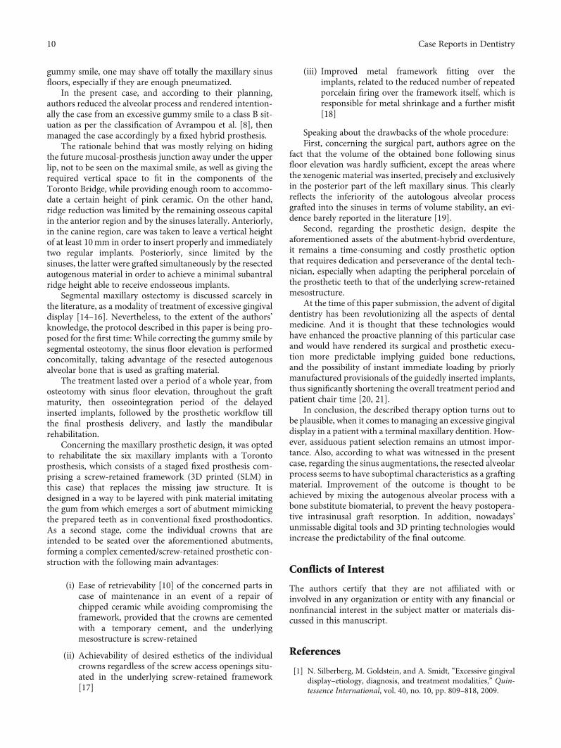
Speaking about the drawbacks of the whole procedure:First, concerning the surgical part, authors agree on the
fact that the volume of the obtained bone following sinusfloor elevation was hardly sufficient, except the areas wherethe xenogenic material was inserted, precisely and exclusivelyin the posterior part of the left maxillary sinus. This clearlyreflects the inferiority of the autologous alveolar processgrafted into the sinuses in terms of volume stability, an evi-dence barely reported in the literature [19].
Second, regarding the prosthetic design, despite theaforementioned assets of the abutment-hybrid overdenture,it remains a time-consuming and costly prosthetic optionthat requires dedication and perseverance of the dental tech-nician, especially when adapting the peripheral porcelain ofthe prosthetic teeth to that of the underlying screw-retainedmesostructure.
At the time of this paper submission, the advent of digitaldentistry has been revolutionizing all the aspects of dentalmedicine. And it is thought that these technologies wouldhave enhanced the proactive planning of this particular caseand would have rendered its surgical and prosthetic execu-tion more predictable implying guided bone reductions,and the possibility of instant immediate loading by priorlymanufactured provisionals of the guidedly inserted implants,thus significantly shortening the overall treatment period andpatient chair time [20, 21].
In conclusion, the described therapy option turns out tobe plausible, when it comes to managing an excessive gingivaldisplay in a patient with a terminal maxillary dentition. How-ever, assiduous patient selection remains an utmost impor-tance. Also, according to what was witnessed in the presentcase, regarding the sinus augmentations, the resected alveolarprocess seems to have suboptimal characteristics as a graftingmaterial. Improvement of the outcome is thought to beachieved by mixing the autogenous alveolar process with abone substitute biomaterial, to prevent the heavy postopera-tive intrasinusal graft resorption. In addition, nowadays’unmissable digital tools and 3D printing technologies wouldincrease the predictability of the final outcome.
Conflicts of Interest
The authors certify that they are not affiliated with orinvolved in any organization or entity with any financial ornonfinancial interest in the subject matter or materials dis-cussed in this manuscript.
References
[1] N. Silberberg, M. Goldstein, and A. Smidt, “Excessive gingivaldisplay–etiology, diagnosis, and treatment modalities,” Quin-tessence International, vol. 40, no. 10, pp. 809–818, 2009.
10 Case Reports in Dentistry

[2] N. Nakib and S. S. Ashrafi, “Drug-induced gingival over-growth,” Disease-a-Month, vol. 57, no. 4, pp. 225–230, 2011.
[3] J. G. Coslet, R. Vanarsdall, and A. Weisgold, “Diagnosis andclassification of delayed passive eruption of the dentogingivaljunction in the adult,” The Alpha Omegan, vol. 70, no. 3,pp. 24–28, 1977.
[4] S. A. Schendel, J. Eisenfeld, W. H. Bell, B. N. Epker, and D. J.Mishelevich, “The long face syndrome: vertical maxillaryexcess,” American Journal of Orthodontics, vol. 70, no. 4,pp. 398–408, 1976.
[5] D. A. Garber andM. A. Salama, “The aesthetic smile: diagnosisand treatment,” Periodontology, vol. 11, no. 1, pp. 18–28, 1996.
[6] M. Bhola, P. Fairbairn, S. Kolhatkar, S. Chu, T. Morris, andM. De Campos, “LipStaT: the lip stabilization technique - indi-cations and guidelines for case selection and classification ofexcessive gingival display,” The International Journal of Peri-odontics & Restorative Dentistry, vol. 35, no. 4, pp. 549–559,2015.
[7] I. Ahmad, “Anterior dental aesthetics: dentofacial perspec-tive,” British Dental Journal, vol. 199, no. 2, pp. 81–88, 2005.
[8] M. Avrampou, R. Mericske-Stern, M. B. Blatz, and J. Katsoulis,“Virtual implant planning in the edentulous maxilla: criteriafor decision-making of prosthesis design,” Clinical OralImplants Research, vol. 24, Supplement A100, pp. 152–159,2013.
[9] S. Hansson and A. Halldin, “Alveolar ridge resorption aftertooth extraction: a consequence of a fundamental principleof bone physiology,” Journal of Dental Biomechanics, vol. 3,2012.
[10] J. Montero, C. Macedo de Paula, and A. Albaladejo, “The"Toronto prosthesis", an appealing method for restoringpatients candidates for hybrid overdentures: a case report,”Journal of Clinical and Experimental Dentistry, vol. 4, no. 5,pp. e309–e312, 2012.
[11] F. Khoury and T. Hanser, “Mandibular bone block harvestingfrom the retromolar region: a 10-year prospective clinicalstudy,” International Journal of Oral & Maxillofacial Implants,vol. 30, no. 3, pp. 688–697, 2015.
[12] A. Sharan and D. Madjar, “Maxillary sinus pneumatizationfollowing extractions: a radiographic study,” The InternationalJournal of Oral &Maxillofacial Implants, vol. 23, no. 1, pp. 48–56, 2008.
[13] M. C. Cavalcanti, T. E. Guirado, V. M. Sapata et al., “Maxillarysinus floor pneumatization and alveolar ridge resorption aftertooth loss: a cross-sectional study,” Brazilian Oral Research,vol. 32, article e64, 2018.
[14] O. T. Jensen, M. W. Adams, J. R. Cottam, S. M. Parel, andW. R. Phillips, “The All-on-4 shelf: Maxilla,” Journal of OralandMaxillofacial Surgery, vol. 68, no. 10, pp. 2520–2527, 2010.
[15] A. S. Bidra, J. R. Agar, and S. M. Parel, “Management ofpatients with excessive gingival display for maxillary completearch fixed implant-supported prostheses,” The Journal of Pros-thetic Dentistry, vol. 108, no. 5, pp. 324–331, 2012.
[16] R. Calixto, F. Faleiros, E. Marangoni, F. Medeiros, A. Wady,and A. Paleari, “Surgical and prosthetic approach for oralrehabilitation of patient with excessive gingival display andgummy smile,” British Journal of Medicine & MedicalResearch, vol. 21, no. 11, pp. 1–7, 2017.
[17] B. Uludag, O. Ozturk, G. Celik, and G. Goktug, “Fabrication ofa retrievable cement- and screw-retained implant-supported
zirconium fixed partial denture: a case report,” The Journalof Oral Implantology, vol. 34, no. 1, pp. 59–62, 2008.
[18] J. Piermatti, “Using CAD-CAM technology for the full-mouth,fixed, retrievable implant restoration: a clinical report,” TheJournal of Oral Implantology, vol. 33, no. 1, pp. 23–27, 2007.
[19] W. Peng, I. K. Kim, H. Y. Cho et al., “Assessment of the autog-enous bone graft for sinus elevation,” Journal of the KoreanAssociation of Oral and Maxillofacial Surgeons, vol. 39, no. 6,pp. 274–282, 2013.
[20] M. A. Pikos, C. W. Magyar, and D. R. Llop, “Guided full-archimmediate-function treatment modality for the edentulousand terminal dentition patient,” The Compendium of Continu-ing Education in Dentistry, vol. 36, no. 2, 2015.
[21] F. Alzoubi, N. Massoomi, and A. Nattestad, “Bone reduction tofacilitate immediate implant placement and loading usingCAD/CAM surgical guides for patients with terminal denti-tion,” The Journal of Oral Implantology, vol. 42, no. 5,pp. 406–410, 2016.
11Case Reports in Dentistry