A STUDY ON THE ROLE OF SUPEROXIDE DISMUTASE ENTRAPPED
12
Volume: 2: Issue-1: Jan-Mar -2011 ISSN 0976-4550 A STUDY ON THE ROLE OF SUPEROXIDE DISMUTASE ENTRAPPED LIPOSOMES IN CIRCUMVENTING FREE RADICAL ONSLAUGHT IN RAT BRAIN Manju Antony* 1 , James TJ 2 1 Lecturer, Department on Life sciences, Kristu Jayanti College Bangalore-560077 2 Department of Zoology, Sacred Heart College Thevara Kerala ABSTRACT: The shift in equilibrium of free radical generation and antioxidant defence enzyme activity has been proved to be the reason for oxidative stress in cells. This fact suggested the need to supplement antioxidant defence enzymes to the cells to circumvent free radical onslaught. The present work deals with administration of the most important defense enzyme, superoxide dismutase into the brain to circumvent hypoxia associated pathological changes in brain. Due to the inability of the free enzyme to cross the blood brain barrier, this enzyme was entrapped in liposomes and was used as a carrier to smuggle past the blood brain barrier to reach the deeper brain regions. The administration of SOD entrapped liposomes brought about a significant increase in Superoxide dismutase and Glutathione peroxidase activity all the brain regions (P<0.05) and also succeeded in bringing down the lipid peroxidation levels in the rat brain. MDA content showed significant (P<0.05) decrease in all the brain regions. Analysis of ultra thin hippocampal sections of SOD administered-hypoxia subjected rat brain revealed few damaged mitochondria, and lesser degree of vacuolization, suggesting a decrease in oxidative injury. Key words: Hypoxia, Hippocampus, Superoxide dismutase, Lipid peroxidation Abbreviations: CBR- cerebrum, BL- Cerebellum, MED- Medulla, HC- Hippocampus HY- hypothalamus, SOD – Superoxide dismutase,GPx – Glutathione peroxidase, MDA- Malondialdehyde , N- Nucleus, M-Mitochondria, Mv-Vacuolated mitochondria, LG-Lipid granules, ER-Endoplasmic reticulum,V- Vacuoles, LY-Lysosomes,L-Liposomes INTRODUCTION Reactive oxygen species generated in body organs including brain comprise superoxide anion (O 2 - ), hydrogen peroxide (H 2 O 2 ), and hydroxyl radicals (.OH). Extremely high level of interest conferred recently on reactive oxygen species in neurobiology arises from a substantial body of information which suggest that they can be involved in post-hypoxic, post-ischemic and post-traumatic brain damage, in the ageing process and in the pathogenesis of several neurodegenerative disorders, Parkinson’s disease and Alzheimer’s disease 1 . The various reactive oxygen species exhibit different degrees of toxicity against their targets, which include lipids, proteins, nucleic acids and even carbohydrates. Lipid peroxidation and formation of peroxide radical and aldehydes damages membranes, membrane-bound enzymes and receptors. Attacks on proteins may need to their unfolding, fragmentation and polymerization, while damage to DNA can result in mutations or activation of certain enzymes. Antioxidant defense mechanism in the body involving defense enzymes superoxide dismutase, catalase and glutathione peroxidase; and other antioxidants like alpha-tocopherol and reduced glutathione plays an important role in keeping these deleterious radicals under check during normal conditions. An increase in the reactive oxygen species during post ischemia/hypoxia might have altered the equilibrium between the free radical generation and its scavenging system leading to cell injury. International Journal of Applied Biology and Pharmaceutical Technology Page: 61 Available online at www.ijabpt.com
Transcript of A STUDY ON THE ROLE OF SUPEROXIDE DISMUTASE ENTRAPPED
Volume: 2: Issue-1: Jan-Mar -2011 ISSN 0976-4550
A STUDY ON THE ROLE OF SUPEROXIDE DISMUTASE ENTRAPPED LIPOSOMES IN CIRCUMVENTING FREE RADICAL ONSLAUGHT IN RAT BRAIN
Manju Antony*1, James TJ2
1 Lecturer, Department on Life sciences, Kristu Jayanti College Bangalore-5600772 Department of Zoology, Sacred Heart College Thevara KeralaABSTRACT: The shift in equilibrium of free radical generation and antioxidant defence enzyme activity has been proved to be the reason for oxidative stress in cells. This fact suggested the need to supplement antioxidant defence enzymes to the cells to circumvent free radical onslaught. The present work deals with administration of the most important defense enzyme, superoxide dismutase into the brain to circumvent hypoxia associated pathological changes in brain. Due to the inability of the free enzyme to cross the blood brain barrier, this enzyme was entrapped in liposomes and was used as a carrier to smuggle past the blood brain barrier to reach the deeper brain regions. The administration of SOD entrapped liposomes brought about a significant increase in Superoxide dismutase and Glutathione peroxidase activity all the brain regions (P<0.05) and also succeeded in bringing down the lipid peroxidation levels in the rat brain. MDA content showed significant (P<0.05) decrease in all the brain regions. Analysis of ultra thin hippocampal sections of SOD administered-hypoxia subjected rat brain revealed few damaged mitochondria, and lesser degree of vacuolization, suggesting a decrease in oxidative injury. Key words: Hypoxia, Hippocampus, Superoxide dismutase, Lipid peroxidation
Abbreviations: CBR- cerebrum, BL- Cerebellum, MED- Medulla, HC- Hippocampus HY- hypothalamus, SOD – Superoxide dismutase,GPx – Glutathione peroxidase, MDA- Malondialdehyde , N-Nucleus, M-Mitochondria, Mv-Vacuolated mitochondria, LG-Lipid granules, ER-Endoplasmic reticulum,V-Vacuoles, LY-Lysosomes,L-Liposomes INTRODUCTIONReactive oxygen species generated in body organs including brain comprise superoxide anion (O2
-), hydrogen peroxide (H2O2), and hydroxyl radicals (.OH). Extremely high level of interest conferred recently on reactive oxygen species in neurobiology arises from a substantial body of information which suggest that they can be involved in post-hypoxic, post-ischemic and post-traumatic brain damage, in the ageing process and in the pathogenesis of several neurodegenerative disorders, Parkinson’s disease and Alzheimer’s disease1.The various reactive oxygen species exhibit different degrees of toxicity against their targets, which include lipids, proteins, nucleic acids and even carbohydrates. Lipid peroxidation and formation of peroxide radical and aldehydes damages membranes, membrane-bound enzymes and receptors. Attacks on proteins may need to their unfolding, fragmentation and polymerization, while damage to DNA can result in mutations or activation of certain enzymes. Antioxidant defense mechanism in the body involving defense enzymes superoxide dismutase, catalase and glutathione peroxidase; and other antioxidants like alpha-tocopherol and reduced glutathione plays an important role in keeping these deleterious radicals under check during normal conditions. An increase in the reactive oxygen species during post ischemia/hypoxia might have altered the equilibrium between the free radical generation and its scavenging system leading to cell injury.
International Journal of Applied Biology and Pharmaceutical Technology Page: 61 Available online at www.ijabpt.com

Antony et al ISSN 0976-4550
It has been observed by a previous experiment by the same author2 that rats when subjected to Isobaric hypoxia, showed drastic increase in lipid peroxidation levels in rat brain indicating free radical onslaught. Hippocampus region of the brain which observed the lowest level of superoxide dismutase enzyme activity observed the highest damage2. These findings point towards shift in equilibrium free radical generation and antioxidant defence enzymes and the need to supplement antioxidant defence enzymes to circumvent free radical onslaught. In the present work an attempt was made to circumvent hypoxia associated pathological changes in brain via administration of the most important defense enzyme, superoxide dismutase into the brain. Due to the inability of the free enzyme to cross the blood brain barrier, this enzyme was entrapped in liposomes and was used as a carrier to smuggle past the blood brain barrier to reach the deeper brain regions.Use of carriers for the transport of drugs to the target areas is now recognized as a promising method of improving drugs selectivity. Many types of carriers such as macromolecules, cells, viruses and synthetic particles have been proposed and, undoubtedly the usefulness of each of these will depend on particular needs. It is however apparent that, most known carriers are limited in the range and quantity of drugs they can accommodate and also on their ability to prevent contact of their drug moiety with the normal biological environment or to promote its access to areas in need of treatment. In addition, there are difficulties related to the toxicity of the carrier’s components, to their availability or cost and the preparation of the carrier -drug unit. All these drawbacks to a large extent overcome in liposomes. Since its discovery, it has gained wide acceptance as an efficient carrier in biological systems and has found application in various fields of medicine and biology, including enzyme replacement therapy, cancer chemotherapy and antimicrobial therapy. Its efficiency as a carrier lies in its ability to ‘smuggle’ entrapped molecules past the selective barriers imposed by membranes, protection of its contents from rapid inactivation and ability of the liposomes to carry its contents to target sites without any on toward harm to the neighboring tissues. And hence for these reasons this multifunctional carrier was used in the present work as carrier of SOD into rat brain. The lipid composition of this carrier system was found as an additional advantage to cross the blood brain barrier, which otherwise is impermeable to most of the compounds.
MATERIAL AND METHODSMATERIALS Experiments were performed on male wistar rats (Ratus nonvergicus), obtained from the inbred colony of the animal house of Department of Nutrition and Biochemistry, Central institute of Fisheries Technology (CIFT), Kochi. Male rats of 3 months old were used for the experiment. The rats weighing 150-180g were kept in polypropylene cages of size 43.5x29.0x16cm and maintained on standard pellet diet and water ad libitum. The cages were maintained in a well-ventilated room at room temperature of 28+/-2oC.EXPERIMENTAL DESIGNOnly healthy male rats were used for the experiments. A total of 80 rats of 3 months weighing 150-180 gms were used for the study. Rats were subjected to 10% isobaric hypoxia in the chamber followed by reoxygenation. After hypoxia for duration of 3 hours the rats were kept in 21% oxygen for 30 minutes. Rats administered with buffer entrapped liposomes (Blank liposomes) formed the control set and SOD entrapped liposomes were administered into the experimental set of rats. Both control and experimental sets were subjected to hypoxia-reoxygenation following which biochemical and ultrastructural studies were carried out on the rat brain.
International Journal of Applied Biology and Pharmaceutical Technology Page: 62 Available online at www.ijabpt.com

Antony et al ISSN 0976-4550
EXPERIMENTAL HYPOXIAA gadget for creating hypoxia was fabricated by modifying an inhalation chamber with an outlet and two inlets one for the entry of O2 and the other for N2. A gauge cum regulator, which monitors the volume and pressure of gases entering the chamber, controls the flow of gases through the inlet. During the experiments rats were placed inside the chamber and by controlling the flow of N2 and O2 the amount of O2 in the chamber is maintained to 10%.
METHODS
BIOCHEMICAL ASSAYS
After subjecting to hypoxia rats were anesthetized with ketamin (0.5 ml). The whole brain was removed by opening the cranium. The brain was washed in 0.90% cold saline and immediately weighed in a single pan electric balance. The brain is then regionalized into cerebrum, cerebellum, medulla, hippocampus and hypothalamus. Each region is weighed separately and 10% homogenate was prepared using phosphate buffer of pH 7.4. Homogenates are then centrifuged in a REMI refrigerator centrifuge.
Following procedures were adopted for the biochemical analysis for:
Superoxide dismutase: Marklund and Marklund(1974)3
Catalase: Kar and Mishra (1976)4
Glutathione peroxidase: Hafeman et al (1974) 5
Lipid peroxidation: Placer et al (1966) 6
LIPOSOME PREPERATION AND ENTRAPMENT OF SOD
Multilamellar liposomes were prepared by the method of Gregoriadis & Ryman (1972)7 with some modifications.
Phosphatidyl choline and cholesterol in the ratio 7:2 is dissolved in 5 ml of chloroform: methanol (2:1) in a round bottom flask and dried under vacuum on a Buchi rotatory evaporator to form a thin film of lipid. The lipid mixture is then dispersed in 2 ml of PBS (Phosphate Buffer 50 mM, pH 7.4, saline 0.15 M) containing 10 mg of protein (SOD). The emulsion thus obtained by continuous stirring is to be left for 1 hour at room temperature. In order to reduce the hydrated lipids to vesicles of the smallest size possible the lipid suspension was exposed to high-energy ultrasonic irradiation using MSE 150 W probe Ultrasonic Disintegrator. The hydrated lipid was sonicated for a short duration (30seconds+30seconds). Small multilamellar liposomes formed are sedimented at 105,000 XG for 45 minutes in an ultra centrifuge. The unentrapped protein shall be removed by washing liposome pellets twice with PBS. Liposomes are then tested for protein. Simultaneously, blank liposomes were prepared by the same procedure except 2ml of PBS (Phosphate Buffer 50 mM, pH 7.4, saline 0.15 M) was added instead of SOD. CONFIRMATION OF LIPOSOME FORMATION BY ELECTRON MICROSCOPY
After the preparation of liposomes, they were further analyzed using electron microscopy to confirm the presence of liposomes and to detect the size of the vesicles. The sample was negatively stained with 2.15% ammonium molybdate at pH 7.2 for electron microscopy (4000X). The vesicles were not uniform in size. They varied in size from 0.25 microns to 2.5 microns. Each liposome demonstrated a darkened area in the center sighting the presence of aqueous inclusion in them (Fig. 1). The electron micrographs obtained were compared with other electron micrographs of negatively stained liposomes published by Gregoriadis (1980)8 and the formation of liposomes were confirmed.
International Journal of Applied Biology and Pharmaceutical Technology Page: 63 Available online at www.ijabpt.com

Antony et al ISSN 0976-4550
ESTIMATION OF PROTEIN ENTRAPMENT IN LPOSOMES
The protein entrapped in liposomes was measured by the method of Lowry et al (1951)9. Protein estimation was carried out after disrupting the pellet using 0.9% sodium dodecyl sulphate. Liposomes entrapped with PBS (Phosphate Buffer 50 mM, pH 7.4, saline 0.15 M) were used as homogenate blank.
Fig:1- Electron micrograph of the liposomes negatively stained with 2.15% ammonium molybdate X4000
SOD TREATMENT The prepared liposome encapsulated SOD (0.5ml) was injected through juglar vein to reach the rat brain.
STATISTICAL ANALYSISThe statistical package SPSS PC+ (statistical package for social science, version 4.0.1) was used for statistical analysis. Mean and standard deviation were estimated from the sample for each study group. Mean values were compared by Student’s independent t-test.RESULTS
Effect of SOD Entrapped Liposomes administration on Enzyme activity and MDA content in the Rat Brain under Hypoxic Condition
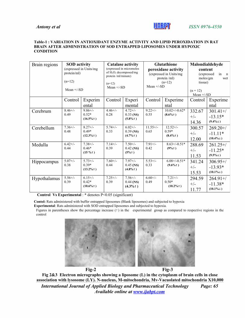
After the administration of SOD entrapped liposomes, Superoxide dismutase activity showed significant increase in all the brain regions (P<0.05) (Table:1). Cerebrum depicted an increase of 17% and cerebellum, medulla, hippocampus, hypothalamus showed 12%, 15%, 13% and 11% increase in SOD activity respectively. The glutathione peroxidase activity in the brain showed significant (P<0.05) increase after SOD administration. The catalase activity in the rat brain did not depict a significant rise however administration of the SOD entrapped liposomes succeeded in bringing down the lipid peroxidation levels in the young rat brain. MDA content showed significant (P<0.05) decrease in all the regions (Table:1). A 9-10% decrease in MDA content was noticed in the rat brain regions.
DETECTION OF LIPOSOME IN RAT BRAIN BY ELECTRON MICROSCOPY
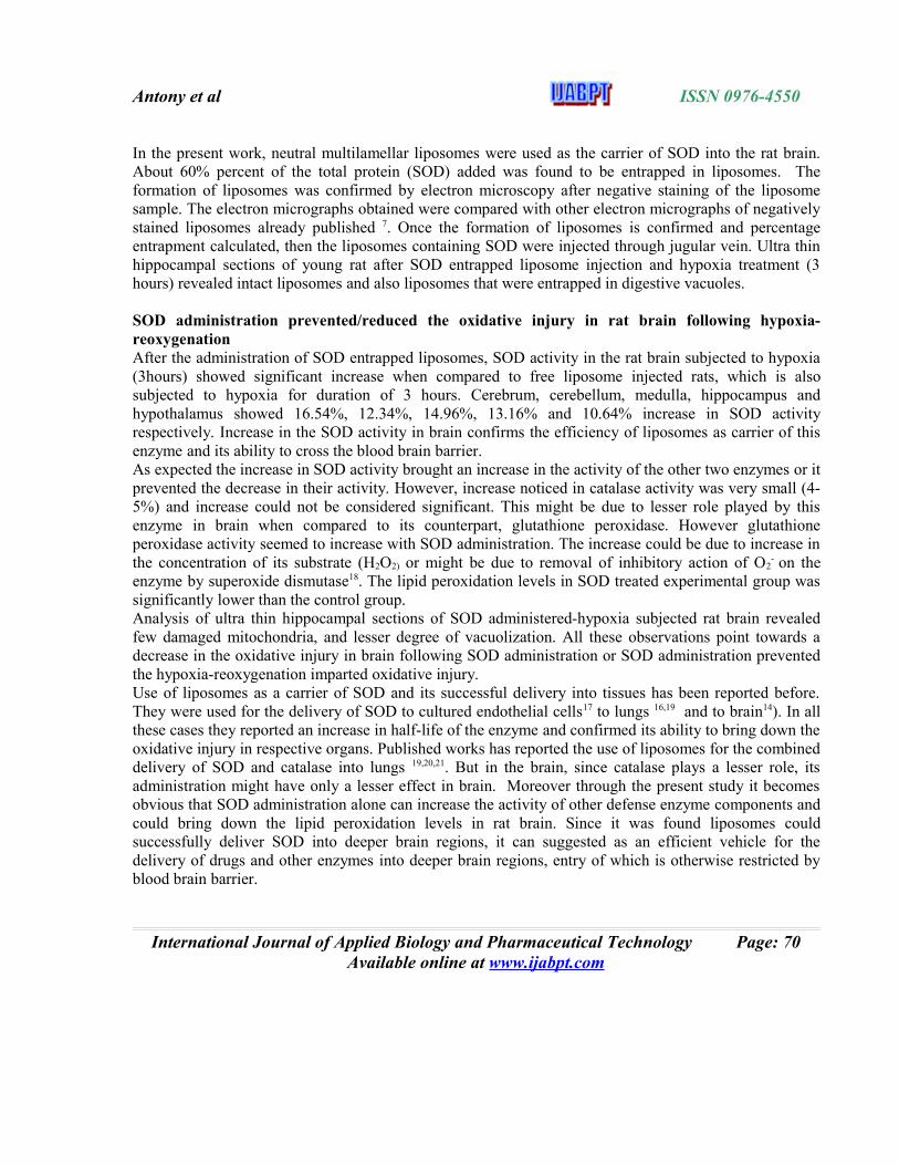
Hippocampus region was used as the sample for the detection of liposomes in brain. In both the control and experimental samples, liposomes were detected even after 3 hours of administration. They were detected in the cytoplasm of the neurons and glial cells (Fig 2,3 & 4) and near the blood vessels (Fig 5). Most of the liposomes (L) were found in association with lysosomes (LY). The liposomes could be identified by their size and lipid nature.
International Journal of Applied Biology and Pharmaceutical Technology Page: 64 Available online at www.ijabpt.com
Antony et al ISSN 0976-4550
Table-1 : VARIATION IN ANTIOXIDANT ENZYME ACTIVITY AND LIPID PEROXIDATION IN RAT BRAIN AFTER ADMINISTRATION OF SOD ENTRAPPED LIPOSOMES UNDER HYPOXIC CONDITION
Control Vs Experimental : * denotes P<0.05 (significant)
Contol: Rats administered with buffer entrapped liposomes (Blank liposomes) and subjected to hypoxia Experimental: Rats administered with SOD entrapped liposomes and subjected to hypoxia.
Figures in parentheses show the percentage increase (↑) in the experimental group as compared to respective regions in the control
Fig-2 Fig-3
Fig 2&3 Electron micrographs showing a liposome (L) in the cytoplasm of brain cells in close association with lysosome (LY). N-nucleus, M-mitochondria, Mv-Vacuolated mitochondria X10,000
International Journal of Applied Biology and Pharmaceutical Technology Page: 65 Available online at www.ijabpt.com
Brain regions SOD activity (expressed in Units/mg protein/ml) (n=12)
Mean +/-SD
Catalase activity (expressed in micromoles of H2O2 decomposed/mg protein /ml/minute) (n=12) Mean +/-SD
Glutathione peroxidase activity (expressed in Units/mg
protein /ml) (n=12)
Mean +/-SD
Malondialdehyde content (expressed in n moles/gm wet tissue)
(n = 12) Mean +/-SD
Control Experimental
Control Experimental
Control Experimental
Control Experimental
Cerebrum 8.46+/-0.49
9.86+/-0.52*(16.5%↑)
4.46+/-0.28
4.72+/-0.33 (NS)(5.8%↑)
9.22+/-0.55
10.02+/-0.62* (8.6%↑)
332.67+/-14.36
301.41+/-13.15*(9.4%↓)
Cerebellum 7.36+/-0.48
8.27+/-0.49*(12.3%↑)
5.74+/-0.33
6.02+/-0.39 (NS)(4.7%↑)
11.55+/-0.65
12.52+/-0.59* (8.4%↑)
300.57+/-12.00
269.20+/-11.11*(10.4%↓)
Medulla 6.42+/-0.44
7.38+/-0.46*(15 %↑)
7.14+/-0.39
7.50+/-0.42 (NS)(5%↑)
7.91+/-0.42
8.63+/-0.51* (9%↑)
288.69+/-11.53
261.25+/-11.25*(9.5%↓)
Hippocampus 5.07+/-0.38
5.73+/-0.39*(13.2%↑)
7.60+/-0.44
7.97+/-0.45 (NS)(4.8%↑)
5.53+/-0.33
6.08+/-0.51* (9.6%↑)
341.24+/-15.53
306.95+/-13.93*(10.1%↓)
Hypothalamus 5.56+/-0.39
6.15+/-0.42*(10.6%↑)
7.25+/-0.39
7.56+/-0.44 (NS) (4.3%↑)
6.60+/-0.49
7.21+/-0.50* (10.2%↑)
294.59+/-11.77
264.91+/-11.38*(10.1%↓)

Antony et al ISSN 0976-4550
Fig: 4 Electron micrograph- A portion of neuronal cytoplasm showing liposomes (L) as engulfed by lysosomes (LY). N-nucleus, M-mitochondria, Mv-Vacuolated mitochondria X 8000
Fig: 5 Electron micrograph of hippocampal region of rat (control) showing a capillary with liposomes (→) in association with lysosomes (>)in the swollen astrocytic processes (ASP). EL-
Endothelial lining, BM- Basal membrane,V-vacuole. X 5000
Structural modifications in the rat brain after administration of SOD entrapped liposomes under hypoxic conditions
Hippocampus region was chosen as the sample for the electron microscopic analysis of structural changes following administration of SOD entrapped liposomes. Structural changes noticed in the hippocampus region of the experimental set of rats (SOD entrapped liposome administered and hypoxia subjected) were compared with the hippocampus region of the control set of rats (Blank liposome administered and hypoxia subjected).
Hippocampal sections of the control rat brain depicted most of the structural changes associated with hypoxia. These sections showed a high degree of vacuolization (V). Mitochondria in these sections showed signs of disintegration of cristae and internal membrane (Mv). The granular endoplasmic reticulum (RER) has become swollen with the reduction in the number of ribosomes attached to them (Fig :6). Even lipid granules (LG) could be detected in the neuronal cytoplasm.
International Journal of Applied Biology and Pharmaceutical Technology Page: 66 Available online at www.ijabpt.com

Antony et al ISSN 0976-4550
In addition, neuronal processes depicted a decrease in the number of organelles and the mitochondria in them showed signs of disintegration. Capillaries showed intense pericapillary swelling and endothelial cells showed all the features seen in the hypoxia exposed samples (Fig:7).
Fig: 6 Electron microgrph- A portion of neuronal cytoplasm in the hippocampal region (control sections) N-nucleus, M-mitochondria, Mv-Vacuolated mitochondria, LG-lipid
granules, ER-Endoplasmic reticulum,V-vacuoles X6000
Fig:7 Electron micrograph -A capillary surrounded by swollen astrocytic processes (ASP) ,
capillary wall shows presence of vacuoles(V) and damaged mitochondria(mv). EL-Endothelial lining, BM- Basal membrane (Control sections) X10,000
However, after administration of SOD entrapped liposomes, the above-mentioned structural modifications in brain cells, which is often associated with a pathological situation in tissues was found to dwindle. Vacuolization (V) was observed in few cells but to a lesser degree. Number of damaged mitochondria (Mv) decreased and mitochondria (M) with normal cristae were common (Fig 8&9). Lipid granules (LG) were uncommon. Thus neuronal and glial cytoplasm depicted a situation more similar to that of normal cells.
International Journal of Applied Biology and Pharmaceutical Technology Page: 67 Available online at www.ijabpt.com

Antony et al ISSN 0976-4550
The distortion of mylein along the nerve processes, a phenomenon common in the hypoxia exposed brain sections were rarely seen after the administration of the SOD into brain (Fig:10).Capillaries after SOD administration depicted a normal ultrastructure with intact basal membrane (BM) and endothelial lining (EL). Endothelial cells showed more normal mitochondria (M) and less no of vacuoles than the control sections. However, astrocytic processes were seen clustered around capillaries (Fig 11).
Fig:8 EM- A portion of neuronal cytoplasm after administration of SOD entrapped liposomes. M-mitochondria, Mv-Vacuolated mitochondria, N-nucleus (experimental) X15,000
Fig:9 EM - A portion of glial cytoplasm after administration of SOD entrapped liposomes. M-mitochondria, Mv-Vacuolated mitochondria, N-nucleus, ER- Endoplasmic reticulum
(experimental) X6000
Fig 10: EM Neuronal processes (NP) with fewer mylein distortion (→), in the brain section after SOD administration x10,000
International Journal of Applied Biology and Pharmaceutical Technology Page: 68 Available online at www.ijabpt.com

Antony et al ISSN 0976-4550
administered rat brain. EL-Endothelial lining, BM- Basal membrane,x 5000
Fig 11 Electron micrograph showing capillary structure in the hippocampal section of the SOD
DISCUSSION
Biochemical and ultrastructural studies on rat brain confirmed that 3 hours of hypoxia followed by reoxygenation is able to impart oxidative injury in the brain tissue. Hypoxia-reoxygenation situation in brain is associated with a decrease in the activity of the antioxidant defense enzymes as reported by the earlier works of the present investigator2. At this juncture supplementation of the components of defense system must be an apt solution to prevent oxidative injury. Accordingly, there are reports of administration of chain-breaking antioxidant alpha- tocopherol being successful in bringing down lipid peroxidation levels in human brain. However, it takes considerable time to increase the alpha-tocopherol content of brain tissue in mammals supplemented with this vitamin 10 Administration of antioxidant defense enzymes to curb the free radical damage is a new approach, which is found to be effective as they can remove the free radicals before they could impart damage in tissues. The study by the present investigator on the effect of isobaric hypoxia on rat brain revealed superoxide dismutase as the enzyme most effected, as it depicted highest percentage decrease in all the 5 brain regions in comparison with other antioxidant enzymes studied2. Moreover SOD forms the first enzyme in the line of defense against free radicals, and the changes in the activity of this enzyme can bring changes in the other two enzymes involved in free radical removal. Significance of this enzyme in bringing about diminution in oxidative stress imparted tissue injury has been proved in transgenic mice over-expressing this enzyme 11&12. However, administration of free enzyme in vivo failed to bring about any significant decrease in oxidative stress mediated tissue injury 13&14. Among the reasons cited for the failure of the free enzyme to bring down the tissue injury include its net negative charge and large molecular weight (31,000) making it unable to cross the blood brain barrier15. Moreover, it has also observed that half-life of SOD in plasma is only 6minutes16. And hence for the efficient delivery of SOD into brain cells, entrapment of this enzyme into a carrier is sought. Liposomes as a carrier for SOD has been used earlier by many workers17 and found it as an efficient carrier which could penetrate cells by endocytosis allowing increase of intracellular enzyme levels.
International Journal of Applied Biology and Pharmaceutical Technology Page: 69 Available online at www.ijabpt.com
Antony et al ISSN 0976-4550
In the present work, neutral multilamellar liposomes were used as the carrier of SOD into the rat brain. About 60% percent of the total protein (SOD) added was found to be entrapped in liposomes. The formation of liposomes was confirmed by electron microscopy after negative staining of the liposome sample. The electron micrographs obtained were compared with other electron micrographs of negatively stained liposomes already published 7. Once the formation of liposomes is confirmed and percentage entrapment calculated, then the liposomes containing SOD were injected through jugular vein. Ultra thin hippocampal sections of young rat after SOD entrapped liposome injection and hypoxia treatment (3 hours) revealed intact liposomes and also liposomes that were entrapped in digestive vacuoles.
SOD administration prevented/reduced the oxidative injury in rat brain following hypoxia-reoxygenationAfter the administration of SOD entrapped liposomes, SOD activity in the rat brain subjected to hypoxia (3hours) showed significant increase when compared to free liposome injected rats, which is also subjected to hypoxia for duration of 3 hours. Cerebrum, cerebellum, medulla, hippocampus and hypothalamus showed 16.54%, 12.34%, 14.96%, 13.16% and 10.64% increase in SOD activity respectively. Increase in the SOD activity in brain confirms the efficiency of liposomes as carrier of this enzyme and its ability to cross the blood brain barrier. As expected the increase in SOD activity brought an increase in the activity of the other two enzymes or it prevented the decrease in their activity. However, increase noticed in catalase activity was very small (4-5%) and increase could not be considered significant. This might be due to lesser role played by this enzyme in brain when compared to its counterpart, glutathione peroxidase. However glutathione peroxidase activity seemed to increase with SOD administration. The increase could be due to increase in the concentration of its substrate (H2O2) or might be due to removal of inhibitory action of O2
- on the enzyme by superoxide dismutase18. The lipid peroxidation levels in SOD treated experimental group was significantly lower than the control group. Analysis of ultra thin hippocampal sections of SOD administered-hypoxia subjected rat brain revealed few damaged mitochondria, and lesser degree of vacuolization. All these observations point towards a decrease in the oxidative injury in brain following SOD administration or SOD administration prevented the hypoxia-reoxygenation imparted oxidative injury.Use of liposomes as a carrier of SOD and its successful delivery into tissues has been reported before. They were used for the delivery of SOD to cultured endothelial cells17 to lungs 16,19 and to brain14). In all these cases they reported an increase in half-life of the enzyme and confirmed its ability to bring down the oxidative injury in respective organs. Published works has reported the use of liposomes for the combined delivery of SOD and catalase into lungs 19,20,21. But in the brain, since catalase plays a lesser role, its administration might have only a lesser effect in brain. Moreover through the present study it becomes obvious that SOD administration alone can increase the activity of other defense enzyme components and could bring down the lipid peroxidation levels in rat brain. Since it was found liposomes could successfully deliver SOD into deeper brain regions, it can suggested as an efficient vehicle for the delivery of drugs and other enzymes into deeper brain regions, entry of which is otherwise restricted by blood brain barrier.
International Journal of Applied Biology and Pharmaceutical Technology Page: 70 Available online at www.ijabpt.com
Antony et al ISSN 0976-4550
REFERENCES
1. Facchinetti F, Dawson VL, Dawson TM (1998): Free radicals as mediators of neuronal injury. Cell. Mol. Neurosci : 18: 667-682.
2. Manju Antony, T. J. James (2010): A study on the effect of Isobaric hypoxia antioxidant enzyme activity and lipid peroxidation in rat brain. IJPSR, Vol. 1, Issue 9 (Suppl.) 67-75
3. Marklund S, Marklund G (1974): Involvment of superoxide anion radical in the autoxidation of pyrogallol and a convenient assay for superoxide dismutase. Eur. J. Biochem.: 4; 47:468-473.
4. Kar HI, Mishra BK (1999): quoted from Vohra BPS, Sharma SP Kansal VK Maharishi Amrit Kalash rejuvenates ageing central nervous system’s antioxidant defence system: an in vivo study. Pharmacol. Res.: 40(6): 497-502.
5. Hafeman DC, Sundae RA, Hoekstra WG (1974): Effect of Dietary selenium on erythrocyte and liver glutathione peroxidase in the rat. J .Nutr: 104: 580-587.
6. Placer ZA, Cusmann LL, Johnson BC (1996): Estimation of product of lipid peroxidation, malodialdehyde, in biological systems. Analyt. Biochem:16: 359-361.
7. Gregoriadis G, Ryman BE (1972): Lysosomal localization of beta-fructofuranosidase-containing liposomes injected into rats. Biochem J:129: 123-133
8. Gregoriadis G(1980): The liposome drug - carrier concept: Its development and future. In Gregoriadis G, Allison AC ed. Liposomes in biological systems. New York, John Wiley& Sons Ltd.,pp.25-79
9. Lowry OH, Roserbrough NS, Farr AL, Randall RJ (1951): Protein measurement with phenol reagent. J Biol chem.: 193: 265-267.
10. Muller DPR, Goss Sampson (1990): Neurochemical, neurophysiological and neuropathological studies in vitamin E deficiency. Crit rev Neurobiol 5:239-265.
11. Kinouchi H, Epstein CJ, Mizui T, Carlson EJ, Chen SF, Chan PH (1991): Attenuation of focal cerebral ischemic injury in transgenic mice overexpressing CuZn superoxide dismutase. Proc Natl Acad Sci USA 88: 11158-11162
12. Chan PH, Kawase M ,Murakami K, Chen SF, LiY, Epstein CJ (1998): Overexpression of SOD1 in transgenic rats protects vulnerable neurons against ischemic damage after global cerebral ischemiaand reperfusion. Neuroscience 18:8292-8299
13. Chan PH, Chen S,Yu ACH, Fishman RA (1986): Superoxide formation induced by arachidonic acid inastocytes. Trans Am Soc Neurochem:17:281
14. Chan PH, Longar S, Fishman RA (1987): Protective effects of liposome-entrapped superoxide dismutase on posttraumatic brain edema. Ann Neurol: 21:540-547
15. Steinman HM (1982): Superoxide dismutase: protein chemisty and structure-function relationships. In Oberley LW ed.Superoxide dismutase.Boca Raton, FL,CRC press,Vol1,pp.12-68
16. Turrens JF, Carpo JD, Freeman BA (1984): Protection against oxygen toxicity by intravenous injection of liposome-entrapped catalase and superoxide dismutase. J clin Invest :73:87-95
17. Freeman BA, Stephen LY, James DC (1983): Liposome-mediated agumentation of superoxide dismutase in endothelial cells prevents oxygen injury. J Biol Chem: 258:12534-12542
18. Blum J and Fridovich I (1985): Inactivation of glutathione peroxidase by superoxide radical. Arch Biochem Biophys 240:500-508
19. Freeman BA,Turrens JF, Mirza Z, Carpo JD, Young SL (1985) : Modulation of Oxidant lung injury by using liposome-entrapped superoxide dismutase and catalase. Fed Proc: 44 (10): 2591-2595
20. Padmanabhan RV, Gudapaty IE, Liener BA, Schwartz, Hidal JR (1985): Protection against pulmonary oxygen toxicity in rats by intratracheal administration of liposome -encapsulated superoxide dismutase and catalase. Am Rev Respir Dis: 132: 164-167.
21. Tanswell AK and Freeman BA (1987): Liposome –entrapped antioxidant enzymes prevent lethal oxygen toxicity in new born rat. J Appl Physiol 63:347-352.
International Journal of Applied Biology and Pharmaceutical Technology Page: 71 Available online at www.ijabpt.com