
A Study on Evalution of Orcun Isler, Nesrin Turaçlar and ... · Profile in Turk Patients with...
9
ARCHIVES IN CANCER RESEARCH ISSN 2254-6081 1 2014 Vol. 2 No. 1:1 doi: 10.3823/902 iMedPub Journals http://journals.imed.pub This article is available from: www.acanceresearch.com / www.medbrary.com © Under License of Creative Commons Attribution 3.0 License A Study on Evalution of Caspase Gene Expression Profile in Turk Patients with Primer Prostate Cancer 1 Selcuk University, Department of Biology, Molecular Biology Department, 42079 Selçuklu, Konya, Turkey, 2 Vocational School of Health Services, Selcuk University, Konya, Turkey,[email protected] Corresponding author: Assoc. Prof. Dr. Hasibe Cingilli Vural [email protected] Orcun Isler 1 , Nesrin Turaçlar 2 and Hasibe Cingilli Vural 1 * Abstract Harmful cells’ , such as cancerous cells, growth depends on evasion of apoptosis, which is considered as one of the hallmarks of cancer. Apoptosis is ultimately carried out by the sequential activation of initia- tor and executioner caspases, which constitute a family of intracellular proteases involved in dismantling the cell in an ordered fashion. In can- cer, therefore, one would anticipate caspases to be frequently rendered inactive, either by gene silencing or by somatic mutations. From clinical data, however, there is little evidence that caspase genes are impaired in cancer. Executioner caspases have only rarely been found mutated or silenced, and also initiator caspases are only affected in particular types of cancer. There is experimental evidence from transgenic mice that certain initiator caspases, such as caspase-8 and -2, might act as tumor suppressors. Caspase-2, the most conserved member of the caspase family, has long been recognized as an important protein in the regulation of apoptosis. Caspase-2 is activated upon genotoxic stress in a large protein complex termed the PIDDosome (Tinel and Tschopp, 2004). For this reason, we study and analyzed 2 exons of the human caspase-2 gene, using a strategy combining gene expression based RT-PCR amplification to investigate the associations between prostate cancer and caspase-2 gene expression in 9 individulas with prostate cancer. As a result of working, the level of variant-1 and variant-2 of caspase-2 gene expression in prostate cancer tissues was shown to be lower than in the control specimens and β-actin gene. Ex- pression levels of β-actin were used as internal positive control. It has been demonstrated that the restoration of caspase-2 deficient cancer tissues augments their sensitivity to undergo apoptosis in response to chemotherapeutic agents and to other apoptotic inducers. Consistent
Transcript of A Study on Evalution of Orcun Isler, Nesrin Turaçlar and ... · Profile in Turk Patients with...
Archives in cAncer reseArchissn 2254-6081
1
2014Vol. 2 No. 1:1
doi: 10.3823/902
iMedPub Journalshttp://journals.imed.pub
This article is available from: www.acanceresearch.com / www.medbrary.com© Under License of Creative Commons Attribution 3.0 License
A Study on Evalution of Caspase Gene Expression
Profile in Turk Patients with Primer Prostate Cancer
1 Selcuk University, Department of Biology, Molecular Biology Department, 42079 Selçuklu, Konya, Turkey,
2 Vocational School of Health Services, Selcuk University, Konya, Turkey,[email protected]
Corresponding author:
Assoc. Prof. Dr. Hasibe Cingilli Vural
Orcun Isler1, Nesrin Turaçlar2 and Hasibe Cingilli Vural1*
Abstract
Harmful cells’, such as cancerous cells, growth depends on evasion of apoptosis, which is considered as one of the hallmarks of cancer. Apoptosis is ultimately carried out by the sequential activation of initia-tor and executioner caspases, which constitute a family of intracellular proteases involved in dismantling the cell in an ordered fashion. In can-cer, therefore, one would anticipate caspases to be frequently rendered inactive, either by gene silencing or by somatic mutations. From clinical data, however, there is little evidence that caspase genes are impaired in cancer. Executioner caspases have only rarely been found mutated or silenced, and also initiator caspases are only affected in particular types of cancer. There is experimental evidence from transgenic mice that certain initiator caspases, such as caspase-8 and -2, might act as tumor suppressors. Caspase-2, the most conserved member of the caspase family, has long been recognized as an important protein in the regulation of apoptosis. Caspase-2 is activated upon genotoxic stress in a large protein complex termed the PIDDosome (Tinel and Tschopp, 2004). For this reason, we study and analyzed 2 exons of the human caspase-2 gene, using a strategy combining gene expression based RT-PCR amplification to investigate the associations between prostate cancer and caspase-2 gene expression in 9 individulas with prostate cancer. As a result of working, the level of variant-1 and variant-2 of caspase-2 gene expression in prostate cancer tissues was shown to be lower than in the control specimens and β-actin gene. Ex-pression levels of β-actin were used as internal positive control. It has been demonstrated that the restoration of caspase-2 deficient cancer tissues augments their sensitivity to undergo apoptosis in response to chemotherapeutic agents and to other apoptotic inducers. Consistent

Archives in cAncer reseArchissn 2254-6081
2014Vol. 2 No. 1:1
doi: 10.3823/902
This article is available from: www.acanceresearch.com / www.medbrary.com2
with this, evidence is accumulating for potential roles of caspase-2 in non-apoptotic processes, including cell cycle regulation and DNA re-pair. In addition, a tumor-suppressor function has been suggested for caspase-2. Here we discuss the various defects in caspases dependent cell death machinery identified in the prostate cancer specimens.
Keyword: apoptosis, cancer patient, caspases, gene expression
Introduction
Cancer results from the accumulation of several ge-netic and epigenetic events, arising over a long time interval. It is clear that identifying the molecular al-terations that distinguish cancer cells from normal cells will ultimately help to define the nature and understand the pathologic behavior of a cancer cell. Prostate cancer is the most frequently diagnosed male cancer in developed countries. Despite recent advancements in the treatment and management of prostate cancer, it still remains the most common malignancy and second leading cause of cancer-related deaths among men in the Turkey. Devel-opment of effective therapeutic modalities for the treatment of human cancer relies heavily upon un-derstanding the molecular alterations that result in initiation and progression of the tumorigenic pro-cess. Many of the molecular changes identified in human prostate tumorigenesis so far play key roles in apoptosis regulation. Apoptotic signaling path-ways are dependent on the activation, by proteolytic cleavage after key aspartic acid residues, of a family of cysteine proteases, termed caspases, which me-diate cleavage and functional destruction of various essential intracellular proteins. This gene encodes a member of the cysteine-aspartic acid protease (cas-
pase) family. Caspases play important roles in regu-lating apoptotic signaling pathways. The encoded protein may function in stress-induced cell death pathways, cell cycle maintenance, and the suppres-sion of tumorigenesis. Interestingly, each caspase-2 mRNA is initiated from separate promoter regions, and the casp-2L promoter is much stronger than the casp-2S promoter, in agreement with the respective transcript levels of the two variants (Logette et al., 2003).
The present studies were initiated in order to deter-mine the mechanism of activation of caspase-2 in apoptosis of cancer cells. The aim of this article is to summarize the various defects in caspases depen-dent cell death machinery identified in the prostate cancer specimens. Development of effective thera-peutic modalities for the treatment of human can-cer relies heavily upon understanding the molecular alterations that result in initiation and progression of the tumorigenic process. Many of the molecular changes identified in human prostate tumorigenesis so far play key roles in apoptosis regulation. As a result, the overall activity of caspases in malignant tissues decreases and the activation threshold in-creases, thus leading to the prevention of apoptosis.

Archives in cAncer reseArchissn 2254-6081
2014Vol. 2 No. 1:1
doi: 10.3823/902
© Under License of Creative Commons Attribution 3.0 License 3
Experimental procedures
Material and Methods
Tumor Samples
Paraffine blocks of prostate pathologies were de-rived from the archives of the Department of Pa-thology in Faculty of Medicine at the University of Konya, Turkey. Namely, paraffine-embedded pros-tate cancer tissue specimens of 9 prostate cancer patients were used in this study. Age range of all patients was 50-74 years. These patients went to physicians to be demonstrate a variety of serious symptoms of prostate cancer, e.g, difficulty in void-ing, urodynia, urgent and frequent urination, and hematuria. Their prostates were examined by one or more of the following means: rectal ultrasound detection, digital rectal examination, computed tomography, and magnetic resonance imaging. Biopsy was performed for the patients who were suspected to have prostate cancer, and all speci-
mens were from archived paraffine blocks that were collected specifically for this study. Control samples were obtained from natural or healthy tissue of the same cases.
Molecular material
Variant-1 and variant-2 belonging to caspase-2 of primers and β- actin primer were obtained from the Primer design (Table 1 and Table 2).
Deparaffinization of Samples
Deparaffinization removes the bulk of paraffin from the paraffin-embedded sample. A number of tech-niques for deparaffinization are known and any suitable technique can be used with the present invention. In this study, we used a method of the invention utilizes washing with an organic solvent to dissolve the paraffin. A xylene was used remove paraffin effectively from the tissue sample without adversely affecting DNA isolation. A xylene is the
Table 1. Variant-1 and variant-2 belonging to caspase-2 of primers.
Gene Oligonucleotide series
Caspase-2 Variant- 1F 5’-CGC GGG GTC TTG GTC C-3’
R 5’-AGG ATG CAT GCC ACA CAC T-3’
Caspase-2 Variant- 2F 5’- GGA AGA AAT CTG CTG CAC CAC-3’
R 5’- TTC TAA CAA TTC GCT CAA CAA CAG-3’
Table 2. β- actin primer.
Gen No Oligonucleotide series Tm bp
β- actin NM_001101.2
F 5’- CGCAAAGACCTGTACGCCAAC -3’ 63
164
R 5’- GAGCCGCCGATCCACACG -3’ 63

Archives in cAncer reseArchissn 2254-6081
2014Vol. 2 No. 1:1
doi: 10.3823/902
This article is available from: www.acanceresearch.com / www.medbrary.com4
preferred solvent for use in the methods of the in-vention. Paraffin was typically removed by washing with xylene vigorous mixing followed by centrifu-gation. Samples are centrifuged at a speed suffi-cient to cause the tissue to pellet in the tube, about 20,000×g. After centrifugation, the organic xylene supernatant is discarded. And then, tissue samples were passed a series of alcohol (Absolute alcohol and 75% ethyl alcohol) and were centrifuged in each of steps. Thus, the paraffin was removed from tissue samples.
Patient Anamnesis
The study was approved by the local ethics com-mittee, all patients gave written informed consent for participation in the study. Patients with either prostate cancer and other primary cancer, and fam-ily history of cancer in terms of patient age and gleason score (see Table 2) and healthy individuals without prostate cancer were included in the study. In addition to working in an individual’s age and
eligibility criteria in terms of gleason scores already published in previous studies. Tissue samples taken from patients according to pathological findings were recruited between 2010 and 2011.
RNA extraction
Total RNA from 1 dpp prostate was extracted using the RNeasy Plus Mini Kit (Qiagen) Absorbance mea-surements at 260 nm in water were used to adjust the stock concentration of all RNA samples to 1 µg/µl assuming an absorbance of 1 is equivalent to RNA at 40 µg/ml. Quality control standards were applied to all RNA samples in this study. Briefly, these were that the purity (A260:A280) was at 1.7.
Reverse Transcription
5 μl of total mRNA were reverse transcribed using the Precision nanoScriptTM Reverse Transcription kit (Primer Design Ltd.) according to the kit instructions.
Table 3. Prostate parameters.
Prostate Cancer Characteristics
Case No Patient Age Gleason Score (ng/dl) (PSA)
TNM (Tumor-lymph nodes-
metastasis)
Family history of cancer and other cancers
Personal history of cancer and
the other cancers
13 49 5 NA No No
19 69 6 T1a testes No
28 89 5 NA No No
30 62 8 T1c Bladder No
31 51 5 T2a No No
35 58 7 T1c Bladder No
36 65 7 T2a Testis, colon Colon
42 72 9 N0 Kidney Lung
48 67 7 T1c Bladder No
Archives in cAncer reseArchissn 2254-6081
2014Vol. 2 No. 1:1
doi: 10.3823/902
© Under License of Creative Commons Attribution 3.0 License 5
RT-PCR
The amount and quality of RNA were measured by Nanodrop Photospectrometer (NanoDrop 2000c THERMO, USA). 5 μg of total mRNA were reverse transcribed using the Precision nanoScriptTM Re-verse Transcription kit (Primer Design Ltd.) After in-cubation at 55 °C for 30 second, the reactions were stopped at 95 °C for 10 min. The resulting total cDNA was then used to determine expression levels of variants of caspase 2. Expression levels of β-actin were used as internal positive control. Samples were heated for 10 min at 95°C (enzyme activation), fol-lowed by 50 cycles of 15 s at 95°C (denaturation), 30 s at both of 55-60°C (annelaing) and then 15 s 72°C (binding). The statistical significance of dif-ferences in mRNA expression was analyzed by the Relative Expression Software Tool (REST). PCR ef-ficiencies for caspase-2 and b-actin were 2.07, 1.98 and 1.89, respectively.
Statistical Analyses
Cell viability, apoptosis and necrotic in rates of changes were evaluated “one-way ANOVA” test us-ing SPSS 15.0 statistical program. With the level of caspase-2 gene expression in primer prostate can-cer patients and healthy examples of differences in levels of beta-actin gene expression “REST (2009 V2.0.13)” compared with the statistical program. P values that is less than 0.05, was considered statis-tically significant. In addition, with increased cas-pase-2 mRNA expression levels in prostate cancer cells showed apoptosis while using the caspase cas-cade.
Results and Discussion
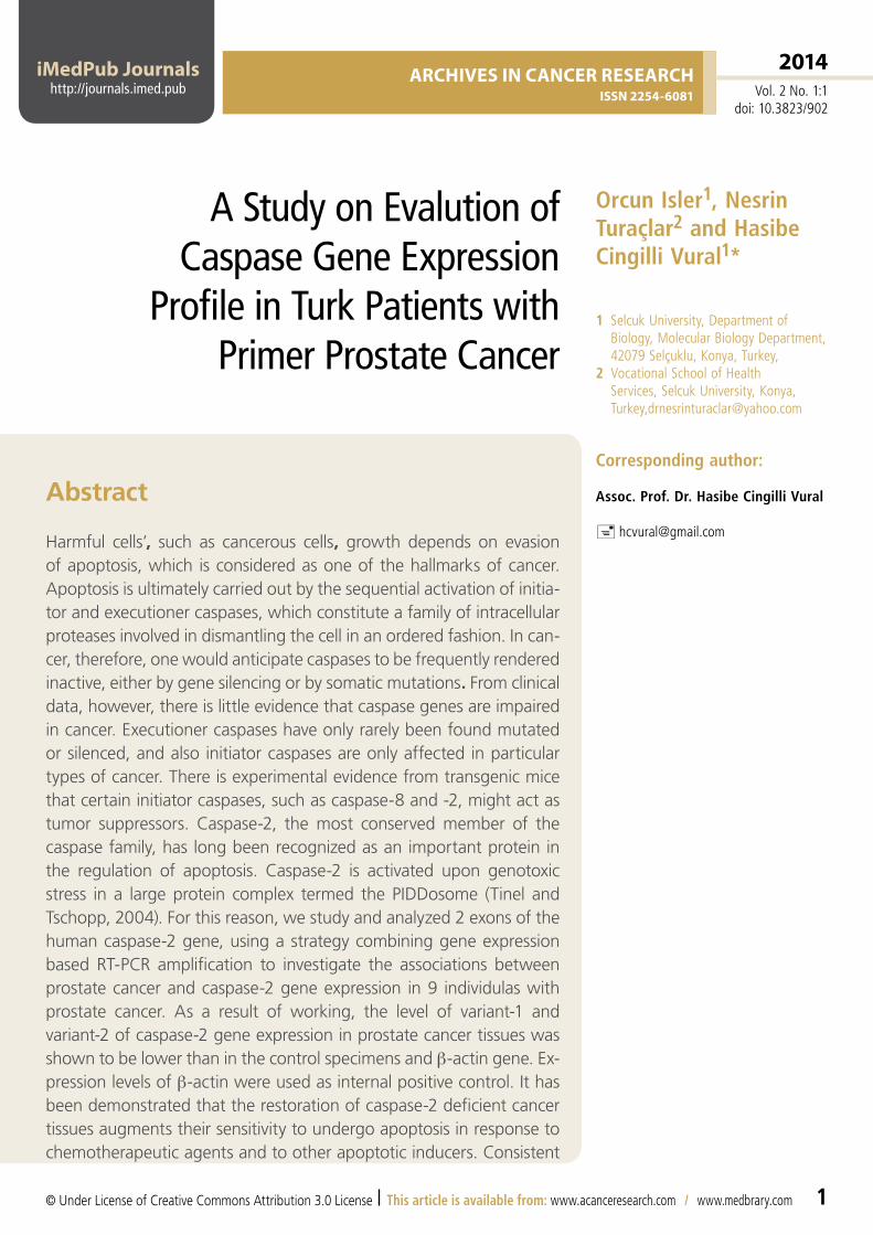
RT–PCR analysis of caspase-2 expression was carried out using a primer set for detection of caspase-2L and -2S. The two forms of caspase-2 mRNAs were
detected in the control blood, but we observed that caspase-2L mRNA predominated, and caspase-2S mRNA was barely detectable. This was true wheth-er caspase-2 expression was normalized according to beta-actin. Our results suggest that testing cas-pase-2 expression by RT-PCR technique seems to be a reliable method in detection of patients with primer prostate cancer because of its high speci-ficity. Apoptosis-related genes down regulated in NSCLC compared to non tumor lung tissue (p <0.05) included representatives of the tumors. The potential of apoptosis-related genes as prognostic and predictive markers should be validated in future studies. A larger group of patients is needed in or-der to confirm the above results, while their clinical significance as potential prognostic and predictive markers should be validated in future studies.
Apoptosis occurs via an extrinsic and an intrinsic pathway. The extrinsic pathway is initiated by bind-ing of cell surface death receptors (via death do-mains) to adaptor proteins [e.g., Fas-associated via death domain (FADD)] in a death-induced signal-ing complex. The intrinsic pathway acts through generation of mitochondrial permeability transition leading to the establishment of the ‘apoptosome’ protein complex. Both pathways converge into a common cascade that consists of proteolytic en-zymes-caspases (Shinno et al., 2005).
Caspase-2 is the earliest identified caspase in mam-mals. This enzyme is unique for its features of both initiator and effector caspases. Caspase-2 appears to be necessary for the onset of apoptosis triggered by several insults, including DNA damage, adminis-tration of TNF, and different pathogens and viruses (Logette et al., 2003). Many studies have shown that caspase-2 serves as an apoptosis inducer in some types of cells. Read et al. (2002) reported the spontaneous recruitment of procaspase-2 into a protein complex without cytochrome c or Apaf-1 in some cells.
Archives in cAncer reseArchissn 2254-6081
2014Vol. 2 No. 1:1
doi: 10.3823/902
This article is available from: www.acanceresearch.com / www.medbrary.com6
Figure 1. Prostate Ca samples at 55 ° C in the image of β-actin gene expression
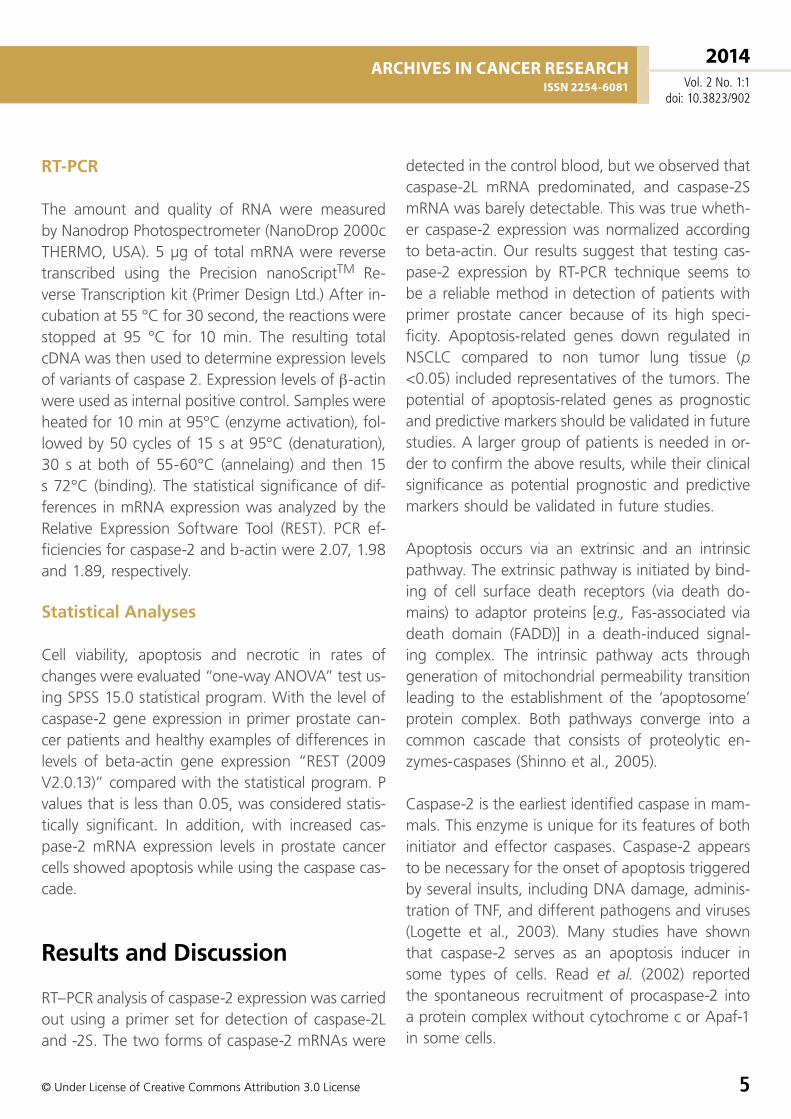
Figure 2. Prostate Ca samples at 55 ° C in the image of caspase-2 Variant 1 gene expression
Archives in cAncer reseArchissn 2254-6081
2014Vol. 2 No. 1:1
doi: 10.3823/902
© Under License of Creative Commons Attribution 3.0 License 7
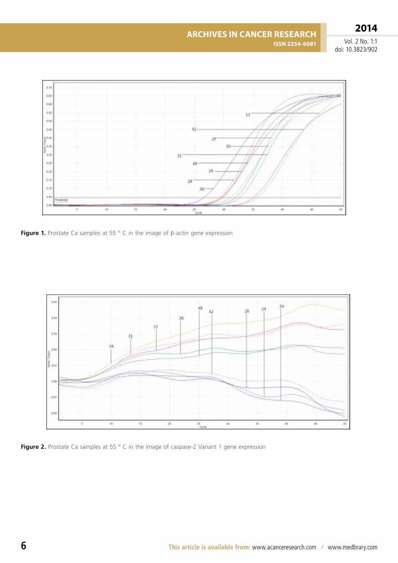
Figure 3. Prostate Ca samples at 55 °C in the image of caspase-2 Variant 2 gene expression
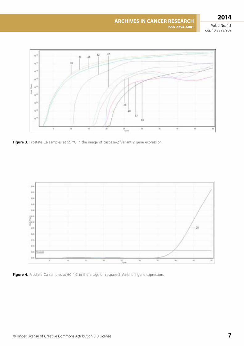
Figure 4. Prostate Ca samples at 60 ° C in the image of caspase-2 Variant 1 gene expression.

Archives in cAncer reseArchissn 2254-6081
2014Vol. 2 No. 1:1
doi: 10.3823/902
This article is available from: www.acanceresearch.com / www.medbrary.com8
In the absence of active caspases cells do not un-dergo caspase independent cell death and instead could survive an insult and promote clonogenic tu-mor growth (Fischer et al., 2007). Thus, it has been suggested that caspases can be viewed as tumor-suppressor proteins (Schulz, 2005). Although the effector caspase-6 was suggested as a putative candidate tumor suppressor gene, the question of whether other caspases (i.e. the initiator caspases) are acting as tumor suppressors remained to be specifically addressed.
The caspase-2 gene generates two main splice vari-ants (caspase-2L and caspase-2S) that fulfill either pro- or anti-apoptotic functions, respectively (Cote et al., 2001). Binding of caspase-2 pre-mRNA to RBM5 protein results in the accumulation of caspase-2L, whereas deletion or mutation of the RBM5-binding site in the caspase-2 pre-mRNA leads to accumula-tion of the caspase-2S isoform (Fushimi et al., 2008).
Over the last few years intense investigation of the function and activation mechanisms of caspase-2 by several groups has clearly implicated this enzyme in apoptosis (Krumschnabel et al., 2009; Zhivotovsky and Orrenius, 2005), but despite the large amount of data accrued, inconsistencies in findings have not allowed solid conclusions to be drawn. In addition, several recent publications have described previous-ly unknown mechanisms relating the activation of caspase-2 to its potential functions, and that might shed light on the roles played by this enzyme (An-dersen et al., 2009; Nutt et al., 2005). These new
findings suggest the involvement of several kinases in the regulation of caspase-2 activation, as well as the presence of various activation platforms for this enzyme, some of which contain the p53-inducible death domain-containing protein, PIDD. Further-more, novel roles of caspase-2 in non-apoptotic processes, such as cell cycle regulation and DNA repair have been suggested (Shi et al., 2009).
The absence of a loss-of-function phenotype of caspase-2 does not necessarily indicate that these proteins have trivial functions. It is possible that cer-tain phenotypes of any of these deficiencies could be observed in certain pathological conditions, such as cancer, deficient fertilization, or premature aging. In fact, a tumor-suppressor function for caspase-2 has now been described. In addition, caspase-2 was recently implicated in cell cycle regulation. Because many of the tumor-suppressor proteins are known to play a role in cell cycle regulation, it is likely that these two newly assigned functions for caspase-2 are interrelated (Vakifahmetoglu-Norberg and Zhi-votovsky 2010).
Acknowledgements
This work was supported by S.Ü. BAP. We are grate-ful for the help of Qiagen Research Labratory and Firm for extracting the gene expression studies.

Archives in cAncer reseArchissn 2254-6081
2014Vol. 2 No. 1:1
doi: 10.3823/902
© Under License of Creative Commons Attribution 3.0 License 9
References
1. Andersen JL, Johnson CE, Freel CD, et al (2009). Restraint of apoptosis during mitosis through interdomain phosphorylation of caspase-2. Embo J. 28, 3216–27.
2. Cote J, Dupuis S, Jiang Z, et al (2001). Caspase-2 pre-mRNA alternative splicing: identification of an intronic element containing a decoy 3’ acceptor site. Proc. Natl. Acad. Sci. U. S. A. 98, 938–43.
3. Fischer U, Janssen K, Schulze-Osthoff K (2007). Does caspase inhibition promote clonogenic tumor growth. Cell Cycle, 6, 3048–53.
4. Fushimi K, Ray P, Kar A, et al (2008). Up-regulation of the proapoptotic caspase 2 splicing isoform by a candidate tumor suppressor, RBM5. Proc. Natl. acad. Sci. U. S. A. 105, 15708–13.
5. Krumschnabel G, Sohm B, Bock F, et al (2009). The enigma of caspase-2: the laymen’s view. Cell Death Differ. 16, 195–207.
6. Logette E, Wotawa A, Solier S, et al (2003). The human caspase-2 gene: alternative promoters, pre-mRNA splicing and AUG usage direct isoform-specific expression. Oncogene, 22, 935– 46.
7. Nutt LK, Margolis SS, Jensen M, et al. (2005). Metabolic regulation of oocyte cell death through the CaMKII-mediated phosphorylation of caspase-2. Cell, 123, 89–103.
8. Read SH, Baliga BC, Ekert PG, et al (2002). A novel apaf-1- independent putative caspase-2 activation complex. J Cell Biol, 159, 739−45.
9. Schulz WA (2005) Apoptosis and replicative senescence in cancer. In: Schulz WA, Ed. Molecular biology of Human Cancers. Dordrecht: Springer science + Business Media. 145.
10. Shi M, Vivian CJ, Lee KJ, et al. (2009) DNA-PKcs-PIDDosome: a nuclear caspase-2- activating complex with role in G2/M checkpoint maintenance. Cell, 136, 508–20.
11. Shinno Y, Gunduz M, Gunduz E, et al. (2005) Fine deletional mapping of chromosome 4q22-35 region in oral cancer. Int. J. Mol. Med. 16, 93–98.
12. Tinel A, Tschopp J (2004). The PIDDosome, a protein complex implicated in activation of caspase-2 in response to genotoxic stress. Science, 304, 843–46.
13. Vakifahmetoglu-Norberg H, Zhivotovsky B (2010). The unpredictable caspase-2: what can it do. Trends Cell Biol. 20:150–59.
14. Zhivotovsky B, Orrenius S (2005) Caspase-2 function in response to DNA damage. Biochem. Biophys. Res. Commun. 331, 859–867
Comment on this article:
JBS publishes peer reviewed articles of contemporary research in the broad field of biomedical sciences. Scope of this journal includes: Biochemistry, Biomedical sciences, Biotechnology, Microbiology, Molecular biology and Genetics. Secondary research including narrative reviews, systematic reviews, evidencebased articles, meta-analysis, practice guidelines will also be considered for publication. From time to time invited articles, editorials and review of selected topics will be published. The editorial board of JBS shall strive to maintain highest standards of quality and ethics in its publication.”
Submit your manuscript here:
http://www.acanceresearch.com
Publish with iMedPub
http://www.imed.pub
Where Doctors exchange clinical experiences, review their cases and share clinical knowledge. You can also access lots of medical publications for free. Join Now!
http://medicalia.org/