A Stem Cell-Based Screening Platform Identifies Compounds ... · Original Article A Stem Cell-Based...
15
Original Article A Stem Cell-Based Screening Platform Identifies Compounds that Desensitize Motor Neurons to Endoplasmic Reticulum Stress Sebastian Thams, 1,9 Emily Rhodes Lowry, 1,7 Marie-Hélène Larraufie, 2,7,10 Krista J. Spiller, 1,8 Hai Li, 1 Damian J. Williams, 3 Phuong Hoang, 1 Elise Jiang, 2 Luis A. Williams, 4 Jackson Sandoe, 4 Kevin Eggan, 4 Ivo Lieberam, 5 Kevin C. Kanning, 1 Brent R. Stockwell, 2 Christopher E. Henderson, 1,11 and Hynek Wichterle 1,6 1 Department of Pathology and Cell Biology, Center for Motor Neuron Biology and Disease, Columbia Stem Cell Initiative, Columbia University Irving Medical Center, 630 West 168th Street, New York, NY 10032, USA; 2 Department of Biological Sciences and Department of Chemistry, Columbia University, Northwest Corner Building, MC4846, 550 West 120th Street, New York, NY 10027, USA; 3 Department of Physiology and Cellular Biophysics, Columbia University Irving Medical Center, 650 West 168th Street, New York, NY, USA; 4 Department of Stem Cell and Regenerative Biology, Harvard University, MA 02138, USA; 5 Centre for Stem Cells and Regenerative Medicine and MRC Centre for Neurodevelopmental Disorders, King’s College London, London SE1 9RT, UK; 6 Departments of Neuroscience, Rehabilitation and Regenerative Medicine, and Neurology, Columbia University Irving Medical Center, 630 West 168th Street, New York, NY 10032, USA Amyotrophic lateral sclerosis (ALS) is a neurodegenerative dis- ease selectively targeting motor neurons in the brain and spinal cord. The reasons for differential motor neuron susceptibility remain elusive. We developed a stem cell-based motor neuron assay to study cell-autonomous mechanisms causing motor neuron degeneration, with implications for ALS. A small-mole- cule screen identified cyclopiazonic acid (CPA) as a stressor to which stem cell-derived motor neurons were more sensitive than interneurons. CPA induced endoplasmic reticulum stress and the unfolded protein response. Furthermore, CPA resulted in an accelerated degeneration of motor neurons expressing human superoxide dismutase 1 (hSOD1) carrying the ALS-causing G93A mutation, compared to motor neurons ex- pressing wild-type hSOD1. A secondary screen identified compounds that alleviated CPA-mediated motor neuron degeneration: three kinase inhibitors and tauroursodeoxy- cholic acid (TUDCA), a bile acid derivative. The neuroprotec- tive effects of these compounds were validated in human stem cell-derived motor neurons carrying a mutated SOD1 allele (hSOD1 A4V ). Moreover, we found that the administra- tion of TUDCA in an hSOD1 G93A mouse model of ALS reduced muscle denervation. Jointly, these results provide insights into the mechanisms contributing to the preferential susceptibility of ALS motor neurons, and they demonstrate the utility of stem cell-derived motor neurons for the discovery of new neu- roprotective compounds. INTRODUCTION Amyotrophic lateral sclerosis (ALS) is a late-onset neurodegenerative disease that preferentially targets motor neurons in the cortex, brain stem, and spinal cord. Despite the discovery of >40 ALS-related genes, 1,2 the pathological processes leading to motor neuron degener- ation and the reasons for differential neuronal subtype susceptibility to broadly expressed mutant proteins remain poorly understood. Even though many different cell types are involved in ALS pathogen- esis, 3 cell-autonomous factors are believed to play a critical role dur- ing the early stages of the disease. 4–8 Effective modeling of motor neuron degeneration is hindered by the limited accessibility of motor neurons in patients and animal models and by the fact that ALS is a late-onset disorder. The development of stem cell technologies that facilitate large-scale production of motor neurons carrying disease-causing mutations has circumvented the first challenge, and it has enabled biochemical analysis and drug screening in a relevant cellular context. 9–13 However, stem cell- derived motor neurons are transcriptionally and electrophysiologi- cally immature, resembling embryonic or early post-natal motor neurons. 14–17 Most importantly, stem cell-derived motor neurons carrying ALS-causing mutations do not exhibit key hallmarks of motor neuron disease, such as aggregates of mutant proteins or p62-immunoreactive inclusions. 18–20 Received 8 May 2018; accepted 16 October 2018; https://doi.org/10.1016/j.ymthe.2018.10.010. 7 These authors contributed equally to this work. 8 Present address: Center for Neurodegenerative Disease Research, Department of Pathology and Laboratory Medicine, University of Pennsylvania, 3600 Spruce Street, Philadelphia, PA 19104, USA 9 Present address: Department of Clinical Neuroscience, Division of Neurology, Karolinska Universitetssjukhuset, 171 76 Stockholm, Sweden. 10 Present address: Novartis Institutes for BioMedical Research, Cambridge, MA 02139, USA. 11 Present address: Biogen Inc., Cambridge, MA 02142, USA. Correspondence: Hynek Wichterle, Departments of Neuroscience, Rehabilitation and Regenerative Medicine, and Neurology, Columbia University Irving Medical Center, 630 West 168th Street, New York, NY 10032, USA. E-mail: [email protected] Correspondence: Sebastian Thams, Department of Clinical Neuroscience, Division of Neurology, Karolinska Universitetssjukhuset, 171 76 Stockholm, Sweden. E-mail: [email protected] Molecular Therapy Vol. 27 No 1 January 2019 ª 2018 The American Society of Gene and Cell Therapy. 1 Please cite this article in press as: Thams et al., A Stem Cell-Based Screening Platform Identifies Compounds that Desensitize Motor Neurons to Endo- plasmic Reticulum Stress, Molecular Therapy (2018), https://doi.org/10.1016/j.ymthe.2018.10.010
Transcript of A Stem Cell-Based Screening Platform Identifies Compounds ... · Original Article A Stem Cell-Based...
Please cite this article in press as: Thams et al., A Stem Cell-Based Screening Platform Identifies Compounds that Desensitize Motor Neurons to Endo-plasmic Reticulum Stress, Molecular Therapy (2018), https://doi.org/10.1016/j.ymthe.2018.10.010
Original Article
A Stem Cell-Based Screening PlatformIdentifies Compounds that Desensitize MotorNeurons to Endoplasmic Reticulum StressSebastian Thams,1,9 Emily Rhodes Lowry,1,7 Marie-Hélène Larraufie,2,7,10 Krista J. Spiller,1,8 Hai Li,1
Damian J. Williams,3 Phuong Hoang,1 Elise Jiang,2 Luis A. Williams,4 Jackson Sandoe,4 Kevin Eggan,4 Ivo Lieberam,5
Kevin C. Kanning,1 Brent R. Stockwell,2 Christopher E. Henderson,1,11 and Hynek Wichterle1,6
1Department of Pathology and Cell Biology, Center for Motor Neuron Biology and Disease, Columbia Stem Cell Initiative, Columbia University Irving Medical Center, 630
West 168th Street, New York, NY 10032, USA; 2Department of Biological Sciences and Department of Chemistry, Columbia University, Northwest Corner Building,
MC4846, 550 West 120th Street, New York, NY 10027, USA; 3Department of Physiology and Cellular Biophysics, Columbia University Irving Medical Center, 650
West 168th Street, New York, NY, USA; 4Department of Stem Cell and Regenerative Biology, Harvard University, MA 02138, USA; 5Centre for Stem Cells and
Regenerative Medicine and MRC Centre for Neurodevelopmental Disorders, King’s College London, London SE1 9RT, UK; 6Departments of Neuroscience,
Rehabilitation and Regenerative Medicine, and Neurology, Columbia University Irving Medical Center, 630 West 168th Street, New York, NY 10032, USA
Received 8 May 2018; accepted 16 October 2018;https://doi.org/10.1016/j.ymthe.2018.10.010.7These authors contributed equally to this work.8Present address: Center for Neurodegenerative Disease Research, Department ofPathology and Laboratory Medicine, University of Pennsylvania, 3600 SpruceStreet, Philadelphia, PA 19104, USA9Present address: Department of Clinical Neuroscience, Division of Neurology,Karolinska Universitetssjukhuset, 171 76 Stockholm, Sweden.10Present address: Novartis Institutes for BioMedical Research, Cambridge, MA02139, USA.11Present address: Biogen Inc., Cambridge, MA 02142, USA.
Correspondence: Hynek Wichterle, Departments of Neuroscience, Rehabilitationand Regenerative Medicine, and Neurology, Columbia University Irving MedicalCenter, 630 West 168th Street, New York, NY 10032, USA.E-mail: [email protected]: Sebastian Thams, Department of Clinical Neuroscience, Divisionof Neurology, Karolinska Universitetssjukhuset, 171 76 Stockholm, Sweden.E-mail: [email protected]
Amyotrophic lateral sclerosis (ALS) is a neurodegenerative dis-ease selectively targeting motor neurons in the brain and spinalcord. The reasons for differential motor neuron susceptibilityremain elusive. We developed a stem cell-based motor neuronassay to study cell-autonomous mechanisms causing motorneuron degeneration, with implications for ALS. A small-mole-cule screen identified cyclopiazonic acid (CPA) as a stressor towhich stem cell-derived motor neurons were more sensitivethan interneurons. CPA induced endoplasmic reticulum stressand the unfolded protein response. Furthermore, CPA resultedin an accelerated degeneration of motor neurons expressinghuman superoxide dismutase 1 (hSOD1) carrying theALS-causing G93A mutation, compared to motor neurons ex-pressing wild-type hSOD1. A secondary screen identifiedcompounds that alleviated CPA-mediated motor neurondegeneration: three kinase inhibitors and tauroursodeoxy-cholic acid (TUDCA), a bile acid derivative. The neuroprotec-tive effects of these compounds were validated in humanstem cell-derived motor neurons carrying a mutated SOD1allele (hSOD1A4V). Moreover, we found that the administra-tion of TUDCA in an hSOD1G93Amouse model of ALS reducedmuscle denervation. Jointly, these results provide insights intothe mechanisms contributing to the preferential susceptibilityof ALS motor neurons, and they demonstrate the utility ofstem cell-derived motor neurons for the discovery of new neu-roprotective compounds.
INTRODUCTIONAmyotrophic lateral sclerosis (ALS) is a late-onset neurodegenerativedisease that preferentially targets motor neurons in the cortex, brainstem, and spinal cord. Despite the discovery of >40 ALS-relatedgenes,1,2 the pathological processes leading to motor neuron degener-ation and the reasons for differential neuronal subtype susceptibilityto broadly expressed mutant proteins remain poorly understood.
Molecular Therapy Vol. 27 No 1 Janu
Even though many different cell types are involved in ALS pathogen-esis,3 cell-autonomous factors are believed to play a critical role dur-ing the early stages of the disease.4–8
Effective modeling of motor neuron degeneration is hindered by thelimited accessibility of motor neurons in patients and animal modelsand by the fact that ALS is a late-onset disorder. The development ofstem cell technologies that facilitate large-scale production of motorneurons carrying disease-causing mutations has circumvented thefirst challenge, and it has enabled biochemical analysis and drugscreening in a relevant cellular context.9–13 However, stem cell-derived motor neurons are transcriptionally and electrophysiologi-cally immature, resembling embryonic or early post-natal motorneurons.14–17 Most importantly, stem cell-derived motor neuronscarrying ALS-causing mutations do not exhibit key hallmarks ofmotor neuron disease, such as aggregates of mutant proteins orp62-immunoreactive inclusions.18–20
ary 2019 ª 2018 The American Society of Gene and Cell Therapy. 1
Molecular Therapy
Please cite this article in press as: Thams et al., A Stem Cell-Based Screening Platform Identifies Compounds that Desensitize Motor Neurons to Endo-plasmic Reticulum Stress, Molecular Therapy (2018), https://doi.org/10.1016/j.ymthe.2018.10.010
Despite their relatively immature state, several studies have reporteddifferences in the survival, physiology, and biochemistry of culturedhuman and mouse stem cell-derived ALS motor neurons.5,6,19,21–28
It has been suggested that many of these phenotypes result from astressful in vitro environment that elicits premature or aberrant man-ifestations of pathological processes in cultured cells, yet the nature ofthese culture-related stressors remains ill defined. Understandingwhich specific stressors potentiate disease-relevant motor neuronpathology would enable the development of more faithful and repro-ducible models of ALS and, in turn, better tools to understand diseaseonset and progression. Ultimately, such models can be used to screenfor neuroprotective drugs.
Here we describe the development of a highly sensitive motor neuronsurvival assay and how it was used to screen a library of bioactivecompounds for stressors that accelerate the degeneration of mousemotor neurons carrying an ALS-causing human superoxide dismut-ase 1 (hSOD1)G93A transgene.29 The screen identified cyclopiazonicacid (CPA), an inhibitor of a calcium ATPase expressed in the endo-plasmic reticulum (sarcoendoplasmic reticulum-associated calciumATPase [SERCA]),30 as a compound to which motor neurons arehighly sensitive, particularly those expressing hSOD1G93A.
In accordance with the literature, we demonstrate that CPA inducesendoplasmic reticulum (ER) stress and activates the downstreamcascades referred to as the unfolded protein response (UPR).31 Thiscellular stress response is induced by unfolded and/or misfolded pro-teins in the ER lumen, and it is mediated by three ER sensors: IRE1a(Ern1), PERK (Eif2ak3), and ATF6. In turn, these sensors activateseparate signaling cascades aiming to alleviate protein misfolding.Despite the initial adaptive response, prolonged activation of ERstress leads to the activation of apoptotic pathways and cell death.32
The accumulation of misfolded proteins is a hallmark of many neuro-degenerative diseases, and it has been described in conjunction withthe activation of ER stress in animal and stem cell-based models ofALS,19,33–36 as well as in post mortem spinal cord samples fromALS patients.33,37
Studies in animal models of ALS show that certain motor neuronpopulations degenerate early during the course of the disease whileothers remain unaffected up until end stage.38,39 Even though the un-derlying causes for this vulnerability are not fully understood, it wassuggested that protein misfolding and ER stress in vulnerable motorneurons are early and crucial events that distinguish vulnerable frommore resistant motor neurons.34,40
Based on our observation that CPA was selectively toxic to motorneurons, we developed an accelerated neurodegeneration assay, andwe used it to screen for compounds that could attenuate the effectsof ER stress. We demonstrate that kenpaullone, a protein kinaseinhibitor that was recently shown to protect motor neurons from aneurotrophic factor withdrawal and to increase survival of humanALS motor neurons,13,41 also protects motor neurons from ER stress.
2 Molecular Therapy Vol. 27 No 1 January 2019
In addition to kenpaullone, we identified several other protectivecompounds, including additional kinase inhibitors and a bile acid de-rivative, tauroursodeoxycholic acid (TUDCA). In summary, wedeveloped a novel, scalable, stem cell-based discovery platform thatcan be used for the evaluation of existing drugs and for the discoveryof new compounds that protect motor neurons from ER stress-induced degeneration.
RESULTSA Screen for Stressors Inducing Preferential Degeneration of
Stem Cell-Derived hSOD1G93A Motor Neurons
To gain insight into the cell-autonomous pathological mechanismscontributing to the onset of motor neuron degeneration in cellsexpressing mutant SOD1 protein, we developed a dual-color motorneuron survival in vitro assay. This robust, sensitive, and scalable sys-tem is ideal for the discovery of cell-autonomous motor neuronphenotypes.
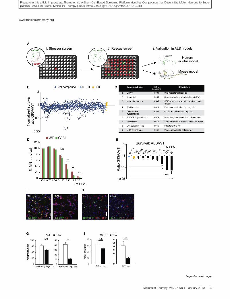
To minimize well-to-well variation and to increase scalability, wedesigned an assay in which hSOD1WT and hSOD1G93A motor neu-rons (referred to hereafter as wild-type [WT] and ALS, respectively)expressing different fluorescent reporters were mixed in the same well(Figure 1A). For this purpose, we derived a set of new embryonic stemcell (ESC) lines by crossing mice carrying hSOD1WT (WT control) orhSOD1G93A (ALS mutant) transgenes29 with mice expressing EGFP12
or tagRFP under the control of a motor neuron-specific Hb9 (Mnx1)promoter (Figure S1A). Immunostaining with antibodies against Hb9and the motor neuron transcription factor Islet1 confirmed that thenew cell lines differentiated into motor neurons with comparableefficiency (Figures S1B–S1E), and immunoprecipitation confirmedthe presence of misfolded SOD1 protein in mutant motor neurons(Figures S1F and S1G). RFP-expressing WT motor neuronswere mixed with GFP-expressing ALS motor neurons in equalproportions, and they were cultured in 96-well plates (Figure 1A)in the presence of glial cell-derived neurotrophic factor (GDNF; G)and the cyclic AMP (cAMP)-elevating compounds IBMX (I) and for-skolin (F).42 Under these basal conditions, we observed a smalldecrease (�15%) in ALS motor neuron survival compared to WTcontrols (Figure S1H).
To identify stressors that potentiate ALS pathology, plated motorneurons were treated with a library of 1,275 biologically active smallmolecules (Tocris Screen Mini and Custom Collection, Tocris Biosci-ence). Compounds were added 24 hr after motor neuron plating at afinal concentration of 10 mM using an automated robot-assistedliquid-handling platform. The ratio of surviving GFP (ALS):RFP(WT) motor neurons was determined 48 hr later, using whole-wellimaging in conjunction with automated image analysis software (Fig-ures S1I and S1J). The screen identified several compounds thatpreferentially decreased the survival of mutant motor neurons. Thesecompounds included agonists and antagonists of membranereceptors, ion pump and channel inhibitors, an anti-mitotic drug,and general pro-apoptotic agents (Figures 1B and 1C).
(legend on next page)
www.moleculartherapy.org
Molecular Therapy Vol. 27 No 1 January 2019 3
Please cite this article in press as: Thams et al., A Stem Cell-Based Screening Platform Identifies Compounds that Desensitize Motor Neurons to Endo-plasmic Reticulum Stress, Molecular Therapy (2018), https://doi.org/10.1016/j.ymthe.2018.10.010
Molecular Therapy
Please cite this article in press as: Thams et al., A Stem Cell-Based Screening Platform Identifies Compounds that Desensitize Motor Neurons to Endo-plasmic Reticulum Stress, Molecular Therapy (2018), https://doi.org/10.1016/j.ymthe.2018.10.010
One of the selective stressors identified in the screen was CPA, amycotoxin that reversibly blocks SERCA. SERCA is responsible forsequestering calcium from the cytoplasm into the ER.30 Since calciumis an essential co-factor for protein-folding chaperones, SERCAblockade with subsequent depletion of calcium from the ER leadsto the accumulation of misfolded proteins and activation of theUPR, ER stress, and apoptotic pathways.31 We titrated CPA usingtwo independent pairs of ALS-WT cell lines (Figures S2A and S2B)to establish the effective concentration range (6.25–12.5 mM; unlessstated otherwise, all subsequent experiments were performed with7.5 mMCPA) at which motor neurons show a reproducible cell deathresponse. We found that ALS motor neurons exhibited reduced sur-vival compared to WT motor neurons (Figures 1D and 1E).
To further investigate the effects of CPA, we examined whether it actsdirectly on motor neurons. We found that motor neurons purified byfluorescence-activated cell sorting (FACS) (Figures S2C–S2F) were assensitive to CPA as motor neurons in mixed cultures, indicating thatCPA acts directly on motor neurons rather than on other cell typesthat then produce secondary toxins.5,6,8,43 These findings also suggestthat the other cell types present in mixed cultures do not provide sig-nificant protection to CPA-treated motor neurons.
Preferential degeneration of motor neurons in the spinal cord, brainstem, and motor cortex is a hallmark of ALS.44 To determine whetherstem cell-derived motor neurons of spinal identity are more sensitiveto CPA treatment than other spinal neurons, we immunostained sur-viving cells for pan-neuronal marker Tuj-1. Quantitative analysis ofimmunostained cultures revealed that, while the survival of GFP-ex-pressing ALS motor neurons was reduced by �71%, the survival ofGFP� Tuj-1+ non-motor neurons of the same genotype was reducedonly by �23% (Figures 1F and 1G). The increased sensitivity of ALSmotor neurons to CPA prompted us to ask whether even WT motorneurons are more sensitive to CPA than other nerve cells.
For this analysis, we generated a new ESC line that expressestdTomato in dorsal spinal inhibitory interneurons derived fromPtf1a-expressing progenitors.45 Following differentiation of this cellline under conditions that promote the specification of dorsal inter-neuron identity, tdTomato-expressing interneurons were co-cultured
Figure 1. Results from a Dual-Color Motor Neuron Stressor Screen: Dose-Res
Neuronal Survival
(A) Overview of the experimental design from primary stressor screen to secondary rescu
48 hr of exposure. Dark blue data points denote normalized G93A:WT survival ratio fo
IBMX + vehicle) and yellow data points (positive control, forskolin + IBMX + vehicle)
compounds after secondary screening. (D and E) Dose-response curve (D) for cyclopiaz
motor neurons and normalized G93A:WT survival ratio (E). Results were compiled us
Figures S2A and S2B. Bars denote average, and error bars indicate SEM; *p < 0.05,
comparison). (F and G) Light microscope micrographs (F) showing control and CPA-tr
hSOD1G93A GFP+ motor neurons and Tuj-1+ GFP� neurons was quantified (G) at 48 hr (
non-purified motor neuron-interneuron co-cultures. Survival of GFP+ hSOD1WT motor n
Survival of GFP+ hSOD1WT motor neurons and Ptf1a+ interneurons was quantified at 48
0.01 and ***p < 0.001 (n = 3, unpaired two-tailed Student’s t test).
4 Molecular Therapy Vol. 27 No 1 January 2019
with GFP-expressing stem cell-derived motor neurons of spinal iden-tity (Figures 1H and 1I). Quantification of RFP- versus GFP-positiveneurons revealed that CPA treatment reduced dorsal spinal inter-neuron survival by only �17%, compared to a �70% decrease inthe survival of co-cultured motor neurons. Together these datademonstrate that motor neurons expressingWT SOD1 too are signif-icantly more sensitive to CPA than other spinal neurons of the sameregional identity.
Effects of CPA on Cytosolic Calcium Levels
CPA is a reversible inhibitor of the SERCA pump, which is impor-tant for sequestration of cytosolic calcium into the ER. Indeed, CPAtreatment resulted in an attenuated clearance of cytosolic calciumfollowing motor neuron depolarization with kainic acid (FiguresS3A–S3E). Elevated cytosolic calcium may activate multiple in-tracellular signaling processes, including cell death pathways.46–48
Moreover, calcium dysregulation has been implicated in manyneurodegenerative conditions, including ALS.19,49–57 To determinewhether motor neuron degeneration following CPA treatment isprimarily caused by increased cytosolic calcium, we evaluated apanel of compounds with known effects on cytosolic calciumhandling and/or signaling. These included BAPTA-am, a cell-permeable calcium chelator; dantrolene, an inhibitor of the ryano-dine receptor that releases calcium from ER stores into the cyto-plasm; three inhibitors of calpains, a family of calcium-dependentcysteine proteases; and three inhibitors of the calcium-activatedkinase CaMKK/II. Notably, none of these treatments improvedmotor neuron degeneration or neurite retraction elicited by CPAexposure (Figures S4A and S4B). These data suggested that a cyto-solic calcium overload is unlikely to be the primary cause of CPA-induced motor neuron death.
StemCell-DerivedMotor Neurons Are Sensitive to the Activation
of ER Stress Pathways
In addition to its effects on cytosolic calcium, CPA treatment has beenshown to decrease calcium levels in the ER, leading to the activation ofER stress pathways.31,58 These pathways are initiated by the bindingimmunoglobulin protein (BiP; HSPA5; GRP-78), an ER-residentchaperone, which translocates from its binding site on ER mem-brane-bound stress sensors upon detection of unfolded proteins in
pone Characterization of a Lead Compound and Subtype-Dependent
e screen and subsequent validation models. (B) Results of small molecule screen at
r well exposed to compounds, and light blue (negative control, GDNF + forskolin +
denote G93A:WT survival ratio for the controls. (C) Circles mark confirmed lead
onic acid (CPA), showing survival of Hb9::RFP hSOD1WT and Hb9::GFP hSOD1G93A
ing two independent pairs of WT-G93A cell lines; individual results are shown in
**p < 0.01, and ***p < 0.001 (n = 9, one-way ANOVA, post hoc Dunnett’s multiple
eated motor neuron cultures. Scale bar, 50 mm. Survival of ctrl and CPA-exposed
n = 3). (H and I) Light microscope micrographs (H) showing control and CPA-treated
eurons and Ptf1a+ interneurons was quantified (I) at 48 hr (n = 3). Scale bar, 50 mm.
hr (n = 3 for both analyses). Bars denote average, and error bars indicate SEM; **p <
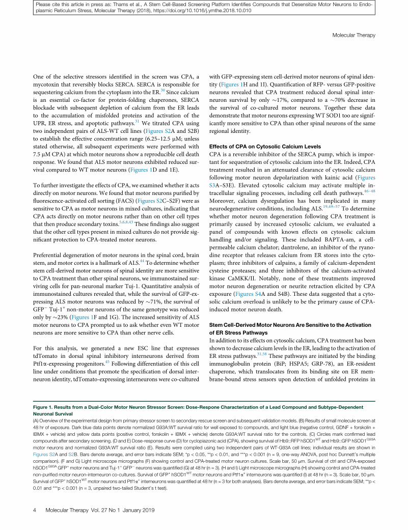
Figure 2. Characterization of ER Stress Markers in Motor Neuron Cultures Treated with Cyclopiazonic Acid
(A) Histogram showing qPCR results points to genes of particular interest at an earlier time after CPA exposure. RNA was extracted from unpurified hSOD1G93A motor
neurons (n = 3, independent culture dishes). Bars denote average, and error bars indicate SEM. CPA was compared to control for each gene and time point; *p < 0.05, **p <
0.01, and ***p < 0.001 (unpaired two-tailed Student’s t test). (B) Immunoblots showing expression of ER stress-related proteins and their loading controls at different time
points after CPA exposure (asterisks denote lanes originating from the same gel). (C) Histogram and inverted gel image showing XBP1 splicing in vehicle and CPA-treated
hSOD1G93A motor neurons at different time points after CPA exposure (n = 3). Bars denote average ratio s/u, and error bars indicate SEM. Note that no XBP1 splicing was
detected in the vehicle-treated group (ctrl). (D and E) Confocal micrographs (D) and histograms (E) showing phospho-c-jun+ motor neurons in ctrls and CPA-treated motor
neuron cultures (n = 5). Scale bar, 50 mm. Bars denote average, and error bars indicate SEM; **p < 0.01 (one-way ANOVA, post hoc Dunnett’s multiple comparison test).
(F) Confocal micrograph showing phospho-c-jun staining (blue) in co-cultures of ALS motor neurons and dorsal interneurons (Ptf1a+). Empty arrowheads indicate
CPA-treated interneurons negative for phospho-c-jun, and filled arrowheads indicate motor neurons with strong nuclear staining for phospho-c-jun. Scale bar, 50 mm.
(G) Immunoblots showing SOD1 expression and input loading control protein (a-tubulin) in lysates from CPA-treated hSOD1G93A cells. Middle lanes show panSOD1
expression. Lower lanes show immunoprecipitated lysates using antibodies specific for misfolded hSOD1 species (C4F6 clone).
www.moleculartherapy.org
Please cite this article in press as: Thams et al., A Stem Cell-Based Screening Platform Identifies Compounds that Desensitize Motor Neurons to Endo-plasmic Reticulum Stress, Molecular Therapy (2018), https://doi.org/10.1016/j.ymthe.2018.10.010
the ER lumen. Unbound BiP is involved in the activation of threeseparate signaling pathways associated with the UPR: the PERK,ATF-6, and IRE1a pathways.59–62
To assess the activation of these pathways in motor neurons exposedto CPA, we used RT-PCR to examine the expression levels of 15stress-associated genes at three time points following CPA treatment
Molecular Therapy Vol. 27 No 1 January 2019 5
Molecular Therapy
Please cite this article in press as: Thams et al., A Stem Cell-Based Screening Platform Identifies Compounds that Desensitize Motor Neurons to Endo-plasmic Reticulum Stress, Molecular Therapy (2018), https://doi.org/10.1016/j.ymthe.2018.10.010
in hSOD1G93A motor neurons (Figure 2A). We observed a rapid in-crease in the expression of Bip and the key downstream effectorChop (Ddit3). Bip increased 2-fold after 1 hr of CPA exposure, andit continued to increase to approximately 5-fold by 8 hr. Chop wasinduced 4-fold after 1 hr of CPA treatment, and it reached 16-fold in-duction after 4 and 8 hr. Other genes with >2-fold induction includedthe following: p58IPK (6.5-fold at 8 hr), an ER stress-induced proteinkinase; Growth arrest and DNA damage-inducible protein 45 alpha(Gadd45a, 5-fold at 4 hr), which has been shown to be upregulatedin the spinal cord of presymptomatic SOD1G93A mice;34 Erdj4, aBip cofactor with involvement in ER-associated protein degradation(ERAD) (5-fold at 4–8 hr); Atf4, a downstream mediator of thePERK axis of the UPR (3-fold at 8 hr); Calreticulin, an ER-associatedchaperone (3-fold at 8 hr), which was linked to nitric oxide (NO)-mediated motor neuron degeneration in hSOD1G93A mice;49 andNrf2, a PERK substrate (2-fold at 8 hr). Taken together, these expres-sion changes pointed to strong activation of multiple axes of the UPRin motor neurons exposed to CPA.
Western blot analysis of protein extracts from control and mutantmotor neurons exposed to CPA for 1, 2, 4, 8, and 24 hr confirmedthe early activation of the PERK pathway: an increase in Eif2a phos-phorylation was already detectable after only 1 hr of CPA exposure,followed by the induction of CHOP (Figure 2B; Figures S6A–S6C).An accumulation of the active cleaved form of ATF-6 was detectableat 8 hr (Figure 2B). Activation of the IRE1a branch was assessed byqPCR analysis of X-box-binding protein 1 (XBP1) splicing, whichwas already induced by 1 hr of CPA treatment and persisted at 4and 8 hr of exposure. Splicing of XBP1 was not detected in vehicle-treated controls (Figure 2C; Figure S6D).We further evaluated activa-tion of the IRE1a branch by immunocytochemical analysis of c-junphosphorylation,63 which peaked after 2 hr of CPA treatment (Fig-ures 2D and 2E). Notably, reactive c-jun phosphorylation was absentin Ptf1a-expressing interneurons exposed to CPA (Figure 2F).Finally, we detected increased levels of cleaved caspase-3 after CPAexposure, with a peak at 8 hr, indicating an apoptotic mechanismfor cell death.
We considered the possibility that the effects of CPA treatment mightreflect increased levels of the proximal disease trigger: accumulationof misfolded SOD1 protein in cultured motor neurons.18,33,34,65,66
We treated mutant motor neurons with CPA or vehicle, and weimmunoprecipitated misfolded SOD1 using two different conforma-tion-specific hSOD1 antibodies. Western blot analysis revealed aCPA-dependent increase in the accumulation of misfolded SOD1(Figure 2G; Figure S6E), potentially explaining the accelerateddeath-inducing effects of CPA in ALS motor neurons.
Compounds that Protect Motor Neurons from CPA-Induced
Degeneration
The realization that motor neurons are more sensitive to the activa-tion of ER stress pathways than other spinal neurons prompted usto set up a candidate molecule screen to identify compounds that in-crease motor neurons’ resistance to CPA. Such compounds might
6 Molecular Therapy Vol. 27 No 1 January 2019
alleviate neurodegeneration in ALS, as well as other conditions asso-ciated with protein misfolding and ER stress activation.32 Wescreened a panel of >100 compounds that was compiled from in-house libraries and supplemented with compounds that emergedfrom a literature search (Table S1). Compounds in the panel areknown to modulate different branches of the UPR, influence calciumsequestration, act as neurotrophic factors, and/or promote motorneuron survival.
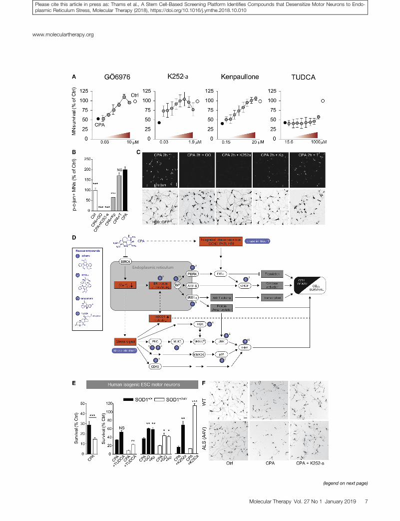
hSOD1G93A motor neuron cultures were treated with rescue com-pounds for 45 min prior to the addition of 7.5 mM CPA. Survivaland neurite growth were assessed after 24 and 48 hr. The screenyielded several compounds that prevented more than 50% of motorneuron degeneration in response to CPA (Figure S4A): the c-JunN-terminal kinase (JNK) inhibitor SP600125; the tyrosine kinase in-hibitor sunitinib; and the broad-spectrum kinase inhibitors Ro 31-8220 mesylate, kenpaullone, GÖ6976, H-7, and K252a.67–69 Com-pounds that rescued over 50% of neurite growth included the neuro-trophic factor Cardiotrophin-1; the p38 inhibitors SB293063 andSB203580; SP600125; the bile acids taurine-conjugated cholic acid(TCA), taurine-glycine-conjugated cholic acid (TGCA), andTUDCA; and the kinase inhibitors Ro 31-8220mesylate, GÖ6976, su-nitinib, kenpaullone, H-7, and K252a (Figure S4B). Overall, GÖ6976,kenpaullone, K252a, and TUDCA appeared to be the most promisingcandidates (Figure 3A), due to their strong survival-promoting effectsat low concentrations (GÖ6976, kenpaullone, K252a) or strong neu-rite outgrowth-promoting effects (TUDCA).
By testing the ER stress gene panel presented in Figure 2A in culturestreated with CPA and rescue compounds, we confirmed that two ofthe protein kinase inhibitors, GÖ6976 and kenpaullone, attenuatedthe cell stress-signaling cascade at different levels (Figures S7A andS7B).
Furthermore, GÖ6976 and K252a treatments suppressed c-jun phos-phorylation in CPA-exposed cultures more effectively than kenpaul-lone (Figures 3B and 3C), indicating that the latter inhibitor acts, atleast in part, on a different target pathway (Figure 3D). TUDCA, anambiphilic bile acid component that functions as a chemical chap-erone, rescued neurite outgrowth (Figure 4A), but it only showed amoderate effect on motor neuron survival and failed to suppress c-jun phosphorylation (Figures 3B and 3C; Figure S4A). TUDCA,which can be expected to act at the protein level, did not result inany major changes in the expression of ER stress-related genes, asshown by selected results from an RNA sequence screen (Figure S7C).
Validating Protective Compounds in Human Stem Cell-Derived
Motor Neurons
To adapt the assay to human cells, we generated a new isogenic pair ofESC lines derived from a human ESC line expressing GFP under thecontrol of the Hb9motor neuron promoter (HUES3HB9::GFP5). TheALS-causing A4V mutation was introduced into a single allele of thehuman SOD1 gene using zinc-finger nuclease (ZFN)-based genomeengineering to recapitulate human patient genotypes (Figures S5A–
(legend on next page)
www.moleculartherapy.org
Molecular Therapy Vol. 27 No 1 January 2019 7
Please cite this article in press as: Thams et al., A Stem Cell-Based Screening Platform Identifies Compounds that Desensitize Motor Neurons to Endo-plasmic Reticulum Stress, Molecular Therapy (2018), https://doi.org/10.1016/j.ymthe.2018.10.010
Molecular Therapy
Please cite this article in press as: Thams et al., A Stem Cell-Based Screening Platform Identifies Compounds that Desensitize Motor Neurons to Endo-plasmic Reticulum Stress, Molecular Therapy (2018), https://doi.org/10.1016/j.ymthe.2018.10.010
S5C). The pair of cell lines was differentiated into motor neurons us-ing previously published protocols;9,70 their relative susceptibility toER stress-mediated neurodegeneration was assessed under increasingconcentrations of CPA. While human motor neurons were less sen-sitive to CPA than mouse motor neurons (Figure S5K), we detecteda significantly increased sensitivity of mutant human SOD1A4Vmotorneurons exposed to 33 mMCPA (�14% survival) compared to controlneurons (�29% survival) (Figure 3E), thereby recapitulating the ge-notype-dependent effects of CPA in mouse motor neurons.
Next, we used the assay to test whether compounds protective tomouse motor neurons would be also able to protect human motorneurons exposed to 33 mMCPA. Remarkably, all of the top protectivecompounds identified in the mouse motor neuron screen were alsoeffective in protecting human motor neurons against CPA (Fig-ure 3E). Pretreatment of human motor neurons with kenpaullonerescued 35% of CPA-induced cell death in WT motor neurons and26% in SOD1+/A4V motor neurons (Figure 3E), but it had no signifi-cant effects on neurite growth (Figure S5L). GÖ6976 rescued 35% ofcell death in hSOD1+/+ motor neurons and 30% in SOD1+/A4V motorneurons (Figure 3E), and it also significantly rescued the decrease inneurite outgrowth (Figure S5L). K252a was overall the most prom-ising compound, rescuing 63% of cell death in hSOD1+/+ and 100%of cell death in hSOD1+/A4V motor neurons (Figures 3E and 3F),with significant effects on neurite growth for both genotypes (Fig-ure S5L). Finally, TUDCA reduced cell death moderately inhSOD1+/+ and hSOD1+/A4V motor neurons by 29% and 15%, respec-tively, with a small significant effect on neurite growth only inhSOD1+/A4V motor neurons (Figure 3E; Figure S5L).
TUDCA Treatment Attenuates ALS-Associated Muscle
Denervation In Vivo
TUDCA is a dietary supplement, and its effects on diverse patholog-ical conditions have been the focus of multiple clinical trials (GEO:NCT00877604, NCT02218619, NCT00771901, and NCT01829698;71). TUDCA is generally safe, has very few side effects, and exhibitsgood blood-brain barrier penetrance when administered subcutane-ously or orally.71,72 Denervation of neuromuscular junctions(NMJs) is one of the earliest phenotypes observed in mouse modelsof ALS,73–76 and, in the light of the in vitro results, we reasoned
Figure 3. Characterization of Rescue Compounds, Including Their Effects on c-
Neurons
(A) Dose-reponse curves for the lead compounds from the rescue screen (GO6976, n =
cultures; and TUDCA, n = 2 independent cultures). Bars denote average, and error bars
effects of rescue compounds on phospho-c-jun expression in CPA-treated motor neuro
groups were compared to CPA, ***p < 0.001 (one-way ANOVA, post hoc Dunnett’s mu
pathways for CPA and targets for rescue compounds. Blue numbered labels indicate r
show reported signaling pathways, and dashed lines show suggested pathways. Abbre
PKR; and Heme-regulated inhibitor, HRI. Superscript numbers denote supporting refe
et al.69; 5, Sun et al.86; 6, Yang et al.13; 7, Meares et al.79; 8, Uppala et al.93; 9, Ozcan et a
of CPA and in the absence or presence of rescue compounds (GO, GO6967; Kp, ken
SOD1+/+ and an isogenic genetically modified SOD1+/A4V human ESC (hESC) line (n = 9 f
genotypes). Bars denote average, and error bars indicate SEM; *p < 0.05, **p < 0.01,
whole-well images showing calcein+ motor neurons. Scale bar, 50 mm.
8 Molecular Therapy Vol. 27 No 1 January 2019
that TUDCA might promote the maintenance of motor axon termi-nal integrity and delay the denervation process.
To compare the effectiveness of TUDCA to its analogs, we screened itin parallel with 10 conjugated bile acids in CPA-treated motor neu-rons. TGCA matched the moderate effects of TUDCA on motorneuron survival, and it worked at lower concentrations; however, ithad smaller effects than TUDCA on neurite extension (Figures S4Cand S4D). TCA exhibited similar effects to TUDCA on both motorneuron survival and neurite extension, but it did not offer any advan-tages in terms of drug development. Thus, we decided to proceed withTUDCA for further evaluation in vivo.
To test the ability of TUDCA to preserve motor axons in ALS modelsin vivo, we designed a small-scale study in which we evaluated thedenervation of the fast fatigable hind limb muscle tibialis anterior(TA) in early disease-stage hSOD1G93A ALS mice (Figure 4C). Wehave previously determined that TA motor neurons in fast-progress-ing hSOD1G93A ALS mice undergo a period of presymptomaticevents, including ER stress, beginning at post-natal day (P)30, fol-lowed by muscle denervation that extends to P50.40 During thisperiod, the TA muscles display 25%–40% denervation beforebecoming substantially atrophied at later time points. To target thiswindow of early cell stress events,34 we treated hSOD1G93A micewith subcutaneous TUDCA or vehicle injections every 3 days betweenP30 and P50. Mice expressing mouse WT SOD1 were treated onlywith TUDCA, and they served as reference for the analysis. At theend of the experiment, we counted total NMJs by staining for acetyl-choline receptors in the TAmuscles with Alexa Fluor 555-conjugateda-bungarotoxin, and we assessed their innervation by staining formotor axons with antibodies against vesicular acetylcholine trans-ferase (VAChT) (Figures 4D and 4E). Despite the fact that the micereceived only seven injections over the course of 21 days of treatment,we observed a moderate, but statistically significant increase in NMJinnervation in TUDCA-treated hSOD1G93A mice compared tovehicle-treated animals (Figure 4D).
DISCUSSIONIn this study, we used a novel stem cell-based discovery platform todetect compounds rescuing human and mouse motor neurons from
jun Phosphorylation in Mouse Motor Neurons and Survival in HumanMotor
6 culture wells; K252a, n = 2 independent cultures; kenpaullone, n = 3 independent
indicate SEM. (B and C) Histogram (B) and confocal micrographs (C), showing the
n cultures at 2 hr of exposure. Bars denote average, and error bars indicate SEM; all
ltiple comparison test). Scale bar, 50 mm. (D) Proposed model for putative signaling
eported upstream or direct effects for the different rescue compounds. Intact lines
viations are as follows: General control non-depressible 2, GCN2; Protein kinase R,
rences as follows: 1, Sakaki et al.67; 2, Lemonnier et al.85; 3, Kase et al.68; 4, Roux
l.92; 10, Castro-Caldas et al.91; 11, present study. (E) Histogram showing the effects
paullone) in FACS-purified Hb9::GFP+ human motor neurons differentiated from an
or CPA versus CTRL for both genotypes, and n = 3 for all rescue compounds for both
and ***p < 0.001 (unpaired two-tailed Student’s t test). (F) Representative cropped
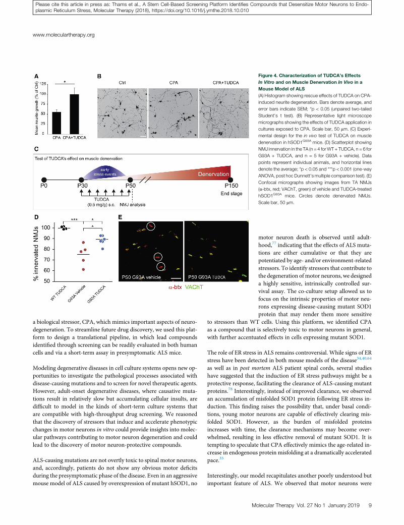
Figure 4. Characterization of TUDCA’s Effects
In Vitro and on Muscle Denervation In Vivo in a
Mouse Model of ALS
(A) Histogram showing rescue effects of TUDCA on CPA-
induced neurite degeneration. Bars denote average, and
error bars indicate SEM; *p < 0.05 (unpaired two-tailed
Student’s t test). (B) Representative light microscope
micrographs showing the effects of TUDCA application in
cultures exposed to CPA. Scale bar, 50 mm. (C) Experi-
mental design for the in vivo test of TUDCA on muscle
denervation in hSOD1G93A mice. (D) Scatterplot showing
NMJ innervation in the TA (n = 4 forWT + TUDCA, n = 6 for
G93A + TUDCA, and n = 5 for G93A + vehicle). Data
points represent individual animals, and horizontal lines
denote the average; *p < 0.05 and ***p < 0.001 (one-way
ANOVA, post hoc Dunnett’s multiple comparison test). (E)
Confocal micrographs showing images from TA NMJs
(a-btx, red; VAChT, green) of vehicle and TUDCA-treated
hSOD1G93A mice. Circles denote denervated NMJs.
Scale bar, 50 mm.
www.moleculartherapy.org
Please cite this article in press as: Thams et al., A Stem Cell-Based Screening Platform Identifies Compounds that Desensitize Motor Neurons to Endo-plasmic Reticulum Stress, Molecular Therapy (2018), https://doi.org/10.1016/j.ymthe.2018.10.010
a biological stressor, CPA, which mimics important aspects of neuro-degeneration. To streamline future drug discovery, we used this plat-form to design a translational pipeline, in which lead compoundsidentified through screening can be readily evaluated in both humancells and via a short-term assay in presymptomatic ALS mice.
Modeling degenerative diseases in cell culture systems opens new op-portunities to investigate the pathological processes associated withdisease-causing mutations and to screen for novel therapeutic agents.However, adult-onset degenerative diseases, where causative muta-tions result in relatively slow but accumulating cellular insults, aredifficult to model in the kinds of short-term culture systems thatare compatible with high-throughput drug screening. We reasonedthat the discovery of stressors that induce and accelerate phenotypicchanges in motor neurons in vitro could provide insights into molec-ular pathways contributing to motor neuron degeneration and couldlead to the discovery of motor neuron-protective compounds.
ALS-causing mutations are not overtly toxic to spinal motor neurons,and, accordingly, patients do not show any obvious motor deficitsduring the presymptomatic phase of the disease. Even in an aggressivemouse model of ALS caused by overexpression of mutant hSOD1, no
M
motor neuron death is observed until adult-hood,77 indicating that the effects of ALS muta-tions are either cumulative or that they arepotentiated by age- and/or environment-relatedstressors. To identify stressors that contribute tothe degeneration of motor neurons, we designeda highly sensitive, intrinsically controlled sur-vival assay. The co-culture setup allowed us tofocus on the intrinsic properties of motor neu-rons expressing disease-causing mutant SOD1protein that may render them more sensitive
to stressors than WT cells. Using this platform, we identified CPAas a compound that is selectively toxic to motor neurons in general,with further accentuated effects in cells expressing mutant SOD1.
The role of ER stress in ALS remains controversial. While signs of ERstress have been detected in both mouse models of the disease34,40,64
as well as in post mortem ALS patient spinal cords, several studieshave suggested that the induction of ER stress pathways might be aprotective response, facilitating the clearance of ALS-causing mutantproteins.78 Interestingly, instead of improved clearance, we observedan accumulation of misfolded SOD1 protein following ER stress in-duction. This finding raises the possibility that, under basal condi-tions, young motor neurons are capable of effectively clearing mis-folded SOD1. However, as the burden of misfolded proteinsincreases with time, the clearance mechanisms may become over-whelmed, resulting in less effective removal of mutant SOD1. It istempting to speculate that CPA effectively mimics the age-related in-crease in endogenous protein misfolding at a dramatically acceleratedpace.35
Interestingly, our model recapitulates another poorly understood butimportant feature of ALS. We observed that motor neurons were
olecular Therapy Vol. 27 No 1 January 2019 9
Molecular Therapy
Please cite this article in press as: Thams et al., A Stem Cell-Based Screening Platform Identifies Compounds that Desensitize Motor Neurons to Endo-plasmic Reticulum Stress, Molecular Therapy (2018), https://doi.org/10.1016/j.ymthe.2018.10.010
considerably more sensitive to ER stress-inducing compounds thanother types of neurons. While we do not know what mechanisms un-derlie such cell type-specific sensitivity to ER stress, it might explainthe preferential degeneration of spinal motor neurons in familial casesof ALS, despite broad expression of misfolded proteins in all types ofneurons.
A screen of candidate neuroactive compounds identified severalpotent drugs that could reverse the harmful effects of CPA. Two clas-ses of compounds were of particular interest: kinase inhibitors andbile acid derivatives. Kinase inhibitors exhibited a remarkable abilityto protect motor neurons from CPA toxicity. One compound, ken-paullone, was previously shown to protect motor neurons fromneurotrophic deprivation,13 improve survival, reverse electrophysio-logical deficits in human stem cell-derived motor neurons from apatient carrying a mutation in the FUS gene,41 and decrease the levelsof the UPR mediator CHOP in neural cells exposed to the ER stressortunicamycin.79 In addition to kenpaullone, we identified twostaurosporine analogs, K252a and GÖ6976, that were previously re-ported to increase neuronal survival in other in vitro models ofneurodegeneration.69,80
By integrating our results with published studies, we propose a modelin which CPA induces a stress response that activates a cascade ofintracellular protein kinase-regulated pathways,81,82 including a pro-tein kinase C (PKC)-JNK-signaling pathway67,68 (Figure 3D).GÖ6976 and K252a strongly inhibit PKC, as well as its downstreamtarget mixed lineage kinase 3 (MLK3).69 Kenpaullone acts primarilyas an inhibitor of HPK1/GCK-like kinase (HGK)13 and cyclin-depen-dent kinases (CDKs).83,84 Due to the unselective nature of these com-pounds, additional stress-activated kinase pathways may be involved,such as p38.85,86 Consequently, we did not discern a consistentpattern that would point to a common upstream target mechanism.Our conclusion is that the kinase targets differ between the com-pounds and act on different branches of the same cell stress-inducedcascade, ultimately converging on the suppression of c-jun phosphor-ylation (Figure 3D).
Testing these kinase inhibitors in vivo will require further optimiza-tion of their pharmacokinetic and pharmacodynamics properties.Kenpaullone is insoluble in aqueous solutions at its most effectiveconcentrations, effectively preventing its testing in vivo. WhileK252a and GÖ6976 are more potent and more soluble than kenpaul-lone (data not shown), these compounds are broad-spectrum inhibi-tors, each targeting >100 different kinases, raising the concern ofadverse secondary effects in vivo. Future drug development andmechanistic target studies will, therefore, require the design ofmore selective inhibitors.
The second class of neuroprotective compounds that emerged fromour screen was derivatives of mammalian bile acids, which havebeen used extensively in traditional Tibetan and Chinese medicine.Notably, it shows potentially beneficial results in ALS patients.71
While this class of compounds protected <30% of dying motor neu-
10 Molecular Therapy Vol. 27 No 1 January 2019
rons after CPA exposure, it completely restored neurite outgrowth. Incontrast to the kinase inhibitors that are not approved for human use,TUDCA is a widely available dietary supplement, and its analogUDCA is a water-soluble FDA-approved drug for treating pruritusand liver disease. TUDCA has previously been shown to have benefi-cial effects in mouse models of Huntington’s, Parkinson’s, and Alz-heimer’s diseases.87–91 TUDCA has also been shown to reduce theexpression of markers of the UPR in amousemodel of type 2 diabetes,in part by acting as a chaperone for misfolded proteins92,93 (Fig-ure 3F). We therefore wanted to validate our results in an in vivomodel, and we tested whether treatment with TUDCA was sufficientto delay muscle denervation in early-stage ALS mice. A brief treat-ment period showed an encouraging effect on denervation in theTA muscle, raising the possibility that TUDCA alone or in combina-tion with other treatments might delay motor disease onset orprogression.
In conclusion, the dual-color motor neuron-screening approachdescribed herein revealed that stem cell-derived motor neurons areselectively sensitive to ER stress pathway activation. Our findingsadd to the mounting evidence that ER stress contributes to motorneuron cell death in ALS. The scalable stem cell-based screening sys-tem identified several compounds that effectively desensitize motorneurons to ER stress, providing new tool compounds for mappingpathways involved in motor neuron degeneration and for the devel-opment of analogs compatible with in vivo testing. This system canbe easily adapted to other neurodegenerative conditions associatedwith ER stress activation, such as Parkinson’s disease, Huntington’sdisease, prion disease, or Alzheimer’s disease.32
MATERIALS AND METHODSDerivation of Mouse Transgenic ESC Lines
Heterozygous Tg(Hlxb9-GFP)1Tmj or Tg(Hlxb9-tagRFP) reportermice were crossed with mice expressing a mutated (B6.Cg-Tg(SOD1*G93A)1Gur/J) or WT form (B6SJL-Tg(SOD1)2Gur/J) ofhuman SOD1. Blastocysts were collected at embryonic day 3.5. MouseESC lines were derived as previously described.12 New lines were gen-otyped and sequenced to confirm the presence of both transgenes andthe G93A point mutation.
For interneuron differentiations, mouse ESC lines were derived fromPtf1a::cremice (kindly provided by Dr. Kaltschmidt) crossed to Rosa-LSL-tdTomato fluorescent reporter mice.94,95 All animal work wasperformed in compliance with Columbia University Institutional An-imal Care and Use Committee (IACUC) protocols.
Generation of Isogenic Human ESC Lines by Genetic Targeting
To extrapolate results from the mouse assays, we generated an inde-pendent set of SOD1+/A4V and SOD1+/+ isogenic cell lines by intro-ducing the A4V mutation into the WT SOD1 locus of the humanESC line HUES3 Hb9::GFP5 (Figure S5). Using again a two-stepnuclease-mediated gene-targeting strategy,96 we introduced the SO-D1A4V mutation into the HUES3 Hb9::GFP genetic background(Figure S5A).
www.moleculartherapy.org
Please cite this article in press as: Thams et al., A Stem Cell-Based Screening Platform Identifies Compounds that Desensitize Motor Neurons to Endo-plasmic Reticulum Stress, Molecular Therapy (2018), https://doi.org/10.1016/j.ymthe.2018.10.010
Mouse and HumanDifferentiation into Spinal Neuronal Lineages
Motor neuron differentiation of transgenic mouse ESCs was per-formed as previously described.12 Briefly, cells were dissociated onday 6 of differentiation and plated on a surface coated with poly-orni-thine (Sigma, 100 mg/mL) and laminin (4 mg/mL). Cells were culturedin the presence of the cAMP-elevating compounds forskolin (10 mM)and IBMX (100 mM) in combination with 500 mM GDNF. For themajority of all experiments, mouse cultures containing motor neu-rons, interneurons, and glial progenitors were used (referred to asmotor neuron cultures); in a few experiments, motor neurons werepurified by FACS (see the Supplemental Materials and Methods).
For differentiation into dI4 interneurons, Ptf1a-tdTomato ESCs weredissociated and cultured in suspension as embryoid bodies (EBs) at adensity of 8.0� 105 cells/10-cm culture-treated Petri dish. On day 2 ofdifferentiation, EBs were collected, spun down, and split 1:4 into newPetri dishes and supplemented with 1 mM retinoic acid (RA). Mediawere exchanged on days 4 and 6 of differentiation. The endpoint ofdI4 interneuron (IN) differentiation was day 8, when EBs werecollected for co-culture studies.
Differentiation of human isogenic HUES3 ESC HB9::GFP reporterlines into motor neurons was performed as previously described.70
Cells were dissociated on day 16 of differentiation, sorted via FACS,and plated on poly-ornithine- and laminin-coated surfaces as above.Serum-free human motor neuron plating media were supplementedwith the antimitotic UFdU and the neurotrophic factors GDNF,brain-derived neurotrophic factor (BDNF), ciliary neurotrophic fac-tor (CNTF), and insulin-like growth factor 1 (IGF1) (all at 10 ng/mL) as described.97
All cell lines used were routinely tested for mycoplasma.
Dual-Color Motor Neuron Co-culture Assay
Dissociated fluorescent Hb9::RFP-hSOD1WT cells were counted byhemacytometer and mixed with an equal number of Hb9::GFP-hSOD1G93A motor neurons, such that 500 fluorescent cells of each ge-notype were plated per well. Cells were plated in coated 96-well platesin amedium containing FSK and IBMX (low trophic support, positivecontrol for cell death) or FSK, IBMX, and 250 pg/mLGDNF (mediumtrophic support, positive control for survival).
Automated Image Analysis
Whole-well images of live GFP+ cells were acquired using a PlateRunnerHD system (Trophos). Images were analyzed using Meta-morph software (Molecular Devices). A healthy cell criterion, i.e.,neurons with a significant neurite (5� cell body diameter), wasused to distinguish live neurons from GFP+ debris (Figure S1K).The endogenous Hb9::GFP reporters in the human lines were notbright enough to be faithfully detected on our automated imagingplatform. Cells were treated immediately prior to imaging with thelive-cell dye calcein-AM (1.33 mM) for 10min, followed by quenchingwith a 10% solution of hemoglobin in PBS.
Small Molecule Screen
Approximately 1,300 biologically active compounds from the TocrisMini Screen and Custom collection were added to screening plates ata final concentration of 10 mM in singletons. The final concentrationof DMSO was 0.5%. A survival ratio was calculated by dividing thenumber of surviving GFP+ cells by the number of RFP+ cells after48 hr of exposure to the compounds.
FACS
Cells were sorted based on GFP or RFP expression using a 5-laserARIA-IIu ROU Cell Sorter (BD BioSciences) configured with a100-mm ceramic nozzle and operating at 20 psi.
ER Stress Rescue Screen
Dissociated and plated cells were allowed to recover for 24 hr,following 45-min incubation with rescue compounds or medium +0.5% DMSO as control. Compounds were screened in triplicates atthree different concentrations with 5-fold dilution steps; hits werefurther evaluated in 6- to 8-point serial dilutions in 3–6 replicates.Rescue compounds were selected from the initial dual-color screenor from a literature search focusing on compounds with documentedeffects on ER stress in other models. Cells were then exposed to7.5 mMCPA for mouse cells and 33 mM for human cells or medium +vehicle 0.5% DMSO, which we referred to as control (ctrl)throughout.
Immunocytochemistry
Live cultures were pre-fixed with 4% paraformaldehyde (PFA) on ice,by adding fixative directly to the medium for 2 min, then fixed anadditional 15 min by replacing the well content with 4% PFA andincubating at 4�C. Fixed cultures were blocked for 1 hr at room tem-perature with 0.01 M PBS containing 0.3% Triton-X and 20% donkeyserum. Primary antibodies were diluted in blocking solution andincubated overnight at 4�C, followed by incubation with secondaryantibodies (Alexa donkey 488/555/647) for 60 min at roomtemperature.
Biochemistry
Day 6 EBs were lysed in TNG-T lysis buffer65 containing protease(Complete Mini) and phosphatase (PhoStop) inhibitors for 30 min,followed by mechanical trituration with a 26G syringe. C4F6 andB8H10 antibodies (MediMabs) were coupled to protein-GDynabeadsand used for the immunoprecipitation of misfolded hSOD1, asdescribed.65 A control immunoglobulin G (IgG) antibody was useda negative control (Figure S1I). A pan-SOD1 antibody (Novus Biolog-icals) was used for immunoblotting; 5% of the input was used as aloading control. For western blotting, the following antibodies wereused: Caspase-3 (1:1,000), CHOP (1:500), phospho-Eif2a (1:1,000)(Cell Signaling Technology), SOD1 and ATF-6 (1:200, Novus Biolog-icals), and a-tubulin (1:50,000, Abcam). Representative gels are crop-ped from scanned images of the original films. Cropped parts withoutrelevance to the present study are indicated by a dashed line in thefigure.
Molecular Therapy Vol. 27 No 1 January 2019 11
Molecular Therapy
Please cite this article in press as: Thams et al., A Stem Cell-Based Screening Platform Identifies Compounds that Desensitize Motor Neurons to Endo-plasmic Reticulum Stress, Molecular Therapy (2018), https://doi.org/10.1016/j.ymthe.2018.10.010
qPCR
Cultures were treated with vehicle or CPA, and samples werecollected at 1, 4, and 8 hr. In addition, the combinations CPA + ken-paullone and CPA + GÖ6976 were evaluated at 4 and 8 hr. Sampleswere lysed in TRIzol and frozen at �80�C until further processing.RNA was extracted using the Qiashredder and QIAGEN RNeasyMini kits (QIAGEN), according to the manufacturer’s protocol. 1–2 mg total RNA was used for each reverse transcription reaction,and reactions were performed using the TaqMan RT kit (Applied Bio-systems, Grand Island, NY, USA). Primer pairs were designed fortarget transcripts using Primer Express 3.0 (Applied Biosystems).qPCR reactions were performed using the Power SYBR Green PCRMaster Mix (Applied Biosystems). Reactions were run and analyzedon a ViiA 7 (Life Technologies) qPCR instrument using absolutequantification settings. Statistics were performed using delta-CTvalues, and data were visualized using fold change values.
XBP1 Splicing
PCR was performed in a 50-mL jumpstart Taq (Sigma-Aldrich,D9307) reaction containing 10 pmol XBP-1-specific primers to detectsplicing (forward: 50-GAATGCCCAAAAGGATATCAGACTC-30,reverse: 50-GGCCTTGTGGTTGAGAACCAGGAG-30). PCR condi-tions were as follows: 1 cycle of 94�C for 1 min; 30 cycles of 94�Cfor 30 s, 60�C for 30 s, and 72�C for 1 min; and one cycle of 1 minat 72�C. PCR products were run for 30 min on 2.5% agarose gels con-taining ethidium bromide. Bands were observed and quantified usingthe Syngene G:Box and Genesis software. Band intensity wasmeasured using the Analyze-Gels application in ImageJ (NIH).
Calcium Imaging
Hb9::RFP WT and ALS motor neurons were dissociated on day 6 ofdifferentiation, and they were cultured 3 days on glass coverslips. Thecoverslips were incubated with 5 mM Fura-2 AM, ratiometric calciumindicator dye (Life Sciences, USA), for 30 min at room temperature.Coverslips were then exposed to a 1-s pulse of 100 mM kainic acid(KA), and one image per second was acquired for 1 min. After a re-covery period of 2min, the coverslips were then continuously exposedto 75 mM CPA for 20 min, and one image was acquired every 30 s.After another 2-min recovery period, a second pulse of KA wasapplied, with the same image acquisition as the first application. A340:380 ratio was calculated for all image series using FIJI (http://fiji.sc/). Quantification was carried out using Igor Pro version (v.)6(Wavemetrics, USA). The rate at which the evoked calcium transientsreturned to the baseline was calculated from the tau (time constant) ofa single exponential curve fitted to the falling part of the Ca intensitytrace from 80% to 20% of the peak.
In Vivo Administration of TUDCA
P30 mice were divided into three cohorts:1 hSOD1G93A mice (B6.Cg-Tg(SOD1*G93A)1Gur/J) receiving 0.5 mg/g TUDCA in 0.01 M PBSsubcutaneously;2 WTmice (C57BL/6J) receiving 0.5 mg/g TUDCA in0.01 M PBS subcutaneously, to evaluate the mutation-specific effectsof NMJ denervation and of the drug; and3 hSOD1G93A mice receiving0.01 M PBS subcutaneously, as a vehicle control. The drug was
12 Molecular Therapy Vol. 27 No 1 January 2019
administered every 3 days from P30 to P51 for a total of 7 injections,after with animals were euthanized. The TA muscles were dissectedout and processed for staining, following transcardiac perfusion. Pre-synaptic terminals were stained with an antibody to VAChT (raisedin rabbit, Covance, 1:32,000), and postsynaptic clusters were stainedwith a-bungarotoxin conjugated to Alexa Fluor 488 (1:500; Invitro-gen). NMJs lacking presynaptic staining were considered denervated.Every third section throughout the whole muscle was analyzed fromone TA per animal (n = 4–6). All animal work was performed incompliance with Columbia University IACUC protocols.
Statistics
Statistical analyses were performed with GraphPad Prism v.7 or R’(www.r-project.org). Datasets are expressed as mean value ± SEMthroughout the paper. If normal distribution and equal variance couldbe assumed, analysis of significance was performed with an unpairedtwo-tailed Student’s t test for pairwise comparison or a one-wayANOVA with post hoc Dunnett’s multiple comparison test. Other-wise, analysis was instead performed by Mann-Whitney rank-sumtest or Kruskal-Wallis test with Dunn’s multiple comparison posthoc test. Statistical significance is indicated by *p < 0.05, **p < 0.01,and ***p < 0.001.
SUPPLEMENTAL INFORMATIONSupplemental Information includes Supplemental Materials andMethods, seven figures, and one table and can be found with thisarticle online at https://doi.org/10.1016/j.ymthe.2018.10.010.
AUTHOR CONTRIBUTIONSConceptualization, H.W., C.E.H., and S.T.; Methodology, H.W.,C.E.H., S.T., E.R.L., M.-H.L., K.J.S., H.L., D.J.W., P.H., L.A.W., J.S.,K.C.K., and E.J.; Validation, H.W., S.T., and E.R.L.; Formal Analysis,H.W., S.T., E.R.L., M.-H.L., K.J.S., D.J.W., and L.A.W.; Investigation,H.W., S.T., E.R.L., M.-H.L., K.J.S., D.J.W., and L.A.W.; Resources, I.L.,K.C.K., P.H., L.A.W., J.S., and K.E.; Writing – Original Draft, H.W.,S.T., and E.R.L.; Writing – Review and Editing, H.W., S.T., E.R.L.,C.E.H., and B.R.S.; Visualization, S.T., K.J.S., D.J.W., and L.A.W.; Su-pervision, H.W., C.E.H., and B.R.S.; Funding Acquisition, H.W.,C.E.H., B.R.S., and K.E.
CONFLICTS OF INTERESTH.W., C.E.H., and S.T. have filed an application for a patent regardingthe use of TUDCA and related compounds in the prospective treat-ment of neurodegenerative disease (CU13092-0379639-TB.JK).
ACKNOWLEDGMENTSWe would like to thank Dr. Julia Kaltschmidt, for kindly providingtransgenic mice used for the derivation of Ptf1a embryonic stemcell lines, and Caroline Lindblad and Arvid Frostell, for assistancewith statistical analysis. This work was funded by Project ALS, TargetALS, the NIH (NS078097), and DoD (W81XWH-16-1-0204). S.T.received additional funding from the SwedishWenner-Gren Founda-tion and The Foundation BLANCEFLOR Boncompagni Ludovisi, néeBildt.
www.moleculartherapy.org
Please cite this article in press as: Thams et al., A Stem Cell-Based Screening Platform Identifies Compounds that Desensitize Motor Neurons to Endo-plasmic Reticulum Stress, Molecular Therapy (2018), https://doi.org/10.1016/j.ymthe.2018.10.010
REFERENCES1. Peters, O.M., Ghasemi, M., and Brown, R.H., Jr. (2015). Emerging mechanisms of
molecular pathology in ALS. J. Clin. Invest. 125, 2548.
2. Chia, R., Chiò, A., and Traynor, B.J. (2018). Novel genes associated with amyotrophiclateral sclerosis: diagnostic and clinical implications. Lancet Neurol. 17, 94–102.
3. Ilieva, H., Polymenidou, M., and Cleveland, D.W. (2009). Non-cell autonomoustoxicity in neurodegenerative disorders: ALS and beyond. J. Cell Biol. 187, 761–772.
4. Boillée, S., Yamanaka, K., Lobsiger, C.S., Copeland, N.G., Jenkins, N.A., Kassiotis, G.,Kollias, G., and Cleveland, D.W. (2006). Onset and progression in inherited ALSdetermined by motor neurons and microglia. Science 312, 1389–1392.
5. Di Giorgio, F.P., Boulting, G.L., Bobrowicz, S., and Eggan, K.C. (2008). Human em-bryonic stem cell-derived motor neurons are sensitive to the toxic effect of glial cellscarrying an ALS-causing mutation. Cell Stem Cell 3, 637–648.
6. Di Giorgio, F.P., Carrasco, M.A., Siao, M.C., Maniatis, T., and Eggan, K. (2007). Non-cell autonomous effect of glia onmotor neurons in an embryonic stem cell-based ALSmodel. Nat. Neurosci. 10, 608–614.
7. Kang, S.H., Li, Y., Fukaya, M., Lorenzini, I., Cleveland, D.W., Ostrow, L.W.,Rothstein, J.D., and Bergles, D.E. (2013). Degeneration and impaired regenerationof gray matter oligodendrocytes in amyotrophic lateral sclerosis. Nat. Neurosci. 16,571–579.
8. Nagai, M., Re, D.B., Nagata, T., Chalazonitis, A., Jessell, T.M., Wichterle, H., andPrzedborski, S. (2007). Astrocytes expressing ALS-linked mutated SOD1 release fac-tors selectively toxic to motor neurons. Nat. Neurosci. 10, 615–622.
9. Amoroso, M.W., Croft, G.F., Williams, D.J., O’Keeffe, S., Carrasco, M.A., Davis, A.R.,Roybon, L., Oakley, D.H., Maniatis, T., Henderson, C.E., and Wichterle, H. (2013).Accelerated high-yield generation of limb-innervating motor neurons from humanstem cells. J. Neurosci. 33, 574–586.
10. Dimos, J.T., Rodolfa, K.T., Niakan, K.K., Weisenthal, L.M., Mitsumoto, H., Chung,W., Croft, G.F., Saphier, G., Leibel, R., Goland, R., et al. (2008). Induced pluripotentstem cells generated from patients with ALS can be differentiated into motor neurons.Science 321, 1218–1221.
11. Höing, S., Rudhard, Y., Reinhardt, P., Glatza, M., Stehling, M., Wu, G., Peiker, C.,Böcker, A., Parga, J.A., Bunk, E., et al. (2012). Discovery of inhibitors of microglialneurotoxicity acting through multiple mechanisms using a stem-cell-based pheno-typic assay. Cell Stem Cell 11, 620–632.
12. Wichterle, H., Lieberam, I., Porter, J.A., and Jessell, T.M. (2002). Directed differenti-ation of embryonic stem cells into motor neurons. Cell 110, 385–397.
13. Yang, Y.M., Gupta, S.K., Kim, K.J., Powers, B.E., Cerqueira, A., Wainger, B.J., Ngo,H.D., Rosowski, K.A., Schein, P.A., Ackeifi, C.A., et al. (2013). A small moleculescreen in stem-cell-derived motor neurons identifies a kinase inhibitor as a candidatetherapeutic for ALS. Cell Stem Cell 12, 713–726.
14. Patterson, M., Chan, D.N., Ha, I., Case, D., Cui, Y., Van Handel, B., Mikkola, H.K.,and Lowry,W.E. (2012). Defining the nature of human pluripotent stem cell progeny.Cell Res. 22, 178–193.
15. Stein, J.L., de la Torre-Ubieta, L., Tian, Y., Parikshak, N.N., Hernández, I.A.,Marchetto, M.C., Baker, D.K., Lu, D., Hinman, C.R., Lowe, J.K., et al. (2014). A quan-titative framework to evaluate modeling of cortical development by neural stem cells.Neuron 83, 69–86.
16. Miles, G.B., Yohn, D.C., Wichterle, H., Jessell, T.M., Rafuse, V.F., and Brownstone,R.M. (2004). Functional properties of motoneurons derived from mouse embryonicstem cells. J. Neurosci. 24, 7848–7858.
17. Jacko, M., Weyn-Vanhentenryck, S.M., Smerdon, J.W., Yan, R., Feng, H., Williams,D.J., Pai, J., Xu, K., Wichterle, H., and Zhang, C. (2018). Rbfox Splicing FactorsPromote Neuronal Maturation and Axon Initial Segment Assembly. Neuron 97,853–868.e6.
18. Bosco, D.A., Morfini, G., Karabacak, N.M., Song, Y., Gros-Louis, F., Pasinelli, P.,Goolsby, H., Fontaine, B.A., Lemay, N., McKenna-Yasek, D., et al. (2010). Wild-type and mutant SOD1 share an aberrant conformation and a common pathogenicpathway in ALS. Nat. Neurosci. 13, 1396–1403.
19. Kiskinis, E., Sandoe, J., Williams, L.A., Boulting, G.L., Moccia, R., Wainger, B.J., Han,S., Peng, T., Thams, S., Mikkilineni, S., et al. (2014). Pathways disrupted in human
ALS motor neurons identified through genetic correction of mutant SOD1. CellStem Cell 14, 781–795.
20. Wang, J., Farr, G.W., Zeiss, C.J., Rodriguez-Gil, D.J., Wilson, J.H., Furtak, K.,Rutkowski, D.T., Kaufman, R.J., Ruse, C.I., Yates, J.R., 3rd, et al. (2009).Progressive aggregation despite chaperone associations of a mutant SOD1-YFP intransgenic mice that develop ALS. Proc. Natl. Acad. Sci. USA 106, 1392–1397.
21. Alami, N.H., Smith, R.B., Carrasco, M.A., Williams, L.A., Winborn, C.S., Han, S.S.W.,Kiskinis, E., Winborn, B., Freibaum, B.D., Kanagaraj, A., et al. (2014). Axonal trans-port of TDP-43 mRNA granules is impaired by ALS-causing mutations. Neuron 81,536–543.
22. Devlin, A.C., Burr, K., Borooah, S., Foster, J.D., Cleary, E.M., Geti, I., Vallier, L., Shaw,C.E., Chandran, S., and Miles, G.B. (2015). Human iPSC-derived motoneurons har-bouring TARDBP or C9ORF72 ALSmutations are dysfunctional despite maintainingviability. Nat. Commun. 6, 5999.
23. Donnelly, C.J., Zhang, P.W., Pham, J.T., Haeusler, A.R., Mistry, N.A., Vidensky, S.,Daley, E.L., Poth, E.M., Hoover, B., Fines, D.M., et al. (2013). RNA toxicity fromthe ALS/FTD C9ORF72 expansion is mitigated by antisense intervention. Neuron80, 415–428.
24. Egawa, N., Kitaoka, S., Tsukita, K., Naitoh, M., Takahashi, K., Yamamoto, T., Adachi,F., Kondo, T., Okita, K., Asaka, I., et al. (2012). Drug screening for ALS using patient-specific induced pluripotent stem cells. Sci. Transl. Med. 4, 145ra104.
25. Naujock, M., Stanslowsky, N., Bufler, S., Naumann, M., Reinhardt, P., Sterneckert, J.,Kefalakes, E., Kassebaum, C., Bursch, F., Lojewski, X., et al. (2016). 4-AminopyridineInduced Activity Rescues Hypoexcitable Motor Neurons from Amyotrophic LateralSclerosis Patient-Derived Induced Pluripotent Stem Cells. Stem Cells 34, 1563–1575.
26. Sareen, D., O’Rourke, J.G., Meera, P., Muhammad, A.K., Grant, S., Simpkinson, M.,Bell, S., Carmona, S., Ornelas, L., Sahabian, A., et al. (2013). Targeting RNA foci iniPSC-derived motor neurons from ALS patients with a C9ORF72 repeat expansion.Sci. Transl. Med. 5, 208ra149.
27. Sivadasan, R., Hornburg, D., Drepper, C., Frank, N., Jablonka, S., Hansel, A.,Lojewski, X., Sterneckert, J., Hermann, A., Shaw, P.J., et al. (2016). C9ORF72 interac-tion with cofilin modulates actin dynamics in motor neurons. Nat. Neurosci. 19,1610–1618.
28. Wainger, B.J., Kiskinis, E., Mellin, C., Wiskow, O., Han, S.S., Sandoe, J., Perez, N.P.,Williams, L.A., Lee, S., Boulting, G., et al. (2014). Intrinsic membrane hyperexcitabil-ity of amyotrophic lateral sclerosis patient-derived motor neurons. Cell Rep. 7, 1–11.
29. Gurney, M.E., Pu, H., Chiu, A.Y., Dal Canto, M.C., Polchow, C.Y., Alexander, D.D.,Caliendo, J., Hentati, A., Kwon, Y.W., Deng, H.X., et al. (1994). Motor neuron degen-eration in mice that express a human Cu,Zn superoxide dismutase mutation. Science264, 1772–1775.
30. Goeger, D.E., Riley, R.T., Dorner, J.W., and Cole, R.J. (1988). Cyclopiazonic acid in-hibition of the Ca2+-transport ATPase in rat skeletal muscle sarcoplasmic reticulumvesicles. Biochem. Pharmacol. 37, 978–981.
31. Doutheil, J., Gissel, C., Oschlies, U., Hossmann, K.A., and Paschen, W. (1997).Relation of neuronal endoplasmic reticulum calcium homeostasis to ribosomal aggre-gation and protein synthesis: implications for stress-induced suppression of proteinsynthesis. Brain Res. 775, 43–51.
32. Hetz, C., and Saxena, S. (2017). ER stress and the unfolded protein response in neuro-degeneration. Nat. Rev. Neurol. 13, 477–491.
33. Hetz, C., Thielen, P., Matus, S., Nassif, M., Court, F., Kiffin, R., Martinez, G., Cuervo,A.M., Brown, R.H., and Glimcher, L.H. (2009). XBP-1 deficiency in the nervous sys-tem protects against amyotrophic lateral sclerosis by increasing autophagy. GenesDev. 23, 2294–2306.
34. Saxena, S., Cabuy, E., and Caroni, P. (2009). A role for motoneuron subtype-selectiveER stress in disease manifestations of FALS mice. Nat. Neurosci. 12, 627–636.
35. Saxena, S., Roselli, F., Singh, K., Leptien, K., Julien, J.P., Gros-Louis, F., and Caroni, P.(2013). Neuroprotection through excitability and mTOR required in ALS motoneu-rons to delay disease and extend survival. Neuron 80, 80–96.
36. Kramer, N.J., Haney, M.S., Morgens, D.W., Jovi�ci�c, A., Couthouis, J., Li, A., Ousey, J.,Ma, R., Bieri, G., Tsui, C.K., et al. (2018). CRISPR-Cas9 screens in human cells andprimary neurons identify modifiers of C9ORF72 dipeptide-repeat-protein toxicity.Nat. Genet. 50, 603–612.
Molecular Therapy Vol. 27 No 1 January 2019 13
Molecular Therapy
Please cite this article in press as: Thams et al., A Stem Cell-Based Screening Platform Identifies Compounds that Desensitize Motor Neurons to Endo-plasmic Reticulum Stress, Molecular Therapy (2018), https://doi.org/10.1016/j.ymthe.2018.10.010
37. Atkin, J.D., Farg, M.A., Walker, A.K., McLean, C., Tomas, D., and Horne, M.K.(2008). Endoplasmic reticulum stress and induction of the unfolded protein responsein human sporadic amyotrophic lateral sclerosis. Neurobiol. Dis. 30, 400–407.
38. Kanning, K.C., Kaplan, A., and Henderson, C.E. (2010). Motor neuron diversity indevelopment and disease. Annu. Rev. Neurosci. 33, 409–440.
39. Nijssen, J., Comley, L.H., and Hedlund, E. (2017). Motor neuron vulnerability andresistance in amyotrophic lateral sclerosis. Acta Neuropathol. 133, 863–885.
40. Kaplan, A., Spiller, K.J., Towne, C., Kanning, K.C., Choe, G.T., Geber, A., Akay, T.,Aebischer, P., and Henderson, C.E. (2014). Neuronal matrix metalloproteinase-9 isa determinant of selective neurodegeneration. Neuron 81, 333–348.
41. Liu, M.L., Zang, T., and Zhang, C.L. (2016). Direct Lineage Reprogramming RevealsDisease-Specific Phenotypes of Motor Neurons from Human ALS Patients. Cell Rep.14, 115–128.
42. Hanson, M.G., Jr., Shen, S., Wiemelt, A.P., McMorris, F.A., and Barres, B.A. (1998).Cyclic AMP elevation is sufficient to promote the survival of spinal motor neuronsin vitro. J. Neurosci. 18, 7361–7371.
43. Haidet-Phillips, A.M., Hester, M.E., Miranda, C.J., Meyer, K., Braun, L., Frakes, A.,Song, S., Likhite, S., Murtha, M.J., Foust, K.D., et al. (2011). Astrocytes from familialand sporadic ALS patients are toxic to motor neurons. Nat. Biotechnol. 29, 824–828.
44. Rowland, L.P., and Shneider, N.A. (2001). Amyotrophic lateral sclerosis. N. Engl. J.Med. 344, 1688–1700.
45. Hoang, P.T., Chalif, J.I., Bikoff, J.B., Jessell, T.M., Mentis, G.Z., and Wichterle, H.(2018). Subtype Diversification and Synaptic Specificity of Stem Cell-DerivedSpinal Interneurons. Neuron 100, 135–149.e7.
46. De Stefani, D., Bononi, A., Romagnoli, A., Messina, A., De Pinto, V., Pinton, P., andRizzuto, R. (2012). VDAC1 selectively transfers apoptotic Ca2+ signals to mitochon-dria. Cell Death Differ. 19, 267–273.
47. Jayaraman, T., andMarks, A.R. (1997). T cells deficient in inositol 1,4,5-trisphosphatereceptor are resistant to apoptosis. Mol. Cell. Biol. 17, 3005–3012.
48. Pinton, P., Giorgi, C., Siviero, R., Zecchini, E., and Rizzuto, R. (2008). Calcium andapoptosis: ER-mitochondria Ca2+ transfer in the control of apoptosis. Oncogene27, 6407–6418.
49. Bernard-Marissal, N., Moumen, A., Sunyach, C., Pellegrino, C., Dudley, K.,Henderson, C.E., Raoul, C., and Pettmann, B. (2012). Reduced calreticulin levelslink endoplasmic reticulum stress and Fas-triggered cell death in motoneuronsvulnerable to ALS. J. Neurosci. 32, 4901–4912.
50. Jaiswal, M.K., and Keller, B.U. (2009). Cu/Zn superoxide dismutase typical for famil-ial amyotrophic lateral sclerosis increases the vulnerability of mitochondria and per-turbs Ca2+ homeostasis in SOD1G93A mice. Mol. Pharmacol. 75, 478–489.
51. Jaiswal, M.K., Zech, W.D., Goos, M., Leutbecher, C., Ferri, A., Zippelius, A., Carrì,M.T., Nau, R., and Keller, B.U. (2009). Impairment of mitochondrial calciumhandling in a mtSOD1 cell culture model of motoneuron disease. BMC Neurosci.10, 64.
52. Kawamata, H., and Manfredi, G. (2010). Mitochondrial dysfunction and intracellularcalcium dysregulation in ALS. Mech. Ageing Dev. 131, 517–526.
53. Kim, H.J., Magranè, J., Starkov, A.A., and Manfredi, G. (2012). The mitochondrialcalcium regulator cyclophilin D is an essential component of oestrogen-mediatedneuroprotection in amyotrophic lateral sclerosis. Brain 135, 2865–2874.
54. Rothstein, J.D., Tsai, G., Kuncl, R.W., Clawson, L., Cornblath, D.R., Drachman, D.B.,Pestronk, A., Stauch, B.L., and Coyle, J.T. (1990). Abnormal excitatory amino acidmetabolism in amyotrophic lateral sclerosis. Ann. Neurol. 28, 18–25.
55. Takuma, H., Kwak, S., Yoshizawa, T., and Kanazawa, I. (1999). Reduction of GluR2RNA editing, a molecular change that increases calcium influx through AMPA recep-tors, selective in the spinal ventral gray of patients with amyotrophic lateral sclerosis.Ann. Neurol. 46, 806–815.
56. Tradewell, M.L., Cooper, L.A., Minotti, S., and Durham, H.D. (2011). Calcium dys-regulation, mitochondrial pathology and protein aggregation in a culture model ofamyotrophic lateral sclerosis: mechanistic relationship and differential sensitivity tointervention. Neurobiol. Dis. 42, 265–275.
57. von Lewinski, F., and Keller, B.U. (2005). Ca2+, mitochondria and selective moto-neuron vulnerability: implications for ALS. Trends Neurosci. 28, 494–500.
14 Molecular Therapy Vol. 27 No 1 January 2019
58. Pirot, P., Eizirik, D.L., and Cardozo, A.K. (2006). Interferon-gamma potentiatesendoplasmic reticulum stress-induced death by reducing pancreatic beta cell defencemechanisms. Diabetologia 49, 1229–1236.
59. Gardner, B.M., Pincus, D., Gotthardt, K., Gallagher, C.M., and Walter, P. (2013).Endoplasmic reticulum stress sensing in the unfolded protein response. ColdSpring Harb. Perspect. Biol. 5, a013169.
60. Hetz, C. (2012). The unfolded protein response: controlling cell fate decisions underER stress and beyond. Nat. Rev. Mol. Cell Biol. 13, 89–102.
61. Iurlaro, R., and Muñoz-Pinedo, C. (2016). Cell death induced by endoplasmic retic-ulum stress. FEBS J. 283, 2640–2652.
62. Kozutsumi, Y., Segal, M., Normington, K., Gething, M.J., and Sambrook, J. (1988).The presence of malfolded proteins in the endoplasmic reticulum signals the induc-tion of glucose-regulated proteins. Nature 332, 462–464.
63. Urano, F., Wang, X., Bertolotti, A., Zhang, Y., Chung, P., Harding, H.P., and Ron, D.(2000). Coupling of stress in the ER to activation of JNK protein kinases by trans-membrane protein kinase IRE1. Science 287, 664–666.
64. Sun, S., Sun, Y., Ling, S.C., Ferraiuolo, L., McAlonis-Downes, M., Zou, Y., Drenner,K., Wang, Y., Ditsworth, D., Tokunaga, S., et al. (2015). Translational profiling iden-tifies a cascade of damage initiated in motor neurons and spreading to glia in mutantSOD1-mediated ALS. Proc. Natl. Acad. Sci. USA 112, E6993–E7002.
65. Gros-Louis, F., Soucy, G., Larivière, R., and Julien, J.P. (2010). Intracerebroventricularinfusion of monoclonal antibody or its derived Fab fragment against misfolded formsof SOD1mutant delays mortality in a mouse model of ALS. J. Neurochem. 113, 1188–1199.
66. Nishitoh, H., Kadowaki, H., Nagai, A., Maruyama, T., Yokota, T., Fukutomi, H.,Noguchi, T., Matsuzawa, A., Takeda, K., and Ichijo, H. (2008). ALS-linked mutantSOD1 induces ER stress- and ASK1-dependent motor neuron death by targetingDerlin-1. Genes Dev. 22, 1451–1464.
67. Sakaki, K., Wu, J., and Kaufman, R.J. (2008). Protein kinase Ctheta is required forautophagy in response to stress in the endoplasmic reticulum. J. Biol. Chem. 283,15370–15380.
68. Kase, H., Iwahashi, K., Nakanishi, S., Matsuda, Y., Yamada, K., Takahashi, M.,Murakata, C., Sato, A., and Kaneko, M. (1987). K-252 compounds, novel and potentinhibitors of protein kinase C and cyclic nucleotide-dependent protein kinases.Biochem. Biophys. Res. Commun. 142, 436–440.
69. Roux, P.P., Dorval, G., Boudreau, M., Angers-Loustau, A., Morris, S.J., Makkerh, J.,and Barker, P.A. (2002). K252a and CEP1347 are neuroprotective compounds thatinhibit mixed-lineage kinase-3 and induce activation of Akt and ERK. J. Biol.Chem. 277, 49473–49480.
70. Maury, Y., Côme, J., Piskorowski, R.A., Salah-Mohellibi, N., Chevaleyre, V.,Peschanski, M., Martinat, C., and Nedelec, S. (2015). Combinatorial analysis of devel-opmental cues efficiently converts human pluripotent stem cells into multipleneuronal subtypes. Nat. Biotechnol. 33, 89–96.
71. Elia, A.E., Lalli, S., Monsurrò, M.R., Sagnelli, A., Taiello, A.C., Reggiori, B., La Bella,V., Tedeschi, G., and Albanese, A. (2016). Tauroursodeoxycholic acid in the treat-ment of patients with amyotrophic lateral sclerosis. Eur. J. Neurol. 23, 45–52.
72. Kaemmerer, W.F., Rodrigues, C.M., Steer, C.J., and Low, W.C. (2001). Creatine-sup-plemented diet extends Purkinje cell survival in spinocerebellar ataxia type 1 trans-genic mice but does not prevent the ataxic phenotype. Neuroscience 103, 713–724.
73. Azzouz, M., Leclerc, N., Gurney, M., Warter, J.M., Poindron, P., and Borg, J. (1997).Progressive motor neuron impairment in an animal model of familial amyotrophiclateral sclerosis. Muscle Nerve 20, 45–51.
74. Maselli, R.A., Wollman, R.L., Leung, C., Distad, B., Palombi, S., Richman, D.P.,Salazar-Grueso, E.F., and Roos, R.P. (1993). Neuromuscular transmission in amyo-trophic lateral sclerosis. Muscle Nerve 16, 1193–1203.
75. Tsujihata, M., Hazama, R., Yoshimura, T., Satoh, A., Mori, M., and Nagataki, S.(1984). The motor end-plate fine structure and ultrastructural localization of acetyl-choline receptors in amyotrophic lateral sclerosis. Muscle Nerve 7, 243–249.
76. Sharma, A., Lyashchenko, A.K., Lu, L., Nasrabady, S.E., Elmaleh, M., Mendelsohn,M., Nemes, A., Tapia, J.C., Mentis, G.Z., and Shneider, N.A. (2016). ALS-associatedmutant FUS induces selective motor neuron degeneration through toxic gain of func-tion. Nat. Commun. 7, 10465.
www.moleculartherapy.org
Please cite this article in press as: Thams et al., A Stem Cell-Based Screening Platform Identifies Compounds that Desensitize Motor Neurons to Endo-plasmic Reticulum Stress, Molecular Therapy (2018), https://doi.org/10.1016/j.ymthe.2018.10.010
77. Chiu, A.Y., Zhai, P., Dal Canto, M.C., Peters, T.M., Kwon, Y.W., Prattis, S.M., andGurney, M.E. (1995). Age-dependent penetrance of disease in a transgenic mousemodel of familial amyotrophic lateral sclerosis. Mol. Cell. Neurosci. 6, 349–362.
78. Cai, Y., Arikkath, J., Yang, L., Guo, M.L., Periyasamy, P., and Buch, S. (2016).Interplay of endoplasmic reticulum stress and autophagy in neurodegenerative disor-ders. Autophagy 12, 225–244.
79. Meares, G.P., Mines, M.A., Beurel, E., Eom, T.Y., Song, L., Zmijewska, A.A., and Jope,R.S. (2011). Glycogen synthase kinase-3 regulates endoplasmic reticulum (ER) stress-induced CHOP expression in neuronal cells. Exp. Cell Res. 317, 1621–1628.
80. Jeohn, G.H., Wilson, B., Wetsel, W.C., and Hong, J.S. (2000). The indolocarbazoleGö6976 protects neurons from lipopolysaccharide/interferon-gamma-induced cyto-toxicity in murine neuron/glia co-cultures. Brain Res. Mol. Brain Res. 79, 32–44.
81. Pakos-Zebrucka, K., Koryga, I., Mnich, K., Ljujic, M., Samali, A., and Gorman, A.M.(2016). The integrated stress response. EMBO Rep. 17, 1374–1395.
82. Wek, R.C. (2018). Role of eIF2a Kinases in Translational Control and Adaptation toCellular Stress. Cold Spring Harb. Perspect. Biol. 10, a032870.
83. Leost, M., Schultz, C., Link, A., Wu, Y.Z., Biernat, J., Mandelkow, E.M., Bibb, J.A.,Snyder, G.L., Greengard, P., Zaharevitz, D.W., et al. (2000). Paullones are potent in-hibitors of glycogen synthase kinase-3beta and cyclin-dependent kinase 5/p25. Eur. J.Biochem. 267, 5983–5994.
84. Schultz, C., Link, A., Leost, M., Zaharevitz, D.W., Gussio, R., Sausville, E.A., Meijer,L., and Kunick, C. (1999). Paullones, a series of cyclin-dependent kinase inhibitors:synthesis, evaluation of CDK1/cyclin B inhibition, and in vitro antitumor activity.J. Med. Chem. 42, 2909–2919.
85. Lemonnier, J., Ghayor, C., Guicheux, J., and Caverzasio, J. (2004). Protein kinase C-independent activation of protein kinase D is involved in BMP-2-induced activationof stress mitogen-activated protein kinases JNK and p38 and osteoblastic cell differ-entiation. J. Biol. Chem. 279, 259–264.
86. Sun, K.H., Lee, H.G., Smith, M.A., and Shah, K. (2009). Direct and indirect roles ofcyclin-dependent kinase 5 as an upstream regulator in the c-Jun NH2-terminal kinasecascade: relevance to neurotoxic insults in Alzheimer’s disease. Mol. Biol. Cell 20,4611–4619.
87. Keene, C.D., Rodrigues, C.M., Eich, T., Chhabra, M.S., Steer, C.J., and Low, W.C.(2002). Tauroursodeoxycholic acid, a bile acid, is neuroprotective in a transgenic an-imal model of Huntington’s disease. Proc. Natl. Acad. Sci. USA 99, 10671–10676.
88. Keene, C.D., Rodrigues, C.M., Eich, T., Linehan-Stieers, C., Abt, A., Kren, B.T., Steer,C.J., and Low, W.C. (2001). A bile acid protects against motor and cognitive deficitsand reduces striatal degeneration in the 3-nitropropionic acid model of Huntington’sdisease. Exp. Neurol. 171, 351–360.
89. Nunes, A.F., Amaral, J.D., Lo, A.C., Fonseca, M.B., Viana, R.J., Callaerts-Vegh, Z.,D’Hooge, R., and Rodrigues, C.M. (2012). TUDCA, a bile acid, attenuates amyloidprecursor protein processing and amyloid-b deposition in APP/PS1 mice. Mol.Neurobiol. 45, 440–454.
90. Ackerman, H.D., and Gerhard, G.S. (2016). Bile Acids in NeurodegenerativeDisorders. Front. Aging Neurosci. 8, 263.
91. Castro-Caldas, M., Carvalho, A.N., Rodrigues, E., Henderson, C.J., Wolf, C.R.,Rodrigues, C.M., and Gama, M.J. (2012). Tauroursodeoxycholic acid preventsMPTP-induced dopaminergic cell death in a mouse model of Parkinson’s disease.Mol. Neurobiol. 46, 475–486.
92. Ozcan, U., Yilmaz, E., Ozcan, L., Furuhashi, M., Vaillancourt, E., Smith, R.O.,Görgün, C.Z., and Hotamisligil, G.S. (2006). Chemical chaperones reduce ER stressand restore glucose homeostasis in a mouse model of type 2 diabetes. Science 313,1137–1140.
93. Uppala, J.K., Gani, A.R., and Ramaiah, K.V.A. (2017). Chemical chaperone, TUDCAunlike PBA, mitigates protein aggregation efficiently and resists ER and non-ER stressinduced HepG2 cell death. Sci. Rep. 7, 3831.
94. Kawaguchi, Y., Cooper, B., Gannon, M., Ray, M., MacDonald, R.J., and Wright, C.V.(2002). The role of the transcriptional regulator Ptf1a in converting intestinal topancreatic progenitors. Nat. Genet. 32, 128–134.
95. Madisen, L., Zwingman, T.A., Sunkin, S.M., Oh, S.W., Zariwala, H.A., Gu, H., Ng,L.L., Palmiter, R.D., Hawrylycz, M.J., Jones, A.R., et al. (2010). A robust and high-throughput Cre reporting and characterization system for the whole mouse brain.Nat. Neurosci. 13, 133–140.
96. Maeder, M.L., Thibodeau-Beganny, S., Osiak, A., Wright, D.A., Anthony, R.M.,Eichtinger, M., Jiang, T., Foley, J.E., Winfrey, R.J., Townsend, J.A., et al. (2008).Rapid “open-source” engineering of customized zinc-finger nucleases for highly effi-cient gene modification. Mol. Cell 31, 294–301.
97. Johnson-Kerner, B.L., Ahmad, F.S., Diaz, A.G., Greene, J.P., Gray, S.J., Samulski, R.J.,Chung, W.K., Van Coster, R., Maertens, P., Noggle, S.A., et al. (2015). Intermediatefilament protein accumulation in motor neurons derived from giant axonal neurop-athy iPSCs rescued by restoration of gigaxonin. Hum. Mol. Genet. 24, 1420–1431.
Molecular Therapy Vol. 27 No 1 January 2019 15


![Ionic Compounds: Naming. Naming Binary Ionic Compounds 1.Always name metal [(+) cation] first stem 2. Write stem of non-metal [(-) anion] 3. Add ending.](https://static.fdocuments.in/doc/165x107/5a4d1b717f8b9ab0599b5930/ionic-compounds-naming-naming-binary-ionic-compounds-1always-name-metal-.jpg)