A single point mutation in ricin A-chain increases toxin ... · Biochem. J. (2011) 436, 371–385...
18
Biochem. J. (2011) 436, 371–385 (Printed in Great Britain) doi:10.1042/BJ20101493 371 A single point mutation in ricin A-chain increases toxin degradation and inhibits EDEM1-dependent ER retrotranslocation Iwona SOKOLOWSKA*, S´ ebastien W ¨ ALCHLI†, Grzegorz W EGRZYN*, Kirsten SANDVIG‡ and Monika SLOMI ´ NSKA-WOJEW ´ ODZKA* 1 * Department of Molecular Biology, University of Gda´ nsk, Kladki 24, 80-822 Gda´ nsk, Poland, †Department of Immunology, Institute for Cancer Research, The Norwegian Radium Hospital, Oslo University Hospital, Montebello, N-0310 Oslo, Norway, and ‡Department of Biochemistry, Institute for Cancer Research, The Norwegian Radium Hospital, Oslo University Hospital, Montebello, N-0310 Oslo, Norway Ricin is a potent plant cytotoxin composed of an A-chain [RTA (ricin A-chain)] connected by a disulfide bond to a cell binding lectin B-chain [RTB (ricin B-chain)]. After endocytic uptake, the toxin is transported retrogradely to the ER (endoplasmic reticulum) from where enzymatically active RTA is translocated to the cytosol. This transport is promoted by the EDEM1 (ER degradation-enhancing α-mannosidase I-like protein 1), which is also responsible for directing aberrant proteins for ERAD (ER-associated protein degradation). RTA contains a 12- residue hydrophobic C-terminal region that becomes exposed after reduction of ricin in the ER. This region, especially Pro 250 , plays a crucial role in ricin cytotoxicity. In the present study, we introduced a point mutation [P250A (substitution of Pro 250 with alanine)] in the hydrophobic region of RTA to study the intracellular transport of the modified toxin. The introduced mutation alters the secondary structure of RTA into a more helical structure. Mutation P250A increases endosomal– lysosomal degradation of the toxin, as well as reducing its transport from the ER to the cytosol. Transport of modified RTA to the cytosol, in contrast to wild-type RTA, appears to be EDEM1- independent. Importantly, the interaction between EDEM1 and RTA P250A is reduced. This is the first reported evidence that EDEM1 protein recognition might be determined by the structure of the ERAD substrate. Key words: endoplasmic reticulum degradation-enhancing α-mannosidase I-like protein 1 (EDEM1), endoplasmic reticulum (ER), endosomal–lysosomal degradation, P250A mutation, retrotranslocation, ricin A-chain (RTA). INTRODUCTION Ricin, the most toxic member of the RIPs (ribosome-inactivating proteins), accumulates to high levels in the endosperm of Ricinus communis seeds. Cell trafficking of this toxin has been extensively studied due to the variety of medical applications in which it can be used. As an example, ricin has been employed in the construction of ITs (immunotoxins) that show specific anti-cancer and anti- AIDS activities in vitro and in vivo [1–4]. Ricin is a heterodimeric glycoprotein composed of an RNA-specific N-glycosidase A- chain [RTA (ricin A-chain)] connected by a disulfide bond to a cell binding lectin B-chain [RTB (ricin B-chain)]. RTA inhibits protein synthesis by enzymatically depurinating a specific adenine residue at the sarcin–ricin loop of the 28S rRNA, thereby preventing the binding of elongation factors to the GTPase activation centre of the ribosome [5]. RTB recognizes cell surface glycolipids or glycoproteins with β -1,4-linked galactose residues. On binding to the cell surface, the toxin is taken up by endocytosis and enters early endosomes. From here, the majority of the endocytosed toxin recycles back to the cell surface, starts to be degraded or proceeds to late endosomes and lysosomes where further degradation is conducted [6–8]. A minor fraction (∼ 5 %) of ricin is transported from early endosomes to the Golgi apparatus, and from there it is further transported retrogradely to the ER (endoplasmic reticulum). In the ER, the disulfide bond of the holotoxin is reduced to liberate RTA. Upon A- and B- chain dissociation, a 12-amino-acid (Val 245 to Val 256 ) hydrophobic region of the RTA, which is hidden in the holotoxin, becomes exposed. It has been demonstrated that P250A (substitution of Pro 250 with alanine) in this region results in a dramatic decrease in RTA P250A cytotoxicity in Vero cells [9]. It is possible that exposure of the RTA hydrophobic region in the ER triggers an interaction between RTA and membranes, ER chaperones or even ER translocons. Partial unfolding of RTA renders it competent to cross the ER membrane in a similar manner as misfolded ER proteins that are dispatched by proteasomal degradation via the ERAD (ER-associated protein degradation) pathway [10,11]. Retrotranslocation of RTA from the ER to the cytosol is believed to occur via the Sec61p translocon [12] with assistance of an ER chaperone, EDEM1 (ER degradation-enhancing α-mannosidase I-like protein 1) [13]. In the cytosol RTA must refold into its biologically active conformation to inactivate the ribosomes. It has been demonstrated that Hsc70 (heat-shock cognate 70 stress protein) and Hsp90 (heat-shock protein 90) cytosolic chaperone machines are involved in RTA folding after retrotranslocation to the cytosol [14]. There is evidence suggesting that RTA can partially disconnect from the ERAD pathway by virtue of its low lysine content [15]. ERAD is a component of the protein quality control system ensuring that misfolded ER proteins are recognized and targeted for degradation. EDEM1 accelerates ERAD of misfolded glycoproteins; its overexpression results in faster release of folding-incompetent proteins from the cnx (calnexin) cycle and earlier onset of their degradation, whereas EDEM1 Abbreviations used: cnx, calnexin; crt, calreticulin; DTT, dithiothreitiol; ER, endoplasmic reticulum; EDEM1, ER degradation-enhancing α-mannosidase I-like protein 1; ERAD, ER-associated protein degradation; FBS, fetal bovine serum; HA, haemagglutinin; HBS, Hepes-buffered saline; HEK-293 cells, human embryonic kidney cells; HRP, horseradish peroxidase; IT, immunotoxin; Ni-NTA, Ni 2 + -nitrilotriacetate; RTA, ricin A-chain; RTB, ricin B-chain; siRNA, small interfering RNA; TGN, trans-Golgi network; TGN46, trans-Golgi network protein, 46 kDa; VLS, vascular leak syndrome. 1 To whom correspondence should be addressed (email [email protected]). c The Authors Journal compilation c 2011 Biochemical Society
Transcript of A single point mutation in ricin A-chain increases toxin ... · Biochem. J. (2011) 436, 371–385...
Biochem. J. (2011) 436, 371–385 (Printed in Great Britain) doi:10.1042/BJ20101493 371
A single point mutation in ricin A-chain increases toxin degradationand inhibits EDEM1-dependent ER retrotranslocationIwona SOKOŁOWSKA*, Sebastien WALCHLI†, Grzegorz W
↪EGRZYN*, Kirsten SANDVIG‡ and
Monika SŁOMINSKA-WOJEWODZKA*1
*Department of Molecular Biology, University of Gdansk, Kładki 24, 80-822 Gdansk, Poland, †Department of Immunology, Institute for Cancer Research, The Norwegian RadiumHospital, Oslo University Hospital, Montebello, N-0310 Oslo, Norway, and ‡Department of Biochemistry, Institute for Cancer Research, The Norwegian Radium Hospital, Oslo UniversityHospital, Montebello, N-0310 Oslo, Norway
Ricin is a potent plant cytotoxin composed of an A-chain [RTA(ricin A-chain)] connected by a disulfide bond to a cell bindinglectin B-chain [RTB (ricin B-chain)]. After endocytic uptake,the toxin is transported retrogradely to the ER (endoplasmicreticulum) from where enzymatically active RTA is translocatedto the cytosol. This transport is promoted by the EDEM1(ER degradation-enhancing α-mannosidase I-like protein 1),which is also responsible for directing aberrant proteins forERAD (ER-associated protein degradation). RTA contains a 12-residue hydrophobic C-terminal region that becomes exposedafter reduction of ricin in the ER. This region, especiallyPro250, plays a crucial role in ricin cytotoxicity. In the presentstudy, we introduced a point mutation [P250A (substitutionof Pro250 with alanine)] in the hydrophobic region of RTAto study the intracellular transport of the modified toxin. The
introduced mutation alters the secondary structure of RTA into amore helical structure. Mutation P250A increases endosomal–lysosomal degradation of the toxin, as well as reducing itstransport from the ER to the cytosol. Transport of modified RTA tothe cytosol, in contrast to wild-type RTA, appears to be EDEM1-independent. Importantly, the interaction between EDEM1 andRTAP250A is reduced. This is the first reported evidence thatEDEM1 protein recognition might be determined by the structureof the ERAD substrate.
Key words: endoplasmic reticulum degradation-enhancingα-mannosidase I-like protein 1 (EDEM1), endoplasmic reticulum(ER), endosomal–lysosomal degradation, P250A mutation,retrotranslocation, ricin A-chain (RTA).
INTRODUCTION
Ricin, the most toxic member of the RIPs (ribosome-inactivatingproteins), accumulates to high levels in the endosperm of Ricinuscommunis seeds. Cell trafficking of this toxin has been extensivelystudied due to the variety of medical applications in which it can beused. As an example, ricin has been employed in the constructionof ITs (immunotoxins) that show specific anti-cancer and anti-AIDS activities in vitro and in vivo [1–4]. Ricin is a heterodimericglycoprotein composed of an RNA-specific N-glycosidase A-chain [RTA (ricin A-chain)] connected by a disulfide bondto a cell binding lectin B-chain [RTB (ricin B-chain)]. RTAinhibits protein synthesis by enzymatically depurinating a specificadenine residue at the sarcin–ricin loop of the 28S rRNA,thereby preventing the binding of elongation factors to theGTPase activation centre of the ribosome [5]. RTB recognizes cellsurface glycolipids or glycoproteins with β-1,4-linked galactoseresidues. On binding to the cell surface, the toxin is taken up byendocytosis and enters early endosomes. From here, the majorityof the endocytosed toxin recycles back to the cell surface, startsto be degraded or proceeds to late endosomes and lysosomeswhere further degradation is conducted [6–8]. A minor fraction(∼ 5%) of ricin is transported from early endosomes to the Golgiapparatus, and from there it is further transported retrogradely tothe ER (endoplasmic reticulum). In the ER, the disulfide bondof the holotoxin is reduced to liberate RTA. Upon A- and B-chain dissociation, a 12-amino-acid (Val245 to Val256) hydrophobic
region of the RTA, which is hidden in the holotoxin, becomesexposed. It has been demonstrated that P250A (substitution ofPro250 with alanine) in this region results in a dramatic decreasein RTAP250A cytotoxicity in Vero cells [9]. It is possible thatexposure of the RTA hydrophobic region in the ER triggers aninteraction between RTA and membranes, ER chaperones or evenER translocons. Partial unfolding of RTA renders it competentto cross the ER membrane in a similar manner as misfoldedER proteins that are dispatched by proteasomal degradation viathe ERAD (ER-associated protein degradation) pathway [10,11].Retrotranslocation of RTA from the ER to the cytosol is believedto occur via the Sec61p translocon [12] with assistance of an ERchaperone, EDEM1 (ER degradation-enhancing α-mannosidaseI-like protein 1) [13]. In the cytosol RTA must refold into itsbiologically active conformation to inactivate the ribosomes. Ithas been demonstrated that Hsc70 (heat-shock cognate 70 stressprotein) and Hsp90 (heat-shock protein 90) cytosolic chaperonemachines are involved in RTA folding after retrotranslocationto the cytosol [14]. There is evidence suggesting that RTA canpartially disconnect from the ERAD pathway by virtue of its lowlysine content [15].
ERAD is a component of the protein quality controlsystem ensuring that misfolded ER proteins are recognizedand targeted for degradation. EDEM1 accelerates ERAD ofmisfolded glycoproteins; its overexpression results in fasterrelease of folding-incompetent proteins from the cnx (calnexin)cycle and earlier onset of their degradation, whereas EDEM1
Abbreviations used: cnx, calnexin; crt, calreticulin; DTT, dithiothreitiol; ER, endoplasmic reticulum; EDEM1, ER degradation-enhancing α-mannosidaseI-like protein 1; ERAD, ER-associated protein degradation; FBS, fetal bovine serum; HA, haemagglutinin; HBS, Hepes-buffered saline; HEK-293 cells,human embryonic kidney cells; HRP, horseradish peroxidase; IT, immunotoxin; Ni-NTA, Ni2 +-nitrilotriacetate; RTA, ricin A-chain; RTB, ricin B-chain; siRNA,small interfering RNA; TGN, trans-Golgi network; TGN46, trans-Golgi network protein, 46 kDa; VLS, vascular leak syndrome.
1 To whom correspondence should be addressed (email [email protected]).
c© The Authors Journal compilation c© 2011 Biochemical Society

372 I. Sokołowska and others
down-regulation delays ERAD by prolonging folding attempts[16,17]. It has been demonstrated that EDEM1-dependentdisposal of glycoprotein is regulated by accelerating substratede-mannosylation by this protein. EDEM1 removes mannoseresidues from branches A and C of glycoproteins, thereby pre-venting their re-glucosylation and return of folding-incompetentproteins to the cnx chaperone system [18,19]. Moreover, itwas shown that EDEM1 accelerates ERAD by inhibiting theformation of disulfide-bonded dimers [20] or covalent aggregateson release of terminally misfolded ERAD candidates from cnx[18]. Non-glycan-mediated interaction of EDEM1 and proteinsubstrates has also been reported for the following: the proteintoxin ricin [13], NHK (null Hong Kong, mutant variant of α1-antitrypsin), the mutant variant of α1-AT (α1-antitrypsin) [21]and the mutant P23H rod opsin [22]. Despite the suggestionsthat EDEM1 recognizes both glycans and putative misfoldedregions of aberrant proteins, little is known about the generalmechanisms of substrate recognition by EDEM1 and sorting to theERAD pathway. This knowledge is crucial in understandingthe quality control system operating in the ER, knowledge thatmay give a better understanding of a broad variety of humandiseases caused by defective protein folding or trafficking.
In the present study, we produced ricin with a point mutationin the hydrophobic region of RTAP250A in order to investigatethe effect of this mutation on vesicular transport of ricin, itsretrotranslocation from the ER to the cytosol and the interactionbetween EDEM1 and RTAP250A. The P250A mutation alters thesecondary structure of the toxin that results in a more helicalstructure of the protein. We have demonstrated that modifiedricin is more extensively degraded in endosomes/lysosomesthan the wild-type protein; moreover, we show here that asmaller fraction of P250A ricin is retrotranslocated from the ERto the cytosol when compared with its wild-type counterpart.Importantly, our study revealed that in contrast to wild-typericin, retrotranslocation of the ricin P250A mutant to the cytosolis EDEM1-independent. Pull-down and co-immunoprecipitationexperiments showed that the interaction between EDEM1 andRTAP250A was significantly decreased. The implications of thesefindings for medical applications involving ricin, as well as forour understanding of ERAD, are discussed.
EXPERIMENTAL
Materials
RTB was obtained from Vector Labs (Burlingame, CA, U.S.A.),Hepes, α-lactose monohydrate, trypsin, proteinase K, pronase,bafilomycin A1, brefeldin A, lactacystin, pepstatin A, CA074methyl ester and digitonin came from Sigma–Aldrich (St.Louis, MO, U.S.A.). [3H]Leucine was purchased from GEHealthcare (Princeton, NJ, U.S.A.), Na2
35SO4 came fromHartmann Analytic (Braunschweig, Germany), whereas Na-125I from DuPont (Brussels, Belgium). The mouse monoclonalanti-HA antibodies were obtained from Covance ResearchProducts (Denver, CO, U.S.A.), rabbit anti-Ricinus communis-lectin antibodies were obtained from Sigma–Aldrich, whereasmouse monoclonal anti-RTA and sheep anti-TGN46 (TGN46is trans-Golgi network protein, 46 kDa) antibodies werepurchased from Serotec (Oxford, U.K.). The rabbit anti-Hisantibodies came from Santa Cruz Biotechnology (Santa Cruz,CA, U.S.A.). The mouse anti-cnx (BD Biosciences, PaloAlto, CA, U.S.A.), anti-crt (calreticulin) (BioSite, San Diego,CA, U.S.A.) and anti-α-tubulin (Sigma–Aldrich) were usedin Western blots. The secondary anti-rabbit HRP (horseradishperoxidase) and anti-mouse HRP antibodies were obtained
from Sigma–Aldrich, whereas anti-rabbit Alexa Fluor® 555 andanti-sheep Cy-2 were obtained from Jackson laboratories (BarHarbour, MA, U.S.A.).
Cell culture
HEK-293 cells (human embryonic kidney cells) and African greenmonkey kidney (Vero) cells were grown under 5 % CO2 in DMEM(Dulbecco’s modified Eagle’s medium; Invitrogen, Carlsbad,CA, U.S.A.) supplemented with 10% FBS (fetal bovine serum)(PAA, Pasching, Austria), 25 units/ml penicillin and 25 μg/mlstreptomycin (all from Invitrogen) in a 37 ◦C incubator.
Cloning, mutagenesis, cDNA constructs and transfections
Ricin A was amplified from pRA (a gift from ProfessorSjur Olsnes, Department of Biochemistry, Norwegian RadiumHospital, Oslo, Norway) [23]. Detailed cloning and mutagenesisare described in the Supplementary online data (available athttp://www.BiochemJ.org/bj/436/bj4360371add.htm)
cDNA encoding the mouse EDEM1 fused to a HA (haemagglu-tinin) tag in the pCMV-SPORT2 vector was a gift from ProfessorK. Nagata and Dr N. Hosokawa (Kyoto, Japan). For detailson the cloning, refer to Hosokawa et al. [24].
HEK-293 cells were transiently transfected with FuGENETM
transfection reagent (Roche Diagnostics, Basel, Switzerland) acc-ording to the manufacturer’s procedure or TurboFect (Fermatas,Vilnius, Lithuania).
Purification of RTA proteins and reconstitution with RTB
His-tagged RTA and His-tagged modified P250A RTA wereexpressed in Escherichia coli Rosetta cells (Merck) and purifiedusing Ni-NTA (Ni2 +-nitrilotriacetate) agarose beads (Qiagen,Germantown, MD, U.S.A.) according to the manufacturer’smanual. The eluate was finally dialysed overnight in PBS.
RTA sulf-1 and mutant P250A RTA sulf-1 fused to MBP(maltose-binding protein) were applied to a column with amyloseresin and purified as previously described [23]. Free wild-typeRTA and RTAP250A were cleaved off with Factor Xa (New EnglandBiolabs, Ipswich, MA, U.S.A.). For further purification, wild-type RTA and RTAP250A proteins were applied on to a MonoScolumn (GE Healthcare) and purified using GE Pharmacia ActaPurifier (GE Healthcare). Then 25 mM phosphate buffer, pH 6.5,was used as the column equilibrating buffer and the wash buffer,and proteins were eluted with 0–500 mM NaCl gradient, and thefractions containing wild-type RTA or RTAP250A were identified byCoomassie Blue staining of SDS/PAGE gels. Purified wild-typeRTA and mutant P250A were mixed with the RTB and dialysedextensively against PBS to remove reducing agents.
Protease digestion assay
RTA or RTAP250A (500 ng) were incubated with increasingconcentrations of trypsin (0–100 μg/ml) in NaCl/Pi (137 mMNaCl, 2.7 mM KCl, 10 mM Na2HPO4 and 1.8 mM KH2PO4,pH 7.4) at 37 ◦C for 15 min and then visualized by SDS/PAGEand Coomassie Blue staining. For proteinase K digestion, 500 ngof RTA or RTAP250A were incubated with the protease at a finalconcentration of 0.25 or 0.5 mg/ml in a buffer containing 20 mMTris/HCl, pH 8.0, and 100 mM NaCl, at 0 ◦C for 40 min. Proteinswere precipitated with cold 10 % (w/v) trichloroacetic acid,washed with 100% acetone and visualized by SDS/PAGE andCoomassie Blue staining. Pronase digestion was performed at40 ◦C for 20 min in a buffer containing 0.1 M Tris/HCl, pH 5.5 or6.5, and 0.5% SDS with increasing concentrations of the protease
c© The Authors Journal compilation c© 2011 Biochemical Society

Intracellular transport of RTA (P250A) 373
(0–1.5 mg/ml); inactivation of the protease was carried out at80 ◦C for 10 min. Proteins were visualized by SDS/PAGE andCoomassie Blue staining.
Interchain disulfide bond stability
An investigation of disulfide bond stability was performedaccording to the previously published protocol [9].
CD
Far-UV CD was measured on Jasco J-815 spectrapolarimeter(Jasco, Tokyo, Japan). Experiments were performed in 25 mMphosphate buffer, pH 6.5, using a 1-mm-pathlength cuvette. Theconcentration of the protein solutions was 30 μg/ml. Spectrarecorded from 190 to 240 nm with a 1-nm step size were averagedfrom three accumulations and were corrected against the buffer.Secondary structure analysis of RTA and RTAP250A were performedby using DichroWeb [25] with analysis programs SELCON3 andCDSSTR. Unfolded RTA P250A was prepared by incubation ofRTAP250A (30 μg/ml) at 65 ◦C for 20 min in 25 mM phosphatebuffer (pH 6.5).
Measurements of protein synthesis
HEK-293 cells or Vero cells were washed in leucine-free Hepes-buffered medium and incubated with the same type of mediumwith different concentrations of wild-type or mutant P250Aricin for 3 or 12 h. Optionally, the cells were preincubatedwith lactacystin for 30 min before toxins were added. Theconcentration of lactacystin is indicated in the figure legend(Figure 2). The cells were then incubated in leucine-free mediumsupplemented with 1 μCi/ml [3H]leucine for 20 min or 2 h at37 ◦C. Cells were extracted with 5% trichloroacetic acid for20 min, followed by a wash (5 min) in 5% trichloroacetic acidand subsequently dissolved in 0.1 M KOH. The cell-associatedradioactivity was measured. The results are expressed as thepercentage of [3H]leucine incorporated into cells incubatedwithout toxin. Deviations between duplicates did not vary bymore than 10%.
Sulfation of wild-type ricin sulf-1 and mutant P250Aand permeabilization of cells
Sulfation of wild-type ricin sulf-1 and mutant P250A andpermeabilization of cells were performed according to thepreviously published protocol [13]. Details on this method, aswell as Western blot analysis of RTA, are described in theSupplementary online data.
Measurements of binding, endocytosis and degradationof wild-type ricin and mutant P250A
Ricin was 125I-labelled according to [26], to a specific activity of3×104–5×10⁴ cpm/ng. Methods used to measure the binding andendocytosis of wild-type and mutant P250A ricin are describedin the Supplementary online data.
For degradation measurements, cells were incubated withor without bafilomycin A1 (0.1 μM) for 30 min, then 125I-labelled wild-type ricin or mutant P250A was added andincubation was continued for additional 15 min. After this, thecells were washed with Hepes medium containing 0.1 M lactose:15 min incubation and three rapid washes. To determine thedegradation of the toxin, incubation was continued for the next
3 h with or without bafilomycin A1 (0.1 μM). Degradation ofwild-type ricin and mutant P250A was measured as the amount ofradioactivity that could not be precipitated by trichloroacetic acid.In a second experiment, cells were treated either with bafilomycinA1, pepstatin A, CA074 methyl ester or a combination of pepstatinA and CA074 methyl ester, for 30 min, and then incubated withunlabelled wild-type ricin or mutant P250A for 3 h. To determinethe total amount of ricin remaining in the cells after degradation,Western blot with anti-RTA antibodies was performed. Concen-trations of inhibitors are indicated in the figure legend (Figure 5).
Confocal fluorescence microscopy
HEK-293 cells were grown on coverslips and incubated withwild-type or P250A ricin for 30 min or 3 h. The cells werethen incubated twice (5 min) with a 0.1 M lactose solutionat 37 ◦C, then washed once with PBS and fixed in 3%(w/v) paraformaldehyde (Sigma–Aldrich). Cells were thenpermeabilized in 0.1% Triton X-100 and blocked in 5% FBSbefore labelling with rabbit anti-ricin together with sheep anti-TGN46 and treated with the appropriate secondary antibodies.DRAQ5 (Alexis Biochemicals, San Diego, CA, U.S.A.) wasused to stain the nuclei. The cells were mounted in Mowiol(Molecular Probes, Eugene, OR, U.S.A.) and examined witha laser-scanning confocal microscope LSM 510 META (CarlZeiss, Jena, Germany). Images were prepared with the LSMImage Browser software (Carl Zeiss) and analysed by the JaCoPplugin [27] in the ImageJ software. Mander’s co-efficient wasused for reporting co-localization between ricin and TGN46.Mander’s co-efficient ranged from 0 to 1, corresponding to non-overlapping images and 100% co-localization between the twoimages respectively.
Measurements of the amount of wild-type ricin and mutant P250Arecycled back to the cell surface
Cells were incubated with 125I-labelled wild-type ricin or mutantP250A (∼100 ng/ml) at 37 ◦C for 80 min. Ricin associated withthe cell surface was then removed by washing the cells four timeswith 0.1 M lactose in Hepes at 37 ◦C: one long, 15-min wash andthree rapid washes. The amount of wild-type ricin and mutantP250A recycled back to the cell surface was measured after a40 min incubation in the presence of 1 mM lactose to preventrebinding of recycled ricin to the cell surface. The trichloroaceticacid precipitation and non-precipitable radioactivity in the lastincubation medium was measured. The obtained results werecompared with the amount of endocytosed ricin.
His-tag pull-down assay
HEK-293 cells (105/plate) were seeded in 6 cm plates andtransfected with either EDEM1-HA cDNA or an empty vector.Three days post-transfection cells were lysed (lysis buffer: 50 mMHepes, pH 7.5, 150 mM NaCl, 10 % glycerol and 1% TritonX-100) in the presence of a protease inhibitor mixture (RocheDiagnostics) and sonicated (5 s, 20% output). His-tag fusionproteins, wild-type RTA or RTAP250A (0.5 μg), were incubatedat 37 ◦C for 2 h with lysates from cells transfected with theindicated constructs and then incubated with Ni-NTA–agarosebeads (Qiagen). Beads were washed three times with lysis buffersupplemented with 30 mM imidazole and resuspended into anSDS/PAGE sample buffer. Amounts of toxin-bound EDEM1–HA were detected after Western blot with anti-HA antibodies.Signal intensities of the bands were quantified using ImageQuant5.0 software (GE Healthcare). Membranes were re-probed with
c© The Authors Journal compilation c© 2011 Biochemical Society
374 I. Sokołowska and others
Figure 1 Biochemical characteristics of P250A ricin
(A) Coomassie Blue-stained SDS/PAGE (12 % gel) showing the effect of the incubation of 500 ng of wild-type RTA and modified RTA (P250A), with increasing concentrations of trypsin. (B) FarUV CD spectra of wild-type RTA, wt; RTAP250A, P250A and thermally unfolded P250A. The scans were corrected by the subtraction of blanks containing only the buffer. Averaged spectra from threemeasurements; S.D.�3 %. (C) Coomassie Blue-stained SDS/PAGE (12 % gel) showing the effect of increasing concentrations of DTT on the reduction of wild-type holotoxin and P250A holotoxin.MW, molecular mass markars; wt, wild-type.
anti-His or anti-RTA antibodies to confirm equal amounts of RTA-His and RTAP250A–His used in the experiments.
Co-immunoprecipitation assay
EDEM1-transfected cells or cells transfected with an empty vectorwere incubated with 80 ng/ml wild-type ricin or mutant P250Afor 3 h, washed with 0.1 M lactose solution at 37 ◦C, followed bya wash with cold HBS (Hepes-buffered saline; 20 mM Hepesand 150 mM NaCl, pH 6.8) and lysed in a buffer containing2% CHAPS in HBS (pH 6.8), 20 mM N-ethylmalemide andprotease inhibitor mixture (Roche Diagnostics). The supernatantswere centrifuged to remove cell debris and nuclei at 10000 g.
The co-immunoprecipitation assay was performed as describedpreviously [13]. Signal intensities of the bands were quantifiedusing ImageQuant 5.0 software (GE Healthcare). For the controlof an equal amount of EDEM1–HA in the probes, the samemembranes were re-probed with anti-HA antibodies.
Statistics
All experiments were performed independently at least threetimes. Values of three or more parallel experiments were given asa means +− S.D. A P-value of 0.05 or less was considered to bestatistically significant and determined by the Student’s t test orANOVA tests.
c© The Authors Journal compilation c© 2011 Biochemical Society
Intracellular transport of RTA (P250A) 375
Figure 2 P250A ricin has reduced toxicity to target cells
Effect of increasing concentrations of mutant P250A and wild-type ricin on Vero cells (A) and HEK-293 cells incubated with the toxin for 12 h (B) and for 3 h (C). Wild-type ricin, wt, (closed circles);modified ricin, P250A, (closed squers). Results with S.D. from five independent experiments. (D) Effect of the proteasomal inhibitor lactacystin (10 μM) on the toxicity of wild-type ricin and mutantP250A. Wild-type ricin, wt (closed circles); wild-type ricin treated with lactacystin, wt Lac (closed squares); modified ricin, P250A (open circles); modified ricin treated with lactacystin, P250A Lac(open squares). Results with S.D. from five independent experiments.
RESULTS
Biophysical characteristics of mutant P250A RTA
To investigate the significance of the C-terminal hydrophobicregion of the RTA on intracellular transport of the toxin,we produced a mutated form of RTA containing an alaninesubstitution at amino acid position 250 to replace the naturallyoccurring proline residue (referred to as P250A, see theExperimental section).
To characterize the correct folding and overall stability ofRTAP250A, its sensitivity to proteases was compared with thatof wild-type ricin. Digestion patterns for the ricin P250Amutant and wild-type counterpart were the same for both trypsin(Figure 1A) and proteinase K (results not shown) confirmingequal in vitro stability of mutant P250A and the wild-typeprotein. The secondary structure of RTAP250A was examinedby CD and compared with the spectrum for unmodified RTA.The CD spectrum of RTAP250A indicates a higher amount of α-helices in comparison with wild-type RTA (Figure 1B). Thisincrease in the amount of α-helical structures is concomitant witha decrease in β-sheet structures (DichroWeb analysis [25]; see theExperimental section). Wild-type RTA achieves accuracies of 0.19for helices, 0.32 for β-sheet, 0.21 for turns and 0.21 for unorderedstructures, whereas RTAP250A has 0.28 for helices, 0.26 for β-sheet,0.20 for turns and 0.26 for unordered structures. Normalized rootmean square deviation for the measurements was <0.05, R<0.1.Additional CD spectrum for unfolded RTAP250A is also shown(Figure 1B). Thus mutation in the hydrophobic region of RTAslightly changes the secondary structure of RTA by introducingadditional α-helical structures.
The obtained modified RTA was re-associated with RTB to forma holotoxin (RTAP250A:RTB) (see Experimental section). To ensurethat the introduced mutation was not affecting the reduction of thedisulfide bond, either by increasing or decreasing the possibilityof reductive cleavage, mutant P250A and wild-type holotoxinwere incubated with increasing amounts of DTT (dithiothreitiol).The results showed no significant difference in the RTAP250A:RTBreducibility in comparison with wild-type holotoxin(Figure 1C).
Ricin P250A mutant reveals decreased cytotoxicity in Veroand HEK-293 cells
To investigate whether a mutation in the hydrophobic regionof RTA influences its cytotoxicity both in Vero and HEK-293cells, cells were incubated with increasing concentrations ofwild-type or modified ricin for 12 h (Figures 2A and 2B) orfor 3 h (Figure 2C and results not shown for Vero cells) and theprotein synthesis was then measured. Results show that the P250Amutation reduces the cytotoxicity of ricin 9-fold (IC50) in both celllines regardless of the incubation time with the toxin. These resultsreveal a decreased cytotoxic effect of the ricin P250A mutant;moreover, the observed effect is not limited to only a single cellline. A lower cytotoxic effect of the modified P250A toxin wasalso reported previously in Vero cells incubated for 12 h with ricin[9].
There was a possibility that the observed protection againstricin containing the P250A mutation might be due to theincreased proteasomal degradation of the ricin P250A mutant
c© The Authors Journal compilation c© 2011 Biochemical Society
376 I. Sokołowska and others
after retrotranslocation to the cytosol. It is known that, despitelow lysine content, ricin is partially degraded by proteasomesand this degradation can be inhibited by lactacystin [12]. In thepresence of this proteasomal inhibitor cells were sensitized toboth wild-type ricin and mutant P250A (Figure 2D). However,even in the presence of lactacystin, the cells were still 9-fold lesssensitive to the P250A mutant than to the wild-type toxin. Thusthese results suggest that the lower cytotoxicity of ricin with amutation in the hydrophobic region of RTA was not due to anincreased degradation of this toxin after retrotranslocation to thecytosol.
Mutation in the hydrophobic region of RTA does not influence thecellular reductive release of RTA from the holotoxin, but affectsthe total amount of the holotoxin P250A mutant in the cell
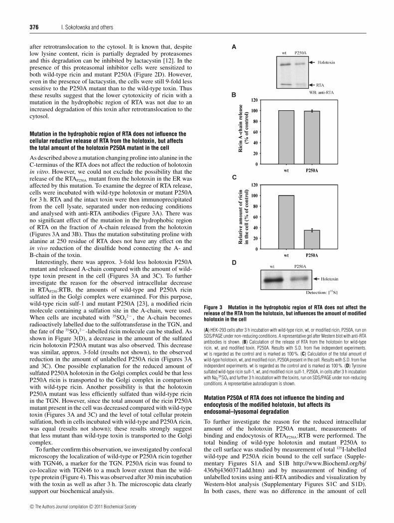
As described above a mutation changing proline into alanine in theC-terminus of the RTA does not affect the reduction of holotoxinin vitro. However, we could not exclude the possibility that therelease of the RTAP250A mutant from the holotoxin in the ER wasaffected by this mutation. To examine the degree of RTA release,cells were incubated with wild-type holotoxin or mutant P250Afor 3 h. RTA and the intact toxin were then immunoprecipitatedfrom the cell lysate, separated under non-reducing conditionsand analysed with anti-RTA antibodies (Figure 3A). There wasno significant effect of the mutation in the hydrophobic regionof RTA on the fraction of A-chain released from the holotoxin(Figures 3A and 3B). Thus the mutation substituting proline withalanine at 250 residue of RTA does not have any effect on thein vivo reduction of the disulfide bond connecting the A- andB-chain of the toxin.
Interestingly, there was approx. 3-fold less holotoxin P250Amutant and released A-chain compared with the amount of wild-type toxin present in the cell (Figures 3A and 3C). To furtherinvestigate the reason for the observed intracellular decreasein RTAP250:RTB, the amounts of wild-type and P250A ricinsulfated in the Golgi complex were examined. For this purpose,wild-type ricin sulf-1 and mutant P250A [23], a modified ricinmolecule containing a sulfation site in the A-chain, were used.When cells are incubated with 35SO4
2 − , the A-chain becomesradioactively labelled due to the sulfotransferase in the TGN, andthe fate of the 35SO4
2 − -labelled ricin molecule can be studied. Asshown in Figure 3(D), a decrease in the amount of the sulfatedricin holotoxin P250A mutant was also observed. This decreasewas similar, approx. 3-fold (results not shown), to the observedreduction in the amount of unlabelled P250A ricin (Figures 3Aand 3C). One possible explanation for the reduced amount ofsulfated P250A holotoxin in the Golgi complex could be that lessP250A ricin is transported to the Golgi complex in comparisonwith wild-type ricin. Another possibility is that the holotoxinP250A mutant was less efficiently sulfated than wild-type ricinin the TGN. However, since the total amount of the ricin P250Amutant present in the cell was decreased compared with wild-typetoxin (Figures 3A and 3C) and the level of total cellular proteinsulfation, both in cells incubated with wild-type and P250A ricin,was equal (results not shown); these results strongly suggestthat less mutant than wild-type toxin is transported to the Golgicomplex.
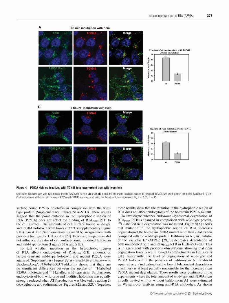
To further confirm this observation, we investigated by confocalmicroscopy the localization of wild-type or P250A ricin togetherwith TGN46, a marker for the TGN. P250A ricin was found toco-localize with TGN46 to a much lower extent than the wild-type protein (Figure 4). This was observed after 30 min incubationwith the toxin as well as after 3 h. The microscopic data clearlysupport our biochemical analysis.
Figure 3 Mutation in the hydrophobic region of RTA does not affect therelease of the RTA from the holotoxin, but influences the amount of modifiedholotoxin in the cell
(A) HEK-293 cells after 3 h incubation with wild-type ricin, wt, or modified ricin, P250A, run onSDS/PAGE under non-reducing conditions. A representative gel after Western blot with anti-RTAantibodies is shown. (B) Calculation of the release of RTA from the holotoxin for wild-typericin, wt, and modified toxin, P250A. Results with S.D. from five independent experiments.wt is regarded as the control and is marked as 100 %. (C) Calculation of the total amount ofwild-type holotoxin, wt, and modified ricin, P250A present in the cell. Results with S.D. from fiveindependent experiments. wt is regarded as the control and is marked as 100 %. (D) Tyrosinesulfated wild-type ricin sulf-1, wt, and modified ricin sulf-1, P250A, in cells after 3 h incubationwith Na2
34SO4 and further 3 h incubation with the toxins, run on SDS/PAGE under non-reducingconditions. A representative autoradiogram is shown.
Mutation P250A of RTA does not influence the binding andendocytosis of the modified holotoxin, but affects itsendosomal–lysosomal degradation
To further investigate the reason for the reduced intracellularamount of the holotoxin P250A mutant, measurements ofbinding and endocytosis of RTAP250A:RTB were performed. Thetotal binding of wild-type holotoxin and mutant P250A tothe cell surface was studied by measurement of total 125I-labelledwild-type and P250A ricin bound to the cell surface (Supple-mentary Figures S1A and S1B http://www.BiochemJ.org/bj/436/bj4360371add.htm) and by measurement of binding ofunlabelled toxins using anti-RTA antibodies and visualization byWestern-blot analysis (Supplementary Figures S1C and S1D).In both cases, there was no difference in the amount of cell
c© The Authors Journal compilation c© 2011 Biochemical Society
Intracellular transport of RTA (P250A) 377
Figure 4 P250A ricin co-localizes with TGN46 to a lower extent than wild-type ricin
Cells were incubated with wild-type ricin or mutant P250A for 30 min (A) or 3 h (B) before the cells were fixed and stained as indicated. DRAQ5 was used to stain the nuclei. Scale bars 10 μm.Co-localization of wild-type ricin or mutant P250A with TGN46 was measured using the JaCoP tool. Bars represent S.D.; P < 0.05, n = 15.
surface bound P250A holotoxin in comparison with the wild-type protein (Supplementary Figures S1A–S1D). These resultssuggest that the point mutation in the hydrophobic region ofRTA (P250A) does not affect the binding of RTAP250A:RTB tothe cell surface. The amounts of cell surface bound wild-typeand P250A holotoxin were lower at 37 ◦C (Supplementary FigureS1B) than at 0 ◦C (Supplementary Figure S1A), in agreement withprevious findings for HeLa cells [28]. However, temperature didnot influence the ratio of cell surface-bound modified holotoxinand wild-type protein (Figures S1A and S1B).
To test whether mutation in the hydrophobic regionof RTA affects endocytosis of RTAP250A:RTB, amounts oflactose-resistant wild-type holotoxin and mutant P250A wereanalysed. Supplementary Figure S2(A) (available at http://www.BiochemJ.org/bj/436/bj4360371add.htm) shows that there areno significant differences between the uptake of 125I-labelledP250A holotoxin and 125I-labelled wild-type ricin. Furthermore,endocytosis of both wild-type and modified holotoxin was equallystrongly reduced when ATP production was blocked by adding 2-deoxyglucose and sodium azide (Figures S2B and S2C). Together,
these results show that the mutation in the hydrophobic region ofRTA does not affect endocytosis of the holotoxin P250A mutant.
To investigate whether endosomal–lysosomal degradation ofRTAP250A:RTB is changed in comparison with wild-type protein,125I -labelled ricin degradation was measured. Figure 5(A) showsthat mutation in the hydrophobic region of RTA increasesdegradation of the holotoxin P250A mutant more than 2-fold whencompared with the wild-type protein. Bafilomycin A1, an inhibitorof the vacuolar H+ -ATPase [29,30] decreases degradation ofboth unmodified ricin and RTAP250A:RTB in HEK-293 cells. Thisis in agreement with previous observations, showing that ricindegradation takes place in low-pH compartments in HeLa cells[31]. Importantly, the level of degradation of wild-type andP250A holotoxin in the presence of bafilomycin A1 is almostequal, strongly indicating that the low-pH-dependent degradationmachinery is at least partially responsible for the increased ricinP250A mutant degradation. These results were confirmed in theexperiments where the total amount of wild-type and P250A ricinin cells treated with or without bafilomycin A1 were estimatedby Western-blot analysis using anti-RTA antibodies. As shown
c© The Authors Journal compilation c© 2011 Biochemical Society
378 I. Sokołowska and others
Figure 5 P250A ricin shows increased degradation in low pH vesicles
(A) Cells were pre-incubated with (+ baf) or without bafilomycin A1 (0.1 μM) and 125I-labelled wild-type ricin, wt, or modified 125I-labelled ricin, P250A, were added. Ricin degradation was measuredas described in the Experimental section. Results are means +− S.D. from five independent experiments. wt is regarded as the control and is marked as 100 %. Other results are relative to this control.Cells were treated with or without bafilomycin A1 (0.1 μM) (B), pepstatin A (pep) (10 μg/ml) (D); CA074 methyl ester (CA074-Me) (50 μM) (F) or a combination of pepstatin A and CA074 methylester (inhibitors) (H) and unlabelled RTA:RTB or RTAP250A:RTB. The amount of wild-type and P250A ricin that remain in the cell after degradation was analysed after SDS/PAGE under non-reducingconditions (see the Experimental section). Representative membranes after Western blot with anti-RTA antibodies are shown. (C, E, G, I) Results are means +− S.D. from five independent experimentspresented in (B, D, F, H) respectively. wt is regarded as the control and is marked as 1. Other results are relative to this control.
in Figures 5(B) and 5(C), bafilomycin A1 increases intracellularamounts of wild-type ricin and mutant P250A approximately tothe same level, strengthening the suggestion that the observeddecrease in the amount of the ricin holotoxin P250A mutant inthe cell (Figure 3) is caused by increased endosomal–lysosomaldegradation of holotoxin containing a mutation in RTA.
It has been demonstrated that the aspartyl protease cathepsinD and cysteine protease cathepsin B both participate in ricincleavage [6]. To estimate if these proteases are responsible for
increased P250A ricin degradation, specific inhibitors wereemployed. Pepstatin A is a potent inhibitor of aspartyl proteasesincluding cathepsin D; CA074 methyl ester blocks cysteinecathepsins, especially cathepsin B. Figures 5(D)–5(I) show thatboth types of proteases are involved in ricin P250A degradation.However, it seems that cathepsin B is more important in thisprocess (Figures 5F and 5G). In the presence of the CA074methyl ester the level of P250A ricin is significantly increased.The combination of pepstatin A and CA074 methyl ester seems to
c© The Authors Journal compilation c© 2011 Biochemical Society
Intracellular transport of RTA (P250A) 379
Figure 6 Differences in the intracellular amount of wild-type ricin andmutant P250A increase with the time of toxin incubation
Lower amount of the ricin P250A mutant is recycled back to the cell surface. (A) Cells wereincubated with wild-type ricin, wt, or modified ricin, P250A. Times of incubation at 37◦C areindicated. SDS/PAGE under non-reducing conditions was performed. A representative membraneafter Western blotting (WB) with anti-RTA antibodies is shown. (B) Results are means +− S.D.from five independent experiments presented in (A). (C) The amount of 125I-labelled wild-type,wt, or 125I-labelled modified ricin, P250A, recycled back to the cell surface was measuredas described in the Experimental section. Results are means +− S.D. from five independentexperiments.
increase the cellular level of P250A ricin even further (Figures 5Hand 5I), but not much more than in cells treated only with theCA074 methyl ester (Figures 5F and 5G). Thus mainly cathepsinB is responsible for the endosomal–lysosomal degradation of thericin holotoxin P250A mutant in the cell.
To study degradation time dependency, the amount of wild-typeholotoxin and mutant P250A in the cells were analysed after10, 30, 45 and 120 min incubations with the toxins (Figures 6Aand 6B). Membranes were probed with anti-RTA antibodies andWestern-blot analysis was performed. As shown in Figures 6(A)and 6(B), the differences between intracellular amounts ofwild-type and P250A ricin increase with the time of incubation.Statistical analysis confirmed that the intracellular amount ofricin is dependent on the time and type of ricin (ANOVA II, time– F3,72 = 24, P < 0.001; type of ricin – F1,72 = 37, P < 0.001;
for both factors F3,72 = 8, P < 0.001). Moreover, differencesbetween amounts of wild-type and P250A ricin after indicatedtimes of toxin incubation are statistically significant (P < 0.001);however, differences after 10 min are marginal.
It has been reported previously that proteolytic cleavage of theRTA and ricin holotoxin takes place mainly in early endosomes,additionally in late endosomes [6,7]. If degradation of the ricinP250A mutant is principally dependent on endosomal proteases,one could possibly observe differences in the amount of wild-type and P250A ricin recycled back to the cell surface. Toanalyse recycling, 125I-labelled wild-type and mutant P250Aricin were used. The amount of the holotoxin P250A mutantrecycled back to the cell surface was significantly decreasedin comparison with wild-type ricin (Figure 6C). These resultssupport the hypothesis that ricin containing mutation in the RTAcould be more extensively degraded in endosomes than wild-type ricin and therefore less P250A mutant is recycled back tothe cell surface. This observation also supports the hypothesisthat the lower amount of the sulfated holotoxin P250A mutantresults from the decreased transport of RTAP250A:RTB to theGolgi apparatus, caused by a lower endosomal amount of P250Aholotoxin accessible for endosome-to-Golgi transport.
Mutation in the hydrophobic region of RTA affects itsretrotranslocation from the ER to the cytosol
To investigate whether retrotranslocation of RTA from the ERto the cytosol is affected by the mutation in the C-terminalregion of the ricin A-chain, transport of the P250A mutant to thecytosol was studied. Cells incubated with wild-type or modifiedricin for 3 h were subjected to permeabilization with digitoninto separate the cytosolic fraction from the ER membranes. In acontrol experiment, we tested that ER proteins remained in theER after permeabilization. We studied the distribution of cnx, anER membrane protein, and crt, a soluble ER protein. As shown inFigure 7(H), these protein markers were not released from the ERto the cytosolic fraction, indicating that ER membranes remainedintact during digitonin treatment.
Figures 7(A) and 7(B) show an 8–9-fold reduction in the amountof modified RTA present in the cytosol fraction in comparison withwild-type RTA. Approx. 3-fold less efficient retrotranslocation ofRTAP250A from the ER to the cytosol was observed in comparisonwith wild-type RTA (Figures 7C and 7D). Importantly, the differ-ence in the amount of free wild-type RTA and RTAP250A in the ERis not higher than 3-fold (Figures 3A, 7C and 7E), similar to dif-ferences observed for the holotoxin present in the Golgi complex.Considering the fact that the reduction of wild-type and modifiedholotoxin in the ER is the same (Figures 3A and 3B), we concludethat the efficiency of the transport of modified RTA from the Golgicomplex to the ER is the same as for wild-type ricin. Thus themutation in the hydrophobic region of RTA affects its transportfrom the ER to the cytosol, and the observed decrease in cytosolicRTA P250A cannot be explained by reduced transport to the ER.
The difference in the cytoplasmic accumulation of wild-type and P250A RTA is not due to increased proteasomaldegradation of RTAP250A. A proteasomal inhibitor, lactacystin,increases the amounts of both wild-type and modified ricinequally (Figures 7F and 7G). These results are consistent withthe results of cytotoxicity experiments performed in the presenceof lactacystin (Figure 2D).
The combination of a higher degradation level of the ricinP250A mutant in endosomes and the lower retrotranslocation ofRTAP250A from the ER to the cytosol might explain the estimated9-fold lower toxic effect of the holotoxin P250A mutant than ofwild-type ricin (Figure 2).
c© The Authors Journal compilation c© 2011 Biochemical Society
380 I. Sokołowska and others
Figure 7 Decreased retrotranslocation of the RTA P250A mutant from the ER to the cytosol
Cells incubated with wild-type ricin, wt, or modified ricin, P250A, were subjected to permeabilization (see the Experimental section). (A) A representative membrane after Western blotting (WB)with anti-RTA antibodies is shown. SDS/PAGE was performed under reducing conditions. The experiments were done in duplicates. (B) Average data with S.D. from five independent experimentspresented in (A). Graph shows relative amount of wild-type RTA, wt, and modified RTA, P250A, present in the cytosolic fraction. wt is marked as 1, and the amount of P250A is relative to thiscontrol. (C) A representative membrane after Western blotting with anti-RTA antibodies is shown. SDS/PAGE was performed under non-reducing conditions. The membrane represents RTA presentin the ER. The images have been grouped from different parts of the same gel, which is indicated by the dividing line. (D) Average data with S.D. from five independent experiments presented in (C).The graph shows percentage of wild-type RTA, wt, and modified RTA, P250A, retrotranslocated to the cytosol. (E) Average data with S.D. from five independent experiments presented in (C). Graphshows differences between wild-type RTA, wt, and modified RTA, P250A, present in the ER. wt is marked as 1, and the amount of P250A is relative to this control. (F) A representative membrane afterWestern blot with anti-RTA antibodies is shown. SDS/PAGE was performed under non-reducing conditions. Cells were preincubated with lactacystin (10 μM) before the addition of ricin. (G) Averagedata with S.D. from five independent experiments are presented in (F). The graph shows a relative amount of wild-type RTA, wt, and modified RTA, P250A, present in the cytosolic fraction. Amountof wt RTA is marked as 1, and other results are relative to this control. Lac, lactacystin. (H) Distribution of the resident ER proteins, cnx and crt, after permeabilization with digitonin. A representativemembrane after Western blotting with anti-cnx and anti-crt is shown. Such permeabilization control was performed for experiments presented in Figures 7–9.
Retrotranslocation of the RTA mutant from the ER to the cytosol isEDEM1-independent
It has been shown previously that the ER chaperone protein,EDEM1 plays a direct role in ricin transport from the ER tothe cytosol [13]. To further investigate the mechanism behindthe reduced translocation of the P250A mutant to the cytosol,
experiments with EDEM1-transfected cells and cells transfectedwith siRNA (small interfering RNA) vectors against EDEM1were performed. Retrotranslocation of the wild-type and RTAP250A mutant was studied in cells permeabilized with digitonin,as described above. Two different vectors expressing shRNA(short hairpin RNA), against both mouse and human EDEM1,have been described previously [13]. Figure 8(C) demonstrates a
c© The Authors Journal compilation c© 2011 Biochemical Society

Intracellular transport of RTA (P250A) 381
Figure 8 P250A RTA retrotranslocates in an EDEM1-independent manner: EDEM1-knockdown studies
Cells transfected with siRNA constructs against EDEM1 or with an unspecific siRNA construct (ctrl) incubated with wild-type ricin, wt, or modified ricin, P250A, were subjected to permeabilization(see the Experimental section). (A) A representative experiment is shown with siRNA construct number 1 (siRNA1). SDS/PAGE was performed under reducing conditions. (B) Average data with S.D.from five independent experiments with siRNA1 and siRNA construct number 2 (siRNA2). Graphs show relative amount of wild-type RTA, wt, and modified RTA, P250A, present in the cytosolicfraction. Amounts of wt RTA are marked as 1, and other results are relative to this control. (C) The presence of EDEM1–HA was determined by Western blotting (WB) with anti-HA antibodies. Cellswere co-transfected with HA-tagged mouse EDEM1 and siRNA constructs against EDEM1. Membranes were re-probed with anti-tubulin antibodies for equal loading control. (D) Protein synthesiswas measured after 12 h incubation with the toxins (see the Experimental section). Wild-type ricin, wt (closed circles); wild-type ricin in siRNA-transfected cells, wt siRNA1 (closed squares); modifiedricin, P250A (open circles); modified ricin in siRNA-transfected cells, P250A siRNA1 (open squares). (E) Average data with S.D. from five independent experiments with siRNA1 and siRNA2. Thelevel of 50 % incorporation of [3H]leucine with wild-type ricin, wt, is marked as 1 and the concentration of wt and P250A required to induce a similar inhibition under other conditions are plotted asrelative values compared with this control.
very efficient knockdown of mouse EDEM1 for both constructs.Reduction in the expression of endogenous human EDEM1 wasconfirmed by real-time RT–PCR (reverse transcription–PCR),which revealed a 70–95% reduction in the mRNA level of thisgene (results not shown).
In cells with reduced amounts of EDEM1, the level of wild-typeRTA present in the cytosol was decreased (Figures 8A and 8B), inagreement with previous findings [13]. This confirms a direct roleof EDEM1 in RTA transport to the cytosol. Importantly, the trans-port of the RTA P250A mutant from the ER to the cytosol seemsto be EDEM1 independent, as retrotranslocation assays showedno significant differences in the cytosolic amount of RTAP250A incells with reduced amounts of EDEM1 in comparison with cellstransfected with an empty control vector (Figures 8A and 8B).
To determine whether the results from the retrotranslocationassays correspond to the inhibition of protein synthesis causedby the translocated toxin, control cells and cells transfected with
siRNA vectors against EDEM1 were incubated with increasingconcentrations of wild-type ricin or mutant P250A. We observedan ∼2-fold protection (statistically significant, P < 0.001) ofsiRNA-transfected cells against wild-type ricin in comparisonwith control cells and no significant influence of EDEM1 down-regulation on the cytotoxicity of P250A ricin (Figures 8D and8E). These results are in agreement with the suggestion that theRTA P250A mutant lacks EDEM1 contribution in the transportto the cytosol. This might explain the observed decrease in theretrotranslocation of RTAP250A from the ER to the cytosol incomparison with wild-type ricin (Figure 7).
Permeabilization assays in cells with overproduction ofEDEM1 revealed a decrease in both wild-type RTA andmutant P250A present in the cytosol, when compared withcells transfected with control cDNA (Figures 9A and 9B). Itwas shown previously that high expression of EDEM1 increasesERAD [24] by promoting extraction of misfolded proteins from
c© The Authors Journal compilation c© 2011 Biochemical Society
382 I. Sokołowska and others
Figure 9 P250A RTA retrotranslocation: EDEM1 overexpression studies
(A) EDEM1-transfected cells or cells transfected with an empty control vector (ctrl) incubated with wild-type ricin, wt, or modified ricin, P250A, were subjected to permeabilization (see the Experimentalsection). A representative membrane after Western blotting (WB) with anti-RTA antibodies is shown. SDS/PAGE was performed under reducing conditions. (B) Average data with S.D. from fiveindependent experiments are presented in (A). The graph shows relative amount of wild-type RTA, wt and modified RTA, P250A, present in the cytosolic fraction. Amount of wt RTA is marked as 1,other results are relative to this control. (C) Protein synthesis was measured after 12 h incubation with toxins (see the Experimental section). Wild-type ricin, wt (closed circles); wild-type ricin inEDEM1-transfected cells, wt EDEM1 (closed squares); modified ricin, P250A (open circles); modified ricin in EDEM1-transfected cells, P250A EDEM1 (open squares). A representative experiment isshown. (D) Average data with S.D. from five independent experiments. The level of 50 % incorporation of [3H]leucine with wild-type ricin, wt, is marked as 1 and the concentration of wt and P250Arequired to induce a similar inhibition under other conditions are plotted as relative values compared with this control.
the cnx cycle [16,24]. Thus in EDEM1-transfected cells, thetranslocon is mainly occupied by misfolded proteins trans-ported to the cytosol for degradation and access of ricin to thetranslocon might be inhibited. This suggestion was confirmedin experiments with overproduction of model misfolded proteins[13]. EDEM1 promotes RTA transport from the ER to the cytosol,but retrotranslocation assays showed a decreased amount of RTApresent in the cytosol in EDEM1-transfected cells due to ERADacceleration [13]. Therefore, in cells with high levels of EDEM1,the availability of the translocon might be limited not only forwild-type but also for RTAP250A, despite the different influence ofEDEM1 on RTA and RTAP250A retrotranslocation to the cytosol.In agreement with this idea, we observed protection against bothwild-type ricin and mutant P250A in cells with overproduction ofEDEM1 (Figures 9C and 9D).
Together, these results support the hypothesis that mutationin the hydrophobic region of RTA disrupts EDEM1-dependenttransport of RTA to the cytosol.
P250A mutation impairs interaction between EDEM1 and RTA
It was shown previously that EDEM1 interacts directly withthe ricin holotoxin [13]. To study the interactions betweenRTA and EDEM1 and between RTAP250A and EDEM1, we
cloned and purified the RTA mutant with a His-tag (see theExperimental section). As shown in Figure 10, EDEM1 interactswith the RTA. This new finding more precisely characterizes thepreviously observed interaction between EDEM1 and holotoxin[13]. Interestingly, a significant decrease in RTAP250A bindingto EDEM1 was observed when compared with wild-type RTA(Figure 10). These results might explain EDEM1-independenttransport of RTAP250A to the cytosol. Importantly, CD analysisof RTA and RTAP250A with His-tags confirmed altered secondarystructure of RTAP250A, presented above (Figure 1B).
Our pull-down assays were confirmed by in vivo experiments.We co-immunoprecipitated wild-type ricin or P250A ricin withanti-HA antibodies from lysates of EDEM1-HA-transfectedcells or cells transfected with a control empty vector. As shown inFigure 10(C), both types of ricin co-immunoprecipitate withEDEM1. Importantly, much more wild-type ricin immunoprecip-itates with EDEM1 in comparison with P250A ricin (Figures 10Cand 10D). The level of interaction between wild-type ricin andEDEM1 and P250A ricin and EDEM1 differs in comparison withpull-down assays. This is due to the already decreased amountof P250A reaching the Golgi complex and consequently the ER(Figure 3).
These results additionally confirm that the interaction betweenP250A ricin and EDEM1 is decreased in comparison withwild-type ricin. Since the P250A mutation slightly changes the
c© The Authors Journal compilation c© 2011 Biochemical Society
Intracellular transport of RTA (P250A) 383
Figure 10 Point mutation P250A in the hydrophobic region of the RTAimpairs interaction between EDEM1 and RTA
(A) Protein interactions were examined by His-tag pull-down assay using RTAP250A–His andRTA–His (0.5 μg each) and lysates from HEK-293 cells overexpressing EDEM1-HA or cellstransfected with an empty control vector, ctrl (see the Experimental section). Proteins bound tothe beads were analysed by SDS/PAGE and Western blotting (WB) with anti-HA antibodies(upper panel). The same membranes were re-probed with anti-His or anti-RTA anti-bodies (middle panel, Western blot with anti-RTA is shown). Whole cell lysates (WCL) analysedwith anti-HA antibodies are shown in the bottom panel. Lines: 1–3 empty vector-transfected cells,ctrl; 4–6 EDEM1-HA-transfected cells, 1 and 4 without toxin; 2 and 5 with RTA-His; 3 and 6 withRTAP250A-His. A representative experiment is shown. (B) Average data from His-tag pull-downwith S.D. from five independent experiments. The amount of EDEM1 bound to RTAP250A is relativeto the amount of EDEM1 interacting with wild-type RTA marked as 1. (C) Immunoprecipitation (IP)of wild-type ricin or P250A ricin with anti-HA antibodies. Representative example of experimentsis shown. SDS/PAGE was performed under non-reducing conditions. Membranes were re-probedwith anti-HA antibodies to confirm equal immunoprecipitation. (D) Average data with S.D. fromfive independent experiments are presented in (C). The level of immunoprecipitated wild-typericin from EDEM1-transfected cells is marked as 1, the level of immunoprecipitated P250A ricinis relative to this value.
conformation of the ricin A-chain into a more helical structure,it is likely that the precise structure of an ERAD substrate mightdetermine its recognition by EDEM1.
DISCUSSION
Increased knowledge about protein toxins from plants and bacteriais important both for the development of new therapeutic strategiesand for characterization of basal mechanisms in cell biology.The results in the present study show that point mutation(P250A) in the hydrophobic region of the RTA influencesendosomal–lysosomal degradation of the toxin, as well as RTAretrotranslocation from the ER to the cytosol. Our results indicatethat the interactions between structurally changed RTAP250A
and EDEM1 are impaired. This observation may significantlycontribute to the general understanding of the recognition ofprotein substrates by EDEM1.
Our results show that mutated P250A ricin is 9-fold less toxicthan wild-type ricin to Vero and HEK-293 cells. It has beendemonstrated previously that the P250A mutant has significantlylower activity than wild-type ricin in Vero cells [9], but thereason behind the reduced cytotoxicity was not investigated.Differences in the activity of P250A and wild-type ricin reportedpreviously [9] were higher than those described in the presentstudy. Slightly different cell culture conditions, some variability inprotein synthesis measurements (e.g. in the present study, resultsfor single radioactivity measurement come from much higheramounts of cells) and different sublines of Vero cells may explainsuch a discrepancy.
Results from experiments performed in the presence ofbafilomycin A, which prevents the acidification of endosomesand lysosomes, suggested an increased endosomal–lysosomaldegradation of the mutated P250A holotoxin. Furthermore, thekinetics of degradation suggested that P250A ricin degradationstarts already in the endosomes, as previously reported for wild-type ricin [6,7]. It has been demonstrated that the proteasescathepsin B and cathepsin D are responsible for RTA degradationin early endosomes in macrophages; cathepsin B is responsiblefor toxin cleavage at both neutral and acidic pH [6]. Our resultsclearly indicate that cathepsin B and additionally cathepsin Dare involved in increased P250A ricin degradation. Distinctendosomal proteases appear to be active in different cell types. Inour study with HEK-293 cells, both wild-type ricin and mutatedP250A degradation is at least partially dependent on low pH, sincebafilomycin A treatment increased intracellular amounts of bothtoxins and decreased their degradation.
It was observed previously that addition of the RTB resulted inthe protection of RTA from proteolytic activities of lysosomesand cathepsins [32]. This protective role of RTB is partiallyresponsible for the higher toxicity of ITs containing ricinholotoxin in comparison with ITs containing only RTA. Re-association of RTAP250A with RTB produces a holotoxin with areducible disulfide bond; it produces a toxin that is equally boundto the cell surface and equally endocytosed as wild-type ricin.However, due to the structural changes in the mutated P250ARTA, RTAP250A:RTB can have altered conformation in comparisonwith the wild-type holotoxin. This altered conformation and/orlocalization of RTB in relation to RTAP250A might influence itsdegradation. It should be noted that in vitro degradation of RTAand RTAP250A by pronase at either pH 5.5 and 6.5 gives similardegradation patterns (results not shown).
Our results demonstrate that a point mutation (P250A) inthe RTA decreases its retrotranslocation from the ER to thecytosol. A lack of EDEM1 contribution in this transport might be
c© The Authors Journal compilation c© 2011 Biochemical Society
384 I. Sokołowska and others
partially responsible for the observed effect. EDEM1 promotesRTA retrotranslocation to the cytosol [13]; however, this proteinmay not be the only one that facilitates RTA transport to thecytosol. In addition to the EDEM1 variant protein [24], EDEM2and EDEM3 homologues also exist in the ER lumen [33–35]. BothEDEM2 and EDEM3 could be involved in ricin retrotranslocation.It is also possible that ricin can use not only Sec61p but alsoother channels/translocons for its transport to the cytosol. Thusone can expect that RTAP250A retrotranslocation from the ER tothe cytosol might be influenced by additional factors; a lack ofthe assistance of EDEM1 may not be the only reason for thereduction of RTAP250A transport to the cytosol.
It has been demonstrated that RTA binds directly to themembrane surface; at the physiologically relevant temperature of37 ◦C, the membrane-bound RTA loses some α-helical structures,undergoing the conformational change that exposes its C-terminalregion to the membrane interior [36]. Such insertion into the lipidbilayer might represent an early step in RTA translocation throughthe ER membrane. It is possible that the P250A mutant possessingan elevated level of α-helices is unable to undergo additionalconformational changes allowing it to be stably inserted into theER membrane. This might be another limiting step in RTAP250A
retrotranslocation to the cytosol.EDEM1 substrate recognition and sorting to the ERAD
pathway are still poorly defined. It has been proposed that EDEM1acts either as mannosidase that produces de-mannosylatedglycoproteins or as the receptor that recognizes, binds and directsmannose-trimmed proteins for ERAD by extracting them fromthe cnx cycle [16–19]. On the other hand, it has been proposedthat EDEM1 substrate recognition might be glycan independent.EDEM1 directly interacts with non-glycosylated ricin, thisinteraction is independent of glycans presented on RTB [13].Hebert and co-workers [21] demonstrated that EDEM1 bindingdoes not require the trimming of substrate glycans or even ERADsubstrate glycosylation, thus suggesting that EDEM1 probablyrecognizes misfolded regions of aberrant proteins. Similarly,EDEM1 binding to mutant P23H rod opsin was independent ofmannose trimming [22]. In the present study, we demonstrate thatpoint mutation in the hydrophobic region of the RTA decreases theinteraction between RTA and EDEM1. It has been suggested thatEDEM1 interacts with proteins via hydrophobic domains [37].Hydrophobicity of the mutated P250A C-terminal domain of RTAis not changed dramatically in comparison with wild-type RTA.However, a point mutation in this region changes the secondarystructure of RTA into more a helical one. This suggests that proteinstructure might influence EDEM1–protein substrate recognition.This hypothesis has to be confirmed in further experiments.Understanding the mechanisms of recognition and degradation ofmisfolded proteins synthesized in the ER is one of the fundamentalissues in cell biology.
The potency of the protein toxin ricin can be combined withthe specificity of various targeting moieties to yield ITs usedin cancer therapy. However, a major disadvantage is the dose-limiting toxicity associated with ricin-conjugated ITs, whichleads to VLS (vascular leak syndrome). It is characterized byhypoalbuminaemia, peripheral oedema and pulmonary oedemain most severe cases [38]. VLS is caused by RTA, which damagesvascular endothelial cells by increasing their permeability in atime- and dose-dependent manner [39]. The exact mechanism bywhich RTA mediates VLS in vivo remains to be determined, butthere is evidence suggesting that direct cytotoxicity of RTA leadsto VLS in vivo [39]. Thus, as an alternative, the use of toxins thatare less cytotoxic to endothelial cells are proposed. Ricin with amutated hydrophobic region of RTA seems to be a good candidatefor such studies. It is still potent enough to be used in ITs, on the
other hand it is possible that VLS caused by this toxin would bestrongly limited. The influence of RTAP250A on endothelial cellpermeability should be determined.
In conclusion, the results presented in the present studycontribute to our general understanding of ricin intracellulartransport and mechanism of the recognition of misfolded proteinsin the ER. This knowledge might bring new solutions intotherapies of severe human diseases.
AUTHOR CONTRIBUTION
Monika Słominska-Wojewodzka and Kirsten Sandvig conceived the project and designedthe experiments. Monika Słominska-Wojewodzka and Iwona Sokołowska performed allexperiments and collected the data. Sebastien Walchli cloned RTA-His and established thepurification protocol. Sebastien Walchli and Monika Słominska-Wojewodzka performedthe confocal microscopy experiments. Monika Słominska-Wojewodzka, Kirsten Sandvigand Iwona Sokołowska analysed the data. Monika Słominska-Wojewodzka and KirstenSandvig wrote the manuscript together with Sebastien Walchli, Grzegorz W
↪egrzyn and
Iwona Sokołowska. All authors read and approved the final version of the manuscript.
ACKNOWLEDGEMENTS
We are grateful to Professor Kazuhiro Nagata and Dr Nobuko Hosokawa (Institute forFrontier Medical Sciences, Kyoto University, Kyoto, Japan) for cDNA encoding the mouseEDEM1. We would like to thank Dr Danuta Augustin-Nowacka (Faculty of Chemistry,University of Gdansk, Gdansk, Poland) for CD measurements and analysis. We aregrateful to Dr Sigrid S. Skanland (Institute of Biochemistry II, Goethe University School ofMedicine, University Hospital, Frankfurt, Germany) and to Dr Anna Herman-Antosiewicz(Department of Molecular Biology, University of Gdansk, Gdansk, Poland) for help inthe confocal microscopy images preparation. We thank Dr Anna Kawiak (Department ofBiotechnology, Intercollegiate Faculty of Biotechnology, University of Gdansk, Gdansk,Poland) for critical reading of the manuscript.
FUNDING
This work was supported by the Ministry of Science and Higher Education [grantnumber 3682/P01/2006/32], Foundation for Polish Science [grant number ‘Homing’HOM/12/2007] and by the University of Gdansk [grant number 1480-5-0344-6].
REFERENCES
1 Vitetta, E. S. and Thorpe, P. E. (1991) Immunotoxins containing ricin or it’s A chain.Semin. Cell Biol. 2, 47–58
2 Schnell, R., Borchmann, P., Staak, L. O., Schindler, J., Ghetie, V., Vitetta, E. S. andEngert, A. (2003) Clinical evaluation of ricin A-chain immunotoxins in patients withHodgkin’s lymphoma. Ann. Oncol. 14, 729–736
3 Youn, Y. S., Na, D. H., Yoo, S. D., Song, S. C. and Lee, K. C. (2005) Carbohydrate-specifically polyethylene glycol-modified ricin A-chain with improved therapeuticpotential. Int. J. Biochem. Cell Biol. 37, 1525–1533
4 Polito, L., Bortolotti, M., Farini, V., Battelli, M. G., Barbieri, L. and Bolognesi, A. (2009)Saporin induces multiple death pathways in lymphoma cells with different intensity andtiming as compared to ricin. Int. J. Biochem. Cell Biol. 41, 1055–1061
5 Sandvig, K., Lauvrak, S. U. and van Deurs, B. (2005) Host cell penetration and traffickingof protein toxins. In Microbial Toxins: Molecular and Cellular Biology (Norfolk, T. P., ed.),pp. 473–469, Horizon Press, Wymondham
6 Blum, J. S., Fiani, M. L. and Stahl, P. D. (1991) Proteolytic cleavage of ricin A chain inendosomal vesicles. J. Biol. Chem. 266, 22091–22095
7 Brech, A., Kjeken, R., Synnes, M., Berg, T., Roos, N. and Prydz, K. (1998) Endocytosedricin and asialoorosomucoid follow different intracellular pathways in hepatocytes.Biochim. Biophys. Acta 1373, 195–208
8 Sandvig, K. and van Deurs, B. (2002) Membrane traffic exploited by protein toxins. Annu.Rev. Cell Biol. 18, 1–14
9 Simpson, J. C., Lord, J. M. and Roberts, L. M. (1995) Point mutations in the hydrophobicC-terminal region of ricin A chain indicate that Pro250 plays a key role in membranetranslocation. Eur. J. Biochem. 232, 458–463
10 Romisch, K. (2005) Endoplasmic reticulum-associated degradation. Annu. Rev. Cell Dev.Biol. 21, 435–456
11 Bukau, B., Weissman, J. and Horwich, A. (2006) Molecular chaperones and proteinquality control. Cell 125, 443–451
c© The Authors Journal compilation c© 2011 Biochemical Society

Intracellular transport of RTA (P250A) 385
12 Wesche, J., Rapak, A. and Olsnes, S. (1999) Dependence of ricin toxicity on translocationof the toxin A-chain from the endoplasmic reticulum to the cytosol. J. Biol. Chem. 274,34443–34449
13 Słominska-Wojewodzka, M., Gregers, T. F., Walchli, S. and Sandvig, K. (2006) EDEM isinvolved in retrotranslocation of ricin from the endoplasmic reticulum to the cytosol. Mol.Biol. Cell 17, 1664–1975
14 Spooner, R. A., Hart, P. J., Cook, J. P., Pietroni, P., Rogon, C., Hohfeld, J., Roberts, L. M.and Lord, J. M. (2008) Cytosolic chaperones influence the fate of a toxin dislocated fromthe endoplasmic reticulum. Proc. Natl. Acad. Sci. U.S.A. 105, 17408–17413
15 Deeks, E. D., Cook, J. P., Day, P. J., Smith, D. C., Roberts, L. M. and Lord, J. M. (2002)The low lysine content of ricin A chain reduces the risk of proteolytic degradation aftertranslocation from the endoplasmic reticulum to the cytosol. Biochemistry 41, 3405–3413
16 Molinari, M., Calanca, V., Galli, C., Lucca, P. and Paganetti, P. (2003) Role of EDEM in therelease of misfolded glycoproteins from the calnexin cycle. Science 299, 1397–1400
17 Oda, Y., Hosokawa, N., Wada, I. and Nagata, K. (2003) EDEM as an acceptor of terminallymisfolded glycoproteins released from calnexin. Science 299, 1394–1397
18 Olivari, S., Cali, T., Salo, K. E., Paganetti, P., Ruddock, L. W. and Molinari, M. (2006)EDEM1 regulates ER-associated degradation by accelerating de-mannosylation offolding-defective polypeptides and by inhibiting their covalent aggregation. Biochem.Biophys. Res. Commun. 349, 1278–1284
19 Hosokawa, N., Tremblay, L. O., Sleno, B., Kamiya, Y., Wada, I., Nagata, K., Kato, K. andHerscovics, A. (2010) EDEM1 accelerates the trimming of a 1,2-linked mannose on the Cbranch of N-glycans. Glycobiology 20, 567–575
20 Hosokawa, N., Wada, I., Natsuka, Y. and Nagata, K. (2006) EDEM accelerates ERAD bypreventing aberrant dimer formation of misfolded α1-antitrypsin. Genes Cells 11,465–476
21 Cormier, J. H., Tamura, T., Sunryd, J. C. and Hebert, D. N. (2009) EDEM1 recognition anddelivery of misfolded proteins to the SEL1L-containing ERAD complex. Mol. Cell 34,627–633
22 Kosmaoglou, M., Kanuga, N., Aguila, M., Garriga, P. and Cheetham, M. E. (2009) A dualrole for EDEM1 in the processing of rod opsin. J. Cell Sci. 122, 4465–4472
23 Rapak, A., Falnes, P. O. and Olsnes, S. (1997) Retrograde transport of mutant ricin to theendoplasmic reticulum with subsequent translocation to cytosol. Proc. Natl. Acad. Sci.U.S.A. 94, 3783–3788
24 Hosokawa, N., Wada, I., Hasegawa, K., Yorihuzi, T., Tremblay, L. O., Herscovics, A. andNagata, K. (2001) A novel ER alpha-mannosidase-like protein accelerates ER-associateddegradation. EMBO Rep. 2, 415–422
25 Whitmore, L. and Wallace, B. A. (2008) Protein secondary structure analyses fromcircular dichroism spectroscopy: methods and reference databases. Biopolymers 89,392–400
26 Fraker, P. J. and Speck, Jr, J. C. (1978) Protein and cell membrane iodinations with asparingly soluble chloroamide, 1,3,4,6-tetrachloro-3a,6a-diphrenylglycoluril. Biochem.Biophys. Res. Commun. 80, 849–857
27 Bolte, S. and Cordelieres, F. P. (2006) A guided tour into subcellular colocalizationanalysis in light microscopy. J Microsc. 224, 213–232
28 Sandvig, K. and Olsnes, S. (1979) Effect of temperature on the uptake, excretion anddegradation of abrin and ricin by HeLa cells. Exp. Cell Res. 121, 15–25
29 Bowman, E. J., Siebers, A. and Altendorf, K. (1998) Bafilomycins: a class of inhibitors ofmembrane ATPases from microorganisms, animal cells, and plant cells. Proc. Natl. Acad.Sci. U.S.A. 85, 7972–7976
30 Yoshimori, T., Yamamoto, Y., Moriyama, M., Futai, M. and Tashito, Y. (1991) BafilomycinA1, a specific inhibitor of vacuolar-type H( + )-ATPase, inhibits acidification and proteindegradation in lysosomes of cultured cells. J. Biol. Chem. 266, 17707–17712
31 Llorente, A., Rapak, A., Schmid, S. L., van Deurs, B. and Sandvig, K. (1998) Expression ofmutant dynamin inhibits toxicity and transport of endocytosed ricin to the Golgiapparatus. J. Cell Biol. 140, 553–563
32 Bilge, A., Howell-Clark, J., Ramakrishnan, S. and Press, O. W. (1994) Degradation of ricinA chain by endosomal and lysosomal enzymes – the protective role of ricin B chain. Ther.Immunol. 1, 197–204
33 Mast, S. W., Diekman, K., Karaveg, K., Davis, A., Sifers, R. N. and Moremen, K. W. (2004)Human EDEM2, a novel homolog of family 47 glycosidases, is involved in ER-associateddegradation of glycoproteins. Glycobiology 15, 421–436
34 Olivari, S., Galli, C., Alanen, H., Ruddock, L. and Molinari, M. (2005) A novelstress-induced EDEM variant regulating endoplasmic reticulum-associated glycoproteindegradation. J. Biol. Chem. 280, 2424–2428
35 Hirao, K., Natsuka, Y., Tamura, T., Wada, I., Morito, D., Natsuka, S., Romero, P., Sleno, B.,Tremblay, L. O., Herscovics, A. et al. (2006) EDEM3, a soluble EDEM homolog, enhancesglycoprotein endoplasmic reticulum-associated degradation and mannose trimming.J. Biol. Chem. 281, 9650–9658
36 Mayerhofer, P. U., Cook, J. P., Wahlman, J., Pinheiro, T. T., Moore, K. A., Lord, J. M.,Johnson, A. E. and Roberts, L. M. (2009) Ricin A chain insertion into endoplasmicreticulum membranes is triggered by a temperature increase to 37◦C. J. Biol. Chem.284, 10232–10242
37 Wang, T. and Hebert, D. N. (2003) EDEM an ER quality control receptor. Nat. Struct. Biol.10, 319–321
38 Vitetta, E. S., Thorpe, P. E. and Uhr, J. W. (1993) Immunotoxins: magic bullets ormisguided missles? Immunol. Today 14, 252–259
39 Lindstrom, A. L., Erlandsen, S. L., Kersey, J. H. and Pennell, C. A. (1997) An in vitromodel for toxin-mediated vascular leak syndrome: ricin toxin A chain increases thepermability of human endothelial cell monolayers. Blood 90, 2323–2334
Received 16 September 2010/7 March 2011; accepted 9 March 2011Published as BJ Immediate Publication 9 March 2011, doi:10.1042/BJ20101493
c© The Authors Journal compilation c© 2011 Biochemical Society

Biochem. J. (2011) 436, 371–385 (Printed in Great Britain) doi:10.1042/BJ20101493
SUPPLEMENTARY ONLINE DATAA single point mutation in ricin A-chain increases toxin degradationand inhibits EDEM1-dependent ER retrotranslocationIwona SOKOŁOWSKA*, Sebastien WALCHLI†, Grzegorz W
↪EGRZYN*, Kirsten SANDVIG‡ and
Monika SŁOMINSKA-WOJEWODZKA*1
*Department of Molecular Biology, University of Gdansk, Kładki 24, 80-822 Gdansk, Poland, †Department of Immunology, Institute for Cancer Research, The Norwegian RadiumHospital, Oslo University Hospital, Montebello, N-0310 Oslo, Norway, and ‡Department of Biochemistry, Institute for Cancer Research, The Norwegian Radium Hospital, Oslo UniversityHospital, Montebello, N-0310 Oslo, Norway
EXPERIMENTAL
Cloning, mutagenesis and cDNA constructs
RTA (ricin A-chain) was amplified from pRA using the followingprimers: 5′-CATGCCATGGCAATATTCCCCAAACAATACCC-3′ and 5′-CCGCTCGAGAAACTGTGACGACGGTGG-3′. Theamplicon was then cloned into pET21d (Merck, Darmstadt,Germany) using NcoI and XhoI. The first primer created an ATGand an NcoI site and the second primer introduced an XhoI sitein frame with the vector-encoded His6-tag. The resulting productwas the RTA fused to His6 in its C-terminal end. Clones weresequenced and a positive construct was transformed into Rosettabacteria (Merck) for further expression.
The RTA P250A mutant was obtained by site direct mutagenesisusing primers: 5′-GTATATTAATCGCGATCATAGCTCTCATG-3′ and 5′-CATGAGAGCTATGATCGCGATTAATATAC-3′. TheRTA sulf-1 cDNA was used as a template [1] and the reaction wasperformed by the standard procedure.
Measurements of binding and endocytosis of wild-type ricinand mutant P250A
Cell surface binding of wild-type ricin and mutant P250A wasmeasured after 30 min incubation with 125I-labelled wild-typericin or mutant P250A (∼100 ng/ml) or unlabelled counterpartsat 37 ◦C or at 0 ◦C in Hepes medium. Ricin was 125I-labelledaccording to [2], to a specific activity of 3 × 10⁴–5 × 10⁴
cpm/ng. After incubation, cells were washed four times with ice-cold Hepes-buffered medium. The cell-bound radioactivity wasmeasured using a gamma counter. For the unlabelled wild-typericin and mutant P250A visualization, Western blotting with anti-RTA antibodies was employed.
Endocytosis of wild-type ricin and mutant P250A wasmeasured as the amounts of 125I-labelled or unlabelled toxin thatcould not be removed by incubating the cells with lactose solution.Briefly, after 20 min incubation with wild-type ricin or mutantP250A (∼100 ng/ml) at 37 ◦C in Hepes medium, cells wereincubated (15 min) in Hepes medium containing 0.1 M lactose,followed by three fast washes with this medium. Optionally,inhibitors of ATP production and endocytosis were added 30 minbefore the toxin. Concentrations of inhibitors are indicated in theFigure legend (Figure S2).
Sulfation of wild-type ricin sulf-1 and mutant P250A andpermeabilization of cells
HEK-293 cells (human embryonic kidney cells) were incubatedwith approx. 500 ng/ml of wild-type ricin sulf-1 or mutantP250A in Hepes medium for 3 h at 37 ◦C. Optionally, cells
were preincubated with lactacystin for 30 min before toxin wasadded. The concentration of lactacystin is indicated in the Figurelegend (see Figure 7 of the main paper). For the sulfated wild-type ricin and mutant P250A analysis, cells were incubated with0.2 mCi/ml Na2
35SO4 in DMEM (Dulbecco’s modified Eagle’smedium) without sulfate for 3 h before toxin was added, andthe incubation was continued for the next 3 h at 37 ◦C. Cellswere then incubated twice (5 min) in a 0.1 M lactose solution at37 ◦C, then washed once with cold PBS, and lysed (lysis buffer:0.1 M NaCl, 10 mM Na2HPO4, 1 mM EDTA and 1% TritonX-100, pH 7.4) in the presence of a protease inhibitor mixture(Roche Diagnostics). For permeabilization (see Figures 7–9 ofthe main paper), cell lysis was preceded by digitonin treatment(3 μg/ml) as previously described [3]. The supernatant(containing cytosolic fraction) and lysates (obtained from theremaining membrane fraction) were centrifuged to remove celldebris and nuclei for 10 min at 10000 g. Wild-type ricin or mutantP250A were immunoprecipitated from the fractions with rabbitanti-ricin antibodies and immobilized on Protein A–SepharoseCL-4B (GE Healthcare). Finally, the beads were washed with coldPBS supplemented with 0.35% Triton X-100, and the adsorbedmaterial was analysed by SDS/PAGE (12% gel) under reducingor non-reducing conditions. For total ricin analysis in the celland for the permeabilization procedures, Western blot with anti-RTA antibodies was performed. Holotoxin and the RTA weredetected by chemiluminescence reagent SuperSignal WestPicoChemiluminescent Substrate (Thermo Scientific, Waltham, MA,U.S.A.). For the detection of 35SO4
2 − -labelled ricin, driedmembranes were exposed to Kodak BioMax MR film (Kodak,Rochester, NY, U.S.A.) at room temperature (25 ◦C). Signalintensities of the bands were quantified using ImageQuant5.0 software (GE Healthcare). The radioactivity in the cell lysateswas measured to detect possible differences in the total amountof isotope incorporated under different conditions. All Western-blot chemiluminescence analysis from the experiments withand without permeabilization were confirmed by experimentsperformed with 35SO4
2 − -labelled toxin.The effect of cell permeabilization on the release of the ER
(endoplasmic reticulum) resident proteins has been studied byWestern-blot analysis. Absence of the ER proteins calnexin andcalreticulin in the cytosolic fraction confirmed that digitonin, usedat an appropriate low concentration, forms pores selectively in theplasma membrane without disturbing the ER membrane.
RTA present in the cytosolic fraction, due to the digitonin-induced release does not represent the total amount ofA-chain translocated to the cytosol. RTA present in the‘membrane fraction’ consists of RTA not only in the ERbut also of RTA partially or completely translocated to thecytosol.
1 To whom correspondence should be addressed (email [email protected]).
c© The Authors Journal compilation c© 2011 Biochemical Society
I. Sokołowska and others
Figure S1 Mutation P250A of RTA does not affect the binding of ricin to the HEK-293 cell surface
Cells were incubated with 125I-labelled wild-type ricin or 125I-labelled P250A ricin at 37◦C (A) or 0◦C (B) for 30 min. Binding was measured as described in the Experimental section. Resultsare means +− S.D. from five independent experiments. (C) Cells were incubated with wild-type ricin, wt, or modified ricin, P250A, for 30 min at 0◦C. Binding was measured as described in theExperimental section. A representative membrane after Western blotting (WB) with anti-RTA antibodies is shown. Results are shown as means +− S.D. from five independent experiments (D).
Figure S2 Mutation in the hydrophobic region of RTA does not inhibit endocytosis of the ricin P250A mutant
(A) Cells were incubated with 125I-labelled wild-type ricin, wt, or 125I-labelled modified ricin, P250A. Endocytosis was measured as described in the Experimental section. Results are means +− S.D.from five independent experiments. (B) Cells were pre-incubated with or without the combination of 2-deoxyglucose (50 mM) and sodium azide (10 mM) before wild-type ricin, wt, or modifiedricin, P250A, was added. Endocytosis was measured as described in the Experimental section. A representative membrane after Western blot with anti-RTA antibodies is shown. 2-Deoxyglucose andsodium azide are marked as inhibitors. Results are shown as means +− S.D. from five independent experiments (C). Amount of wild-type ricin without inhibitors is marked as 1. Other results arerelative to this control.
c© The Authors Journal compilation c© 2011 Biochemical Society
Intracellular transport of RTA (P250A)
REFERENCES
1 Rapak, A., Falnes, P. O. and Olsnes, S. (1997) Retrograde transport of mutant ricin to theendoplasmic reticulum with subsequent translocation to cytosol. Proc. Natl. Acad. Sci.U.S.A. 94, 3783–3788
2 Fraker, P. J. and Speck, Jr, J. C. (1978) Protein and cell membrane iodinations with asparingly soluble chloroamide, 1,3,4,6-tetrachloro-3a,6a-diphrenylglycoluril. Biochem.Biophys. Res. Commun. 80, 849–857
3 Słominska-Wojewodzka, M., Gregers, T. F., Walchli, S. and Sandvig, K. (2006) EDEM isinvolved in retrotranslocation of ricin from the endoplasmic reticulum to the cytosol. Mol.Biol. Cell 17, 1664–1975
Received 16 September 2010/7 March 2011; accepted 9 March 2011Published as BJ Immediate Publication 9 March 2011, doi:10.1042/BJ20101493
c© The Authors Journal compilation c© 2011 Biochemical Society


![371 [Vol. CORPORATE AND BUSINESS LAW JOURNAL 2:371: 2021]](https://static.fdocuments.in/doc/165x107/617a02a951808a692b5a5087/371-vol-corporate-and-business-law-journal-2371-2021.jpg)