A Simplified Approach for Interpreting Principal Component Images

Case ReportA Simplified Digital Approach to the Treatment of a PostpubertyPatient with a Class III Malocclusion and Bilateral Crossbite
Domenico Aiello ,1 Riccardo Nucera,2 Stefania Costa,2 Michele Mario Figliuzzi ,1
and Sergio Paduano1
1Department of Health, University Magna Graecia of Catanzaro, Viale Europa, Loc Germaneto, 88100 Catanzaro, Italy2Department of Biomedical and Dental Sciences and Morphofunctional Imaging, Section of Orthodontics,University of Messina, Italy
Correspondence should be addressed to Domenico Aiello; [email protected]
Received 30 May 2021; Accepted 25 August 2021; Published 30 September 2021
Academic Editor: Sukumaran Anil
Copyright © 2021 Domenico Aiello et al. This is an open access article distributed under the Creative Commons AttributionLicense, which permits unrestricted use, distribution, and reproduction in any medium, provided the original work isproperly cited.
Monolateral and bilateral crossbites are amongst the most frequent forms of malocclusion in the world population. The lack ofearly correction of this type of malocclusion leads to the partial or total ossification of the sutures which then require surgicaltreatment in adult patients. In recent years, devices on minipalatal screws have noticeably increased the time window in whichit is possible to correct these types of alterations. In this case report, we show how it is possible to correct a third-class skeletalmalocclusion associated with a posterior bilateral crossbite in a young woman using a rapid expander on miniscrews and fixedorthodontic device to finalise the process. The procedure for the insertion of the palatal screws was aided by the use of adigitally printed surgical guide, and the appliance was applied in the same sitting thanks to the use of a digital flow softwareand a systematic easy driver. The CBCT scans show how the orthopaedic expansion of the upper maxilla was obtained withoutany important alterations that damaged the permanent teeth. This case report wishes to demonstrate how easy and predictableit can be to resolve cases of this type with optimal aesthetic and functional results even when body growth has ended.
1. Introduction
According to an earlier study of ours, about 13% of youngpeople suffer from bilateral posterior crossbites [1]. Correc-tion of this problem, in the cases where there is a maxillarydefect, is by following a therapy with a rapid palatal expander[2–5]. However, this type of appliance is much more effectivewhen used before the peak of growth occurs, but they lead tominor success rates and increased negative consequences tothe permanent teeth at postpuberty [6]. In recent years,research has been concentrated on the use of appliances thatare partially or completely anchored to the bone (MARPE).These devices seem to reduce to a minimum any dental alter-ations arising from this form of therapy [7], and from a studyof the finished elements carried out by Seong et al., it seemsthat the distribution of the forces is decisively better in the
hybrid types compared with the types that are purely skeletalor purely dental [8]. This type of appliance seems to consid-erably reduce the therapeutic indications for the surgicallyassisted expansion (SARPE) reducing the operational risksof this kind of therapy [9]. Based on the information availableabout implant surgery [10, 11] and the new concepts ofdigital odontology [12], many firms have begun to developsurgical guidance systems to insert the TADS with a greatercontrol over the positioning of the same [13, 14], thusguaranteeing bicorticality [15, 16] and allowing greater safetyof the anatomic structures present. However, these methodsstill require the taking of an impression that can lead toimprecisions in the manufacture of the devices and do notpermit an immediate fitting of the TADs as is usually advisedin orthodontics [17, 18]. Methods that permit the insertion ofminiscrews and appliances in one sitting with digital
HindawiCase Reports in DentistryVolume 2021, Article ID 3883187, 12 pageshttps://doi.org/10.1155/2021/3883187

planning have been created only very recently, and thesereduce the therapeutic risks and speed up the start of thetherapy [19].
2. Case Report
This case report discusses the treatment of a girl who hadreached the end of her growth and was a third-class skeletalcase with bilateral crossbite. She was treated with the help ofa hybrid expander positioned in one sitting and finishedwith a fixed therapy extra torque type multibracket followingthe prescription by Prof. R. H. Roth.
2.1. Diagnosis and Aetiology. The patient, female, aged 14years and 6 months whose anamnesis showed that menarchehad occurred 2 years and 3 months earlier, did not reportany signs or symptoms of gnathological problems [20] andon the OSAS questionnaire was not considered a patient atrisk [21]. She presented a dentally compensated Class III skel-etal malocclusion and a bilateral crossbite with a completeinversion of the cusp-pit rapport. The cephalometric analysis(Figure 1 and Table 1, carried out using Delta-Dent software,outside format, Spino d’Adda, Italy) brought to light thethird-class skeletal malocclusion (AN/Pog-5°) brachyfacial(SN/Go-Gn: 28.7°; Ans-Pns/Go-Gn: 17.5°) characterised by adevelopment deficit of the upper maxilla whilst the mandiblewas positioned normally and presented the correct dimen-sions. At a dental level (Figure 2), the patient presented afirst-class molar rapport and a canine with upper incisor posi-tioned normally, an important retro inclination of the lowerincisors (-1/Go-Gn: 80°) and an increase in the interincisorangle (144.4°). Aesthetically (Figure 3), the patient had a goodprofile with aminor increase of the nasolabial angle although aslight protrusion of the chin was noticeable given the anatomyof the mandibular symphysis. The upper interincisor line wascentred with the median of the face whilst the lower one wasdeviated by about 2mm towards the right. The initial OPTdid not show any relevant problems (Figure 4). The resultsof the CBCT scan (Figure 5) showed a partial ossification ofthe mediopalatine suture rated as category B according tothe article by Angelieri et al. [22] but with 2 well-defined linesof ossification for about half of the posterior suture line. All inall, from a dental and periodontal point of view, the patient didnot show signs of dental disease or periodontal problems.
2.2. Treatment Objectives. In treating this patient, the follow-ing objectives were fixed: correction of the bilateral posteriorcrossbite using orthopaedic expansion procedures avoidingdental compensations; achievement of a first-class canineand molar occlusion with good compensation of the skeletalproblems; correction of the overbite and overjet; improve-ment, or at least nondeterioration of the facial aesthetics;and visibility of the upper incisors when smiling.
2.3. The Sequence of the Treatment. Given the type of mesio-palatine suture found in the patient [22], in order to ensurethat the suture expansion was as controlled as possible toreduce dental effects, we opted for using a hybrid rapidpalatal expander on 2 miniscrews, thus allowing the mostuniform discharge of the expanding forces following the
Easy Driver® protocol; this digitalised procedure gives a clini-cian the possibility of using a surgical guide for the placementof the screws and a rapid palatal expander which can be posi-tioned and activated right from the first sitting, thus avoidingthe need for intermediate dental impressions and consequentimperfections due to the same as well as increase of therapytime. The protocol makes use of a superimposition of Stan-dard Triangulation Language (STL), obtained in this casefrom the scan of the initial impression and superimposedon the Digital Imaging and Communication in Medicine(DICOM) obtained from the CBTC using the specific EasyDriver® software (Figure 6). With this software (EasyDriver®, Uniontech, via R. Bormioli, 5/A, 43122 Parma,Italy), it is possible to insert the 2 miniscrews following theprincipal of bicorticality [23] and, using specific 3D stereo-lithographic dental printers, to print the surgical guide for
Figure 1: Initial lateral cephalogram.
Table 1: Initial cephalometric values.
Sagittal skeletal relations
Maxillary position S-N-A 78.8° 82° ± 3:5°
Mandibular position S-N-Pg 83.9° 80° ± 3:5°
Sagittal jaw relation A-N-Pg -5.1° 2° ± 2:5°
Vertical skeletal relations
Maxillary inclination S-N/ANS-PNS 11.2° 8° ± 3:0°
Mandibular inclination S-N/Go-Gn 28.7° 33° ± 2:5°
Vertical jaw relation ANS-PNS/Go-Gn 17.5° 25° ± 6:0°
Dentobasal relations
Maxillary incisor inclination 1-ANS-PNS 118.1° 110° ± 6:0°
Mandibular incisor inclination 1-Go-Gn 80.0° 94° ± 7:0°
Mandibular incisor compensation1-A-Pg (mm)
-0.6 2 ± 2:0
Dental relations
Overjet (mm) 3.0 3:5 ± 2:5Overbite (mm) 1.4 2 ± 2:5Interincisal angle 1/1 144.4° 132° ± 6:0°
2 Case Reports in Dentistry

the insertion of the screws supplied with specifically placedholes for the bushings (as supplied in the implant kit)together with a model to which analogous implants areapplied which leads to the preparation of the definitive devicetogether with its fixing screws. During the first operationalsitting, the medial and distal separating elastics on the first
upper molars were removed, the device was tried out in theoral cavity, and the two screws were positioned (length11mm × 2mm Ø Benefit, PSM, Gunningen, Germany)using an implant micromotor (Surgic XT plus, NakanishiInc., 700 Shimohinata, Kanuma-shi, Tochigi, 322-8666,Japan) set at 20 rpm and 25N/cm torque using the free-drill
Figure 2: Initial intraoral photographs.
Figure 3: Initial extraoral photographs.
3Case Reports in Dentistry

Figure 4: Initial OPT.
Figure 5: CBCT suture evaluation.
Figure 6: Implants digital planning.
4 Case Reports in Dentistry

method without any need for irrigation (Figures 7 and 8). Ina separate sitting, the appliance was cemented to the firstupper molars using GIC—glass ionomer cement (KetakCem, 3M, Maplewood, Minnesota, USA), and following this,it was anchored, using specific tightening screws, onto theTADs. The expansion procedure included 4 turns of thescrews immediately after this (0.8mm) followed by 2 turnsevery day (=0.4mm) for a total of 23 days. Fifteen days afterthe initial activation, a check-up X-ray was taken to assess theopening of the suture (Figure 9). At the end of the expansionprocedure, the appliance was blocked with composite resininside the expansion screws, and the patient was monitoredfor three months. Once the control period was ended, thebands on the molars and the arms were removed leaving onlythe expansion screws as the means of restraint. Then, amultibracket appliance was inserted (Ovation, DentsplySirona, Charlotte, NC USA) with an extratorque prescriptionaccording to the technique of Prof. R.H. Roth to correct theimperfections caused by a slight crowding and the rotationsfound in the arch, thus favouring a complete and correct
engagement between the arches. The extratorque prescrip-tion turned out to be a high performer in this case of com-pensation aimed at increasing the torque of the incisors andeasing the dental compensation of the skeletal problem. Forposttherapeutic restraint, we used a 33-43 retainer in thelower region and a thermoplastic mask in the upper levels.
3. Results of the Treatment
The total duration of the therapy was about 18 months. Thebilateral crossbite was completely resolved with the skeletalexpansion procedure. Analysis on superimposition of theCBCT prior to treatment and after treatment (Figure 10)shows a real skeletal expansion with a minimum effect onthe dental elements. The patient finished the therapy witha correct rapport for the molars and the canines and anexcellent engagement of the arches. The cephalometric data(Figure 11 and Table 2) report a vertical increase (SN/Ans-Pns from 11,2° to 6,7°; Ans-Pns/Go-Gn from 17,5° to 21,3°)but most of all an excellent dental compensation with an
Figure 7: Operative sequence.
Figure 8: Implants and hybrid RME placed.
5Case Reports in Dentistry

Figure 9: Before expansion treatment.
Figure 10: Superimposition of CBTC, light blue pre- and violet postexpansion.
6 Case Reports in Dentistry

increase in the inclination of the inferior incisors, normalisa-tion of the position of the upper incisors, and a reduction ofthe interincisal angle (from 147° to 138.9°). However, thepatient presented a minor dental compensation such as aminor reduction of the overbite due to an increased Boltonratio with lateral parts that are reduced and the profileremained unchanged compared with that at the start of thetherapy (Figures 12 and 13). The final opt shows good rootparallelism (Figure 14). On virtual models, it is possible toevaluate the perfect interdigitation between the arches(Figure 15). To encourage a further aesthetic improvement,
the patient was advised to have reductive mentoplasty sur-gery. This procedure would have promoted a noticeablereduction of the projection of the chin creating greater har-mony in the profile. Unfortunately, the patient did notaccept this proposal.
4. Discussion
The rapid expansion of the palate is certainly one of themost used and studied practices in orthodontics. The epi-demiological prevalence of monoliteral crossbites has beenestimated between 8.5 and 11.6% [24, 25] whilst that ofbilateral crossbites stands between 1.19 and 5% [25, 26],and specifically, in the Italian population, the prevalencefor monolateral crossbites stands at 10.2% and for bilateralones at 3.1% [1]. If this alteration is not diagnosed andtreated early in life, it can provoke skeletal defects thatthen become structural ones. According to Baccetti et al.[6] the correction of a crossbite achieves the best resultswhen carried out early, before the growth peak occurs.In adults and older adolescents, clinicians very often requirethe auxiliary of an expansion therapy that is surgicallyassisted in order to guarantee certain opening of the sutureand to avoid excessive collateral effects on the dentition,especially in female patients [27]. In recent years, however,there has been an exponential increase in the use ofexpanders on miniscrews as an alternative to surgicallyassisted expansion in adult patients [28] and patients at theend of their growth with reasonably acceptable results evenin cases of patients aged 35 years when associated with corti-copuncture [29]. Thanks to the Easy Driver® protocol, it was,in this case, possible to insert the TADs and the appliance inone sitting without any risks to the anatomic structures, thusconsiderably speeding up the start of the therapy, reducingdiscomforts and promoting a procedure that was notoperator-dependent that could be used by less expert people.The complete resolution of the other problems of dentalnature were obtained with a finishing phase which, thanksto an extratorque prescription, increases the torque to 17°
on the central incisors, 10° on the lateral ones, and 3° onthe canines, allowing a more rapid compensation of the ante-rior sector restoring a correct rapport of the overbite andoverjet, the correction of the rotations, the recentring of thelower median line, and the achievement of a good occlusionand aesthetics in only a few months (about 12). The superim-position of the CBTC prior to and immediately after the ther-apy was carried out with the “global registration” function ofthe software 3-matic Medical (Mimics Innovation Suite,Materialise Technologielaan 15 3001, Leuven, Belgium),and a clear expansion of the upper mandible can be clearlyshown as being the results of this treatment protocol.Although the dental assessment was affected by the subse-quent orthodontic therapy, it shows the resolution of thecrossbite due to the widening of the diameter of the maxillarybone, a sign of a clear orthopaedic expansion without creat-ing any fenestrations at the root levels of the upper molars.Following the article by Lin et al. [30], the linear assessmentof expansion was carried out using the Mimics software(Mimics Innovation Suite, Materialise Technologielaan 15
Figure 11: Final lateral cephalogram.
Table 2: Final cephalometric values.
Sagittal skeletal relations
Maxillary position S-N-A 76.9° 82° ± 3:5°
Mandibular position S-N-Pg 83.9° 80° ± 3:5°
Sagittal jaw relation A-N-Pg -7.1° 2° ± 2:5°
Vertical skeletal relations
Maxillary inclination S-N/ANS-PNS 6.7° 8° ± 3:0°
Mandibular inclination S-N/Go-Gn 28.1° 33° ± 2:5°
Vertical jaw relation ANS-PNS/Go-Gn 21.3° 25° ± 6:0°
Dentobasal relations
Maxillary incisor inclination 1-ANS-PNS 117.4° 110° ± 6:0°
Mandibular incisor inclination 1-Go-Gn 82.4° 94° ± 7:0°
Mandibular incisor compensation1-A-Pg (mm)
+0.8 2 ± 2:0
Dental relations
Overjet (mm) 3.4 3:5 ± 2:5Overbite (mm) 0.4 2 ± 2:5Interincisal angle 1/1 138.9° 132° ± 6:0°
7Case Reports in Dentistry

3001, Leuven, Belgium); this was carried out on the coronalsection level with the first upper molars along the NF line(maxillary width tangent to the nasal floor), along the HP line(maxillary width level with the lower tangent of the hard pal-ate), and along the HP5 line (5mm lower than the HP line).
The values obtained (Figure 16) show a marked increase atthe level of the HP lines which pass from 58 to 62.5mm withan increase of about 5mm and the HP5 line that passes from54 to 59.7mm with an increase of about 6mm, whilst therewas a paradoxical reduction in the NF line that changed from
Figure 12: Final intraoral photographs.
Figure 13: Final extraoral photographs.
8 Case Reports in Dentistry
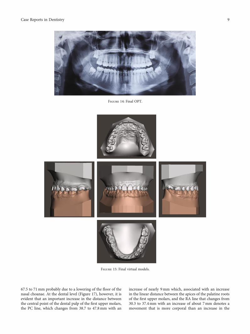
67.5 to 71mm probably due to a lowering of the floor of thenasal choanae. At the dental level (Figure 17), however, it isevident that an important increase in the distance betweenthe central point of the dental pulp of the first upper molars,the PC line, which changes from 38.7 to 47.8mm with an
increase of nearly 9mm which, associated with an increasein the linear distance between the apices of the palatine rootsof the first upper molars, and the RA line that changes from30.3 to 37.4mm with an increase of about 7mm denotes amovement that is more corporal than an increase in the
Figure 14: Final OPT.
Figure 15: Final virtual models.
9Case Reports in Dentistry

molar torque. The superimposition of the traces shows thatthere was a mainly dental effect without great verticalitychanges (Figure 18).
5. Conclusions
The presentation of this case shows how simple and prac-tical the treatment of cases with contracted palates can beeven in patients who have passed their peak in growth along time before starting treatment, going on to manage,in a predictable and practical way, skeletal class III malocclu-sions with the presence of bilateral crossbites and allowing anorthopaedic resolution of the same without transversal com-pensations. Today, the use of protocols that are not operator-dependent, such as these, makes therapies of this kind bothpredictable and trustworthy even for operators with littlesurgical experience. This makes the operation simple, and itrespects the anatomic limits, promoting an adequate expan-sion without compensatory risks or treatments that are surgi-cally assisted.
Conflicts of Interest
The authors have no conflicts of interest to declare.
References
[1] S. Paduano, R. Rongo, R. Bucci et al., “Is there an associationbetween various aspects of oral health in southern Italy chil-dren? An epidemiological study assessing dental decays, peri-odontal status, malocclusions and temporomandibular jointfunction,” European Journal of Paediatric Dentistry, vol. 19,no. 3, pp. 176–180, 2018.
Figure 16: Expansion virtual evaluation.
Figure 17: First molar torque evaluation.
Figure 18: Cephalometric superimposition.
10 Case Reports in Dentistry

[2] A. L. Giudice, P. Spinuzza, L. Rustico, G. Messina, andR. Nucera, “Short-term treatment effects produced by rapidmaxillary expansion evaluated with computed tomography: asystematic review with meta-analysis,” The Korean Journal ofOrthodontics, vol. 50, no. 5, pp. 314–323, 2020.
[3] G. Zuccati, S. Casci, T. Doldo, and C. Clauser, “Expansion ofmaxillary arches with crossbite: a systematic review of RCTsin the last 12 years,” European Journal of Orthodontics,vol. 35, no. 1, pp. 29–37, 2013.
[4] J. Huang, C. Y. Li, and J. H. Jiang, “Facial soft tissue changesafter nonsurgical rapid maxillary expansion: a systematicreview and meta-analysis,” Head & Face Medicine, vol. 14,no. 1, p. 6, 2018.
[5] M. O. Lagravere, P. W. Major, and C. Flores-Mir, “Long-termskeletal changes with rapid maxillary expansion: a systematicreview,” The Angle Orthodontist, vol. 75, no. 6, pp. 1046–1052, 2005.
[6] T. Baccetti, L. Franchi, C. G. Cameron, and J. A. McNamara,“Treatment timing for rapid maxillary expansion,” The AngleOrthodontist, vol. 75, no. 6, pp. 1046–1052, 2001.
[7] F. M. Copello, G. A. Marañón-Vásquez, D. P. Brunetto et al.,“Is the buccal alveolar bone less affected by mini-implantassisted rapid palatal expansion than by conventional rapidpalatal expansion?- A systematic review and meta-analysis,”Orthodontics & Craniofacial Research, vol. 23, no. 3, pp. 237–249, 2020.
[8] E. H. Seong, S. H. Choi, H. J. Kim, H. S. Yu, Y. C. Park, andK. J. Lee, “Evaluation of the effects of miniscrew incorporationin palatal expanders for young adults using finite element anal-ysis,” The Korean Journal of Orthodontics, vol. 48, no. 2,pp. 81–89, 2018.
[9] P. H. A. Carvalho, L. B. Moura, G. S. Trento et al., “Surgicallyassisted rapid maxillary expansion: a systematic review ofcomplications,” International Journal of Oral and Maxillofa-cial Surgery, vol. 49, no. 3, pp. 325–332, 2020.
[10] F. Bover-Ramos, J. Viña-Almunia, J. Cervera-Ballester,M. Peñarrocha-Diago, and B. García-Mira, “Accuracy ofimplant placement with computer-guided surgery: a system-atic review and meta-analysis comparing cadaver, clinical,and in vitro studies,” The International Journal of Oral &Max-illofacial Implants, vol. 33, no. 1, pp. 101–115, 2018.
[11] I. Laleman, L. Bernard, M. Vercruyssen, R. Jacobs,M. Bornstein, and M. Quirynen, “Guided implant surgery inthe edentulous maxilla: a systematic review,” The InternationalJournal of Oral & Maxillofacial Implants, vol. 31, Suppl,pp. s103–s117, 2017.
[12] A. Lo Giudice, L. Rustico, V. Ronsivalle et al., “A full diagnosticprocess for the orthodontic treatment strategy: a documentedcase report,” Dental Journal, vol. 8, no. 2, p. 41, 2020.
[13] M. Cassetta, F. Altieri, R. Di Giorgio, and E. Barbato, “Palatalorthodontic miniscrew insertion using a CAD-CAM surgicalguide: description of a technique,” International Journal ofOral and Maxillofacial Surgery, vol. 47, no. 9, pp. 1195–1198,2018.
[14] B. G. Maino, E. Paoletto, Lombardo L 3rd, and G. Siciliani, “Athree-dimensional digital insertion guide for palatal miniscrewplacement,” Journal of Clinical Orthodontics, vol. 50, no. 1,pp. 12–22, 2016.
[15] B. T. Brettin, N. M. Grosland, F. Qian et al., “Bicortical vsmonocortical orthodontic skeletal anchorage,” American Jour-nal of Orthodontics and Dentofacial Orthopedics, vol. 134,no. 5, pp. 625–635, 2008.
[16] A. Poorsattar-Bejeh Mir, “Monocortical versus bicortical hardpalate anchorage with the same total available cortical thick-ness: a finite element study,” Journal of Investigative and Clin-ical Dentistry, vol. 8, no. 3, 2017.
[17] A. Manni, M. Cozzani, F. Tamborrino, S. De Rinaldis, andA. Menini, “Factors influencing the stability of miniscrews.A retrospective study on 300 miniscrews,” European Journalof Orthodontics, vol. 33, no. 4, pp. 388–395, 2011.
[18] M. Migliorati, S. Drago, F. Gallo et al., “Immediate versusdelayed loading: comparison of primary stability loss afterminiscrew placement in orthodontic patients-a single-centreblinded randomized clinical trial,” European Journal of Ortho-dontics, vol. 38, no. 6, pp. 652–659, 2016.
[19] O. de Gabriele, G. Dallatana, R. Riva, S. Vasudavan, andB. Wilmes, “The easy driver for placement of palatal mini-implants and a maxillary expander in a single appointment,”Journal of Clinical Orthodontics, vol. 51, no. 11, pp. 728–737,2017.
[20] G. Iodice, G. Danzi, R. Cimino, S. Paduano, andA. Michelotti, “Association between posterior crossbite,masticatory muscle pain, and disc displacement: a system-atic review,” European Journal of Orthodontics, vol. 35,no. 6, pp. 737–744, 2013.
[21] S. Paduano, F. P. Paduano, D. Aiello et al., “OSAS in develop-ing age: screening of a southern Italy population,” EuropeanJournal of Paediatric Dentistry, vol. 20, no. 4, pp. 302–305,2019.
[22] F. Angelieri, L. H. S. Cevidanes, L. Franchi, J. R. Gonçalves,E. Benavides, and J. A. McNamara Jr, “Midpalatal suture mat-uration: classification method for individual assessment beforerapid maxillary expansion,” American Journal of Orthodonticsand Dentofacial Orthopedics, vol. 144, no. 5, pp. 759–769,2013.
[23] R. J. Lee, W. Moon, and C. Hong, “Effects of monocortical andbicortical mini-implant anchorage on bone-borne palatalexpansion using finite element analysis,” American Journal ofOrthodontics and Dentofacial Orthopedics, vol. 151, no. 5,pp. 887–897, 2017.
[24] K. Gungor, L. Taner, and E. Kaygisiz, “Prevalence of poste-rior crossbite for orthodontic treatment timing,” The Jour-nal of Clinical Pediatric Dentistry, vol. 40, no. 5, pp. 422–424, 2016.
[25] O. G. da Silva Filho, M. Santamaria, and L. Capelozza Filho,“Epidemiology of posterior crossbite in the primary dentition,”The Journal of Clinical Pediatric Dentistry, vol. 32, no. 1,pp. 73–78, 2007.
[26] L. A. Souza, T. R. Elmadjian, R. B. . Dias, and N. P. Coto,“Prevalence of malocclusions in the 13-20-year-old categoriesof football athletes,” Brazilian Oral Research, vol. 25, no. 1,pp. 19–22, 2011.
[27] L. M. Jimenez-Valdivia, V. Malpartida-Carrillo, Y. A. Rodrí-guez-Cárdenas, H. L. Dias-Da Silveira, and L. E. Arriola-Guil-lén, “Midpalatal suture maturation stage assessment inadolescents and young adults using cone-beam computedtomography,” Progress in Orthodontics, vol. 20, no. 1, p. 38,2019.
[28] D. P. Brunetto, E. F. Sant’Anna, A. W. Machado, andW. Moon, “Non-surgical treatment of transverse deficiencyin adults using microimplant-assisted rapid palatal expansion(MARPE),” Dental Press Journal of Orthodontics, vol. 22,no. 1, pp. 110–125, 2017.
11Case Reports in Dentistry

[29] S. S. Suzuki, L. F. S. Braga, D. N. Fujii, W. Moon, andH. Suzuki, “Corticopuncture facilitated microimplant-assisted rapid palatal expansion,” Case Reports in Dentistry,vol. 2018, Article ID 1392895, 12 pages, 2018.
[30] L. Lin, H. W. Ahn, S. J. Kim, S. C. Moon, S. H. Kim, andG. Nelson, “Tooth-borne vs bone-borne rapid maxillaryexpanders in late adolescence,” The Angle Orthodontist,vol. 85, no. 2, pp. 253–262, 2015.
12 Case Reports in Dentistry