A simple fabricated thickness-based stiffness gradient for ...
7
Article A simple fabricated thickness-based stiffness gradient for cell studies Yiwei Shu a,1 , Ho Nam Chan b,1 , Dongshi Guan c , Hongkai Wu b,⇑ , Lan Ma a,⇑ a Division of Life Science & Health, Graduate School at Shenzhen, Tsinghua University, Shenzhen 518055, China b Department of Chemistry, The Hong Kong University of Science and Technology, Hong Kong, China c Department of Physics, The Hong Kong University of Science and Technology, Hong Kong, China article info Article history: Received 16 August 2016 Received in revised form 23 September 2016 Accepted 12 October 2016 Available online 27 December 2016 Keywords: Stiffness gradient Cell analysis PDMS Etoposide abstract In this work, we developed a simple method to fabricate a thickness-based continuous stiffness gradient for biological studies. It was made by glass slides, polydimethylsiloxane (PDMS) pre-polymer, spacer and clips only, without any sophisticated equipment. It is easy to fabricate in any general biological and phar- maceutical laboratories. The stiffness gradient was characterized in terms of apparent Young’s modulus by atomic force microscopy (AFM) and the Young’s modulus along the gradient was found to be 8.5– 120 kPa, which is within the physiological relevant range. HeLa-C3 cells were cultured on the gradient to study their morphological behavior according to the substrate stiffness. Furthermore, the drug effi- ciency of etoposide, an anti-cancer drug, was studied along the substrate stiffness gradient. It was found that HeLa-C3 cells cultured on the soft region of the gradient (8.5–11 kPa) are more sensitive to etopo- side. We believe the proposed device could promote cell investigations and drug screenings on a sub- strate with comparable stiffness to the native tissue. Ó 2016 Science China Press. Published by Elsevier B.V. and Science China Press. All rights reserved. 1. Introduction Cancer is the leading cause of death worldwide. Tremendous works have been done for the prevention, diagnosis, and treatment of cancers [1–6]. To date, more than 50,000 natural extracts and 100,000 compounds have been screened for assessing their poten- tial to be anti-cancer drug [7]. However, the successful rate of anti- cancer drug development is extremely low, with only 1 or 2 out of 1,000 candidate compounds successfully qualify phase III trials and being used as anti-cancer drugs on the market [8–10]. This is because of the inherent drawbacks of the traditional drug screen- ing setup. Plastic culture dishes made of polystyrene are routinely used as the substrate for drug screening, but the elasticity of cul- ture dish is around 1 GPa, roughly six orders of magnitude stiffer than the epithelium and the extracellular matrix (ECM) in vivo. In human body, the elasticity of ECM in different tissues varies greatly, from 1 kPa of brain, 10 kPa of muscle to 100 kPa of pre- calcified bone [11–13]. Also, the stiffness of the ECM was found to affect cell behaviors including migration [14,15], adhesion [16,17], proliferation [18], differentiation [19], and cancer metasta- sis [20]. Conventional plastic culture dish based platforms cannot address these concerns, thus, there is an urge to build alternative platform with physiological relevant substrate stiffness for study- ing biological problems. Instead of building culture platform with pre-defined substrate stiffness, building a stiffness gradient can gather more interesting information such as the critical stiffness value of stiffness regulated cell behaviors and morphological changes during stiffness-induced migration. Hence, some methods were reported to build stiffness gradient, such as approaches based on photopolymerization [21–24], microfluidics [25–27], temperature gradient [28] and freezing-thawing of hydrogel [29]. However, these methods are still limited in the following aspects. Firstly, for photopolymeriza- tion based approaches, the toxicity of monomers, photoinitiators and crosslinkers residues and UV radiation are discouraging their wide adoption. Secondly, the surface chemistry of the substrate often change along with the substrate stiffness gradient, which unfavorably coupled the influence of surface chemistry with sub- strate stiffness in cell studies. Recently, a group has reported that osteogenic differentiation is dependent on substrate stiffness only but not surface adhesive protein tethering which demonstrates the importance of considering surface chemistry together with sub- strate stiffness in cell studies [30]. Moreover, other studies reported that cell behaviors including migration, morphology and adhesion can be altered by substrate surface chemistry [31–33]. Therefore, having a uniform surface chemistry along the stiffness gradient is important when studying stiffness influence. Thirdly, most of them are complicated to fabricate, which require long http://dx.doi.org/10.1016/j.scib.2016.12.012 2095-9273/Ó 2016 Science China Press. Published by Elsevier B.V. and Science China Press. All rights reserved. ⇑ Corresponding authors. E-mail addresses: [email protected] (H. Wu), [email protected] (L. Ma). 1 Yiwei Shu and Ho Nam Chan have equal contribution. Science Bulletin 62 (2017) 222–228 Contents lists available at ScienceDirect Science Bulletin journal homepage: www.elsevier.com/locate/scib
Transcript of A simple fabricated thickness-based stiffness gradient for ...

Science Bulletin 62 (2017) 222–228
Contents lists available at ScienceDirect
Science Bulletin
journal homepage: www.elsevier .com/locate /sc ib
Article
A simple fabricated thickness-based stiffness gradient for cell studies
Yiwei Shu a,1, Ho Nam Chan b,1, Dongshi Guan c, Hongkai Wub,⇑, Lan Ma a,⇑aDivision of Life Science & Health, Graduate School at Shenzhen, Tsinghua University, Shenzhen 518055, ChinabDepartment of Chemistry, The Hong Kong University of Science and Technology, Hong Kong, ChinacDepartment of Physics, The Hong Kong University of Science and Technology, Hong Kong, China
a r t i c l e i n f o a b s t r a c t
Article history:Received 16 August 2016Received in revised form 23 September2016Accepted 12 October 2016Available online 27 December 2016
Keywords:Stiffness gradientCell analysisPDMSEtoposide
http://dx.doi.org/10.1016/j.scib.2016.12.0122095-9273/� 2016 Science China Press. Published by
⇑ Corresponding authors.E-mail addresses: [email protected] (H. Wu), malan@
1 Yiwei Shu and Ho Nam Chan have equal contribut
In this work, we developed a simple method to fabricate a thickness-based continuous stiffness gradientfor biological studies. It was made by glass slides, polydimethylsiloxane (PDMS) pre-polymer, spacer andclips only, without any sophisticated equipment. It is easy to fabricate in any general biological and phar-maceutical laboratories. The stiffness gradient was characterized in terms of apparent Young’s modulusby atomic force microscopy (AFM) and the Young’s modulus along the gradient was found to be 8.5–120 kPa, which is within the physiological relevant range. HeLa-C3 cells were cultured on the gradientto study their morphological behavior according to the substrate stiffness. Furthermore, the drug effi-ciency of etoposide, an anti-cancer drug, was studied along the substrate stiffness gradient. It was foundthat HeLa-C3 cells cultured on the soft region of the gradient (8.5–11 kPa) are more sensitive to etopo-side. We believe the proposed device could promote cell investigations and drug screenings on a sub-strate with comparable stiffness to the native tissue.
� 2016 Science China Press. Published by Elsevier B.V. and Science China Press. All rights reserved.
1. Introduction
Cancer is the leading cause of death worldwide. Tremendousworks have been done for the prevention, diagnosis, and treatmentof cancers [1–6]. To date, more than 50,000 natural extracts and100,000 compounds have been screened for assessing their poten-tial to be anti-cancer drug [7]. However, the successful rate of anti-cancer drug development is extremely low, with only 1 or 2 out of1,000 candidate compounds successfully qualify phase III trials andbeing used as anti-cancer drugs on the market [8–10]. This isbecause of the inherent drawbacks of the traditional drug screen-ing setup. Plastic culture dishes made of polystyrene are routinelyused as the substrate for drug screening, but the elasticity of cul-ture dish is around 1 GPa, roughly six orders of magnitude stifferthan the epithelium and the extracellular matrix (ECM) in vivo.In human body, the elasticity of ECM in different tissues variesgreatly, from 1 kPa of brain, 10 kPa of muscle to 100 kPa of pre-calcified bone [11–13]. Also, the stiffness of the ECM was foundto affect cell behaviors including migration [14,15], adhesion[16,17], proliferation [18], differentiation [19], and cancer metasta-sis [20]. Conventional plastic culture dish based platforms cannotaddress these concerns, thus, there is an urge to build alternative
Elsevier B.V. and Science China Pr
sz.tsinghua.edu.cn (L. Ma).ion.
platform with physiological relevant substrate stiffness for study-ing biological problems.
Instead of building culture platform with pre-defined substratestiffness, building a stiffness gradient can gather more interestinginformation such as the critical stiffness value of stiffness regulatedcell behaviors and morphological changes during stiffness-inducedmigration. Hence, some methods were reported to build stiffnessgradient, such as approaches based on photopolymerization[21–24], microfluidics [25–27], temperature gradient [28] andfreezing-thawing of hydrogel [29]. However, these methods arestill limited in the following aspects. Firstly, for photopolymeriza-tion based approaches, the toxicity of monomers, photoinitiatorsand crosslinkers residues and UV radiation are discouraging theirwide adoption. Secondly, the surface chemistry of the substrateoften change along with the substrate stiffness gradient, whichunfavorably coupled the influence of surface chemistry with sub-strate stiffness in cell studies. Recently, a group has reported thatosteogenic differentiation is dependent on substrate stiffness onlybut not surface adhesive protein tethering which demonstrates theimportance of considering surface chemistry together with sub-strate stiffness in cell studies [30]. Moreover, other studiesreported that cell behaviors including migration, morphology andadhesion can be altered by substrate surface chemistry [31–33].Therefore, having a uniform surface chemistry along the stiffnessgradient is important when studying stiffness influence. Thirdly,most of them are complicated to fabricate, which require long
ess. All rights reserved.

Y. Shu et al. / Science Bulletin 62 (2017) 222–228 223
processing time and sophisticated equipment. As a result, culturingand studying cells on petri dish and culture flask are still the dom-inated practice in the current biological and pharmaceutical com-munities, neglecting the substrate stiffness influences on cells.
Alternatively, by exploiting the rigidity of a hard substrate canbe sensed through a soft material, a stiffness gradient can be gen-erated by controlling the thickness of soft material on top of a rigidsubstrate. This approach was used to create a stiffness gradient byincorporating rigid microspheres in a thin layer of hydrogel [34].However, the gradient generated was very narrow and difficultto control because of the tiny size and geometrical constrain ofmicrospheres.
In this work, we prepared a tunable stiffness gradient withoutany sophisticated equipment. The stiffness gradient was obtainedthrough a thickness gradient (i.e., a wedge structure), which wasmade by polydimethylsiloxane (PDMS), glass slides, clips and spac-ers only. The fabrication is simple that can be quickly prepared bygeneral biomedical laboratories. The surface chemistry along thegradient is homogeneous, which is difficult to achieve by othermethods. The stiffness gradient of the PDMS wedge structurewas characterized in terms of reduced Young’s modulus of thecomposite material by atomic force microscopy (AFM) and theYoung’s modulus prepared along the gradient was from 8.5 to120 kPa, which was within the physiological relevant range. Onthis platform, the response of cancer cells to etoposide (a cancerdrug) was found to be dependent on the stiffness of culture sub-strate. We believe our newly developed platform with continuousstiffness gradient can better mimic the microenvironment thatcells experienced in vivo and provide a simple and economic toolfor drug screening and other cellular activities studies, which areunable to be revealed by traditional petri-dish based biologicalplatforms [7].
2. Material and methods
2.1. Fabrication of the PDMS wedge structure
The fabrication scheme of the wedge structure is shown inFig. 1a. A glass slide that was cleaned by piranha solution and trea-ted with oxygen plasma (Harrick Plasma PDC-32G, USA) was usedas a substrate. A 50 lm spacer was placed on one side of the sub-strate. PDMS (GE Silicones RTV 615, Wilton, CT, USA) prepolymer(A:B = 50:1, where part A and B are the base and curing agentrespectively) was poured onto the substrate and then covered byanother glass slide that was treated with trichloro (1H,1H,2H,2H-perfluorooctyl)silane (Sigma-Aldrich, USA). Two clips were usedto clip the cover glass and substrate from two side to form thewedge structure. The whole setup was put in a 70 �C oven over-night to cure the PDMS prepolymer. After the clips and cover glassslide were removed, the wedge-shaped PDMS slab always adheredon the glass substrate because the cover glass was grafted with alayer of fluorinated molecules. Finally, the PDMS wedge structurewas placed on a 200 �C hot plate for 3 h to complete thepolymerization.
2.2. Fabrication of the PDMS perforated culture wells
PDMS perforated culture wells were fabricated by soft lithogra-phy using a 3D printed (Miicraft) master (Fig. S1 online). Beforemolding, the 3D printed master was treated with the procedurereported previously [35]. Briefly, the freshly printed master washeated at 130 �C overnight, followed by oxygen plasma treatmentfor 3 min. Afterwards, the 3D printed master was grafted with alayer of fluorinated molecules by reacting with trichloro(1H,1H,2H,2H-perfluorooctyl)silane. Hence, the PDMS replica can
be peeled off easily. The positive master of the culture well wasprinted with sufficient height (8 mm), so that the perforated cul-ture wells can be molded simply by pouring limited amount ofPDMS on to the master structure.
2.3. HeLa-C3 cells culture
The HeLa-C3 cells, which stably expressed cyan fluorescent pro-tein (CFP) and yellow fluorescent protein (YFP) [36], were seededat density of 4 � 105 cells/mL and cultured in Eagle’s minimumessential medium (MEM) with 10% fetal bovine serum (FBS) andsupplemented with 100 U/mL penicillin, 100 lg/mL streptomycin(Gibco, Invitrogen, NY, USA), and 50 lg/mL G-418 solution (Roche,Switzerland). Cells were cultured in 37 �C incubator with 5% CO2.Before seeding cells on the gradient, the PDMS gradient was steril-ized under UV radiation for 30 min and then washed three timeswith sterilized PBS.
2.4. Drug treatment and data analysis
Etoposide was diluted to the desired concentrations (50, 100and 200 lmol/L) with culture medium before dosing. The drugwas added to the cells 12 h after seeding by replacing the culturemedium to a drug-dosed medium. Fluorescent images of theHeLa-C3 cells were captured by an inverse fluorescence micro-scope (Nikon Eclipse TE2000-U, Japan) at different time (0, 12,24, 36 and 48 h) with excitation filter at (436 ± 20) nm, a dichroicmirror at 455 nm, and emission at (535 ± 15) nm (YFP) and(480 ± 20) nm (CFP). Images were captured by a cooled CCD cam-era. The fluorescence signals from the FRET probes (CFP and YFPfusion protein) were analyzed by image processing and analysissoftware (AlphaEaseFC, CA). The fluorescence intensity of each cellwas corrected by subtracting the averaged local background.
2.5. Stiffness measurement and analysis
An AFM (MFP-3D, Asylum Research Inc., USA) with the colloidalprobe cantilever was used for the Young’s modulus measurement.The assembly of the glass sphere (Microspheres-Nanospheres,r = 16 lm) to the cantilever (MikroMasch, tipless, spring constantk = 0.08 N/m) was performed following the procedure describedpreviously [37]. Fig. S2 (online) shows the scanning electron micro-scope (SEM) image of the bottom view of the assembled colloidalcantilever. The exact spring constant of the cantilever was cali-brated by the thermal noise method [38], following the standardprocedures provided by the AFM manufacturer. The AFM measure-ments were carried out in contact mode with 2 lm indentationdepth. The measured force–distance curves were recorded and fit-ted with Hertz model F = (4/3)r1/2E * h3/2 to calculate the reducedYoung’s modulus E⁄ [39], where F is the measured force, r is theprobe radius and h is the indentation distance.
3. Results and discussion
3.1. Stiffness gradient design, fabrication and its characterization
The stiffness gradient design is based on the fact that cells cansense the stiffness of a rigid substrate even without direct contact[40]. When a soft material is placed onto a hard substrate, the stiff-ness of the complex system is determined by the thickness of thesoft material and the magnitude of its deformation [34]. The thin-ner part of the wedge structure is apparently stiffer than thethicker part as illustrated in Fig. 1a. For the thick part, the apparentYoung’s modulus is mostly contributed by the Young’s modulus ofthe soft material. To let the fabricated stiffness gradient stay within

Fig. 1. (Color online) PDMS-based stiffness gradient. (a) Schematic representation of the fabrication procedures. The schematic is not drawn to scale. The actual slope is muchsmaller on the device. (b) Plot of the apparent elastic modulus E⁄ as a function of the PDMS thickness. (c) Photograph of the fabricated platform.
224 Y. Shu et al. / Science Bulletin 62 (2017) 222–228
the physiological relevant range, 50:1 (A:B ratio) PDMS was usedto fabricate the wedge structure, which its Young’s modulus wasreported to be c.a. 8 kPa [41].
To characterize the fabricated platform, AFM was used to mea-sure the apparent Young’s modulus along the gradient. Here, wedid the measurement with two types of cantilever—the conven-tional cantilever with sharp tip and the colloidal probe cantilever.Preliminary results revealed that the cantilever with sharp tipcould not measure the Young’s modulus because the tip penetratedinto the PDMS during the measurement. As a result, the colloidalprobe cantilever is used for this soft contact measurement.Fig. 1b shows the plot of measured apparent elastic modulus (E⁄)as a function of the PDMS thickness.
The fabricated stiffness gradient is shown in Fig. 1c, its lengthwas 9.4 cm and the height of the thickest part was 50 lm, whichwas controlled by the spacer. Thus, the slope of the wedge struc-ture was 5.3 � 10�4 and the stiffness of every region could beobtained from the plot (Fig. 1b). The steepness of the stiffness gra-dient can be adjusted by the slope of the wedge structure, which iseasy to change by modifying the length of glass and the thicknessof spacer. With the current fabrication parameters, our platformcan provide continuous stiffness gradient within a broad rangewith apparent Young’s modulus of 8.5–120 kPa, which can mimicthe in vivo mechanical microenvironment of different tissues. Itshould be noted that the stiffness of softest part in the gradientis entirely contributed by the stiffness of the bulk material. As aresult, the lowest achievable stiffness can be adjusted by selectingthe appropriate soft substrate material. Compared with thereported method using microsphere as substrate, our method cre-ates much wider gradient with easy controllable steepness. Here,although the cells were cultured on a very small slope(5.3 � 10�4), its influence is believed to be negligible. This isbecause in most of the studies, unless very carefully controlled,cells are not cultured on a perfectly horizontal plane. Such errorcould be the result of the imperfectly leveled cell incubator ortable. However, if researchers want the cells to be cultured on aperfectly horizontal plane, the slope effect can be compensatedby placing a spacer under the glass substrate.
For the cells to correctly sense the apparent stiffness gradient,the soft material must be in direct contact with the rigid substrate.Here, the glass was cleaned with piranha solution followed by cast-
ing PDMS on it. There was no chemical bonding between the PDMSand the glass, but the physical adhesion force (van der Waals’force) between them was strong enough to prevent the PDMS fromdetaching or sliding on the substrate. Thus, at the interface, thePDMS was in direct contact with and adhered on the glass sub-strate firmly. However, for other systems such as placing softhydrogel on a plastic dish. It is important to let the hydrogel chem-ically bonded to the substrate to prevent the existent of a thinwater layer (i.e., floating) at the interface between the hydrogeland substrate which will interfere the cell sensed apparent stiff-ness gradient.
3.2. The influence of substrate stiffness on HeLa-C3 cells morphology
To investigate the effect of substrate stiffness gradient on cellbehavior, HeLa-C3 cells were cultured on the wedge-shaped PDMSplatform with a cell density of 4 � 105 cells/mL. The PDMS surfacewas treated with oxygen plasma to enhance cell adhesion. After 6 hof culture, most of the cells were attached on the substrate. InFig. 2, it was observed that the HeLa-C3 cells on the softest region(8.5 kPa) spread less which showed a more spherical morphology.Cell-spreading area was measured statistically on four regions withapparent Young’s modulus ranging from 8.5 to 120 kPa. The cellspreading area on the substrate with apparent Young’s modulusof 11, 27 and 120 kPa is significantly larger than that on the8.5 kPa region (Fig. 2e), suggesting the cells were able to sensethe stiffness difference within the gradient. This observation iscoherent with the previous studies showing that cell spreadingbehavior can be enhanced with increasing substrate stiffness[17,42]. Although each type of cells has the preferred substratestiffness (e.g. neurite extend more on substrate below 0.5 kPa[43] and fibroblasts achieve maximum spreading on substrate withabout 10 kPa [44]), our findings are not statistically significant todraw the conclusion that the HeLa-C3 cells prefer a substrate withYoung’s modulus of 11 kPa than the 27 and 120 kPa counterparts.
3.3. Anti-cancer drug screening
Next, the stiffness gradient platform was used to study theeffect of substrate stiffness on anti-cancer drug efficiency. Etopo-side was chosen as the drug model. It can activate cellular
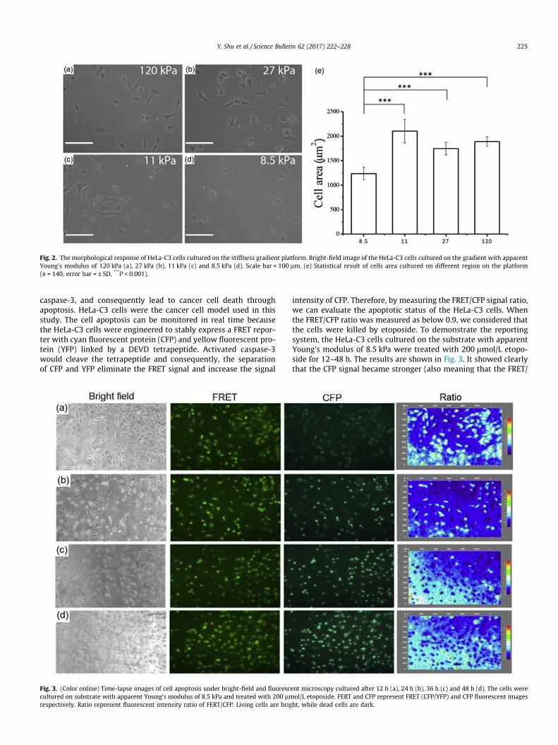
Fig. 2. The morphological response of HeLa-C3 cells cultured on the stiffness gradient platform. Bright-field image of the HeLa-C3 cells cultured on the gradient with apparentYoung’s modulus of 120 kPa (a), 27 kPa (b), 11 kPa (c) and 8.5 kPa (d). Scale bar = 100 lm. (e) Statistical result of cells area cultured on different region on the platform(n = 140, error bar = ± SD, ***P < 0.001).
Y. Shu et al. / Science Bulletin 62 (2017) 222–228 225
caspase-3, and consequently lead to cancer cell death throughapoptosis. HeLa-C3 cells were the cancer cell model used in thisstudy. The cell apoptosis can be monitored in real time becausethe HeLa-C3 cells were engineered to stably express a FRET repor-ter with cyan fluorescent protein (CFP) and yellow fluorescent pro-tein (YFP) linked by a DEVD tetrapeptide. Activated caspase-3would cleave the tetrapeptide and consequently, the separationof CFP and YFP eliminate the FRET signal and increase the signal
Fig. 3. (Color online) Time-lapse images of cell apoptosis under bright-field and fluoresccultured on substrate with apparent Young’s modulus of 8.5 kPa and treated with 200 lmrespectively. Ratio represent fluorescent intensity ratio of FERT/CFP. Living cells are brig
intensity of CFP. Therefore, by measuring the FRET/CFP signal ratio,we can evaluate the apoptotic status of the HeLa-C3 cells. Whenthe FRET/CFP ratio was measured as below 0.9, we considered thatthe cells were killed by etoposide. To demonstrate the reportingsystem, the HeLa-C3 cells cultured on the substrate with apparentYoung’s modulus of 8.5 kPa were treated with 200 lmol/L etopo-side for 12–48 h. The results are shown in Fig. 3. It showed clearlythat the CFP signal became stronger (also meaning that the FRET/
ent microscopy cultured after 12 h (a), 24 h (b), 36 h (c) and 48 h (d). The cells wereol/L etoposide. FERT and CFP represent FRET (CFP/YFP) and CFP fluorescent imagesht, while dead cells are dark.
226 Y. Shu et al. / Science Bulletin 62 (2017) 222–228
CFP ratio became smaller) when the cells were treated with thedrug for longer time which indicated the reporting system wasfunctional.
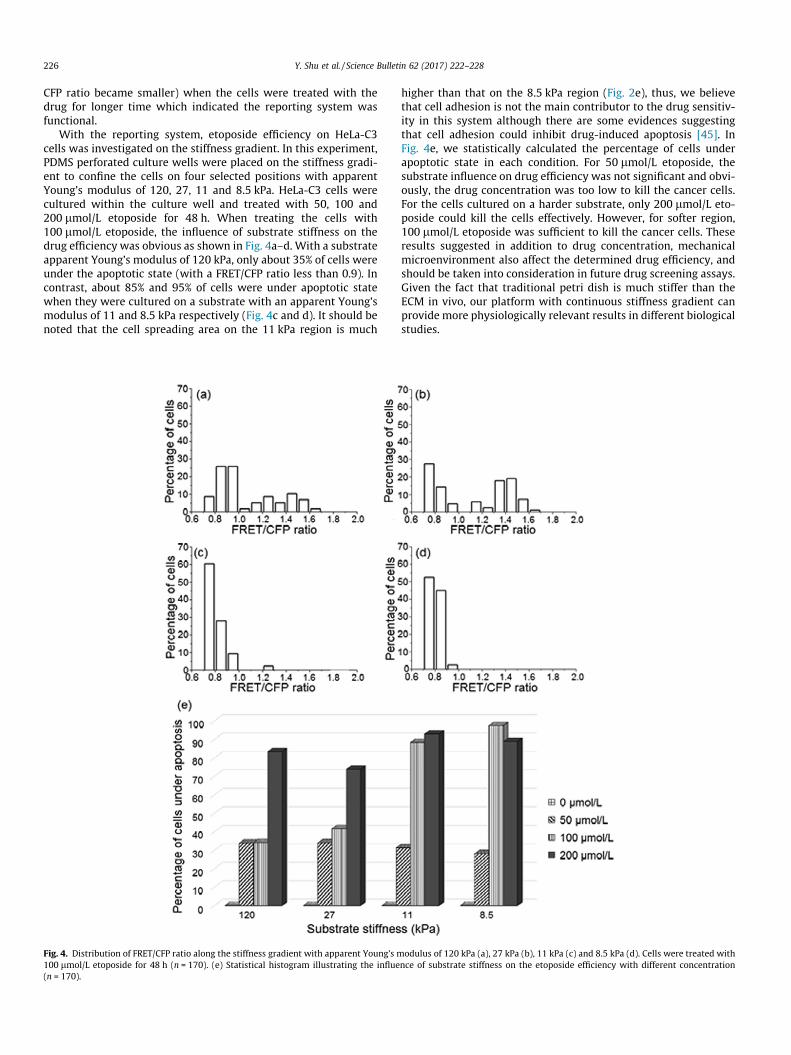
With the reporting system, etoposide efficiency on HeLa-C3cells was investigated on the stiffness gradient. In this experiment,PDMS perforated culture wells were placed on the stiffness gradi-ent to confine the cells on four selected positions with apparentYoung’s modulus of 120, 27, 11 and 8.5 kPa. HeLa-C3 cells werecultured within the culture well and treated with 50, 100 and200 lmol/L etoposide for 48 h. When treating the cells with100 lmol/L etoposide, the influence of substrate stiffness on thedrug efficiency was obvious as shown in Fig. 4a–d. With a substrateapparent Young’s modulus of 120 kPa, only about 35% of cells wereunder the apoptotic state (with a FRET/CFP ratio less than 0.9). Incontrast, about 85% and 95% of cells were under apoptotic statewhen they were cultured on a substrate with an apparent Young’smodulus of 11 and 8.5 kPa respectively (Fig. 4c and d). It should benoted that the cell spreading area on the 11 kPa region is much
Fig. 4. Distribution of FRET/CFP ratio along the stiffness gradient with apparent Young’s m100 lmol/L etoposide for 48 h (n = 170). (e) Statistical histogram illustrating the influe(n = 170).
higher than that on the 8.5 kPa region (Fig. 2e), thus, we believethat cell adhesion is not the main contributor to the drug sensitiv-ity in this system although there are some evidences suggestingthat cell adhesion could inhibit drug-induced apoptosis [45]. InFig. 4e, we statistically calculated the percentage of cells underapoptotic state in each condition. For 50 lmol/L etoposide, thesubstrate influence on drug efficiency was not significant and obvi-ously, the drug concentration was too low to kill the cancer cells.For the cells cultured on a harder substrate, only 200 lmol/L eto-poside could kill the cells effectively. However, for softer region,100 lmol/L etoposide was sufficient to kill the cancer cells. Theseresults suggested in addition to drug concentration, mechanicalmicroenvironment also affect the determined drug efficiency, andshould be taken into consideration in future drug screening assays.Given the fact that traditional petri dish is much stiffer than theECM in vivo, our platform with continuous stiffness gradient canprovide more physiologically relevant results in different biologicalstudies.
odulus of 120 kPa (a), 27 kPa (b), 11 kPa (c) and 8.5 kPa (d). Cells were treated withnce of substrate stiffness on the etoposide efficiency with different concentration

Y. Shu et al. / Science Bulletin 62 (2017) 222–228 227
4. Conclusion
In this work, we developed a thickness-based continuous stiff-ness gradient made by PDMS, glass slides, spacer and clips only.Compared with the previously reported methods to generatedstiffness gradient, our proposed wedge structure is much easierto fabricate for biological laboratories. Moreover, by modifyingthe wedge structure, the steepness of the stiffness gradient canbe adjusted easily. The surface chemistry along the gradient ishomogeneous which can decouple the influence of surface chem-istry from stiffness. Most importantly, the material of the wedgestructure is not limited to PDMS, any elastic and soft material thatare interested to the researchers (e.g., hydrogels) can be used withthis approach to obtain a stiffness gradient. Currently, engineeringphysical microenvironment for 3D cell culturing is getting moreattention [46–52]. To further apply our method in 3D cell cultur-ing, after the cells attached to the PDMS surface, researchers canpour a thin layer of collagen solution (or other scaffolding hydro-gels) on top of the cells and let it gel at 37 �C in the incubator. Asa result, the cells can experience a 3D microenvironment that thebottom face is on a stiffness gradient. This could be interestingbecause some cells in our body are interacting with two types ofstiffness simultaneously. We believe the proposed device couldpromote cell investigations and drug screenings on a substratewith comparable stiffness to the native tissue.
Conflict of interest
The authors declare that they have no conflict of interest.
Acknowledgments
This work was supported by the Hong Kong Research GrantCouncil (GRF#16306115 and #16325116), HKUST SSTSP(#FP701) and the National High Technology Research and Develop-ment Program of China (2013AA032204).
Appendix A. Supplementary data
Supplementary data associated with this article can be found, inthe online version, at http://dx.doi.org/10.1016/j.scib.2016.12.012.
References
[1] Aggarwal BB, Shishodia S. Molecular targets of dietary agents for preventionand therapy of cancer. Biochem Pharmacol 2006;71:1397–421.
[2] Li X, Ling V, Li PCH. Same-single-cell analysis for the study of drug effluxmodulation of multidrug resistant cells using a microfluidic chip. Anal Chem2008;80:4095–102.
[3] Li X, Xue X, Li PCH. Real-time detection of the early event of cytotoxicity ofherbal ingredients on single leukemia cells studied in a microfluidic biochip.Integr Biol 2009;1:90–8.
[4] Li X, Chen Y, Li PCH. A simple and fast microfluidic approach of same-single-cell analysis (SASCA) for the study of multidrug resistance modulation incancer cells. Lab Chip 2011;11:1378–84.
[5] Peer D, Karp JM, Hong S, et al. Nanocarriers as an emerging platform for cancertherapy. Nat Nanotechnol 2007;2:751–60.
[6] Tang DG. Understanding cancer stem cell heterogeneity and plasticity. Cell Res2012;22:457–72.
[7] Feng J, Tang Y, Xu Y, et al. Substrate stiffness influences the outcome ofantitumor drug screening in vitro. Clin Hemorheol Microcirc 2013;55:121–31.
[8] Narang AS, Desai DS. Pharmaceutical perspectives of cancer therapeutics. In:Lu Y, Mahato RI, editors. Pharmaceutical Perspectives of CancerTherapeutics. New York, NY: Springer; 2009.
[9] Kitchen DB, Decornez H, Furr JR, et al. Docking and scoring in virtual screeningfor drug discovery: methods and applications. Nat Rev Drug Discov2004;3:935–49.
[10] White RE. High-throughput screening in drug metabolism andpharmacokinetic support of drug discovery. Annu Rev Pharmacol Toxicol2000;40:133–57.
[11] Andrades JA, Santamaría JA, Nimni ME, et al. Selection and amplification of abone marrow cell population and its induction to the chondro-osteogeniclineage by rhOP-1: an in vitro and in vivo study. Int J Dev Biol 2001;45:689–93.
[12] Ferrari G, Angelis D, Coletta M, et al. Muscle regeneration by bone marrow-derived myogenic progenitors. Science 1998;279:1528–30.
[13] Sǎftoiu A, Vilman P. Endoscopic ultrasound elastography—a new imagingtechnique for the visualization of tissue elasticity distribution. J GastrointestLiver Dis 2006;15:161–5.
[14] Lo CM, Wang HB, Dembo M, et al. Cell movement is guided by the rigidity ofthe substrate. Biophys J 2000;79:144–52.
[15] Pathak A, Kumar S. Independent regulation of tumor cell migration by matrixstiffness and confinement. Proc Natl Acad Sci USA 2012;109:10334–9.
[16] Yeung T, Georges PC, Flanagan LA, et al. Effects of substrate stiffness on cellmorphology, cytoskeletal structure, and adhesion. Cell Motil Cytoskeleton2005;60:24–34.
[17] Pelham Robert J, Wang Y. Cell locomotion and focal adhesions are regulated bysubstrate flexibility. Proc Natl Acad Sci USA 1997;94:13661–5.
[18] Wang N, Naruse K, Stamenovic D, et al. Mechanical behavior in living cellsconsistent with the tensegrity model. Proc Natl Acad Sci USA2001;98:7765–70.
[19] Engler AJ, Sen S, Sweeney HL, et al. Matrix elasticity directs stem cell lineagespecification. Cell 2006;126:677–89.
[20] Egeblad M, Rasch MG, Weaver VM. Dynamic interplay between the collagenscaffold and tumor evolution. Curr Opin Cell Biol 2010;22:697–706.
[21] Nemir S, Hayenga HN, West JL. PEGDA hydrogels with patterned elasticity:novel tools for the study of cell response to substrate rigidity. BiotechnolBioeng 2010;105:636–44.
[22] Wong JY, Velasco A, Rajagopalan P, et al. Directed movement of vascularsmooth muscle cells on gradient-compliant hydrogels. Langmuir2003;19:1908–13.
[23] Johnson PM, Reynolds TB, Stansbury JW, et al. High throughput kinetic analysisof photopolymer conversion using composition and exposure time gradients.Polymer 2005;46:3300–6.
[24] Marklein RA, Burdick JA. Spatially controlled hydrogel mechanics to modulatestem cell interactions. Soft Matter 2010;6:136–43.
[25] Lo CT, Throckmorton DJ, Singh AK, et al. Photopolymerized diffusion-definedpolyacrylamide gradient gels for on-chip protein sizing. Lab Chip2008;8:1273–9.
[26] Burdick JA, Khademhosseini A, Langer R. Fabrication of gradient hydrogelsusing a microfluidics/ photopolymerization process. Langmuir 2004;20:8–11.
[27] Du Y, Hancock MJ, He J, et al. Convection-driven generation of long-rangematerial gradients. Biomaterials 2010;31:2686–94.
[28] Wang PY, Tsai WB, Voelcker NH. Screening of rat mesenchymal stem cellbehaviour on polydimethylsiloxane stiffness gradients. Acta Biomater2012;8:519–30.
[29] Kim TH, An DB, Oh SH, et al. Creating stiffness gradient polyvinyl alcoholhydrogel using a simple gradual freezing-thawing method to investigate stemcell differentiation behaviors. Biomaterials 2015;40:51–60.
[30] Wen JH, Vincent LG, Fuhrmann A, et al. Interplay of matrix stiffness andprotein tethering in stem cell differentiation. Nat Mater 2014;13:979–87.
[31] da Costa DS, Pires RA, Frias AM, et al. Sulfonic groups induce formation offilopodia in mesenchymal stem cells. J Mater Chem 2012;22:7172–8.
[32] Hickman GJ, Rees RC, Boocock DJ, et al. Controlling the dynamics of celltransition in heterogeneous cultures using surface chemistry. Adv HealthcMater 2015;4:593–601.
[33] van Dongen SFM, Maiuri P, Marie E, et al. Triggering cell adhesion, migration orshape change with a dynamic surface coating. Adv Mater 2013;25:1687–91.
[34] Kuo CHR, Xian J, Brenton JD, et al. Complex stiffness gradient substrates forstudying mechanotactic cell migration. Adv Mater 2012;24:6059–64.
[35] Chan HN, Chen Y, Shu Y, et al. Direct, one-step molding of 3D-printedstructures for convenient fabrication of truly 3D PDMS microfluidic chips.Microfluid Nanofluid 2015;19:9–18.
[36] Luo KQ, Yu VC, Pu Y, et al. Application of the fluorescence resonance energytransfer method for studying the dynamics of caspase-3 activation during UV-induced apoptosis in living HeLa cells. Biochem Biophys Res Commun2001;283:1054–60.
[37] Guan D, Hang ZH, Marcet Z, et al. Direct measurement of optical force inducedby near-field plasmonic cavity using dynamic mode AFM. Sci Rep2015;5:16216.
[38] Hutter JL, Bechhoefer J. Calibration of atomic-force microscope tips. Rev SciInstrum 1993;64:1868–73.
[39] Hertz H. On the contact of elastic solids. J Reine Angew Math 1881;92:156–71.[40] Buxboim A, Rajagopal K, Brown AEX, et al. How deeply cells feel: methods for
thin gels. J Phys Condens Matter 2010;22:194116.[41] Ochsner M, Dusseiller MR, Grandin HM, et al. Micro-well arrays for 3D shape
control and high resolution analysis of single cells. Lab Chip 2007;7:1074–7.[42] Engler A, Bacakova L, Newman C, et al. Substrate compliance versus ligand
density in cell on gel responses. Biophys J 2004;86:617–28.[43] Flanagan LA, Ju YE, Marg B, et al. Neurite branching on deformable substrates.
NeuroReport 2002;13:2411–5.[44] Subramanian A, Lin HY. Crosslinked chitosan: its physical properties and the
effects of matrix stiffness on chondrocyte cell morphology and proliferation. JBiomed Mater Res Part A 2005;75:742–53.
[45] Hazlehurst LA, Dalton WS. Mechanisms associated with cell adhesionmediated drug resistance (CAM-DR) in hematopoietic malignancies. CancerMetastasis Rev 2001;20:43–50.

228 Y. Shu et al. / Science Bulletin 62 (2017) 222–228
[46] Jeon O, Alt DS, Linderman SW, et al. Biochemical and physical signal gradientsin hydrogels to control stem cell behavior. Adv Mater 2013;25:6366–72.
[47] Zhang X, Liu M, Li Y, et al. Engineering cell microenvironment using novelfunctional hydrogels. Eur Polym J 2015;72:590–601.
[48] Han YL, Wang S, Zhang X, et al. Engineering physical microenvironment forstem cell based regenerative medicine. Drug Discov Today 2014;19:763–73.
[49] Wang L, Li Y, Huang G, et al. Hydrogel-based methods for engineering cellularmicroenvironment with spatiotemporal gradients. Crit Rev Biotechnol2016;36:553–65.
[50] Tse JR, Engler AJ. Stiffness gradients mimicking in vivo tissue variation regulatemesenchymal stem cell fate. PLoS ONE 2011;6:e15978.
[51] Huang G, Wang L, Wang S, et al. Engineering three-dimensional cellmechanical microenvironment with hydrogels. Biofabrication 2012;4:42001.
[52] Li (James) X, Valadez AV, Zuo P, et al. Microfluidic 3D cell culture: potentialapplication for tissue-based bioassays. Bioanalysis 2012;4:1509–25.