A Sialylated Glycan Microarray Reveals Novel Interactions … · · 2011-07-12A Sialylated Glycan...
25
A Sialylated Glycan Microarray Reveals Novel Interactions of Modified Sialic Acids with Proteins and Viruses Xuezheng Song 1 , Hai Yu 2 , Xi Chen 2 , Yi Lasanajak 1 , Mary M. Tappert 3 , Gillian M. Air 3 , Vinod K. Tiwari 2,4 , Hongzhi Cao 2,5 , Harshal A. Chokhawala 2,6 , Haojie Zheng 2 , Richard D. Cummings §1 and David F. Smith §1 From the 1 Department of Biochemistry and the Glycomics Center; Emory University School of Medicine; Atlanta, GA 30322, USA; 2 Department of Chemistry; University of California, Davis; Davis, CA 95616, USA; 3 Department of Biochemistry & Molecular Biology; University of Oklahoma Health Sciences Center; Oklahoma City, OK 73126, USA; 4 Current address: Department of Chemistry, Banaras Hindu University, Varanasi, India; 5 Current address: National Glycoengineering Research Center, Shandong University, Jinan, Shandong, China; 6 Current address: College of Chemistry & Energy Biosciences Institute, University of Berkeley, California, USA Running Title: A Novel Sialylated Glycan Microarray Keywords: glycan microarray, sialic acid, protein-carbohydrate interaction, functional glycomics § Address correspondence to: Richard D. Cummings, Ph.D., Department of Biochemistry, Emory University School of Medicine, O. Wayne Rollins Research Center, 1510 Clifton Road, Suite 4001, Atlanta, GA 30322; Tel: 404-727-5962 (main office); Fax: 404-727-2738; Email: [email protected] ; or David F. Smith, Ph.D., Department of Biochemistry, Emory University School of Medicine, O. Wayne Rollins Research Center, 1510 Clifton Road, Room 4035, Atlanta, GA 30322; Tel: 404-727-6155; Fax: 404-727-2738; Email: [email protected] . Many glycan-binding proteins in animals and pathogens recognize sialic acid or its modified forms, but their molecular recognition is poorly understood. Here we describe studies on sialic acid recognition using a novel sialylated glycan microarray containing modified sialic acids presented on different glycan backbones. Glycans terminating in β-linked galactose at the non-reducing end and with an alkylamine- containing fluorophore at the reducing end were sialylated by a one-pot-three-enzyme system to generate α2-3- and α2-6-linked sialyl glycans with 16 modified sialic acids. The resulting 77 sialyl glycans were purified and quantified, characterized by mass spectrometry, covalently printed on activated slides, and interrogated with a number of key sialic acid binding proteins and viruses. Sialic acid recognition by the sialic acid-binding lectins SNA and MAL-I, which are routinely used for detecting α2-6- and α2-3-linked sialic acids, is affected by sialic acid modifications and both lectins bind glycans terminating with Kdn and Kdn derivatives stronger than the derivatives of more common Neu5Ac and Neu5Gc. Three human parainfluenza viruses bind to glycans terminating with Neu5Ac or Neu5Gc and some of their derivatives, but not to Kdn and its derivatives. Influenza A virus also does not bind glycans terminating in Kdn or Kdn derivatives. An especially novel aspect of human influenza A virus binding is its ability to equivalently recognize glycans terminated with either α2-6-linked Neu5Ac9Lt or α2-6-linked Neu5Ac. Our results demonstrate the utility of this sialylated glycan microarray to investigate the biological importance of modified sialic acids in protein-glycan interactions. Protein-glycan interactions play important roles in cellular adhesion, signal transduction, and host- pathogen interactions, and are mediated through specific glycan recognition by glycan-binding proteins (GBPs) (1-3). Sialic acids (Sia), which are targets for GBP recognition because they commonly occur at the non-reducing ends of glycans, are uniquely diverse among monosaccharides in that many different modified sialic acids have been identified (4). Sialylation of cell surface glycoconjugates is highly regulated by sialyltransferases and/or sialidases (5) where the most common enzymes add or cleave sialic acid in - 1 - http://www.jbc.org/cgi/doi/10.1074/jbc.M111.274217 The latest version is at JBC Papers in Press. Published on July 12, 2011 as Manuscript M111.274217 Copyright 2011 by The American Society for Biochemistry and Molecular Biology, Inc. by guest on June 14, 2018 http://www.jbc.org/ Downloaded from
-
Upload
nguyendien -
Category
Documents
-
view
223 -
download
2
Transcript of A Sialylated Glycan Microarray Reveals Novel Interactions … · · 2011-07-12A Sialylated Glycan...
A Sialylated Glycan Microarray Reveals Novel Interactions of Modified Sialic Acids with Proteins and Viruses
Xuezheng Song1, Hai Yu2, Xi Chen2, Yi Lasanajak1, Mary M. Tappert3, Gillian M. Air3, Vinod K.
Tiwari2,4, Hongzhi Cao2,5, Harshal A. Chokhawala2,6, Haojie Zheng2, Richard D. Cummings§1 and David F. Smith§1
From the 1Department of Biochemistry and the Glycomics Center; Emory University School of Medicine; Atlanta, GA 30322, USA; 2Department of Chemistry; University of California, Davis; Davis, CA 95616, USA; 3Department of Biochemistry & Molecular Biology; University of Oklahoma Health Sciences Center; Oklahoma City, OK 73126, USA; 4Current address: Department of Chemistry, Banaras Hindu University, Varanasi, India; 5Current address: National Glycoengineering Research Center, Shandong University, Jinan, Shandong, China; 6Current address: College of Chemistry & Energy Biosciences Institute, University of Berkeley, California, USA Running Title: A Novel Sialylated Glycan Microarray Keywords: glycan microarray, sialic acid, protein-carbohydrate interaction, functional glycomics §Address correspondence to: Richard D. Cummings, Ph.D., Department of Biochemistry, Emory University School of Medicine, O. Wayne Rollins Research Center, 1510 Clifton Road, Suite 4001, Atlanta, GA 30322; Tel: 404-727-5962 (main office); Fax: 404-727-2738; Email: [email protected]; or David F. Smith, Ph.D., Department of Biochemistry, Emory University School of Medicine, O. Wayne Rollins Research Center, 1510 Clifton Road, Room 4035, Atlanta, GA 30322; Tel: 404-727-6155; Fax: 404-727-2738; Email: [email protected]. Many glycan-binding proteins in animals and pathogens recognize sialic acid or its modified forms, but their molecular recognition is poorly understood. Here we describe studies on sialic acid recognition using a novel sialylated glycan microarray containing modified sialic acids presented on different glycan backbones. Glycans terminating in β-linked galactose at the non-reducing end and with an alkylamine-containing fluorophore at the reducing end were sialylated by a one-pot-three-enzyme system to generate α2-3- and α2-6-linked sialyl glycans with 16 modified sialic acids. The resulting 77 sialyl glycans were purified and quantified, characterized by mass spectrometry, covalently printed on activated slides, and interrogated with a number of key sialic acid binding proteins and viruses. Sialic acid recognition by the sialic acid-binding lectins SNA and MAL-I, which are routinely used for detecting α2-6- and α2-3-linked sialic acids, is affected by sialic acid modifications and both lectins bind glycans terminating with Kdn and Kdn derivatives stronger than the derivatives of more common Neu5Ac and Neu5Gc. Three human parainfluenza viruses bind to glycans
terminating with Neu5Ac or Neu5Gc and some of their derivatives, but not to Kdn and its derivatives. Influenza A virus also does not bind glycans terminating in Kdn or Kdn derivatives. An especially novel aspect of human influenza A virus binding is its ability to equivalently recognize glycans terminated with either α2-6-linked Neu5Ac9Lt or α2-6-linked Neu5Ac. Our results demonstrate the utility of this sialylated glycan microarray to investigate the biological importance of modified sialic acids in protein-glycan interactions. Protein-glycan interactions play important roles in cellular adhesion, signal transduction, and host-pathogen interactions, and are mediated through specific glycan recognition by glycan-binding proteins (GBPs) (1-3). Sialic acids (Sia), which are targets for GBP recognition because they commonly occur at the non-reducing ends of glycans, are uniquely diverse among monosaccharides in that many different modified sialic acids have been identified (4). Sialylation of cell surface glycoconjugates is highly regulated by sialyltransferases and/or sialidases (5) where the most common enzymes add or cleave sialic acid in
- 1 -
http://www.jbc.org/cgi/doi/10.1074/jbc.M111.274217The latest version is at JBC Papers in Press. Published on July 12, 2011 as Manuscript M111.274217
Copyright 2011 by The American Society for Biochemistry and Molecular Biology, Inc.
by guest on June 14, 2018http://w
ww
.jbc.org/D
ownloaded from

α2-3, α2-6, or α2-8 linkages to underlying glycan structures. Sialylated glycans play important roles in the pathogenesis of many microorganisms. The species specificity of influenza A virus is associated with differential binding of the virus to sialic acids linked either α2-3 or α2-6 to galactose on cell surface glycoconjugates (6). However, modification of sialic acids, such as hydroxylation that converts N-acetylneuraminic acid (Neu5Ac) to N-glycolylneuraminic acid (Neu5Gc), also plays an important role in sialic acid ligand recognition by pathogens and is a component of the surface receptor for certain bacterial toxins (7). Humans, unlike most other mammals including other primates, are unable to naturally synthesize Neu5Gc (8,9) although they can incorporate it from dietary sources (10). Overall, the most commonly studied sialic acids are Neu5Ac, Neu5Gc, and their 9-O-acetylated derivatives (Neu5,9Ac2 and Neu5Gc9Ac) (11-14). The investigation of more diverse sialic acids has been limited due to the difficulty of chemically synthesizing these complex derivatives and the lack of availability of microarrays of glycans containing these diverse sialic acids(15). We hypothesized that the existence of multiple modified sialic acid structures has biological importance related to their interaction with proteins or microorganisms. To further explore the recognition and function of modified sialic acid residues, we exploited the development of new chemoenzymatic methods to synthesize modified sialic acids (5), along with recent development of glycan microarray technology, which provide an innovative approach to studying glycan recognition and function by GBPs. We adapted our recent development of natural glycan microarrays (16-20) to the production of a diverse array of sialylated glycans using fluorescent derivatives of glycans that terminate with β-linked galactose, which are sialyltransferase acceptors, in a one-pot-three-enzyme approach for the microscale chemoenzymatic synthesis of sialosides (21,22). The resultant microarray consists of 77 sialylated glycans incorporating 16 modified sialic acids in α2-3 and α2-6 linkages to different underlying structures. This novel
sialylated glycan microarray is useful for rapidly screening GBPs and viruses for their interactions with a wide variety of modified sialic acids and represents a new glycomic technology to explore glycan function and recognition. Experimental Procedures Free reducing glycans lactose, lactose-N-tetraose (LNT), and lactose-N-neo-tetraose (LNnT) and all chemicals were purchased from Sigma-Aldrich and used without further purification. Asialo, biantennary N-glycan (NA2) was prepared from a chicken egg yolk glycopeptide (23) by mild acid hydrolysis and PNGase F digestion. The bifunctional linker, 2-amino(N-aminoethyl) benzamide (AEAB), was prepared as described previously (20). HPLC solvents were purchased from Fisher Scientific. An Ultraflex-II TOF/TOF system from Bruker Daltonics was used for MALDI-TOF mass spectrometry analysis of glycan conjugates. Biotinylated Con A, RCA-I, SNA, and MAL-I were from Vector Labs, Cy5- and Alexa488-labeled Streptavidin were from Invitrogen, and Arthrobacter ureafaciens sialidase was from Sigma. Glycan-AEAB conjugation, sialylation and purification Free reducing glycans were conjugated with AEAB as described previously (20). Briefly, 1-10 mg glycan was mixed with 50-250 µL AEAB hydrochloride salt solution freshly prepared at 0.35 M in DMSO/AcOH (v/v = 7/3) followed by equal volume of sodium cyanoborohydride solution freshly prepared in the same solvent. The mixture was vortexed briefly and incubated at 65oC for 2 h. The mixture was chilled and the products were precipitated upon addition of 10 volumes of acetonitrile. After bringing the suspension to -20oC and maintaining that temperature for 30 min, the products were separated by centrifugation at 10,000xg for 3 min. The pellet was redissolved in 200-500 µL water and purified by HPLC. AEAB derivatives of Galβ1-4GlcNAcβ1-3Galβ1-4Glc (LNnT), Galβ1-3GlcNAcβ1-3Galβ1-4Glc (LNT), and the biantennary N-glycan, Galβ1-4GlcNAcβ1-2Manβ1-6(Galβ1-4GlcNAcβ1-2Manβ1-3)Manβ1-4GlcNAcβ1-4GlcNAc (NA2) were prepared as precursors for presentation of modified sialic acids.
- 2 -
by guest on June 14, 2018http://w
ww
.jbc.org/D
ownloaded from
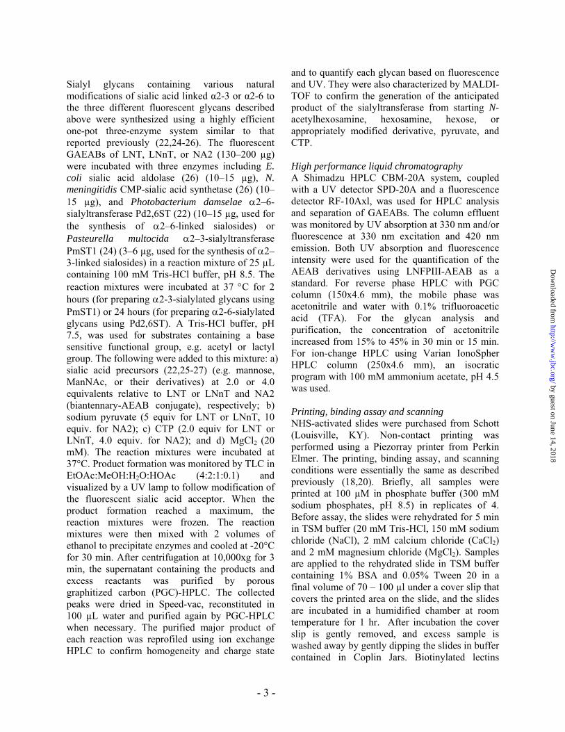
Sialyl glycans containing various natural modifications of sialic acid linked α2-3 or α2-6 to the three different fluorescent glycans described above were synthesized using a highly efficient one-pot three-enzyme system similar to that reported previously (22,24-26). The fluorescent GAEABs of LNT, LNnT, or NA2 (130–200 µg) were incubated with three enzymes including E. coli sialic acid aldolase (26) (10–15 µg), N. meningitidis CMP-sialic acid synthetase (26) (10–15 µg), and Photobacterium damselae α2–6-sialyltransferase Pd2,6ST (22) (10–15 µg, used for the synthesis of α2–6-linked sialosides) or Pasteurella multocida α2–3-sialyltransferase PmST1 (24) (3–6 µg, used for the synthesis of α2–3-linked sialosides) in a reaction mixture of 25 µL containing 100 mM Tris-HCl buffer, pH 8.5. The reaction mixtures were incubated at 37 °C for 2 hours (for preparing α2-3-sialylated glycans using PmST1) or 24 hours (for preparing α2-6-sialylated glycans using Pd2,6ST). A Tris-HCl buffer, pH 7.5, was used for substrates containing a base sensitive functional group, e.g. acetyl or lactyl group. The following were added to this mixture: a) sialic acid precursors (22,25-27) (e.g. mannose, ManNAc, or their derivatives) at 2.0 or 4.0 equivalents relative to LNT or LNnT and NA2 (biantennary-AEAB conjugate), respectively; b) sodium pyruvate (5 equiv for LNT or LNnT, 10 equiv. for NA2); c) CTP (2.0 equiv for LNT or LNnT, 4.0 equiv. for NA2); and d) MgCl2 (20 mM). The reaction mixtures were incubated at 37°C. Product formation was monitored by TLC in EtOAc:MeOH:H2O:HOAc (4:2:1:0.1) and visualized by a UV lamp to follow modification of the fluorescent sialic acid acceptor. When the product formation reached a maximum, the reaction mixtures were frozen. The reaction mixtures were then mixed with 2 volumes of ethanol to precipitate enzymes and cooled at -20°C for 30 min. After centrifugation at 10,000xg for 3 min, the supernatant containing the products and excess reactants was purified by porous graphitized carbon (PGC)-HPLC. The collected peaks were dried in Speed-vac, reconstituted in 100 µL water and purified again by PGC-HPLC when necessary. The purified major product of each reaction was reprofiled using ion exchange HPLC to confirm homogeneity and charge state
and to quantify each glycan based on fluorescence and UV. They were also characterized by MALDI-TOF to confirm the generation of the anticipated product of the sialyltransferase from starting N-acetylhexosamine, hexosamine, hexose, or appropriately modified derivative, pyruvate, and CTP. High performance liquid chromatography A Shimadzu HPLC CBM-20A system, coupled with a UV detector SPD-20A and a fluorescence detector RF-10Axl, was used for HPLC analysis and separation of GAEABs. The column effluent was monitored by UV absorption at 330 nm and/or fluorescence at 330 nm excitation and 420 nm emission. Both UV absorption and fluorescence intensity were used for the quantification of the AEAB derivatives using LNFPIII-AEAB as a standard. For reverse phase HPLC with PGC column (150x4.6 mm), the mobile phase was acetonitrile and water with 0.1% trifluoroacetic acid (TFA). For the glycan analysis and purification, the concentration of acetonitrile increased from 15% to 45% in 30 min or 15 min. For ion-change HPLC using Varian IonoSpher HPLC column (250x4.6 mm), an isocratic program with 100 mM ammonium acetate, pH 4.5 was used. Printing, binding assay and scanning NHS-activated slides were purchased from Schott (Louisville, KY). Non-contact printing was performed using a Piezorray printer from Perkin Elmer. The printing, binding assay, and scanning conditions were essentially the same as described previously (18,20). Briefly, all samples were printed at 100 µM in phosphate buffer (300 mM sodium phosphates, pH 8.5) in replicates of 4. Before assay, the slides were rehydrated for 5 min in TSM buffer (20 mM Tris-HCl, 150 mM sodium chloride (NaCl), 2 mM calcium chloride (CaCl2) and 2 mM magnesium chloride (MgCl2). Samples are applied to the rehydrated slide in TSM buffer containing 1% BSA and 0.05% Tween 20 in a final volume of 70 – 100 µl under a cover slip that covers the printed area on the slide, and the slides are incubated in a humidified chamber at room temperature for 1 hr. After incubation the cover slip is gently removed, and excess sample is washed away by gently dipping the slides in buffer contained in Coplin Jars. Biotinylated lectins
- 3 -
by guest on June 14, 2018http://w
ww
.jbc.org/D
ownloaded from

binding to glycans on the array were detected by a secondary incubation with cyanine 5- or Alexa488-Streptavidin,while labeled viruses were assayed immediately after washing. The slides were scanned with a Perkin Elmer ProScanarray microarray scanner equipped with 4 lasers covering an excitation range from 488 nm to 637 nm. For cyanine 5 fluorescence, 649 nm (Ex) and 670 nm (Em) were used. For Alexa488 fluorescence, 495 nm (Ex) and 510 nm (Em) were used. All images obtained from the scanner are in grayscale and colored for easy discrimination. The scanned images were analyzed with the ScanArray Express software, which provided raw values in Relative Fluorescence Units (RFU) from each spot in an excel spreadsheet. The data from the scanner were processed in the Excel workbook to average and determine the Standard Deviation of the 4 replicates. The %CV (Coefficient of Variation expressed in percent) is calculated as 100 x STDEV/Mean. For any value with a %CV>30, the data are evaluated to determine the presence of an unacceptable value. The range of the acceptance for the raw RFU is the value of one STDEV below and above average. Thus, if a raw RFU value of the 4 replicates is outside the range, it is discarded, and only the raw RFU in the range is used to calculate the final value of average, standard deviation, and %CV. Virus Preparations Human parainfluenza and influenza A viruses were grown in LLC-MK2 cells and MDCK cells, respectively, in infection medium as previously described (28). Virus was purified through a sucrose gradient at 10-60% (hPIV) or 10-40% (influenza), harvested by ultracentrifugation, resuspended in calcium-magnesium saline, and labeled directly with AlexaFluor 488 (Molecular Probes) as previously described (29). Results Preparation of sialylated glycan-AEAB conjugates (GAEABs) We chose a synthetic strategy that combined two earlier developments, the bifunctional fluorescent tag 2-amino-(N-aminoethyl) benzamide (AEAB) (20) and the one-pot-three-enzyme sialylation reaction (22) (Fig. 1). Glycan-AEAB conjugates (GAEABs) are fluorescent and enable us to isolate
and process glycans on a microscale. In addition, they have a primary amino group for immobilization onto activated glass surfaces to generate glycan microarrays. Using GAEABs with terminal galactose residues as acceptors for sialyltransferases, we explored a one-pot-three-enzyme sialylation system to synthesize terminally sialylated glycans with various sialic acid modifications. The GAEAB acceptors included lactose, lacto-N-neo-tetraose (LNnT), lacto-N-tetraose (LNT) and the asialo, galactosylated biantennary glycan (NA2), which were prepared from natural glycans as described previously (20) and purified prior to the multi-enzyme sialylation. Aliquots (50 µg to 1 mg) of the GAEABs were subjected to one-pot-three-enzyme enzymatic sialylation in a combinatorial fashion (30). The application of N-acetylmannosamine (ManNAc), N-glycolylmannosamine (ManNGc), Mannose (Man), or their derivatives in the enzymatic reactions containing a sialic acid aldolase, a CMP-sialic acid synthetase, CTP and a sialyltransferase allows formation of corresponding sialosides containing terminal Neu5Ac, Neu5Gc, 2-Keto-3-deoxy-D-glycero-D-galacto-nononic acid (Kdn), or their derivatives, respectively as summarized in Supplemental Table 1. Since the enzymatic sialylation efficiency varied dramatically among different reactions, the products were purified by HPLC. Profiles of 4 of the 77 reaction mixtures are shown as examples in Fig. 2, and the structures and homogeneity of each purified sialylated glycan were confirmed by MALDI-TOF and HPLC analysis shown for each sialyl glycan in Supplemental Fig. 1. Since the sialylated glycan products are fluorescent, even small amounts of the isolated products (~1 nmol) are accurately quantified for printing on NHS-activated glass slides. As shown in Table 1 and Supplemental Table 2, we generated 77 α2-3- or α2-6-sialylated structures incorporating 16 different terminal sialic acids (Fig. 3a) on 4 different underlying glycan structures (Fig. 3b) that were tagged with a functional fluorescent linker (Fig. 3c). Each glycan was quantified based on its fluorescence and printed in replicates of n=4 on NHS-activated glass slides as described previously (20) to provide a sialylated glycan microarray (SGM). The selection of this set of sialic acid structures is based on structures of known importance, natural occurrence, and higher abundance, along with
- 4 -
by guest on June 14, 2018http://w
ww
.jbc.org/D
ownloaded from

current chemical accessibility. Of the 16 sialic acid structures selected for synthesis, 4 (Neu5Ac, Neu5,9Ac2, Neu5Ac9Lt, and Kdn) are known to occur in humans and only 3 (Neu5Ac9Me, Kdn9Me, and Kdn7Me) are not known to occur or are yet to be discovered in nature. The large diversity of sialic acid structures on common acceptors printed as a glycan microarray facilitates defining the specific interactions of different binding proteins and microorganisms. Lectin binding confirms efficient printing of sialylated glycans To test whether the sialylated glycans were efficiently printed on the SGM and to evaluate its utility, we interrogated it with plant lectins of known specificities (Fig. 4, Lanes 1-4 and Fig. 5a-d). Con A, a lectin that binds high mannose-, hybrid-, and complex-type biantennary N-glycans (31), showed strong binding to all of the sialylated biantennary N-glycans (NA2) possessing the trimannosyl core structure (#18-30, 58-72),the acceptor biantennary N-glycan galactoside used for the sialylation reaction (#79), and the Man5GlcNAc2 N-glycan (#80). RCA-I, which binds to terminal Galβ1-4GlcNAc (31), showed stronger binding, as expected, to bivalent NA2 (#79) compared to monovalent LNnT (#78). Consistent with its known specificity, RCA-I bound glycans with α2-6-linked sialic acid on Galβ1-4GlcNAc (#18-30), but did not bind glycans terminating in α2-3-linked sialic acid, except for the monosialylated, biantennary NA2 structures (#59-61, 66, 68, 70-72). The presence of α2-6-linked sialic acid on Gal residues in Galβ1-4GlcNAc interferes with, but does not prevent binding of RCA-I, as seen by comparing binding to NA2 (#79) and its disialylated (α2-6) derivative (#18). The α2-6 monosialylated derivatives of NA2 (#28-30) show binding that was essentially equivalent to unsubstituted NA2 (#79). Glycan #20, which is a α2-6 monosialylated derivative of NA2 bound RCA-I before and after neuraminidase, although binding was not enhanced after neuraminidase treatment. RCA-I bound weakly to α2-6-sialylated lactose and LNnT (#1–17), unlike the corresponding α2-3-sialylated lactose and LNnT (#41–57), which were not bound. While most sialic acid derivatives moderately decreased RCA-I binding to NA2, alpha2-6-sialylation
with Neu5Ac8Me (#19), and Neu5Gc9Ac (#25) greatly inhibit RCA-I binding. After treatment of the slides with mild acid to remove the terminal sialic acid from the sialylated glycans on the array, RCA-I binding was generally increased over control levels in sialylated type 2 glycans substituted with sialic acids. The acid treatment slightly reduced binding to the control glycans (LNnT and NA2, #78, 79) compared to the untreated slide probably as a result of slight damage to the derivatized surface. Binding of RCA-I to LNnT (glycans #3–17 and #31–40) was low, but detectable on the mild acid-treated slide compared to the untreated slide. When the SGM was treated with Arthrobacter ureafaciens sialidase, we observed increased binding of RCA-I for those glycans cleavable by the enzyme as discussed below. These data demonstrate that the sialylated glycans were efficiently printed on the slides. The Neu5Ac8Me derivatives of NA2 (#19, 59) and all Kdn derivatives (#67–72) showed no significant increase in binding by RCA-I after sialidase compared to the untreated and mild acid treated slides. While Kdn is generally more resistant than Neu5Ac and Neu5Gc derivatives, 9-O-acylation of Neu5Ac (#6, 7, 22) and Neu5Gc (#10, 25) also increase their resistance to sialidase. This finding is important and indicates that caution is needed in studies using plant lectins and sialidase treatments of biological samples to explore expression of sialylated glycoconjugates. Binding of sialic acid-binding plant lectins to the SGM We analyzed binding of the sialic acid-binding lectins Sambucus nigra agglutinin (SNA) and Maackia amurensis lectin (MAL-I) to the SGM. SNA bound to the Siaα2-6Galβ1-4GlcNAc-terminated glycans (LNnT and NA2 backbones) (Fig. 4, Lane 5, Fig. 6a), which is consistent with its known specificity for this sialic acid linkage (32,33). However, it differentiates among the sialyl derivatives and does not bind either LNnT or NA2 derivatized with α2-6-linked Neu5Ac8Me on #4 and 19, respectively, suggesting a requirement of a free 8-OH in sialic acid for SNA binding. Interestingly, SNA binds poorly to Siaα2-6 derivatives of LNT, which contains penultimate Galβ1-3GlcNAc; but there is significant binding to the Kdnα2-6 derivatives of LNT. This is
- 5 -
by guest on June 14, 2018http://w
ww
.jbc.org/D
ownloaded from

consistent with the strong binding of SNA to Kdnα2-6 derivatives of NA2 (#27-30) compared to the corresponding Neu5Ac and Neu5Gc derivatives of NA2 (#18, 20, 23-26). MAL-I, which is specific for Siaα2-3Galβ1-4GlcNAc-R-terminated glycans (34), bound only sialylated glycans terminating with that sequence (Fig. 4, Lane 8; Fig. 6d). MAL-I differentiates among the sialyl derivatives; it does not bind α2-3-sialylated LNnT or NA2 with Kdn5Me on #56 and 71, respectively. This observation and the relatively high binding of MAL-I for glycans α2-3-sialylated with C5-substituted Kdn derivatives (Kdn5Ac, #54, 69 and Kdn5,9Ac2, #55) indicate that an acetyl group on the 5-OH of Kdn is needed for MAL-I binding. Like SNA, MAL-I bound better to O-acetylated derivatives of Kdn than to corresponding glycans terminated in the common sialic acids, Neu5Ac and Neu5Gc. The observation that MAL-I binds poorly to α2-3-sialylated LNT, which contains the sub-terminal sequence Galβ1-3GlcNAc, is consistent with the specificity of MAL-I for the type 2 sequence Galβ1-4GlcNAc. Removal of sialic acids from the glycan array by mild acid hydrolysis eliminated binding by SNA and MAL-I (Fig. 4, Lanes 7 and 10; Fig. 6c and f), and the residual binding after Arthrobacter ureafaciens sialidase digestion (Fig. 4, Lanes 6 and 9; Fig. 6b and e) is consistent with the earlier observation of the sialidase resistance of the Kdn derivatives. To better evaluate the specificity of these sialic acid-binding lectins for this diverse group of sialic acid derivatives, we analyzed the binding of lectins in a dose-dependent manner on the SGM as previously described for SNA on a mammalian cell glycan array (33). Briefly, the binding of each lectin at different concentrations to each glycan was normalized to percentages of the highest RFU value for each analysis, and the % of maximum binding for each glycan at the different lectin concentrations was averaged to obtain an Average Ranking. The detailed ranked analyses for SNA and MAL-I are shown in Supplemental Tables 3 and 4. Assuming that the rankings represent strong binding at high values with rankings below 10% considered weak or non-binders, the influence of sialic acid modification on the SNA
and MAL-I binding can be evaluated. This is, however, a complex analysis due to the number of sialic acid modifications and the fact that significant differences in ranking are observed between monovalent and divalent interactions created by the precursor glycans (LNnT and NA2). To simplify the comparisons of the effects of sialic acid modification on SNA and MAL-I binding, Tables 2 and 3 only show the ranking of the potential monovalent glycan ligands for SNA (Siaα2,6 derivatives of the tetrasaccharides) and MAL-I (Siaα2,3 derivatives of LNnT), respectively. Consistent with the known specificity of SNA for glycans terminating in Siaα2-6Galβ1-4GlcNAc, ranking analysis (Table 2) showed that the highest binding glycans on the array were indeed those possessing sialic acids linked α2-6 to type 2 precursors. Interestingly, the LNnT terminating with the most common sialic acids, Neu5Acα2-6- and Neu5Gcα2-6- (#3 and 8, respectively) were among the weakest of the sialylated glycans bound by SNA at a ranking of 8 compared to glycans terminating in modified Kdns (#15, 16, 6, 14 13, 17, and 38) at rankings of ~30-100, representing a 10-fold higher level of binding for this lectin in certain cases. If Neu5Ac is modified with a methyl group at the C8 position (#4), even this weak interaction is lost; the 8-O-methyl derivative of Neu5Gc is not available (35). On the other hand, if Neu5Ac is derivatized with an O-acetyl group at the 9-position (#6), SNA-binding rank increases to 50. Modification of Neu5Ac with a methyl group at the C9-position (#5) had little effect on binding to SNA while modification at the C9-position with a larger lactyl group (#7) increased binding approximately 3-fold to a ranking of 24. Methylation or acetylation of the N-glycolyl group at C-5 in Neu5Gc (#9 and 11, respectively) resulted in a slight increase in SNA binding, while 9-O-acetylation of Neu5Gc (glycan 10), like 9-O-acetylation of Neu5Ac resulted in significant increase in SNA binding. The α2-6Kdn derivative of LNnT (#12), which is the simplest of the sialic acid derivatives, was bound by SNA almost 3-fold stronger than the corresponding Neu5Ac and Neu5Gc terminated LNnT. This binding increased an additional 4-fold when the Kdn was modified by O-acetylation at the 5- and 9- positions (#15);
- 6 -
by guest on June 14, 2018http://w
ww
.jbc.org/D
ownloaded from

interestingly, this Kdn5,9Ac2 derivative (#15) was still 2-fold more strongly bound than the corresponding Neu5,9Ac2 derivative of LNnT (#6). Other O-acetylated and O-methylated derivatives of Kdn (#13, 14, 16, and 17) showed increased binding over glycans containing Kdn alone. Consistent with the known specificity of MAL-I for glycans terminating in Siaα2-3Galβ1-4GlcNAc, ranking analysis of this lectin (Table 3) showed that the strongest binding glycans on the array were those possessing sialic acids linked α2-3 to type 2 precursors. Similar to what we observed for SNA, LNnT terminating with the most common sialic acids, Neu5Acα2-3- and Neu5Gcα2-3- (#43 and 48, respectively) were among the weaker of the sialylated glycans bound by MAL-I at rankings of 10 and 15, respectively, compared to glycans terminating in modified Kdns at rankings of 37-100, representing a significantly higher binding level for this lectin in certain cases. In the case of MAL-I binding, modification of Neu5Ac with a methyl group at the 8 position (#44), resulted in increased binding Other modifications of Neu5Ac including 9-O-acetylation (#46) and 9-O-methylation (#45) increased binding by MAL-I, while the addition of an O-lactyl group at position 9 (#47) had little effect. While the α2,3-linked Kdn derivative of LNnT (#52) shared similar apparent binding levels with the corresponding Neu5Ac and Neu5Gc derivatives (#43 and 48), MAL-I binding was very sensitive to modifications of Kdn with significant increases associated with Kdn5,9Ac2 (#55), Kdn7Me (#57), and Kdn5Ac (#54), while the binding to 9-O-acetyl derivative of Kdn (#53) was unchanged and the binding to the 5-O-methyl derivative of LNnT (#56) was severely reduced. These results demonstrate that sialic acid modifications affect recognition by SNA and MAL-I in novel ways, and that the SGM provides a new platform for studying the specific effects of sialic acid modifications on sialic acid-recognizing lectins. The binding of viruses to the SGM Influenza viruses bind sialic acid on cell surfaces via hemagglutinin and their specificities towards α6-linked sialic acid and α3-linked sialic acid are associated with their ability to infect humans or
birds, respectively (6). Their specificities for the various modifications of sialic acid, however, have not been systematically studied due to the lack of available glycans. We screened several human viruses, including influenza A/Oklahoma/447/08 H1N1, influenza A/Oklahoma/483/08 H3N2, and human parainfluenza viruses (hPIVs) hPIV1 (strain C-35), hPIV2, and hPIV3 on the SGM (Fig. 4, Lanes 11 -15). While binding of the three strains of hPIV was restricted to sialic acids in α2-3-linkages to type 2 glycan chains (Galβ1-4GlcNAc) of LNnT and NA2 (Fig. 4, Lanes 11-13, Fig. 7a-c), each demonstrated significant differences in binding to the diverse sialic acids on the SGM. These compounds were not available on the glycan microarray of the Consortium of Functional Glycomics (CFG) (http://www.functionalglycomics.org/) on which hPIV1 and hPIV3 were analyzed and reported earlier (36). While hPIV1 and hPIV2 bound glycans with similar efficiency based on the relative fluorescence unit (RFU) values, they demonstrated very different specificities for different sialic acid derivatives. The strictest specificity was observed with hPIV1, which bound to the monovalent Neu5Ac derivative of LNnT (#43) and had stronger interaction with the corresponding Neu5GcMe derivative (#49), whereas no binding was observed with the Neu5Gc derivative (#48). This observation suggests that the virus hemagglutinin prefers the hydrophobic center provided by the CH3 group associated with N-acetyl group of the sialic acid at the 5-position of Neu5Ac and that the methoxy group of the glycolyl function on Neu5Gc may even be more preferred. This is consistent with the observation that the bulkier Neu5GcAc is not bound. Presentation of these sialyl derivatives as divalent structures on NA2 (#58, 64 and 63) did not enhance binding by hPIV1. This is in contrast with commonly observed stronger binding of proteins to ligands presented at higher valency and may be due to interference between the two branches on an N-glycan core. hPIV2 had a much broader specificity and bound to the monovalent Neu5Ac (#43), Neu5Ac9Lt (#47), and Neu5GcMe (#49) derivatives of LNnT that were bound by hPIV1, as well as the
- 7 -
by guest on June 14, 2018http://w
ww
.jbc.org/D
ownloaded from

corresponding Neu5Gc (#48) and Neu5GcAc (#51) and the corresponding divalent (NA2) derivatives (#58, 62, 64). No binding of hPIV2 was observed toward either the monovalent or divalent derivatives of Neu5Ac8Me (#44, 59), Neu5Ac9Me (#45, 60) and Neu5,9Ac2 (#46, 61), suggesting that binding requires a free 9-OH on the sialic acid. In light of this observation, it is not obvious why this virus binds the 9-O-lactyl derivative of Neu5Ac linked α2-3 to LNnT (#47) or α2-3 to NA2 (#62) to approximately the same degree (relative RFU) as the corresponding α2-3-linked Neu5Ac derivatives (#43, 58). The hPIV2 tolerated the hydrophilic HO- on the glycolyl of Neu5Gc on Neu5Gcα2-3LNnT (#48), but the corresponding divalent structure on NA2 was not bound. Unlike hPIV1 specificity, the bulkier Neu5GcAc derivative of LNnT (#51) was bound by hPIV2 but the divalent Neu5GcAc derivative of NA2 was not available for comparison (#66 is a monovalent Neu5GcAc derivative of NA2). By contrast, the binding of hPIV3 to the SGM was approximately an order of magnitude lower in RFU than the other hPIVs (Fig. 7a-c). The lower RFU values are presumably due to variations in the preparation of the viruses and/or differences in the fluorescent labeling efficiency of virus; however, this should not affect the relative binding of virus to different glycans. While somewhat similar to hPIV2, the pattern of binding exhibited broader specificity, showing binding to the Neu5Ac and Neu5Gc derivatives α2-3-linked to type 1 underlying structures (LNT) (#73-75). None of the hPIVs showed binding to Kdn derivatives. Clearly, additional studies are needed using structural approaches to define the interactions of such unusual sialic derivatives with hPIV hemagglutinins. Binding of hPIV2 to Neu5Gc was seen on the CFG array (http://www.functionalglycomics.org/), but the other modified derivatives are not present on the CFG array. Influenza viruses A/Oklahoma/447/08 H1N1 and A/Oklahoma/483/08 H3N2 were analyzed for binding to the SGM and both bound only glycans with α2-6-linked sialic acid. These viruses demonstrated a preference for only two sialic acid derivatives; namely, the common α2-6-linked Neu5Ac (#1, 3, 18, 31) and interestingly the α2-6-linked Neu5Ac9Lt (#7, 22, 33), an equally strong
binder (Fig. 4, Lanes 14 and 15, Fig. 7d and e). The H1N1 had a broader specificity for the underlying glycan, binding the α2-6-sialylated NA2 structures (#18, 22) that were not bound by the H3N2. The broader specificity of the H1N1 compared to H3N2 was observed on the defined glycan array from the CFG (37), but binding to Neu5Ac9Lt, which is not present on the CFG defined array, has not been observed previously. In general, the hydrophobic 5-position and hydrophilic 9-position substitution groups are usually important in determining virus binding (Fig. 8). Discussion Our results demonstrate the development of a novel sialylated glycan microarray that permits studies on the roles of sialic acid modifications on molecular recognition by proteins and viruses. Analyses of specific expression and functional recognition of modified sialic acids have been difficult due to their unavailability. Previous development of sialoside arrays are either limited to a small set of smaller synthetic glycans due to the synthetic challenges, or a larger array of unpurified sialosides generated directly in microplates (15,30). We report here the development of, a comprehensive and well-defined sialylated glycan microarray printed covalently on glass slides. The diversity of the SGM includes 4 type 1 and type 2 underlying structures, α2-3/6-sialyl linkages and 16 different modified sialic acids, 13 of which are known to exist in nature (38) and the natural occurrence of Neu5Ac9Me, Kdn9Me, and Kdn7Me is yet to be explored Although modified sialic acid-containing glycans are challenging to synthesize, we adapted the bifunctional fluorescent labeling of natural glycans and the one-pot-multi-enzyme sialylation in a combinatorial fashion to synthesize this otherwise difficult-to-achieve large microscale library of sialylated glycans. The fluorescent labeled precursor enabled the parallel microscale enzymatic synthesis and facilitated the purification of products by HPLC. The well-characterized one-pot-multi-enzyme sialylation technology enabled the generation of terminal sialic acid diversity. The final products can be directly immobilized on to NHS- or epoxy-activated glass slides without further modification. Although the purification of the final products from the reaction mixtures was
- 8 -
by guest on June 14, 2018http://w
ww
.jbc.org/D
ownloaded from
relatively time-consuming and yields varied, the final product library represents a durable resource for systematic study of sialic-acid binding proteins and organisms in the format of a microarray. Much of our understanding about the occurrence of sialic acids in cells and tissues has come from chemical analysis or histochemical staining of samples with the sialic specific lectins SNA and MAL-I (39,40). Detailed specificity studies on SNA using the glycan array from the CFG, which has a limited number of different sialic acid derivatives, indicated that SNA has high binding activity toward Siaα2-6Galβ1,4GlcNAc- containing glycans (33). However, our results show that sialic acid recognition by SNA and MAL-I are affected by sialic acid modifications and raise new questions as to the use of these lectins to generally define sialic acid expression in tissues. An unexpected observation was that both lectins generally bound well to Kdn, in comparison to the more common Neu5Ac and Neu5Gc derivatives. Kdn is a deaminated sialic acid that occurs widely in all vertebrates and in bacteria (41), and has been observed in human erythrocytes (11,42). Importantly, certain modifications of sialic acid residues interfered with the binding of lectins. For example, SNA bound poorly to Neu5Ac8Meα2-6 and MAL-I bound poorly to Kdn5Meα2-3. Furthermore, inspection of the binding patterns of these sialic acid specific lectins after treatment of sialylated glycans by either mild acid hydrolysis or sialidase indicated that some modifications of sialic acid also result in resistance to sialidase. While glycans with Kdn were more resistant to Arthrobacter ureafaciens sialidase than those with either Neu5Ac or Neu5Gc, several modifications of Neu5Ac and Neu5Gc, including 8-methylation and 9-acylation, inhibited their desialylation by this sialidase. This sialidase was studied since it is routinely used in sialylation-related studies as it is the most general sialidase known and is capable of releasing sialic acids in α2-3, α2-6, and α2-8 linkages (43). Our studies indicate that histological staining using lectins and specific sialidase treatment should include additional methods such as specific antibodies to modified sialylated glycan determinants (44) in order to explore sialic acid
expression on cells and in tissues. In addition, the information provided by this and other glycan microarrays comprised of modified sialic acids will greatly facilitate our understanding of sialidase specificities and perhaps contribute to the design of new sialidase inhibitors and identification of new types of sialidases. An important utility of the SGM will be its use as a discovery platform to detect novel binding specificities of proteins, antibodies, bacteria, and viruses. Here we have described screening assays using several samples of viruses that are known or suspected of binding to sialylated glycans. During the initiation of infection, many viruses such as influenza A, bind to cell surface sialic acids (6,8). While significant data have accumulated that correlate virus binding specificities directed at α2-3- or α2-6-linked sialic acid with infection of avian and human populations, respectively, most studies have focused attention on differences in the underlying glycans. However, very little information is available on the relative binding of such infectious agents to glycans presenting modified sialic acid residues. The SGM, which includes the most comprehensive group of naturally occurring sialic acid structures on a single slide, provides an ideal discovery platform to explore the impact of modified sialic acids on molecular recognition. We found that three hPIVs showed very different binding patterns on the SGM. Interestingly, these viruses bound only to modified Neu5Ac or Neu5Gc found in mammals, but not to Kdn and its derivatives, which are expressed more commonly in prokaryotes and invertebrates. Although most hydroxyl groups and the amide at position-5 of Neu5Ac and Neu5Gc are necessary for hPIV binding (Kdn lacks the 5-amide), the subtle changes at the 5- and 9-positions can result in different binding affinities. In most cases, the hydrophobicity at the 5-position and the hydrophilicity at the 9-position need to be retained for binding by hPIVs (Fig. 8). We observed a similar phenomenon for influenza viruses H1N1 and H3N2, for which α2-6-linked Neu5Ac and Neu5Ac9Lt are the best ligands. It is interesting that for several of the viruses interrogated on the SGM, the best recognized ligands were actually not directed toward Neu5Ac, the most abundant
- 9 -
by guest on June 14, 2018http://w
ww
.jbc.org/D
ownloaded from

- 10 -
form of sialic acid. For example the highest bound ligand for hPIV1 and 2 contained Neu5GcMe, and for both H3N2 and H1N1 influenza A virus the highest bound ligand was Neu5Ac9Lt. This is an exciting finding, since this modified sialic acid occurs in significant amounts in humans; i.e., 20% of the sialic acid in normal human serum is Neu5Ac9Lt (45). It is noteworthy that in previous studies, the motifs recognized by hPIV1 and 3 also include fucosylation of GlcNAc and sulfation of Gal of Neu5Acα2-3Galβ1-3/4GlcNAc motif (36). Furthermore, the difference in N-glycolyl and N-acetyl neuraminic acid has been a source of enigma for antibodies recognizing sialyl Lewis X and 6-sulfo sialyl Lewis X (46,47). While our current modified sialic acid array lacks fucosylation and sulfation, future expansion of this array using fucosylated and sulfated acceptors will
facilitate further investigation into the context of relevant modifications to glycan backbones. The general occurrence and localization of these modified sialic acid structures in human tissues are poorly understood. Our results indicate the need to reexamine modified sialic acid expression in human and animal tissues in regard to virus interactions beyond analyses of the common Neu5Ac and Neu5Gc glycan derivatives. The resulting binding information and specificity analysis could also further the development of pharmacotherapy for virus infections (48). The SGM could serve as a general platform to promote the functional studies of modified sialic acids towards more glycan binding proteins and viruses in the future.
References 1. Avci, F. Y., and Kasper, D. L. (2010) Annu Rev Immunol 28, 107-130 2. Stowell, S. R., Arthur, C. M., Dias-Baruffi, M., Rodrigues, L. C., Gourdine, J. P., Heimburg-
Molinaro, J., Ju, T., Molinaro, R. J., Rivera-Marrero, C., Xia, B., Smith, D. F., and Cummings, R. D. (2010) Nat Med 16(3), 295-301
3. Taylor, M. E., and Drickamer, K. (2007) Curr Opin Cell Biol 19(5), 572-577 4. Varki, A., Cummings, R. D., Esko, J. D., Freeze, H., Stanley, P., Bertozzi, C. R., Hart, G. W., and
Etzler, M. E. (2009) Essentials of Glycobiology, 2nd Ed., Cold Spring Harbor Laboratory Press, Cold Spring Harbor, N.Y.
5. Chen, X., and Varki, A. (2010) ACS Chem Biol 5(2), 163-176 6. Nicholls, J. M., Chan, R. W., Russell, R. J., Air, G. M., and Peiris, J. S. (2008) Trends Microbiol
16(4), 149-157 7. Byres, E., Paton, A. W., Paton, J. C., Lofling, J. C., Smith, D. F., Wilce, M. C., Talbot, U. M.,
Chong, D. C., Yu, H., Huang, S., Chen, X., Varki, N. M., Varki, A., Rossjohn, J., and Beddoe, T. (2008) Nature 456(7222), 648-652
8. Schauer, R. (2009) Curr Opin Struct Biol 19(5), 507-514 9. Gagneux, P., and Varki, A. (2001) Mol Phylogenet Evol 18(1), 2-13 10. Tangvoranuntakul, P., Gagneux, P., Diaz, S., Bardor, M., Varki, N., Varki, A., and Muchmore, E.
(2003) Proc Natl Acad Sci U S A 100(21), 12045-12050 11. Bratosin, D., Palii, C., Moicean, A. D., Zanetta, J. P., and Montreuil, J. (2007) Biochimie 89(3),
355-359 12. Bulai, T., Bratosin, D., Pons, A., Montreuil, J., and Zanetta, J. P. (2003) FEBS Lett 534(1-3), 185-
189 13. Schultze, B., Gross, H. J., Klenk, H. D., Brossmer, R., and Herrler, G. (1990) Adv Exp Med Biol
276, 115-119 14. Varki, A. (2009) Glycoconj J 26(3), 231-245
by guest on June 14, 2018http://w
ww
.jbc.org/D
ownloaded from
15. Padler-Karavani, V., Hurtado-Ziola, N., Pu, M., Yu, H., Huang, S., Muthana, S., Chokhawala, H.
A., Cao, H., Secrest, P., Friedmann-Morvinski, D., Singer, O., Ghaderi, D., Verma, I. M., Liu, Y. T., Messer, K., Chen, X., Varki, A., and Schwab, R. (2011) Cancer Res 71(9), 3352-3363
16. Bohnsack, R. N., Song, X., Olson, L. J., Kudo, M., Gotschall, R. R., Canfield, W. M., Cummings, R. D., Smith, D. F., and Dahms, N. M. (2009) J Biol Chem 284(50), 35215-35226
17. Song, X., Lasanajak, Y., Olson, L. J., Boonen, M., Dahms, N. M., Kornfeld, S., Cummings, R. D., and Smith, D. F. (2009) J Biol Chem 284(50), 35201-35214
18. Song, X., Lasanajak, Y., Xia, B., Heimburg-Molinaro, J., Rhea, J. M., Ju, H., Zhao, C., Molinaro, R. J., Cummings, R. D., and Smith, D. F. (2011) Nat Meth 8, 85-90
19. Song, X., Xia, B., Lasanajak, Y., Smith, D. F., and Cummings, R. D. (2008) Glycoconj J 25(1), 15-25
20. Song, X., Xia, B., Stowell, S. R., Lasanajak, Y., Smith, D. F., and Cummings, R. D. (2009) Chem Biol 16(1), 36-47
21. Cao, H., Muthana, S., Li, Y., Cheng, J., and Chen, X. (2009) Bioorg Med Chem Lett 19(20), 5869-5871
22. Yu, H., Chokhawala, H. A., Huang, S., and Chen, X. (2006) Nat Protoc 1(5), 2485-2492 23. Seko, A., Koketsu, M., Nishizono, M., Enoki, Y., Ibrahim, H. R., Juneja, L. R., Kim, M., and
Yamamoto, T. (1997) Biochim Biophys Acta 1335(1-2), 23-32 24. Yu, H., Chokhawala, H., Karpel, R., Yu, H., Wu, B., Zhang, J., Zhang, Y., Jia, Q., and Chen, X.
(2005) J Am Chem Soc 127(50), 17618-17619 25. Yu, H., Huang, S., Chokhawala, H., Sun, M., Zheng, H., and Chen, X. (2006) Angew Chem Int
Ed Engl 45(24), 3938-3944 26. Yu, H., Yu, H., Karpel, R., and Chen, X. (2004) Bioorg Med Chem 12(24), 6427-6435 27. Li, Y., Yu, H., Cao, H., Lau, K., Muthana, S., Tiwari, V. K., Son, B., and Chen, X. (2008) Appl
Microbiol Biotechnol 79(6), 963-970 28. Liu, C., Eichelberger, M. C., Compans, R. W., and Air, G. M. (1995) J Virol 69(2), 1099-1106 29. Kumari, K., Gulati, S., Smith, D. F., Gulati, U., Cummings, R. D., and Air, G. M. (2007) Virol J
4, 42 30. Chokhawala, H. A., Huang, S., Lau, K., Yu, H., Cheng, J., Thon, V., Hurtado-Ziola, N., Guerrero,
J. A., Varki, A., and Chen, X. (2008) ACS Chem Biol 3(9), 567-576 31. Cummings, R. D. (1994) Methods Enzymol 230, 66-86 32. Shibuya, N., Goldstein, I. J., Broekaert, W. F., Nsimba-Lubaki, M., Peeters, B., and Peumans, W.
J. (1987) J Biol Chem 262(4), 1596-1601 33. Smith, D. F., Song, X., and Cummings, R. D. (2010) Methods Enzymol 480, 417-444 34. Wang, W. C., and Cummings, R. D. (1988) J Biol Chem 263(10), 4576-4585 35. Yu, H., Cao, H., Tiwari, V. K., Li, Y., and Chen, X. (2011) Bioorg Med Chem Lett 36. Amonsen, M., Smith, D. F., Cummings, R. D., and Air, G. M. (2007) J Virol 81(15), 8341-8345 37. Gulati, S., Smith, D. F., and Air, G. M. (2009) Virol J 6, 22 38. Angata, T., and Varki, A. (2002) Chem Rev 102(2), 439-469 39. Nelli, R. K., Kuchipudi, S. V., White, G. A., Perez, B. B., Dunham, S. P., and Chang, K. C. (2010)
BMC Vet Res 6, 4 40. Suzuki, Y., Ito, T., Suzuki, T., Holland, R. E., Jr., Chambers, T. M., Kiso, M., Ishida, H., and
Kawaoka, Y. (2000) J Virol 74(24), 11825-11831 41. Inoue, S., and Kitajima, K. (2006) Glycoconj J 23(5-6), 277-290 42. Inoue, S., Lin, S. L., Chang, T., Wu, S. H., Yao, C. W., Chu, T. Y., Troy, F. A., 2nd, and Inoue,
Y. (1998) J Biol Chem 273(42), 27199-27204 43. Uchida, Y., Tsukada, Y., and Sugimori, T. (1979) J Biochem 86(5), 1573-1585 44. Diaz, S. L., Padler-Karavani, V., Ghaderi, D., Hurtado-Ziola, N., Yu, H., Chen, X., Brinkman-
Van der Linden, E. C., Varki, A., and Varki, N. M. (2009) PLoS One 4(1), e4241 45. Sillanaukee, P., Ponnio, M., and Jaaskelainen, I. P. (1999) Eur J Clin Invest 29(5), 413-425 46. Hirakawa, J., Tsuboi, K., Sato, K., Kobayashi, M., Watanabe, S., Takakura, A., Imai, Y., Ito, Y.,
Fukuda, M., and Kawashima, H. (2010) J Biol Chem 285(52), 40864-40878
- 11 -
by guest on June 14, 2018http://w
ww
.jbc.org/D
ownloaded from

47. Mitoma, J., Miyazaki, T., Sutton-Smith, M., Suzuki, M., Saito, H., Yeh, J. C., Kawano, T.,
Hindsgaul, O., Seeberger, P. H., Panico, M., Haslam, S. M., Morris, H. R., Cummings, R. D., Dell, A., and Fukuda, M. (2009) Glycoconj J 26(5), 511-523
48. Soundararajan, V., Tharakaraman, K., Raman, R., Raguram, S., Shriver, Z., Sasisekharan, V., and Sasisekharan, R. (2009) Nat Biotechnol 27(6), 510-513
Acknowledgements This work was supported by the following: NIH Grant RO1GM085448 to D.F.S., Bridging Grant to R.D.C. from the Consortium for Functional Glycomics under NIGMS, NIH Grant GM62116, DARPA grant HR0011-10-00 to R.D.C., NIH Grant GM076360 to X.C., and OCAST HR09-001 to G.M.A. We thank Shelly Gulati (University of Oklahoma Health Sciences Center) for influenza virus preparations and Dr. Jamie Heimburg-Molinaro (Emory University School of Medicine) for manuscript editing and review. The authors declare they have no financial interest. Abbreviations AEAB -2-amino-N-(2-aminoethyl)-benzamide; GAEAB - Glycan-AEAB conjugate; LNnT - Galβ1-4GlcNAcβ1-3Galβ1-4Glc; LNT - Galβ1-3GlcNAcβ1-3Galβ1-4Glc; NA2 - Galβ1-4GlcNAcβ1-2Manβ1-6(Galβ1-4GlcNAcβ1-2Manβ1-3)Manβ1-4GlcNAcβ1-4GlcNAc; HPLC - high performance liquid chromatography; NHS - N-hydroxysuccinimide; RFU - relative fluorescence unit; GBP - glycan binding protein; RCA-I - Ricinus communis Agglutinin I; Con A - Concanavalin A; SNA - Sambucus nigra Agglutinin; MAL-I - Maackia amurensis lectin-I.
- 12 -
by guest on June 14, 2018http://w
ww
.jbc.org/D
ownloaded from

Table 1. The chart ID and glycan structures of the sialylated glycan microarray (SGM). Structures marked with an asterisk are monosialylated biantennary glycans. The exact branch to which the sialic acids are attached was not determined.
Chart ID Structure 1 Neu5Acα6Galβ4Glcitol-AEAB 2 Neu5Gcα6Galβ4Glcitol-AEAB 3 Neu5Acα6Galβ4GlcNAcβ3Galβ4Glcitol-AEAB 4 Neu5Ac8Me α6Galβ4GlcNAcβ3Galβ4Glcitol-AEAB 5 Neu5Ac9Meα6Galβ4GlcNAcβ3Galβ4Glcitol-AEAB 6 Neu5,9Ac2α6Galβ4GlcNAcβ3Galβ4Glcitol-AEAB 7 Neu5Ac9Ltα6Galβ4GlcNAcβ3Galβ4Glcitol-AEAB 8 Neu5Gcα6Galβ4GlcNAcβ3Galβ4Glcitol-AEAB 9 Neu5GcMeα6Galβ4GlcNAcβ3Galβ4Glcitol-AEAB 10 Neu5Gc9Acα6Galβ4GlcNAcβ3Galβ4Glcitol-AEAB 11 Neu5GcAcα6Galβ4GlcNAcβ3Galβ4Glcitol-AEAB 12 Kdnα6Galβ4GlcNAcβ3Galβ4Glcitol-AEAB 13 Kdn9Acα6Galβ4GlcNAcβ3Galβ4Glcitol-AEAB 14 Kdn5Acα6Galβ4GlcNAcβ3Galβ4Glcitol-AEAB 15 Kdn5,9Ac2α6Galβ4GlcNAcβ3Galβ4Glcitol-AEAB 16 Kdn9Meα6Galβ4GlcNAcβ3Galβ4Glcitol-AEAB 17 Kdn7Meα6Galβ4GlcNAcβ3Galβ4Glcitol-AEAB 18 Neu5Acα6Galβ4GlcNAcβ2Manα3(Neu5Acα6Galβ4GlcNAcβ2Manα6)Manβ4GlcNAcβ4GlcNAcitol-AEAB 19 Neu5Ac8Meα6Galβ4GlcNAcβ2Manα3(Neu5Ac8Meα6Galβ4GlcNAcβ2Manα6)Manβ4GlcNAcβ4GlcNAcitol-AEAB 20 *Neu5Ac9Meα6Galβ4GlcNAcβ2Manα3(Galβ4GlcNAcβ2Manα6)Manβ4GlcNAcβ4GlcNAcitol-AEAB 21 Neu5,9Ac2α6Galβ4GlcNAcβ2Manα3(Neu5,9Ac2α6Galβ4GlcNAcβ2Manα6)Manβ4GlcNAcβ4GlcNAcitol-AEAB 22 Neu5Ac9Ltα6Galβ4GlcNAcβ2Manα3(Neu5Ac9Ltα6Galβ4GlcNAcβ2Manα6)Manβ4GlcNAcβ4GlcNAcitol-AEAB 23 Neu5Gcα6Galβ4GlcNAcβ2Manα3(Neu5Gcα6Galβ4GlcNAcβ2Manα6)Manβ4GlcNAcβ4GlcNAcitol-AEAB 24 Neu5GcMeα6Galβ4GlcNAcβ2Manα3(Neu5GcMeα6Galβ4GlcNAcβ2Manα6)Manβ4GlcNAcβ4GlcNAcitol-AEAB 25 Neu5Gc9Acα6Galβ4GlcNAcβ2Manα3(Neu5Gc9Acα6Galβ4GlcNAcβ2Manα6)Manβ4GlcNAcβ4GlcNAcitol-AEAB 26 Neu5GcAcα6Galβ4GlcNAcβ2Manα3(Neu5GcAcα6Galβ4GlcNAcβ2Manα6)Manβ4GlcNAcβ4GlcNAcitol-AEAB 27 Kdn5Acα6Galβ4GlcNAcβ2Manα3(Kdn5Acα6Galβ4GlcNAcβ2Manα6)Manβ4GlcNAcβ4GlcNAcitol-AEAB 28 *Kdn9Meα6Galβ4GlcNAcβ2Manα3(Galβ4GlcNAcβ2Manα6)Manβ4GlcNAcβ4GlcNAcitol-AEAB 29 *Kdn5Meα6Galβ4GlcNAcβ2Manα3(Galβ4GlcNAcβ2Manα6)Manβ4GlcNAcβ4GlcNAcitol-AEAB 30 *Kdn7Meα6Galβ4GlcNAcβ2Manα3(Galβ4GlcNAcβ2Manα6)Manβ4GlcNAcβ4GlcNAcitol-AEAB 31 Neu5Acα6Galβ3GlcNAcβ3Galβ4Glcitol-AEAB 32 Neu5,9Ac2α6Galβ3GlcNAcβ3Galβ4Glcitol-AEAB 33 Neu5Ac9Ltα6Galβ3GlcNAcβ3Galβ4Glcitol-AEAB 34 Neu5Gcα6Galβ3GlcNAcβ3Galβ4Glcitol-AEAB 35 Neu5GcMeα6Galβ3GlcNAcβ3Galβ4Glcitol-AEAB 36 Neu5GcAcα6Galβ3GlcNAcβ3Galβ4Glcitol-AEAB 37 Kdnα6Galβ3GlcNAcβ3Galβ4Glcitol-AEAB 38 Kdn9Acα6Galβ3GlcNAcβ3Galβ4Glcitol-AEAB 39 Kdn5Acα6Galβ3GlcNAcβ3Galβ4Glcitol-AEAB 40 Kdn5Meα6Galβ3GlcNAcβ3Galβ4Glcitol-AEAB 41 Neu5Acα3Galβ4Glcitol-AEAB 42 Neu5Gcα3Galβ4Glcitol-AEAB 43 Neu5Acα3Galβ4GlcNAcβ3Galβ4Glcitol-AEAB 44 Neu5Ac8Meα3Galβ4GlcNAcβ3Galβ4Glcitol-AEAB 45 Neu5Ac9Meα3Galβ4GlcNAcβ3Galβ4Glcitol-AEAB 46 Neu5,9Ac2α3Galβ4GlcNAcβ3Galβ4Glcitol-AEAB 47 Neu5Ac9Ltα3Galβ4GlcNAcβ3Galβ4Glcitol-AEAB 48 Neu5Gcα3Galβ4GlcNAcβ3Galβ4Glcitol-AEAB 49 Neu5GcMeα3Galβ4GlcNAcβ3Galβ4Glcitol-AEAB 50 Neu5Gc9Acα3Galβ4GlcNAcβ3Galβ4Glcitol-AEAB 51 Neu5GcAcα3Galβ4GlcNAcβ3Galβ4Glcitol-AEAB 52 Kdnα3Galβ4GlcNAcβ3Galβ4Glcitol-AEAB 53 Kdn9Acα3Galβ4GlcNAcβ3Galβ4Glcitol-AEAB 54 Kdn5Acα3Galβ4GlcNAcβ3Galβ4Glcitol-AEAB 55 Kdn5,9Ac2α3Galβ4GlcNAcβ3Galβ4Glcitol-AEAB 56 Kdn5Meα3Galβ4GlcNAcβ3Galβ4Glcitol-AEAB 57 Kdn7Meα3Galβ4GlcNAcβ3Galβ4Glcitol-AEAB 58 Neu5Acα3Galβ4GlcNAcβ2Manα3(Neu5Acα3Galβ4GlcNAcβ2Manα6)Manβ4GlcNAcβ4GlcNAcitol-AEAB 59 *Neu5Ac8Meα3Galβ4GlcNAcβ2Manα3(Galβ4GlcNAcβ2Manα6)Manβ4GlcNAcβ4GlcNAcitol-AEAB 60 *Neu5Ac9Meα3Galβ4GlcNAcβ2Manα3(Galβ4GlcNAcβ2Manα6)Manβ4GlcNAcβ4GlcNAcitol-AEAB 61 * Neu5,9Ac2α3Galβ4GlcNAcβ2Manα3(Galβ4GlcNAcβ2Manα6)Manβ4GlcNAcβ4GlcNAcitol-AEAB 62 Neu5Ac9Ltα3Galβ4GlcNAcβ2Manα3(Neu5Ac9Ltα3Galβ4GlcNAcβ2Manα6)Manβ4GlcNAcβ4GlcNAcitol-AEAB 63 Neu5Gcα3Galβ4GlcNAcβ2Manα3(Neu5Gcα3Galβ4GlcNAcβ2Manα6)Manβ4GlcNAcβ4GlcNAcitol-AEAB 64 Neu5GcMeα3Galβ4GlcNAcβ2Manα3(Neu5GcMeα3Galβ4GlcNAcβ2Manα6)Manβ4GlcNAcβ4GlcNAcitol-AEAB 65 Neu5Gc9Acα3Galβ4GlcNAcβ2Manα3(Neu5Gc9Acα3Galβ4GlcNAcβ2Manα6)Manβ4GlcNAcβ4GlcNAcitol-AEAB 66 *Neu5GcAcα3Galβ4GlcNAcβ2Manα3(Galβ4GlcNAcβ2Manα6)Manβ4GlcNAcβ4GlcNAcitol-AEAB 67 Kdnα3Galβ4GlcNAcβ2Manα3(Kdnα3Galβ4GlcNAcβ2Manα6)Manβ4GlcNAcβ4GlcNAcitol-AEAB 68 * Kdn9Acα3Galβ4GlcNAcβ2Manα3(Galβ4GlcNAcβ2Manα6)Manβ4GlcNAcβ4GlcNAcitol-AEAB 69 Kdn5Acα3Galβ4GlcNAcβ2Manα3(Kdn5Acα3Galβ4GlcNAcβ2Manα6)Manβ4GlcNAcβ4GlcNAcitol-AEAB 70 *Kdn9Meα3Galβ4GlcNAcβ2Manα3(Galβ4GlcNAcβ2Manα6)Manβ4GlcNAcβ4GlcNAcitol-AEAB 71 *Kdn5Meα3Galβ4GlcNAcβ2Manα3(Galβ4GlcNAcβ2Manα6)Manβ4GlcNAcβ4GlcNAcitol-AEAB 72 *Kdn7Meα3Galβ4GlcNAcβ2Manα3(Galβ4GlcNAcβ2Manα6)Manβ4GlcNAcβ4GlcNAcitol-AEAB 73 Neu5Acα3Galβ3GlcNAcβ3Galβ4Glcitol-AEAB 74 Neu5,9Ac2α3Galβ3GlcNAcβ3Galβ4Glcitol-AEAB 75 Neu5Gcα3Galβ3GlcNAcβ3Galβ4Glcitol-AEAB 76 Neu5GcMeα3Galβ3GlcNAcβ3Galβ4Glcitol-AEAB 77 Kdn9Acα3Galβ3GlcNAcβ3Galβ4Glcitol-AEAB 78 Galβ4GlcNAcβ3Galβ4Glcitol-AEAB 79 Galβ4GlcNAcβ2Manα3(Galβ4GlcNAcβ2Manα6)Manβ4GlcNAcβ4GlcNAcitol-AEAB 80 Manα6(Manα3)Manα6(Manα3)Manβ4GlcNAcβ4GlcNAcitol-AEAB
- 13 -
by guest on June 14, 2018http://w
ww
.jbc.org/D
ownloaded from

Table 2. The relative ranking of the sialylated glycans that bind SNA*
Average Rank Structure Chart ID
100 Kdn5,9Ac2α6Galβ4GlcNAcβ3Galβ4Glcitol-AEAB 15 50 Kdn9Meα6Galβ4GlcNAcβ3Galβ4Glcitol-AEAB 16 50 Neu5,9Ac2α6Galβ4GlcNAcβ3Galβ4Glcitol-AEAB 6 33 Kdn5Acα6Galβ4GlcNAcβ3Galβ4Glcitol-AEAB 14 32 Kdn9Acα6Galβ4GlcNAcβ3Galβ4Glcitol-AEAB 13 32 Kdn7Meα6Galβ4GlcNAcβ3Galβ4Glcitol-AEAB 17 27 Kdn9Acα6Galβ3GlcNAcβ3Galβ4Glcitol-AEAB 38 25 Neu5Gc9Acα6Galβ4GlcNAcβ3Galβ4Glcitol-AEAB 10 24 Neu5Ac9Ltα6Galβ4GlcNAcβ3Galβ4Glcitol-AEAB 7 23 Kdnα6Galβ4GlcNAcβ3Galβ4Glcitol-AEAB 12 22 Kdn5Acα6Galβ3GlcNAcβ3Galβ4Glcitol-AEAB 39 20 Neu5GcAcα6Galβ4GlcNAcβ3Galβ4Glcitol-AEAB 11 18 Neu5GcMeα6Galβ4GlcNAcβ3Galβ4Glcitol-AEAB 9 12 Kdn5Meα6Galβ3GlcNAcβ3Galβ4Glcitol-AEAB 40 8 Neu5Gcα6Galβ4GlcNAcβ3Galβ4Glcitol-AEAB 8 8 Neu5Acα6Galβ4GlcNAcβ3Galβ4Glcitol-AEAB 3 7 Neu5Ac9Meα6Galβ4GlcNAcβ3Galβ4Glcitol-AEAB 5 2 Kdnα6Galβ3GlcNAcβ3Galβ4Glcitol-AEAB 37 0 Neu5Ac8Meα6Galβ4GlcNAcβ3Galβ4Glcitol-AEAB 4
1 Manα6(Manα3)Manα6(Manα3)Manβ4GlcNAcβ4GlcNAcitol-AEAB 80
*Binding of biotinylated-SNA to the SGM was carried out at 3 concentrations (10, 1, and 0.1 µg/mL) and each glycan on the array was ranked as a percentage based on its RFU relative to the RFU of the strongest binding glycan at each concentration (Rank = 100 x (Glycan RFU//RFU Max at each concentration). The relative rankings were averaged over at least two lectin concentrations in the linear binding range to obtain the Average Rank of each glycan and then sorted from high to low. An Average Rank of 100 is assigned to the strongest binding glycan.
- 14 -
by guest on June 14, 2018http://w
ww
.jbc.org/D
ownloaded from

Table 3. The relative ranking of the sialylated glycans that bind MAL-I*
Average Rank Structure Chart ID
100 Kdn5,9Ac2α3Galβ4GlcNAcβ3Galβ4Glcitol-AEAB 55 42 Kdn7Meα3Galβ4GlcNAcβ3Galβ4Glcitol-AEAB 57 37 Kdn5Acα3Galβ4GlcNAcβ3Galβ4Glcitol-AEAB 54 32 Neu5Ac9Meα3Galβ4GlcNAcβ3Galβ4Glcitol-AEAB 45 27 Neu5Ac8Meα3Galβ4GlcNAcβ3Galβ4Glcitol-AEAB 44 23 Neu5GcAcα3Galβ4GlcNAcβ3Galβ4Glcitol-AEAB 51 18 Neu5Gc9Acα3Galβ4GlcNAcβ3Galβ4Glcitol-AEAB 50 15 Neu5Gcα3Galβ4GlcNAcβ3Galβ4Glcitol-AEAB 48 14 Kdn9Acα3Galβ4GlcNAcβ3Galβ4Glcitol-AEAB 53 13 Neu5GcMeα3Galβ4GlcNAcβ3Galβ4Glcitol-AEAB 49 10 Neu5Acα3Galβ4GlcNAcβ3Galβ4Glcitol-AEAB 43 10 Neu5,9Ac2α3Galβ4GlcNAcβ3Galβ4Glcitol-AEAB 46 9 Kdnα3Galβ4GlcNAcβ3Galβ4Glcitol-AEAB 52 8 Neu5Ac9Ltα3Galβ4GlcNAcβ3Galβ4Glcitol-AEAB 47 1 Kdn5Meα3Galβ4GlcNAcβ3Galβ4Glcitol-AEAB 56 3 Manα6(Manα3)Manα6(Manα3)Manβ4GlcNAcβ4GlcNAcitol-AEAB 80
*Binding of biotinylated-MAL-I to the SGM was carried out at 2 concentrations (10 and 1 µg/mL) and each glycan on the array was ranked as a percentage based on its RFU relative to the RFU of the strongest binding glycan at each concentration (Rank = 100 x (Glycan RFU//RFU Max at each concentration). The relative rankings were averaged over at least two lectin concentrations in the linear binding range to obtain the Average Rank of each glycan and then sorted from high to low.
Figure Legends Figure 1. The design and preparation of a sialylated glycan microarray (SGM) – Fluorescent glycans (GAEABs) terminating in β-linked galactose were acceptors in a one-pot-three-enzyme synthetic scheme with pyruvate, CTP and mannose, ManNAc, or various derivatives to generate fluorescent sialyl glycans containing α2-3- and α2-6-linked sialic acid forms. The purified fluorescent sialyl glycans were structurally confirmed and printed as a Sialylated Glycan Microarray (SGM). Figure 2. HPLC profiles of LNnT-AEAB and several sialylated products, including Neu5Acα2-6-LNnT-AEAB, Neu5Gcα2-6-LNnT-AEAB, Neu5Acα2-3-LNnT-AEAB, Neu5Gcα2-3-LNnT-AEAB (from bottom to top). The position of elution of the precursor, LNnT-AEAB is indicated in the lower profile. Figure 3. Structures of the sialic acid derivatives – a) Sixteen different sialic acid forms were obtained. b) Four different fluorescent glycans were used as sialyltransferase acceptors. c) The structure of the bi-functional fluorescent tag used as the linker to covalently couple the glycan to the NHS-derivatized glass slide. Figure 4. Binding of lectins and viruses to the SGM – The y axis represents 77 sialylated glycans and three controls (LNnT, NA2, and Man5), corresponding to chart ID 1-80. All glycans are AEAB
- 15 -
by guest on June 14, 2018http://w
ww
.jbc.org/D
ownloaded from

- 16 -
conjugates. The glycans are sorted for discrimination of α2-6 and α2-3 linkages; the sialic acids Neu5Ac, Neu5Gc, Kdn, and derivatives; and the underlying structures lactose, LNnT, NA2, and LNT. The x axis represents lectins and viruses interrogated on the SGM, shown in individual lanes. All lectins are biotinylated and detected with cyanine 5- or Alexa488-labeled streptavidin. All viruses are labeled with Alexa488. A heat map format is used to demonstrate the relative binding strength with red indicating strong and yellow indicating weak. The binding signals of all lectins and viruses are normalized individually and shown in individual lanes, except for the sialidase treatments (lane 3, 6, 9) and mild acid treatments (lane 4, 7, 10), which are normalized together with corresponding lectins (lane 2, 5, 8). Figure 5. Binding of lectin ConA and RCA-I to the SGM – a) The SGM was interrogated with biotinylated Con A (10 µg/ml) followed by Alexa 488-labeled Streptavidin at 1 µg/ml. b) The SGM was interrogated with biotinylated RCA-I (10 µg/ml) followed by Alexa 488-labeled Streptavidin at 1 µg/ml. c) As a control, the SGM was digested with Arthrobacter ureafaciens sialidase (150 mU/ml, pH 5.0, 37° C for 4 hr). Then the SGM was interrogated with biotinylated RCA-I (10 µg/ml) followed by Alexa 488-labeled Streptavidin at 1 µg/ml. d) As a separate control, after mild acid treatment (0.1 N HCl, 80°C, 1 hr) of the SGM to remove the acid-labile sialic acid, the SGM was interrogated with biotinylated RCA-I (10 µg/ml) followed by Alexa 488-labeled Streptavidin at 1 µg/ml. Figure 6. Binding of sialic acid binding lectins, SNA and MAL-I, to the SGM – a)The SGM was interrogated with biotinylated SNA (10 µg/ml) followed by Alexa 488-labeled Streptavidin at 1 µg/ml. b) The SGM was treated with Arthrobacter ureafaciens sialidase and then interrogated with biotinylated SNA (10 µg/ml) followed by Alexa 488-labeled Streptavidin at 1 µg/ml. c) The SGM was treated with mild acid and then interrogated with biotinylated SNA (10 µg/ml) followed by Alexa 488-labeled Streptavidin at 1 µg/ml. d)The SGM was interrogated with biotinylated MAL-I (10 µg/ml) followed by Alexa 488-labeled Streptavidin at 1 µg/ml. e) The SGM was treated with Arthrobacter ureafaciens sialidase and then interrogated with biotinylated MAL-I (10 µg/ml) followed by Alexa 488-labeled Streptavidin at 1 µg/ml. f) The SGM was treated with mild acid and then interrogated with biotinylated MAL-I (10 µg/ml) followed by Alexa 488-labeled Streptavidin at 1 µg/ml. Figure 7. Binding of viruses to the SGM – Various preparations of fluorescently labeled virus, a) hPIV1, b) hPIV2, c) hPIV3, d) Influenza A viruses A/Oklahoma/447/08 H1N1 and e) A/Oklahoma/483/08 H3N2 were suspended in binding buffer and applied to the SGM. Figure 8. The general structure of a modified sialic acid with 5-position and 9-position substitution group noted.
by guest on June 14, 2018http://w
ww
.jbc.org/D
ownloaded from

David F. SmithandK. Tiwari, Hongzhi Cao, Harshal A. Chokhawala, Haojie Zheng, Richard D. Cummings
Xuezheng Song, Hai Yu, Xi Chen, Yi Lasanajak, Mary M. Tappert, Gillian M. Air, Vinodproteins and viruses
A sialylated glycan microarray reveals novel interactions of modified sialic acids with
published online July 12, 2011J. Biol. Chem.
10.1074/jbc.M111.274217Access the most updated version of this article at doi:
Alerts:
When a correction for this article is posted•
When this article is cited•
to choose from all of JBC's e-mail alertsClick here
Supplemental material:
http://www.jbc.org/content/suppl/2011/07/12/M111.274217.DC1
by guest on June 14, 2018http://w
ww
.jbc.org/D
ownloaded from