A Self Instructional Text - Amazon Simple Storage Servicepdfs... · A Self Instructional Text. 4th...
51
Histotechnology A Self Instructional Text 4th Edition Freida L Carson PhD, HT(ASCP) Department of Pathology (retired) Baylor University Medical Center Dallas, Texas Christa Hladik Cappellano BS, HT(ASCP) cm , QIHC Manager, Workflow Consultant Roche Tissue Diagnostics Roche Diagnostics Corporation Fishers, IN
Transcript of A Self Instructional Text - Amazon Simple Storage Servicepdfs... · A Self Instructional Text. 4th...

HistotechnologyA Self Instructional Text
4th Edition
Freida L CarsonPhD, HT(ASCP)Department of Pathology (retired)Baylor University Medical CenterDallas, Texas
Christa Hladik CappellanoBS, HT(ASCP)cm, QIHCManager, Workflow ConsultantRoche Tissue DiagnosticsRoche Diagnostics CorporationFishers, IN

©ASCP 2015 ISBN 978 ‑ 089189‑6319iii
Histotechnology 4e
Table of Contents
xi Prefacexi Glossary
Chapter 01 | Fixation
2 Definition2 Functions of fixatives2 Actions of fixatives4 Factors affecting fixation4 Temperature4 Size5 Volume ratio5 Time7 Choice of fixative7 Penetration8 Tissue storage8 pH8 Osmolality
9 Reactions of the cell with fixatives9 The nucleus9 Proteins9 Lipids9 Carbohydrates
9 Simple aqueous fixatives or fixative ingredients
9 Acetic acid 10 Formaldehyde 12 10% aqueous formalin12 10% formalin saline12 Calcium formalin12 Formalin ammonium bromide13 Acetate formalin13 10% neutralized formalin13 10% neutral buffered formalin13 Modified Millonig formalin13 Phosphate buffered paraformaldehyde13 Alcoholic formalin14 Glutaraldehyde 14 Phosphate buffered glutaraldehyde
15 Glyoxal15 Mercuric chloride16 Osmium tetroxide16 Phosphate buffered osmium tetroxide16 Picric acid 17 Potassium dichromate 17 Zinc salts 19 Other fixative ingredients
19 Compound or combined fixatives19 B-5 fixative20 Bouin solution20 Gendre solution21 Hollande solution21 Formaldehyde-glutaraldehyde (4CF-1G)21 Zenker & Helly (Zenker-Formol) solutions22 Orth solution22 Zamboni solution (buffered picric acid-
formaldehyde or PAF)22 Zinc formalin solutions23 Aqueous zinc formalin (original formula)23 Unbuffered aqueous zinc formalin23 Alcoholic zinc chloride formalin
23 Nonaqueous fixatives23 Acetone24 Alcohol24 Carnoy solution24 Clarke fluid
24 Transport solutions24 Michel transport medium25 PBS buffer stock solution (also used in immunohistochemistry)25 PBS-10% sucrose solution
25 Fixatives for electron microscopy25 Advantages of primary osmium tetroxide fixation25 Disadvantages of primary osmium tetroxide fixation25 Advantages of primary aldehyde fixation25 Disadvantages of primary aldehyde fixation25 Advantages of primary buffered PAF fixation25 Disadvantages of primary buffered PAF fixation
25 Removal of fixation pigments26 Lugol Iodine Solution

Contents
ISBN 978 ‑ 089189‑6319 ©ASCP 2015iv
Histotechnology 4e
26 Hallmarks of good fixation26 Troubleshooting fixation problems26 Autolysis26 Incomplete fixation
29 Learning activities29 References
Chapter 02 | Processing
32 Dehydration32 Alcohols33 Ethyl alcohol (ethanol)33 Methyl alcohol (methanol)33 Isopropyl alcohol (isopropanol)33 Butyl alcohol (butanol)33 Acetone34 Universal solvents
34 Clearing34 Xylene34 Toluene35 Benzene35 Chloroform35 Acetone35 Essential oils35 Limonene reagents (xylene substitute)36 Aliphatic hydrocarbons (xylene substitutes)36 Other clearing agents
36 Infiltration37 Paraffin38 Protocol 138 Protocol 238 Quality control of paraffin processing39 Microwave oven processing39 Water soluble waxes40 Celloidin40 Plastics40 Glycol methacrylate40 Epoxy resins41 Agar & gelatin
41 Troubleshooting processing41 Precipitate in the processor chamber & in the tub-
ing41 Overdehydration41 Poor processing42 Sponge artifact42 Tissue accidentally desiccated42 Embedding & specimen orientation45 Troubleshooting embedding
45 Soft mushy tissue45 Incorrect orientation46 Tissue carryover46 Tissue not embedded at the same level46 Pieces of tissue missing from the block
46 Special techniques in processing46 Decalcification47 Acid methods48 Chelating agents48 End point of decalcification49 Undecalcified bone49 Troubleshooting decalcification50 Frozen sections51 Frozen sectioning formalin fixed tissue51 Troubleshooting processing tissue for frozen sections
52 Learning activities52 References
Chapter 03 | Instrumentation
54 Microscopes54 Light microscope55 Polarizing microscope56 Phase-contrast microscope56 Darkfield microscope56 Fluorescence microscope57 Electron microscope
58 Microtomes58 Rotary microtome58 Sliding microtome58 Clinical freezing microtome58 Microtome blades60 Troubleshooting microtomy60 Crooked ribbons61 Block face unevenly sectioned61 Holes in the section62 Failure of ribbon to form62 Lifting of the section from the blade as the block is raised62 Washboarding or undulations in the section64 Chatter, or microscopic vibration, in the section64 Skipped or varied thickness of sections (thick & thin sections)64 Compressed, wrinkled, or jammed sections64 Lengthwise scratches or splits in the ribbon65 Fragmented or torn sections66 Sections flying and sticking to nearby objects or other parts of the
microtome

Contents
©ASCP 2015 ISBN 978 ‑ 089189‑6319v
Histotechnology 4e
66 Cryostat67 Troubleshooting cryotomy67 Poorly adjusted antiroll plate67 Incomplete sections
67 Tissue processors67 Conventional processor68 Microwave processor
69 Stainers & coverslippers69 Automatic stainer70 Microwave staining oven71 Automatic coverslipper
71 Miscellaneous equipment71 Flotation baths73 Chromium potassium sulfate coated slides73 Poly-L-lysine coated slides73 Aminoalkylsilane treated slides76 Dryers & ovens77 Slide & cassette printers78 Circulating water bath78 Freezers & refrigerators78 pH meters79 Balances & scales79 Embedding center79 Micrometer pipettes79 Solvent recycler
80 Problems incurred with instruments80 Equipment malfunction80 Automated stainers and/or tissue processors80 Miscellaneous problems80 Instrument quality control80 New instrument verification80 Quality control program
84 Learning activities84 References
Chapter 04 | Safety
86 Biological or infectious hazards86 Tuberculosis exposure87 Cryogenic sprays87 HIV, hepatitis C virus (HCV) & HBV87 Creutzfeldt-Jakob disease (CJD)
87 Handling tissue waste88 Mechanical hazards88 Ergonomics
88 Chemical hazards90 Carcinogens90 Corrosive substances91 Fire & explosive hazards92 Hazardous chemical spills & storage92 Chemical storage92 Hazardous chemical disposal93 Hazard identification
95 General safety practices95 Employees95 Supervisors
95 Learning activities96 References
Chapter 05 |
Laboratory Mathematics & Solution Preparation
98 Percentage solutions98 Problems & examples
99 Use of the gravimetric factor in solution preparation
99 Problems & examples99 Hydrates
100 Normal & molar solutions100 Problems
101 The metric system101 Problems
101 Temperature conversion101 Problems
102 Buffers102 General guidelines for solution preparation,
use & storage102 Stability of solutions
104 Answers to problems104 Learning activities104 References

Contents
ISBN 978 ‑ 089189‑6319 ©ASCP 2015vi
Histotechnology 4e
Chapter 06 |
Nuclear & Cytoplasmic Staining
106 Ultrastructure of the cell106 The nucleus106 Nuclear membrane106 Nuclear pores106 Nucleolus107 Chromatin107 The cytoplasm107 Plasmalemma108 Mitochondria108 Ribosomes108 Endoplasmic reticulum108 Golgi apparatus108 Centriole108 Lysosomes109 Staining mechanisms109 Nuclear staining109 Cytoplasmic staining
110 The dyes111 Factors affecting dye binding111 Differentiation112 The nuclear dyes113 Harris hematoxylin114 Mayer hematoxylin114 Ehrlich hematoxylin114 Gill hematoxylin 1115 Scott solution115 Weigert hematoxylin115 Hematoxylin substitutes116 Celestine blue116 Gallein iron hematoxylin116 Plasma stains116 Eosin solution117 Eosin-phloxine B solution117 H&E staining117 Manual progressive staining method118 Manual regressive staining method118 Automated staining119 Note on results of H&E staining119 Hints to help achieve good H&E staining120 Restoring tissue basophilia121 Troubleshooting the H&E stain
121 Incomplete deparaffinization121 Nuclear staining is not crisp122 Pale nuclear staining122 Dark nuclear staining
122 Red or red-brown nuclei122 Pale cytoplasmic staining123 Dark cytoplasmic staining124 Eosin not properly differentiated124 Blue-black precipitate on top of sections124 Water & slides turn milky when slides are placed in water following
alcohol during deparaffinization124 Slides are hazy or milky in last xylene before applying cover glass124 Uneven H&E staining125 Dark basophilic staining of nuclei & cytoplasm, especially around
tissue edges125 Poor contrast between nucleus & cytoplasm126 Nucleic acid stains126 Feulgen reaction 127 Methyl green-pyronin Y 129 Polychromatic stains130 May-Grunwald Giemsa stain
131 Mounting stained sections131 Resinous media131 Aqueous mounting media132 Coverslips132 Troubleshooting mounted stained sections132 Water bubbles noted in mounted sections132 All areas of section cannot be brought into focus134 Cornflaking artifact seen on mounted sections134 Mounted stained sections are not as crisp as usual when viewed
microscopically135 Retracted mounting medium
135 Learning activities136 References
Chapter 07 | Carbohydrates & Amyloid
138 Carbohydrates138 Group 1: neutral polysaccharides (nonionic
homoglycans)138 Group II: acid mucopolysaccharides (anionic
heteroglycans)138 Group III: glycoproteins (mucins, mucoid,
mucoprotein, mucosubstances)138 Group IV: glycolipids139 Special staining techniques139 PAS reaction 139 Test for quality of Schiff reagent 141 PAS reaction with diastase digestion 143 Best carmine 144 Mayer mucicarmine 147 Alcian blue, pH 2.5 149 Alcian blue with hyaluronidase 150 Alcian blue-PAS-hematoxylin

Contents
©ASCP 2015 ISBN 978 ‑ 089189‑6319vii
Histotechnology 4e
151 Müller-Mowry colloidal iron
154 Amyloid154 Alkaline Congo red method 156 Crystal violet 157 Thioflavine T fluorescent method
158 Learning activities158 References
Chapter 08 |
Connective & Muscle Tissue
160 Connective tissue161 Basement membrane
161 Muscle162 Staining techniques for connective tissue fibers162 Masson trichrome stain 165 Gomori 1-step trichrome stain 166 van Gieson picric acid-acid fuchsin stain 167 Verhoeff elastic stain 169 Aldehyde fuchsin elastic stain 171 Russell modification of the Movat pentachrome stain 173 Silver techniques for reticular fibers174 Gomori stain for reticular fibers 176 Gordon & Sweets stain for reticular fibers 178 Staining techniques for muscle178 Mallory PTAH technique for cross-striations & fibrin 180 PTAH without mercuric solutions180 Staining technique for basement membranes180 Periodic acid-methenamine silver microwave procedure for
basement membranes 183 Staining techniques for lipid183 Oil red O method for neutral fats 184 Sudan black B in propylene glycol 185 Osmium tetroxide paraffin procedure for fat 186 Staining techniques for connective tissue cells186 Toluidine blue for mast cells 188 Methyl green-pyronin Y
188 Learning activities188 References
Chapter 09 | Nerve
190 The nervous system190 Neurons190 Nissl substance
190 Nerve cell processes
190 Neuroglia190 Oligodendroglia191 Astrocytes191 Microglia191 Ependymal cells
191 Myelin191 Special staining techniques191 Nissl substance: cresyl echt violet method I 192 Nissl substance: cresyl echt violet method II 193 Nerve fibers, nerve endings, neurofibrils: Bodian method 194 Nerve fibers & neurofibrils: Holmes silver nitrate method 196 Nerve fibers, neurofibrillary tangles & senile plaques: Bielschowsky-
PAS stain 197 Nerve fibers, neurofibrillary tangles & senile plaques: microwave
modification of Bielschowsky method 199 Nerve fibers, neurofibrillary tangles & senile plaques: the Sevier-
Munger modification of Bielschowsky method 200 Neurofibrillary tangles & senile plaques: thioflavin S (modified) 201 Glial fibers: Mallory phosphotungstic acid hematoxylin (PTAH) stain 202 Glial fibers: Holzer method 203 Astrocytes: Cajal stain 204 Myelin sheath: Weil method 206 Myelin sheath: Luxol fast blue method 207 Myelin sheath & Nissl substance combined: Luxol fast blue-cresyl
echt violet Stain 209 Myelin sheaths & nerve fibers combined: Luxol fast blue-Holmes
silver nitrate method 210 Luxol fast blue-PAS-hematoxylin
212 Learning activities212 References
Chapter 10 | Microorganisms
214 Bacteria214 Fungi215 Viruses216 Protozoans216 Special staining techniques216 Kinyoun acid-fast stain 218 Ziehl-Neelsen method for acid-fast bacteria (AFIP
modification) 219 Microwave Ziehl-Neelsen method for acid-fast bacteria 220 Fite acid-fast stain for leprosy organisms 221 Microwave auramine-rhodamine fluorescence technique 222 Brown-Hopps modification of the Gram stain 224 Giemsa methods224 Modified Diff-Quik Giemsa stain for Helicobacter pylori

Contents
ISBN 978 ‑ 089189‑6319 ©ASCP 2015viii
Histotechnology 4e
225 Alcian yellow-toluidine blue method for H pylori 226 Hotchkiss-McManus periodic acid-Schiff (PAS) reaction for fungi 227 Chromic acid-Schiff stain for fungi 228 Gridley fungus stain 230 Grocott methenamine-silver nitrate fungus stain 233 Microwave methenamine-silver nitrate procedure for fungi 234 Mayer mucicarmine & alcian blue techniques for C neoformans234 Warthin-Starry technique for spirochetes 235 Microwave modification of the Warthin-Starry method for bacteria 237 Dieterle method for spirochetes & Legionella organisms 238 Microwave Steiner & Steiner procedure for spirochetes, Helicobacter
& Legionella organisms
240 Learning activities240 References
Chapter 11 |
Pigments, Minerals & Cytoplasmic Granules
242 Pigments242 Artifact pigments242 Exogenous pigments242 Endogenous hematogenous pigments243 Endogenous nonhematogenous pigment244 Endogenous deposits244 Minerals
244 Cytoplasmic granules245 Special staining techniques245 Prussian blue stain for ferric iron 246 Turnbull blue stain for ferrous iron 246 Schmorl technique for reducing substances 248 Fontana-Masson stain for melanin & argentaffin granules 250 Microwave Fontana-Masson stain 251 Grimelius argyrophil stain 252 Churukian-Schenk method for argyrophil granules 253 Microwave Churukian-Schenk method for argyrophil granules 254 Gomori methenamine-silver method for urates 255 Bile stain 256 von Kossa calcium stain 257 Alizarin red S calcium stain258 Rhodanine method for copper 259 Microwave rhodanine copper method
261 Learning activities261 References
Chapter 12 | Immunohistochemistry
264 General immunology264 Antibody264 Antigen264 Polyclonal antisera265 Monoclonal antibodies265 Rabbit monoclonal antibodies
265 Tissue handling265 Frozen tissue fixation & processing266 Fixatives for paraffin processed tissue267 Processing267 Microtomy268 Epitope enhancement or retrieval
269 Methods of visualization269 Immunofluorescence269 Enzyme immunohistochemistry270 Immunohistochemical staining methods270 Direct method271 Indirect method271 Unlabeled or soluble enzyme immune complex method271 Avidin-biotin methods271 Polymeric detection
272 Controls272 Positive controls272 Negative controls
272 Antibody evaluation & validation272 Antibody specification sheet272 Prediluted & concentrated antibodies273 Antibody validation274 Storage of antibodies274 Blocking reactions276 Multilink biotinylated secondary antisera276 DAB reaction product intensification276 Buffer solutions
276 Commonly used antibodies & their applications
276 Neoplastic terminology
279 Quality control279 Recommended QC steps for an antibody279 Positive & negative tissue controls280 Recommended QC steps for a tissue block280 Daily QC of immunohistochemistry282 Storage of control slides
282 Standardization286 Troubleshooting immunoperoxidase
techniques

Contents
©ASCP 2015 ISBN 978 ‑ 089189‑6319ix
Histotechnology 4e
287 Staining techniques287 Basic PAP immunoperoxidase procedure 289 ABC-immunoperoxidase procedure290 HRP enzyme labeled polymer procedure
292 Learning activities292 References
Chapter 13 | Enzyme Histochemistry
294 Muscle histology294 Pathologic changes in muscle
295 Enzyme histochemistry296 Oxidation & reduction296 Properties of enzymes296 Preservation of enzymes296 Classification of enzymes297 Hydrolases298 Oxidoreductases298 Transferases298 Freezing muscle biopsy specimens301 α-naphthyl acetate esterase stain for muscle biopsies 302 Naphthol AS-D chloroacetate esterase technique 303 ATPase stain 306 Acid phosphatase in muscle biopsies 307 Alkaline phosphatase stain for muscle biopsies 308 NADH diaphorase 310 Succinic dehydrogenase (SDH) 311 Phosphorylase stain for muscle 312 Nonenzymatic procedures for muscle disorders312 Modified Gomori trichrome
314 Learning activities314 References
Chapter 14 |
Cytopreparatory Techniques
316 Cytopreparation316 Collection316 Gynecologic cytology316 Nongynecologic cytology317 Fixation318 Prefixatives318 Smear preparation318 Direct smears319 Fluids
320 Mucoid specimens321 Sparsely cellular specimens322 Fine needle aspiration322 Special problems322 Bloody specimens322 Poorly adhesive specimens, particularly urine & breast fluids322 Choosing the best method
322 Liquid based cytology324 Cell blocks324 Methods324 For specimens with spontaneous clots 324 For cellular material entrapped in mucus 324 For loose cellular material: plasma-thrombin method
326 Cytology staining326 Hematoxylin326 OG-6326 EA326 EA-36, EA-50, EA-65, modified EA (Gill)327 Papanicolaou (Pap) stain 328 Toluidine blue wet film 328 Cross contamination328 Special stains328 Immunohistochemistry
328 Learning activities328 References
Chapter 15 | Laboratory Informatics
330 General informatics331 LIS333 Anatomic pathology laboratory process333 Security & patient safety333 Electronic chain of custody333 Barcode & tracking335 Electronic ordering & testing335 Electronic documentation
336 Quality control documentation336 Learning activities336 References

ISBN 978 ‑ 089189‑6319 ©ASCP 2015x
Histotechnology 4e
After each edition was done, I have vowed that it would be my last one; however, something usually comes up that entices me to work on another one. Laboratory information systems and the ability to track specimens from accessioning to sign-out have led to this edition. The current texts for the histotechnology laboratory don’t touch upon informatics, so a chapter on labora-tory informatics has been added to this edition. Since the certifi-cation exams no longer include electron microscopic techniques, we have deleted this chapter. Fixatives for electron microscopy are discussed in the fixation chapter. Other chapters are updated, many new images have been added, as well as a glossary.
Our thanks go to Donna Willis, HT(SCP)HTL, Anatomic Pathology Manager, Baylor University Medical Center, Dallas, Texas for her help in obtaining the many of the new images; to Maureen Doran, HTL(ASCP), Chair of the Health and Safety Committee of the National Society for Histology for reviewing the safety chapter and keeping me from making incor-rect statements, while adding new information; Lori Schmitt, BS, HT(ASCP)QIHC, Anatomic Pathology Manager at the Children’s Hospital of Pittsburgh for information on the inter-ference of eosin with FISH, and its replacement with methy-lene blue; and to Amy Watts, HT(ASCP), Field Application Specialist, Roche Tissue Diagnostics for reviewing the chapter on Laboratory Informatics.
While this edition, again, is written with histotechnology students foremost in mind, I also hope that practicing techni-cians, technologists, residents, and pathologists will find it useful. Finally, I give my thanks to all of the students who have shared their appreciation for the help that they have received from the previous 3 editions.
Freida Carson, HT(ASCP), MASCP, PhD
Preface

On completing this chapter, the student should be able to do the following:
ObjectivesChapter 8
©ASCP 2015 ISBN 978 ‑ 089189‑6319159
Histotechnology 4e
1. Identify the 3 connective tissue fibers and briefly describe each
2. Identify the 3 types of muscle fibers and briefly describe each
3. Identify 5 connective tissue cells and briefly describe each
4. Classify the following techniques as to the fiber(s) or cell demonstrated:a. Masson trichromeb. Gomori 1-step trichromec. van Giesond. Verhoeffe. Gomori aldehyde fuchsinf. orceing. Russell modification of the
Movat pentachrome stainh. Gomori silver impregnationi. Gordon & Sweets silver
impregnationj. methenamine-silverk. Mallory phosphotungstic
acid-hematoxylinl. oil red Om. Sudan black Bn. osmium tetroxideo. toluidine bluep. methyl green-pyronin
5. Outline each of the above staining procedures, considering the following characteristics:a. most desirable fixativeb. if another fixative has been used,
what can be donec. primary reagents or dyes and
their purposed. results of the staine. appropriate control materialf. sources of error and appropriate
correctiong. mode of actionh. special requirements
(eg, chemically clean glassware)i. microscope used
6. Identify reasons for using each of the techniques listed in objective 4
7. Describe the tissue preparatory techniques(s) used for the following stains:a. oil red Ob. osmium tetroxidec. Sudan black B
8. Identify a unique feature of mast cells that aids in their demonstration
9. Identify the cytoplasmic substance stained by pyronin
10. Define impregnation
11. Identify the name of a modification of the periodic acid-methenamine silver technique for demonstrating basement membranes that uses a hematoxylin and eosin counterstain
Connective & Muscle Tissue

Chapter 8: Connective & Muscle Tissue
ISBN 978 ‑ 089189‑6319 ©ASCP 2015160
Histotechnology 4e
Connective tissueConnective tissue is one of the 4 basic tissue types, and it
functions to provide structural and metabolic support for the other tissues and organs in the body. It consists of 3 different components: fibers, cells, and amorphous ground substance. These components vary in amount within the different con-nective tissue types, which are (1) connective tissue proper, (2) cartilage, and (3) bone. Blood also is sometimes classified as a connective tissue.
Most commonly, when we use special stains to demonstrate connective tissue elements in the histopathology laboratory, we are interested in the fibers or cells of connective tissue proper. The ground substance of connective tissue proper is a muco-polysaccharide and is demonstrated with carbohydrate staining techniques. The fibers of connective tissue proper are described below:1. Collagen fibers provide strength; the more collagen
present, the stronger the tissue is. A dense regular arrangement of collagen fibers is found in tendons, organ capsules, and the dermis. At least 27 different types of collagen have been identified [Young 2006], with type 1 being the most common in humans. The differences in the different types are unnecessary for our understanding of the affinity of collagen for certain stains. Type 1 collagen is found in fibrous supporting tissue, the dermis of skin, tendons, ligaments, and bone. It is very eosinophilic, readily visible with light microscopy birefringent upon polarization, and reveals a characteristic pattern of cross-striations with the electron microscope. When necessary, collagen may be differentiated from smooth muscle using the Masson and Gomori trichrome techniques, and the van Gieson stain. The procedures are given later in the chapter.
2. Elastic fibers are present in most fibrous connective tissue, but are most abundant in tissue requiring flexibility, because the elastic fibers allow tissues to stretch. The size and arrangement vary among different tissues, from fenestrated sheets or lamellae in the aorta to scattered fibers in loose connective tissue. These fibers usually cannot be seen on hematoxylin-eosin (H&E) stained sections but require special stains such as the Verhoeff iron hematoxylin, Weigert resorcin fuchsin, orcein, or Gomori aldehyde fuchsin stains for demonstration.
3. Reticulin fibers have been identified as a type of collagen (type III). They form a delicate supporting network for many highly cellular organs such as endocrine glands, lymph nodes, and liver. These fibers are not apparent in ordinary H&E stained sections but may be demonstrated with an argyrophilic reaction, because they have the ability to adsorb silver from solution. The silver may then be reduced chemically to its visible metallic form. Reticular fibers form delicate networks and are much smaller than most collagen fibers.
The cells found in connective tissue proper, whether fixed, or free and transient, are:1. Fibroblasts, the most common cell in connective tissue,
produce the connective tissue fibers (extracellular, nonliving elements). Frequently, only flattened fibroblast nuclei can be distinguished among bundles of collagen. These cells are not commonly of interest in tissues; rather, it is the product of fibroblasts that we demonstrate with special techniques.
2. Mesenchymal cells, which may be indistinguishable from fibroblasts. These are primitive, relatively undifferentiated cells that may develop into various differentiated cell types if the need arises for replacement.
3. Adipose or fat cells, which synthesize and store lipid and are common in most loose connective tissue. In some areas of the body, this is the predominant cell type and the tissue is known as adipose tissue. The nucleus in an adipose cell becomes very flattened as the lipid accumulation of the cell grows. These are very long lived cells, and even though the fat store may be depleted, the cell remains, awaiting a new accumulation. Fat, or simple lipid, is not preserved in paraffin sections, but can be demonstrated in frozen sections with special techniques. Lipids are discussed separately in most special staining manuals, but because adipose tissue is a type of connective tissue, I have elected to present the techniques for simple lipids in the connective tissue section.
4. Mast cells contain abundant secretory granules that, with special stains, frequently obscure the nucleus. Mast cell granules contain histamine and heparin, and exhibit metachromasia when stained with toluidine blue, a reaction that primarily is the result of the heparin content of the granules. Mast cells are most prominent along small blood vessels, and they closely resemble the basophilic leukocyte found in blood. Both mast cells and basophils can degranulate to increase vascular permeability; however, there are structural differences in these 2 cells, which suggests that mast cells are not merely basophils resident in the tissues [Young 2006].
5. Macrophages are “big eaters” or scavenger cells that are found not only in connective tissue proper but in various other tissues such as liver, and myeloid and lymphatic tissues. Monocytes (blood leukocytes) are the precursor of macrophages, and are also known as histiocytes. These cells play an important role in immune mechanisms, processing antigenic material for presentation to the lymphocytes. We are not asked to demonstrate this cell in routine histopathology.
6. Plasma cells, which are derived from B lymphocytes, produce immunoglobulins. Before immunoenzyme techniques were used for the demonstration of immunoglobulins, the methyl green-pyronin stain was frequently used to help identify immunoblastic sarcomas, a type of B cell lymphoma.
Chapter 8: Connective & Muscle Tissue
©ASCP 2015 ISBN 978 ‑ 089189‑6319161
Histotechnology 4e
7. Blood cells of all types may be found in tissue, but these will not be discussed in this chapter.
Basement membraneThe basement membrane, frequently referred to as the basal
lamina, is found beneath epithelium and separates the epithe-lium from the underlying connective tissue. The basement mem-brane consists of type IV collagen, the glycoproteins laminin, fibronectin, and entactin, and a proteoglycan rich in heparin sul-fate. A similar structure surrounds muscle cells, Schwann cells, and other cells of mesenchymal origin. Basement membranes are illustrated by techniques that demonstrate the carbohydrate component. This is due to the glycoproteins present in the mem-brane, and to the fact that the collagen present in the basement membrane contains much more sugar in some of its side chains than is normally present in ordinary collagen.
The primary function of the basement membrane is to provide physical support for epithelium; it also provides for cell attachment and for ultrafiltration. In the kidney, the basement membrane of capillary endothelium acts as a sieve by holding back molecules on the basis of size, shape, and electrostatic charge. The glomerular basement membrane stained with peri-odic acid-Schiff (PAS) is demonstrated in Chapter 7 i7.2 and stained with a silver technique in this chapter.
MuscleMuscle is also 1 of the 4 basic types of tissue and, based
on the differences in structure and in function, is classified as follows:1. Skeletal muscle. This type of muscle also may be classified
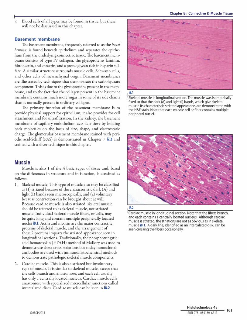
as (1) striated because of the characteristic dark (A) and light (I) bands seen microscopically, and (2) voluntary because contraction can be brought about at will. Because cardiac muscle is also striated, skeletal muscle should be referred to as skeletal muscle, not striated muscle. Individual skeletal muscle fibers, or cells, may be quite long and contain multiple peripherally located nuclei i8.1. Actin and myosin are the major contractile proteins of skeletal muscle, and the arrangement of these 2 proteins imparts the striated appearance seen in longitudinal sections. Traditionally, the phosphotungstic acid-hematoxylin (PTAH) method of Mallory was used to demonstrate these cross-striations but today monoclonal antibodies are used with immunohistochemical methods to demonstrate pathologic skeletal muscle components.
2. Cardiac muscle. This is also a striated but involuntary type of muscle. It is similar to skeletal muscle, except that the cells branch and anastomose, and each cell usually has only 1 centrally located nucleus. Cardiac muscle cells anastomose with specialized intercellular junctions called intercalated discs. Cardiac muscle can be seen in i8.2.
i8.1 | Skeletal muscle in longitudinal section. The muscle was isometrically fixed so that the dark (A) and light (I) bands, which give skeletal muscle its characteristic striated appearance, are demonstrated with the H&E stain. Note that each muscle cell or fiber contains multiple peripheral nuclei.
i8.2 | Cardiac muscle in longitudinal section. Note that the fibers branch, and each contains 1 centrally located nucleus. Although cardiac muscle is striated, the striations are not as obvious as in skeletal muscle i8.1. A dark line, identified as an intercalated disk, can be seen crossing the fibers occasionally.

Chapter 8: Connective & Muscle Tissue
ISBN 978 ‑ 089189‑6319 ©ASCP 2015162
Histotechnology 4e
3. Smooth muscle. This is a nonstriated, involuntary type of muscle that is commonly arranged in layers. The muscle fibers are long and tapered and contain a single centrally located nucleus i8.3. Actin and myosin are also present in smooth muscle, but they are present in a different ratio than in striated muscle, and these proteins also are organized very differently. Smooth muscle regulates luminal size in hollow organs and tubular structures. i8.4, i8.5 show smooth muscle fibers in the wall of a blood vessel stained red.
Staining techniques for connective tissue fibersMasson trichrome stain [Luna 1968, Sheehan 1980]
PurposeTrichrome stains are frequently used to differentiate col-
lagen from smooth muscle in tumors and to identify increases in collagenous tissue in diseases such as cirrhosis of the liver.
PrincipleTrichrome procedures are so named because 3 dyes, which
may or may not include the nuclear stain, are used. The mecha-nism of the stain is not totally understood and may be related in part to the size of different dye molecules. Sections are first stained with an acid dye such as Biebrich scarlet; all acidophilic tissue elements such as cytoplasm, muscle, and collagen will bind the acid dyes. The sections are then treated with phosphotung-stic and/or phosphomolybdic acid. Because cytoplasm is much less permeable than collagen, phosphotungstic and phosphomo-lybdic acids cause Biebrich scarlet to diffuse out of the collagen but not out of the cytoplasm of cells. Phosphotungstic and phos-phomolybdic acid have numerous acidic groups that most likely act as a link between the decolorized collagen and aniline blue dye. Also the pH of the phosphotungstic/phosphomolybdic acid solution probably increases selective collagen staining and aids in the diffusion or removal of Biebrich scarlet.
FixativeBouin solution is preferred, but 10% neutral buffered for-
malin may be used.
Equipment56°C to 58°C oven, Coplin jars, Erlenmeyer flasks, gradu-
ated cylinders, pipettes.
i8.3 | Smooth muscle in cross (top left) and longitudinal (bottom right) section, characteristic of the external muscle layers of the gastrointestinal tract. The single nuclei are elongated and centrally placed in the longitudinally sectioned fibers, and round and centrally placed in the cross-sectioned fibers. The arrow is located in a ganglion or a collection of nerve cells occurring outside the central nervous system.
i8.4 | Masson trichrome stained section of a large vein. The lumen can be seen at the top. Smooth muscle cells are bright red and collagen fibers are blue.
i8.5 | Masson trichrome stained section of liver shows the portal vein containing a few layers of smooth muscle in a portion of the portal triad. A large vein or a muscular artery in a section is best for judging the quality of staining. Red staining of epithelial cells but not of smooth muscle cells indicates a problem with the procedure or reagents.

Chapter 8: Connective & Muscle Tissue
©ASCP 2015 ISBN 978 ‑ 089189‑6319163
Histotechnology 4e
TechniqueCut paraffin sections at 4 - 5 μm.
Quality controlPractically every tissue has an internal control, so no other
control sections are needed; however, if a control is desired, uterus, small intestine, appendix, or Fallopian tube will provide good material
Reagents
Bouin solutionPicric acid, saturated aqueous solution 75 mLFormaldehyde, 37% - 40% 25 mLGlacial acetic acid 5 mL
Weigert iron hematoxylin solutionSolution AHematoxylin 10 gAlcohol, 95% 1,000 mL
Solution BFerric chloride, 29% aqueous solution 20 mLDistilled water 475 mLGlacial acetic acid 5 mL
Working solutionMix equal parts of solutions A and B
Biebrich scarlet-acid fuchsin solutionBiebrich scarlet, 1% aqueous solution 360 mLAcid fuchsin, 1% aqueous solution 40 mLGlacial acetic acid 4 mL
Phosphomolybdic/phosphotungstic acid solutionPhosphomolybdic acid 25 gPhosphotungstic acid 25 gDistilled water 1,000 mL
Aniline blue solutionAniline blue 25 gGlacial acetic acid 20 mLDistilled water 1,000 mL
Acetic acid, 1% solutionGlacial acetic acid 1 mLDistilled water 99 mL
Conventional procedure1. Deparaffinize sections and hydrate to distilled water2. Rinse well in distilled water3. Mordant formalin fixed sections in Bouin solution for 1
hour at 56°C4. Remove slides from oven, allow to cool, and wash in
running water until the yellow color disappears5. Rinse in distilled water6. Stain sections in Weigert iron hematoxylin for 10 minutes7. Wash in running water for 10 minutes8. Rinse in distilled water
9. Stain sections in Biebrich scarlet-acid fuchsin solution for 2 minutes. If desired, the solution may be saved for 1 more run only
10. Rinse in distilled water11. Place the slides in phosphomolybdic/phosphotungstic acid
solution for 10 - 15 minutes; discard this solution12. Stain sections in aniline blue solution for 5 minutes; if
desired, the solution may be saved for 1 more run only13. Rinse the slides in distilled water14. Place the slides in 1% acetic acid solution for 3 - 5 minutes;
discard this solution15. Dehydrate with 95% and absolute alcohols, 2 changes
each16. Clear with 2 or 3 changes of xylene and mount with
synthetic resin
Results i8.4, i8.5
Nuclei BlackCytoplasm, keratin, muscle fibers RedCollagen and mucin Blue
Technical notes1. If desired, collagen may be counterstained with light
green instead of aniline blue i8.6. The following changes are made:
Step 11: Place the sections in a 5% aqueous solution of phosphotungstic acidStep 12: Stain 5 minutes in 2% light green
i8.6 | A section of skin stained with Masson trichrome. Light green has been used as the counterstain instead of aniline blue. An erector pili muscle and the epithelium are stained red, and the collagen is stained green.

Chapter 8: Connective & Muscle Tissue
ISBN 978 ‑ 089189‑6319 ©ASCP 2015164
Histotechnology 4e
Light green, SF yellowish 2 gDistilled water 99 mLGlacial acetic acid 1 mL
Light green is a better counterstain when collagen is predominant, however, when only small amounts are to be demonstrated, the aniline blue is the better counterstain.2. Decreased red staining usually indicates that the staining
solution has aged or been overused and should be discarded i8.7. If blue staining of connective tissue appears faded, the section has probably been overdifferentiated in the acetic acid solution. Pathologically altered collagen, such as that seen in burns, may lose its affinity for aniline blue and bind the acid dye instead [Vacca 1985].
3. Sections fixed in 10% neutral buffered formalin will stain poorly and unevenly. Cross-linking fixatives such as formaldehyde and glutaraldehyde mask reactive chemical groups that would otherwise bind the acid dyes used in trichrome techniques. Formalin fixed tissues will stain brilliantly when mordanted in either Bouin or a mercuric chloride solution; however, mercuric fixatives should not be used because of the toxicity. Zinc formalin has been shown to give satisfactory results [Della Speranza 2009].
4. An iron hematoxylin solution is used for nuclear staining in the trichrome procedures because iron hematoxylin is more resistant than aluminum hematoxylin to decolorization in the subsequently used acidic dye solution.
5. Although most texts state that Weigert iron hematoxylin should be prepared fresh, I find that it is good for several days.
6. Picric acid containing less that 10% water is very explosive; therefore, it is important that solutions not be spilled in the oven and then allowed to evaporate. For this reason, the staining jar containing picric acid (Bouin solution) should be placed inside another container while in the oven.
Microwave procedure [Crowder 1991]
1. Deparaffinize the sections and hydrate to distilled water.2. If the sections were not originally fixed in Bouin solution,
they should be mordanted by placing in a vented plastic Coplin jar of Bouin solution. Microwave at 70% power for 45 seconds. If originally fixed in Bouin solution, skip to step 4.
3. Rinse sections in running water until colorless.4. Place slides in Gill hematoxylin III in a vented plastic
Coplin jar. Microwave on high for 15 seconds.5. Rinse slides in tap water.6. Blue the nuclei with an alkaline blueing solution.7. Rinse well with tap water.8. Rinse slides in distilled water.
9. Place sections in Biebrich scarlet-acid fuchsin solution in a vented plastic Coplin jar. Microwave at 70% power for 30 seconds.
10. Rinse sections in distilled water.11. Place sections in phosphomolybdic/phosphotungstic acid
solution in a vented plastic Coplin jar. Microwave at full power for 15 seconds. Do not rinse.
12. Place in aniline blue solution in a vented Coplin jar. Microwave at 70% power for 30 seconds.
13. Rinse slides in distilled water.14. Place slides in 1% acetic acid for 2 - 3 minutes.15. Dehydrate sections with 95% and absolute alcohol, 2
changes each.16. Clear with xylene and mount with synthetic resin.
Results i8.4, i8.5
Nuclei Dark blueCytoplasm, keratin, muscle fibers RedCollagen and mucin Blue
Technical notes1. All reagents should be discarded after use.2. Because picric acid is explosive when allowed to dry, the
Coplin jar should be place in a partially closed plastic bag to trap any boil-over or spill of this reagent.
i8.7 | A section of fallopian tube stained with Masson trichrome. Note that neither the smooth muscle surrounding the blood vessels nor the epithelium is stained red, but the red blood cells do show red staining. Red blood cells should never be used to judge the quality of the stain. This is a bad stain and should be repeated with new reagents.

Chapter 8: Connective & Muscle Tissue
©ASCP 2015 ISBN 978 ‑ 089189‑6319165
Histotechnology 4e
Gomori 1-step trichrome stain [Gomori 1950b, Sheehan & Hrapchak 1980]
PurposeTo identify an increase in collagenous connective tissue
fibers or to differentiate between collagen and smooth muscle fibers.
PrincipleIn the 1-step trichrome procedure, a plasma stain (chromo-
trope 2R) and a connective tissue fiber stain (fast green FCF, light green, or aniline blue) are combined in a solution of phos-photungstic acid to which glacial acetic acid has been added. Phosphotungstic acid favors the red staining of muscle and cytoplasm. The tungstate ion is specifically taken up by collagen, and the connective tissue fiber stain is subsequently bound to this complex.
FixativeAny well fixed tissue may be used. Bouin solution is used as
a mordant to intensify the color reactions.
Equipment56°C - 58°C oven, Coplin jars, Erlenmeyer flasks, graduated
cylinders, pipettes.
TechniqueCut paraffin sections at 4 - 5 μm.
Quality controlPractically every tissue has an internal control, so no other
control sections are needed; however, if a control is desired, uterus, small intestine, appendix, or Fallopian tube will provide good material.
Reagents
Bouin solutionPicric acid, saturated aqueous solution 75 mLFormaldehyde, 37% - 40% 25 mLGlacial acetic acid 5 mLWeigert iron hematoxylin solution
Solution AHematoxylin 10 gAlcohol, 95% 1,000 mL
Solution BFerric chloride, 29% aqueous solution 20 mLDistilled water 475 mLGlacial acetic acid 5 mL
Working solutionMix equal parts of solutions A and B.
Gomori trichrome stainChromotrope 2R 0.6 gFast green FCF, light green, or aniline blue 0.3 gPhosphotungstic acid 0.8 gGlacial acetic acid 1 mLDistilled water 100 mLStore this solution in the refrigerator.
Acetic acid, 0.5% solutionGlacial acetic acid 0.5 mLDistilled water 99.5 mL
Procedure1. Deparaffinize sections and hydrate to distilled water2. Rinse well in distilled water3. Mordant sections in Bouin solution for 1 hour at 56°C4. Remove slides from the oven, allow to cool, and wash in
running water until the yellow color disappears5. Rinse in distilled water6. Stain sections in Weigert iron hematoxylin for 10 minutes7. Wash in running water for 10 minutes8. Stain sections for 15 - 20 minutes in Gomori trichrome
stain9. Differentiate for 2 minutes in 0.5% acetic acid10. Dehydrate, clear, and mount with synthetic resin
Results i8.8
Nuclei BlackCytoplasm, keratin, muscle fibers RedCollagen and mucin Green or blue
i8.8 | Gomori 1-step trichrome stained section of a large vein. Light green was used instead of aniline blue in preparing the solution. Smooth muscle cells are stained bright red and collagen fibers are stained green.
Chapter 8: Connective & Muscle Tissue
ISBN 978 ‑ 089189‑6319 ©ASCP 2015166
Histotechnology 4e
Technical notes1. Sweat [1968] states that coloration of fine connective tissue
fibers is affected by the dye solution pH, with maximum binding occurring around pH 1.3. The pH of Gomori trichrome is ~2.5, which decreases affinity for anions by ~50%, so these investigators suggest that by replacing the acetic acid with hydrochloric acid, a pH of ~1.3 can be obtained. The intensity of coloration of the fine connective tissue fibers can be varied by altering the pH.
2. Churukian [1993] finds that zinc formalin allows good trichrome staining without mordanting in Bouin solution.
van Gieson picric acid-acid fuchsin stain [Mallory 1942, Sheehan 1980]
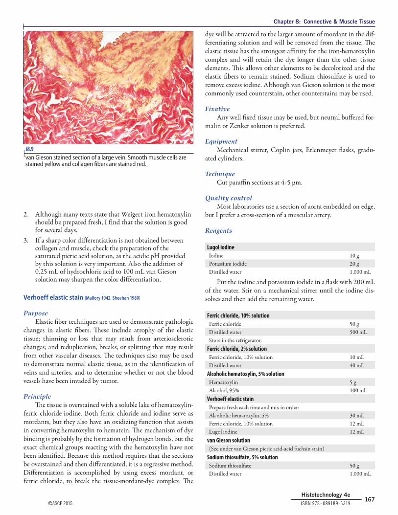
PurposeAlthough the van Gieson technique may be considered a
primary connective tissue stain, it is rarely used as such; however, it serves as an excellent counterstain for other methods such as the Verhoeff elastic technique, referred to in many institutions as the Verhoeff-van Gieson (VVG) stain. Other institutions refer to it as the elastic-van Gieson (EVG) stain.
PrincipleIn a strongly acidic solution, collagen is selectively stained by
acid fuchsin, an acid aniline dye. Picric acid provides the acidic pH necessary and also acts as a stain for muscle and cytoplasm. The low pH is very important, as selective staining of collagen will not occur at higher pH levels. The addition of 0.25 mL of hydrochloric acid to 100 mL of van Gieson solution will sharpen the differentiation between collagen and muscle [Lillie 1976]. Saturated picric acid solutions are important in the preparation of the stain, and again for the selective staining of collagen. If the picric acid solution is not saturated, collagen may stain pale pink to pale orange, and collagen, cytoplasm, and muscle may all stain the same color.
FixativeAny well fixed tissue may be used.
EquipmentMechanical stirrer, Coplin jars, Erlenmeyer flasks, gradu-
ated cylinders, pipettes.
TechniqueCut paraffin sections at 4 - 5 μm.
Quality controlPractically every tissue has an internal control, so no other
control sections are needed; however, if a control is desired, uterus, small intestine, appendix, or Fallopian tube will provide good material.
Reagents
Weigert iron hematoxylin solutionSolution AHematoxylin 10 gAlcohol, 95% 1,000 mL
Solution BFerric chloride, 29% aqueous solution 20 mLDistilled water 475 mLGlacial acetic acid 5 mL
Working solutionMix equal parts of solutions A and B
Acid fuchsin, 1% solutionAcid fuchsin 1 gDistilled water 100 mL
Picric acid, saturated solutionPicric acid 13 gDistilled water 1,000 mL
Stir the solution on the mechanical stirrer for several hours. Some picric acid should remain undissolved in the bottom of the flask. The solubility of picric acid is 1.23 g/100 mL water at 20°C. The amount used may have to be adjusted, depending on whether water has been added to the stock powder to ensure that the water content does not drop below at least 10%.
van Gieson solutionAcid fuchsin, 1% solution 5 mLPicric acid, saturated solution 95 mL
Procedure1. Deparaffinize sections and hydrate to distilled water2. Stain sections with Weigert iron hematoxylin for 10 - 20
minutes; sections should be overstained, as they will be slightly decolorized by the picric acid
3. Wash in running tap water for 10 minutes4. Stain sections in van Gieson stain for 5 minutes; discard
solution5. Place slides in 95% alcohol6. Dehydrate as usual, clear with xylene, and mount with
synthetic resin
Results i8.9
Nuclei BlackCollagen Brilliant redMuscle and cytoplasm Yellow
Technical notes1. An iron hematoxylin solution is used for nuclear staining
in the trichrome procedures because iron hematoxylin is more resistant than aluminum hematoxylin to decolorization in subsequent acidic dye solutions.
Chapter 8: Connective & Muscle Tissue
©ASCP 2015 ISBN 978 ‑ 089189‑6319167
Histotechnology 4e
2. Although many texts state that Weigert iron hematoxylin should be prepared fresh, I find that the solution is good for several days.
3. If a sharp color differentiation is not obtained between collagen and muscle, check the preparation of the saturated picric acid solution, as the acidic pH provided by this solution is very important. Also the addition of 0.25 mL of hydrochloric acid to 100 mL van Gieson solution may sharpen the color differentiation.
Verhoeff elastic stain [Mallory 1942, Sheehan 1980]
PurposeElastic fiber techniques are used to demonstrate pathologic
changes in elastic fibers. These include atrophy of the elastic tissue; thinning or loss that may result from arteriosclerotic changes; and reduplication, breaks, or splitting that may result from other vascular diseases. The techniques also may be used to demonstrate normal elastic tissue, as in the identification of veins and arteries, and to determine whether or not the blood vessels have been invaded by tumor.
PrincipleThe tissue is overstained with a soluble lake of hematoxylin-
ferric chloride-iodine. Both ferric chloride and iodine serve as mordants, but they also have an oxidizing function that assists in converting hematoxylin to hematein. The mechanism of dye binding is probably by the formation of hydrogen bonds, but the exact chemical groups reacting with the hematoxylin have not been identified. Because this method requires that the sections be overstained and then differentiated, it is a regressive method. Differentiation is accomplished by using excess mordant, or ferric chloride, to break the tissue-mordant-dye complex. The
dye will be attracted to the larger amount of mordant in the dif-ferentiating solution and will be removed from the tissue. The elastic tissue has the strongest affinity for the iron-hematoxylin complex and will retain the dye longer than the other tissue elements. This allows other elements to be decolorized and the elastic fibers to remain stained. Sodium thiosulfate is used to remove excess iodine. Although van Gieson solution is the most commonly used counterstain, other counterstains may be used.
FixativeAny well fixed tissue may be used, but neutral buffered for-
malin or Zenker solution is preferred.
EquipmentMechanical stirrer, Coplin jars, Erlenmeyer flasks, gradu-
ated cylinders.
TechniqueCut paraffin sections at 4 - 5 μm.
Quality controlMost laboratories use a section of aorta embedded on edge,
but I prefer a cross-section of a muscular artery.
Reagents
Lugol iodineIodine 10 gPotassium iodide 20 gDistilled water 1,000 mL
Put the iodine and potassium iodide in a flask with 200 mL of the water. Stir on a mechanical stirrer until the iodine dis-solves and then add the remaining water.
Ferric chloride, 10% solutionFerric chloride 50 gDistilled water 500 mLStore in the refrigerator.
Ferric chloride, 2% solutionFerric chloride, 10% solution 10 mLDistilled water 40 mL
Alcoholic hematoxylin, 5% solutionHematoxylin 5 gAlcohol, 95% 100 mL
Verhoeff elastic stainPrepare fresh each time and mix in order:Alcoholic hematoxylin, 5% 30 mLFerric chloride, 10% solution 12 mLLugol iodine 12 mL
van Gieson solution(See under van Gieson picric acid-acid fuchsin stain)
Sodium thiosulfate, 5% solutionSodium thiosulfate 50 gDistilled water 1,000 mL
i8.9 | van Gieson stained section of a large vein. Smooth muscle cells are stained yellow and collagen fibers are stained red.

Chapter 8: Connective & Muscle Tissue
ISBN 978 ‑ 089189‑6319 ©ASCP 2015168
Histotechnology 4e
Procedure1. Deparaffinize sections and hydrate to distilled water2. Place sections in Verhoeff elastic tissue stain for 1 hour3. Wash in 2 changes of distilled water4. Differentiate sections microscopically in 2% ferric
chloride until the elastic fibers are distinct and the background is colorless to light gray; if the sections are differentiated too far, restain
5. Rinse sections in distilled water6. Place section in sodium thiosulfate for 1 minute7. Wash in running tap water for 5 minutes8. Counterstain sections in van Gieson stain for 1 minute9. Differentiate in 95% alcohol10. Dehydrate in absolute alcohol, clear in xylene, and mount
with synthetic resin
Results i8.10, i8.11
Elastic fibers Blue-black to blackNuclei Blue to blackCollagen RedOther tissue elements Yellow
Technical notes1. It is easy to overdifferentiate this stain i8.12. If the
background is completely colorless, so that a clear yellow counterstain is obtained, the section may be overdifferentiated. It is probably better to err on the side of underdifferentiation.
2. Overdifferentiated sections may be restained at any step, provided they have not been treated with alcohol.
3. Do not prolong staining with van Gieson solution, because picric acid also will differentiate the stain further.
4. It is not necessary to remove mercury deposits before staining, because they will be removed by the staining solution; however, because of the toxicity, mercuric fixatives should not be used.
5. The preparation of van Gieson solution is critical for proper differentiation of muscle and collagen. If the picric acid is not saturated, collagen will not stain red, and cytoplasm, muscle, and collagen may all stain the same color i8.13.
i8.10 | A Verhoeff-van Gieson stained section from the wall of the aorta, an elastic artery. The tunica media (middle layer) is composed of concentrically arranged perforated laminae of elastic tissue stained black. The spaces between the elastic laminae are occupied by smooth muscle cells (yellow)
i8.11 | Part of a cross-section of a muscular artery stained with the Verhoeff-van Gieson stain. Both the internal and external elastic laminae are very prominent, and very fine elastic fibers (black) can be seen in the yellow muscular tunica media. The collagen fibers of the tunica adventitia are stained red.
i8.12 | A duplicate of i8.10 stained with the Verhoeff-van Gieson stain, but overdifferentiated. Note how much less elastic tissue is demonstrated. The lack of the slightly grayed background, along with very clear, bright yellow muscle fiber staining, is frequently indicative of overdifferentiation. It is imperative that each section be differentiated microscopically.

Chapter 8: Connective & Muscle Tissue
©ASCP 2015 ISBN 978 ‑ 089189‑6319169
Histotechnology 4e
6. To prepare Verhoeff elastic staining solution, the reagents must be added in the order given, with mixing after each addition, or poor staining may result.
7. The staining jar that contained Verhoeff solution may be cleaned easily by transferring the 2% ferric chloride to the jar for a few minutes before discarding the solution.
8. For optimum results, slides must be individually differentiated, because the time of differentiation is somewhat dependent on the amount of elastic tissue present. Do not depend on the control for timing the differentiation of all sections.
9. Because proper differentiation is sometimes difficult, it is helpful to use duplicate sections differentiated to a slightly different end point.
Aldehyde fuchsin elastic stain [Gomori 1950a, Sheehan & Hrapchak1980]
PurposeRefer to the Verhoeff elastic stain.
PrincipleHydrochloric acid and paraldehyde are added to an alco-
holic solution of basic fuchsin to form aldehyde fuchsin. Schiff bases are formed by the aldehyde and the fuchsin, but the affinity of elastic fibers for this solution is not understood. A number of other tissue elements will also stain with aldehyde fuchsin. These elements include pancreatic β cell granules and sulfated mucosubstances. Staining is intensified by prior oxida-tion. With variations in the technique, aldehyde fuchsin also has been used to stain highly sulfated mucosubstances, pancreatic β cells, and hepatitis b antigen.
Fixative10% neutral buffered formalin is preferred; chromate fixa-
tives should be avoided. Formalin and Bouin fixed tissues will show a colorless background and mercury fixed tissue show a pale lilac background [Sheehan & Hrapchak 1980].
EquipmentCoplin jars, Erlenmeyer flasks, graduated cylinders, pipettes,
and Whatman No. 2 filter paper.
TechniqueCut paraffin sections at 4 - 5 μm.
Quality controlUse a section of aorta embedded on edge or a cross section
of a muscular artery. Skin also provides a good control.
Reagents
Aldehyde fuchsin solutionPararosaniline (basic fuchsin, CI 42500) 1 gEthyl alcohol, 70% 200 mLHydrochloric acid, concentrated 2 mLParaldehyde (must be fresh) 2 mLMix well and let stand at room temperature for 2 - 3 days or until the stain is deep purple. Store in the refrigerator.
Light green stock solutionLight green SF yellowish 0.2 gDistilled water 100 mLGlacial acetic acid 0.2 mLMix well.
Light green working solutionLight green stock solution 10 mLDistilled water 50 mL
Procedure1. Deparaffinize sections and hydrate to 70% alcohol2. Stain sections in aldehyde fuchsin solution for 10 - 40
minutes; with good solutions, 10 minutes is usually sufficient for staining
3. Rinse off the excess stain with 70% alcohol4. Wash the sections in water and check microscopically for
staining of elastic fibers. If a deeper stain is desired, rinse sections briefly in 70% alcohol and return to the aldehyde fuchsin. If further differentiation is needed, return sections to the 70% alcohol. Differentiation is stopped by rinsing the sections with distilled water. The stain may be filtered and reused.
5. Rinse the sections with distilled water6. Counterstain sections with the light green working
solution for 1 - 2 minutes; discard the solution7. Dehydrate in 2 changes each of 95% and absolute
alcohols, clear in xylene, and mount with synthetic resin
i8.13 | A section of skin stained with the Verhoeff-van Gieson stain. The collagen should be stained bright red in a correctly done stain, but it is yellow-orange in this section, indicating that the picric acid used to prepare the van Gieson stain was not saturated. This stain should be repeated with correctly prepared van Gieson stain.

Chapter 8: Connective & Muscle Tissue
ISBN 978 ‑ 089189‑6319 ©ASCP 2015170
Histotechnology 4e
Results i8.14
Elastic fibers Deep blue to purpleOther tissue elements Green
Technical notes1. The paraldehyde used for preparation of the aldehyde
fuchsin reagent should be fresh. Do not use reagent that was opened previously.
2. Old solutions of aldehyde fuchsin may not stain well, and the staining time may need to be prolonged.
3. Do not use rosaniline (CI 42510) for the preparation of this reagent, it is not satisfactory [Mowry 1979].
4. The shelf life of aldehyde fuchsin may be prolonged by refrigerating a small amount and freezing aliquots of the remainder.
5. Acetaldehyde is cheaper and may be obtained without a DEA number. The solution can be prepared as follows [Wenk 1995 ]:
Alcoholic basic fuchsin, 0.5% solutionBasic fuchsin (CI 42500) 2.5 gEthyl alcohol, 70% 500 mLStir until dissolved and store at room temperature. Stable for several months.
Aldehyde fuchsin solutionAlcoholic basic fuchsin, 0.5% solution 50 mLAcetaldehyde 2.5 mLHydrochloric acid, concentrated 1.0 mL
Stir until dissolved. Cover tightly and allow to stand over-night at room temperature. Filter and store at 4°C. Allow to warm to room temperature and shake before using. The solution may be reused until staining is weak. It is stable for ~3 weeks.
Notes on other elastic stainsIn the United States, the Verhoeff-van Gieson stain is
probably the most widely used for the demonstration of elastic fibers. The second most widely used is the aldehyde fuchsin stain. Other stains for elastic tissue that are not used often in the United States, and not given in this text, are orcein and res-orcin fuchsin. Orcein is 1 of the oldest methods for elastic fibers; however, it gives a less intense color than the Verhoeff. Orcein is used in an acidified alcoholic solution, and elastic fibers are stained brown with this stain i8.15. This procedure may be found in Kiernan [2008]. Resorcin fuchsin uses an acidified alcoholic solution of resorcin fuchsin and usually the counterstain is van Gieson; the results look very much like the Verhoeff-van Gieson. This procedure may be found in Vacca [1985].1. In Europe, the Miller technique for elastic tissue is
widely used. This is 1 of the modifications of the original Weigert resorcin fuchsin method. Miller elastic stain is composed of a mixture of Victoria blue 4R, new fuchsin, and crystal violet dissolved in water, to which the following is added in order: resorcin, dextrin, and ferric chloride. Finally hydrochloric acid is added. This procedure was described by Luna [1992].
i8.14 | A section of skin stained with the Gomori aldehyde fuchsin procedure. Elastic tissue is stained a dark purple. The keratinized stratified squamous epithelium, or the epidermis, and the dense irregular connective tissue of the dermis are all stained green.
i8.15 | Orcein stained sections of a small artery demonstrating the elastic fibers.

Chapter 8: Connective & Muscle Tissue
©ASCP 2015 ISBN 978 ‑ 089189‑6319171
Histotechnology 4e
Russell modification of the Movat pentachrome stain [Luna 1992]
PurposeThe demonstration of mucin, fibrin, elastic fibers, muscle,
and collagen.
PrincipleAcidic mucosubstances are stained by alcian blue. The
alkaline alcohol solution that follows converts the alcian blue to monastral fast blue, which is insoluble. Complete conversion is necessary because alcian blue will be decolorized during the remainder of the procedure. Iron hematoxylin is used to stain the elastic fibers, which are then differentiated with ferric chlo-ride (see the Verhoeff method). Sodium thiosulfate removes any residual iodine. Crocein scarlet and acid fuchsin are acid dyes that stain muscle, cell cytoplasm, collagen, and ground sub-stance. Phosphotungstic acid differentiation removes the stain from the collagen and ground substance. The acetic acid removes the phosphotungstic acid, and collagen is then counterstained with alcoholic safran.
Fixative10% neutral buffered formalin is preferred.
EquipmentCoplin jars, Erlenmeyer flasks, graduated cylinders, pipettes.
TechniqueCut paraffin sections at 4 - 5 μm.
Quality controlUse a section of lung, skin, or colon.
Reagents
Alcian blue, 1% solutionAlcian blue-GS 1 gDistilled water 100 mLGlacial acetic acid 1 mLMix well and store at room temperature.
Alkaline alcohol solutionAmmonium hydroxide 10 mLAlcohol, 95% 90 mLPrepare fresh.
Iodine-iodide solutionIodine 2 gPotassium iodide 4 gDistilled water 100 mLAdd the iodine and potassium iodide to ~25 mL of water and mix until dissolved. Add the remaining water.
Absolute alcoholic hematoxylin, 10% solutionHematoxylin 10 gAbsolute alcohol 100 mLMix until dissolved. Cap tightly and store at room temperature.
Ferric chloride, 10% solutionFerric chloride 10 gDistilled water 100 mLMix until dissolved and store at room temperature.
Working hematoxylin solutionAbsolute alcoholic hematoxylin, 10% 25 mLAbsolute alcohol 25 mLFerric chloride, 10% aqueous 25 mLIodine-iodide solution 25 mLPrepare just before use.
Ferric chloride, 2% (for differentiation)Ferric chloride, 10% solution 10 mLDistilled water 40 mLPrepare just before use.
Sodium thiosulfate, 5% solutionSodium thiosulfate 5 gDistilled water 100 mLMix until dissolved and store at room temperature.
Crocein scarlet-acid fuchsin solutionSolution A (stock)Crocein scarlet 1 gDistilled water 99.5 mLGlacial acetic acid 0.5 mLMix until dissolved and store at room temperature.
Solution B (stock)Acid fuchsin 0.1 gDistilled water 99.5 mLGlacial acetic acid 0.5 mLMix until dissolved, and store at room temperature.
Working solutionSolution A 40 mLSolution B 10 mLPrepare just before use.
Phosphotungstic acid, 5% solutionPhosphotungstic acid 5 gDistilled water 100 mLMix until dissolved and store at room temperature.
Alcoholic safran solutionSafran du Gatinais 6 gAbsolute alcohol 100 mLPlace in an air-tight container, and mix well. Keep at 56°C-58°C for 48 hours, and keep tightly closed in a brown bottle to prevent hydration or evaporation.

Chapter 8: Connective & Muscle Tissue
ISBN 978 ‑ 089189‑6319 ©ASCP 2015172
Histotechnology 4e
Procedure1. Deparaffinize and hydrate to distilled water2. Stain in alcian blue for 20 minutes3. Wash in running tap water for 5 minutes4. Place slides in alkaline alcohol for 30 minutes5. Wash in running tap water for 10 minutes6. Rinse in distilled water7. Stain in working hematoxylin solution for 15 minutes8. Rinse in several changes of distilled water9. Differentiate in 2% aqueous ferric chloride until the
elastic fibers contrast sharply with the background10. Rinse in distilled water11. Place slides in sodium thiosulfate for 1 minute12. Wash in running tap water for 5 minutes; rinse in
distilled water13. Stain in Crocein scarlet-acid fuchsin solution for 1 minute14. Rinse in several changes of distilled water15. Rinse in 0.5% acetic acid solution for 30 seconds16. Place slides in 5% phosphotungstic acid solution, 2
changes of 5 minutes each17. Rinse in 0.5% acetic acid solution18. Rinse in 3 changes of absolute alcohol19. Stain in alcoholic safran solution for 15 minutes20. Rinse in 3 changes of absolute alcohol21. Clear in 2 or 3 changes of xylene and mount with
synthetic resin
Results i8.16, i8.17
Nuclei and elastic fibers BlackCollagen YellowGround substance and mucin BlueFibrinoid, fibrin Intense redMuscle Red
Technical notes1. Allison [2014] prepares the stock Crocein scarlet
(solution A) as follows:
Crocein scarlet MOO 0.1 gDistilled water 99.5 mLGlacial acetic acid 0.5 mL
She recommends staining with the Crocein scarlet MOO-acid fuchsin working solution for 90 seconds to 3 minutes depending on desired redness i8.17.
2. Allison [2014] suggests pouring the used safran solution back into the stock bottle (will last ~6 months). Store the safran solution in a 60°C-65°C oven and also stain at this temperature. Use very pure Safran du Gatinais and keep tightly capped at all times. The heat helps give proper staining for collagen and reticular fibers.
3. The differentiation of the elastic fibers is usually complete in 2 - 3 minutes.
4. The complete removal of alkaline alcohol with running water is very important. Failure to remove all of the alkaline alcohol will inhibit the subsequent staining steps.
5. This stain also may be used to demonstrate Cryptococcus neoformans, staining the organism a bright blue.
i8.16 | A section of colon stained with the Movat pentachrome procedure. Note the blue stained mucin in the goblet cells, red stained smooth muscle, yellow stained collagen, and black elastic fibers.
i8.17 | A Movat pentachrome stained section, showing blue stained mucin, red smooth muscle, yellow collagen, and black elastic fibers. Cartilage is seen on the right [image courtesy of Akemi Allison].

Chapter 8: Connective & Muscle Tissue
©ASCP 2015 ISBN 978 ‑ 089189‑6319173
Histotechnology 4e
Silver techniques for reticular fibersMany variations of silver techniques can be used for the
demonstration of reticular fibers; the principles, however, are the same for most of the techniques, and closely resemble those of the PAS technique. The major steps are:1. Oxidation of the adjacent glycol groups of the hexose
sugars in reticulin to aldehydes. Reagents vary with the technique used, but the most frequently used oxidizers are phosphomolybdic acid, potassium permanganate, and periodic acid.
2. Sensitization, which usually is a metallic impregnation step. Impregnation is the deposition of metallic salts on or around, but not in, the tissue element to be demonstrated. The exact chemical reaction of sensitizers is not known, but according to Sheehan & Hrapchak [1980], the metallic salt used in this step probably forms a metal-organic compound with the reticulin. The sensitizing metal is then replaced by silver. Commonly used sensitizers are uranyl nitrate, ferric ammonium sulfate, and dilute solutions of silver nitrate.
3. Silver impregnation involves treating tissue with an ammoniacal or diamine silver complex, [Ag(NH3)2]+. Kiernan [2008] states that 4 atoms of silver will be deposited at the site of each reactive sugar residue in the reticulin, and that the aldehyde groups present will reduce the diamine silver to metallic silver; however, this is not enough silver to provide adequate visibility. Further deposition occurs when incompletely washed sections are transferred to formaldehyde.
4. Reduction uses formaldehyde in all methods. Residual silver diamine ions are reduced to metallic silver by the formaldehyde. Kiernan [2008] states that metallic silver catalyzes the reaction, so the metal precipitates due to formaldehyde reduction mainly at the original sites of the sugar molecules of the reticulin. This further
deposition of silver by the formaldehyde reduction gives a highly visible precipitate. The reduction step in silver impregnation techniques is sometimes termed developing.
5. Toning is the term used when bound metallic silver is treated with gold chloride, and the color of the impregnated component is changed from brown to black. The metallic silver is replaced by metallic gold in the following reaction:
3Ag° + AuCl3 → Au° ↓ + 3AgCl
A more stable compound is formed, and section contrast and clarity are improved. The yellow color is removed from the background by this step; however, toning can be overdone and a violet to red background instead of the desired gray one will result.
6. Unreduced silver is removed by treating the sections with sodium thiosulfate (hypo). This step will prevent any nonspecifically bound silver remaining in the section from being reduced by later exposure to light.
7. A counterstain may or may not be used, depending on the type of tissue stained and personal preferences of the pathologists.Only 2 representative, reliable methods for reticular fibers
will be presented. Silver stains are notoriously capricious, and the method that consistently gives the best results in your laboratory should be used. If 1 of these methods does not work well in your hands, refer to the literature for other techniques. In my experi-ence, the results of the 2 methods presented are consistently better than those obtained with either the Wilder or the Snook technique. It is very important that the amount of reticulin to be demonstrated in the control section be known and that it be checked carefully each time to determine how completely the reticulin is stained. A summary of the most common reticulin methods and the variations in the first 4 steps is given in t8.1.
t8.1 Variations in the first 4 steps of 6 of the most common methods for reticulin
Reagent Snook Gordon & Sweet Gomori Laidlaw Nasher & Shanklin WilderOxidizer Potassium
permanganatePotassium permanganate
Potassium permanganate
Potassium permanganate
Potassium permanganate plus sulfuric acid
Phosphomolybdic acid
Sensitizer Uranyl nitrate Ferric ammonium sulfate
Ferric ammonium sulfate
None Silver nitrate Uranyl nitrate
Impregnating solution Ammoniacal silver (silver nitrate plus sodium hydroxide, precipitate almost dissolved with ammonium hydroxide)
Ammoniacal silver (silver nitrate plus sodium hydroxide, precipitate almost dissolved with ammonium hydroxide)
Ammoniacal silver (silver nitrate plus sodium hydroxide, precipitate almost dissolved with ammonium hydroxide)
Lithium silver (silver nitrate plus lithium carbonate, precipitate almost dissolved with ammonium hydroxide)
Ammoniacal silver (ammonium hydroxide plus silver nitrate until slight turbidity; pyridine added at end)
Ammoniacal silver (silver nitrate plus sodium hydroxide, precipitate almost dissolved with ammonium hydroxide)
Reducing solution Formaldehyde Formaldehyde Formaldehyde Formaldehyde Formaldehyde plus absolute alcohol
Formaldehyde plus uranyl nitrate
The remaining steps (gold chloride, sodium thiosulfate, and counterstain) may vary only in reagent concentration or counterstain choice [table courtesy of NSH/CAP HQIP Final Critique 2004A]

Chapter 8: Connective & Muscle Tissue
ISBN 978 ‑ 089189‑6319 ©ASCP 2015174
Histotechnology 4e
Gomori stain for reticular fibers [Luna 1968]
PurposeThe demonstration of reticular fibers in tissue sections
can be important in the differential diagnosis of certain types of tumors. A change from the normal reticular fiber pattern, as seen in some liver diseases, is also an important diagnostic finding.
PrincipleThe hexose sugars of reticulin are demonstrated by oxida-
tion to aldehydes. Potassium permanganate is the oxidizing agent in this procedure and the excess is removed by potassium metabisulfite. Ferric ammonium sulfate acts as the sensitizer and is subsequently replaced by silver from the diamine silver solution. Following impregnation, formalin is used to reduce the silver to its visible metallic form. Toning with gold chloride and removal of unreacted silver with sodium thiosulfate follow. The final step is to counterstain if desired.
Fixative10% neutral buffered formalin is preferred.
EquipmentNonmetallic forceps, chemically clean glassware (Coplin
jars, graduated cylinders, Erlenmeyer flasks, and pipettes), filter paper.
TechniqueCut paraffin sections at 4 - 5 μm.
Quality controlLiver is a very good control tissue.
Reagents
Silver nitrate, 10% solutionSilver nitrate 10 gDistilled water 100 mLPotassium hydroxide, 10% solutionPotassium hydroxide 10 gDistilled water 100 mLAmmoniacal silver solution
Combine 20 mL of 10% silver nitrate solution and 4 - 5 mL of 10% aqueous solution of potassium hydroxide. Add concen-trated ammonium hydroxide, drop by drop, while shaking the container continuously, until the precipitate is completely dis-solved. Cautiously add 10% silver nitrate solution, drop by drop, until 1 drop causes the solution to become permanently cloudy. Only a faint cloudiness is desirable. Measure the resulting solu-tion, dilute with an equal amount of distilled water, and filter into a chemically clean Coplin jar.
Potassium permanganate, 0.5% solutionPotassium permanganate 2.5 gDistilled water 500 mL
Potassium metabisulfite, 2% solutionPotassium metabisulfite 10 gDistilled water 500 mL
Ferric ammonium sulfate, 2% solutionFerric ammonium sulfate 10 gDistilled water 500 mL
Formalin solutionFormaldehyde, 37% - 40% 10 mLDistilled water 40 mL
Gold chloride, 0.2% solutionStock gold chloride solution (1%) 10 mLDistilled water 40 mL
Sodium thiosulfate, 2% solutionSodium thiosulfate 10 gDistilled water 500 mL
Nuclear fast red (Kernechtrot) solutionNuclear fast red (Kernechtrot) 0.5 gAluminum sulfate 25 gDistilled water 500 mL
Dissolve the aluminum sulfate in the distilled water and then dissolve the nuclear fast red in this solution using heat. Cool, filter, and add a few grains of thymol as a preservative.
Procedure1. Deparaffinize sections and hydrate to distilled water2. Oxidize sections in 0.5% potassium permanganate
solution for 1 minute3. Rinse in tap water for 2 minutes4. Differentiate in 2% potassium metabisulfite for 1 minute5. Wash in tap water for 2 minutes6. Sensitize sections in 2% ferric ammonium sulfate for 1
minute7. Wash slides in tap water for 2 minutes followed by 2
changes of distilled water for 30 second each8. Impregnate sections with the ammoniacal silver solution
for 1 minute9. Rinse in distilled water for 20 seconds10. Reduce for 3 minutes in the 20% formalin solution11. Wash in tap water for 3 minutes12. Tone in 0.2% gold chloride solution for 10 minutes13. Rinse in distilled water14. Place sections in 2% potassium metabisulfite for 1 minute15. Place sections in 2% sodium thiosulfate for 1 minute16. Wash in tap water for 2 minutes17. Counterstain if desired with nuclear fast red for 5 minutes

Chapter 8: Connective & Muscle Tissue
©ASCP 2015 ISBN 978 ‑ 089189‑6319175
Histotechnology 4e
18. Wash well in tap water19. Dehydrate in 95% and absolute alcohols20. Clear in xylene and mount with synthetic resin
Results i8.18, i8.19Reticulin BlackCollagen TaupeOther tissue elements reflect the counterstain used.
Technical notes1. It is important that a hint of turbidity remain in the silver
solution. An excess of ammonia decreases the sensitivity and results in incomplete impregnation of reticular fibers.
2. The rinse between the diamine silver solution and the formaldehyde is critical for good reticulin demonstration. If the wash is prolonged, the staining of the reticulin will be reduced, and if it is insufficient, there will be excessive background staining.
3. The glassware must be chemically cleaned with commercial cleaning agents or bleach. The older method of using a mixture of sulfuric acid and potassium dichromate is not recommended because of the hazards involved in the preparation and use of this solution. Contaminated glassware, metallic instruments, salts from tap water, unfiltered silver solution, incorrect pH, and impregnation solution temperature incorrect [Brown 2009] are all possible causes of nonspecific silver staining and precipitation i8.20.
4. Metal forceps should not be used in any silver staining solutions. If chemicals are contaminated by metal forceps or metal measuring instruments, silver nitrate will be nonselectively precipitated onto the tissue [Swisher 1997]. Many types of plastic or coated (eg, Teflon) forceps are available commercially and should be used in this method. Metallic forceps can be coated with paraffin, but this is less than satisfactory because heated solutions or paraffin solvents must be avoided.
5. Some texts refer to the ferric ammonium sulfate as iron alum. Alum is a term used for salts that are double or bisulfates, such as potassium aluminum sulfate, ammonium aluminum sulfate, chromium potassium sulfate, and ferric ammonium sulfate.
i8.18 | A section of liver stained with the Gomori reticulin stain and lightly counterstained with nuclear-fast red. As in this photograph, the pattern should be readily assessed with the 10× objective.
i8.20 | Marked precipitate is present in this reticulin stained liver section.
i8.19 | The same section as shown in i8.18 viewed with the 40× objective. The reticulin fibers stand out sharply in i8.18, i8.19. Some cellular organelles have stained black with the silver, and rare suggestions of silver precipitate can be seen in the sinusoids.

Chapter 8: Connective & Muscle Tissue
ISBN 978 ‑ 089189‑6319 ©ASCP 2015176
Histotechnology 4e
6. If nuclear fast red is used as a counterstain, wash the slides well with water after staining. If the slides are not washed well, or are transferred from the counterstain directly to alcohol, they will develop a cloudiness that can be removed only by backing the slides up to water.
7. Many times the pattern of reticular fiber staining is very important, as in liver biopsy specimens, and should be seen easily with the scanning lens of the microscope. The easy visualization of the reticulin pattern with the 10× objective is a guide to the quality of the stain. The counterstain, however, may obscure this easy visualization and therefore should not be used, or should be extremely light, when visualization of the pattern is important.
8. If silver staining of the nuclei is a problem, this may be helped by using acetified potassium permanganate:
Sulfuric acid, 3% 2.5 mLPotassium permanganate, 0.5% 47.5 mL
9. When the ammonium hydroxide has lost strength because of the loss of ammonia from the solution, a fresh bottle should be opened. The decrease in strength is commonly noticed because more than the usual amount of ammonium hydroxide is required, and the reticulin may stain gray-black rather than a sharp black.
10. Churukian [1993] used the ammoniacal silver solution for several days, storing it in the refrigerator, and bringing it to room temperature before use. He feels that the reticulin stains better after the ammoniacal silver has aged for a few days.
11. The deposition of silver should be in a linear pattern. Stains exhibiting a granular deposition, as seen in i8.21, should be repeated.
12. Diamine silver is a very alkaline solution, with a pH in the range of 11 - 12, and sections will sometimes wash off the slides. It may be helpful to use special coated slides or charged slides for picking up the sections. If this is not sufficient, sections may be treated with celloidin before staining.
13. Ammoniacal silver solutions may form explosive compounds, so extreme care must be exercised in the preparation, use, and storage of these solutions. Storage of these solutions in the refrigerator inhibits the formation of explosive compounds; exposure to direct sunlight should be avoided.
Gordon & Sweets stain for reticular fibers [Gordon 1936]
PurposeThe demonstration of reticular fibers in tissue sections can
be important in the differential diagnosis of certain types of tumors. A change from the normal reticular fiber pattern, as seen in liver diseases such as cirrhosis, hepatocellular fibrosis, and/or necrotic liver disease, is also an important diagnostic finding.
PrincipleThe tissue is first oxidized by potassium permanganate to
enhance subsequent staining of reticular fibers, and excess per-manganate is removed by oxalic acid. Ferric ammonium sulfate acts as the sensitizer and is subsequently replaced by silver from the diamine silver solution. After impregnation, formalin is used to reduce the silver to its visible metallic form. Before toning with gold chloride, unreacted silver is removed with sodium thiosulfate. The final step is to counterstain if desired.
Fixative10% neutral buffered formalin is preferred.
EquipmentNonmetallic forceps, chemically clean glassware (Coplin
jars, graduated cylinders, Erlenmeyer flasks, and pipettes), filter paper.
TechniqueCut paraffin sections at 4 - 5 μm.
Quality controlLiver is a very good control tissue.
Reagents Silver nitrate, 10% solutionSilver nitrate 10 gDistilled water 100 mL
Sodium hydroxide, 3% solutionSodium hydroxide 3 gDistilled water 100 mL
i8.21 | A section of liver stained with the Snook reticulin stain [Sheehan 1980] shows granular, rather than linear, staining of the reticulin. This may be because of the use of old reagents, especially old uranium nitrate. This stain should be repeated.

Chapter 8: Connective & Muscle Tissue
©ASCP 2015 ISBN 978 ‑ 089189‑6319177
Histotechnology 4e
Ammoniacal silver solutionPlace 5 mL of 10% silver nitrate solution in an Erlenmeyer
flask and add concentrated ammonium hydroxide, drop by drop, while shaking the container continuously, until the precipitate that forms is completely dissolved. Do not add any excess ammo-nium hydroxide. Add 5 mL of 3% sodium hydroxide solution and cautiously redissolve the precipitate with concentrated ammonium hydroxide until only a faint cloudiness remains. If this step is carried too far and no cloudiness remains, add 10% silver nitrate solution, drop by drop, until 1 drop causes the solu-tion to become permanently cloudy. Only a faint cloudiness is desirable. Dilute the resulting solution to 50 mL with distilled water, and filter into a chemically clean Coplin jar.
Potassium permanganate, 1% solutionPotassium permanganate 1 gDistilled water 100 mLOxalic acid, 1% solutionOxalic acid 1 gDistilled water 100 mLFerric ammonium sulfate, 2.5% solutionFerric ammonium sulfate 2.5 gDistilled water 100 mLFormalin, 10% solutionFormaldehyde, 37% - 40% 10 mLDistilled water 90 mLGold chloride, 0.2% solutionStock gold chloride solution (1%) 10 mLDistilled water 40 mLSodium thiosulfate, 5% solutionSodium thiosulfate 5 gDistilled water 100 mLNuclear fast red (Kernechtrot) solutionNuclear fast red (Kernechtrot) 0.5 gAluminum sulfate 25 gDistilled water 500 mL
Dissolve the aluminum sulfate in the distilled water and then dissolve the nuclear fast red in this solution using heat. Cool, filter, and add a few grains of thymol as a preservative.
Procedure1. Deparaffinize sections and hydrate to distilled water2. Oxidize sections in 1% potassium permanganate solution
for 5 minutes3. Rinse in tap water for 2 minutes4. Bleach in 1% oxalic acid for 2 minutes or until sections are
colorless5. Wash in tap water for 2 minutes6. Sensitize sections in 2.5% ferric ammonium sulfate for at
least 15 minutes7. Wash in several changes of distilled water
8. Impregnate sections with the silver solution for 2 minutes9. Rinse well with distilled water10. Reduce sections for 2 minutes in the 10% formalin
solution11. Wash in tap water for 3 minutes12. Tone in 0.2% gold chloride solution for 10 minutes13. Wash in tap water for 2 minutes14. Place slides in 5% sodium thiosulfate for 1 minute15. Rinse in distilled water16. Counterstain, if desired, with nuclear fast red for 5
minutes17. Wash well in distilled water18. Dehydrate in 2 changes each of 95% and absolute
alcohols, clear in xylene, and mount with synthetic resin
Results i8.22
Reticulin BlackOther tissue elements depend on counterstain used.
Technical notes1. See the technical notes under the Gomori technique for
reticular fibers.2. The Gordon and Sweets method gives much less
background and nuclear staining than most of the more common methods.
i8.22 |A section of liver stained with the Gordon and Sweets technique. No counterstain has been applied, the reticular network lining the sinusoids is well demonstrated, and the reticulin pattern can be evaluated easily with lower magnification.
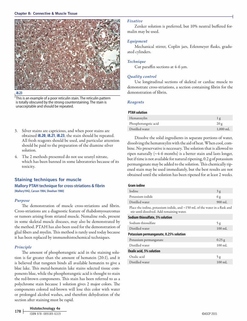
Chapter 8: Connective & Muscle Tissue
ISBN 978 ‑ 089189‑6319 ©ASCP 2015178
Histotechnology 4e
3. Silver stains are capricious, and when poor stains are obtained i8.20, i8.21, i8.23, the stain should be repeated. All fresh reagents should be used, and particular attention should be paid to the preparation of the diamine silver solution.
4. The 2 methods presented do not use uranyl nitrate, which has been banned in some laboratories because of its toxicity.
Staining techniques for muscleMallory PTAH technique for cross-striations & fibrin [Mallory1942, Carson 1984, Sheehan 1980]
PurposeThe demonstration of muscle cross-striations and fibrin.
Cross-striations are a diagnostic feature of rhabdomyosarcomas or tumors arising from striated muscle. Nemaline rods, present in some skeletal muscle diseases, may also be demonstrated by the method. PTAH has also been used for the demonstration of glial fibers and myelin. This method is rarely used today because it has been replaced by immunohistochemical techniques.
PrincipleThe amount of phosphotungstic acid in the staining solu-
tion is far greater than the amount of hematein (20:1), and it is believed that tungsten binds all available hematein to give a blue lake. This metal-hematein lake stains selected tissue com-ponents blue, while the phosphotungstic acid is thought to stain the red-brown components. This stain has been referred to as a polychrome stain because 1 solution gives 2 major colors. The components colored red-brown will lose this color with water or prolonged alcohol washes, and therefore dehydration of the section after staining must be rapid.
FixativeZenker solution is preferred, but 10% neutral buffered for-
malin may be used.
EquipmentMechanical stirrer, Coplin jars, Erlenmeyer flasks, gradu-
ated cylinders.
TechniqueCut paraffin sections at 4 - 6 μm.
Quality controlUse longitudinal sections of skeletal or cardiac muscle to
demonstrate cross-striations, a section containing fibrin for the demonstration of fibrin.
Reagents
PTAH solutionHematoxylin 1 gPhosphotungstic acid 20 gDistilled water 1,000 mL
Dissolve the solid ingredients in separate portions of water, dissolving the hematoxylin with the aid of heat. When cool, com-bine. No preservative is necessary. The solution that is allowed to ripen naturally (~4 - 6 months) is a better stain and lasts longer, but if time is not available for natural ripening, 0.2 g of potassium permanganate may be added to the solution. This chemically rip-ened stain may be used immediately, but the best results are not obtained until the solution has been ripened for at least 2 weeks.
Gram iodineIodine 3 gPotassium iodide 6 gDistilled water 900 mLPlace the iodine, potassium iodide, and ~150 mL of the water in a flask and stir until dissolved. Add remaining water.
Sodium thiosulfate, 5% solutionSodium thiosulfate 5 gDistilled water 100 mL
Potassium permanganate, 0.25% solutionPotassium permanganate 0.25 gDistilled water 100 mL
Oxalic acid, 5% solutionOxalic acid 5 gDistilled water 100 mL
i8.23 | This is an example of a poor reticulin stain. The reticulin pattern is totally obscured by the strong counterstaining. The stain is unacceptable and should be repeated.
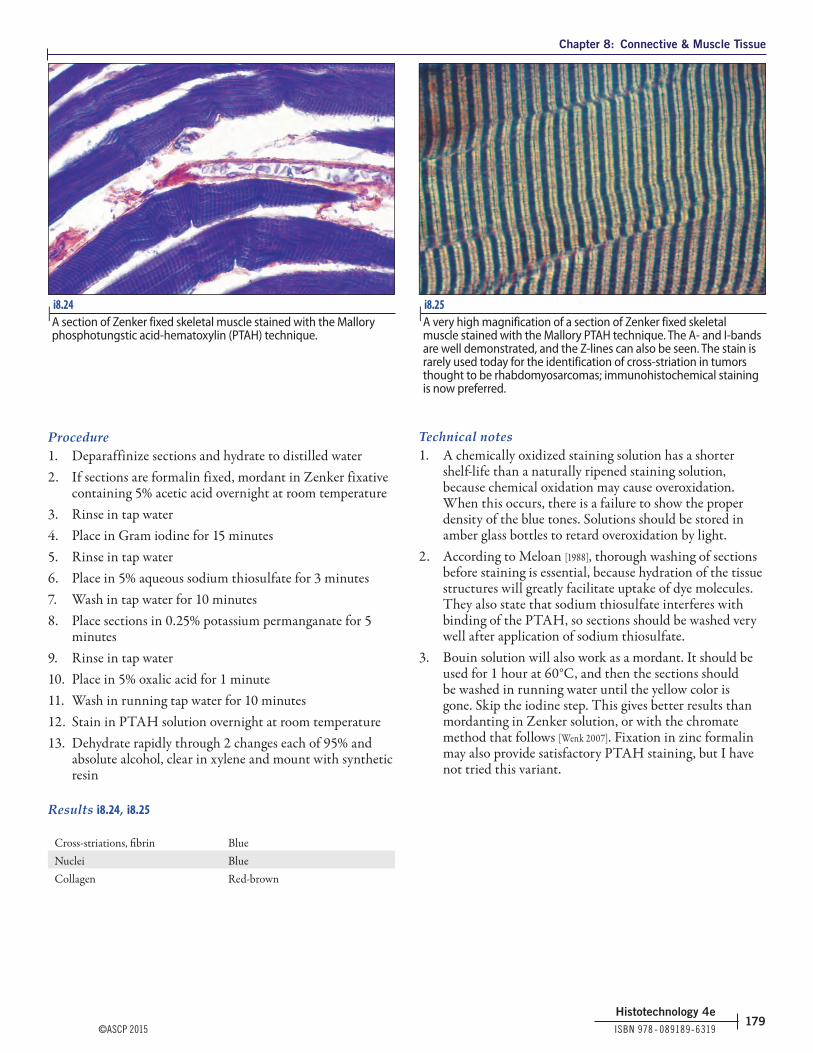
Chapter 8: Connective & Muscle Tissue
©ASCP 2015 ISBN 978 ‑ 089189‑6319179
Histotechnology 4e
Procedure1. Deparaffinize sections and hydrate to distilled water2. If sections are formalin fixed, mordant in Zenker fixative
containing 5% acetic acid overnight at room temperature3. Rinse in tap water4. Place in Gram iodine for 15 minutes5. Rinse in tap water6. Place in 5% aqueous sodium thiosulfate for 3 minutes7. Wash in tap water for 10 minutes8. Place sections in 0.25% potassium permanganate for 5
minutes9. Rinse in tap water10. Place in 5% oxalic acid for 1 minute11. Wash in running tap water for 10 minutes12. Stain in PTAH solution overnight at room temperature13. Dehydrate rapidly through 2 changes each of 95% and
absolute alcohol, clear in xylene and mount with synthetic resin
Results i8.24, i8.25
Cross-striations, fibrin BlueNuclei BlueCollagen Red-brown
Technical notes1. A chemically oxidized staining solution has a shorter
shelf-life than a naturally ripened staining solution, because chemical oxidation may cause overoxidation. When this occurs, there is a failure to show the proper density of the blue tones. Solutions should be stored in amber glass bottles to retard overoxidation by light.
2. According to Meloan [1988], thorough washing of sections before staining is essential, because hydration of the tissue structures will greatly facilitate uptake of dye molecules. They also state that sodium thiosulfate interferes with binding of the PTAH, so sections should be washed very well after application of sodium thiosulfate.
3. Bouin solution will also work as a mordant. It should be used for 1 hour at 60°C, and then the sections should be washed in running water until the yellow color is gone. Skip the iodine step. This gives better results than mordanting in Zenker solution, or with the chromate method that follows [Wenk 2007]. Fixation in zinc formalin may also provide satisfactory PTAH staining, but I have not tried this variant.
i8.24 | A section of Zenker fixed skeletal muscle stained with the Mallory phosphotungstic acid-hematoxylin (PTAH) technique.
i8.25 | A very high magnification of a section of Zenker fixed skeletal muscle stained with the Mallory PTAH technique. The A- and I-bands are well demonstrated, and the Z-lines can also be seen. The stain is rarely used today for the identification of cross-striation in tumors thought to be rhabdomyosarcomas; immunohistochemical staining is now preferred.

Chapter 8: Connective & Muscle Tissue
ISBN 978 ‑ 089189‑6319 ©ASCP 2015180
Histotechnology 4e
4. Fortunately, the PTAH technique has been largely replaced by immunohistochemical methods; although fibrin will stain very well after formaldehyde fixation, we consistently get better results when staining for muscle cross-striations if the tissue is mordanted, or postfixed, in a mercuric solution. The stain is not as good when formalin fixed sections are mordanted as when the original fixative is mercuric; very uneven staining frequently occurs in tissue originally fixed in formalin i8.26. Stevens and Wilson [1996] described the following technique, which avoids mercuric solutions and uses acidic dichromate treatment instead.
PTAH without mercuric solutions
Reagents
Acidic dichromate solutionHydrochloric acid, 10% in absolute alcohol 12 mLPotassium dichromate, 3% aqueous solution 36 mL
Acidic potassium permanganate solutionPotassium permanganate, 0.5% aqueous solution 50 mLSulfuric acid, 3% solution 2.5 mL
Oxalic acid, 1% solutionOxalic acid 1 gDistilled water 100 mL
Procedure1. Deparaffinize sections and hydrate to distilled water2. Place slides in acidic dichromate solution for 30 minutes3. Wash in tap water4. Place slides in acidic permanganate solution for 1 minute
5. Wash in tap water6. Bleach sections in 1% oxalic acid solution7. Rinse in tap water8. Stain in PTAH solution overnight9. Dehydrate rapidly through 95% and absolute alcohols,
clear in xylene, and mount with synthetic resinThe results of this stain should be the same as those
described for the technique using mercuric solutions for either primary fixation or postfixation mordanting.
Technical notePotassium dichromate is a hazardous chemical that is toxic
on inhalation and ingestion. It is corrosive to eyes, skin, and mucous membranes, and is also a carcinogen; however, with care, the solutions pose little risk when used under normal con-ditions. It cannot be put down the drain.
Staining technique for basement membranesPeriodic acid-methenamine silver microwave procedure for basement membranes [Brinn 1983, Sheehan 1980]
PurposeThis procedure best delineates basement membranes, and is
most often used in the histopathology laboratory for the detec-tion of abnormalities or diseases manifested in the glomerular basement membrane.
PrincipleThe carbohydrate component of basement membranes is
oxidized to aldehydes by periodic acid. The aldehydes formed by oxidation bind the silver ions from the methenamine silver complex and reduce the silver to its metallic form. Methenamine gives the solution the alkaline properties necessary for the proper reaction, and the sodium borate acts as a buffer. Toning is with gold chloride, and any unreduced silver is removed by sodium thiosulfate.
Fixative10% neutral buffered formalin is preferred. Mercury-
containing fixatives are not recommended.
EquipmentMicrowave oven or 50°C-60°C water bath, vented plastic
staining jars, graduated cylinders, Erlenmeyer flasks.
TechniqueParaffin sections cut at 2 μm.
Quality controlKidney has an internal control. No other control slide is
necessary.
i8.26 | A section of formalin fixed skeletal muscle stained with PTAH. The sections were mordanted overnight in Zenker fixative. This uneven staining appears to be characteristic of formalin fixed tissue. Note that the collagen is stained red in i8.24, i8.25, i8.26. This is an important differentiation in correctly performed PTAH staining.

Chapter 8: Connective & Muscle Tissue
©ASCP 2015 ISBN 978 ‑ 089189‑6319181
Histotechnology 4e
Reagents
Stock methenamine silverMethenamine, 3% aqueous (15 g/500 mL) 400 mLSilver nitrate, 5% aqueous (5 g/100 mL) 20 mLMix and keep solution refrigerated at 4°C.
Borax (sodium borate), 5% solutionSodium borate 5 gDistilled water 100 mL
Working methenamine silver solutionStock methenamine silver 25 mLDistilled water 25 mLBorax (sodium borate), 5% solution 2 mLPrepare just before use.
Periodic acid, 1% solutionPeriodic acid 1 gDistilled water 100 mL
Sodium thiosulfate, 2% solutionSodium thiosulfate 10 gDistilled water 500 mL
Gold chloride, 0.02% solutionGold chloride, 1% solution 1 mLDistilled water 49 mL
Stock light green, 0.2% solutionLight green SF (yellowish) 1 gDistilled water 500 mLGlacial acetic acid 1 mL
Working light green solutionLight green stock solution 10 mLDistilled water 50 mL
ProcedureThis procedure is for 5 slides. If you do not have 5 slides,
then include blank slides. Do not use >5 slides. This procedure should be followed exactly for optimum results.1. Deparaffinize sections and hydrate to distilled water2. Place sections in 1% periodic acid solution for 15 minutes
at room temperature3. Rinse in distilled water4. Place slides (5) in a vented plastic Coplin jar containing
50 mL of methenamine working solution.* Apply the vented cap and place in the microwave oven. Also place a vented plastic Coplin jar containing exactly 50 mL (measured) of distilled water in the oven. Microwave on full power for exactly 70 seconds (see technical note 2). Remove both jars from the oven, mix the staining solution with a plastic Pasteur pipette, and let stand on the counter. Check the slides frequently until the desire staining intensity is achieved. This will take ~15 - 20 minutes.
5. Rinse slides in the heated distilled water
6. Tone sections in 0.02% gold chloride*7. Rinse slides in distilled water8. Treat sections with 2% sodium thiosulfate for 1 minute9. Wash in tap water10. Counterstain in the working light green solution for 1½
minutes11. Dehydrate with 2 changes each of 95% and absolute
alcohols12. Clear with xylene and mount with synthetic resin
*If for some reason, the microwave oven cannot be used, substitute the following solutions and staining times:
Methenamine silver solutionStock methenamine silver solution 50 mLBorax, 5% solution 5 mLPreheat the solution and stain slides at 56°C-60°C for 40 - 90 minutes.
Gold chloride, 0.2% solutionGold chloride, 1% solution 10 mLDistilled water 40 mLTone for 30 seconds to 1 minute.
Results i8.27
Basement membrane BlackBackground Green
i8.27 | A kidney glomerulus stained with a periodic acid-methenamine silver technique. A good stain should look almost as if the basement membrane had been drawn with ink. Although the tubular basement membranes are well stained, they should not be used to judge the end point of the impregnation; use the glomerular basement membrane only.

Chapter 8: Connective & Muscle Tissue
ISBN 978 ‑ 089189‑6319 ©ASCP 2015182
Histotechnology 4e
Technical notes1. This nonmicrowave technique is a modification of the
Jones method [1951], which heats the methenamine silver in a 60°C-70°C water bath for ~60 - 75 minutes with periodic checks on the depth of staining. When the basement membranes show a medium brown color, remove from the silver and rinse in 70°C distilled water. The other difference is in the counterstain used, usually hematoxylin and eosin i8.28. Nuclear fast red may also be used i8.29.
2. Sharper staining of the basement membrane and less background staining can be obtained with the use of the microwave oven for silver techniques.
3. The temperature is critical and should be just below boiling or ~95°C, immediately after removal from the microwave oven. Each oven should be calibrated for the time required to reach the correct temperature.
4. This is a very difficult stain to perform correctly. Although the tubular membranes will stain, the endpoint should be determined by the glomerular basement only i8.30. When well stained, the glomerular basement membrane should appear as a continuous black line. Stopping the silver impregnation too soon will result in uneven or interrupted staining i8.31. The application
i8.28 | i8.28 A kidney glomerulus stained with the Jones periodic acid-methenamine silver technique. Hematoxylin & eosin are the counterstains.
i8.30 | Only the glomerular basement membrane should be used to determine the endpoint of staining. Although many tubular basement membranes are well stained in this section, no staining is seen in the glomerular basement membrane, and the stain is worthless.
i8.29 | A section of kidney stained with the Jones periodic acid-methenamine silver technique and counterstained with nuclear fast red.
i8.31 | This kidney glomerulus has been understained with a periodic acid-methenamine silver technique. Compare the staining here with that in i8.27. Note that the basement membrane exhibits continuous staining in i8.27 and interrupted staining here.

Chapter 8: Connective & Muscle Tissue
©ASCP 2015 ISBN 978 ‑ 089189‑6319183
Histotechnology 4e
of too much counterstain will mask the silver stain and decrease contrast i8.32.
5. As with all silver stains, chemically cleaned glassware and plastic forceps should be used.
6. The PAS technique is also use to stain basement membranes and can be found in Chapter 7, “Carbohydrates & Amyloid.”
Staining techniques for lipidBecause the lipid stains requested in the routine histopa-
thology laboratory are for simple lipids, only a selection of tech-niques for simple fats will be presented.
Oil red O method for neutral fats [Pearse 1968]
PurposeThe demonstration of neutral lipids in frozen tissue sec-
tions. Fat occurring in an abnormal place such as fatty emboli that may develop after either a bone fracture or an injury that crushes a fatty body area may be demonstrated. The fat stain may verify that the emboli caused death. Degenerating material con-taining fat, such as cell membranes or myelin, may coalesce into fat droplets that are demonstrable with fat stains, and tumors arising from fat cells (liposarcomas) can be differentiated from other types of tumors.
PrincipleStaining with oil soluble dyes is based on the greater
solubility of the dye in the lipoid substances than in the usual hydroalcoholic dye solvents. This is a physical method of staining and the dye used must (1) be more soluble in the tissue lipid than in the solvent in which it is dissolved, (2) not be water soluble, (3)
be strongly colored, and (4) act with tissue constituents only by solution. The solvent used is critical, with isopropanol removing a minimal amount of lipid and propylene glycol not extracting any lipid.
Fixative10% neutral buffered formalin or calcium-formol. Because
of the lipid-dissolving ability, no alcoholic fixative should be used.
EquipmentCryostat, Coplin jars, Erlenmeyer flasks, graduated cylin-
ders, filter paper.
TechniqueCut frozen sections at 10 μm. Paraffin sections cannot be
used because dehydrating and clearing agents dissolve the fat. If free-floating sections are not used, then sections of fixed tissue should be picked up on coated, charged, or subbed slides.
Quality controlMost tissue contains some fat, so normally a control is not
used.
Reagents
Oil red O stock solutionOil red O 2.5 gIsopropanol, 98% 500 mLMix well.
Oil red O working solutionOil red O, stock solution 24 mLDistilled water 16 mLMix well and let stand for 10 minutes. Filter. The filtrate can be used for several hours.
Procedure1. Cut frozen sections, fix in 40% formaldehyde for 1
minute, and wash well in tap water; blot off excess water; if the tissue has been previously fixed, there is no need to refix
2. Stain sections in oil red O for 10 minutes3. Wash sections in tap water4. Stain for 1 minute in Harris hematoxylin containing
acetic acid (glacial acetic acid, 2 mL/hematoxylin, 48 mL)5. Wash section in tap water6. Blue in ammonia water7. Wash in tap water8. Mount sections with an aqueous mounting medium9. Seal the edges of the coverslip with fingernail polish
i8.32 | Too much counterstain has been applied to this section of kidney, and the periodic acid-methenamine silver staining is masked. The section is also too thick; sections should be 2 μm thick for demonstration of the glomerular basement membrane.

On completing this chapter, the student should be able to do the following:
Objectives
©ASCP 2015 ISBN 978 ‑ 089189‑6319263
Histotechnology 4e
Chapter 12
1. Define:a. antigenb. antibodyc. substrated. chromogene. fluorochromef. epitopeg. secondary antibodyh. multilink secondary antiserum
2. Differentiate between polyclonal antisera and monoclonal antibodies
3. Explain each of the following immunohistochemical techniques:a. directb. indirectc. peroxidase-antiperoxidased. avidin-biotin complexe. polymeric detection system
4. List 2 fluorochromes
5. Identify 2 common chromogens used with immunoperoxidase techniques
6. Identify 2 chromogens used with immunoalkaline phosphatase techniques
7. Identify at least 3 enzyme systems used in enzyme immunohistochemistry
8. Describe the preferred method of specimen preparation for immunofluorescence
9. Identify problems that may be encountered with formaldehyde fixation of specimens for immunohistochemical staining
10. Identify fixatives other than formalin and describe any special issues with respect to immunohistochemistry
11. Describe 2 blocking reactions and identify the purpose of each
12. Identify the 5 heavy chains and 2 light chains that make up the different classes of immunoglobulins
13. Describe the use of negative tissue and reagent controls
14. List at least 6 problems that may occur with immunohistochemical staining and state a corrective action for each
15. State why preparing positive controls in the laboratory is preferred to purchasing them
16. List 2 methods of epitope retrieval
17. List at least 3 solutions that have been used for heat induced epitope retrieval
18. List at least 3 ways of heating in heat induced epitope retrieval methods (HIER)
19. List at least 3 solutions that have been used for enzyme induced epitope retrieval (EIER)
20. Identify alcohol soluble and nonalcohol soluble chromogens
21. List 4 chemicals used to intensify the 3,3′-diaminobenzidine (DAB) reaction
22. List common antibodies and the tumors they are used to identify
Immunohistochemistry

Chapter 12: Immunohistochemistry
ISBN 978 ‑ 089189‑6319 ©ASCP 2015264
Histotechnology 4e
In the clinical laboratory, immunohistochemistry is a heavily used technique for assisting the pathologist in making a diagnosis. Immunohistochemistry is used to determine the origin of a tumor, prognosis, and treatment. It is important to understand the basics of immunology and the reagents and methods used, to achieve reproducible, high-quality patient care. Rigorous quality control (QC) is vital to ensure that results are accurate and consistent with expected staining patterns. The major role an immunohistochemist plays is troubleshooting for variables introduced by fixation, processing, and tissue types. The immunohistochemist should be familiar with both the con-trols used and the staining patterns of an antibody to trouble-shoot unexpected results.
Techniques such as immunofluorescence and enzyme immunoassays have been available as diagnostic tools for over 50 years, but the development of immunodiagnostic methods, including immunohistochemistry, was much slower because of the unpredictable quality of polyclonal antibodies.
Since the immunohistochemistry revolution began in the 1980s, a number of dramatic advances have been made in immu-nology and immunodiagnostic methods; these advances have had a substantial impact on all phases of laboratory medicine. In most laboratories, the use of immunohistochemical stains has become as routine as the use of any other special stain, and in many instances, these stains have completely replaced the older histochemical or empirical methods.
The development of immunohistochemical staining stan-dards and guidelines set by the College of American Pathologists (CAP) is related to predictive markers that are used for thera-peutic patient care, and which qualify the patient for specific drug treatment. The laboratory’s role is pivotal in ensuring that all specimens are processed and treated according to the recom-mended guidelines.
General immunologyThe immune system exerts its control through humoral
(antibody) and cellular components. The overview of the immune system presented here will emphasize only the humoral or antibody system and how it relates to the discussion of immu-nohistochemical techniques.
AntibodyAn antibody is a host protein (immunoglobulin) produced
in response to the presence of foreign molecules, organisms, or other agents in the body. Antibodies, commonly known as immunoglobulins, are proteins that are produced by B lympho-cytes in response to antigenic stimulation. An immunoglobulin is a “Y”-shaped protein molecule that is composed of both heavy and light chains. Classes of antibodies differ in structure and function, with each immunoglobulin antigenically distinct. An immunoglobulin (Ig) is composed of 2 identical heavy chains (γ, α, mu, δ, or ε) that determine the Ig subclass (IgG, IgA, IgE,
IgD, and IgM). The light chain is either κ or λ. These proteins are expressed on the cell surface or membrane and secreted into blood and other fluids by plasma cells. The upper arms of the Y are the regions of the antibody that bind to the short arms of their specific antigen. This region of the antigen is known as an epitope, and is the site at which the antibody attaches to tissue. An antibody may target more that 1 antigen, but it is specific for only 1 epitope. Proteins are very good antigens because of their large size. The fact that antibodies (eg, immunoglobulins) are proteins makes them very potent antigens.
AntigenAn antigen is a molecule made up of proteins, carbo-
hydrates, or other polymers, and is capable of producing an immune response in animals or cell cultures for the production of antibodies. Antigens in tissues bind antibodies at different cell sites: the membrane, cytoplasm, nucleus, organelles, or a combination of sites. Many compounds are antigenic. Because of their large size, proteins are ideal for immunization and for binding, but polysaccharides, nucleic acids, and other polymers also can be antigenic. The most common antigens that induce antibody production by the body are bacteria and viruses.
Polyclonal antiseraIf blood was drawn from your arm and IgM antibody was
isolated from it and injected into a rabbit, your IgM antibody would act as an antigen and stimulate the rabbit to make anti-human IgM antibody. Serum from the immunized rabbit could then be used as a polyclonal antihuman IgM antibody reagent. When lymphocytes are exposed to antigen, some of the lympho-cytes proliferate, and each lymphocyte forms a cell line. All cells in a single clone, or cell line, produce an identical antibody, but various clones produce antibodies of different classes with speci-ficities to different molecular sites (epitopes) on the antigen. The result of antigenic stimulation is the production of a mixture of antibodies from many clones of lymphocytes. This pool of anti-bodies is known as polyclonal antiserum.
Polyclonal antiserum is highly sensitive because it binds to multiple epitopes, but this also means that it is not as selec-tive, resulting in some nonspecific staining (background). The titers used to stain tissue must be carefully selected to ensure no false positive staining. Examples of hosts that are used to pro-duce polyclonal antiserum include rabbit, goat, horse, sheep, and human. Because of the variability of the immune response from 1 animal to another, polyclonal antibodies are difficult to stan-dardize and can be used only in a limited number of immunoas-says. Pooled antibodies made from many immunized animals of the same species are less likely to exhibit major batch-to-batch variations than pools made from only a few animals.

Chapter 12: Immunohistochemistry
©ASCP 2015 ISBN 978 ‑ 089189‑6319265
Histotechnology 4e
Monoclonal antibodiesThe development of monoclonal antibody techniques revo-
lutionized the whole science of immunology. Monoclonal antibodies are prepared by injecting mice with an antigen. B lymphocytes are fused with nonsecreting myeloma cells (non-secreting plasma cell tumor). This in vitro fusion yields hybrid cells (hybridoma) that retain the antibody secretion capability of the B cell and the immortality of the tumor cells. Hybridoma cells can be cloned, and a single clone is capable of producing antibody that is identical in molecular structure to the original. The antibody can be produced in unlimited quantities by tissue culture or by transplantation into the peritoneal cavities of mice. Although other animal species can be used, monoclonal antibodies for the histopathology laboratory are produced most commonly in mice. Monoclonal antibodies can be character-ized, standardized, and produced in unlimited quantities. The advantages of monoclonal antibodies include high homoge-neity, the absence of nonspecific antibodies, and no batch-to-batch or lot-to-lot variability. Mouse monoclonal antibodies are purer than polyclonal antibodies, and monoclonal antibodies display the most desirable attributes (high affinity and selec-tivity) of polyclonals without displaying most of the undesir-able characteristics (lack of chemical homogeneity and lack of continuous supply).
Rabbit monoclonal antibodiesRabbit monoclonal antibody production was first reported
by Spieker-Polet in 1995. Since that initial report, the produc-tion of rabbit monoclonal antibodies is making its way into the clinical laboratory. This technology follows the same principal of monoclonal hybridoma production, but uses “rabbit fusion partner cells,” which fuse with rabbit B cells. These antibodies
are proven to provide both sensitivity of a rabbit antibody and specificity of targeting a single epitope i12.1, i12.2.
Tissue handlingFrozen tissue fixation & processing
Before performing any immunologic procedure, the sta-bility of the antigen must be considered. Because most antigens are soluble in aqueous solutions, the antigens must be fixed in situ when the technique permits.
Immunofluorescence, by today’s standards, is a relatively insensitive method, and antigenic reactivity must be preserved to the maximal extent possible. Frozen sections of unfixed tissue constitute the classic preparation of tissue for immunofluores-cence, because antigenic reactivity is minimally impaired and fluorescent antibody staining is strongest; however, in such sec-tions, soluble antigens are lost [Sternberger 1979]. Formalin fixed paraffin sections are rarely used in immunofluorescence because of inconsistent results; it is suggested that, fixation and pro-cessing impair antigenic reactivity beyond the sensitivity of immunofluorescence [Sternberger 1979].
Dehydration, or drying, and fixation by chemical reagents are the 2 major categories of fixation. Dehydration unfolds and changes the solubility of the protein. Chemical fixation denatures and stabilizes proteins by coagulation, by forming additive compounds, or by a combination of the 2 actions. Acetone and alcohol are coagulating, nonadditive reagents; formaldehyde is a noncoagulating, additive fixative that cross-links proteins; and mercuric chloride is both a coagulating and additive fixative. The coagulant fixatives that do not form additive compounds permit good penetration of the antibody
i12.1 | A section stained with CyclinD1 mouse monoclonal antibody demonstrating poor signal-to-noise ratio and nonspecific background staining.
i12.2 | A section stained with CyclinD1 rabbit monoclonal antibody demonstrating greater sensitivity and eliminating the nonspecific staining. Compare with i12.1.

Chapter 12: Immunohistochemistry
ISBN 978 ‑ 089189‑6319 ©ASCP 2015266
Histotechnology 4e
and do not block immunoreactive determinants. However, the treatment of cryostat sections with acetone, as currently prac-ticed, does not achieve complete fixation. Extended immuno-histochemical procedures may produce deleterious morphologic changes, including chromatolysis and loss of membranes [Farmilo 1989]. Allowing thin cryostat sections or cytospin preparations to dry for up to 48 hours before acetone fixation will improve morphologic preservation [Farmilo 1989]. It is my experience that frozen sections can be cut at 4 - 5 µm, placed in cold 10% neutral buffered formalin for 5 minutes, then transferred to phosphate buffered saline (PBS; pH 7.4) and held for routine immunohistochemical staining. Muscle sections should be cut, air-dried for 1 hour, and immediately stained with antibodies targeted for dystrophin or other muscular dystrophy related proteins. Zenker and Bouin fixatives are also good for the preservation of some antigens, and alcohol based fixatives such as methacarn (methanol-Carnoy solution) or alcohol based proprietary reagents are reported to preserve antigens very well [Battifora1986]. Elias [1985] states that there appears to be an inverse relationship with regard to preservation of antigenicity and morphology. Glutaraldehyde preserves morphology best, but it irreversibly blocks tissue antigenic determinants.
Fixatives for paraffin processed tissueProper tissue handling, beginning with fixation and
continuing through processing and microtomy, is pivotal to producing consistent results when performing immunohisto-chemical staining on paraffin sections. It is important for the immunohistochemist to understand the basics of how fixation, processing, temperature, and pH can affect the quality of a stain.
Regulations regarding predictive marker staining of tissue places responsibility on the clinical laboratory for documenting the time in fixative. For example, it is recommended that breast tissue from all invasive carcinomas be formalin fixed at room temperature for a minimum of 6 hours and a maximum of 48 hours [Wolff 2007] prior to HER2 testing. Breast tissue should be grossed and sectioned promptly to begin formalin fixation, and the time of fixation must be documented.
A fixative can positively or negatively influence the staining result with a given antibody. Not all antibodies are affected in the same way. Dr Timothy Drevyanko [1983] communicated that delayed fixation may have an effect on the staining results when diagnosing colorectal carcinoma with microsatellite testing. Specific concern related to false negative colon microsatellite marker results, when negative staining is interpreted as a posi-tive result. It was suggested that there is a correlation between extended cold ischemic time and loss of antigen stability. The immunohistochemist should know which fixative was used before staining with an antibody. If the laboratory uses multiple fixatives, each antibody protocol should be validated with each type of fixative before staining patient tissue. If this is the case, it is desirable to have a multiple tissue control block containing tissue fixed with the different fixatives to ensure adequate
results. It is also likely that different antibody dilutions and staining times may be required based on the tissue fixative.
The procedures commonly used in diagnostic laboratories are based on tissue fixed in 10% neutral buffered formalin for an average of 6 - 48 hours. During fixation in neutral buffered for-malin, hydrogen bonds are formed that must be reversed to have successful antibody binding to the antigen site. In rare cases, the epitope cannot be retrieved and frozen sections may be required. With the introduction of methods for unmasking or enhancing antigens, especially the microwave antigen retrieval methods, the time of exposure to formalin fixation is not emphasized as much as in the past. However, Taylor [1994] states that the first and perhaps most important thing that a laboratory can do is to standardize specimen fixation time. In most tissues, antige-nicity can be restored by using 1 or a combination of several epitope retrieval methods. Using the mildest retrieval method with minimal damage to tissue morphology to achieve optimal staining is the goal. It is important to retain tissue morphology for diagnostic interpretation.
Herman [1988] demonstrated that zinc formalin preserved immunoreactivity remarkably well and that it can be used suc-cessfully on automated tissue processors. This report, and the search for better antigen retention and preservation, has led many laboratories to switch from 10% neutral buffered formalin to some formulation of a zinc formalin solution for routine processing. Zinc formalin is discussed in Chapter 1, “Fixation.”
Less common fixatives such as mercuric chloride (B5) and Bouins are still used. B5 is noted for producing excellent nuclear morphologic detail in hematopoietic tissues such as bone marrow or lymph nodes. Because of concerns about mercury as an environmental hazard, it is being replaced with substitutes-like acetic zinc formalin. Acetic zinc formalin is found to yield
i12.3 | A formalin fixed thymus stained with CK19 demonstrates an alcohol fixed center. This results from incomplete formalin fixation before beginning processing.

Chapter 12: Immunohistochemistry
©ASCP 2015 ISBN 978 ‑ 089189‑6319267
Histotechnology 4e
comparable results to B5 in immunohistochemical staining [Bonds 2005].
Bouin fixative, composed of glacial acetic acid, formalin, and picric acid, is not ideal for immunohistochemical staining. The complete removal of picric acid is difficult yet vital for adequate staining. Crisp staining detail is lost with this fixative.
Because fixation varies markedly from laboratory to labora-tory, vimentin provides an excellent way of determining when tissue has been overfixed. Vimentin staining is usually excellent in paraffin sections of tissue that has been optimally fixed in formalin but is progressively lost as the length of time in the fixative increases. When the vimentin stain is completely nega-tive, the tissue is most likely overfixed and all antibody reactions should be interpreted with caution; if preservation of vimentin is uneven, then immunostains on parallel sections should be read in the area of most intense vimentin staining [Battifora 1991].
ProcessingComplete fixation before processing is necessary to achieve
optimal results for stains performed at either a protein or molec-ular level. Thorough dehydration and infiltration are necessary for consistent staining in the tissue section. False negative or variable staining in the middle of a section in contrast to proper staining at the edges is a common artifact produced when the outer edge is formalin fixed and the center is alcohol fixed, the result of underfixation before processing i12.3.
The paraffin that is used in both the processor and embed-ding center should have a low melting point. Paraffin containing dimethyl sulfoxide may require extended deparaffinization times to ensure complete removal. Incomplete removal of par-affin may obstruct antibody-binding sites, producing variation of staining intensity. Antigens are primarily proteins that can be altered by high temperatures. Tissue exposed to high tempera-tures during processing can accelerate the damage. Reprocessing tissue may alter the epitope sites, causing irreversible damage and leading to false negative results.
MicrotomyThe water bath should contain clean deionized water for
immunohistochemical slide preparation. Albumin and other waterbath adhesives should be avoided because of potential non-specific staining over the slide and tissue. Gloves should be worn to avoid squamous cell contamination. Positively charged slides are recommended to ensure tissue adhesion during the epitope retrieval and rinsing steps.
Staining quality is strongly affected by the quality of the tissue section. Sections should be free of wrinkles and folds. Good technique will decrease potential staining artifacts pro-duced by reagents being trapped underneath the tissue or incom-plete rinsing during staining i12.4, i12.5, i12.6.
Section thickness can affect staining intensity, therefore the patient tissue and control tissue should be cut at the same thickness; 3-4 µm sections are recommended. Tissue sections
i12.4 | Wavy section, poor tissue adherence from poor microtomy technique is demonstrated in this section.
i12.5 | MIB-1 antibody, 3,3′-diaminobenzidine chromogen with nonspecific staining due to tissue lifting.
i12.6 | 3,3′-diaminobenzidine chromogen is trapped underneath the tissue in this section.
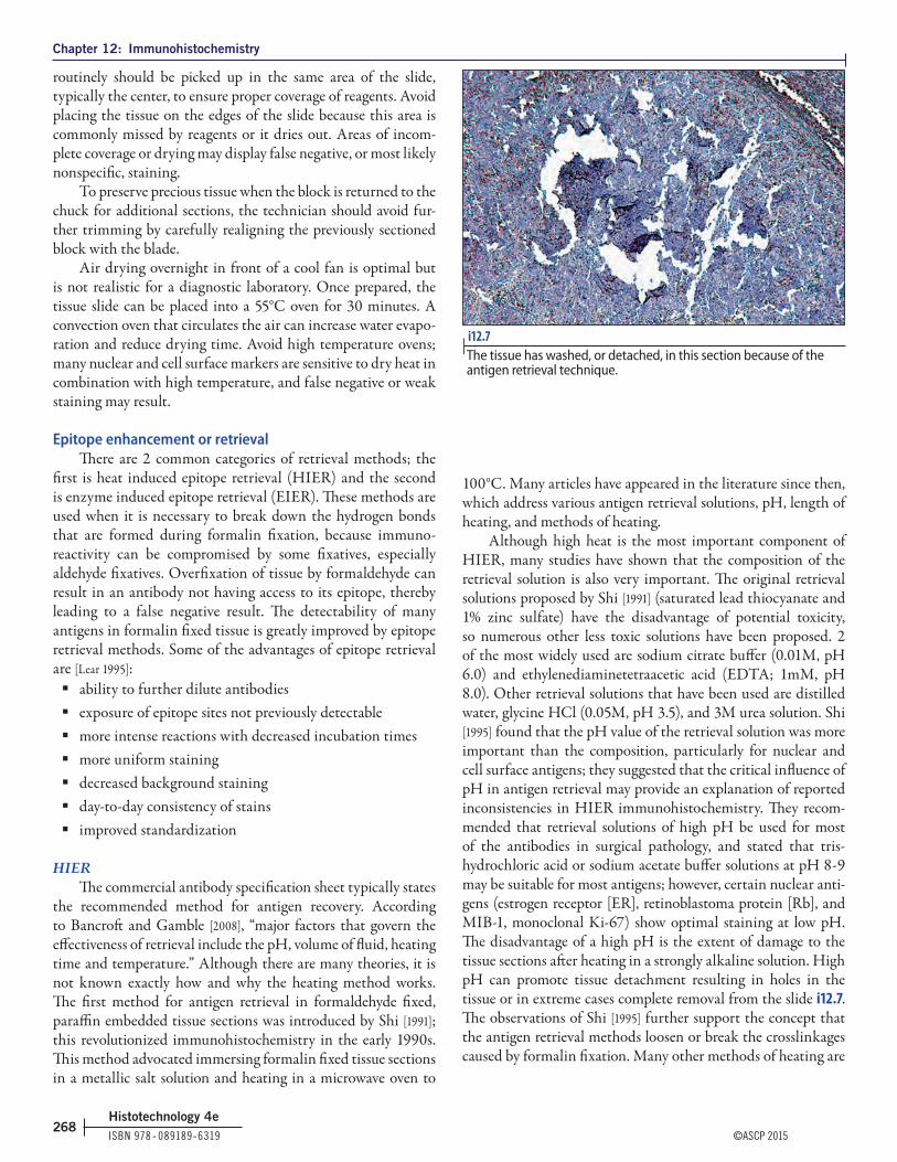
Chapter 12: Immunohistochemistry
ISBN 978 ‑ 089189‑6319 ©ASCP 2015268
Histotechnology 4e
routinely should be picked up in the same area of the slide, typically the center, to ensure proper coverage of reagents. Avoid placing the tissue on the edges of the slide because this area is commonly missed by reagents or it dries out. Areas of incom-plete coverage or drying may display false negative, or most likely nonspecific, staining.
To preserve precious tissue when the block is returned to the chuck for additional sections, the technician should avoid fur-ther trimming by carefully realigning the previously sectioned block with the blade.
Air drying overnight in front of a cool fan is optimal but is not realistic for a diagnostic laboratory. Once prepared, the tissue slide can be placed into a 55°C oven for 30 minutes. A convection oven that circulates the air can increase water evapo-ration and reduce drying time. Avoid high temperature ovens; many nuclear and cell surface markers are sensitive to dry heat in combination with high temperature, and false negative or weak staining may result.
Epitope enhancement or retrievalThere are 2 common categories of retrieval methods; the
first is heat induced epitope retrieval (HIER) and the second is enzyme induced epitope retrieval (EIER). These methods are used when it is necessary to break down the hydrogen bonds that are formed during formalin fixation, because immuno-reactivity can be compromised by some fixatives, especially aldehyde fixatives. Overfixation of tissue by formaldehyde can result in an antibody not having access to its epitope, thereby leading to a false negative result. The detectability of many antigens in formalin fixed tissue is greatly improved by epitope retrieval methods. Some of the advantages of epitope retrieval are [Lear 1995]:
� ability to further dilute antibodies � exposure of epitope sites not previously detectable � more intense reactions with decreased incubation times � more uniform staining � decreased background staining � day-to-day consistency of stains � improved standardization
HIERThe commercial antibody specification sheet typically states
the recommended method for antigen recovery. According to Bancroft and Gamble [2008], “major factors that govern the effectiveness of retrieval include the pH, volume of fluid, heating time and temperature.” Although there are many theories, it is not known exactly how and why the heating method works. The first method for antigen retrieval in formaldehyde fixed, paraffin embedded tissue sections was introduced by Shi [1991]; this revolutionized immunohistochemistry in the early 1990s. This method advocated immersing formalin fixed tissue sections in a metallic salt solution and heating in a microwave oven to
100°C. Many articles have appeared in the literature since then, which address various antigen retrieval solutions, pH, length of heating, and methods of heating.
Although high heat is the most important component of HIER, many studies have shown that the composition of the retrieval solution is also very important. The original retrieval solutions proposed by Shi [1991] (saturated lead thiocyanate and 1% zinc sulfate) have the disadvantage of potential toxicity, so numerous other less toxic solutions have been proposed. 2 of the most widely used are sodium citrate buffer (0.01M, pH 6.0) and ethylenediaminetetraacetic acid (EDTA; 1mM, pH 8.0). Other retrieval solutions that have been used are distilled water, glycine HCl (0.05M, pH 3.5), and 3M urea solution. Shi [1995] found that the pH value of the retrieval solution was more important than the composition, particularly for nuclear and cell surface antigens; they suggested that the critical influence of pH in antigen retrieval may provide an explanation of reported inconsistencies in HIER immunohistochemistry. They recom-mended that retrieval solutions of high pH be used for most of the antibodies in surgical pathology, and stated that tris-hydrochloric acid or sodium acetate buffer solutions at pH 8 - 9 may be suitable for most antigens; however, certain nuclear anti-gens (estrogen receptor [ER], retinoblastoma protein [Rb], and MIB-1, monoclonal Ki-67) show optimal staining at low pH. The disadvantage of a high pH is the extent of damage to the tissue sections after heating in a strongly alkaline solution. High pH can promote tissue detachment resulting in holes in the tissue or in extreme cases complete removal from the slide i12.7. The observations of Shi [1995] further support the concept that the antigen retrieval methods loosen or break the crosslinkages caused by formalin fixation. Many other methods of heating are
i12.7 | The tissue has washed, or detached, in this section because of the antigen retrieval technique.

Chapter 12: Immunohistochemistry
©ASCP 2015 ISBN 978 ‑ 089189‑6319269
Histotechnology 4e
also available, including modified pressure cooker, laboratory microwave oven, vegetable steamer, or circulating waterbath.
A standard method should be established in each labora-tory, but at least 2 methods should be available. A higher-volume laboratory will most likely require multiple retrieval solutions and methods, as the increased variety of antibodies will require different pH solutions and retrieval techniques.
Technology has allowed for automated immunohistochem-ical stainers to perform the steps of heating, deparaffinization, and antigen retrieval, reducing human error and ensuring that the temperature and exposure time are consistent each time a stain is performed, thus improving reproducibility.
EIERIn the EIER method, a proteolytic enzyme is used to expose
epitope sites. Proteolytic enzyme digestion is the older of the 2 epitope enhancement methods, but is used much less frequently since the advent of the heat induced epitope enhancement methods. As Elias [1990] stated, there is no universal proteolytic agent, so a number of enzymes have been used. Some of the solutions that have been used are 0.1% pronase in 0.5M tris buffer, pH 7.5; 0.6% ficin; 0.1% protease in PBS, pH 7.4; and 0.4% pepsin in 0.01N hydrochloric acid. Different proteolytic enzymes may be needed for epitope enhancement of various antigens. Digestion times for these proteolytic enzymes vary from 1 - 60 minutes or more. It is important to know the concen-tration of the enzyme solution being used.
Although proteolytic enzyme digestion usually reduces nonspecific staining, it may increase nonspecific staining if not used carefully. It may weaken specific staining and create false negative results, and also cause fragmentation or loss of tissue sections. The term “overdigestion” of the tissue is used to describe distorted tissue morphology and poor staining results. This can be caused when the incubation time is extended or concentration of the enzyme is high. Commonly the result is loss of cellular detail, in combination with nonspecific staining or no staining. Excessive digestion can promote loss of tissue from the slide during the staining process.
Measures should be taken to ensure that when EIER is per-formed manually, the digestion time is controlled for staining consistency. Temperature can influence the rate at which the digestion is achieved; the temperature should be the same each time an enzyme is used. A temperature of 37°C is commonly used for enzyme incubation. This step should be performed in a humidity chamber to avoid drying of the tissue.
Combined HIER & EIERThe use of enzyme digestion alone for epitope retrieval can
reduce the quality of tissue morphology because of a lengthy exposure time. Therefore it may be useful to combine both methods of epitope retrieval to minimize the extent of enzy-matic pretreatment required. In general, HIER is performed first, followed by rinsing, and then enzyme digestion for a short time (from 1 - 5 minutes).
Methods of visualizationImmunofluorescence
Immunofluorescence, the oldest of pathology’s immuno-histochemical techniques, is still widely used. This technique makes it possible to visualize antigens in tissue sections or in live cell suspensions.
A fluorochrome is a dye that absorbs light and then emits its own light at a longer wavelength. This phenomenon of absorption and emission of light is called fluorescence. When the fluorochrome is attached or conjugated to an antibody, the sites of reaction between antigen and the labeled antibody can be visualized easily.
The most commonly used fluorochromes in immunofluo-rescence techniques are fluorescein isothiocyanate (FITC) and rhodamine. Both of these dyes absorb light that is not visible to the human eye (UV) and emit light that is visible using a fluorescence microscope. Virtually any antigen can be detected with immunofluorescence. The combination of sensitivity, specificity, and simplicity makes the method very useful. In most routine histopathology laboratories, frozen kidney and skin biopsy specimens are examined with immunofluorescence techniques, but the differentiation of tumors is performed using enzyme immunohistochemical techniques suitable for light microscopy.
Enzyme immunohistochemistryThe fundamental principle of enzyme labeled antibodies
is similar to fluorescein labeled antibodies. The enzyme, in the presence of a substrate and a chromogen (may be the same or separate reagents), provides the indicator system to visualize the location of the antibody. Various enzymes such as alkaline phosphatase, β-galactosidase, glucose oxidase, and horseradish peroxidase (HRP) are used as markers.
Horseradish peroxidase and alkaline phosphatase are the enzymes most commonly chosen for antibody visualization. HRP, in the presence of hydrogen peroxide (substrate) and a chromogen such as 3,3′-diaminobenzidine (DAB) or 3-amino-9-ethylcarbazole (AEC) will identify the sites of antibody binding by forming a colored compound. An HRP system rou-tinely serves as a “work horse” in a clinical laboratory because it is consistent when producing large quantities of slides. AEC is soluble in organic solvents and requires an aqueous mounting medium, whereas DAB stained slides may be mounted with synthetic resins.
Alkaline phosphatase is demonstrated by the use of naph-thol-AS-phosphate (substrate) and a chromogen such as fast red-violet LB, fast-red TR, or fast-blue BBN. Alkaline phos-phatase is sensitive to heat and light which can lead to staining inconsistency. Semipermanent chromogens compatible with alkaline phosphatase allow the use of a permanent mounting media but require either no dehydration with air drying before coverslipping, or a quick dehydration to xylene. If not properly dehydrated, the alkaline phosphatase chromogen can break
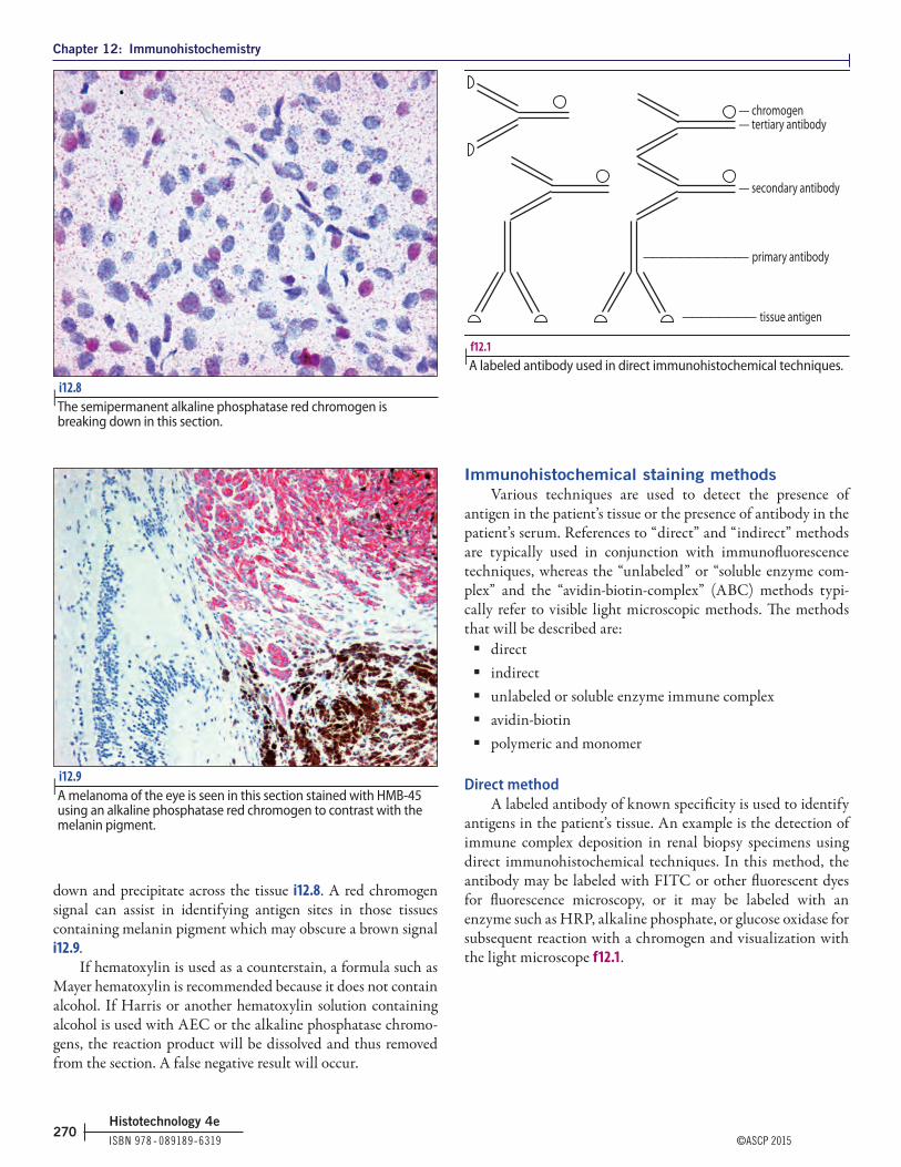
Chapter 12: Immunohistochemistry
ISBN 978 ‑ 089189‑6319 ©ASCP 2015270
Histotechnology 4e
down and precipitate across the tissue i12.8. A red chromogen signal can assist in identifying antigen sites in those tissues containing melanin pigment which may obscure a brown signal i12.9.
If hematoxylin is used as a counterstain, a formula such as Mayer hematoxylin is recommended because it does not contain alcohol. If Harris or another hematoxylin solution containing alcohol is used with AEC or the alkaline phosphatase chromo-gens, the reaction product will be dissolved and thus removed from the section. A false negative result will occur.
Immunohistochemical staining methodsVarious techniques are used to detect the presence of
antigen in the patient’s tissue or the presence of antibody in the patient’s serum. References to “direct” and “indirect” methods are typically used in conjunction with immunofluorescence techniques, whereas the “unlabeled” or “soluble enzyme com-plex” and the “avidin-biotin-complex” (ABC) methods typi-cally refer to visible light microscopic methods. The methods that will be described are:
� direct � indirect � unlabeled or soluble enzyme immune complex � avidin-biotin � polymeric and monomer
Direct methodA labeled antibody of known specificity is used to identify
antigens in the patient’s tissue. An example is the detection of immune complex deposition in renal biopsy specimens using direct immunohistochemical techniques. In this method, the antibody may be labeled with FITC or other fluorescent dyes for fluorescence microscopy, or it may be labeled with an enzyme such as HRP, alkaline phosphate, or glucose oxidase for subsequent reaction with a chromogen and visualization with the light microscope f12.1.
i12.8 | The semipermanent alkaline phosphatase red chromogen is breaking down in this section.
i12.9 | A melanoma of the eye is seen in this section stained with HMB-45 using an alkaline phosphatase red chromogen to contrast with the melanin pigment.
f12.1 |A labeled antibody used in direct immunohistochemical techniques.
— chromogen— tertiary antibody
— secondary antibody
——————————–— primary antibody
———————— tissue antigen

Chapter 12: Immunohistochemistry
©ASCP 2015 ISBN 978 ‑ 089189‑6319271
Histotechnology 4e
Indirect methodIn the indirect method, the patient’s serum is added to
tissue sections containing known antigens to test the patient for the presence of antibodies to those antigens. An example is the test for the presence of antinuclear antibodies. The patient’s serum may also be added to a known bacterium to detect the presence of bacterial antibodies in the patient. A labeled anti-body (anti-human Ig) must be used to detect the bound anti-bodies from the patient’s serum. These indirect methods most frequently involve immunofluorescence microscopy.
Unlabeled or soluble enzyme immune complex methodThis is a 3-step method using primary antibody, linking or
secondary antibody, and soluble enzyme-antienzyme complex. The primary and the enzyme-antienzyme complexes must be made in the same animal species for the secondary antibody to link them together. These techniques are based on the orig-inal peroxidase-antiperoxidase (PAP) technique of Sternberger [1979]. The most common techniques in this category involve the use of either a PAP or alkaline phosphatase-antialkaline phosphatase immune complex f12.2.
Avidin-biotin methodsAvidin has a very high affinity for the vitamin biotin (>1
million times greater than the affinity of antibody for most antigens) and the binding is essentially irreversible. Avidin isolated from egg white was used first in the ABC methods, but streptavidin, isolated from the microorganism Streptomyces avidinii, is more commonly used today.
In the ABC method, the primary antibody is followed by a biotinylated secondary antibody (linking antibody). The third step in the ABC method is the application of a preformed avidin-biotin enzyme complex. This method has low back-ground economy, and sensitivity reported to be up to 40× that of other immunoperoxidase methods [Hsu 1981]. The antibodies may be used at higher dilutions than in other techniques f12.3.
Polymeric detectionThe dextran polymer and monomer technology has quickly
become a standard for clinical immunohistochemical staining. The polymer enzyme molecules such as HRP or alkaline phos-phatase have been fused with the secondary antibody. Monomer technology uses a single strand, allowing for greater sensitivity and penetration at the antibody binding site. Turnaround times have been improved with the elimination of serum and avidin-biotin blocking steps necessary for the ABC method. In addition, this chemistry provides sensitivity without the risk of nonspecific staining when using the ABC method. Staining steps would be: antibody, polymer, and chromogen.
f12.3 |The avidin-biotin-complex (ABC) immunohistochemical technique.
f12.2 |The peroxidase-antiperoxidase (PAP) immunohistochemical technique.
–— chromogen
APAAP –——
PAP
—— secondary antibody ———
——— primary antibody —–——
–— peroxidase conjugated avidin
–— peroxidase conjugated avidin-biotin complex
–————— biotin conjugated ——————— secondary antibody
——————— primary antibody —–———–——

Chapter 12: Immunohistochemistry
ISBN 978 ‑ 089189‑6319 ©ASCP 2015272
Histotechnology 4e
ControlsPositive controls
A positive control must be run with each antibody stain each time it is performed. This applies to both manual and automated methods. Although commercially prepared control slides can be used to check reliability of the reagents, these purchased slides should not be used as routine patient controls. The optimum control is one prepared in the laboratory under the exact same conditions as the diagnostic tissue, including type and timing of fixation and processing. Because fixation varies and affects the reactions so dramatically, commercially prepared control slides are not ideal to determine if a negative reaction on the diagnostic slide is a true negative or if it is the result of overfixation. The method described by Battifora [1986] of preparing sausage controls is excellent, though somewhat time consuming and difficult to execute. With careful plan-ning the laboratory can make the most efficient use of available tissue to fulfill requirements of using controls containing both positive and negative tissue types. The author has found that a tissue microarray technique such as that available from Beecher Instruments (Sun Prairie, WI) is a more effective means for constructing multitissue control blocks (both positive and negative tissues) than previously reported manual methods. Technology can reduce the distortion of the tissue block and extend the use of a control tissue due to smaller precise core sizes [Bancroft 2008].
Best practice is to use a multitissue control that contains both positive and negative tissue for an antibody. This control specimen should be placed on the same slide as the patient’s specimen to ensure that all of the steps, reagents, and tempera-ture are exactly the same for the patient and control specimens during the staining process. Control specimens can be precut before cutting the patient specimen. If precut patient slides are used then the multitissue control block can be cut and placed on the patient slide. When cutting controls, the end of the slide that is for the patient should not be placed into the water bath, this can reduce the charge of the slide.
Negative controlsIn most laboratories, negative controls are run by substi-
tuting for the primary antibody either nonimmune serum from the same species as the primary antibody or the diluent used for the primary antibody. If diluent buffer is used, the negative control will not detect nonspecific binding of animal serum components to the tissue, and any staining observed will be the result of either endogenous peroxidase activity or binding of other antibody reagents to the specimen [Bourne 1989].
Antibody evaluation & validationAntibody specification sheet
The antibody specification sheet provided with a com-mercial antibody contains valuable information regarding the antibody or antiserum. The immunohistochemist should be familiar with the specification sheet for many reasons.
The following are typically included, but are not always available:
� a monoclonal antibody will list the clone and the isotope (IgG or IgM) to ensure that the secondary antibody will bind to the primary antibody
� the expiration date under recommended storage conditions
� long term or short term storage temperatures; some antibodies require long term storage at –20°C; measures to ensure that the antibody storage condition is optimal will assist with antibody stability and maximum reactivity for the life of the antibody
� the protein concentration and suggested working dilution ranges
� pretreatment (antigen retrieval) solution, pH, and/or method, if recommended
� expected positive and negative tissue types, including normal and tumor.
� reference to publications and applications for the antibody; these are useful if additional staining methods are needed
� antibody type: in vitro (IVD), research use only (RUO), or analyte specific reagent (ASR)
Prediluted & concentrated antibodiesPrediluted antibody refers to a commercially available
“ready-to-use” solution. The vendor has already taken a concen-trated antibody and diluted it down to an “optimal” dilution. Because it has been diluted, the shelf-life is routinely shorter and it cannot be frozen to extend it, thus prediluted antibodies are less economical if not used before the date of expiration. Prediluted antibodies must be validated for reactivity before use on patient tissue. The laboratory cannot assume that the antibody is ready to use with that laboratory’s method and must perform serial dilutions (1:2, 1:4, 1:8, and 1:16) to validate. The exception is a Food and Drug Administration (FDA) approved antibody test, which mandates the antibody concentration in addition to times, temperature, and detection reagents. This is usually manufactured and distributed as an FDA approved kit or the manufacturer provides specific protocols to follow.
A concentrated antibody is one that is supplied in an “undi-luted” form. Dilutions must be performed to find the optimal dilution for antigen detection, and the user has more control of fine tuning the dilution to be compatible with the laboratory’s fixation and processing protocols. These undiluted antibodies are more economical because they allow for the modification of

Chapter 12: Immunohistochemistry
©ASCP 2015 ISBN 978 ‑ 089189‑6319273
Histotechnology 4e
titers used in staining. In addition, they routinely have a longer shelf-life when stored properly; most concentrated antibodies can be “snap” frozen using liquid nitrogen and stored at –70°C indefinitely.
If undiluted antibodies come in solution form, it is best to dilute only the amount of antibody needed for the current diagnostic slides or the smallest amount feasible when very high dilutions are needed. Dilutions are expressed as ratios, as in a 1:20 dilution. This is equivalent to 1 part antibody solu-tion in a total of 20 parts, or 1 part antibody plus 19 parts of diluent. Pipettes capable of measuring in microliters (µL) are a must in the immunohistochemistry laboratory. Microliter (µL) and lambda (λ) are used interchangeably, with both equal to 0.001 mL. Pipettes that will cover a range, such as 0.5 - 10 µL, 10 - 100 µL, and 100 - 1,000 µL, are the most useful. For higher dilutions, it is best to prepare a more concentrated stock dilution, and then prepare further dilutions from this stock dilution.
A basic formula to use to make 1 mL (1,000 µL) of a 1:x dilution is [Chlipala 1985]:
This formula can be changed easily by changing 1,000 µL (1 mL) to whatever volume is required.
Antibody validationRoutinely a standard staining protocol will be used for
most formalin fixed tissue samples. This will also assist with laboratory reproducibility and consistency. Although it will be necessary to have unique protocols, the number of antibodies requiring unique detection, special pretreatments, and incuba-tion times should be kept to a minimum.
An example of a standard laboratory protocol is as follows:1. Retrieval: EDTA, pH 8.02. Heating method: modified pressure cooker (115°C for 25
minutes or 124°C for 40 minutes). Combination of time and temperature are based on the stringency of retrieval needed.
3. Peroxidase quenching: 3% hydrogen peroxide (H2O2), 10 minutes
4. Detection system: universal polymer, HRP, DAB5. Staining times: antibody 30 minutes, polymer 10 minutes,
and chromogen 5 minutes6. Counterstain: Mayer hematoxylin, 3 minutes
There are 2 approaches to new antibody validation, the first using no retrieval, multiple retrieval solutions, and multiple enzyme pretreatments; this approach may be necessary when validating a “homegrown” antibody or an antibody that has no previously documented use in the clinical laboratory. The second approach is researching the antibody and planning a validation protocol using the wealth of information available. Resources for a starting point are the antibody or detection vendor, antibody specification, journal articles, and text books [Dabbs 2006, Shi 2000]. These resources provide basic methods before staining, thus knowledge is gained of the recommended retrieval solution, pH and method, type of detection used, and positive and negative tissue types. Knowing expected staining patterns and location of expression, such as nuclear or cyto-plasmic, can assist when reviewing staining results.
Upon receiving the new undiluted antibody, the labora-tory will typically begin with a series of testing variables. First perform serial dilutions (1:100, 1:200, 1:400, and 1:800) of the antibody using a known positive and negative tissue control. Usually the manufacturer will indicate an approximate dilu-tion; however, several different dilutions should be used with the standard laboratory protocol to determine the correct dilution for your laboratory. It is important to use a diluent that contains carrier proteins and the proper pH for antibody stability.
Review the results for an optimal dilution looking for the best signal-to-noise ratio. If the 1:100 targeted cells are staining faintly, with no nonspecific staining, then stain further with more concentrated dilutions of 1:25, 1:50, and 1:100. To ensure no errors in dilution preparation and reproducibility, always repeat in the new series the previous dilution that is closest to optimal. If the 1:800 appears overstained with high signal and high background, then start with 1:800, 1:1,600, and 1:3,200 to further validate If the initial staining yields negative or suboptimal results, then the laboratory should begin with changing only 1 variable at a time. Routinely changing the pH of the retrieval solution is first; if unsuccessful, the intro-duction of an enzyme step omitting the retrieval solution is recommended. The introduction of an enzyme will require not only a redetermination of the antibody concentration but also an evaluation of the time in the enzyme solution. It is recom-mended that when an enzyme is introduced, times of 5, 10, 20, and 30 minutes in the enzyme solution be evaluated to ensure that adequate retrieval is performed. Some antibodies do not require any type of retrieval, and using retrieval may introduce false positive staining i12.10, i12.11, i12.12.
Once the appropriate protocol and dilution are determined for a given antibody, at least 10 positive and 10 negative neo-plasms should be tested to ensure the targeted cells are staining appropriately [CAP 2013] to validate sensitivity, specificity, accu-racy, and reproducibility before using on patient tissue. After
1,000 = µL of raw antisera required x
1,000 – 1,000 = µL of antibody dilution buffer required x

Chapter 12: Immunohistochemistry
ISBN 978 ‑ 089189‑6319 ©ASCP 2015274
Histotechnology 4e
complete validation, the slides should be retained in a perma-nent file for reference. A written and/or electronic record of optimum antibody dilution and retrieval procedure, if appli-cable, should be maintained for each antibody f12.4. Antibodies that fall under the category of “predictive markers” are required to follow a different validation process.
Storage of antibodiesBoth the storage container and the temperature at which
the antibody is stored are very important. Polypropylene, poly-carbonate, or borosilicate glass is recommended for storage because these materials have low protein absorptive properties. If the antibody solution contains very low concentrations of protein, 0.1%-1.0% bovine serum albumin may be added to
reduce loss through polymerization and absorption on the con-tainer [Boenisch 2001].
The recommendations of the manufacturer as to the storage temperature should be carefully followed. Most prediluted anti-bodies and kits are stored at 4°C-8°C to avoid repeated freezing and thawing. Once lyophilized antibodies have been diluted, they are usually divided into aliquots and stored at –70°C.
Blocking reactionsMost immunoperoxidase methods include 2 blocking
reactions. The first is the use of hydrogen peroxide, usually prepared in absolute methanol, to block tissue endogenous per-oxidase activity. If the tissue contains many red blood cells, this blocking step is essential i12.13.
i12.10 | Expected staining patterns with SMI-31 antibody are seen on this positive control tissue from the cerebellum.
i12.11 | A brain biopsy specimen stained with SMI-31 antibody that demonstrates false nuclear staining because of unnecessary heat-induced epitope retrieval.
i12.12 | A brain biopsy specimen stained with SMI-31 antibody demonstrating expected staining patterns after no pretreatment of the sections. Compare with i12.11.
i12.13 | Red blood cells demonstrating positive staining after insufficiently blocked peroxidase activity.

Chapter 12: Immunohistochemistry
©ASCP 2015 ISBN 978 ‑ 089189‑6319275
Histotechnology 4e
Valid
atio
n Fo
rm fo
r Ant
ibod
ies
& Tis
sue C
ontro
lsD
ate r
eque
sted
:
Purp
ose o
f dev
elop
men
t:A
ntib
ody v
alid
atio
n P
ositi
ve ti
ssue
cont
rol
Neg
ativ
e tiss
ue co
ntro
l
Ret
riev
al in
stru
men
t: (c
ircl
e one
) M
odifi
ed p
ress
ure c
ooke
r (12
5°) o
r (11
5°),
wat
erba
th,
mic
row
ave,
stea
mer
, or o
ther
Ret
riev
al so
lutio
n:
Lot#
Exp
irat
ion
pH
:
Enzy
me p
retr
eatm
ent s
olut
ion:
Lot#
Exp
irat
ion
Tim
e:
Det
ectio
n:
Lot
# E
xpir
atio
n
Chr
omog
en:
Lot
#Ex
pira
tion
A
utom
ated
: y/
n (i
f yes
) in
stru
men
tatio
n:
Ant
ibod
y na
me
Ant
ibod
ylo
t#A
ntib
ody e
xp d
ate
Tiss
uety
peC
ontr
ol
bloc
k ID
Dilu
tion
Dat
e st
aine
dC
omm
ents
/res
ults
Rev
iew
ed b
y (M
edic
al D
irec
tor o
r Des
igne
e) F
inal
appr
oval
dat
e:
f12.4
| An
tibod
y &
tissu
e co
ntro
l val
idat
ion
form

Chapter 12: Immunohistochemistry
ISBN 978 ‑ 089189‑6319 ©ASCP 2015276
Histotechnology 4e
The second block is for nonspecific background staining that may occur as a result of antibody (protein) attachment to highly charged collagen and connective tissue elements [Bourne 1989]. If the first protein solution applied to the tissue is the pri-mary antibody, nonspecific binding can occur. The secondary antibody will bind to this nonspecifically bound primary anti-body, and react with the substrate-chromogen, resulting in nonspecific positive staining. The best way to prevent this type of nonspecific staining is to add an innocuous protein solution to the tissue before the primary antibody is applied. The protein will bind to the charged sites and then nonspecific binding of the primary will not occur. Nonimmune serum from the same animal species in which the secondary antibody is produced is the most common source of the blocking reagent. The nonim-mune serum is applied just before the primary antibody. After 10 - 20 minutes of incubation, the excess is tapped off (do not rinse off) and the primary antibody is applied. Casein and nondry fat milk have also been used as a blocking agent.
Multilink biotinylated secondary antiseraSecondary antibodies that are typically called “multilink”
or “universal” refer to cocktails of antibodies raised in different species. These secondary antibody mixtures are commonly bio-tinylated antimouse IgG and IgM and antirabbit IgG. Other species such as goat can also be added. The laboratory technician should know the species in the secondary antibody mixture to ensure that the targeted antibody is compatible. One advantage of this type of secondary antibody is that it avoids the problem of having to stock various specific link antibodies (eg, biotinylated goat antirabbit or goat antimouse IgG and IgM).
DAB reaction product intensificationSeveral methods have been used to intensify the DAB reac-
tion product. Heavy metals can be used for intensification of the DAB reaction product by supplementing, or following, the incubation medium with reagents such as 1% cobalt chloride. However, there is a risk of increasing background staining with heavy metal intensification, and some of the metals should not be used with the immunohistochemical detection of nuclear antigens [Elias 1990], especially if weak hematoxylin counter-staining is used.
Imidazole is an effective intensification reagent. Elias [1990] states that the addition of 0.01M imidazole to the DAB incu-bation medium at pH 7.6 is superior to standard DAB and DAB-cobalt chloride-nickel sulfate or DAB-cobalt chloride pro-cedures, both in sensitivity and detection efficiency. Imidazole also significantly inhibits the pseudoperoxidase activity of hemoglobin.
Osmium tetroxide may also be used to intensify the DAB reaction product, but unlike the other reagents mentioned, it is always used after the DAB reaction. The DAB reaction product is osmiophilic and will be darkened by mild poststaining osmifica-tion. This also helps prevent fading of the final reaction product
during long term storage; however, unless carefully done, this reagent has the possibility of darkening any background staining present as well as intensifying the reaction product.
Elias [1990] warns that increased sensitivity is not always commensurate with enhancement of staining intensity. For example, post-treatment osmium boosts staining intensity without increasing detection efficiency.
Buffer solutionsFor reproducible results, the pH of the buffer used during
staining must be maintained and checked often. For each new lot of commercially prepared buffer, or each time a buffer is pre-pared, the immunohistochemist should check and document its pH before use. Typically, 2 buffers are used during the staining process: PBS and tris-base solution (TBS). Both can be used with an HRP detection system, but only TBS should be used with an alkaline phosphatase detection system. The addition of a wetting agent will assist with even distribution of the reagents during staining and improve rinsing steps; at the same time, it will slow down bacterial growth. Common wetting agents used are Tween-20, Triton-X, and brij-35. The buffer should be used at room temperature during staining.
Commonly used antibodies & their applicationsAntibody stains are routinely used by the pathologist to
identify a neoplasm and define whether it is benign or malig-nant, and whether it is metastatic or primary. The pathologist identifies the extent to which cells in a neoplasm resemble normal cells (differentiation) both morphologically and func-tionally. The type of neoplasm is defined by the germ cell layer (endoderm, mesoderm, ectoderm) from which it derives. Types of neoplasms include carcinomas, sarcomas, lymphomas, mela-nomas, mesotheliomas, neuroendocrine tumors, gliomas, and germ cell tumors.
Neoplastic terminologyNeoplasm: A new growth of tissue in which cell multipli-
cation is uncontrolled and progressive; can be either benign or malignant.
Tumor: Swelling; one of the cardinal signs of inflammation and morbid enlargement. Often used to mean “neoplasm.”
Anaplasia: Lack of differentiation. Some features include variation in size and shape of cells and nuclei, dark nuclei, giant cells, and disturbed orientation.
Differentiation: The distinguishing of 1 thing from another and the extent to which neoplastic cells resemble com-parable normal cells, both morphologically and functionally.
Benign: Shows a lesser degree of anaplasia than malignant tumors; often surrounded by a fibrous capsule; does not produce metastasis or significant invasion.
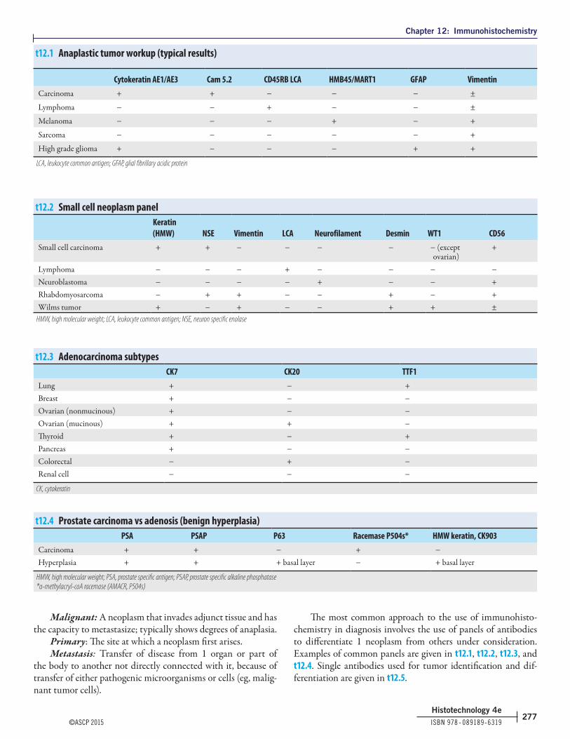
Chapter 12: Immunohistochemistry
©ASCP 2015 ISBN 978 ‑ 089189‑6319277
Histotechnology 4e
Malignant: A neoplasm that invades adjunct tissue and has the capacity to metastasize; typically shows degrees of anaplasia.
Primary: The site at which a neoplasm first arises.Metastasis: Transfer of disease from 1 organ or part of
the body to another not directly connected with it, because of transfer of either pathogenic microorganisms or cells (eg, malig-nant tumor cells).
The most common approach to the use of immunohisto-chemistry in diagnosis involves the use of panels of antibodies to differentiate 1 neoplasm from others under consideration. Examples of common panels are given in t12.1, t12.2, t12.3, and t12.4. Single antibodies used for tumor identification and dif-ferentiation are given in t12.5.
t12.1 Anaplastic tumor workup (typical results)
Cytokeratin AE1/AE3 Cam 5.2 CD45RB LCA HMB45/MART1 GFAP VimentinCarcinoma + + − − − ±
Lymphoma − − + − − ±
Melanoma − − − + − +
Sarcoma − − − − − +
High grade glioma + − − − + +
LCA, leukocyte common antigen; GFAP, glial fibrillary acidic protein
t12.2 Small cell neoplasm panelKeratin (HMW) NSE Vimentin LCA Neurofilament Desmin WT1 CD56
Small cell carcinoma + + − − − − − (except ovarian)
+
Lymphoma − − − + − − − −Neuroblastoma − − − − + − − +Rhabdomyosarcoma − + + − − + − +Wilms tumor + − + − − + + ±
HMW, high molecular weight; LCA, leukocyte common antigen; NSE, neuron specific enolase
t12.3 Adenocarcinoma subtypesCK7 CK20 TTF1
Lung + − +Breast + − −Ovarian (nonmucinous) + − −Ovarian (mucinous) + + −Thyroid + − +Pancreas + − −Colorectal − + −Renal cell − − −
CK, cytokeratin
t12.4 Prostate carcinoma vs adenosis (benign hyperplasia)PSA PSAP P63 Racemase P504s* HMW keratin, CK903
Carcinoma + + − + −Hyperplasia + + + basal layer − + basal layer
HMW, high molecular weight; PSA, prostate specific antigen; PSAP, prostate specific alkaline phosphatase*α-methylacryl-coA racemase (AMACR, P504s)

Chapter 12: Immunohistochemistry
ISBN 978 ‑ 089189‑6319 ©ASCP 2015278
Histotechnology 4e
Combinations of antibody expression, both positive and negative assist the pathologist when working up an anaplastic tumor for differential diagnosis. Consideration of cellular mor-phology, tissue source and patient history are key factors for a pathological diagnosis. Antibodies listed in this table are not used individually for diagnosis and are not all inclusive.
Development of new clones and antibodies are constant. Clones can be combined together to create cocktails that may increase the sensitivity or specificity of the test; cocktails were included in this table only when a combination of clones was the standard for the antibody, as with CD45.
t12.5 Antibodies used for the differentiation and identification of tumors*
Antibody Clones mAb, rmAb, or polyclonal pAb†Neoplasm identification & diagnostic differentiation
bcl2 mAb: 124, 100/D5, bcl-2/100/D5, 100 Follicular lymphomaCD117 mAb: T595
rmAb: YR125, 9.7pAb: A4502, RB-9038-P
Gastrointestinal stromal tumor
CD15 mAb: MMA, C3D-1, Carb-3, BY87, H198 Hodgkin lymphomaCD20 mAb: L26, 7D1 or MJ1
rmAb: MJ1B cell lymphoma
CD3 mAb: F7.2.38, LN10, PS1,rmAb: EP41, EP449E, SP7, 2GV6, EP272 MAD-000325QD, MRQ-39 103R
pAb: A0542, IR503/IS503, N1580
T cell lymphoma
CK20 mAb: Ks20.8, PW31rmAb: EP23, SP33pAb: E16444
Colon carcinoma
Cytokeratin (pankeratin) mAb: AE1/AE3, KL1, OSCAR CarcinomaDesmin mAb: D33, DE-R-11, ZC18
pAb: RB-9014-PLeiomyoma, leiomyosarcoma
Estrogen receptor mAb: 1D5, 6F11rmAb: EP1, SP1
Breast carcinoma
Glial fibrillary acidic protein (GFAP) mAb: 6F2, GA-5, SP2/AG14rmAb: EP6724pAb: Z0334, 760 - 2516
Glioblastoma multiforme
Gross cystic disease fluid protein GCDFP-15 mAb: 23A3, D6, SPM135rmAb: EP1582Y, EP95
Breast ductal carcinoma
Her2 mAb: CB11rmAb: 4B5, EP1045Y, SP3pAb: A0485
Breast carcinoma
HMB-45 mAb: HMB-45 MelanomaCD45, leukocyte common antigen mAb: 2B11+PD7/26, X16/99, RP2/18 LymphomaMelan-A mAb: A-103, HMB45 + MC-7C10+M2 - 9E3 + T31, M2 - 7C10 MelanomaProgesterone receptor mAb: 16, 1A6, PgR 636, PgR 1294
rmAb: Ep2, SP2, SP42, Y85Breast ductal carcinoma
Prostate specific antigen mAb: ER-PR8, 35H9pAb: A0562, RB-084, IS514/IR514, N1517, 760 - 2506
Prostate adenocarcinoma
S100 mAb: 4c4.9, 12E2E2rmAb: EP32pAb: Z0311, NCL-S100p, RB-9018p, E031, 760 - 2523, IR/IS504, PA0900
Schwannoma
Thyroid transcription factor 1 (TTF1) mAb: 8G7G3/1, SPT24rmAb: EP1584Y, SP141
Lung adenocarcinoma
Vimentin mAb: V9, Vim 3B4rmAb: SP20
Seminoma
*From [NordiQC http://www.nordiqc.org; ISBN 978 - 0443102790]†Monoclonal antibody (mAb), rabbit monoclonal antibody (rmAb), or polyclonal antibody name (pAb).

Chapter 12: Immunohistochemistry
©ASCP 2015 ISBN 978 ‑ 089189‑6319279
Histotechnology 4e
Quality controlMeasures of QC and thorough documentation are funda-
mental in a diagnostic laboratory. QC measures serve many roles in the laboratory and are especially useful for troubleshooting. Antibody and control tissue validation must be documented and maintained. Each laboratory should define a QC system that enables the immunohistochemist to have easy access to previous validation results and final protocols, especially in a laboratory where manual staining is performed. Tracking of lot numbers, expiration dates, dilutions, protocols, pH monitoring, and vali-dation of controls are basic QC in the immunohistochemistry laboratory. Laboratory accreditation agencies also have specific QC and documentation requirements. Both paper and elec-tronic documentation are useful, but if a final protocol can be maintained electronically it assures the immunohistochemist that the most current information is saved and retrievable.
Recommended QC steps for an antibody1. Verify and document on the bottle and the specification
sheet the antibody name, receipt date, and expiration date2. Check storage temperature and conditions upon receipt3. Use a form to plan initial validation to include
pretreatment, incubation times, detection system, control tissue, dilutions and date f12.4
4. Document the date validation was performed and who approved the final procedure, such as the medical director
5. Make a final copy of the procedure that can be accessed by the technician
6. Keep all records and document and date any modifications to the procedure; historical records may be useful for troubleshooting or revalidating an existing antibody
7. Maintain a separate slide file for each antibody lot and dilution validation; the date on the slides should correlate with the antibody records
Positive & negative tissue controlsPositive controls are those tissues that express the antigen
in either normal tissue components or tumor cells i12.14, i12.15. Negative tissue controls are either normal tissue or tumor cells that do not contain that antigen. When validating an antibody, it is important to optimize the dilution using both positive and negative tissue types. This ensures that a low expressing tumor will not be missed and a false positive result is not misinter-preted. Tissue blocks that contain several different normal and tumor tissues exhibiting positive and negative expression are very helpful to decrease the cost of validation. New technology such as the tissue microarrayer is making a constructive impact on QC measures with antibody validation i12.16, i12.17, i12.18.
i12.14 | CD3 T cell staining pattern is demonstrated in a normal lymph node control.
i12.15 | CD20 B cell staining pattern is demonstrated in a normal lymph node control.
i12.16 | Cam 5.2 antibody staining of a colon adenocarcinoma control specimen.