A self-assembled cyclodextrin nanocarrier for ... · 2535 A self-assembled cyclodextrin nanocarrier...
8
2535 A self-assembled cyclodextrin nanocarrier for photoreactive squaraine Ulrike Kauscher and Bart Jan Ravoo * Full Research Paper Open Access Address: Organic Chemistry Institute and Center for Soft Nanoscience, Westfälische Wilhelms-Universität Münster, Corrensstrasse 40, 48149 Münster, Germany Email: Bart Jan Ravoo * - [email protected] * Corresponding author Keywords: cyclodextrin; host–guest chemistry; photodynamic therapy; self-assembly; squaraine Beilstein J. Org. Chem. 2016, 12, 2535–2542. doi:10.3762/bjoc.12.248 Received: 15 July 2016 Accepted: 04 November 2016 Published: 25 November 2016 This article is part of the Thematic Series "Superstructures with cyclodextrins: Chemistry and applications IV". Guest Editor: G. Wenz © 2016 Kauscher and Ravoo; licensee Beilstein-Institut. License and terms: see end of document. Abstract Photoreactive squaraines produce cytotoxic oxygen species under irradiation and have significant potential for photodynamic therapy. Herein we report that squaraines can be immobilized on a self-assembled nanocarrier composed of amphiphilic cyclo- dextrins to enhance their photochemical activity. To this end, a squaraine was equipped with two adamantane moieties that act as anchors for the cyclodextrin vesicle surface. The supramolecular immobilization was monitored by using fluorescence spectrosco- py and microscopy and the photochemistry of the squaraine was investigated by using absorption spectroscopy. 2535 Introduction Photodynamic therapy (PDT) has become a very attractive al- ternative to traditional cancer therapies due to its efficiency and selectivity [1-4]. PDT is based on a photosensitizer (PS), which is delivered to cancerous tissue followed by the irradiation with light of an appropriate wavelength. Irradiation of the PS causes its transition to the excited singlet state and, via intersystem crossing; the PS reaches its triplet state. Electron transfer occurs upon relaxation to the ground state. This process can take place in two ways distinguished as type I and II mechanisms [5]. The type I mechanism describes a direct interaction between the PS and the surrounding cell material. Electrons are transferred onto the cell material, causing the production of radicals that then react with oxygen to form reactive oxygen species. The type II mechanism, however, describes the interference of the PS with the triplet ground state of oxygen, leading to the production of singlet oxygen. In both mechanisms, the PS harvests the energy of incident light to form reactive oxygen species or singlet oxygen, which are highly cytotoxic [6,7]. Most clinically used PSs are based on porphyrins, which have the disadvantage of an absorption wavelength in the range of
Transcript of A self-assembled cyclodextrin nanocarrier for ... · 2535 A self-assembled cyclodextrin nanocarrier...
2535
A self-assembled cyclodextrin nanocarrier forphotoreactive squaraineUlrike Kauscher and Bart Jan Ravoo*
Full Research Paper Open Access
Address:Organic Chemistry Institute and Center for Soft Nanoscience,Westfälische Wilhelms-Universität Münster, Corrensstrasse 40, 48149Münster, Germany
Email:Bart Jan Ravoo* - [email protected]
* Corresponding author
Keywords:cyclodextrin; host–guest chemistry; photodynamic therapy;self-assembly; squaraine
Beilstein J. Org. Chem. 2016, 12, 2535–2542.doi:10.3762/bjoc.12.248
Received: 15 July 2016Accepted: 04 November 2016Published: 25 November 2016
This article is part of the Thematic Series "Superstructures withcyclodextrins: Chemistry and applications IV".
Guest Editor: G. Wenz
© 2016 Kauscher and Ravoo; licensee Beilstein-Institut.License and terms: see end of document.
AbstractPhotoreactive squaraines produce cytotoxic oxygen species under irradiation and have significant potential for photodynamic
therapy. Herein we report that squaraines can be immobilized on a self-assembled nanocarrier composed of amphiphilic cyclo-
dextrins to enhance their photochemical activity. To this end, a squaraine was equipped with two adamantane moieties that act as
anchors for the cyclodextrin vesicle surface. The supramolecular immobilization was monitored by using fluorescence spectrosco-
py and microscopy and the photochemistry of the squaraine was investigated by using absorption spectroscopy.
2535
IntroductionPhotodynamic therapy (PDT) has become a very attractive al-
ternative to traditional cancer therapies due to its efficiency and
selectivity [1-4]. PDT is based on a photosensitizer (PS), which
is delivered to cancerous tissue followed by the irradiation with
light of an appropriate wavelength. Irradiation of the PS causes
its transition to the excited singlet state and, via intersystem
crossing; the PS reaches its triplet state. Electron transfer occurs
upon relaxation to the ground state. This process can take place
in two ways distinguished as type I and II mechanisms [5]. The
type I mechanism describes a direct interaction between the PS
and the surrounding cell material. Electrons are transferred onto
the cell material, causing the production of radicals that
then react with oxygen to form reactive oxygen species.
The type II mechanism, however, describes the interference of
the PS with the triplet ground state of oxygen, leading to the
production of singlet oxygen. In both mechanisms, the PS
harvests the energy of incident light to form reactive
oxygen species or singlet oxygen, which are highly cytotoxic
[6,7].
Most clinically used PSs are based on porphyrins, which have
the disadvantage of an absorption wavelength in the range of

Beilstein J. Org. Chem. 2016, 12, 2535–2542.
2536
Figure 1: Schematic representation of adamantane-substituted squaraine (AdSq) binding as a divalent guest to the surface of cyclodextrin vesicles(CDV).
visible light [8,9]. Visible light is scattered and absorbed by the
inhomogeneous biological tissue. In order to optimise effi-
ciency, it is vital to skew the absorption range of the PS towards
the near infrared range. Then, the scattering and absorption by
tissue is minimized and a more in-depth therapy becomes
possible.
The search for more effective compounds with absorption in the
near infrared includes amongst others cyanine [10], bodipy [11],
and phthalocyanine [12] photosensitizers and has lately led to
the consideration of a class of compounds known as squaraines
[13-18]. Squaraines are a promising class of fluorescent dyes
for PDT. In the past, squaraines showed a very low intersystem-
crossing efficiency, leading to the premature conclusion that
these dyes cannot be used in PDT [19]. Santos et al. showed
that the intersystem-crossing efficiency of squaraines can be
remarkably increased by incorporation of heavy atoms like sele-
nium or sulfur and the addition of long aliphatic side chains
[20,21]. The enhanced intersystem-crossing efficacy due to
spin-orbital interactions makes modified squaraines a promis-
ing component of PDT [22,23].
Whether squaraines follow the type I or type II mechanism is
discussed controversially in the literature. Santos et al. [19,20],
Salice et al. [24] and Rapozzi et al. [25] investigated how
benzothiazole-squaraines, which are also pursued in this
project, cause cytotoxicity. Santos et al. postulated the produc-
tion of singlet oxygen as a result of interaction between the
squaraines and the ground state of oxygen [19,20]. A few years
later, Salice et al. showed that squaraines are not efficient
singlet oxygen producing agents, but can be applied successful-
ly as singlet oxygen quenchers [23]. The authors discussed that
this behaviour is based on charge-transfer processes between
stacked squaraines as well as oxygen squaraine complexes.
Within the same year, Rapozzi et al. described the photooxida-
tion process of benzothiazol squaraines [24]. They showed that
the irradiation of benzothiazole-squaraines results in a photo-
degradation process. Rapozzi et al. assume that oxidation proba-
bly involves through formation of a π-complex between the
electron-rich enamine double bond and molecular oxygen. In
comparison to the photo-oxidation of enaminon or other elec-
tron-rich double bonds, the complex can react further to
produce 3-HBT (3-hexylbenzo[d]thiazol-2(3H)-one). The
photogenerated oxidation products can then induce radical-
chain reactions with the surrounding cell material, leading to
cell toxicity.
However, like most PS, squaraines have poor water solubility,
which leads to aggregation and inactivation under physiologi-
cal conditions, causing a reduced production of reactive oxygen
species [26]. To overcome this drawback, we considered to
immobilize the squaraines onto a platform that provides spatial
separation (Figure 1). Our platforms of choice are vesicles self-
assembled from amphiphilic cyclodextrin [27]. Given their
negligible toxicity cyclodextrins have been utilized as carriers
in a number of studies [28,29]. Amphiphilic cyclodextrins
substituted with hydrophobic alkyl groups on the primary side
and hydrophilic oligo(ethylene glycol) units on the secondary
side can be synthesized in a simple and straightforward three
step synthesis [27,30]. Cyclodextrin vesicles (CDVs) can be
easily prepared by sonication or extrusion in aqueous solution.
CDVs have the unique possibility of versatile surface decora-
tion by the simple addition of guest molecules to the aqueous
solution of the vesicle [30]. The hydrophobic cyclodextrin cavi-
ties are well known to form size-selective inclusion complexes
with apolar compounds such as adamantane [31]. In several
publications, we have shown that CDVs retain this ability by
keeping the cavity available for host–guest complexation. In
that way, it was possible to decorate CDVs with carbohydrates
[32,33], with peptides [34], and also with DNA [35]. Moreover
Beilstein J. Org. Chem. 2016, 12, 2535–2542.
2537
CDVs were also shown to form very dense membranes in com-
bination with phospholipids and cholesterol [36]. Previous
studies also showed promising results for PDT applications
when a well-known PS, phthalocyanine, was used to decorate
the cyclodextrin vesicles [37,38]. The immobilization led to an
increased photoactivity of the phthalocyanines due to suppres-
sion of aggregation and inactivation of the PS at the surface of
the CDVs.
In this contribution we show that squaraines can also be immo-
bilized onto CDVs. For this purpose, the squaraines are
equipped with two adamantane functions that bind into the cavi-
ties of cyclodextrin on the surface of the CDVs (Figure 1). The
resulting divalent host–guest interaction should lead to a higher
binding affinity of the squaraine for the CDV without affecting
its photochemistry. In addition, it is expected that the aggrega-
tion of squaraine will be supressed by immobilization at the
CDV surface. Ultimately, the photoactive squaraine can be
combined with other functional guests, such as targeting
units and tracers, to further enhance the potential of the
nanocarrier.
Results and DiscussionAdamantane-substituted squaraine (AdSq) was synthesized in a
three step synthesis. Benzothiazole rings were introduced to
enlarge the π-system. The sulfur atoms of these moieties
increase the intersystem crossing due to the heavy atom effect.
Additionally hexyl alkane chains were introduced on both
N-termini. Santos et al. showed that these lead to a higher inter-
system crossing compared to shorter alkane chains [20,21]. The
analytical data for AdSq (see Supporting Information File 1) are
consistent with the molecular structure shown in Figure 1.
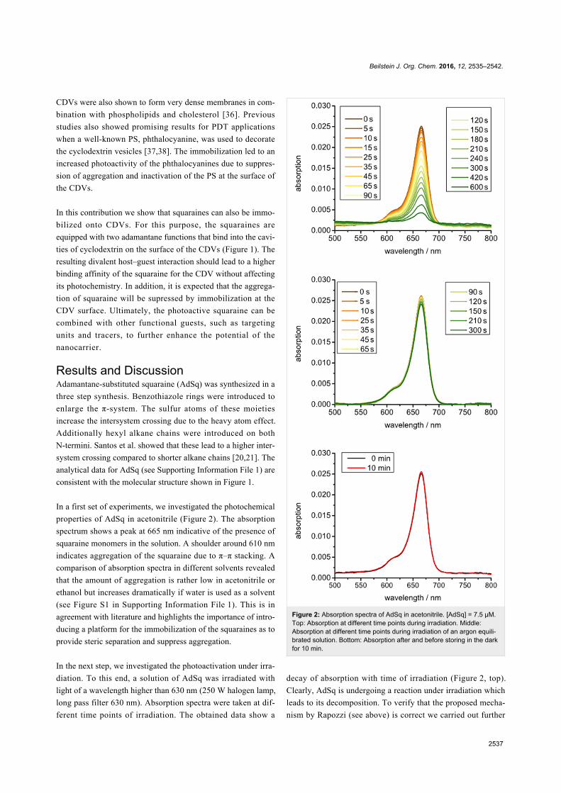
In a first set of experiments, we investigated the photochemical
properties of AdSq in acetonitrile (Figure 2). The absorption
spectrum shows a peak at 665 nm indicative of the presence of
squaraine monomers in the solution. A shoulder around 610 nm
indicates aggregation of the squaraine due to π–π stacking. A
comparison of absorption spectra in different solvents revealed
that the amount of aggregation is rather low in acetonitrile or
ethanol but increases dramatically if water is used as a solvent
(see Figure S1 in Supporting Information File 1). This is in
agreement with literature and highlights the importance of intro-
ducing a platform for the immobilization of the squaraines as to
provide steric separation and suppress aggregation.
In the next step, we investigated the photoactivation under irra-
diation. To this end, a solution of AdSq was irradiated with
light of a wavelength higher than 630 nm (250 W halogen lamp,
long pass filter 630 nm). Absorption spectra were taken at dif-
ferent time points of irradiation. The obtained data show a
Figure 2: Absorption spectra of AdSq in acetonitrile. [AdSq] = 7.5 µM.Top: Absorption at different time points during irradiation. Middle:Absorption at different time points during irradiation of an argon equili-brated solution. Bottom: Absorption after and before storing in the darkfor 10 min.
decay of absorption with time of irradiation (Figure 2, top).
Clearly, AdSq is undergoing a reaction under irradiation which
leads to its decomposition. To verify that the proposed mecha-
nism by Rapozzi (see above) is correct we carried out further

Beilstein J. Org. Chem. 2016, 12, 2535–2542.
2538
experiments. If the solution of AdSq in acetonitrile was equili-
brated in an argon atmosphere to drive away any oxygen, the
absorption spectra obtained show the same absorption intensity
for all time points (Figure 2, middle). This reveals that AdSq
undergoes no decomposition in absence of oxygen and that
photodecomposition is definitely due to interaction with
oxygen. To test if the decomposition mechanism is light driven,
another set of absorption spectra were taken in an oxygen-equi-
librated solution that was stored in the dark for 10 min. In this
case, the absorption spectra show no decrease indicating that
again no decomposition is obtained (Figure 2, bottom). Thus,
AdSq is stable in solution if stored in the dark.
Next, we investigated the immobilization of AdSq by
host–guest interaction with CDVs. Amphiphilic β-cyclo-
dextrins substituted with 7 dodecylsulfide groups on the prima-
ry side and 7 oligo(ethylene glycol) units on the secondary side
were obtained via a straightforward three step synthesis as de-
scribed [27,30]. A thin film of these amphiphiles was obtained
by evaporation of a chloroform solution in a round bottom
flask. Hydration and extrusion with a Liposofast extruder and
membranes with 100 nm pore size yield bilayer vesicles of an
approximate size of 100 nm. CDVs were decorated with AdSq
by simple addition of the AdSq to the aqueous dispersion of the
CDVs.
Interestingly, the immobilization of AdSq on CDV can be
directly observed by fluorescence spectrometry. To this end,
solutions with different concentrations of CDV were prepared.
AdSq was dissolved in acetonitrile in the dark and added to the
vesicle solution. The percentage of acetonitrile in water was
kept under 0.5%, so that any influence on solubility of AdSq,
stability of CDVs or host guest interaction can be neglected.
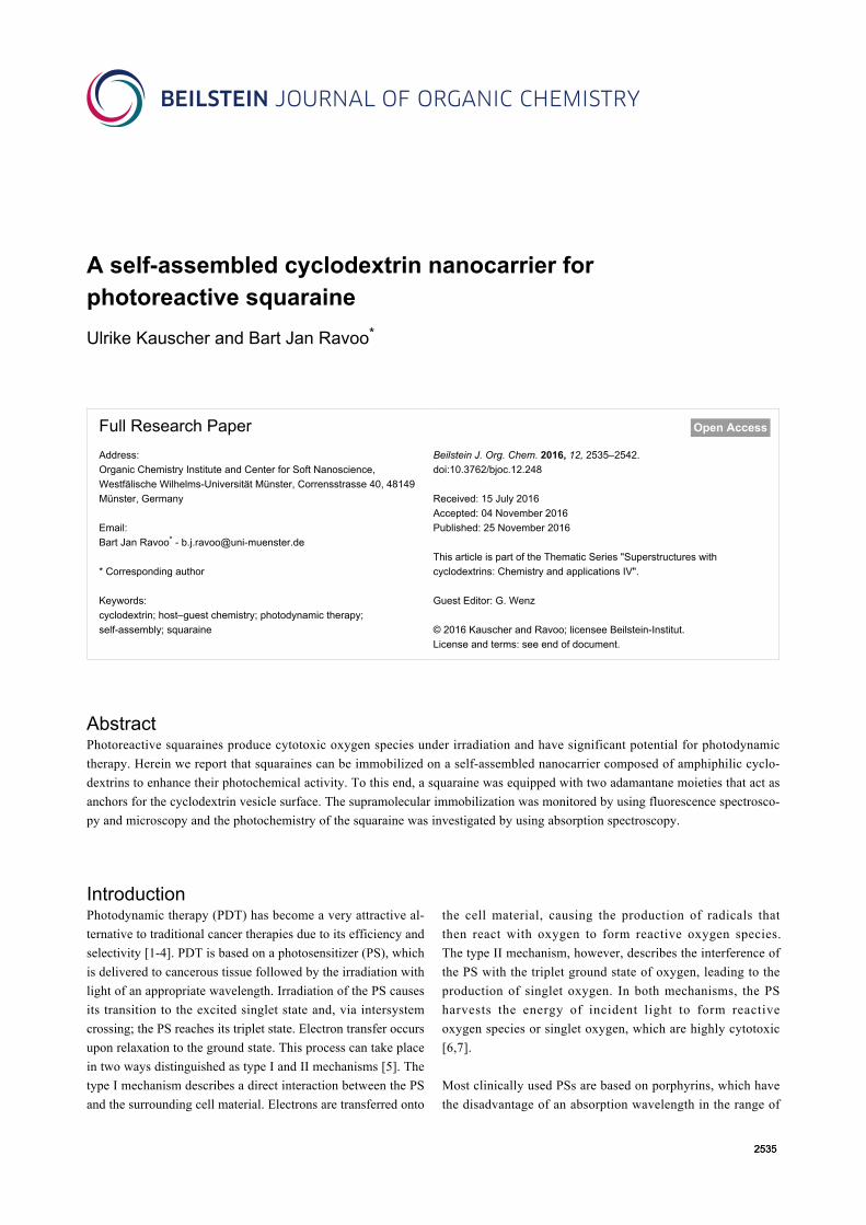
Fluorescence spectra were taken 20 min after addition of AdSq
to assure the complete equilibration of the CDV dispersion. The
fluorescence spectrum of AdSq without CDV shows no fluores-
cence, while increasing fluorescence intensity was detected with
more CDV available (Figure 3, top). These results indicate that
immobilization of the AdSq on the surface of CDV suppresses
the aggregation of AdSq in aqueous solution and that the AdSq
fluorescence is enhanced due to a suppression of intermolecu-
lar π–π stacking. The more CDV is available, the more AdSq is
immobilized on the surface of the vesicles. To support this ex-
planation, absorption spectra were taken of the solutions that
were prepared for the fluorescence experiments. The absorp-
tion spectra show a decrease of the signal at 615 nm that indi-
cates the presence of the aggregated form of AdSq (see
Figure S2 in Supporting Information File 1).
The fluorescence spectra show a saturation of the intensity
maximum for a concentration of 60 µM CDV. Assuming that
Figure 3: Top: Emission spectra (ex: 630 nm) of AdSq immobilized atCDV. [CDV] = 0–100 µM; [AdSq] = 5 µM. Bottom: Langmuir regres-sion of a fluorescence titration of CDV to AdSq. [AdSq] = 10 µM,[CDV] = 0–100 µM. It is assumed that only cyclodextrins on the outersurface of the CDV are available for host–guest interactions.
the accessible concentration of cyclodextrin available for the
complexation of guest is only 30 µM (since approximately 50%
of the amphiphilic cyclodextrins reside at the interior surface of
the CDV and AdSq is too large and too polar to pass the mem-
brane), saturation is reached at a six-fold excess of host to
guest, even though both are present in micromolar concentra-
tions only. The binding constant of AdSq with the cyclo-
dextrins in the CDV could be obtained by plotting the
maximum fluorescence emission (at 676 nm) against the con-
centration of available cyclodextrin (Figure 3, bottom). The re-
sulting Langmuir isotherm was fitted by linear regression (see
Supporting Information File 1 for details) and the binding con-
stant was determined to be Ka = 1.2 × 10⁵ M−1. The high-
affinity binding of AdSq is a clear indication that both adaman-
tanes are involved in binding to the CDV and that the interac-
tion is effectively divalent.
Beilstein J. Org. Chem. 2016, 12, 2535–2542.
2539
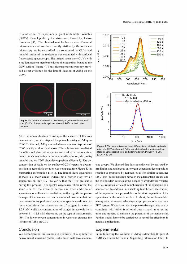
In another set of experiments, giant unilamellar vesicles
(GUVs) of amphiphilic cyclodextrins were formed by electro-
formation [35]. The obtained vesicles have a size of several
micrometers and are thus directly visible by fluorescence
microscopy. AdSq were added to a solution of the GUVs and
immobilization of the molecules was examined with confocal
fluorescence spectroscopy. The images taken show GUVs with
a red luminescent membrane due to the squaraines bound to the
GUV surface (Figure 4). Thus, fluorescence microscopy provi-
ded direct evidence for the immobilization of AdSq on the
CDV.
Figure 4: Confocal fluorescence microscopy of giant unilamellar vesi-cles (GUVs) of amphiphilic cyclodextrins with AdSq on their outersurface.
After the immobilization of AdSq on the surface of CDV was
demonstrated, we investigated the photochemistry of AdSq on
CDV. To this end, AdSq was added to an aqueous dispersion of
CDV exactly as described above. The solution was irradiated
for 600 s and absorption spectra were taken at various time
points. As shown before in the acetonitrile solution, also AdSq
immobilized on CDV photodecomposition (Figure 5). The de-
composition of AdSq on the surface of CDV versus its decom-
position in acetonitrile solution was compared (see Figure S3 in
Supporting Information File 1). The immobilized squaraines
showed a slower decay indicating a higher stability of
squaraines on the CDV. To verify that the CDV are stable
during this process, DLS spectra were taken. These reveal the
same size for the vesicles before and after addition of
squaraines as well as after irradiation, so that significant photo-
damage of the nanocarrier can be excluded. We note that our
measurements are performed under atmospheric conditions. At
these conditions the concentration of oxygen in water is
1.25 mM while the concentration of oxygen in acetonitrile is
between 8.1–12.1 mM, depending on the type of measurement.
[39]. The lower oxygen concentration in water can enhance the
lifetime of AdSq on CDV.
ConclusionWe demonstrated the successful synthesis of a symmetric
benzothiazol squaraine (AdSq) substituted with two adaman-
Figure 5: Top: Absorption spectra at different time points during irradi-ation of a CDV solution with AdSq immobilised on the vesicle surface.Bottom: DLS spectra before and after irradiation. [AdSq] = 7.5 µM,[CDV] = 80 µM.
tane groups. We showed that this squaraine can be activated by
irradiation and undergoes an oxygen-dependent decomposition
reaction as proposed by Rapozzi et al. for similar squaraines
[25]. Host–guest inclusion between the adamantane groups and
the cyclodextrin cavities at the surface of cyclodextrin vesicles
(CDVs) results in efficient immobilization of the squaraine on a
nanocarrier. In addition, π–π stacking (and hence inactivation)
of the squaraine is supressed due to the steric separation of the
squaraines on the vesicle surface. In short, the self-assembled
nanosystem has several advantageous properties to be used as a
PDT system. We envision that the photoactive squaraine can be
combined with other functional guests, such as targeting
units and tracers, to enhance the potential of the nanocarrier.
Further studies have to be carried out to reveal the effectivity in
medical applications.
ExperimentalIn the following the synthesis of AdSq is described (Figure 6).
NMR spectra can be found in Supporting Information File 1. In

Beilstein J. Org. Chem. 2016, 12, 2535–2542.
2540
Figure 6: Synthesis of AdSq (I) NaH, DMF, 1,6-dibromohexane, 24 h, rt. (II) benzothiazole, acetonitrile, 12 h, 90 °C, 5%; (III) squaric acid, benzene,n-butanol, pyridine, 12 h, 120 °C, 26%.
the cases where the experimental protocol required an inert gas
atmosphere, the Schlenk method was used under argon atmo-
sphere.
1-[{(6-Bromohexyl)oxy}methyl]adamantane (1)
Adamantanyl-1-methanol (4.98 g, 29.95 mmol) and NaH (wt
60%, 2.38 g) were dissolved in dry DMF and stirred at room
temperature for 90 minutes. 1,6-Dibromohexane (18.27 mL,
120 mmol) was then added drop-wise to the reaction mixture.
The reaction mixture was stirred at room temperature for
24 hours and the excess NaH was subsequently quenched by ad-
dition of water. The product was then extracted from pentane
(1×) and water (3×). The product was dried over MgSO4 and
excess solvent was removed by rotary evaporation. A fraction
of crude product was then purifiied through silica column chro-
matography (EtOAc/cyclohexane (40:1), Rf: 0.6). Molecular
formula: C17H29BrO. 1H NMR (400 MHz, CDCl3, 298 K)
δ 3.33 (m, 4H, 1,6-H), 2.88 (s, 2H, 7-H), 1.89 (p, J = 3.1 Hz,
3H, 10-H), 1.84–1.75 (m, 2H, 2-H ), 1.68–1.55 (m, 6H, 11-H),
1.54-1.25 (m, 14H, 2-5-9-H) ppm; 13C NMR (75 MHz, CDCl3,
298 K) δ 81.92 (CH2, 7-C), 71.40 (CH2, 6-C), 39.74 (3 CH2,
9-C), 37.24 (3 CH2, 11-C), 34.08 (Cq, 8-C), 33.96 (CH2, 1-C),
32.78 (CH2, 2-C), 29.38 (CH2, 5-C), 28.29 (3 CH, 10-C), 28.01
(CH2, 3-C), 25.37 (CH2, 4-C) ppm; HRMS–ESI (m/z): calcd for
[C17H29BrO]+: 351.1294; found: 351.1299.
3-[6-{(Adamantan-1-yl)methoxy}hexyl]-2-methylben-
zo[d]thiazole (2)
Methylbenzothiazole (0.89 g, 6 mmol) and 1-[{(6-
bromohexyl)oxy}methyl]adamantane (1, 1.97 g, 6 mmol) were
dissolved in 4 mL of acetonitrile and the resulted solution was
stirred at 90 °C overnight. Afterwards the solvent was evaporat-
ed by rotary evaporation. The residue was washed with diethyl
ether. The product was filtered and the precipitate was washed
with diethyl ether. Afterwards the precipitate was redissolved in
ethanol. The solvent was evaporated and the product was ob-
tained as a dark red solid (0.119 g , 0.3 mmol, 5%). Molecular
formula: C25H36NOS; 1H NMR (300 MHz, CDCl3, 298 K)
δ 8.38 (m, 1H, 7-H), 7.98 (d, J = 8.5 Hz, 1H, 4-H), 7.75 (dd, J =
8.5, 1.3 Hz, 1H, 6-H), 7.71–7.56 (m, 1H, 5-H), 4.88 (t, J = 7.8
Hz, 2H, 9-H), 3.44 (s, 3H, 1-H), 3.32 (t, J = 6.0 Hz, 2H, 14-H),
Beilstein J. Org. Chem. 2016, 12, 2535–2542.
2541
2.90 (s, 2H, 15-H), 1.91 (m, 10-/18-H), 1.68–1.44 (m, 16H, 11-/
12-/13-/17-/19-H), 1.22 (m, 2H) ppm; 13C NMR (75 MHz,
CDCl3, 298 K) δ 175.66 (Cq, 2-C), 140.97 (Cq, 8-C), 129.88
(Cq, 3-C), 129.42, 128.58, 125.03, 116.40 (CH, 4-7-C), 82.02
(CH2, 15-C), 71.24 (CH2, 14-C), 51.04 (CH2, 9-C), 39.81 (3
CH2, 17-C), 37.29 (3 CH2, 19-C), 34.16 (Cq, 16-C), 29.38
(CH2, 13-C), 28.81 (CH2, 10-C), 28.33 (3 CH, 18-C), 26.69
(CH2, 11-C), 25.93 (CH2, 12-C), 19.06 (CH3, 1-C) ppm;
HRMS–ESI (m/z): calcd for [C25H36NOS]+: 398.2512; found:
398.2516.
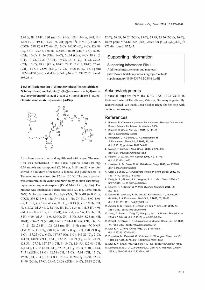
2-((3-(6-((Adamantan-1-yl)methoxy)hexyl)benzo[d]thiazol-
2(3H)-ylidene)methyl)-4-((3-(6-((adamantan-1-yl)meth-
oxy)hexyl)benzo[d]thiazol-3-ium-2-yl)methylene)-3-oxocy-
clobut-1-en-1-olate, squaraine (AdSq)
All solvents were dried and equilibrated with argon. The reac-
tion was performed in the dark. Squaric acid (10 mg,
0.09 mmol) and compound (2, 70 mg, 0.18 mmol) were dis-
solved in a mixture of benzene, n-butanol and pyridine (2:1:1).
The reaction was stirred for 12 h at 120 °C. The crude product
was concentrated in vacuo and purified by column chromatog-
raphy under argon atmosphere (DCM:MeOH 9:1, Rf: 0.6). The
product was obtained as a dark blue solid (20 mg, 0.088 mmol,
26%). Molecular formula: C54H68N2O4S2. 1H NMR (600 MHz,
CDCl3, 298 K) δ 9.41 (dd, J = 18.1, 6.1 Hz, 2H, Har), 8.97–8.91
(m, 1H, Har), 8.55–8.45 (m, 2H, Har), 8.13 (t, J = 6.9 Hz, 2H,
Har), 8.02 (dd, J = 8.0, 5.5 Hz, 1H, Har), 6.34 (s, 1H, 5-H), 4.96
(dd, J = 8.8, 6.2 Hz, 2H, 13-H), 4.63 (td, J = 6.6, 1.3 Hz, 1H,
5-H), 4.19 (qd, J = 11.0, 6.0 Hz, 2H, 13-H), 3.39–3.26 (m, 4H,
18-H), 2.94–2.89 (m, 4H, 19-H), 2.14–1.03 (m, 42H, 14-,16-
,17-,21-,22-,23-H), 1.02–0.81 (m, 4H, 15-H) ppm; 13C NMR
(151 MHz, CDCl3, 298 K) δ 198.35 (Cq, 3-C), 190.29 (Cq,
1-C), 187.25 (Cq, 6-C), 167.87 (Cq, 6-C), 145.25 (Cq, 2-C),
141.94, 140.97 (2CH, 12-C), 132.55, 130.99 (Cq, 7-C), 128.89,
128.59, 127.72, 127.27 (4CH, 9-,10-C), 124.95, 122.46 (CH,
8-,11-C), 112.36 (2CH, 5-C), 82.02 (2CH2, 19-H), 73.01, 71.44,
71.31 (2CH2, 18-C), 62.34 (CH, 13-C), 47.01 (CH, 13-C),
39.86 (CH, 21-C), 37.34 (CH, 23-C), 34.20 (Cq, C-20), 32.05,
31.99 (2CH2, 17-C), 29.47, 29.38 (2CH2, 14-C), 28.38 (2CH,
22-C), 26.09, 26.02 (2CH2, 15-C), 25.99, 25.76 (2CH2, 16-C),
18.69 ppm; MALDI–MS (m/z): calcd for [C54H68N2O4S2]+:
872,46; found: 872,47.
Supporting InformationSupporting Information File 1Additional measurements and methods.
[http://www.beilstein-journals.org/bjoc/content/
supplementary/1860-5397-12-248-S1.pdf]
AcknowledgmentsFinancial support from the DFG EXC 1003 Cells in
Motion–Cluster of Excellence, Münster, Germany is gratefully
acknowledged. We thank Lena Fischer-Riepe for her help with
confocal microscopy.
References1. Bonnett, R. Chemical Aspects of Photodynamic Therapy; Gordon and
Breach Science Publishers: Amsterdam, 2000.2. Bonnett, R. Chem. Soc. Rev. 1995, 24, 19–33.
doi:10.1039/cs99524000193. Robertson, C. A.; Evans, D. H.; Abrahamse, H.
J. Photochem. Photobiol., B 2009, 96, 1–8.doi:10.1016/j.jphotobiol.2009.04.001
4. Maisch, T. Mini-Rev. Med. Chem. 2009, 9, 974–983.doi:10.2174/138955709788681582
5. Felsher, D. W. Nat. Rev. Cancer 2003, 3, 375–379.doi:10.1038/nrc1070
6. Josefsen, L. B.; Boyle, R. W. Met.-Based Drugs 2008, No. 276109.doi:10.1155/2008/276109
7. Ortel, B.; Shea, C. R.; Calzavara-Pinton, P. Front. Biosci. 2009, 14,4157–4172. doi:10.2741/3520
8. Detty, M. R.; Gibson, S. L.; Wagner, S. J. J. Med. Chem. 2004, 47,3897–3915. doi:10.1021/jm040074b
9. Cherno, N. K.; Krusir, G. V. Prikl. Biokhim. Mikrobiol. 2005, 28,297–303.
10. Delaey, E.; van Laar, F.; De Vos, D.; Kamuhabwa, A.; Jacobs, P.;de Witte, P. J. Photochem. Photobiol., B 2000, 55, 27–36.doi:10.1016/S1011-1344(00)00021-X
11. Awuah, S. G.; Polreis, J.; Biradar, V.; You, Y. Org. Lett. 2011, 13,3884–3887. doi:10.1021/ol2014076
12. Jiang, Z.; Shao, J.; Yang, T.; Wang, J.; Jia, L. J. Pharm. Biomed. Anal.2014, 87, 98–104. doi:10.1016/j.jpba.2013.05.014
13. Sreejith, S.; Divya, K. P.; Ajayaghosh, A. Angew. Chem., Int. Ed. 2008,47, 7883–7887. doi:10.1002/anie.200803194
14. Law, K. Y. J. Phys. Chem. 1987, 91, 5184–5193.doi:10.1021/j100304a012
15. Emmelius, M.; Pawloski, G.; Vollmann, H. W. Angew. Chem., Int. Ed.1989, 28, 1445–1471. doi:10.1002/anie.198914453
16. Law, K. Y. Chem. Rev. 1993, 93, 449–486. doi:10.1021/cr00017a02017. Dolmans, D. E. J. G. J.; Fukumura, D.; Jain, R. K. Nat. Rev. Cancer
2003, 3, 380–387. doi:10.1038/nrc1071

Beilstein J. Org. Chem. 2016, 12, 2535–2542.
2542
18. Yagi, S.; Nakazumi, H. Syntheses and Applications of SquaryliumDyes. In Functional Dyes; Kim, S.-H., Ed.; Elsevier B. V.: Amsterdam,2006; pp 215–255. doi:10.1016/b978-044452176-7/50007-3
19. Kamat, P. V.; Das, S.; Thomas, K. G.; George, M. V. J. Phys. Chem.1992, 96, 195–199. doi:10.1021/j100180a038
20. Santos, P. F.; Reis, L. V.; Almeida, P.; Oliveira, A. S.;Vieira Ferreira, L. F. J. Photochem. Photobiol., A 2003, 160, 159–161.doi:10.1016/S1010-6030(03)00203-X
21. Santos, P. F.; Reis, L. V.; Almeida, P.; Serrano, J. P.; Oliveira, A. S.;Vieira Ferreira, L. F. J. Photochem. Photobiol., A 2004, 163, 267–269.doi:10.1016/j.jphotochem.2003.12.007
22. Foote, C. S.; Lin, J. W.-P. Tetrahedron Lett. 1968, 9, 3267–3270.doi:10.1016/S0040-4039(00)89543-X
23. Mazur, S.; Foote, C. S. J. Am. Chem. Soc. 1970, 92, 3225–3226.doi:10.1021/ja00713a073
24. Salice, P.; Arnbjerg, J.; Pedersen, B. W.; Toftegaard, R.; Beverina, L.;Pagani, G. A.; Ogilby, P. R. J. Phys. Chem. A 2010, 114, 2518–2525.doi:10.1021/jp911180n
25. Rapozzi, V.; Beverina, L.; Salice, P.; Pagani, G. A.; Camerin, M.;Xodo, L. E. J. Med. Chem. 2010, 53, 2188–2196.doi:10.1021/jm901727j
26. Monge-Fuentes, V.; Muehlmann, L. A.; de Azevedo, R. B. Nano Rev.2014, 5, No. 24381. doi:10.3402/nano.v5.24381
27. Ravoo, B. J.; Darcy, R. Angew. Chem., Int. Ed. 2000, 39, 4324–4326.doi:10.1002/1521-3773(20001201)39:23<4324::AID-ANIE4324>3.0.CO;2-O
28. Chauhan, P.; Hadad, C.; Sartorelli, A.; Zarattini, M.;Herreros-López, A.; Mba, M.; Maggini, M.; Prato, M.; Carofiglio, T.Chem. Commun. 2013, 49, 8525–8527. doi:10.1039/c3cc44852e
29. Uekama, K.; Hirayama, F.; Irie, T. Chem. Rev. 1998, 98, 2045–2076.doi:10.1021/cr970025p
30. Falvey, P.; Lim, C. W.; Darcy, R.; Revermann, T.; Karst, U.;Giesbers, M.; Marcelis, A. T. M.; Lazar, A.; Coleman, A. W.;Reinhoudt, D. N.; Ravoo, B. J. Chem. – Eur. J. 2005, 11, 1171–1180.doi:10.1002/chem.200400905
31. Chen, Y.; Liu, Y. Chem. Soc. Rev. 2010, 39, 495–505.doi:10.1039/B816354P
32. Voskuhl, J.; Stuart, M. C. A.; Ravoo, B. J. Chem. – Eur. J. 2010, 16,2790–2796. doi:10.1002/chem.200902423
33. Kauscher, U.; Ravoo, B. J. Beilstein J. Org. Chem. 2012, 8,1543–1551. doi:10.3762/bjoc.8.175
34. Versluis, F.; Tomatsu, I.; Kehr, S.; Fregonese, C.; Tepper, A. W. J. W.;Stuart, M. C. A.; Ravoo, B. J.; Koning, R. I.; Kros, A. J. Am. Chem. Soc.2009, 131, 13186–13187. doi:10.1021/ja9026264
35. Nalluri, S. K. M.; Voskuhl, J.; Bultema, J. B.; Boekema, E. J.;Ravoo, B. J. Angew. Chem., Int. Ed. 2011, 50, 9747–9751.doi:10.1002/anie.201103707
36. Kauscher, U.; Stuart, M. C. A.; Drücker, P.; Galla, H.-J.; Ravoo, B. J.Langmuir 2013, 29, 7377–7383. doi:10.1021/la3045434
37. Voskuhl, J.; Kauscher, U.; Gruener, M.; Frisch, H.; Wibbeling, B.;Strassert, C. A.; Ravoo, B. J. Soft Matter 2013, 9, 2453–2457.doi:10.1039/c2sm27353e
38. Galstyan, A.; Kauscher, U.; Block, D.; Ravoo, B. J.; Strassert, C. A.ACS Appl. Mater. Interfaces 2016, 8, 12631–12637.doi:10.1021/acsami.6b02132
39. Li, Q.; Batchelor-McAuley, C.; Lawrence, N. S.; Hartshorne, R. S.;Compton, R. G. J. Electroanal. Chem. 2013, 688, 328–335.doi:10.1016/j.jelechem.2012.07.039
License and TermsThis is an Open Access article under the terms of the
Creative Commons Attribution License
(http://creativecommons.org/licenses/by/4.0), which
permits unrestricted use, distribution, and reproduction in
any medium, provided the original work is properly cited.
The license is subject to the Beilstein Journal of Organic
Chemistry terms and conditions:
(http://www.beilstein-journals.org/bjoc)
The definitive version of this article is the electronic one
which can be found at:
doi:10.3762/bjoc.12.248