
A role for a lithium-inhibited Golgi nucleotidase in …brane domain and was disrupted by brefeldin...
8
A role for a lithium-inhibited Golgi nucleotidase in skeletal development and sulfation Joshua P. Frederick*, A. Tsahai Tafari*, Sheue-Mei Wu* † , Louis C. Megosh*, Shean-Tai Chiou*, Ryan P. Irving*, and John D. York* ‡ *Department of Pharmacology and Cancer Biology, Howard Hughes Medical Institute, and † Department of Psychiatry and Behavioral Sciences, Duke University Medical Center, Durham, NC 27710 This Feature Article is part of a series identified by the Editorial Board as reporting findings of exceptional significance. Edited by Philip W. Majerus, Washington University School of Medicine, St. Louis, MO, and approved June 23, 2008 (received for review February 6, 2008) Sulfation is an important biological process that modulates the function of numerous molecules. It is directly mediated by cytosolic and Golgi sulfotransferases, which use 3-phosphoadenosine 5- phosphosulfate to produce sulfated acceptors and 3-phosphoad- enosine 5-phosphate (PAP). Here, we identify a Golgi-resident PAP 3-phosphatase (gPAPP) and demonstrate that its activity is po- tently inhibited by lithium in vitro. The inactivation of gPAPP in mice led to neonatal lethality, lung abnormalities resembling atelectasis, and dwarfism characterized by aberrant cartilage mor- phology. The phenotypic similarities of gPAPP mutant mice to chondrodysplastic models harboring mutations within compo- nents of the sulfation pathway lead to the discovery of undersul- fated chondroitin in the absence of functional enzyme. Addition- ally, we observed loss of gPAPP leads to perturbations in the levels of heparan sulfate species in lung tissue and whole embryos. Our data are consistent with a model that clearance of the nucleotide product of sulfotransferases within the Golgi plays an important role in glycosaminoglycan sulfation, provide a unique genetic basis for chondrodysplasia, and define a function for gPAPP in the formation of skeletal elements derived through endochondral ossification. chondrodysplasia glycosaminoglycan phosphatase chondroitin sulfate phosphoadenosine phosphate T he formation of bone occurs primarily through intramem- branous and endochondral ossification. Intramembranous ossification is the emergence of bone directly from the mesen- chyme and is responsible for the generation of flat bones. Endochondral ossification is the driving force of longitudinal extension best visualized in the growth plates of developing long bones and relies on a chondrocyte-produced cartilage template. Establishment of the cartilaginous template is a tightly ordered process that involves the function and interplay of: (i) specific transcription factors; (ii) chondrocyte-modulatory ligand path- ways; (iii) extracellular matrix (ECM) structural proteins; (iv) core proteins of proteoglycans and their covalently linked gly- cosaminoglycans (GAGs), such as chondroitin sulfate (CS) chains; and correspondingly (v) gene products essential to the appropriate synthesis and sulfation of GAGs (1–7). Perturba- tions in a variety of genes that encode proteins in each of the above five categories have been implicated in endochondral ossification defects and commensurate development of chon- drodysplasias that vary from subtle growth defects to severe dwarfism and neonatal lethality (7–9). Among the examples of altered genes that produce chondrodysplasias in humans with corresponding mouse models is the diastrophic dysplasia sulfate transporter (DTDST), which is essential for the delivery of extracellular inorganic sulfate into the cytoplasm allowing for subsequent intracellular sulfation reactions (1, 10, 11). Sulfation is an important functional modification of a wide array of endogenous and xenobiotic compounds that is mediated by two structurally related sulfotransferase (SULT) families: cytosolic SULTs and Golgi resident SULTs (gSULTs). gSULTs are mostly single-pass transmembrane domain containing pro- teins that modify lipids, tyrosine residues of proteins, and GAGs (12–14). gSULT-mediated sulfation can alter the activity of certain signaling molecules, such as cholecystokinin, gastrin, and angiotensin II (14). It is also essential for the proper develop- ment, structure, and function of tissues such as cartilage through modulating the sequestration of growth factors and their binding to cognate receptors, as well as moderating appropriate inter- actions among ECM proteins, proteoglycans, and GAGs (14– 17). SULTs modify acceptor moieties through the utilization of the universal sulfate donor 3-phosphoadenosine 5-phospho- sulfate (PAPS) (Fig. 1A). Availability of PAPS enables SULTs to modify targets in a reaction that produces the sulfated substrate and 3-phosphoadenosine 5-phosphate (PAP). Al- though the molecular basis for PAP metabolism within the Golgi lumen is unknown, conversion of PAP to 5-AMP within the cytoplasm is accomplished by a bisphosphate 3-nucleotidase (annotated BPNT1 in mouse and human or RnPIP in rat) that also exhibits activity toward PAPS and inositol phosphate (IP) substrates (18–20). BPNT1 belongs to a structurally conserved, lithium-inhibited family of small-molecule phosphomonoesterases, which in ad- dition to glycogen synthase kinase, have been implicated as possible cellular targets of lithium, a drug used for the treatment of bipolar disease (21, 22). In humans and mice, this phosphatase family consists of seven gene products defined by a consensus active-site motif (Fig. 1B). Six of the seven family members have defined biochemical functions in IP signaling, energy homeosta- sis, and nucleotide metabolism; these include the aforemen- tioned BPNT1, inositol polyphosphate 1-phosphatase (INPP1), fructose-1,6-bisphosphatase (FBP) 1 and 2, and myo-inositol monophosphatase (IMP) A1 and A2. Members of this phospha- tase family are functionally inhibited in vitro and in vivo by clinically appropriate doses of lithium (22–24). Chronic lithium treatment reduces levels of inositol in brain through inhibition of IMPs and INPP1 (25–27), the loss of INPP1 in Drosophila mimics lithium-induced alterations in synaptic transmission (28), and perturbations in cytosolic 3-nucleotidase activity have been shown to regulate lithium toxicity in yeast (29–31). Author contributions: J.P.F., A.T.T., S.-M.W., and J.D.Y. designed research; J.P.F., A.T.T., S.-M.W., L.C.M., S.-T.C., and R.P.I. performed research; J.P.F. contributed new reagents/ analytic tools; J.P.F., A.T.T., S.-M.W., L.C.M., S.-T.C., R.P.I., and J.D.Y. analyzed data; and J.P.F., A.T.T., S.-M.W., R.P.I., and J.D.Y. wrote the paper. The authors declare no conflict of interest. This article is a PNAS Direct Submission. Freely available online through the PNAS open access option. ‡ To whom correspondence should be addressed. E-mail: [email protected]. This article contains supporting information online at www.pnas.org/cgi/content/full/ 0801182105/DCSupplemental. © 2008 by The National Academy of Sciences of the USA www.pnas.orgcgidoi10.1073pnas.0801182105 PNAS August 19, 2008 vol. 105 no. 33 11605–11612 BIOCHEMISTRY FEATURE ARTICLE Downloaded by guest on April 25, 2020
Transcript of A role for a lithium-inhibited Golgi nucleotidase in …brane domain and was disrupted by brefeldin...

A role for a lithium-inhibited Golgi nucleotidasein skeletal development and sulfationJoshua P. Frederick*, A. Tsahai Tafari*, Sheue-Mei Wu*†, Louis C. Megosh*, Shean-Tai Chiou*, Ryan P. Irving*,and John D. York*‡
*Department of Pharmacology and Cancer Biology, Howard Hughes Medical Institute, and †Department of Psychiatry and Behavioral Sciences, DukeUniversity Medical Center, Durham, NC 27710
This Feature Article is part of a series identified by the Editorial Board as reporting findings of exceptional significance.
Edited by Philip W. Majerus, Washington University School of Medicine, St. Louis, MO, and approved June 23, 2008 (received for review February 6, 2008)
Sulfation is an important biological process that modulates thefunction of numerous molecules. It is directly mediated by cytosolicand Golgi sulfotransferases, which use 3�-phosphoadenosine 5�-phosphosulfate to produce sulfated acceptors and 3�-phosphoad-enosine 5�-phosphate (PAP). Here, we identify a Golgi-resident PAP3�-phosphatase (gPAPP) and demonstrate that its activity is po-tently inhibited by lithium in vitro. The inactivation of gPAPP inmice led to neonatal lethality, lung abnormalities resemblingatelectasis, and dwarfism characterized by aberrant cartilage mor-phology. The phenotypic similarities of gPAPP mutant mice tochondrodysplastic models harboring mutations within compo-nents of the sulfation pathway lead to the discovery of undersul-fated chondroitin in the absence of functional enzyme. Addition-ally, we observed loss of gPAPP leads to perturbations in the levelsof heparan sulfate species in lung tissue and whole embryos. Ourdata are consistent with a model that clearance of the nucleotideproduct of sulfotransferases within the Golgi plays an importantrole in glycosaminoglycan sulfation, provide a unique genetic basisfor chondrodysplasia, and define a function for gPAPP in theformation of skeletal elements derived through endochondralossification.
chondrodysplasia � glycosaminoglycan � phosphatase �chondroitin sulfate � phosphoadenosine phosphate
The formation of bone occurs primarily through intramem-branous and endochondral ossification. Intramembranous
ossification is the emergence of bone directly from the mesen-chyme and is responsible for the generation of flat bones.Endochondral ossification is the driving force of longitudinalextension best visualized in the growth plates of developing longbones and relies on a chondrocyte-produced cartilage template.Establishment of the cartilaginous template is a tightly orderedprocess that involves the function and interplay of: (i) specifictranscription factors; (ii) chondrocyte-modulatory ligand path-ways; (iii) extracellular matrix (ECM) structural proteins; (iv)core proteins of proteoglycans and their covalently linked gly-cosaminoglycans (GAGs), such as chondroitin sulfate (CS)chains; and correspondingly (v) gene products essential to theappropriate synthesis and sulfation of GAGs (1–7). Perturba-tions in a variety of genes that encode proteins in each of theabove five categories have been implicated in endochondralossification defects and commensurate development of chon-drodysplasias that vary from subtle growth defects to severedwarfism and neonatal lethality (7–9). Among the examples ofaltered genes that produce chondrodysplasias in humans withcorresponding mouse models is the diastrophic dysplasia sulfatetransporter (DTDST), which is essential for the delivery ofextracellular inorganic sulfate into the cytoplasm allowing forsubsequent intracellular sulfation reactions (1, 10, 11).
Sulfation is an important functional modification of a widearray of endogenous and xenobiotic compounds that is mediatedby two structurally related sulfotransferase (SULT) families:
cytosolic SULTs and Golgi resident SULTs (gSULTs). gSULTsare mostly single-pass transmembrane domain containing pro-teins that modify lipids, tyrosine residues of proteins, and GAGs(12–14). gSULT-mediated sulfation can alter the activity ofcertain signaling molecules, such as cholecystokinin, gastrin, andangiotensin II (14). It is also essential for the proper develop-ment, structure, and function of tissues such as cartilage throughmodulating the sequestration of growth factors and their bindingto cognate receptors, as well as moderating appropriate inter-actions among ECM proteins, proteoglycans, and GAGs (14–17). SULTs modify acceptor moieties through the utilization ofthe universal sulfate donor 3�-phosphoadenosine 5�-phospho-sulfate (PAPS) (Fig. 1A). Availability of PAPS enables SULTsto modify targets in a reaction that produces the sulfatedsubstrate and 3�-phosphoadenosine 5�-phosphate (PAP). Al-though the molecular basis for PAP metabolism within the Golgilumen is unknown, conversion of PAP to 5�-AMP within thecytoplasm is accomplished by a bisphosphate 3�-nucleotidase(annotated BPNT1 in mouse and human or RnPIP in rat) thatalso exhibits activity toward PAPS and inositol phosphate (IP)substrates (18–20).
BPNT1 belongs to a structurally conserved, lithium-inhibitedfamily of small-molecule phosphomonoesterases, which in ad-dition to glycogen synthase kinase, have been implicated aspossible cellular targets of lithium, a drug used for the treatmentof bipolar disease (21, 22). In humans and mice, this phosphatasefamily consists of seven gene products defined by a consensusactive-site motif (Fig. 1B). Six of the seven family members havedefined biochemical functions in IP signaling, energy homeosta-sis, and nucleotide metabolism; these include the aforemen-tioned BPNT1, inositol polyphosphate 1-phosphatase (INPP1),fructose-1,6-bisphosphatase (FBP) 1 and 2, and myo-inositolmonophosphatase (IMP) A1 and A2. Members of this phospha-tase family are functionally inhibited in vitro and in vivo byclinically appropriate doses of lithium (22–24). Chronic lithiumtreatment reduces levels of inositol in brain through inhibition ofIMPs and INPP1 (25–27), the loss of INPP1 in Drosophila mimicslithium-induced alterations in synaptic transmission (28), andperturbations in cytosolic 3�-nucleotidase activity have beenshown to regulate lithium toxicity in yeast (29–31).
Author contributions: J.P.F., A.T.T., S.-M.W., and J.D.Y. designed research; J.P.F., A.T.T.,S.-M.W., L.C.M., S.-T.C., and R.P.I. performed research; J.P.F. contributed new reagents/analytic tools; J.P.F., A.T.T., S.-M.W., L.C.M., S.-T.C., R.P.I., and J.D.Y. analyzed data; andJ.P.F., A.T.T., S.-M.W., R.P.I., and J.D.Y. wrote the paper.
The authors declare no conflict of interest.
This article is a PNAS Direct Submission.
Freely available online through the PNAS open access option.
‡To whom correspondence should be addressed. E-mail: [email protected].
This article contains supporting information online at www.pnas.org/cgi/content/full/0801182105/DCSupplemental.
© 2008 by The National Academy of Sciences of the USA
www.pnas.org�cgi�doi�10.1073�pnas.0801182105 PNAS � August 19, 2008 � vol. 105 � no. 33 � 11605–11612
BIO
CHEM
ISTR
YFE
ATU
REA
RTIC
LE
Dow
nloa
ded
by g
uest
on
Apr
il 25
, 202
0

We now report a function for the seventh member of thisfamily as a gPAPP whose activity is inhibited by lithium in vitro.We find that gPAPP is required in mice for proper endochondralossification, and its genetic inactivation results in neonatallethality, morphological abnormalities in lung, and severe chon-drodysplasia. The biochemical characterization of gPAPP andphenotypic analysis of mutant mice led to the discovery of a rolefor this protein in the process of GAG sulfation. Our studiesprovide insights into skeletal development, an unanticipatedgenetic basis of chondrodysplasia, and a link between loss ofnucleotidase function and undersulfated molecules.
ResultsgPAPP Is a Golgi Resident Protein. Murine gPAPP was first iden-tified by searching metazoan genomes and expressed sequencesfor the presence of an encoded canonical active-site motif (Fig.1B). We and others previously annotated this sequence in theabsence of functional data as lithium-inhibited phosphomonoes-
terase (31), inositol monophosphatase domain containing 1(GenBank accession no. AAH48776), and myo-inositol mono-phosphatase A3 (IMPA3) (GenBank accession no. AAK52336).Despite sharing nearly 20% overall sequence identity to BPNT1and INPP1, initial attempts to identify a phosphatase activity forrecombinant bacterially expressed gPAPP by using candidatenucleotide and IP substrates were unsuccessful. Further analysisof the amino acid sequence revealed a N-terminal, single-passtransmembrane domain (residues 7–29) and a consensus N-linked glycosylation sequence, N257-Q-T [supporting informa-tion (SI) Fig. S1 A], two features that raised the possibility thatgPAPP may reside within the endoplasmic reticulum/Golgi. Totest this hypothesis, we examined the subcellular distribution ofgPAPP. Indirect immunofluorescence in normal human skinfibroblasts revealed that endogenous gPAPP was present in aperinuclear organelle that partially colocalized with the markerprotein GM130, which is enriched in cis- but also present inmedial- and trans-Golgi compartments (Fig. 2A). Similarly, weobserved partially overlapping signals between gPAPP and thetrans-Golgi network resident Golgin-97 (data not shown).gPAPP localization to the Golgi depended on the transmem-
acceptor - sulfate
acceptor - sulfate
A
B
Fig. 1. The sulfation pathway including two members of a structurallyconserved, lithium-sensitive phosphomonoesterase family. (A) Representa-tion of acceptor sulfation mediated by cytosolic SULTs and gSULTs. Theuniversal sulfate donor PAPS is produced in the cytoplasm by PAPS synthetases(PAPSS) from sulfate (SO4
2�) and ATP through the intermediate generation ofadenosine-5�-phosphosulfate (APS). PAPS can be used by SULTs in the sulfa-tion of cytosolic small molecules or transported into the lumen of the Golgi byPAPS transporters (PAPSTs). Once luminal, PAPS is used by gSULTs in thesulfation of Golgi-restricted macromolecules. In addition to sulfate-conjugation to acceptors, sulfation produces PAP, which can be further me-tabolized to 5�-AMP by a predominantly cytoplasmic bisphosphate 3�-nucleotidase (BPNT1) and a gPAPP. (B) The consensus motif that defines themetal-dependent/lithium-sensitive phosphomonoesterase family with link-ing residues (X) in stretches of specified lengths is boxed. Representation ofthe Mus musculus phosphatase family is shown as an unrooted phylogentictree produced by Clustal W and Phylip’s Drawtree software. Members includeFBP1 (GenBank accession no. Q9QXD6), FBP2 (GenBank accession no. P70695),IMPA1 (GenBank accession no. O55023), IMPA2 (GenBank accession no.Q91UZ5), INPP1 (GenBank accession no. P49442), BPNT1 (GenBank accessionno. AAD1733), and gPAPP.
-1000 -500 0 500 1000LiCl (µM)
PAP (µM)9.5
193876
1.0
0.8
0.6
0.4
0.2(µm
ol/m
in/m
g)-1
Km (µM) FL 21.0 9.5 174.0∆55 22.8 2.3 210.8
αgPAPP αGM130 merge
Minutes5 10 15 20 25 30
806040200
mA
bsor
banc
e un
its
259
5’-AMP PAP PAPS
80604020
0
A
B D
C Vmax (µmole/min/mg) Ki of LiCl (µΜ)
Fig. 2. Identification of gPAPP as a Golgi-resident 3� nucleotidase inhibitedby lithium. (A) Indirect immunofluorescence of endogenous gPAPP (red; Left),the known Golgi-associated GM130 (green; Center), and the merge of the twosignals (Right) in normal human skin fibroblasts. gPAPP and GM130 signalspartially overlap (yellow), yet higher magnification displays some distinctlocalization within the Golgi (Inset). Blue is DAPI-stained nuclei. (Scale bar: 10microns.) (B) HPLC traces of resolved reaction constituents from 20 �M of PAPand PAPS treated in buffer A with no recombinant protein (Mock; Upper) or200 ng of �55 gPAPP (Lower) for 1 h. 5�-AMP, PAP, and PAPS were detected byUV absorbance (259 nm) with elution times corresponding with previously runstandards as indicated. Note that gPAPP treatment results in nearly completehydrolysis of PAP to 5�-AMP, yet does not catalyze the conversion of PAPS inthe used conditions. (C) Km, Vmax, and lithium Ki of PAP hydrolysis by full-length (FL) and �55 gPAPP purified from baculovirus-infected cells. Km andVmax values were calculated from a logarithmic equation fitting averagevelocities obtained from at least duplicate data points plotted against varioussubstrate concentrations. Ki of gPAPP-mediated PAP hydrolysis by LiCl arepresented as the average with standard deviations calculated by the equation:�Ki � x � intercept/(1 � (Km/[s])) from plotted lines obtained with eachconcentration of PAP used in Dixon plot analyses. (D) Dixon plot analysis oflithium inhibition of recombinant full-length gPAPP-mediated PAP hydrolysis.The four indicated concentrations of PAP were used to plot 1/velocity averageversus inhibitor concentration, and the parallel lines signify uncompetitiveinhibition.
11606 � www.pnas.org�cgi�doi�10.1073�pnas.0801182105 Frederick et al.
Dow
nloa
ded
by g
uest
on
Apr
il 25
, 202
0
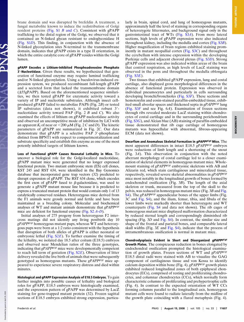
brane domain and was disrupted by brefeldin A treatment, afungal metabolite known to induce the redistribution of Golgiresident proteins (Fig. S1 B and C). Consistent with gPAPPtrafficking to the distal region of the Golgi, we observed that itcontained an N-linked glycan resistant to endoglycosidase H(Fig. S1D). This finding, in conjunction with the absence ofN-linked glycosylation sites N-terminal to the transmembranedomain, indicates that gPAPP exists in a type II orientation, inwhich the entire catalytic core of gPAPP resides within the Golgilumen.
gPAPP Encodes a Lithium-Inhibited Phosphoadenosine Phosphate3�-Nucleotidase. Given these results, we hypothesized that gen-eration of functional enzyme may require luminal traffickingand/or N-linked glycosylation. Using a baculovirus-induced ex-pression system, we produced recombinant full-length gPAPPand a secreted form that lacked the transmembrane domain(�55gPAPP). Based on the aforementioned sequence similari-ties, we then tested gPAPP for enzymatic activity toward avariety of IP and nucleotide substrates. Although insect cell-produced gPAPP failed to metabolize PAPS (Fig. 2B) or testedIP substrates (data not shown), it exhibited robust 3�-nucleotidase activity toward PAP (Fig. 2 B and C). We alsoexamined the effects of lithium on gPAPP nucleotidase activityand observed an uncompetitive mode of inhibition by LiCl withan apparent Ki of near or �200 �M (Fig. 2 C and D). The kineticparameters of gPAPP are summarized in Fig. 2C. Our datademonstrate that gPAPP is a selective PAP 3�-phosphatasedistinct from BPNT1 with respect to compartmentalization andsubstrate specificity and establish this enzyme as one of the mostpotently inhibited targets of lithium known.
Loss of Functional gPAPP Causes Neonatal Lethality in Mice. Touncover a biological role for the Golgi-localized nucleotidase,gPAPP mutant mice were generated that no longer expressedfunctional protein. Two mutant embryonic stem (ES) cell clones,KST 245 and RST 634, were identified in the Bay Genomicsdatabase that incorporated gene trap vectors (32) predicted todisrupt expression of gPAPP. The RST 634 clone, which encodesthe first exon of gPAPP fused to �-galactosidase, was used togenerate a gPAPP mutant mouse line because it is predicted toexpress a truncated mutant protein that would contain only 1 of 13catalytically conserved residues. Heterozygous mice derived fromthe F1 animals were grossly normal and fertile and have beenmaintained as a breeding colony. Molecular and biochemicalanalyses of WT and mutant animals demonstrate that gPAPPgt/gt
mice are deficient for functional enzyme (Fig. S2 A–D).Initial analyses of 275 progeny from heterozygous F2 inter-
cross matings did not identify any living postbirth day 10gPAPPgt/gt homozygous mutant pups, whereas WT and heterozy-gous pups were born at a 1:2 ratio consistent with the hypothesisthat disruption of both alleles of gPAPP is either neonatal orembryonic lethal (Fig. S2E). To further examine the timing ofthe lethality, we isolated day 18.5 after coitum (E18.5) embryosand observed near Mendelian ratios of the three genotypes,indicating that gPAPPgt/gt mice were developmentally competentto reach full term of gestation (Fig. S2E). Observation of litterdelivery revealed the live birth of animals that were subsequentlygenotyped as homozygous mutants. These gPAPPgt/gt mice ap-peared to experience severe respiratory distress and died withinminutes.
Histological and gPAPP Expression Analysis of E18.5 Embryos. To gainfurther insights into possible causes of lethality and biologicalroles for gPAPP, E18.5 embryos were histologically examined,and the expression pattern of gPAPP was determined by LacZstaining for gene-trapped mutant protein (32). Frozen sagittalsections of E18.5 embryos exhibited strong expression, particu-
larly in brain, spinal cord, and lung of homozygous mutants,approximately half the level of staining in corresponding organsof heterozygote littermates, and background signal only in thegastrointestinal tract of WTs (Fig. S3A). From more lateralsections, high levels of gPAPP expression were also indicatedthroughout kidney, including the medulla and cortex (Fig. S3B).Higher magnification of brain regions exhibited staining prom-inently in mutant neopallial cortex (Fig. S3C) and throughoutthe cerebellum with intense expression within the developingPurkinje cells and adjacent choroid plexus (Fig. S3D). StronggPAPP expression was also indicated within areas of the brainthat control respiration, as high levels of LacZ staining wasobserved in the pons and throughout the medulla oblongata(Fig. S3E).
Two tissues that exhibited gPAPP expression, lung and costalcartilage, also displayed gross morphological differences in theabsence of functional protein. Expression was observed inindividual pneumocytes and particularly in cells surroundingdeveloping bronchi/bronchioles (Fig. S3F). These sections andhemotoxylin and eosin-stained paraffin-embedded tissue, exhib-ited small alveolar spaces and thickened septa in gPAPPgt/gt lung(Fig. S3F and see Fig. 6A). Examination of frozen sectionsthrough ribs revealed moderate LacZ staining within chondro-cytes of costal cartilage and in the surrounding perichondrium(Fig. S3G), and Alcian blue (AB) staining of paraffin-embeddedtissue demonstrated that rib cage cartilage of homozygousmutants was hypocellular with abnormal, fibrous-appearingECM (data not shown).
Dwarfism and Anomalous Skeletal Formation in gPAPPgt/gt Mice. Themost apparent differences in intact E18.5 gPAPPgt/gt embryoswere reductions of limb length and a shortening of the snout(Fig. 3A). This observation in conjunction with the notedaberrant morphology of costal cartilage led to a closer exami-nation of skeletal elements in homozygous mutant mice. Whole-mount staining of gPAPPgt/gt embryonic skeletons with AB andAlizarin red, which stain cartilaginous and mineralized tissue,respectively, revealed severe skeletal abnormalities in gPAPPgt/gt
mice most notably in the longitudinal growth of bones formed byendochondral ossification (Fig. 3B). The length of the axialskeleton or trunk, measured from the top of the skull to thepelvis, was reduced in homozygous mutant mice (Fig. 3B and Fig.S4). The gPAPPgt/gt appendicular bones of the upper limbs (Fig.3C and Fig. S4), and the ilium, femur, tibia, and fibula of thelower limbs were markedly shorter than heterozygote and WTcounterparts (Fig. S4 and data not shown). The rib cages ofhomozygous mutant mice displayed malformation characterizedby reduced sternal length and correspondingly diminished ribspacing (Fig. 3D and Fig. S4). In contrast, the similar size andshape of the frontal and parietal bones, and comparable lateralskull widths (Fig. 3E and Fig. S4), indicate that the process ofintramembranous ossification is normal in mutant mice.
Chondrodysplasia Evident in Short and Disorganized gPAPPgt/gt
Growth Plates. The conspicuous reduction in bones elongated byendochondral ossification prompted the histological examina-tion of growth plates. Frozen sections of WT and gPAPPgt/gt
E18.5 distal radii were stained with AB to visualize the GAGcomponent of cartilaginous tissue and von Kossa to identifycalcium deposition within bone (Fig. 4). gPAPPgt/gt growth platesexhibited reduced longitudinal zones of both epiphyseal chon-drocytes (ECs), comprised of resting and proliferating chondro-cytes, and columnar chondrocytes (CCs), which normally formcharacteristic columns of proliferating and prehypertrophic cells(Fig. 4). In contrast to the expected orientation of WT CCsforming columns parallel to the longitudinal axis, homozygousmutant cells were found to radiate laterally from the midline ofthe growth plate coinciding with a flared metaphysis (Fig. 4).
Frederick et al. PNAS � August 19, 2008 � vol. 105 � no. 33 � 11607
BIO
CHEM
ISTR
YFE
ATU
REA
RTIC
LE
Dow
nloa
ded
by g
uest
on
Apr
il 25
, 202
0
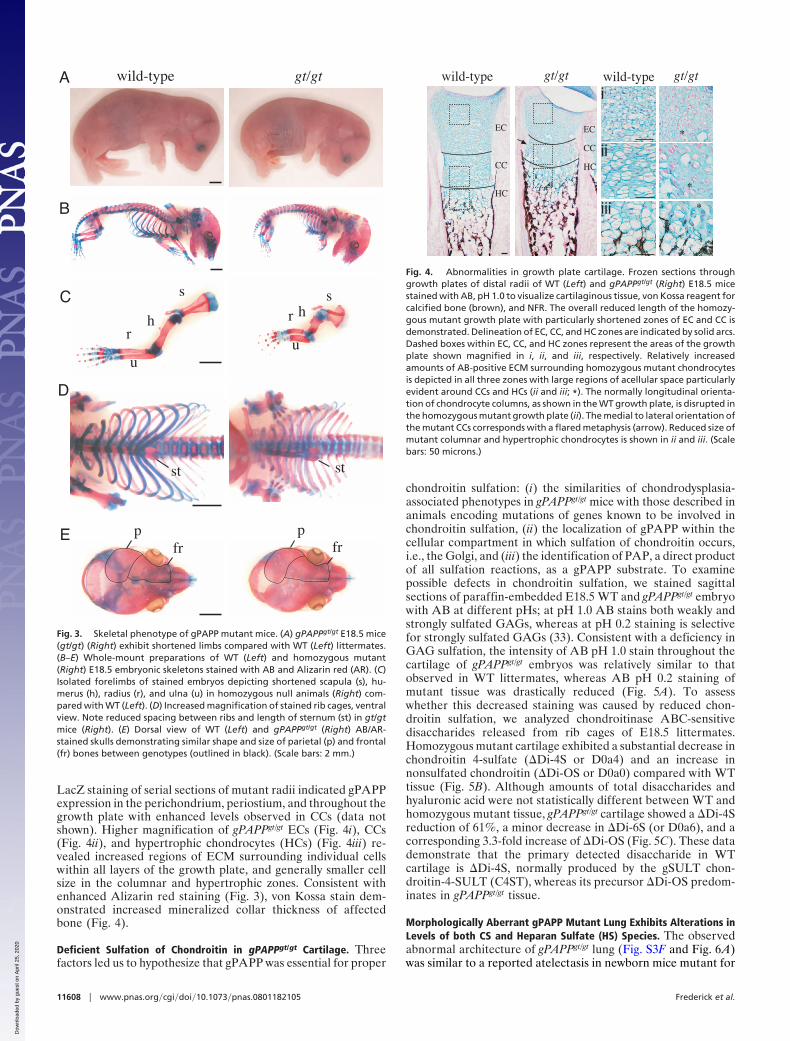
LacZ staining of serial sections of mutant radii indicated gPAPPexpression in the perichondrium, periostium, and throughout thegrowth plate with enhanced levels observed in CCs (data notshown). Higher magnification of gPAPPgt/gt ECs (Fig. 4i), CCs(Fig. 4ii), and hypertrophic chondrocytes (HCs) (Fig. 4iii) re-vealed increased regions of ECM surrounding individual cellswithin all layers of the growth plate, and generally smaller cellsize in the columnar and hypertrophic zones. Consistent withenhanced Alizarin red staining (Fig. 3), von Kossa stain dem-onstrated increased mineralized collar thickness of affectedbone (Fig. 4).
Deficient Sulfation of Chondroitin in gPAPPgt/gt Cartilage. Threefactors led us to hypothesize that gPAPP was essential for proper
chondroitin sulfation: (i) the similarities of chondrodysplasia-associated phenotypes in gPAPPgt/gt mice with those described inanimals encoding mutations of genes known to be involved inchondroitin sulfation, (ii) the localization of gPAPP within thecellular compartment in which sulfation of chondroitin occurs,i.e., the Golgi, and (iii) the identification of PAP, a direct productof all sulfation reactions, as a gPAPP substrate. To examinepossible defects in chondroitin sulfation, we stained sagittalsections of paraffin-embedded E18.5 WT and gPAPPgt/gt embryowith AB at different pHs; at pH 1.0 AB stains both weakly andstrongly sulfated GAGs, whereas at pH 0.2 staining is selectivefor strongly sulfated GAGs (33). Consistent with a deficiency inGAG sulfation, the intensity of AB pH 1.0 stain throughout thecartilage of gPAPPgt/gt embryos was relatively similar to thatobserved in WT littermates, whereas AB pH 0.2 staining ofmutant tissue was drastically reduced (Fig. 5A). To assesswhether this decreased staining was caused by reduced chon-droitin sulfation, we analyzed chondroitinase ABC-sensitivedisaccharides released from rib cages of E18.5 littermates.Homozygous mutant cartilage exhibited a substantial decrease inchondroitin 4-sulfate (�Di-4S or D0a4) and an increase innonsulfated chondroitin (�Di-OS or D0a0) compared with WTtissue (Fig. 5B). Although amounts of total disaccharides andhyaluronic acid were not statistically different between WT andhomozygous mutant tissue, gPAPPgt/gt cartilage showed a �Di-4Sreduction of 61%, a minor decrease in �Di-6S (or D0a6), and acorresponding 3.3-fold increase of �Di-OS (Fig. 5C). These datademonstrate that the primary detected disaccharide in WTcartilage is �Di-4S, normally produced by the gSULT chon-droitin-4-SULT (C4ST), whereas its precursor �Di-OS predom-inates in gPAPPgt/gt tissue.
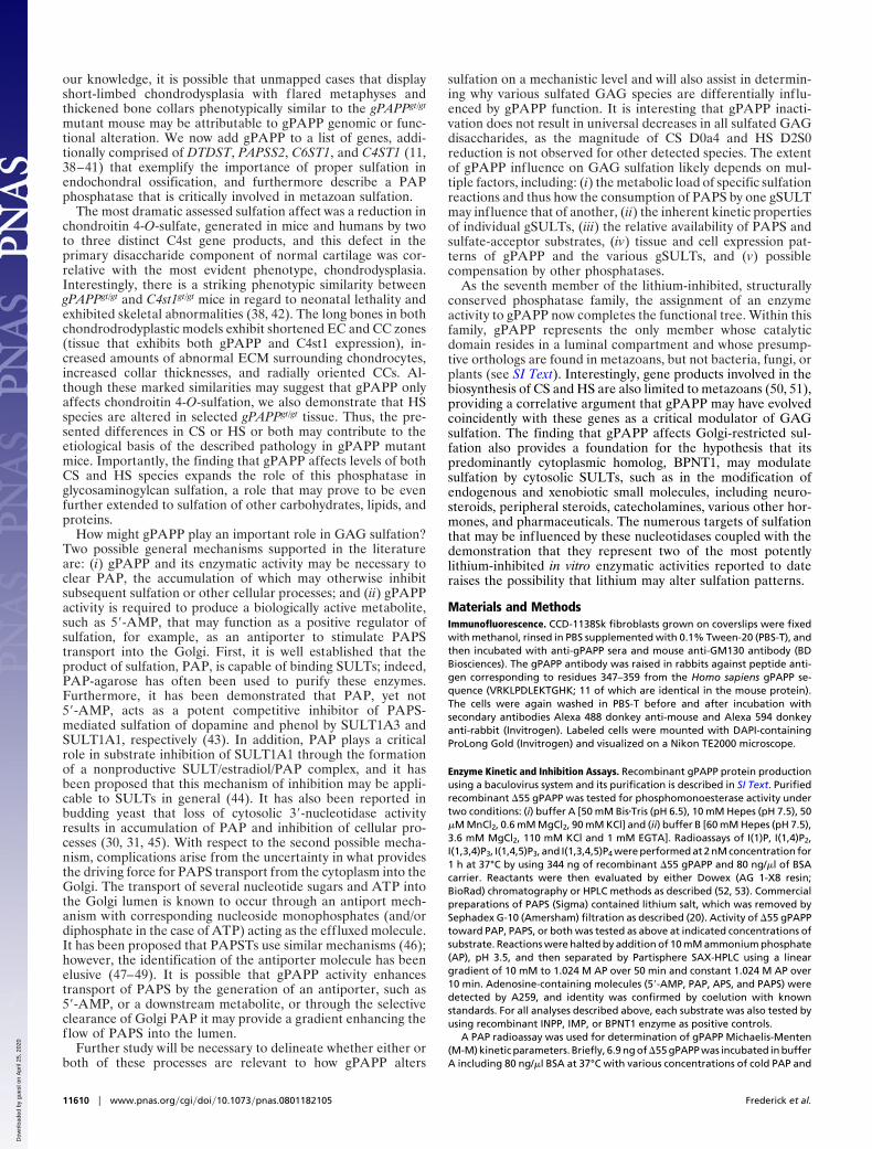
Morphologically Aberrant gPAPP Mutant Lung Exhibits Alterations inLevels of both CS and Heparan Sulfate (HS) Species. The observedabnormal architecture of gPAPPgt/gt lung (Fig. S3F and Fig. 6A)was similar to a reported atelectasis in newborn mice mutant for
hr
u
shr
u
s
st st
pfr
pfr
wild-type gt/gtA
B
C
D
E
Fig. 3. Skeletal phenotype of gPAPP mutant mice. (A) gPAPPgt/gt E18.5 mice(gt/gt) (Right) exhibit shortened limbs compared with WT (Left) littermates.(B–E) Whole-mount preparations of WT (Left) and homozygous mutant(Right) E18.5 embryonic skeletons stained with AB and Alizarin red (AR). (C)Isolated forelimbs of stained embryos depicting shortened scapula (s), hu-merus (h), radius (r), and ulna (u) in homozygous null animals (Right) com-pared with WT (Left). (D) Increased magnification of stained rib cages, ventralview. Note reduced spacing between ribs and length of sternum (st) in gt/gtmice (Right). (E) Dorsal view of WT (Left) and gPAPPgt/gt (Right) AB/AR-stained skulls demonstrating similar shape and size of parietal (p) and frontal(fr) bones between genotypes (outlined in black). (Scale bars: 2 mm.)
wild-type gt/gt
*
*
*
i
ii
iii
EC
CC
HC
EC
CC
HC
wild-type gt/gt
Fig. 4. Abnormalities in growth plate cartilage. Frozen sections throughgrowth plates of distal radii of WT (Left) and gPAPPgt/gt (Right) E18.5 micestained with AB, pH 1.0 to visualize cartilaginous tissue, von Kossa reagent forcalcified bone (brown), and NFR. The overall reduced length of the homozy-gous mutant growth plate with particularly shortened zones of EC and CC isdemonstrated. Delineation of EC, CC, and HC zones are indicated by solid arcs.Dashed boxes within EC, CC, and HC zones represent the areas of the growthplate shown magnified in i, ii, and iii, respectively. Relatively increasedamounts of AB-positive ECM surrounding homozygous mutant chondrocytesis depicted in all three zones with large regions of acellular space particularlyevident around CCs and HCs (ii and iii; *). The normally longitudinal orienta-tion of chondrocyte columns, as shown in the WT growth plate, is disrupted inthe homozygous mutant growth plate (ii). The medial to lateral orientation ofthe mutant CCs corresponds with a flared metaphysis (arrow). Reduced size ofmutant columnar and hypertrophic chondrocytes is shown in ii and iii. (Scalebars: 50 microns.)
11608 � www.pnas.org�cgi�doi�10.1073�pnas.0801182105 Frederick et al.
Dow
nloa
ded
by g
uest
on
Apr
il 25
, 202
0
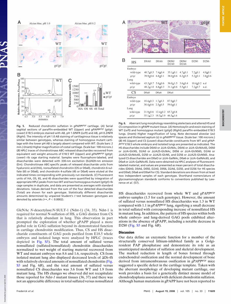
GlcNAc N-deacetylase/N-SULT-1 (Ndst-1) (34, 35). Ndst-1 isrequired for normal N-sulfation of HS, a GAG distinct from CSthat is relatively abundant in lung. This observation in partprompted the exploration of whether gPAPP played a moreglobal role in GAG sulfation beyond its demonstrated functionin cartilage chondroitin modification. Thus, CS and HS disac-charide constituents of GAG pools purified from E18.5 wholeembryos and isolated lungs were analyzed by HPLC (tracesdepicted in Fig. S5). The total amount of sulfated versusnonsulfated (sulfated/nonsulfated) chondroitin disaccharidesnormalized to wet weight of starting material recovered fromWT and mutant embryos was 8.6 and 1.6, respectively. CS fromisolated mutant lung also displayed decreased levels of �Di-4Swith relatively elevated amounts of nonsulfated chondroitin (Fig.S5 and Fig. 6B), and the total amount of sulfated versusnonsulfated CS disaccharides was 3.6 from WT and 1.9 frommutant lung. The HS changes we observed did not recapitulatethose reported for Ndst-1 mutant tissues (36, 37) and there wasnot an appreciable difference in total sulfated versus nonsulfated
HS disaccharides recovered from whole WT and gPAPPgt/gt
embryo samples (1.5 for each genotype). However, the amountof sulfated versus nonsulfated HS disaccharides was 1.3 in WTcompared with 1.1 in gPAPPgt/gt lung, signifying a small decreasein total sulfated with corresponding increase of nonsulfated HSin mutant lung. In addition, the pattern of HS species within bothwhole embryo- and lung-derived GAG pools exhibited alter-ations in the absence of gPAPP, including a notable decrease inD2S0 (Fig. S5 and Fig. 6B).
DiscussionOur data define an enzymatic function for a member of thestructurally conserved lithium-inhibited family as a Golgi-resident PAP phosphatase and demonstrate its role as anunanticipated modulator of sulfation and skeletal development.The marked reduction in length of bones formed throughendochondral ossification and the normal development of bonederived from intramembranous ossification in gPAPPgt/gt micesuggested a specific defect in the cartilage template. Taken withthe aberrant morphology of developing mutant cartilage, ourwork provides a basis for a genetically distinct mouse model ofchondrodysplasia associated with deficient chondroitin sulfation.Although human mutations in gPAPP have not been reported to
minutes
WTWT
gt/gt gt/gt
WTgt/gt
WT
gt/gt
A
BC
Fig. 5. Reduced chondroitin sulfation in gPAPPgt/gt cartilage. (A) Serialsagittal sections of paraffin-embedded WT (Upper) and gPAPPgt/gt (gt/gt;Lower) E18.5 embryos stained with AB, pH 1.0/NFR (Left) and AB, pH 0.2/NFR(Right). The intensity of pH 1.0 AB staining of cartilaginous tissue is relativelysimilar between genotypes, whereas staining of homozygous mutant carti-lage with the lower pH AB is largely absent compared with WT. (Scale bars: 2mm.) (Insets) Higher magnification of costal cartilage. (Scale bar: 100 microns.)(B) HPLC traces of chondroitinase ABC-released disaccharides recovered fromequivalent wet weight amounts of E18.5 WT (Upper) and gPAPPgt/gt (gt/gt;Lower) rib cage starting material. Samples were fluorophore-labeled, anddisaccharides were detected with 330-nm excitation (Ex)/420-nm emission(Em). Chondroitinase ABC-specific peaks of released disaccharide units fromhyaluronic acid (HA), nonsulfated chondroitin (OS or D0a0), chondrotin 6-sul-fate (6S or D0a6), and chondrotin 4-sulfate (4S or D0a4) were eluted at theindicated times corresponding with previously run standards. (C) Fluorescentunits of HA, OS, 6S, and 4S disaccharides were quantified by integration ofappropriate HPLC peaks from two WT and two homozygous mutant (gt/gt) ribcage samples in duplicate, and data are presented as averages with standarddeviations. Values derived from the sum of the four detected disaccharides(Total) are shown for each genotype. Statistically different disaccharideamounts determined by unpaired Student’s t test between genotypes aredenoted by asterisks (**, P � 0.0002).
A
B
av avss
WT gt/gt
wild-typegt/gt
Embryo
Lungwild-typegt/gt
D0A0 D0A6 D0S0/D2A0
D0S6/D2A6
D2S0 D2S6
40.7+0.5 7.4+0.4 35.1+0.1 4.7+0.1 7.2+0.1 4.9+0.139.9+0.6 6.8+0.1 38.0+0.6 6.2+0.5 5.2+0.1 3.8+0.6
43.3+0.7 5.6+0.6 36.0+1.5 3.6+0.4 10.4+0.2 n.d48.2+1.0 6.8+0.3 31.1+0.6 5.5+0.1 7.2+0.7 n.d.
wild-typegt/gt
Embryo
Lungwild-typegt/gt
D0a0 D0a6 D0a4
10.4+0.3 1.7+0.3 87.9+0.738.4+0.6 2.5+0.5 59.0+0.1
21.9+0.6 12.4+0.2 65.7+0.835.1+2.3 18.5+3.9 46.5+1.6
HS
CS
Fig. 6. Aberrant lung morphology resembling atelectasis and altered HS andCS composition in gPAPP mutant tissue. (A) Hemotoxylin and eosin staining ofWT (Left) and homozygous mutant (gt/gt) (Right) paraffin-embedded E18.5lungs. (Insets) Higher magnification of lung. Note decreased alveolar (av)spaces and thickened septum (s) in gPAPPgt/gt tissue. (Scale bar: 100 microns.)(B) HS (Upper) and CS (Lower) disaccharide constituents from WT and gPAP-Pgt/gt E18.5 whole embryos and isolated lungs are presented as indicated. TheHS disaccharides include D0A0 or �UA-GlcNAc, D0A6 or �UA-GlcNAc6S, D0S0or �UA-GlcNS, D2A0 or �UA2S-GlcNAc, D0S6 or �UA-GlcNS6S, D2A6 or�UA2S-GlcNAc6S, D2S0 or �UA2S-GlcNS, and D2S6 or �UA2S-GlcNS6S. Ana-lyzed CS disaccharides are D0aO or �UA-GalNAc, D0a6 or �UA-GalNAc6S, andD0a4 or �UA-GalNAc4S. Data were obtained via HPLC analyses of fluorescent-labeled material, and values are presented as mean percent of total disaccha-rides (D0A0, D0A6, D0S0, D2A0, D0S6, D2A6, D2S0, and D2S6 for HS speciesand D0a0, D0a6 and D0a4 for CS). Standard deviations are shown from at leasttwo independent samples of each genotype. Shorthand nomenclature ofglycosamonoglycan standards conforms to conventions published by Law-rence et al. (57).
Frederick et al. PNAS � August 19, 2008 � vol. 105 � no. 33 � 11609
BIO
CHEM
ISTR
YFE
ATU
REA
RTIC
LE
Dow
nloa
ded
by g
uest
on
Apr
il 25
, 202
0
our knowledge, it is possible that unmapped cases that displayshort-limbed chondrodysplasia with flared metaphyses andthickened bone collars phenotypically similar to the gPAPPgt/gt
mutant mouse may be attributable to gPAPP genomic or func-tional alteration. We now add gPAPP to a list of genes, addi-tionally comprised of DTDST, PAPSS2, C6ST1, and C4ST1 (11,38–41) that exemplify the importance of proper sulfation inendochondral ossification, and furthermore describe a PAPphosphatase that is critically involved in metazoan sulfation.
The most dramatic assessed sulfation affect was a reduction inchondroitin 4-O-sulfate, generated in mice and humans by twoto three distinct C4st gene products, and this defect in theprimary disaccharide component of normal cartilage was cor-relative with the most evident phenotype, chondrodysplasia.Interestingly, there is a striking phenotypic similarity betweengPAPPgt/gt and C4st1gt/gt mice in regard to neonatal lethality andexhibited skeletal abnormalities (38, 42). The long bones in bothchondrodrodyplastic models exhibit shortened EC and CC zones(tissue that exhibits both gPAPP and C4st1 expression), in-creased amounts of abnormal ECM surrounding chondrocytes,increased collar thicknesses, and radially oriented CCs. Al-though these marked similarities may suggest that gPAPP onlyaffects chondroitin 4-O-sulfation, we also demonstrate that HSspecies are altered in selected gPAPPgt/gt tissue. Thus, the pre-sented differences in CS or HS or both may contribute to theetiological basis of the described pathology in gPAPP mutantmice. Importantly, the finding that gPAPP affects levels of bothCS and HS species expands the role of this phosphatase inglycosaminogylcan sulfation, a role that may prove to be evenfurther extended to sulfation of other carbohydrates, lipids, andproteins.
How might gPAPP play an important role in GAG sulfation?Two possible general mechanisms supported in the literatureare: (i) gPAPP and its enzymatic activity may be necessary toclear PAP, the accumulation of which may otherwise inhibitsubsequent sulfation or other cellular processes; and (ii) gPAPPactivity is required to produce a biologically active metabolite,such as 5�-AMP, that may function as a positive regulator ofsulfation, for example, as an antiporter to stimulate PAPStransport into the Golgi. First, it is well established that theproduct of sulfation, PAP, is capable of binding SULTs; indeed,PAP-agarose has often been used to purify these enzymes.Furthermore, it has been demonstrated that PAP, yet not5�-AMP, acts as a potent competitive inhibitor of PAPS-mediated sulfation of dopamine and phenol by SULT1A3 andSULT1A1, respectively (43). In addition, PAP plays a criticalrole in substrate inhibition of SULT1A1 through the formationof a nonproductive SULT/estradiol/PAP complex, and it hasbeen proposed that this mechanism of inhibition may be appli-cable to SULTs in general (44). It has also been reported inbudding yeast that loss of cytosolic 3�-nucleotidase activityresults in accumulation of PAP and inhibition of cellular pro-cesses (30, 31, 45). With respect to the second possible mecha-nism, complications arise from the uncertainty in what providesthe driving force for PAPS transport from the cytoplasm into theGolgi. The transport of several nucleotide sugars and ATP intothe Golgi lumen is known to occur through an antiport mech-anism with corresponding nucleoside monophosphates (and/ordiphosphate in the case of ATP) acting as the effluxed molecule.It has been proposed that PAPSTs use similar mechanisms (46);however, the identification of the antiporter molecule has beenelusive (47–49). It is possible that gPAPP activity enhancestransport of PAPS by the generation of an antiporter, such as5�-AMP, or a downstream metabolite, or through the selectiveclearance of Golgi PAP it may provide a gradient enhancing theflow of PAPS into the lumen.
Further study will be necessary to delineate whether either orboth of these processes are relevant to how gPAPP alters
sulfation on a mechanistic level and will also assist in determin-ing why various sulfated GAG species are differentially influ-enced by gPAPP function. It is interesting that gPAPP inacti-vation does not result in universal decreases in all sulfated GAGdisaccharides, as the magnitude of CS D0a4 and HS D2S0reduction is not observed for other detected species. The extentof gPAPP influence on GAG sulfation likely depends on mul-tiple factors, including: (i) the metabolic load of specific sulfationreactions and thus how the consumption of PAPS by one gSULTmay influence that of another, (ii) the inherent kinetic propertiesof individual gSULTs, (iii) the relative availability of PAPS andsulfate-acceptor substrates, (iv) tissue and cell expression pat-terns of gPAPP and the various gSULTs, and (v) possiblecompensation by other phosphatases.
As the seventh member of the lithium-inhibited, structurallyconserved phosphatase family, the assignment of an enzymeactivity to gPAPP now completes the functional tree. Within thisfamily, gPAPP represents the only member whose catalyticdomain resides in a luminal compartment and whose presump-tive orthologs are found in metazoans, but not bacteria, fungi, orplants (see SI Text). Interestingly, gene products involved in thebiosynthesis of CS and HS are also limited to metazoans (50, 51),providing a correlative argument that gPAPP may have evolvedcoincidently with these genes as a critical modulator of GAGsulfation. The finding that gPAPP affects Golgi-restricted sul-fation also provides a foundation for the hypothesis that itspredominantly cytoplasmic homolog, BPNT1, may modulatesulfation by cytosolic SULTs, such as in the modification ofendogenous and xenobiotic small molecules, including neuro-steroids, peripheral steroids, catecholamines, various other hor-mones, and pharmaceuticals. The numerous targets of sulfationthat may be influenced by these nucleotidases coupled with thedemonstration that they represent two of the most potentlylithium-inhibited in vitro enzymatic activities reported to dateraises the possibility that lithium may alter sulfation patterns.
Materials and MethodsImmunofluorescence. CCD-1138Sk fibroblasts grown on coverslips were fixedwith methanol, rinsed in PBS supplemented with 0.1% Tween-20 (PBS-T), andthen incubated with anti-gPAPP sera and mouse anti-GM130 antibody (BDBiosciences). The gPAPP antibody was raised in rabbits against peptide anti-gen corresponding to residues 347–359 from the Homo sapiens gPAPP se-quence (VRKLPDLEKTGHK; 11 of which are identical in the mouse protein).The cells were again washed in PBS-T before and after incubation withsecondary antibodies Alexa 488 donkey anti-mouse and Alexa 594 donkeyanti-rabbit (Invitrogen). Labeled cells were mounted with DAPI-containingProLong Gold (Invitrogen) and visualized on a Nikon TE2000 microscope.
Enzyme Kinetic and Inhibition Assays. Recombinant gPAPP protein productionusing a baculovirus system and its purification is described in SI Text. Purifiedrecombinant �55 gPAPP was tested for phosphomonoesterase activity undertwo conditions: (i) buffer A [50 mM Bis�Tris (pH 6.5), 10 mM Hepes (pH 7.5), 50�M MnCl2, 0.6 mM MgCl2, 90 mM KCl] and (ii) buffer B [60 mM Hepes (pH 7.5),3.6 mM MgCl2, 110 mM KCl and 1 mM EGTA]. Radioassays of I(1)P, I(1,4)P2,I(1,3,4)P3, I(1,4,5)P3, and I(1,3,4,5)P4 were performed at 2 nM concentration for1 h at 37°C by using 344 ng of recombinant �55 gPAPP and 80 ng/�l of BSAcarrier. Reactants were then evaluated by either Dowex (AG 1-X8 resin;BioRad) chromatography or HPLC methods as described (52, 53). Commercialpreparations of PAPS (Sigma) contained lithium salt, which was removed bySephadex G-10 (Amersham) filtration as described (20). Activity of �55 gPAPPtoward PAP, PAPS, or both was tested as above at indicated concentrations ofsubstrate. Reactions were halted by addition of 10 mM ammonium phosphate(AP), pH 3.5, and then separated by Partisphere SAX-HPLC using a lineargradient of 10 mM to 1.024 M AP over 50 min and constant 1.024 M AP over10 min. Adenosine-containing molecules (5�-AMP, PAP, APS, and PAPS) weredetected by A259, and identity was confirmed by coelution with knownstandards. For all analyses described above, each substrate was also tested byusing recombinant INPP, IMP, or BPNT1 enzyme as positive controls.
A PAP radioassay was used for determination of gPAPP Michaelis-Menten(M-M) kinetic parameters. Briefly, 6.9 ng of �55 gPAPP was incubated in bufferA including 80 ng/�l BSA at 37°C with various concentrations of cold PAP and
11610 � www.pnas.org�cgi�doi�10.1073�pnas.0801182105 Frederick et al.
Dow
nloa
ded
by g
uest
on
Apr
il 25
, 202
0
10,000 dpm of 32[P]-labeled PAP that was synthesized as described (20).Reactions were terminated with formate buffer and separated by Dowexchromatography, and [32P]-5�-AMP product was quantified as described (20).The activity of full-length gPAPP on PAP was evaluated as described above,except that enzyme was preincubated with 0.25% Triton X-100 and 0.25%phosphatidylcholine on ice for 10 min, and reactions were carried out in 50mM Bis�Tris (pH 6.5), 50 �M MnCl2, 100 mM KCl, 0.1% Triton X-100, and 0.1%phosphatidylcholine. The presented Km and Vmax figures were determined byfitting data through the use of SigmaPlot into the M-M equation v � Vmax[S]/(Km�[S]) and using nonlinear regression analysis. The Ki and mode of inhibi-tion of LiCl with respect to PAP hydrolysis by both �55 and full-length gPAPPwas determined by Dixon plot analyses.
Histology and Staining. Timed-pregnant females with E18.5 litters were eu-thanized with an overdose of pentobarbital sodium (Abbott Laboratories),followed by �-galactosidase fixative [0.1% gluteraldehyde, 1.5% formalde-hyde, 5 mM EGTA (pH 8.0), 2 mM MgCl2, 100 mM sodium phosphate (pH 8.0)]transcardially perfused. Tails were removed for genotyping, and embryoswere equilibrated overnight in 30% sucrose, 0.1 M phosphate buffer, pH 7.4.Embryos and limbs frozen in Tissue-Tek OCT compound (Sakura) were sec-tioned with a Leica CM 3050S cryostat onto gelatin-coated slides. Frozenembryo and limb sections were air-dried, fixed in 0.2% gluteraldehyde/PBS,washed, and then stained at 37°C with 1 mg/ml X-Gal (Invitrogen), 5 mMK4Fe(CN)6, 5 mM K3Fe(CN)6 in 2 mM MgCl2, 0.02% Nonidet P-40, 0.01%deoxycholate, and 100 mM sodium phosphate, pH 8.0 for 24 and 44 h,respectively. Stained sections were washed with PBS and fixed in formalinovernight at 4°C, and embryo samples were counterstained with Nuclear FastRed (NFR; Vector). Serial limb sections were also stained with von Kossa, AB8GX (pH 1.0), and NFR. Briefly, sections through distal radii were fixed in 4%paraformaldehyde for 5 min, placed in 1% silver nitrate under UV light for 20min, then incubated with 5% sodium thiosulfate with intervening H20 washes.Sections were then stained in AB, pH 1.0 (1% AB in 0.1 M HCl) and counter-stained in NFR. Paraffin-embedded sections of E18.5 embryos were preparedand hematoxylin and eosin-stained by the Duke University Medical CenterImmunohistology Research Laboratory. Serial, paraffin-embedded sections ofE18.5 mice were stained with AB at different pHs essentially as described (33)and NFR-counterstained. Briefly, paraffin was removed, and sections wererehydrated and then stained for 15 min with 1% AB in either 0.1 M HCl (pH 1.0)or 10% sulfuric acid (pH 0.2). The sections stained with AB, pH 0.2 were thenrinsed in 10% sulfuric acid and blot-dried. All sections were dehydrated,cleared in Histo-Clear (National Diagnostics), and mounted with Krystalon(EMD). AB/Alizarin red staining of skeletons were performed as described (54).
Bone measurements from three skeletons were made with digital calipers (L.S.Starrett) and the averages were presented. Histological reagents were ob-tained from Sigma unless otherwise noted.
GAG Analysis. GAG isolation and analysis is described in detail in SI Text.Rib cage CS. Briefly, rib cages of E18.5 littermates were homogenized anddigested with chondroitinase ABC (Seikagaku). Released disaccharides werepurified, fluorophore-labeled with a GlycoProfile 2-AB labeling kit (Sigma),and resolved by HPLC using a Supelcosil LC-NH2 250 � 4.6-mm column andlinear gradient of sodium phosphate, pH 6.5. Peak identity was confirmed bycoelution with known standards and �Di-4S susceptibility to chondro-4-sulfatase.Whole embryo and lung GAGs. E18.5 litters were euthanized, and individualembryos were either processed whole or individual organs were isolated forhomogenization. E18.5 embryos were pulverized by using a liquid nitrogenfreezer mill, and isolated lungs were homogenized by hard-tissue generator.GAGs were purified from the homogenized tissue, then treated and analyzedessentially as described (55) with modifications as detailed in SI Text. Resultsare presented as a ratio of total sulfated disaccharides versus nonsulfateddisaccharides (sulfated/nonsulfated as well as percent of total disaccharidedetected), and the values were obtained from integrated fluorescent HPLCpeaks (as depicted in Fig. S5) normalized to wet weight of starting material.
Note. During the completion of our study we became aware of a report thatvalidated the use of alternative gene trap vectors in the generation of 60mouse lines mutant for secreted/transmembrane gene products, includingone harboring ‘‘insertion in a novel membrane protein with similarity toinositol monophosphates’’ (56). Craniofacial abnormalities and shortenedlimbs were noted in these mice generated from the aforementioned KST 245ES clone, which can now be designated as mutant gPAPP. Further study of theKST245-derived line has recently been published by Sohaskey, et al. (58).
ACKNOWLEDGMENTS. We thank B. Hudson, B. Mutamba and Dr. W. Bai fortechnical assistance; Drs. I. Esko and J. Liu, and members of J.D.Y.’s laboratory,especially S. Rozenman, for helpful discussions; Drs. J. Esko, R. Lawrence(University of California San Diego, La Jolla, CA), and C. Nicchitta (DukeUniversity Medical Center, Durham, NC) for providing reagents; and Drs. P.Majerus, S. York, and S. Kornfeld for critical reading of the manuscript andconstructive comments. This work was supported by funds from the HowardHughes Medical Institute and the National Institutes of Health R01 grantnumber HL-55672.
1. Schwartz NB, Domowicz M (2002) Chondrodysplasias due to proteoglycan defects.Glycobiology 12:57R–68R.
2. Mundlos S, Olsen BR (1997) Heritable diseases of the skeleton. Part II: Molecularinsights into skeletal development-matrix components and their homeostasis. FASEBJ 11:227–233.
3. Mundlos S, Olsen BR (1997) Heritable diseases of the skeleton. Part I: Molecular insightsinto skeletal development-transcription factors and signaling pathways. FASEB J11:125–132.
4. Knudson CB, Knudson W (2001) Cartilage proteoglycans. Semin Cell Dev Biol 12:69–78.5. Lefebvre V, Smits P (2005) Transcriptional control of chondrocyte fate and differenti-
ation. Birth Defects Res C Embryo Today 75:200–212.6. Goldring MB, Tsuchimochi K, Ijiri K (2006) The control of chondrogenesis. J Cell
Biochem 97:33–44.7. Cohen MM, Jr (2006) The new bone biology: Pathologic, molecular, and clinical
correlates. Am J Med Genet A 140:2646–2706.8. Superti-Furga A, Bonafe L, Rimoin DL (2001) Molecular-pathogenetic classification of
genetic disorders of the skeleton. Am J Med Genet 106:282–293.9. Horton WA (2003) The evolving definition of a chondrodysplasia? Pediatr Pathol Mol
Med 22:47–52.10. McLean W, Olsen BR (2001) Mouse models of abnormal skeletal development and
homeostasis. Trends Genet 17:S38–S43.11. Forlino A, et al. (2005) A diastrophic dysplasia sulfate transporter (SLC26A2) mutant
mouse: Morphological and biochemical characterization of the resulting chondrodys-plasia phenotype. Hum Mol Genet 14:859–871.
12. Kusche-Gullberg M, Kjellen L (2003) Sulfotransferases in glycosaminoglycan biosyn-thesis. Curr Opin Struct Biol 13:605–611.
13. Honke K, Taniguchi N (2002) Sulfotransferases and sulfated oligosaccharides. Med ResRev 22:637–654.
14. Strott CA (2002) Sulfonation and molecular action. Endocr Rev 23:703–732.15. Gama CI, et al. (2006) Sulfation patterns of glycosaminoglycans encode molecular
recognition and activity. Nat Chem Biol 2:467–473.16. Silbert JE, Sugumaran G (2002) Biosynthesis of chondroitin/dermatan sulfate. IUBMB
Life 54:177–186.17. Esko JD, Selleck SB (2002) Order out of chaos: Assembly of ligand binding sites in
heparan sulfate. Annu Rev Biochem 71:435–471.
18. Ramaswamy SG, Jakoby WB (1987) (2�)3�,5�-Bisphosphate nucleotidase. J Biol Chem262:10044–10047.
19. Lopez-Coronado JM, Belles JM, Lesage F, Serrano R, Rodriguez PL (1999) A novelmammalian lithium-sensitive enzyme with a dual enzymatic activity, 3�-phosphoad-enosine 5�-phosphate phosphatase, and inositol-polyphosphate 1-phosphatase. J BiolChem 274:16034–16039.
20. Spiegelberg BD, Xiong JP, Smith JJ, Gu RF, York JD (1999) Cloning and characterizationof a mammalian lithium-sensitive bisphosphate 3� nucleotidase inhibited by inositol1,4-bisphosphate. J Biol Chem 274:13619–13628.
21. York JD, Ponder JW, Majerus PW (1995) Definition of a metal-dependent/Li(�)-inhibited phosphomonoesterase protein family based upon a conserved three-dimensional core structure. Proc Natl Acad Sci USA 92:5149–5153.
22. Gould TD, Quiroz JA, Singh J, Zarate CA, Manji HK (2004) Emerging experimentaltherapeutics for bipolar disorder: Insights from the molecular and cellular actions ofcurrent mood stabilizers. Mol Psychiatry 9:734–755.
23. Phiel CJ, Klein PS (2001) Molecular targets of lithium action. Annu Rev PharmacolToxicol 41:789–813.
24. Harwood AJ (2005) Lithium and bipolar mood disorder: The inositol-depletion hypoth-esis revisited. Mol Psychiatry 10:117–126.
25. Allison JH, Boshans RL, Hallcher LM, Packman PM, Sherman WR (1980) The effects oflithium on myo-inositol levels in layers of frontal cerebral cortex, in cerebellum, and incorpus callosum of the rat. J Neurochem 34:456–458.
26. Hallcher LM, Sherman WR (1980) The effects of lithium ion and other agents on theactivity of myo-inositol-1-phosphatase from bovine brain. J Biol Chem 255:10896–10901.
27. Inhorn RC, Majerus PW (1987) Inositol polyphosphate 1-phosphatase from calf brain.Purification and inhibition by Li�, Ca2�, and Mn2�. J Biol Chem 262:15946–15952.
28. Acharya JK, Labarca P, Delgado R, Jalink K, Zuker CS (1998) Synaptic defects andcompensatory regulation of inositol metabolism in inositol polyphosphate 1-phospha-tase mutants. Neuron 20:1219–1229.
29. Glaser HU, et al. (1993) Salt tolerance and methionine biosynthesis in Saccharomycescerevisiae involve a putative phosphatase gene. EMBO J 12:3105–3110.
30. Dichtl B, Stevens A, Tollervey D (1997) Lithium toxicity in yeast is due to the inhibitionof RNA processing enzymes. EMBO J 16:7184–7195.
Frederick et al. PNAS � August 19, 2008 � vol. 105 � no. 33 � 11611
BIO
CHEM
ISTR
YFE
ATU
REA
RTIC
LE
Dow
nloa
ded
by g
uest
on
Apr
il 25
, 202
0
31. Spiegelberg BD, Dela Cruz J, Law TH, York JD (2005) Alteration of lithium pharmacol-ogy through manipulation of phosphoadenosine phosphate metabolism. J Biol Chem280:5400–5405.
32. Leighton PA, et al. (2001) Defining brain wiring patterns and mechanisms throughgene trapping in mice. Nature 410:174–179.
33. Bancroft JD, Stevens A (1996) in Theory and Practice of Histological Techniques(Churchill Livingstone, New York), pp 189–191.
34. Fan G, et al. (2000) Targeted disruption of NDST-1 gene leads to pulmonary hypoplasiaand neonatal respiratory distress in mice. FEBS Lett 467:7–11.
35. Ringvall M, et al. (2000) Defective heparan sulfate biosynthesis and neonatal lethalityin mice lacking N-deacetylase/N-sulfotransferase-1. J Biol Chem 275:25926–25930.
36. Ledin J, et al. (2004) Heparan sulfate structure in mice with genetically modifiedheparan sulfate production. J Biol Chem 279:42732–42741.
37. Grobe K, et al. (2005) Cerebral hypoplasia and craniofacial defects in mice lackingheparan sulfate Ndst1 gene function. Development 132:3777–3786.
38. Kluppel M, Wight TN, Chan C, Hinek A, Wrana JL (2005) Maintenance of chondroitinsulfation balance by chondroitin-4-sulfotransferase 1 is required for chondrocytedevelopment and growth factor signaling during cartilage morphogenesis. Develop-ment 132:3989–4003.
39. Thiele H, et al. (2004) Loss of chondroitin 6-O-sulfotransferase-1 function results insevere human chondrodysplasia with progressive spinal involvement. Proc Natl AcadSci USA 101:10155–10160.
40. ul Haque MF, et al. (1998) Mutations in orthologous genes in human spondyloepime-taphyseal dysplasia and the brachymorphic mouse. Nat Genet 20:157–162.
41. Kurima K, et al. (1998) A member of a family of sulfate-activating enzymes causesmurine brachymorphism. Proc Natl Acad Sci USA 95:8681–8685.
42. Kluppel M, Vallis KA, Wrana JL (2002) A high-throughput induction gene trap ap-proach defines C4ST as a target of BMP signaling. Mech Dev 118:77–89.
43. Rens-Domiano SS, Roth JA (1987) Inhibition of M and P phenol sulfotransferase byanalogues of 3�-phosphoadenosine-5�-phosphosulfate. J Neurochem 48:1411–1415.
44. Gamage N, et al. (2006) Human sulfotransferases and their role in chemical metabo-lism. Toxicol Sci 90:5–22.
45. Murguia JR, Belles JM, Serrano R (1995) A salt-sensitive 3�(2�),5�-bisphosphate nucle-otidase involved in sulfate activation. Science 267:232–234.
46. Hirschberg CB, Robbins PW, Abeijon C (1998) Transporters of nucleotide sugars, ATP,and nucleotide sulfate in the endoplasmic reticulum and Golgi apparatus. Annu RevBiochem 67:49–69.
47. Mandon EC, Milla ME, Kempner E, Hirschberg CB (1994) Purification of the Golgiadenosine 3�-phosphate 5�-phosphosulfate transporter, a homodimer within themembrane. Proc Natl Acad Sci USA 91:10707–10711.
48. Ozeran JD, Westley J, Schwartz NB (1996) Kinetics of PAPS translocase: Evidence for anantiport mechanism. Biochemistry 35:3685–3694.
49. Capasso JM, Hirschberg CB (1984) Mechanisms of glycosylation and sulfation in theGolgi apparatus: Evidence for nucleotide sugar/nucleoside monophosphate and nu-cleotide sulfate/nucleoside monophosphate antiports in the Golgi apparatus mem-brane. Proc Natl Acad Sci USA 81:7051–7055.
50. Perrimon N, Bernfield M (2001) Cellular functions of proteoglycans: An overview.Semin Cell Dev Biol 12:65–67.
51. Bishop JR, Gagneux P (2007) Evolution of carbohydrate antigens–microbial forcesshaping host glycomes? Glycobiology 17:23R–34R.
52. Frederick JP, et al. (2005) An essential role for an inositol polyphosphate multikinase,Ipk2, in mouse embryogenesis and second messenger production. Proc Natl Acad SciUSA 102:8454–8459.
53. Inhorn RC, Majerus PW (1988) Properties of inositol polyphosphate 1-phosphatase.J Biol Chem 263:14559–14565.
54. Scheijen B, Bronk M, van der Meer T, Bernards R (2003) Constitutive E2F1 overexpres-sion delays endochondral bone formation by inhibiting chondrocyte differentiation.Mol Cell Biol 23:3656–3668.
55. MacArthur JM, et al. (2007) Liver heparan sulfate proteoglycans mediate clearance oftriglyceride-rich lipoproteins independently of LDL receptor family members. J ClinInvest 117:153–164.
56. Mitchell KJ, et al. (2001) Functional analysis of secreted and transmembrane proteinscritical to mouse development. Nat Genet 28:241–249.
57. Lawrence R, Lu H, Rosenberg RD, Esko JD, Zhang L (2008) Disaccharide structure codefor the easy representation of constituent oligosaccharides from glycosaminoglycans.Nat Methods 5:291–292.
58. Sohaskey ML, Yu J, Diaz MA, Plaas AH, Harland RM (2008) JAWS coordinates chon-drogenesis and synovial joint positioning. Development 135:2215–2220.
11612 � www.pnas.org�cgi�doi�10.1073�pnas.0801182105 Frederick et al.
Dow
nloa
ded
by g
uest
on
Apr
il 25
, 202
0


![A Golgi-Released Subpopulation of the Trans-Golgi · A Golgi-Released Subpopulation of the Trans-Golgi Network Mediates Protein Secretion in Arabidopsis1[OPEN] Tomohiro Uemura,a,b,2,3,4](https://static.fdocuments.in/doc/165x107/5eda9f5a09f66a09130ba5a1/a-golgi-released-subpopulation-of-the-trans-golgi-a-golgi-released-subpopulation.jpg)