A revision of the Devonian Malvinokaffric dalmanitid ... · Museo de Ciencias Naturales,...
21
Palaeontologia Electronica http://palaeo-electronica.org PE Article Number: 15.1.11A Copyright: Palaeontological Association March 2012 Submission: 18 March 2011. Acceptance: 6 February 2012 Rustán, Juan J. and Vaccari, N. Emilio. 2012. A revision of the Devonian Malvinokaffric dalmanitid trilobite Dalmanitoides Delo, 1935, on the basis of new data from Argentina. Palaeontologia Electronica Vol. 15, Issue 1;11A,21p; palaeo-electronica.org/content/2012-issue-1-articles/194-the-trilobite-dalmanitoides A revision of the Devonian Malvinokaffric dalmanitid trilobite Dalmanitoides Delo, 1935, on the basis of new data from Argentina Juan J. Rustán and N. Emilio Vaccari ABSTRACT In light of new information on holotypes and additional material from the Lower Devonian type areas from Argentina, the dalmanitid trilobite Dalmanitoides Delo, 1935, is rediagnosed and considered a dalmanitine rather than a synphoriine. Comparisons suggest that Gamonedaspis Braniša and Vaněk, 1973, is a junior synonym of Dalman- itoides, so that this Early-Middle Devonian genus includes at least five species: four formally named from South America, D. drevermanni (Delo, 1935), D. boehmi (Knod, 1908), D. scutata (Braniša and Vaněk, 1973), and D. accola (Clarke, 1913), together with a species from South Africa herein treated in open nomenclature (D. sp. A). Although the number of Dalmanitoides species suggests a diversification of cosmopol- itan dalmanitines already present in Malvinokaffric basins, a close relationship with the boreal and slightly older Roncellia Lespérance and Bourque, 1971, would suggest migration from the Eastern Americas Paleobiogeographical Realm during the Early Devonian as origin of the clade, which is in accordance with paleobiogeographic pat- terns recognized on the basis of evidence from synphoriine distributions. Juan José Rustán. CIPAL - CICTERRA (Facultad de Ciencias Exactas Físicas y Naturales, Universidad Nacional de Córdoba - CONICET), Av. Vélez Sársfield 299, 5000, Córdoba, Argentina and Museo de Ciencias Naturales, Universidad Nacional de La Rioja, Av. René Favaloro s/n 5300- La Rioja, Argentina, [email protected] N. Emilio Vaccari. CIPAL - CICTERRA (Facultad de Ciencias Exactas Físicas y Naturales, Universidad Nacional de Córdoba - CONICET), Av. Vélez Sársfield 299, 5000, Córdoba, Argentina and Museo de Ciencias Naturales, Universidad Nacional de La Rioja, Av. René Favaloro s/n 5300- La Rioja, Argentina, [email protected] KEYWORDS: dalmanitid trilobites; Devonian; Argentina; Malvinokaffric; Dalmanitoides; Gamonedaspis; systematics; paleobiogeography
Transcript of A revision of the Devonian Malvinokaffric dalmanitid ... · Museo de Ciencias Naturales,...

Palaeontologia Electronica http://palaeo-electronica.org
A revision of the Devonian Malvinokaffric dalmanitid trilobite Dalmanitoides Delo, 1935,
on the basis of new data from Argentina
Juan J. Rustán and N. Emilio Vaccari
ABSTRACT
In light of new information on holotypes and additional material from the LowerDevonian type areas from Argentina, the dalmanitid trilobite Dalmanitoides Delo, 1935,is rediagnosed and considered a dalmanitine rather than a synphoriine. Comparisonssuggest that Gamonedaspis Braniša and Vaněk, 1973, is a junior synonym of Dalman-itoides, so that this Early-Middle Devonian genus includes at least five species: fourformally named from South America, D. drevermanni (Delo, 1935), D. boehmi (Knod,1908), D. scutata (Braniša and Vaněk, 1973), and D. accola (Clarke, 1913), togetherwith a species from South Africa herein treated in open nomenclature (D. sp. A).Although the number of Dalmanitoides species suggests a diversification of cosmopol-itan dalmanitines already present in Malvinokaffric basins, a close relationship with theboreal and slightly older Roncellia Lespérance and Bourque, 1971, would suggestmigration from the Eastern Americas Paleobiogeographical Realm during the EarlyDevonian as origin of the clade, which is in accordance with paleobiogeographic pat-terns recognized on the basis of evidence from synphoriine distributions.
Juan José Rustán. CIPAL - CICTERRA (Facultad de Ciencias Exactas Físicas y Naturales, Universidad Nacional de Córdoba - CONICET), Av. Vélez Sársfield 299, 5000, Córdoba, Argentina and Museo de Ciencias Naturales, Universidad Nacional de La Rioja, Av. René Favaloro s/n 5300- La Rioja, Argentina, [email protected] N. Emilio Vaccari. CIPAL - CICTERRA (Facultad de Ciencias Exactas Físicas y Naturales, Universidad Nacional de Córdoba - CONICET), Av. Vélez Sársfield 299, 5000, Córdoba, Argentina and Museo de Ciencias Naturales, Universidad Nacional de La Rioja, Av. René Favaloro s/n 5300- La Rioja, Argentina, [email protected]
KEYWORDS: dalmanitid trilobites; Devonian; Argentina; Malvinokaffric; Dalmanitoides; Gamonedaspis;systematics; paleobiogeography
PE Article Number: 15.1.11ACopyright: Palaeontological Association March 2012Submission: 18 March 2011. Acceptance: 6 February 2012
Rustán, Juan J. and Vaccari, N. Emilio. 2012. A revision of the Devonian Malvinokaffric dalmanitid trilobite Dalmanitoides Delo, 1935, on the basis of new data from Argentina. Palaeontologia Electronica Vol. 15, Issue 1;11A,21p; palaeo-electronica.org/content/2012-issue-1-articles/194-the-trilobite-dalmanitoides

Rustán & VACCARI: THE TRILOBITE DALMANITOIDES
INTRODUCTION
Pioneering geological and paleontological sur-veys were carried out in western Argentina duringthe 19th and early 20th centuries. As a result ofthese works, two new dalmanitid trilobite specieswere recognized in the Lower Devonian sedimen-tary succession known as the Talacasto Formation(Figure 1) that crops out a few kilometers to thesouthwest of Jáchal, San Juan Province. Subse-quently cited as “Cerro del Agua Negra,” “Agua deFelipe,” or “west of Jáchal Valley,” the present-dayname for these localities corresponds to the neigh-borhood of the Loma de los Piojos locality (Figure1).
Dalmanitoides drevermanni (Thomas, 1906),was erected on the basis of a single specimencomposed of a cephalon, pygidium and a few frag-mentary thoracic segments associated in a singlecarbonate nodule. This holotype (housed in Ger-many) is from an undetermined stratigraphic leveland was poorly illustrated. No more specimens
were collected, nor were further details on itsoccurrence provided, except in a recent brief treat-ment of the type specimen by Holloway and Car-valho (2009).
Dalmanitoides boehmi (Knod, 1908) is basedon a nearly complete and outstretched articulatedspecimen preserved in a sandstone slab collectedoriginally by Hauthal. This specimen was neverillustrated photographically and until now, it lackeda diagnosis, detailed description, precise strati-graphic occurrence data, and repository informa-tion. Although this taxon was subsequently citedfrom Bolivia (Kozłowski, 1923) and South Africa(Cooper, 1982), its original provenance from west-ern Argentina was overlooked (Cooper, 1982,p.61).
Apart from discussions on Gamonedaspis(Edgecombe, 1993), both species have been ofinterest in discussing several other closely relateddalmanitid taxa such as Amazonaspis (Carvalhoand Fonseca, 2007) and Fenestraspis (Hollowayand Carvalho, 2009). However, the lack of suffi-
Pampa deUmango
1,5 km
Jáchal
Route 150
Jáchal River
HornoEl Refugio
Loma deLos Piojos
La Usina
N
N
Cerro delFuerte
Niquivil
Tucunuco
Talacasto
Jáchal River
San Juan River
La Chilca Creek
La Cortadera Creek
Ullum
Tala
cast
o R
ange
TalacastoCreek
Jáchal
SAN JUAN
69° W
31° S
Huaco
150
436
40
149
149
12
40
40 20 km
Silurian-Devonianoutcrops
MENDOZAMENDOZA
SAN JUANSAN JUAN
LA RIOJALA RIOJA
CH
ILE
50 km
Iglesia
Las Flores
Pismanta
Rodeo
1000 km
ARG
ENTI
NA
ARG
ENTI
NA
Fosiliferous locality
Fm. La Chilca (Silurian)Fm. Los Espejos (Silurian)Fm. Talacasto (L. Devonian)
Fm. Guandacol (Pennsyl.)Fm. L. de los Piojos (Mississip.)
Unnamed unitFm. Punta Negra (M. Devonian)Fm. Tupe (Pennsyl-Perm)
References
FIGURE 1. Left: location map of the Central Argentine Precordillera basin, San Juan Province, western Argentina,showing Silurian-Devonian outcrops and fossiliferous localities. Right: the type area of Dalmanitoides drevermanniand D. boehmi at the Loma de los Piojos locality, to the southwest of Jáchal, shown in more detail.
2

PALAEO-ELECTRONICA.ORG
cient material, revised diagnosis, and exhaustivedescriptions, has precluded a reappraisal of theirsystematic, phylogenetic, and paleobiogeographicsignificance.
In this contribution, we report several addi-tional specimens of both taxa coming from Lomade los Piojos and from some other localities of thePrecordillera Basin of San Juan Province; as wellas repository information of the holotype of D. boe-hmi. On the basis of this new material, with strati-graphic and geographic data, a completedescription is provided for each species, and thediagnoses are emended to allow better systematiccomparisons and to provide new insights on thebiostratigraphic rank of the taxa. The new informa-tion sheds light on the systematic, phylogenetic,and paleobiogeographic proposals discussed forrelated Malvinokaffric Devonian trilobites.
GEOLOGICAL SETTING
Aspects of the geologic setting relevant herehave been adequately treated in several previousworks (Baldis, 1975; Astini, 1991; Herrera, 1993,1995a, 1995b; Edgecombe et al., 1994; Vaccari etal., 1994 ; Waisfeld et al., 1994; Bustos, 1996; Her-rera and Bustos, 2001; Rustán and Vaccari, 2010).Fossils studied come from the Talacasto Formation(Padula et al., 1967), which is extensively exposedin the Central Argentine Precordillera of San JuanProvince, western Argentina (Figure 1). This LowerDevonian unit is composed of a marine successionof intensely bioturbated greenish-gray mudstoneswith intercalated beds of sandstone. It starts withdark muddy levels basally, passing upwards intosandy levels with fossiliferous nodules (Figure 2).The Talacasto Formation reaches a maximumthickness of more than 1000 m in the northern partof the basin. It represents a muddy shelf deposi-tional system developed during a highstand, andoverlies the (mainly) Silurian shelf sequence of theLos Espejos Formation (Astini, 1991). The strati-graphic record of the Silurian-Devonian boundaryin Central Precordillera is poor due to the presenceof a discontinuity between Los Espejos and Talac-asto Formations. The turbiditic system of the upperLower-lower Upper Devonian Punta Negra Forma-tion (Bracaccini, 1949) overlies the Talacasto For-mation (Bustos, 1996; Bustos and Astini, 1997).According to the studies of the brachiopod fauna(Herrera, 1993, Racheboeuf and Herrera, 1994,Herrera and Bustos, 2001) the Talacasto Forma-tion spans the early Lochkovian-Emsian. The topof the unit is considered diachronous, with late Pra-gian to earliest Emsian levels recorded in the
Lithologic referencesShales and mudstones
Siltstones and fine sandstones
Wackes and siltstones
Coquinas
Guide horizon of Keidel (1921)
Level of typeD. drevermanni ’s
Sandstones
Fossiliferous nodules
Dalman
itoides
dreverman
ni
Dalman
itoides
boeh
mi
100 m
Low
er D
evon
ian
Tala
cast
o Fo
rmat
ion
Los E
spej
os F
m.
PNe
gra
Fm.
Mid
dle
Dev
onia
nLo
chko
vian
Emsia
nPr
agia
nU
pper
Silu
rian
?
?
FIGURE 2. Stratigraphic column of the Talacasto For-mation at Loma de los Piojos section, where it is mostdeveloped. Column shows the stratigraphic position ofthe studied Dalmanitoides species in Devonian sedi-mentary succession. Dashed lines represent low abun-dance, complete lines correspond to most fossiliferouslevels. Star shows probable location of type levels ofDalmanitoides drevermanni.
3

Rustán & VACCARI: THE TRILOBITE DALMANITOIDES
southern sections and younger late Emsian levelsfound in the northernmost sections. This can berecognized in the field, thanks to a distinctive latestPragian to earliest Emsian stratigraphic lightmarker horizon, observed initially by Keidel (1921)and then by Astini (1991), which is continuous formore than 100 km, from the Talacasto Creek (Her-rera and Bustos, 2001, p. 369) to the Loma de losPiojos section to the north. The strata overlying thisguide horizon, which are mainly recorded in thethicker northernmost sections of the basin, areoverall considered of Emsian age.
MATERIALS
Devonian materials studied herein derive fromclassical sections of the Talacasto Formation, inSan Juan Province, as follows (Figure 1): Talac-asto Creek, the type section, that runs in an E-SWtrend 60 km N-NW of San Juan city; La ChilcaCreek section, located approximately 16 km to thewest of Tucunuco town; Loma de los Piojos sec-tion, located 7 km to the Southwest of Jáchal city(about 170 km N-NW of San Juan city); and LaCortadera Creek, located 20 km to the Southwestof Talacasto, about 50 km N-NW of San Juan city.
Illustrated specimens are housed in the pale-ontological repository of the Centro de Investiga-ciones Paleobiológicas (CIPAL), numbered withthe prefix CEGH-UNC (Cátedra de Estratigrafía yGeología Histórica-Universidad Nacional de Cór-doba), and in the collection of the Museo de Pale-ontología, numbered with the prefix CORD-PZ(Córdoba- Paleozoología) both belonging to theUniversidad Nacional de Córdoba, Córdoba,Argentina.
Specimens of D. accola, were donated by J.C.White in 1906 to the New York State Museum,Albany, USA, housed there with the prefix NYSM.
The holotype of D. drevermanni is housed inGöttingen, Germany (GeowissenschaftlichesZentrum der Universität Göttingen, under reposi-tory number: GZG 15239).
The holotype of D. boehmi is housed inFreiburg, Germany (Institut für Geowissen-schaften -Geologie, Albert-Ludwigs-UniversitätFreiburg, under repository number: 2778).
SYSTEMATIC PALEONTOLOGY
Superfamily DALMANITOIDEA Vogdes, 1890Family DALMANITIDAE Vogdes, 1890
Subfamily DALMANITINAE Vogdes, 1890Genus Dalmanitoides Delo, 1935
Type species. Dalmanites drevermanni Thomas,1906
Emended diagnosis of the genus. Dalmanitinewith short (sag., exsag.) anterior cephalic border,with short (sag.) and broad (tr.) anterior process;frontal glabellar lobe approximately rhombic in out-line, bearing a delicate and narrow (tr.) depressedband of subtly more effaced ornamentation posteri-orly located (sag.); glabella coarsely tuberculated;S3 broadening and shallowing at junction with axialfurrow and anterior part of eye; S2 more convexforwards and apodemal adaxially, overall postero-laterally oriented and slightly concave forwardsadaxially; S1 gently concave forwards, slightly pos-terolaterally directed and deepest adaxially; SOgently concave forwards, slightly posterolaterallydirected; epiborder furrow on the lateral bordercontinued along the external (lower) side of thegenal spine; long and flattened genal spine; trendto develop some more swollen pygidial axial ringswhich variably exhibit either paired strong infla-tions, coarse tubercules or spines; delicate andnarrow (tr.) sagittal band of effaced segmentationin the last several pygidial axial rings, progressivelybroader (tr.) backwards; pygidial pleural ribs strongand continuous, tending to be sinuous and todevelop expansions and constrictions, andabruptly deflected backwards distally; posteriorpygidial pleural bands bearing bulbous thickeningsirregularly, which variably develop coarse tubercu-les or spines, the bases of which invade the follow-ing anterior pleural bands backwards; pygidial axismerging with a broad postaxial ridge passing intoan upturned mucro.
Species included. D. drevermanni (Thomas,1906), D. boehmi (Knod, 1908), D. accola (Clarke,1913), D. scutata (Braniša and Vaněk, 1973), D. spA (from South Africa, after Cooper, 1982).
Discussion. Gamonedaspis scutata Braniša andVaněk, 1973, from the Pragian-Emsian of theGamoneda Formation, Bolivia, is the type speciesof the genus Gamonedaspis. According to illustra-tions by Edgecombe (1993), this species shareswith Dalmanitoides all of characters mentioned inthe emended diagnosis of this latter genus above.Therefore we interpret Gamonedaspis as a juniorsynonym of the genus Dalmanitoides. The diagno-sis of Dalmanitoides put forward by Delo (1935, p.413) corresponds mainly to the type species D.drevermanni, so that it was extended to includeGamonedaspis species.
4

PALAEO-ELECTRONICA.ORG
This diagnosis comprises a unique charactercombination rather than an exhaustive list ofgeneric apomorphies.
Species of Gamonedaspis were interpreted asdalmanitines by Edgecombe (1993), an opinionwith which we agree. Meanwhile D. drevermanni,was originally considered a synphoriine (Delo,1935), but was excluded from this subfamily byLespérance and Bourque (1971, p. 186), althoughit was again regarded as a synphoriine by Hollo-way and Carvalho (2009, p. 940).
The recognition and definition of subfamilieswithin Dalmanitidae has been long discussed, andbrief reviews of the literature (focused on Malvi-nokaffric dalmanitids), can be found in Carvalhoand Fonseca (2007) and Holloway and Carvalho(2009). Herein, we follow considerations given byCampbell (1977) and Holloway (1981), togetherwith additional taxonomic criteria drawn by Hollo-way and Carvalho (2009).
Besides previously available illustrations of D.drevermanni provided by Thomas (1906, plate 11,figure 1, 1a, 2, 2a, 3), and drawings based on themby Delo, 1935 (figures 28–29); systematic observa-tions herein are based on new material collected inthe type locality, together with high quality reillus-trations of the holotype by Holloway and Carvalho(2009, figure 3 A-G).
Dalmanitoides drevermanni is now interpretedas a dalmanitine, based on the distance betweenS1 and S2 apodemes being less than 1.5 times thedistance between SO and S1 apodemes; apode-mal submesial part of S2 not equidimensional buttransversely elongated; lateral border relativelybroad (tr.) and bearing an epiborder furrow, nearlyflat-topped and meeting the doublure in a sharpangle (Figure 3.10); dominating thoracic pleuralfurrow continued as a postfacetal furrow (Figure4.3); pygidial pleural furrows tending to be lanceo-late (Figure 5.4, 5.9) and gently asymmetrical incross-section, with the posterior side more weaklyinclined (Figure 5.15) (particularly when observedon internal moulds); posterior pleural bands subtlyweaker than the anterior ones, fading towards thelateral border (Figure 5.4); and long and flattenedgenal spine exhibiting a continuation of the epibor-der furrow (Figure 6.11).
Contrasting opinions on the subfamilial statusof this species may be due to intraspecific variabil-ity together with a slightly different morphologicalexpression of anatomical structures between inter-nal and external moulds, observed for examplebetween the exfoliated left side and the well-pre-served right side of the holotype pygidium illus-
trated by Holloway and Carvalho (2009, figure 3G); as well as in Figure 5 herein.
Dalmanitoides (specially D. drevermanni),closely resembles Fenestraspis amauta Branišaand Vaněk, 1973, as noted by Holloway and Car-valho (2009). Shared features include: median pro-cesses anteriorly; a short (sag., exsag.) cephalicborder with small crenulations laterally; the patternof coarse tubercles on the glabella; coarse tuber-cles or small spines on the thoracic axial rings; aconvex projection of the most adaxial part of L2invading forwards the apodemal area of S2; anelliptical expansion abaxially on S3 at the junctionwith the axial furrow and the anteriormost extremityof the palpebral lobe, accompanied by an abruptshallowing; an effaced sagittal band of pygidialaxial rings progressively broader (tr.) backwards;an upturned terminus; a broad postaxial ridge; anda similar distribution pattern of paired spines onsome more inflated pygidial axial rings.
However, the intersegmental fenestrae, verytall eyes with a very different lens formula, a con-spicuous palpebral rim, morphology and arrange-ment of glabellar lobes and furrows, almost straightmorphology of pygidial pleural ribs, conspicuousindentations of the apodemal pits exsagitally alongthe pygidial axis, among many other differenceswith Dalmanitoides cited by Holloway and Carvalho(2009, p. 939-940), justify the generic separation ofFenestraspis.
So many similarities suggest a sister taxonrelationship rather than a convergence betweenDalmanitoides and Fenestraspis. Nevertheless,since Dalmanitoides (including Gamonedaspis) isreliably considered a dalmanitine, whereas Fenes-traspis has most recently been interpreted as asynphoriine, our reasoning presents a major sys-tematic problem: the subfamilial status of Fenes-traspis. Certainly, Fenestraspis exhibitssynphoriine characters i.e.: distance between S1and S2 apodemes more than 1.5 times the dis-tance between occipital and S1 apodemes; genalspine lacking a longitudinal furrow close to its loweredge, pleural bands of equal strength, the posteriorone diminishing in height towards the border, andsteep sided and symmetrical pygidial pleural furrowin cross section (Holloway and Carvalho, 2009).
These observations challenge the distinctive-ness of subfamilies within dalmanitids, resurrectingan old and classic discussion (see Delo, 1935; Les-pérance and Bourque, 1971; Lespérance, 1975;Campbell, 1977; Holloway, 1981; Carvalho andFonseca, 2007; Holloway and Carvalho, 2009).Unraveling this major taxonomic issue requires an
5

Rustán & VACCARI: THE TRILOBITE DALMANITOIDES
6
FIGURE 3. Cephala of Dalmanitoides drevermanni (Thomas, 1906). 1, 4, 6-7, 10-11 specimen CEGH-UNC 10499,partial cephalon: 1, internal mould dorsal view, 4, idem lateral view, 7, idem anterolateral dorsal view, 10, idem frontalview, 6, latex mould, dorsal view, arrows point to spiny processes on anterior border, 11, idem frontal view. 2-3, 5, 8,12 specimen CORD-PZ 8615, partial cephalon: 2, internal mould, dorsal view, 5, idem lateral view, 8, idem anterolat-eral dorsal view, 12, detail of the anterior doublure and anterior margin, ventral view, 3, latex mould, dorsal view. 9specimen CEGH-UNC 24304, partial cephalon, internal mould, dorsal view, arrow shows a spine on posterior border.Loma de los Piojos Section, Talacasto Formation, type early Emsian levels, San Juan Province, Argentina. Scale barequals 5 mm.

PALAEO-ELECTRONICA.ORG
exhaustive revision of dalmanitids, which is beyondthe aim of the present contribution, but whichshould be explored in future works.
Dalmanitoides exhibits a high intraspecificvariability, and the overlapping wide range of mor-phologies complicate the recognition of species.Hence, most useful characters separating speciesof Dalmanitoides include: morphology of anteriorcephalic margin, ornamentation of the occipital ringand posterior border, the proportional size andangle degree of the upturned mucro, and, althoughless clearly, pygidial ornamentation.
Dalmanitoides is considered to be closelyrelated to Roncellia Lespérance and Bourque,1971, from the Eastern Americas Paleobiogeo-graphic Realm, mainly on the basis of pygidialcharacters, which was discussed at length byEdgecombe (1993).
Dalmanitoides drevermanni (Thomas, 1906)Figures 3-6
Material. Loma de los Piojos: GZG 15239, CORD-PZ 8098, 8608, 8610, 8615, CEGH-UNC 10499-10501, 10503, 10504, 12738, 24300-24310,24312-24314, 24316, Cerro La Chilca section24318-24329 (tentatively).
Emended diagnosis. Dalmanitoides with pairedanterior spiny projections medially and little spinycrenulations anterolaterally on anterior border,stout median spine on occipital ring and each tho-racic axial ring with a couple of additional spineslocated laterally; small spines on the posterior bor-der; glabella variably exhibiting paired large tuber-cles or spines on its median part; 16-18 pygidialaxial rings bearing paired spines most frequentlyon strongly swollen rings 1, 3 and 4, 7 or 8, 12 and
FIGURE 4. Articulated specimens of Dalmanitoides drevermanni (Thomas, 1906). 1-6, CEGH-UNC 12738, out-stretched articulated specimen, strongly compressed dorso-ventrally: 1, internal mould nearly complete, dorsal view,2, latex cast, dorsal view, 3, detail of pleural lobes, internal mould, dorsal view, 4, latex cast, dorsolateral view, 5,detail of cephalon internal mould, dorsal view, 6, detail of occipital spine, dorsolateral view. Loma de los Piojos Sec-tion, Talacasto Formation, indeterminate levels, probably Pragian levels according to sandy lithology. 7, specimenCEGH-UNC 24309, two thoracic segments showing dorsal spine bases, type early Emsian levels, same section,San Juan Province, Argentina. Scale bar equals 5 mm.
7

Rustán & VACCARI: THE TRILOBITE DALMANITOIDES
8
FIGURE 5. Pygidia of Dalmanitoides drevermanni (Thomas,1906). 1-5, specimen CEGH-UNC 24307, partial pygid-ium, internal mould: 1, dorsal view, 2, lateral view, 3, posterior view, 4, posterodorsolateral view, 5, dorsolateral view.6-10, 15, specimen CORD-PZ 8608, partial pygidum latex cast, 6, dorsal view, 7, lateral view, 8, posterior view, 9,posterodorsolateral view, 10, dorsolateral view, 15 detail of pleural field. 11-13, specimen CEGH-UNC 24313, partialpygidium latex cast, 11, dorsal view, 12, lateral view, posterior view. 14, specimen CEGH-UNC 24302, partial pygid-ium, internal mould, posterodorsolateral view. Loma de los Piojos Section, Talacasto Formation, 11-13 Pragian lev-els, all remaining type early Emsian levels, San Juan Province, Argentina. Scale bar = 5 mm.

PALAEO-ELECTRONICA.ORG
9
FIGURE 6. Cephala of Dalmanitoides drevermanni? (Thomas, 1906). 1, 3-4, 6-9, 12, specimen CEGH-UNC 24318,1, latex cast of partial cephalon, dorsal view, 3, internal mould, dorsal view, 4, latex cast, anterolateral view, 6, detailof anterior portion of cephalon, internal mould, 7, latex cast, anterolateral view, 8, anterolateral view, 9, lateral view,12, frontal view. 2, 5, specimen CEGH-UNC 24324, partial cephalon: 2, dorsal view, 5, anterolateral view. 10, spec-imen CEGH-UNC 24327, latex cast of librigena, lateral view. 11, specimen CEGH-UNC 24324B, latex cast of a dal-manitine-type cheek showing a flat genal spine bearing a continuation of the epiborder furrow along its lower side.La Chilca section, Talacasto Formation, early Emsian levels, San Juan Province, Argentina. Scale bar equals 5mm.

Rustán & VACCARI: THE TRILOBITE DALMANITOIDES
16; large spinose tubercles or spines on posteriorpygidial pleural bands irregularly disposed buttending to define bilateral symmetry.
Description. Cephalon (Figure 3) widely subtrian-gular in outline, with length (sag.) about 55-60 % ofmaximum width (tr.) at level of the occipital ring;anterolateral margins with successively diminishinglittle spiny crenulations or processes lateral to amedian process, which bears two little anterolateralspiny projections. Anterior cephalic border approxi-mately twice as long (sag., exsag.) medially as lat-erally (excluding processes), flattened to weaklyconcave, inclined outwards, slightly arched (con-vex) upwards in anterior view, in front of lateral partof frontal lobe and less arched upwards medially.
Glabella (excluding occipital ring) approxi-mately as long (sag.) as wide (tr.). Axial furrowwide (tr.) and deep, nearly straight in front of L1and L2, then bending outwards giving a maximumof lateral convexity in front of L3; deepest adjacentto L1, shallowing markedly adjacent to L3 andanteriormost part of L2, then increasing abruptly indepth (sloping downwards rapidly), along the pos-terolateral side of the frontal lobe until the antero-lateral corner where it shallows and meets thelateral border furrow. Preglabellar furrow short(sag., exsag.) and shallow, slightly longer medially.LO (occipital ring) moderately long (sag., exsag.),longer sagittally than exsagittally where the ante-rior side passes from approximately straight tostrongly concave backwards, more extended thanL1 laterally, with lateral ends curving or projectinganterolaterally towards axial furrows at junctionwith posterolateral extremity of L1, slightly higherand more convex than L1 in lateral view, with pos-terior edge rather straight, bearing a stout and tallmedian occipital spine and two (rarely more)smaller spines adjacent laterally on each side (Fig-ure 3.3). SO (occipital furrow) moderately deepand long (sag., exsag.), approximately straightsagittally more concave backwards laterally,where it is gently convex forwards deepening tobecome a long (exsag.) slot-like to commissuralapodemal groove. L1 moderately narrow, shorter(sag.) than LO, with lateral part posterolaterallydirected in dorsal view, anterior side concave andposterior side convex backwards, bearing medium-sized tubercles laterally (often with a more conspic-uous large one), with a medial part (separated fromlateral parts by a depression running exsagittally)which sometimes bears three big spiny tuberclesdisposed in a tight triangle. S1 of overall concaveforwards shape, very weak sagittally, longer(exsag.) and deepest adaxially to became an apo-
demal pit transversely elongated which is broadlysubtriangular to lenticular in outline, then shallow-ing laterally to fade at junction with axial furrow. L2longer sagittally than L1, approximately as long asthe sagittal length of the occipital ring; with medialpart separated from the lateral parts by an elon-gated and weak shallow depression anteromedi-ally-posterolaterally oriented, bearing coarse tospinose tubercles (with one larger tubercle usuallypresent); lateral part barely oriented backwards,longest (exsag.). S2 posterolaterally directed, start-ing adaxially as a deep commissural apodemal pitconvex forwards (defining an exsagittal small con-vex projection coming from the most adaxial partL2 forward), then progressively weakening tobecome inconspicuous laterally at junction withaxial furrow. L2 and L3 tending slightly to be fusedand swollen. L3 with medial part approximately aslong (sag.) as the L1, weakly convex in general,often bearing paired spines or strong spiny tuber-cles medially; lateral part increasing markedly inlength and convexity (exsag.) laterally, almosttwice as long as L2 distally, coarsely tuberculatedbearing one (rarely more) largest tubercle laterally,projecting laterally, bending the axial furrow out-wards. S3 forming an angle of about 60–70degrees between each branch, long (exsag.) anddeep, deepest adaxially, then rapidly broadeningexsagittally to describe a conspicuous ellipticalexpansion (widely fossula-like) accompanied of anabrupt shallowing in front of (yet somewhat later-ally to) the anterior extreme of palpebral lobe atjunction with the axial furrow. Frontal lobe approxi-mately rhombic in outline tending to be of symmet-rical anterior and posterior aspects, comprisingmore than half sagittal length of glabella includingthe occipital ring, of width (tr.) comprising almost45-50 % cephalic width at level of occipital ring;showing a dorsal slightly more depressed areamedially which tends to become a shallow and nar-row (tr.) delicate furrow backwards. Tubercles ofvarious sizes rather densely distributed on frontallobe, very coarse in general and frequently tendingto be elliptical in outline, coarser and moderatelydense in two elongated zones anterolaterally-pos-teromedially oriented, coarsest and more denselydisposed posteromedially, smaller and sparselydeveloped in the sagittal (along the delicatelydepressed median band), anterior and anterolat-eral zones, and particularly small and subdued onthe entire periphery adjacent to the edge of frontallobe. Eye situated with posterior edge opposite ofL1, and anterior edge opposite of outer end of S3.Palpebral furrow deep and broad (tr.) strongly
10

PALAEO-ELECTRONICA.ORG
curved at midlength, with anterior section ratherdeeper than posterior section. Palpebral lobe ele-vated with respect to palpebral area, convex intransverse and exsagittal profile, slightly taller inthe posterior part; palpebral rim weakly defined.Visual surface outline as a semicircle in dorsalview, inclined and slightly convex in frontal view;composed of 48-51? dorsoventral files, with a max-imum of 16-17? lenses in some files (after CEGH-UNC 10499). Anterior section of facial suture run-ning subparallel to and just laterally of axial furrownear lateral corner of frontal lobe of glabella; poste-rior section as a convex curve forwards, crossingthe genal field towards the inner edge of lateralborder opposite posterior part of the eye, thendeflected obliquely backwards to reach thecephalic margin in front of genal angle, with a shal-low furrow-like depression associated anteriorly.Base of the eye limited by a moderately deep andshort (tr.) surrounding furrow, associated with sub-parallel strong and rounded outer ridge.
Anterior portion of fixigena vestigial; interocu-lar fixigena strongly reduced to a slopping and verynarrow (tr.) continuation of the palpebral areatowards axial furrow; posterior portion of fixigenaadjacent to axial furrows as a very small and nearlytriangular, more convex area; lateral portion of fixi-gena broadly (tr.) subtriangular, gently convex,bearing scarce subdued tubercles; continuedtowards the genal angle as a prominent and poste-rolaterally elongated ridge separating the posteriorand lateral border furrows onto the base of thegenal spine.
Posterior border moderately narrow (exsag.),ridge-like, slightly shorter than the lateral part of LOadjacent to axial furrow, then broadening markedlytowards genal angle, bearing a number of postero-dorsally directed small and coarse spines, thelarger ones situated at fulcral level. Posterior bor-der furrow deep and relatively broad (exsag.), lim-ited anteriorly by the ridge of posterior portion offixigena, starting as a broad (exsag.) furrow adja-cent to axial furrow, then gently expanding laterallytowards genal angle, then bending and narrowingrapidly backwards without meeting the lateral bor-der furrow nor continuing along genal spine, poste-rior slope markedly steeper in the portion of thegenal angle.
Librigenal field steeply sloping, moderatelynarrow (tr.), slightly narrower (tr.) anteriorly, gentlyconvex in general, bearing scattered and subduedtubercles. Lateral border weakly concave, approxi-mately as broad (tr.) as the median part of anteriorborder (sag.), bearing an epiborder furrow along
the entire lateral margin of librigena. Epiborder fur-row conspicuous, wide (tr.) and moderately deep,narrowing (tr.) and shallowing towards the genalspine where it continues as a furrow along themost external (lower) side. Lateral border furrowbroad (tr.) and moderately shallow, weakly differen-tiated from librigenal field, and lateral border;slightly shallower and broader (tr.) on fixigena thanon librigena, shallowest at genal angle anddeflected outwards posteriorly onto base of genalspine. Base of genal spine bearing a longitudinal,posterolaterally directed ridge which is a continua-tion of the anterior edge of the posterior border fur-row, and the effaced extension of lateral borderfurrow, herein abruptly interrupted. Genal spineflattened, relatively narrow (tr.), reaching at leastthe fourth thoracic segment (estimated becausethe spine is distally broken in the observed speci-men CEGH-UNC 12738, Figure 4), bearing thecontinuation of the epiborder furrow along the outerside. Cephalic doublure incompletely known yetapparently composed of a slightly inflated anteriorband and a more depressed posterior region.
Hypostome without material for description.Thorax (Figure 4) composed of 11 segments.
Wide (tr.) axis (about 55 % of total segment widthin anteriormost segments), with gentle dorsal con-vexity. Axial ring short (sag., exsag.), about 15 %of total axial ring wide (tr.) medially, bearing several(typically four) large spiny tubercles dorsally, tend-ing to be disposed in longitudinal rows along thethorax. Spiny tubercles defining two median filesadjacent to the sagittal plane, continuing alongpygidial axis, although more irregularly. Articulatinghalf-ring separated from axial ring by a deep andmoderately short (sag., exsag.) furrow. Lateralaxial lobe 25 % of total width (tr.) of axial ring, onlyweakly inflated and separated from median part ofring by means of a gentle depression. Axial furrowshallow and moderately narrow (tr.), slightly convexlaterally along the thorax, with a small apodemalfossula posteriorly in each segment. Pleuraeslightly oriented backwards with respect of theaxis, with subhorizontal adaxial part wider (tr.) thanabaxial part, this latter gradually inclined towardspleural tip so that the fulcrum is not sharplydefined. Anterior pleural band ridge-like, muchshorter (exsag.) than posterior pleural band adaxi-ally, widening (exsag.) laterally towards the pleuraltip, tallest at fulcrum, almost transversely directedadaxially then flexed backwards and narrowing(exsag.) beyond fulcrum, not bearing tubercles.Deep and wide (exsag.) pleural furrow of broad(exsag.), angular cross-section, starting as a fine
11

Rustán & VACCARI: THE TRILOBITE DALMANITOIDES
line anteriorly at junction with axial furrow, runningposterolaterally in a gentle curve, widening(exsag.) and deepening noticeably at fulcrum, thenshallowing and narrowing to become an incisedfurrow most laterally, ending before the pleural tip.Posterior pleural band convex-topped, long(exsag.) about 2.5 times the length (exsag.) ofanterior pleural band, longest at junction with axialfurrows then narrowing (exsag.) laterally, barelyflexed backwards and posterolaterally directed,bearing variably two or three (to four) large spinytubercles of which the most adaxially located andthat at fulcrum are the largest and tend to definelongitudinal rows along the thorax towards pygid-ium. Distal part of pleurae acuminate. Articulatingfacet smooth and barely concave anterolaterally.
Pygidium (Figure 5), approximately as long(sag.) as wide (tr.), subtriangular in outline, withslightly convex lateral margins. Pygidial axis about30 % maximum pigidial width (tr.) anteriorly, dimin-ishing to approximately 25 % at level of seventhpygidial segment, then increasing again backwards(reaching almost 30 %) in the posteriormost seg-ments. Total of 16-18 distinguishable axial rings,convex to barely flattened, bearing two longitudinalrows of subrounded projections defined by themore inflated portions of axial rings located imme-diately adjacent to the sagittal plane, whichdevelop spines in some axial rings. Paired spineson axial rings distributed variably on axial rings 1, 3to 5, 7 to 9, 11 or 12, and 16; most frequently pres-ent on rings 1, 3 and 4, 7 or 8, 12, and 16. Short(sag., exsag.) axial rings, tending to be moreinflated laterally. Strongly marked axial segmenta-tion except in the last few (2-3) effaced axial rings.Inter-ring furrows moderately long (sag., exsag.)and deep, deepest laterally where developed aselliptical to elongated (tr.) apodemal pits, withoutcontacting the axial furrow. Elongated dorsal sagit-tal band of effaced segmentation present on thelast 6-7 axial rings, which is progressively broader(tr.) and more conspicuous backwards, sometimesforming a subtle longitudinal furrow distally back-wards. Terminal piece of effaced ornamentationcontinued as a long (sag.) and upturned mucro.Axial furrows broad (tr.), straight and moderatelyshallow, converging posteriorly gradually, showingdelicate apodemal notches posteriorly in each seg-ment. Pleural field moderately convex in posteriorview, steeply inclined beyond fulcrum, with 12-13(excluding the first anterior “half rib”) stronglyexpressed pleural ribs sinuously (sigmoidally) bentbackwards, abruptly deflected backwards at ful-crum (mainly the most anterior ones), exhibiting
constrictions and enlargements; becoming moreweakly defined, narrower (exsag.), straighter andmore posteriorly directed gradually backwards,where trend to be subparallel to the axis. Anteriorpleural band of slightly more height and width(exsag.) than posterior pleural band, narrowest(exsag.) at junction with axial furrow then broaden-ing laterally, without tubercles. Posterior pleuralband widest (exsag.) at junction with axial furrowthen narrowing laterally, bearing one or twospinose tubercles or spines, diminishing slightly inheight towards lateral margin. Tubercles or spinesof posterior pleural band, irregularly disposedalong pleural field, usually large and strong yetsometimes small or weakly defined in each seg-ment, bending the interpleural furrow backwards soas to invade the anterior part of the adjacent ante-rior pleural band when they are very large. 13-14pygidial pleural furrows well impressed, deep andwide (exsag.), slightly sinuous to sigmoidal, tend-ing to be asymmetrical in cross section (exsag.)with anterior slope only subtly steeper than poste-rior one, abruptly deflected backwards and shal-lowing next to the lateral margin at fulcrum,gradually becoming narrower (exsag.), straighterand more posteriorly directed backwards. Inter-pleural furrows located barely more posteriorlythan the midpoint (exsag.) of pleural ribs, as a deli-cately incised, fine (exsag.) and shallow line sub-parallel to the pleural rib, curving to surroundbackwards tubercles from the posterior pleuralband. Neither a lateral border nor a lateral borderfurrow is conspicuously developed. Narrow andeven pygidial doublure. Terrace lines could not beobserved.
Remarks. The dalmanitine affinities of D. drever-manni have been commented on above in the dis-cussion on the genus Dalmanitoides as well assimilarities that support a close relationshipbetween Fenestraspis and Dalmanitoides.
Fossiliferous nodules bearing D. drevermanni(Figure 3 and Figure 5 except pictures 11-14) in thetype locality of Loma de los Piojos, occur sparselyin a more finely grained interval of greenish silt-stones to mudstones of about 30 m thick, locatedapproximately 110 to 140 m above the markerstratigraphic horizon of Keidel (Figure 2). Accord-ing to the illustrations of Holloway and Carvalho(2009), these fossiliferous carbonate nodules andthe preservation of trilobites contained, are identi-cal to that of the holotype of D. drevermanni, sothat we suspect that the type specimen studied byThomas (1906) and collected by Hauthal wasobtained from these same levels. This stratigraphic
12

PALAEO-ELECTRONICA.ORG
position corresponds to a probable early Emsianage, according to brachiopod data (Herrera, 1993,1995a; Herrera and Bustos, 2001). However, D.drevermanni occurs (but infrequently) in wackes upto 40 m below Keidel’s horizon (Figure 5, 11-13),as well as in overlying sandstone beds towards theupper part of the Talacasto Formation. Hence, aPragian-Emsian biostratigraphic range is sug-gested for this species.
Strong tuberculation and spinosity seems be atypical character of D. drevermanni. Although theholotype appears to have a smooth posterior bor-der (Holloway and Carvalho, 2009, p. 940), newmaterials demonstrate the presence of smallspines there (Figure 3.9). Along the posteromedianpart of the glabella, the holotype and additionalmaterial from the type levels exhibit strong tuber-cles or paired spines that on L1 are posteriorly dis-posed in a tight triangle (Figure 3.1, 3.3). However,this feature is considered of little diagnostic valuefor the species because it seems to exhibit continu-ous variation toward smaller and more irregularlydistributed tubercles in some specimens, espe-cially those coming from the La Chilca locality (Fig-ure 6), which exhibit a number of diagnosticfeatures of D. drevermanni (e.g., similar spinosityon the axial ring, posterior border and pygidium).Lack of spinose tubercules arranged in a triangle isalso exhibited by the specimen CEGH-UNC 12738(Figure 4.1, 4.2, 4.5) from the type locality of Lomade los Piojos, so that materials from La Chilcashould not be considered a new species, nor asubspecies because of lack of geographic isolation(see Figure 1 for map). Nevertheless, as speci-mens from La Chilca section do not clearly pre-serve the anterior border, a character consideredof diagnostic value, they are herein assigned toDalmanitoides drevermanni with question (Figure6), since more information is necessary to decidewhether they represent a new taxon.
Dalmanitoides boehmi (Knod, 1908)Figures 7-10
Material. Loma de los Piojos, Freiburg 2778,CORD-PZ 8609, 8611, CEGH-UNC 10496, 10502,24315, 24331-24419, 24429.
Diagnosis of the species. Dalmanitoides with awide (tr.) subrounded and flattened median pro-cess on anterior border, and two similar progres-sively smaller ones laterally, followed posteriorly bya long (exsag.) embayment of the lateral border,and two flattened and subrounded processes;large and very faint occipital tubercle; pygidiumwith subtle to rather coarse tubercles on posterior
pleural bands irregularly distributed and variablyexpressed; stout but rapidly acuminate upturnedmucro; small depressed area located sagitallybetween the end of the pygidial axis and a verycoarse postaxial ridge.
Description. Because of the similarities with D.drevermanni, description of D. boehmi is givenherein in comparative terms by highlighting differ-ences. Only the hypostome is described in detail,since it is unknown in D. drevermanni.
Characters of D. boehmi, differing from D.drevermanni, includes: a cephalon whithout spinesbut coarse tubercles (Figure 7); subrounded andflattened median process without spiny projectionson anterior border, two progressively smaller androunded ones laterally, and a long (exsag.) embay-ment of the crenulated border more laterally, LO(occipital ring) bearing a large, rounded and slightlyfaint median occipital tubercle, visual surface com-posed of 46 dorsoventral files, with a maximum of13 lenses in several files, lens formula approxi-mated from the best preserved eyes (integratedfrom specimens CEGH-UNC 24394 and 24419),from anterior: 4 7 8 9 10 11 12 12 12 12 12? 12?12? 12? ? ? ? 13 13 13 13 13 13 13 13 13 13 1312 12 12 11 10 10 10 10 10 10 10 9 9 8 7 ? ? ?;anterior cephalic doublure composed medially of aslightly inflated ridge-like anterior band (excludingmarginal processes) highest medially, and a moredepressed posterior region, longer (sag.) medially;thoracic axial ring bearing tubercles tending to bedisposed in longitudinal rows; posterior pleuralband bearing variably two or three (to four) largespiny tubercles, pygidium (Figure 8) of dorsalaspect even and very finely granulated, withslightly convex lateral margins, pygidial axis includ-ing 16-17 axial rings bearing two longitudinalexsagittal rows defined by subrounded moreinflated portions presents in several rings, whichvariably develop paired coarse tubercles in somemore swollen axial rings (developed variably fromconspicuous to more faint, and from regularly pres-ent in almost each ring to occurring irregularly,describing gaps of axial rings -commonly one tothree- without conspicuous tubercles); depressionbetween end of pygidial axis and the base of acoarse postaxial ridge; gently upturned mucro rap-idly acuminate like a short spine whose base isslightly inflated; pygidial pleural field gently convexin posterior view.
Hypostome (Figure 9) with anterior part isincompletely known, so that wings and shouldercould not be observed. Anterior lobe of the middlebody narrows backwards, moderately inflated,
13

Rustán & VACCARI: THE TRILOBITE DALMANITOIDES
14
FIGURE 7. Cephala of Dalmanitoides boehmi (Knod, 1908). 1-4, specimen CEGH-UNC 24429, partial cephalon: 1,internal mould, dorsal view, note left S2 is effaced due to a malformation, 2, idem dorsolateral view, 3, idem anterodor-solateral view, 4, idem frontal view.5-9, specimen CEGH-UNC 24419, partial cephalon: 5, dorsal view, 6, idem lateralview, 7, detail of crenulated anterior margin, and anterior doublure, ventral view, 8, idem anterodorsolateral view, 9,frontal view. 10, specimen CEGH-UNC 24315, partial cephalon, internal mould, dorsal view. 11-12, specimen CEGH-UNC 24412, librigena showing the eye socle and the lateral doublure separated in a nearly sharp angle from the bor-der (in cross section), 12 idem, dorsal view showing facial sutures. 13, specimen CEGH-UNC 24402, cheek showingthe epiborder furrow, dorsal view. Loma de los Piojos Section, Talacasto Formation, Pragian levels under the guidehorizon of Keidel (1921), San Juan Province, Argentina. Scale bar equals 5 mm.

PALAEO-ELECTRONICA.ORG
15
FIGURE 8. Pygidia of Dalmanitoides boehmi (Knod, 1908). 1-4, specimen CEGH-UNC 24379, internal mould, 1, dor-sal view, 2, idem lateral view, 3, idem posterior view, 4, idem posterodorsolateral view. 5-8, specimen CEGH-UNC24331, internal mould, 5, dorsal view, 6, idem lateral view, 7, posterior view, 8, posterodorsolateral view. 9-12, speci-men CEGH-UNC 24374, internal mould, dorsal view. 10, lateral view, 11, detail of mucro, posterolateral view, 12,posterodorsolateral view. 13, specimen CEGH-UNC 24380, partial pygidium, latex cast. 14, specimen CEGH-UNC24378, partial pygidium, dorsal view, note evenly granulated surface of the carapace and tubercules on posteriorpleural bands invading following anterior pleural bands backwards. 15, specimen CEGH-UNC 24377, pygidium withsome thoracic segments attached, dorsal view, note tubercles on thoracic axial rings and long (exsag.) thoracic pleu-ral furrows. Loma de los Piojos Section, Talacasto Formation, Pragian levels of the guide horizon of Keidel (1921)and immediately underlying, San Juan Province, Argentina. Scale bar equals= 5 mm.

Rustán & VACCARI: THE TRILOBITE DALMANITOIDES
bearing only subdued ornamentation. Posteriorlobe of the middle body, moderately narrow (tr.)and slightly convex dorsally in lateral and posteriorviews. Middle furrow faint and shallowest anterolat-erally near lateral border, deepening slightly andnarrowing (tr.) up to a weak macula posteromedi-ally, becoming very broad (sag., exsag.) andeffaced medially. Lateral margin slightly concaveanteriorly, converging gently backwards up to adistinct lateral projection, then straight and con-verging rapidly up the spiny posterolateral junctionwith posterior margin. Posterior margin convex inoutline, with a conspicuous median spine directedbackwards, and denticles or small spines at theposterolateral corners at junction with lateral mar-gin. Lateral border, moderately inflated and narrow(tr.), widest in the lateral projection located slightlyposterior to the middle furrow. Lateral furrow is pro-gressively deeper and broader (tr.) backwards upto the posterolateral corner. Posterior border verylong (sag., exsag), and approximately flat toslightly convex in posterior view. Posterior borderfurrow shallowing and narrowing (exsag.) from theanterolateral corner at junction with lateral borderfurrow.
Remarks. Although Thomas (1906, pp. 233, 242)said that Bodenbender collected the original mate-rial of D. boehmi, in the original label of the holo-type specimen housed in Freiburg, Germany, it iswritten “Hauthal.” This fact support the statement
of Knod (1908, p. 567-568), who specified that theholotype specimen was collected by Hauthal at“west of Jáchal Valley,” Jáchal, San Juan. This ref-erence indicates that type material comes fromoutcrops somewhat to the west of the Loma de LosPiojos locality, (Figure 1). The species wasdescribed as having typically a couple of tubercleson each pygidial and thoracic axial ring, and basedon this character it was subsequently reported fromBolivia (Kozłowski, 1923, p. 36, figure 2.1) andSouth Africa (Cooper, 1982, p. 55, figures 39.D,40.A and B, 41.A, figure 42.C, figure 43). Accord-ing to photographs of the holotype, however, thesecharacters are not clearly observable. The holo-type differs from the drawing published by Knod(1908, p. figure 21.1, see Figure 10) in having itsleft genal spine broken, the left anterior pleural sideclean (without sediment), the dorsal axial part ofthe thorax eroded, the pygidium broken dorsally,and tubercles present on thoracic and pygidialpleurae (Figure 10). It seems that either the draw-ing published by Knod is “idealized” and/or theholotype suffered damage after it was drawn. Inspite of the fact that missing data obscure the rec-ognizing of diagnostic characters in the holotype,we assigned our material to D. boehmi becauseour specimens from Loma de los Piojos closelyresemble it. The holotype seems to shows anoccipital faint tubercule (a character not exhibitedby any other Dalmanitoides species); the range of
FIGURE 9. Hypostome of D. boehmi. 1-4, specimen CEGH-UNC 24377, 1, latex cast, ventral view, 2, anteroventro-lateral view, 3, detail of denticles on posterior margin, 4, lateral view. Loma de los Piojos Section, Talacasto Forma-tion, Pragian (early Emsian?) levels of the guide horizon of Keidel (1921), preserved in a nodule together with apygidium, San Juan Province, Argentina. Scale bar equals 5 mm.
16
PALAEO-ELECTRONICA.ORG
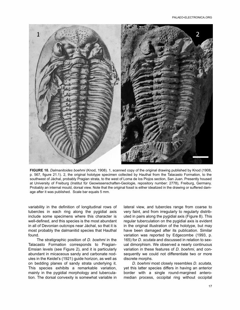
variability in the definition of longitudinal rows oftubercles in each ring along the pygidial axisinclude some specimens where this character iswell-defined, and this species is the most abundantin all of Devonian outcrops near Jáchal, so that it ismost probably the dalmanitid species that Hauthalfound.
The stratigraphic position of D. boehmi in theTalacasto Formation corresponds to Pragian-Emsian levels (see Figure 2), and it is particularlyabundant in micaceous sandy and carbonate nod-ules in the Keidel’s (1921) guide horizon, as well ason bedding planes of sandy strata underlying it.This species exhibits a remarkable variation,mainly in the pygidial morphology and tubercula-tion. The dorsal convexity is somewhat variable in
lateral view, and tubercles range from coarse tovery faint, and from irregularly to regularly distrib-uted in pairs along the pygidial axis (Figure 8). Thisregular tuberculation on the pygidial axis is evidentin the original illustration of the holotype, but mayhave been damaged after its publication. Similarvariation was reported by Edgecombe (1993, p.165) for D. scutata and discussed in relation to sex-ual dimorphism. We observed a nearly continuousvariation in these features of D. boehmi, and con-sequently we could not differentiate two or morediscrete morphs.
D. boehmi most closely resembles D. scutata,yet this latter species differs in having an anteriorborder with a single round-margined antero-median process, occipital ring without occipital
FIGURE 10. Dalmanitoides boehmi (Knod, 1908). 1, scanned copy of the original drawing published by Knod (1908,p. 567, figure 21.1). 2, the original holotype specimen collected by Hauthal from the Talacasto Formation, to thesouthwest of Jáchal, probably Pragian strata, to the west of Loma de los Piojos section, San Juan. Presently housedat University of Freiburg (Institut für Geowissenschaften-Geologie, repository number: 2778), Freiburg, Germany.Probably an internal mould, dorsal view. Note that the original fossil is either idealized in the drawing or suffered dam-age after it was published. Scale bar equals 5 mm.
17

Rustán & VACCARI: THE TRILOBITE DALMANITOIDES
node or tubercule, and a more strongly upturnedmucro which is notably larger.
Eifelian specimens from the Voorstehoek For-mation from South Africa were assigned to D. boe-hmi by Cooper (1982), based on the presence ofpaired axial tubercles on each pygidial axial ring.However, these specimens exhibit a large(spinose?) occipital tubercle and a bluntly roundedmedian process on the anterior border, which doesnot exhibits anterolateral crenulations. These fea-tures distinguish the South African specimens fromD. boehmi, D. scutata, or D. drevermanni, but areshared with D. accola (Clarke, 1913, p. 101, figure4.9-18) from the Ponta Grossa Formation, Paraná,Brazil.
As noted by Edgecombe (1993, p. 165), fea-tures separating South African specimens from D.accola include the regularity in the paired tubercleson the pygidial axial rings, as well as the number ofdorsoventral lens files. These observations sug-gest that South African specimens belong to a dif-ferent species based on the lack of tubercles onthe thoracic axial rings, conspicuous regularity ofpaired tubercles on every pygidial axial ring, and arow of tubercles along the pygidial fulcral line, aswell as the number of dorsoventral lens files (up to40 in D. accola, and near 30-32 in the South Afri-can material). Nevertheless, only a few pygidiafrom the South African form clearly display the dis-tribution of pygidial axial pairs of tubercles, andconsequently we agree with Cooper (1982, p. 59)that the range of variability in this character is notsufficiently documented. Furthermore, taking intoaccount data from Clarke (1913, p. 103) and addi-tional material of D. accola that we examined (Fig-ure 11), paired pygidial tubercles in this speciesseem be present on more rings and are more irreg-ularly distributed than has been reported (e.g.,Edgecombe, 1993, p. 165). Hence, consideringthis character is highly variable in other Dalmani-toides species, the South African specimens couldbe conspecific with the Brazilian D. accola as Coo-per conjectured (1982, p. 61). Differences could beinterpreted being of subspecies level, as Clarke(1913) suggested. Hence, we leave the South Afri-can species in open nomenclature as Dalmani-toides sp. A.
Dalmanitoides sp. A from South Africa, lackstubercles on the axial rings, a feature also noted inthe Bolivian specimen reported by Kozłowski(1923, p. 36, figure 2.1) and referred to Dalmanitesboehmi var. boliviensis, so that it appears to beconspecific according to Cooper (1982). However,this assignment is tentative since we have not
revised the Bolivian material, and characters suchas the anterior border have not been observedfrom illustrations . Due to this fact, we neither pro-vide an opinion on the synonymy of the non-tuber-culate Bolivian species Dalmanitespatacamayensis Kozłowski, 1923, a similar formwhich appears to belong to Dalmanitoides(Kozłowski, 1923, p. 36, figure 2.2).
REMARKS ON PALEOBIOGEOGRAPHYAND PHYLOGENY
Devonian records of Dalmanitoides suggest adiversification involving at least five species duringthe Early-Middle Devonian. This evidence sug-gests no migrations but rather isolation and specia-tion of a stock of widely distributed Dalmanitinetrilobites already present by the earliest Devonian,in Malvinokaffric basins. This pattern is consistentwith that recognized for other Malvinokaffric trilo-bite groups (Eldredge and Ormiston, 1979; Abeand Lieberman, 2009; Rustán and Vaccari, 2010).
Nevertheless, the close similarities betweenDalmanitoides and the slightly earlier genus Ron-cellia from Gaspe, Appalachian region of Canada(Eastern Americas Paleobiogeographic Realm),suggests a southerly dispersal from northern pale-olatitudes during the Early Devonian. Roncellia isconsidered sister taxon of Dalmanitoides, and theancestral area of the clade should be located inboreal regions, the area of most ancient strati-graphic records. To date, the earliest stratigraphicrecord of Dalmanitoides is Pragian, so that takinginto account the slightly older (Lochkovian?) recordof Roncellia (Lespérance and Bourque, 1971), andderived characters of Dalmanitoides (i.e., long andcurvy genal spines and tuberculated ornamenta-tion), migrations from the South to the North seemto be less probable.
As was discussed by Holloway and Carvalho(2010), a similar case occurs with respect to paleo-biogeographical implications of the synphoriinegenus Chacomurus Braniša and Vaněk, 1973, fromthe Pragian-Emsian of the lower member of theBelén Formation from Bolivia, which is hardly dif-ferentiated from the North American Eifelian sistergenus Coronura Hall and Clarke, 1888.
In contrast with dalmanitines, synphoriineswere overall considered of strongest affinities withthe Eastern Americas PaleobiogeographicalRealm, according to Carvalho and Fonseca (2007).Hence, these authors proposed migrations fromthe Appalachian region by the Eifelian as explana-tion of the synphoriine origins in Malvinokaffricbasins, based on the presence of Amazonaspis
18
PALAEO-ELECTRONICA.ORG
19
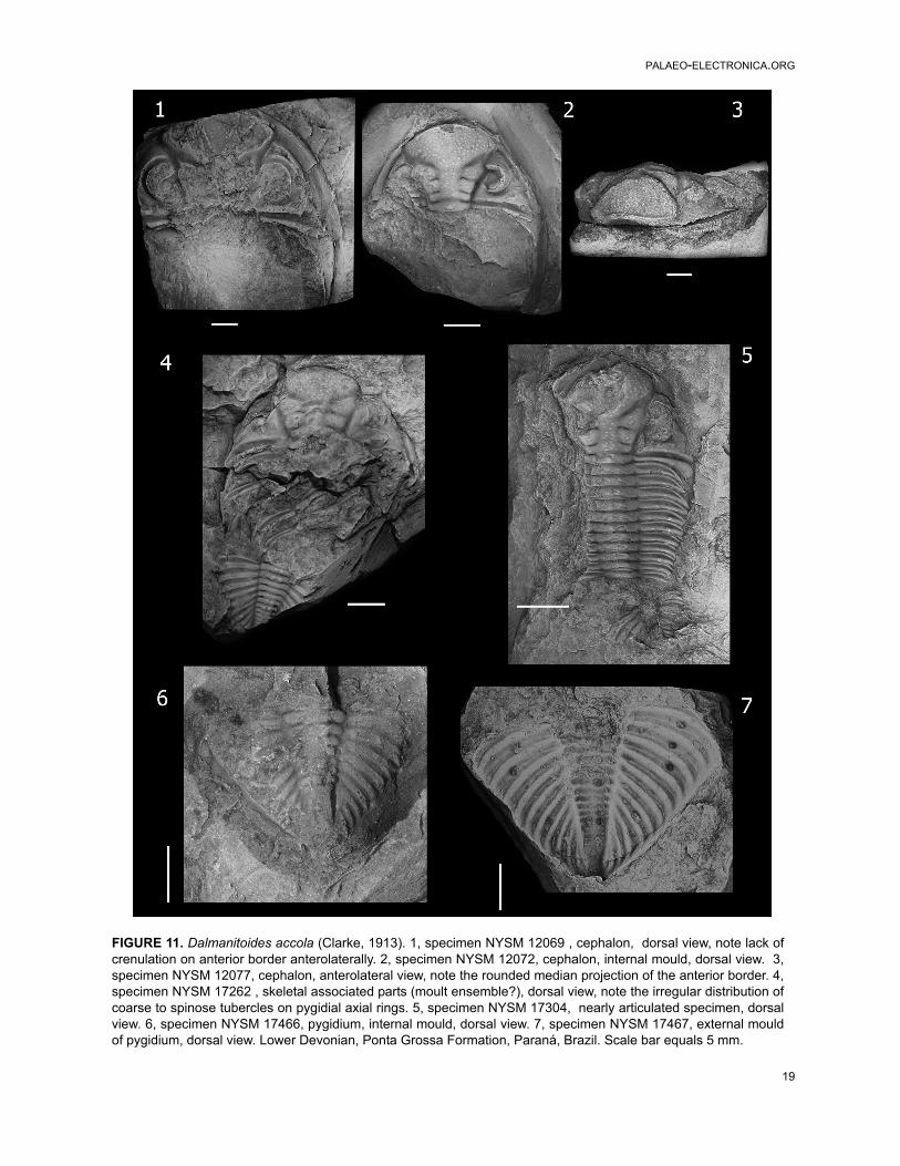
FIGURE 11. Dalmanitoides accola (Clarke, 1913). 1, specimen NYSM 12069 , cephalon, dorsal view, note lack ofcrenulation on anterior border anterolaterally. 2, specimen NYSM 12072, cephalon, internal mould, dorsal view. 3,specimen NYSM 12077, cephalon, anterolateral view, note the rounded median projection of the anterior border. 4,specimen NYSM 17262 , skeletal associated parts (moult ensemble?), dorsal view, note the irregular distribution ofcoarse to spinose tubercles on pygidial axial rings. 5, specimen NYSM 17304, nearly articulated specimen, dorsalview. 6, specimen NYSM 17466, pygidium, internal mould, dorsal view. 7, specimen NYSM 17467, external mouldof pygidium, dorsal view. Lower Devonian, Ponta Grossa Formation, Paraná, Brazil. Scale bar equals 5 mm.

Rustán & VACCARI: THE TRILOBITE DALMANITOIDES
maecurua (Clarke, 1890), from the Middle Devo-nian Maecurú Formation, Amazonas Basin, Pará,Brazil. Subsequently, the presence of Chacomurusin Bolivia (Holloway and Carvalho, 2010) confirmedthe presence of synphoriines in South America bythe Early Devonian, suggesting an older dispersalevent.
Although Dalmanitoides was interpreted inthis synphoriine framework, in accordance withChacomurus’s data, its reassignment to the dal-manitines suggest that the phylo-geographic his-tory of the taxon could not be the same thansynphoriines. A similar conclusion could beextended to Fenestraspis, because of the ques-tioned synphoriine affinities according comparisonswith Dalmanitoides given earlier in the text. Addi-tional, Late Silurian-Lochkovian dalmanitine datafrom Argentina would be of particular interest inthese issues, which we consider open to furtherdiscussions.
ACKNOWLEDGMENTS
R. Haude and B. Waisfeld have facilitated thestudy of material from Loma de los Piojos collectedby them. D. Balseiro, R. Foglia, and A. Rydczewskihave collaborated invaluably in the field works ofthe authors. The curator in charge at University ofFreiburg, Ursula Leppig, successfully localized theholotype of D. boehmi in the repository of that insti-tution, and kindly took photographs. E. Landingand F. Mannolini contributed by taking nice photo-graphs of D. accola specimens housed at NewYork State Museum, Albany, U.S.A. and specifyingrepository numbers. G. Edgecombe and B. Chat-terton revised an earlier version of the manuscript,contributing with fruitful comments. Two anony-mous reviewers spent precious time in providingdetailed suggestions in order to improve this work.Financial support for this study was provided byCONICET and ANPCyT (Agencia Nacional de Pro-moción Científica y Tecnológica) PICT-2006-01272, grant to Claudia Rubinstein.
REFERENCES
Abe, F.R. and Lieberman, B.S. 2009. The nature of evo-lutionary radiations: a case study involving Devoniantrilobites. Evolutionary Biology, 36:225-234.
Astini, A.R. 1991. Sedimentología de la Formación Tala-casto: plataforma fangosa del Devónico precordille-rano, provincia de San Juan. Revista de laAsociación Geológica Argentina, 44:277-294.
Baldis, B.A. 1975. El Devónico Inferior en la Precordil-lera Central. Parte I: Estratigrafía. Revista de la Aso-ciación Geológica Argentina, 30:53-83.
Bracaccini, O.I. 1949. El perfil de Tambolar. Revista de laAsociación Geológica Argentina, 4:165-179.
Braniša, A.L. and Vaněk, J. 1973. Several new trilobitegenera of the superfamily Dalmanitacea Vogdes,1890 in the Devonian of Bolivia. Vestník UstredníhoÚstavu Geologického, 48:97-101.
Bustos, U.D. 1996. Modelo sedimentario alternativo parael Devónico de la Precordillera central sanjuanina:Formación Punta Negra. Revista de la AsociaciónArgentina de Sedimentología, 3:17-30.
Bustos, U.D. and Astini, A.R. 1997. Formación PuntaNegra: análisis secuencial y evolución de la CuencaDevónica Precordillerana. Revista de la AsociaciónArgentina de Sedimentología, 4:97-111.
Campbell, K.S.W. 1977. Trilobites of the Haragan, Boisd'Arc and Frisco Formations (Early Devonian)Arbuckle Mountains Region, Oklahoma. OklahomaGeological Survey Bulletin, 123:1-227.
Carvalho, M.G. and Fonseca, V.M.M.D. 2007. The trilo-bite ‘Dalmanites’ maecurua Clarke, 1890 (MiddleDevonian, Amazon Basin, Brazil) and the new genusAmazonaspis (Synphoriidae). American MuseumNovitates, 3591:1-14.
Clarke, J.M. 1913. Fosseis devonianos do Paraná.Monografias Serviço Geologico e Mineralogico doBrasil, 1:1-353.
Cooper, M.R. 1982. A revision of the Devonian (Emsian–Eifelian) Trilobita from the Bokkeveld Group of SouthAfrica. Annals of the South African Museum, 89:1-174.
Delo, D.M. 1935. A revision of the phacopid trilobites.Journal of Paleontology, 9:402-420.
Edgecombe, G.D. 1993. A revision of the Devonian dal-manitid trilobite Gamonedaspis. Revista Técnica deYacimientos Petrolíferos Fiscales Bolivianos, 13-14:161-166.
Edgecombe G.D., Vaccari N.E., and Waisfeld, B.G. 1994.Lower Devonian calmoniid trilobites from the Argen-tine Precordillera: new taxa of the Bouleia group, andremarks on the tempo of calmoniid radiation. Geolog-ical Magazine, 131:449-464.
Eldredge, N. and Ormiston, L. 1979. Biogeography ofSilurian and Devonian Trilobites of the MalvinokaffricRealm p. 147-167. In Gray, J. and Boucot, H.J.(eds.), Historical Biogeography, Plate Tectonics, andthe changing Environment. Oregon State UniversityPress, Corvallis.
Hall, J.C. and Clarke, J.M. 1888. Descriptions of the trilo-bites and other Crustaceae of the Oriskany, UpperHelderberg, Hamilton, Portage, Chemung andCatskill groups. New York State Geological Survey,Natural history of New York, Palaeontology 7:1-236.
Herrera, Z.A. 1993. Nuevas precisiones sobre la edadde la Formación Talacasto (Precordillera Argentina)en base a su fauna de braquiópodos. Actas del 12doCongreso Geológico Argentino y 2do Congreso deExploración de Hidrocarburos, 2: 289-295.
20

PALAEO-ELECTRONICA.ORG
Herrera, Z.A. 1995a. The first notanopliid brachiopodfrom the South American Devonian sequence. Geo-bios, 28:337-342.
Herrera, Z.A. 1995b. The Lower Devonian chonetoideanbrachiopods from the Argentine Precordillera, p. 101-147. In Racheboeuf, P. (ed.), Four contributions tothe study of chonetoidean brachiopods. Documentsdes Laboratories de Géologie Lyon. Centre desSciences de la Terre, Université Claude-Bernard-Lyon I, Lyon.
Herrera, Z.A. and Bustos, U.D. 2001. Braquiópodosdevónicos de la Formación Punta Negra, en el perfildel Río de las Chacritas, Precordillera Argentina.Ameghiniana, 38:367-374.
Holloway, D. 1981. Silurian dalmanitacean trilobites fromNorth America, and the subfamilies Dalmanitinaeand Synphoriinae. Palaeontology, 24:695-731.
Holloway, D. and Carvalho, M.G. 2009. The extraordi-nary trilobite Fenestraspis (Dalmanitidae, Synphorii-nae) from the Lower Devonian of Bolivia.Palaeontology, 52:933-949.
Holloway, D. and Carvalho, M.G. 2010. The trilobite Cha-comurus (Dalmanitidae, Synphorinae) from theLower Devonian of Bolivia. Memoirs of the Associa-tion of Australasian Palaeontologists, 39:71-83.
Keidel, J. 1921. Observaciones geológicas en la Precor-dillera de San Juan y Mendoza. Anales del Ministeriode Agricultura, Sección Geología, Mineralogía y Min-ería, 15:7-102.
Knod, R. 1908. Devonische Faunen Boliviens, p. 493-600. In Steinmann, G. (ed.), Beiträge zur Geologieund Paläontologie von Südamerika. Neues Jahrbuchfür Mineralogie, Geologie und Paläontologie 25. E.Schweizerbart, Stuttgart.
Kozłowski, R. 1923. Faune devonienne de Bolivie. Anna-les de Paleontologie, 12:1-112.
Lespérance, P. 1975. Stratigraphy and paleontology ofthe Synphoriidae (Lower and Middle Devonian dal-manitacean trilobites). Journal of Paleontology,49:91-137.
Lespérance, P. and Bourque, P.A. 1971. The Synphorii-nae: an evolutionary pattern of Lower and MiddleDevonian trilobites. Journal of Paleontology, 45:182-208.
Padula, E., Rolleri, E., Mingramm, A.R., Criado Roqué,P., Flores, M.A., and Baldis, B.A. 1967. Devonian ofArgentina, p. 165-199. In Oswald, D. (ed.), Proceed-ings of the International Symposium on the DevonianSystem, 2. Canadian Society of Petroleum Geolo-gists, Calgary.
Racheboeuf, P.R. and Herrera, Z.A. 1994. Über einigeneue silurische und devonische Chonetaceen-Arten(Brachiopoda), und Reklassifizierung anderer Arten(On some new malvinokaffric Silurian and Devonianchonetacean brachiopods and reclassification of oth-ers). Neues Jahrbuch für Geologie und Paläontolo-gie, Monateshefte, 9:541-560.
Rustán, J.J. and Vaccari, N.E. 2010. The aulacopleuridtrilobite Maurotarion Alberti, 1969 in the Silurian-Devonian of Argentina: systematic, phylogenetic andpaleobiogeographic significance. Journal of Paleon-tology, 84:1082-1098.
Thomas, I. 1906. Neue Beiträge zur Kenntnis derDevonischen Faunen Argentiniens. Zeitschrift derDeutschen geologischen Gesellschaft, 57: 233-290.
Vaccari, N.E., Waisfeld, B.G., and Edgecombe, G.D.1994. Calmoniid Trilobites of the Lower DevonianScaphiocoelia zone in the Argentine Precordillera.Geobios, 27:591-608.
Vogdes, A.W. 1890. A bibliography of Paleozoic Crusta-cea from 1698 to 1889 including a list of North Amer-ican species and a systematic arrangement ofgenera. United States Geological Survey Bulletin,63:1-177.
Waisfeld, B.G., Edgecombe, G.D., and Vaccari, N.E.1994. Tormesiscus, a new blind calmoniid trilobitefrom the Lower Devonian, Argentine Precordillera.Geologica et Palaeontologica, 28:27- 43.
21