A REVIEW ON TYPES AND TECHNIQUES FOR THE PREPARATION … · Applications of Nanoparticles Different...
18
International Journal of Innovative Research and Review ISSN: 2347 – 4424 (Online) An Online International Journal Available at http://www.cibtech.org/jirr.htm 2018 Vol. 6 (2) April-June, pp.1-18/Syed and Krishnasailaja Review Article Centre for Info Bio Technology (CIBTech) 1 A REVIEW ON TYPES AND TECHNIQUES FOR THE PREPARATION OF NANOPARTICLES Huda Syed and *Abbaraju Krishnasailaja RBVRR Women’s College of Pharmacy, Barkatpura, Hyderabad, India *Author for Correspondence ABSTRACT For the past few decades, there has been a considerable research interest in the area of drug delivery using particulate delivery systems as carriers for small and large molecules. Nanoparticles (NPs) are defined as particles with a diameter smaller than 100 nm, are increasingly used in different applications, including drug carrier systems and to pass organ barriers such as the blood-brain barrier. Via nanotechnology we can achieve better therapeutic action, better bioavailability and better patient compliance. As because of several advantages over to the conventional drug delivery Nanosparticulate drug delivery prepared in several means by several ways of methods have several applications in different discipline of Pharmaceutical science. Several techniques are used for preparation of nanoparticles like Solvent Evaporation, Nanoprecipitation , Salting Out Method, Dialysis and Supercritical fluid technology. Keywords: Nanocrystals, Nanotechnology, Dendrimers, Nanoprecipitation, Solvent Evaporation. INTRODUCTION Nanoparticles (NPs) are defined as particulate dispersions or solid particles drug carrier that may or may not be biodegradable. Nanoparticles are sub -nanosized colloidal structures composed of synthetic or semi-synthetic polymer and the particle size ranges from 10-100 nm in diameter ( N. K. Jain, 2001) . The drug is dissolved, entrapped, encapsulated or attached to a nanoparticle matrix. Nanoparticles are solid core spherical particulates which are nanometric in size. The term nanoparticle is a combined name for both nanospheres and nanocapsules ( N. K. Jain, 2001). Nanospheres: Nanospheres contain drug embedded with the matrix (or) adsorbed on to the surface (Vyas S.P, Khar,2002) .Nanospheres are the spherical particles which have the size between 10-200 nm in diameter and that exhibit some new enhanced size dependent properties in comparison of larger spheres of the same material (N. K. Jain, 2001). Nanocapsules: are vesicular system in which drug is essentially encapsulated with in the central volume surrounded by an embryonic polymeric sheath (N. K. Jain, 2001). Figure 1: Nanoparticle system (Nanomedicine (2010) Future Medicine Ltd).
Transcript of A REVIEW ON TYPES AND TECHNIQUES FOR THE PREPARATION … · Applications of Nanoparticles Different...

International Journal of Innovative Research and Review ISSN: 2347 – 4424 (Online)
An Online International Journal Available at http://www.cibtech.org/jirr.htm
2018 Vol. 6 (2) April-June, pp.1-18/Syed and Krishnasailaja
Review Article
Centre for Info Bio Technology (CIBTech) 1
A REVIEW ON TYPES AND TECHNIQUES FOR THE PREPARATION
OF NANOPARTICLES
Huda Syed and *Abbaraju Krishnasailaja
RBVRR Women’s College of Pharmacy, Barkatpura, Hyderabad, India
*Author for Correspondence
ABSTRACT
For the past few decades, there has been a considerable research interest in the area of drug delivery using
particulate delivery systems as carriers for small and large molecules. Nanoparticles (NPs) are defined as
particles with a diameter smaller than 100 nm, are increasingly used in different applications, including
drug carrier systems and to pass organ barriers such as the blood-brain barrier. Via nanotechnology we
can achieve better therapeutic action, better bioavailability and better patient compliance. As because of
several advantages over to the conventional drug delivery Nanosparticulate drug delivery prepared in
several means by several ways of methods have several applications in different discipline of
Pharmaceutical science. Several techniques are used for preparation of nanoparticles like Solvent
Evaporation, Nanoprecipitation , Salting Out Method, Dialysis and Supercritical fluid technology.
Keywords: Nanocrystals, Nanotechnology, Dendrimers, Nanoprecipitation, Solvent Evaporation.
INTRODUCTION
Nanoparticles (NPs) are defined as particulate dispersions or solid particles drug carrier that may or may
not be biodegradable. Nanoparticles are sub -nanosized colloidal structures composed of synthetic or
semi-synthetic polymer and the particle size ranges from 10-100 nm in diameter ( N. K. Jain, 2001) .
The drug is dissolved, entrapped, encapsulated or attached to a nanoparticle matrix. Nanoparticles are
solid core spherical particulates which are nanometric in size. The term nanoparticle is a combined name
for both nanospheres and nanocapsules ( N. K. Jain, 2001).
Nanospheres: Nanospheres contain drug embedded with the matrix (or) adsorbed on to the surface (Vyas
S.P, Khar,2002) .Nanospheres are the spherical particles which have the size between 10-200 nm in
diameter and that exhibit some new enhanced size dependent properties in comparison of larger spheres
of the same material (N. K. Jain, 2001).
Nanocapsules: are vesicular system in which drug is essentially encapsulated with in the central volume
surrounded by an embryonic polymeric sheath (N. K. Jain, 2001).
Figure 1: Nanoparticle system (Nanomedicine (2010) Future Medicine Ltd).
International Journal of Innovative Research and Review ISSN: 2347 – 4424 (Online)
An Online International Journal Available at http://www.cibtech.org/jirr.htm
2018 Vol. 6 (2) April-June, pp.1-18/Syed and Krishnasailaja
Review Article
Centre for Info Bio Technology (CIBTech) 2
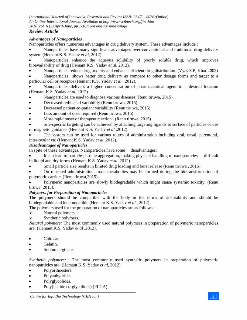
Advantages of Nanoparticles Nanoparticles offers numerous advantages in drug delivery system. These advantages include :
Nanoparticles have many significant advantages over conventional and traditional drug delivery
system (Hemant K.S. Yadav et al, 2012).
Nanoparticles enhance the aqueous solubility of poorly soluble drug, which improves
bioavailability of drug (Hemant K.S. Yadav et al, 2012).
Nanoparticles reduce drug toxicity and enhance efficient drug distribution. (Vyas S.P, Khar,2002)
Nanoparticles shows better drug delivery as compare to other dosage forms and target to a
particular cell or receptor (Hemant K.S. Yadav et al , 2012).
Nanoparticles delivers a higher concentration of pharmaceutical agent to a desired location
(Hemant K.S. Yadav et al, 2012).
Nanoparticles are used to diagnose various diseases (Renu tiruwa, 2015).
Decreased fed/fasted variability (Renu tiruwa, 2015).
Decreased patient-to-patient variability (Renu tiruwa, 2015).
Less amount of dose required (Renu tiruwa, 2015).
More rapid onset of therapeutic action (Renu tiruwa, 2015).
Site-specific targeting can be achieved by attaching targeting ligands to surface of particles or use
of magnetic guidance (Hemant K.S. Yadav et al ,2012).
The system can be used for various routes of administration including oral, nasal, parenteral,
intra-ocular etc (Hemant K.S. Yadav et al ,2012).
Disadvantages of Nanoparticles
In spite of these advantages, Nanoparticles have some disadvantages:
It can lead to particle-particle aggregation, making physical handling of nanoparticles , difficult
in liquid and dry forms (Hemant K.S. Yadav et al ,2012).
Small particle size results in limited drug loading and burst release (Renu tiruwa , 2015).
On repeated administration, toxic metabolites may be formed during the biotransformation of
polymeric carriers (Renu tiruwa,2015).
Polymeric nanoparticles are slowly biodegradable which might cause systemic toxicity. (Renu
tiruwa, 2015).
Polymers for Preparation of Nanoparticles The polymers should be compatible with the body in the terms of adaptability and should be
biodegradable and biocompatible (Hemant K.S. Yadav et al , 2012).
The polymers used for the preparation of nanoparticles are as follows:
Natural polymers.
Synthetic polymers.
Natural polymers: The most commonly used natural polymers in preparation of polymeric nanoparticles
are: (Hemant K.S. Yadav et al ,2012).
Chitosan .
Gelatin.
Sodium alginate.
Synthetic polymers: The most commonly used synthetic polymers in preparation of polymeric
nanoparticles are: (Hemant K.S. Yadav et al, 2012).
Polyorthoesters.
Polyanhydrides.
Polyglycolides.
Poly(lactide co-glycolides) (PLGA) .

International Journal of Innovative Research and Review ISSN: 2347 – 4424 (Online)
An Online International Journal Available at http://www.cibtech.org/jirr.htm
2018 Vol. 6 (2) April-June, pp.1-18/Syed and Krishnasailaja
Review Article
Centre for Info Bio Technology (CIBTech) 3
Applications of Nanoparticles
Different applications of nanoparticles are listed below:
Used in targeted drug delivery (therapy) to brain and cancer therapy. (M. Anto Godwin et al ,2015).
Drug and gene delivery (Saba Hasan et al ,2015).
Detection of proteins (M. Anto Godwin et al ,2015).
Biomarker mapping (Saba Hasan et al ,2015).
Probing of DNA structure and also used in tissue engineering (M. Anto Godwin et al ,2015).
Bio detection of pathogens (M. Anto Godwin et al , 2015).
Separation and purification of biological molecules and cells (P.amareshwar et al, 2011).
MRI contrast enhancement (M. Anto Godwin et al , 2015).
Used in ocular delivery (P. amareshwar et al, 2011).
Nanotechnology in Diabetes (M. Anto Godwin et al , 2015).
Nanotechnology in CVS Disorders (M. Anto Godwin et al , 2015.
Types of Nanoparticles
NPs
Nanoshells
Dendrimers
Nanosuspensions
PNPs
Nanotube
SLN
Magnetic Nps
Nanocrystals
Nanobots
Nanowire
Liposomes
Nanobubbles
Figure 2: Types of Nanoparticles

International Journal of Innovative Research and Review ISSN: 2347 – 4424 (Online)
An Online International Journal Available at http://www.cibtech.org/jirr.htm
2018 Vol. 6 (2) April-June, pp.1-18/Syed and Krishnasailaja
Review Article
Centre for Info Bio Technology (CIBTech) 4
Nanosuspensions The suspension of nanoparticles prepared in a liquid is known as nanosuspenion. The size of nanoparticle
ranges from 200 to 500 nm and incredible character of nanosuspension is the enhanced saturation,
solubility and dissolution rate of the compound (P.Velavan et al ,2015). The saturation and solubility of
compound increases below a particle size of 1 mcm. An outstanding character of nanosuspension is that
they can produce variations in the crystalline structure such as they increase the amorphous fraction in
particle. Nanosuspensions show an increased adherence to tissue (P.Velavan et al ,2015).
For instance: Nanosuspension of ibuprofen is formulated by emulsion solvent diffusion method for
improving ocular availability. The need for nanosuspensions as a dosage form was recognized as a means
to administer therapeutic quantities of water-insoluble dosage forms (P.Velavan et al ,2015).
Nanocrystal
Nanocrystal is any nanomaterial with at least one dimension ≤ 100nm and that is single crystalline
(P.Velavan et al , 2015). More properly, any material with a dimension of less than 1 micrometre, i.e.,
1000 nanometers, should be referred to as a nanoparticle, not a nanocrystal (P.Velavan et al ,2015). For
example, any particle which exhibits regions of crystallinity should be termed nanoparticle or nanocluster
based on dimensions. (P.Velavan et al ,2015). These materials are of huge technological interest since
many of their electrical and thermodynamic properties show strong size dependence and can therefore be
controlled through careful manufacturing processes (P.Velavan et al, 2015).
Drug nanocrystals have to be distinguished from polymeric nanoparticles, which consist of a polymeric
matrix and an incorporated drug. Drug nanocrystals do not consist of any matrix material (Hemant K.S.
Yadav et al , 2012).
Magnetic Nanoparticles
Magnetic nanoparticles are strong and adaptable diagnostic tool in the field of medicine. Magnetic
immunoassay methods have been established in which the main field generated by the magnetically
labelled target detected directly with sensitive magnetometer (P.Velavan et al ,2015). Super magnetic
nanoparticles are used as adverse agents in magnetic resonance imaging. (Hemant K.S. Yadav et al ,
2012).
Magnetic nanoparticles. Indomethacin established selective targeting under magnetic field of 8000 oe-
strength, following normal administration, the concentration of the drug was higher in liver and spleen
where the process of phagocytosis and endocytosis might occur (P.Velavan et al, 2015).
The above Figure indicates that magnetic nanoparticles (orange circles) carrying nucleic acids or drugs
can be magnetically guided into cells (blue circle) using n external magnet. Nucleic acids such as short
interfering RNA can turn off certain genes that cause diseases (P.Velavan et al, 2015).
Figure 3: Magnetic Nanoparticles (Plank, et al).

International Journal of Innovative Research and Review ISSN: 2347 – 4424 (Online)
An Online International Journal Available at http://www.cibtech.org/jirr.htm
2018 Vol. 6 (2) April-June, pp.1-18/Syed and Krishnasailaja
Review Article
Centre for Info Bio Technology (CIBTech) 5
The advantages of magnetic nanoparticles drug delivery sytems include:
The ability to target specific locations in the body (P.Velavan et al, 2015).
The reduction of the quantity of drug needed to attain a particular concentration in the vicinity of
the target (P.Velavan et al, 2015).
The reduction of the concentration of the drug at non target sites minimizing severe side effects
(P.Velavan et al, 2015).
Magnetic nanoparticles can attach to cancer cells in the blood stream. These nanoparticles may
allow doctors to remove cancer cells before they can establish new tumors (P.Velavan et al, 2015).
Many types of magnetic nanoparticles-based biosensors have been surface functionalized to
recognize specific molecular targets, due to their unique magnetic properties which are not found in
biological systems (P.Velavan et al, 2015).
Solid lipid nanoparticles
Solid lipid nanoparticles (SLN) introduced in 1991 represent an alternative carrier system to tradition
colloidal carriers such as - emulsions, liposomes and polymeric micro and nanoparticles. (P.Velavan et al,
2015). Nanoparticles made from solid lipids are attracting major attention as novel colloidal drug carrier
for intravenous applications as they have been proposed as an alternative particulate carrier system .
(Vyas S.P, Khar, 2002).
SLN are sub-micron colloidal carriers ranging from 50 to 1000 nm, which are composed of physiological
lipid, dispersed in water or in aqueous surfactant solution. SLN offer unique properties such as small size,
large surface area, high drug loading and the interaction of phases at the interface and are attractive for
their potential to improve performance of pharmaceuticals (Hemant K.S. Yadav et al , 2012).
In order to overcome the disadvantages associated with the liquid state of the oil droplets, the liquid lipid
was replaced by a solid lipid, which eventually transformed into solid lipid nanoparticles (P.Velavan et al,
2015).
The reasons for the increasing interest in lipid based system are many – fold and include. (Vyas S.P, Khar
2002).
Lipids enhance oral bioavailability and reduce plasma profile variability.
Better characterization of lipoid excipients.
An improved ability to address the key issues of technology transfer and manufacture scale-up.
Solid lipid nanoparticles are one of the novel potential colloidal carrier systems as alternative materials to
polymers which is identical to oil in water emulsion for parenteral nutrition, but the liquid lipid of the
emulsion has been replaced by a solid lipid (Vyas S.P, Khar ,2002). They have many advantages such as
good biocompatibility, low toxicity and lipophilic drugs are better delivered by solid lipid nanoparticles
and the system is physically stable (Vyas S.P, Khar ,2002).
Figure 4: Solid Lipid NPs (Vyas S.P, Khar, 2002)

International Journal of Innovative Research and Review ISSN: 2347 – 4424 (Online)
An Online International Journal Available at http://www.cibtech.org/jirr.htm
2018 Vol. 6 (2) April-June, pp.1-18/Syed and Krishnasailaja
Review Article
Centre for Info Bio Technology (CIBTech) 6
Nanotubes Nanotubes are self-accumulating sheets of atoms organized into tubes (P.Velavan et al, 2015). They
may be organic or inorganic in their constitution, and can be formulated as single or multi walled
structures, carbon nanotubes have recently been introduced to this category (P.Velavan et al, 2015).
Carbon nanotubes (CNT) have shown substantial potential in a variety of biological applications
including use as DNA and protein biosensors, ion channel blockers, bio-separators, and biocatalysts
(Bianco et al,2005). Nanotubes have been constructed with length-to-diameter ratio of up to
132,000,000:1, which is significantly larger than any other material (P.Velavan et al, 2015). These
cylindrical carbon molecules have novel properties which make them potentially useful in many
applications in nanotechnology, electronics, optics, and other fields of materials science, as well as
potential uses in architectural fields (Vyas S.P, Khar, 2002).
The ends of a nanotube may be capped with a hemisphere of the bucky ball structure (Vyas S.P,
Khar,2002) .Their name is derived from their size, since the diameter of a nanotube is on the order of a
few nanometers (approximately 1/50,000th of the width of a human hair), while they can be up to 18
centimeters in length (as of 2010). Nanotubes are categorized as single-walled nanotubes (SWNTs) and
multi-walled nanotubes (MWNTs) (Vyas S.P, Khar, 2002).
Figure 5: Nanotubes (P.Velavan et al, 2015).
Polymeric nanoparticles
Polymeric nanoparticles (PNPs) consists of a biodegradable polymer (Mahmoud Elsabahy, 2012). The
advantages of using PNPs in drug delivery are many, being the most important that they generally
increase the stability of any volatile pharmaceutical agents and that they are easily and cheaply fabricated
in large quantities by a multitude of methods (P.Velavan et al, 2015).
Also, polymeric nanoparticles may have engineered specificity, allowing them to deliver a higher
concentration of pharmaceutical agent to a desired location (Vyas S.P, Khar, 2002).The solid structure of
polymeric nanoparticles gives them higher stability, uniform size distribution, and more sustained drug
release profiles. It involves biodegradable synthetic polymers like polylactic co-glycolic acid (PLGA) and
polycaprolactone (PCL) and natural polymers like poly peptides and polysaccharides (Mahmoud
Elsabahy, 2012).
Figure 6: Polymeric nanoparticles (Mahmoud Elsabahy 2012)

International Journal of Innovative Research and Review ISSN: 2347 – 4424 (Online)
An Online International Journal Available at http://www.cibtech.org/jirr.htm
2018 Vol. 6 (2) April-June, pp.1-18/Syed and Krishnasailaja
Review Article
Centre for Info Bio Technology (CIBTech) 7
Dendrimers
Dendrimers, a unique class of polymers, that presents the polymers in nanometric dimensions and are
highly branched macromolecules whose size and shape can be precisely controlled (Urvashi Singh et al
2014). Dendrimers are fabricated from monomers using either convergent or divergent step growth
polymerization. The well-defined structure, mono dispersity of size, surface functionalization capability,
and stability are properties of dendrimers that make them attractive drug carrier candidates (P.Velavan et
al, 2015). Dendrimers offer enormous capacity for solubilization of hydrophobic compounds, and can be
modified with guest molecules (Urvashi Singh et al 2014).
Dendrimers that are involved in drug delivery and imaging are basically 10-100 nm in diameter having
multiple functional groups on their surface, interpretating them ideal carriers for site specific drug
delivery (V. J. Mohanraj et al , 2006). Studies of biomedical application of dendrimers are becoming
more and more attractive especially in the field of non viral gene vector. (Urvashi Singh et al 2014).
Figure 7: Dendrimers (Urvashi Singh et al 2014).
Nanoshells
Nanoshells are the new modified forms of targeted therapy, having core of silica and a metallic outer
layer. These thin coated core particles of different material have gained considerable attention now days.
The properties of nanoshells can be altered by simply tuning the core to shell ratio. (V. J. Mohanraj et al ,
2006).
With the recent advancement in new techniques it is now possible to synthesize these nanostructures in
desired shape, size and morphology (V. J. Mohanraj et al , 2006) . Nanoshells are synthesized to create
novel structures with different morphologies, since not possible to synthesize all the materials in desired
morphologies (V. J. Mohanraj et al , 2006). For obtaining desirable morphology core particles of
different morphologies such as rods, wires, tubes, rings, cubes, etc can be coated with thin shell in core
shell structures (P.Velavan et al, 2015). Therefore while synthesizing nanoshells expensive material is
required in lesser amount than usual (P.Velavan et al, 2015). Targeting of nanoshells can be achieved by
using immunological methods (P.Velavan et al, 2015). Nanoshells occupies variety of applications in
diverse areas such as providing chemical stability to colloids, enhancing luminescence properties,
engineering band structures, biosensors, drug delivery, etc (P.Velavan et al, 2015).
Nanobubbles Nanobubbles (NBs) are nanoscaled bubble like structures that are generated in the interface of
hydrophobic surfaces in liquids (P.Velavan et al, 2015). These nanobubbles remain stable at room
temperature and when heated to physiological temperature within the body coalesce to form microbubbles
(P.Velavan et al, 2015). The mechanism of NB formation is based on the nucleation of gas at the
hydrophobic surface from a supersaturated solution, leading to trap atmospheric gases (P.Velavan et al,
2015).There are four types of nanobubbles: bulk, interfacial, plasmonic and oscillating nanobubbles (V. J.
Mohanraj et al , 2006). Nanobubbles potentially exhibit advantages in targeting the tumor tissue and
delivering the drug selectively under the influence of ultrasound exposure(V. J. Mohanraj et al , 2006).

International Journal of Innovative Research and Review ISSN: 2347 – 4424 (Online)
An Online International Journal Available at http://www.cibtech.org/jirr.htm
2018 Vol. 6 (2) April-June, pp.1-18/Syed and Krishnasailaja
Review Article
Centre for Info Bio Technology (CIBTech) 8
Nanowire
A nanowire is a nanostructure, with the diameter of 10−9 meters (P.Velavan et al, 2015).Alternatively,
nanowires can be defined as structures that have a thickness or diameter constrained to tens of nanometers
or less and an unconstrained length
(P.Velavan et al, 2015). Many different types of nanowires exist,
including metallic e.g., Ni, Pt, Au, semiconducting e.g., Si, InP, GaN, etc., and insulating e.g., SiO2,
TiO2 (P.Velavan et al, 2015).
Liposomes
Liposomes are concentric bilayered vesicles in which an aqueous volume is entirely enclosed by a
membranous lipid bilayer mainly composed of natural or synthetic phospholipids (P.Velavan et al, 2015).
Liposomes are characterized in terms of size, surface charge and number of bilayers (N. K. Jain, 2001). It
exhibits number of advantages in terms of amphiphilic character, biocompatibility, and ease of surface
modification rendering it a suitable candidate delivery system for biotech drugs (Vyas S.P, Khar, 2002).
Liposomes have been used successfully in the field of biology, biochemistry and medicine since its origin
(Vyas S.P, Khar, 2002).
Liposomes were the first nanoparticle platform applied in medicine since Bangham described them in
1961 (N. K. Jain ,2001).liposomes are nanometric (30–100 nm) versions of liposomes formed by
spontaneous self-organization of phospholipids such as phosphatidylcholine, phosphatidylethanolamine,
phosphatidylglycerol and phosphatidylserine, and other molecules such as cholesterol (Vyas S.P, Khar,
2002).
Nanobots
Nanorobotics is the technology of creating machines or robots at or close to the microscopic scale of a
nanometer 10−9 meters (Deepak N. Kapoor, 2012) . Nanorobots are essentially nanoelectromechanical
devices (NEMS) (Deepak N.Kapoor, 2012).
These nanorobotic devices are comparable to biological cells and organelles in size (Deepak N.Kapoor,
2012). It is a multidisciplinary field requiring advanced level input from different areas of science and
technology including, physics, chemistry, biology, medicine, pharmaceutical sciences, engineering,
biotechnology and other biomedical sciences (Deepak N.Kapoor, 2012).
Figure 8: Liposomes ((N. K. Jain ,2001).

International Journal of Innovative Research and Review ISSN: 2347 – 4424 (Online)
An Online International Journal Available at http://www.cibtech.org/jirr.htm
2018 Vol. 6 (2) April-June, pp.1-18/Syed and Krishnasailaja
Review Article
Centre for Info Bio Technology (CIBTech) 9
Applications for nanorobotics in medicine include early diagnosis and targeted drug-delivery for cancer,
biomedical instrumentation surgery, pharmacokinetics monitoring of diabetes, and health care (Deepak
N.Kapoor, 2012).
Techniques for the preparation of nanoparticles
The appropriate method for the preparation of nanoparticles depends on the characteristics of polymer and
the drug that is to be used in Nano preparations therefore in order to achieve the properties of interest the
mode of preparation plays a vital role (Swati Tyagi et al ,2016).
Nanoparticles can be prepared from a variety of materials such as proteins, polysaccharides and synthetic
polymers. The selection of matrix materials is dependent on factors like: (Swati Tyagi et al, 2016).
Size of nanoparticles required.
Degree of biodegradability,
Biocompatibility and toxicity.
Drug release profile desired.
Nanoparticles can be prepared by following methods:
Emulsion Polymerization Method
Interfacial polymerization
Desolvation Technique
Emulsion interfacial reaction method
Solvent Evaporation Method
Emulsification Or Solvent Diffusion Method
Ionic Gelation Or Coacervation Method
Salting out method
Nanoprecipitation Method
Spray drying method
Dialysis
Supercritical Fluid Technology
Emulsion Polymerization Method:
Emulsion polymerization is one of the fastest methods for nanoparticle preparation and is readily scalable.
The method is classified into two categories, based on the use of an organic or aqueous continuous phase
(Hemant K.S. Yadav et al ,2012). The continuous organic phase methodology involves the dispersion of
monomer into an emulsion or inverse microemulsion, or into a material in which the monomer is not
soluble (nonsolvent) (Hemant K.S. Yadav et al ,2012). Polyacrylamide nanospheres were produced by
this method (Hemant K.S. Yadav et al , 2012). As one of the first methods for production of
nanoparticles, surfactants or protective soluble polymers were used to prevent aggregation in the early
stages of polymerization (Hemant K.S. Yadav et al ,2012). In the aqueous continuous phase the monomer
is dissolved in a continuous phase that is usually an aqueous solution, and the surfactants or emulsifiers
are not needed (Hemant K.S. Yadav et al , 2012).

International Journal of Innovative Research and Review ISSN: 2347 – 4424 (Online)
An Online International Journal Available at http://www.cibtech.org/jirr.htm
2018 Vol. 6 (2) April-June, pp.1-18/Syed and Krishnasailaja
Review Article
Centre for Info Bio Technology (CIBTech) 10
The polymerization process can be initiated by different mechanisms. Initiation occurs when a monomer
molecule dissolved in the continuous phase collides with an initiator molecule that might be an ion or a
free radical (Swati Tyagi et al 2016). Alternatively, the monomer molecule can be transformed into an
initiating radical by high-energy radiation, including g-radiation, or ultraviolet or strong visible light
(Swati Tyagi et al 2016). Chain growth starts when initiated monomer ions or monomer radicals collide
with other monomer molecules according to an anionic polymerization mechanism
(Swati Tyagi et al
2016). Phase separation and formation of solid particles can take place before or after termination of the
polymerization reaction (Hemant K.S. Yadav et al ,2012).
Interfacial Polymerization:
It is one of the well-established methods used for the preparation of polymer nanoparticles (Hemant K.S.
Yadav et al , 2012). It involves step polymerization of two reactive monomers or agents, which are
dissolved respectively in two phases (i.e., continuous- and dispersed-phase), and the reaction takes place
at the interface of the two liquids (Hemant K.S. Yadav et al, 2012). Nanometer-sized hollow polymer
particles were synthesized by employing interfacial cross-linking reactions as polyaddition and
polycondensation or radical polymerization. Oil-containing nanocapsules were obtained by the
polymerization of monomers at the oil/water interface of a very fine oil-in-water micro- emulsion
(Hemant K.S. Yadav et al ,2012). The organic solvent, which was completely miscible with water, served
as a monomer vehicle and the interfacial polymerization of the monomer was believed to occur at the
surface of the oil droplets that formed during emulsification (Hemant K.S. Yadav et al , 2012). To
promote nanocapsule formation, the use of aprotic solvents, such as acetone and acetonitrile was
recommended (Hemant K.S. Yadav et al, 2012).
Desolvation Technique:
The protein or polysaccharide from an aqueous phase can be desolvated by PH changes or change in
temperature or by adding some appropriate counter ions ( P.amareshwar et al, 2011).Aqueous drug
polymer dispersion was prepared and pH was optimized to 7 . (P.amareshwar et al ,2011). The
desolvating agent was added under continuous mechanical stirring.It is added intermittent or by
continuous method till the solution become turbid. Appearance of turbidity in the solution indicates the
end point of the process. ( P.amareshwar et al ,2011).Then add 2 drops of glutaraldehyde , a cross -
linking agent & allowed by continuous stirring for the next 12 hours at 700 rpm ( P.amareshwar et al
,2011).The residual solvent was removed by rota evaporation. Free flowing nanoparticles were obtained
are kept for air drying. ( P.amareshwar et al ,2011).
Desolvation Technique: Is prepared by two methods: ( P.amareshwar et al ,2011).
Figure 9: Poymerization Method (Niba Ibrahim et al 2018)

International Journal of Innovative Research and Review ISSN: 2347 – 4424 (Online)
An Online International Journal Available at http://www.cibtech.org/jirr.htm
2018 Vol. 6 (2) April-June, pp.1-18/Syed and Krishnasailaja
Review Article
Centre for Info Bio Technology (CIBTech) 11
Continuous method: In this method solvent was added continuously 1ml/1 minute till the turbidity
appears.
Intermittent method: In this method solvent was added intermittently 1ml/5 minute till the
turbidity appears.
Emulsion interfacial reaction method:
For the preparation of nanoparticles by emulsion interfacial reaction method, following procedure was
adopted. According to this technique (Vyas S.P, Khar, 2002).
Emulsion A: 1% (polymer) solution and 5% span 80 solution was prepared, then in a beaker 100 mg of
drug in 1 ml of methanol was taken. To this add 4 ml of polymer solution add 0.5ml of span 80 solution.
It was kept for stirring at 2000 rpm for 5 mins (Vyas S.P, Khar, 2002).
Emulsion B: 0.25 ml of 2% Glutraldehyde was taken. To this 0.5 ml of span80 solution was added. Then
kept for stirring at 3000 rpm for 5 mins. Then Emulsion A&B was mixed and kept for stirring at 5000
rpm for 10 mins. Finally the solution was filtered by vacuum filtration and kept for drying
(Vyas S.P,
Khar, 2002).
Solvent Evaporation Method:
Solvent evaporation was the first method that was developed for the preparation of nanoparticles, in this
technique the polymer solutions were prepared in volatile solvents and emulsions were formulated by
employing dichloromethane and chloroform, but now it is replaced with ethyl acetate that shows a much
better toxicological profile to obtain polymeric particles less than 500 nm in size (Hemant K.S. Yadav et
al , 2012). In this method, the polymer is dissolved in an organic solvent such as dichloromethane,
chloroform or ethyl acetate, which is also used as the solvent for dissolving the hydrophobic drug
(Hemant K.S. Yadav et al ,2012). The mixture of polymer and drug solution is then emulsified in an
aqueous solution containing a surfactant or emulsifying agent to form oil in water (o/w) emulsion
(Hemant K.S. Yadav et al ,2012). After the formation of stable emulsion, the organic solvent is
evaporated either by reducing the pressure or by continuous stirring (Swati Tyagi et al 2016). Particle size
was found to be influenced by the type and concentrations of stabilizer, homogenizer speed and polymer
concentration (Swati Tyagi et al 2016). In order to produce small particle size, often a high-speed
Figure 10: Desolvation Technique (Amrit Rai et al, 2017).

International Journal of Innovative Research and Review ISSN: 2347 – 4424 (Online)
An Online International Journal Available at http://www.cibtech.org/jirr.htm
2018 Vol. 6 (2) April-June, pp.1-18/Syed and Krishnasailaja
Review Article
Centre for Info Bio Technology (CIBTech) 12
homogenization or ultrasonication may be employed, followed by evaporation of the solvent, either by
continuous magnetic stirring at room temperature or under reduced pressure resulting in the formation of
solidified nanosized particles, collected by ultracentrifugation followed by washing to remove surfactants
and at last the product is lyophilized (Swati Tyagi et al 2016).
Emulsification Or Solvent Diffusion Method:
This is a modified version of solvent evaporation method. In this method, the water miscible solvent like
acetone along with a small amount of the water immiscible organic solvent like chloroform is used as an
oil phase (Hemant K.S. Yadav et al, 2012). Due to the spontaneous diffusion of solvents an interfacial
turbulence is created between the two phases leading to the formation of small particles (Hemant K.S.
Yadav et al, 2012). As the concentration of water miscible solvent increases, a decrease in the size of
particle can be achieved (Hemant K.S. Yadav et al ,2012). Both solvent evaporation and solvent diffusion
methods can be used for hydrophobic or hydrophilic drugs. (Hemant K.S. Yadav et al, 2012). In the case
of hydrophilic drug, a multiple w/o/w emulsion needs to be formed with the drug dissolved n the internal
aqueous phase (Hemant K.S. Yadav et al, 2012).
As with some of the other techniques, this one is efficient in encapsulating lipophilic drugs (Hemant K.S.
Yadav et al ,2012). Several drug-loaded nanoparticles were produced by this technique, including
mesotetra (hydroxyphenyl) porphyrin-loaded PLGA (p-THPP) nanoparticles, doxorubicin-loaded PLGA
nanoparticles, plasmid DNA-loaded PLA nanoparticles, coumarin-loaded PLA nanoparticles, indocyanine
,cyclosporine (Cy-A)-loaded gelatin and cyclosporin (Cy-A)-loaded sodium glycolate nanoparticles
(Hemant K.S. Yadav et al. 2012).
Figure 11: Solvent evaporation method (Swati Tyagi et al 2016).
Figure 12: Emulsification Or Solvent Diffusion Method (Hemant K.S. Yadav 2012).
International Journal of Innovative Research and Review ISSN: 2347 – 4424 (Online)
An Online International Journal Available at http://www.cibtech.org/jirr.htm
2018 Vol. 6 (2) April-June, pp.1-18/Syed and Krishnasailaja
Review Article
Centre for Info Bio Technology (CIBTech) 13
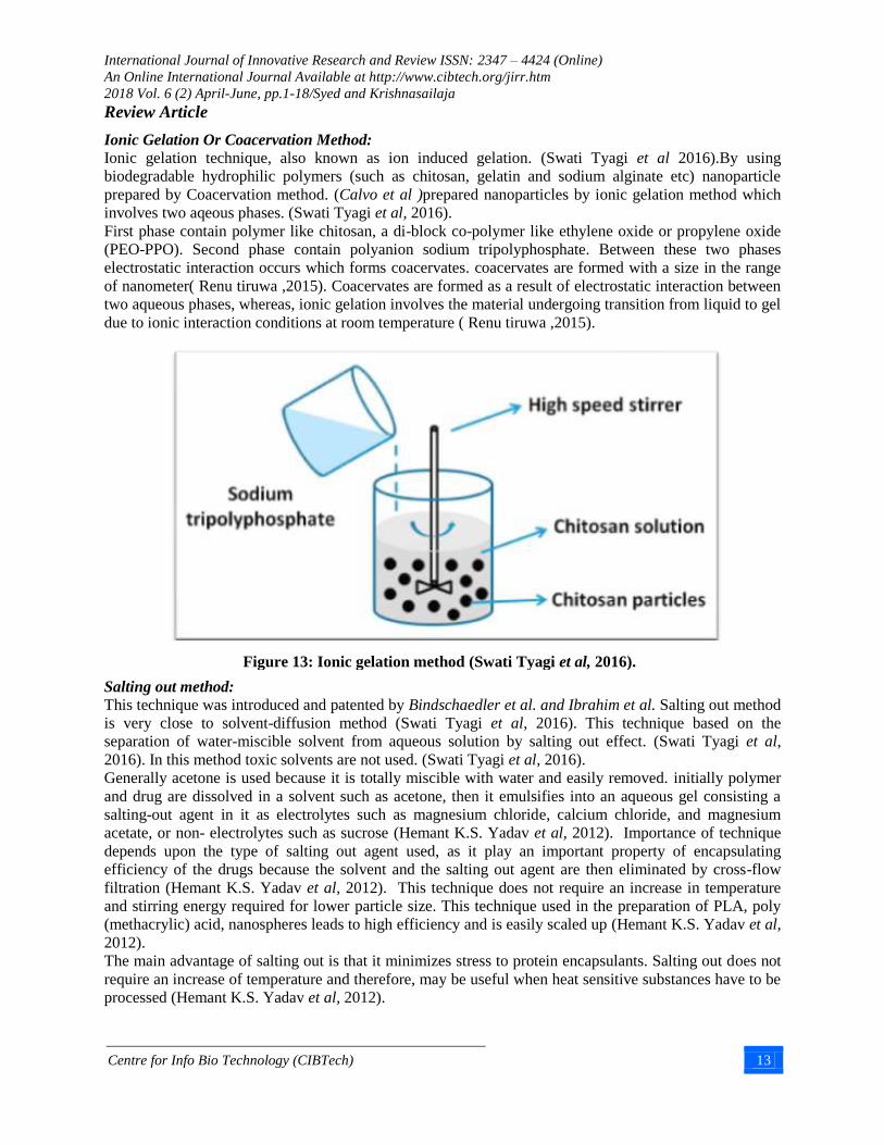
Ionic Gelation Or Coacervation Method:
Ionic gelation technique, also known as ion induced gelation. (Swati Tyagi et al 2016).By using
biodegradable hydrophilic polymers (such as chitosan, gelatin and sodium alginate etc) nanoparticle
prepared by Coacervation method. (Calvo et al )prepared nanoparticles by ionic gelation method which
involves two aqeous phases. (Swati Tyagi et al, 2016).
First phase contain polymer like chitosan, a di-block co-polymer like ethylene oxide or propylene oxide
(PEO-PPO). Second phase contain polyanion sodium tripolyphosphate. Between these two phases
electrostatic interaction occurs which forms coacervates. coacervates are formed with a size in the range
of nanometer( Renu tiruwa ,2015). Coacervates are formed as a result of electrostatic interaction between
two aqueous phases, whereas, ionic gelation involves the material undergoing transition from liquid to gel
due to ionic interaction conditions at room temperature ( Renu tiruwa ,2015).
Salting out method: This technique was introduced and patented by Bindschaedler et al. and Ibrahim et al. Salting out method
is very close to solvent-diffusion method (Swati Tyagi et al, 2016). This technique based on the
separation of water-miscible solvent from aqueous solution by salting out effect. (Swati Tyagi et al,
2016). In this method toxic solvents are not used. (Swati Tyagi et al, 2016).
Generally acetone is used because it is totally miscible with water and easily removed. initially polymer
and drug are dissolved in a solvent such as acetone, then it emulsifies into an aqueous gel consisting a
salting-out agent in it as electrolytes such as magnesium chloride, calcium chloride, and magnesium
acetate, or non- electrolytes such as sucrose (Hemant K.S. Yadav et al, 2012). Importance of technique
depends upon the type of salting out agent used, as it play an important property of encapsulating
efficiency of the drugs because the solvent and the salting out agent are then eliminated by cross-flow
filtration (Hemant K.S. Yadav et al, 2012). This technique does not require an increase in temperature
and stirring energy required for lower particle size. This technique used in the preparation of PLA, poly
(methacrylic) acid, nanospheres leads to high efficiency and is easily scaled up (Hemant K.S. Yadav et al,
2012).
The main advantage of salting out is that it minimizes stress to protein encapsulants. Salting out does not
require an increase of temperature and therefore, may be useful when heat sensitive substances have to be
processed (Hemant K.S. Yadav et al, 2012).
Figure 13: Ionic gelation method (Swati Tyagi et al, 2016).

International Journal of Innovative Research and Review ISSN: 2347 – 4424 (Online)
An Online International Journal Available at http://www.cibtech.org/jirr.htm
2018 Vol. 6 (2) April-June, pp.1-18/Syed and Krishnasailaja
Review Article
Centre for Info Bio Technology (CIBTech) 14
Nanoprecipitation method
This method is widely used for the preparation of nanoparticles, which is also called solvent displacement
method (Hemant K.S. Yadav et al, 2012).
Nanoprecipitation is a facile, mild, and low energy input process to carry out polymeric nanoparticles
synthesis which is also termed as solvent displacement method. (Hemant K.S. Yadav et al, 2012). The
process of preparing involves preformed polymer of organic solution (acetone, ethanol, or methanol) and
then in the presence or absence of surfactant the organic solvent is allowed to diffuse generally using
polymer Poly-Lactic Acid (PLA) (Hemant K.S. Yadav et al, 2012).
The polymer PLA of intermediate polarity is allowed to dissolved in a water-miscible solvent, resulting in
formation of nanospheres and the solution is injected into an aqueous solution containing stabilizer as a
surfactant as to result the formation of nanoparticles due to interaction between the water and the organic
solvent ( Renu tiruwa ,2015).The nanoparticles synthesized through the process are of submicron size
(<210 nm) with of low polydispersity. Biodegradable nanocarriers such as lipid or polymer based
nanoparticles that were designed to enhance the efficacy of nanoparticles and reduce the toxic effects of
drugs that results from therapeutic delivery of drugs for treatment of diseases. ( Renu tiruwa ,2015).
Figure 15: Nanoprecipitation method (Hemant K.S. Yadav 2012)
Figure 14: Salting out technique (Swati Tyagi et al 2016).

International Journal of Innovative Research and Review ISSN: 2347 – 4424 (Online)
An Online International Journal Available at http://www.cibtech.org/jirr.htm
2018 Vol. 6 (2) April-June, pp.1-18/Syed and Krishnasailaja
Review Article
Centre for Info Bio Technology (CIBTech) 15
Spray drying method In this method, chitosan is first dissolved in acetic acid; the drug is dissolved or dispersed in the solution
and then a suitable cross-linking agent is added; this solution or dispersion is then atomised in a stream of
hot air (Vyas S.P, Khar ,2002). Atomization leads to the formation of small droplets, from which the
solvent evaporates, leading to the formation of free-flowing powders1. Particle size depends upon size of
the nozzle, spray flow rate, atomisation pressure and inlet air temperature, and extent of cross-linking (
Vyas S.P, Khar, 2002).
Dialysis
Dialysis is a simple and effective method for the preparation of small, narrow-distributed nanoparticles
synthesis in which polymer is dissolved in an organic solvent and placed inside a dialysis tube with
proper molecular weight cut off and the displacement of solvent inside the membrane is followed by the
progressive aggregation of polymer due to a loss of solubility and the formation of homogeneous
suspensions of nanoparticles (Swati Tyagi et al ,2016). whereas, it is based on the use of a physical
barrier, specifically dialysis membrane or common semi permeable membranes that allow the passive
transport of solvents to slow down the mixing of the polymer solution with a non-solvent; the dialysis
membrane contains the solution of the polymer (Swati Tyagi et al ,2016).
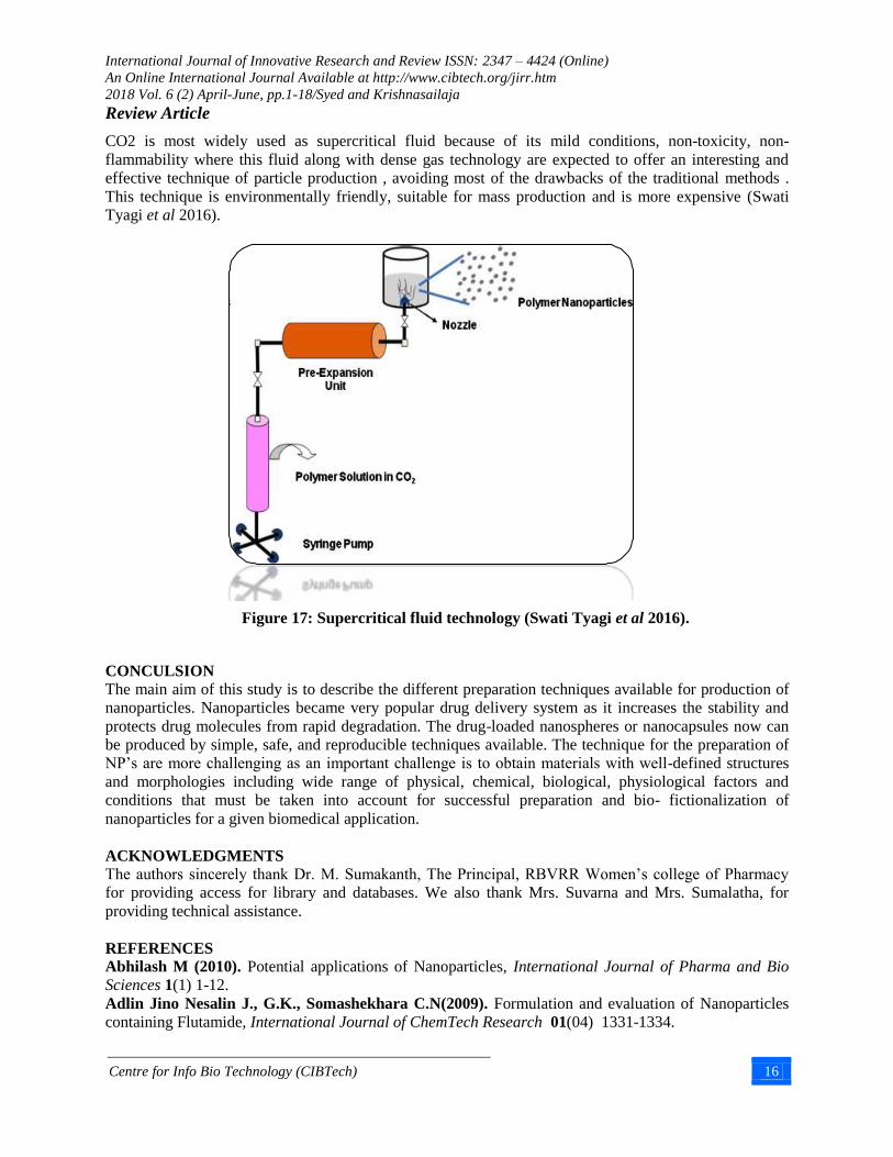
Supercritical fluid technology
Supercritical fluid is defined as a solvent at a temperature above its critical temperature, at which the
single phase regardless of pressure moreover , the technology has been used as an alternative to prepare
biodegradable micro and nanoparticles because supercritical fluids are environmentally safe (Swati Tyagi
et al 2016). The need to develop environmentally safer methods for the production of nanoparticles has
motivated research on the utility of supercritical fluids as more environmental friendly solvents, with the
potential to produce nanoparticles with high purity and without any trace of organic solvent (Swati Tyagi
et al 2016).
Two principles have been developed for the production of nanoparticles using supercritical fluids:
(Hemant K.S. Yadav 2012).
1. Rapid expansion of supercritical solution (RESS) (Hemant K.S. Yadav 2012).
2. Rapid expansion of supercritical solution into liquid solvent (RESOLV) (Hemant K.S. Yadav 2012).
Figure 16: Dialysis (Swati Tyagi et al 2016)
International Journal of Innovative Research and Review ISSN: 2347 – 4424 (Online)
An Online International Journal Available at http://www.cibtech.org/jirr.htm
2018 Vol. 6 (2) April-June, pp.1-18/Syed and Krishnasailaja
Review Article
Centre for Info Bio Technology (CIBTech) 16
CO2 is most widely used as supercritical fluid because of its mild conditions, non-toxicity, non-
flammability where this fluid along with dense gas technology are expected to offer an interesting and
effective technique of particle production , avoiding most of the drawbacks of the traditional methods .
This technique is environmentally friendly, suitable for mass production and is more expensive (Swati
Tyagi et al 2016).
CONCULSION
The main aim of this study is to describe the different preparation techniques available for production of
nanoparticles. Nanoparticles became very popular drug delivery system as it increases the stability and
protects drug molecules from rapid degradation. The drug-loaded nanospheres or nanocapsules now can
be produced by simple, safe, and reproducible techniques available. The technique for the preparation of
NP’s are more challenging as an important challenge is to obtain materials with well-defined structures
and morphologies including wide range of physical, chemical, biological, physiological factors and
conditions that must be taken into account for successful preparation and bio- fictionalization of
nanoparticles for a given biomedical application.
ACKNOWLEDGMENTS
The authors sincerely thank Dr. M. Sumakanth, The Principal, RBVRR Women’s college of Pharmacy
for providing access for library and databases. We also thank Mrs. Suvarna and Mrs. Sumalatha, for
providing technical assistance.
REFERENCES
Abhilash M (2010). Potential applications of Nanoparticles, International Journal of Pharma and Bio
Sciences 1(1) 1-12.
Adlin Jino Nesalin J., G.K., Somashekhara C.N(2009). Formulation and evaluation of Nanoparticles
containing Flutamide, International Journal of ChemTech Research 01(04) 1331-1334.
Figure 17: Supercritical fluid technology (Swati Tyagi et al 2016).

International Journal of Innovative Research and Review ISSN: 2347 – 4424 (Online)
An Online International Journal Available at http://www.cibtech.org/jirr.htm
2018 Vol. 6 (2) April-June, pp.1-18/Syed and Krishnasailaja
Review Article
Centre for Info Bio Technology (CIBTech) 17
Amrit Rai, Josphine Jenifer (2017). Nanoparticles in therapeutic applications and role of albumin and
casein nanoparticles in cancer therapy, sciendo 11 1.
Anto Godwin M and K. Mahitha Shri (2015). Nanoparticles and their applications -A mini review,
International Journal of Research in Engineering and Bioscience 3(5) 11- 29.
Darren Yohan and B. Devika Chithrani (2014). Applications of Nanoparticles in Nanomedicine,
Journal of Biomedical Nanotechnology 10 2371–2392.
Deepak N.Kapoor (2012) . Nanorobotics, DHR International Journal Of Pharmaceutical Sciences
(DHR-IJPS), Vol. 2, ( 1).
Hemant K.S. Yadav( 2012) .Different techniques for preparation of polymeric nanoparticles- A Review,
Asian Journal of Pharmaceutical and Clinical Research 5 Suppl 3.
Jain N. K. (2001). Advances in Controlled and Novel Drug Delivery, (CBS Publishers and Distributers,
New Delhi, India), 408.
Khar R.K., S.P.V (2002). Controlled Drug Delivery System-concepts and advances. 1'st ed. 2002, (CBS
publishers and Distributers, New Delhi, India).
Kreuter J. (1994). Nanoparticles Colloidal Drug Delivery Systems, edited by Kreuter, J. New York:
Marcel Dekker, 261‐276.
Kreuter, J.O (2007). Historical Perspectives Nanoparticles—a historical perspective. International
Journal of Pharmaceutics 331 1-10.
Mahmoud Elsabahy (2012). Design of polymeric nanoparticles for biomedical delivery applications,
Chemical Society Reviews, 41, 2545-2561.
Mohanraj VJ and Y. Chen (2006). Nanoparticles - A Review, Tropical Journal of Pharmaceutical
Research 5 1 561-573.
Nagavarma B V N, H.K.S.Y., Ayaz A, Vasudha L.S, Shivakumar H. G (2012). Different techniques
for preparation of Polymeric Nanoparticles- A review. Asian Journal of Pharmaceutical and Clinical
Research 05(03) 16-23.
Namita Rajput (2015). Methods Of Preparation Of Nanoparticles – A Review, International Journal Of
Advances In Engineering & Technology 7(4) 1806-1811, (2015).
Niba Ibrahim, K. Krishnakumar ( 2018), An overview on nanosphere drug delivery, European Journal
Of Pharmaceutical And Medical Research, ejpmr, 2018,5(4), 192-198.
P.amareshwar and A.Krishna sailaja (2011). Different techniques used for the preparation of
nanoparticles using natural polymers and their application, International Journal of Pharmacy and
Pharmaceutical Sciences 3 Suppl 2.
Plank C (2009). Magnetic nanoparticles, Nanomedicine: Silence the target. Nature Nanotechnology.
2009, 4 544 – 545.) (online) Available:
https://www.researchgate.net/publication/261547783_Engineering_Magnetic_Nanoparticles_for_Thermo
-Ablation_and_Drug_Delivery_in_Neurological_Cancers [accessed Sep 15 2018].
Prof. D. sambha reddy, (2015). International journal of pharmaceutical sciences and nanotechnology
.(Pharma book syndicate, Hyderabad, India), vol.8 issue 2015.
Raval Amit J., M.M.P. (2011). Preparation and Characterization of Nanoparticles for Solubility and
Dissolution Rate Enhancement of Meloxicam. International Journal of Pharmaceuticals,. 01(02): 42-49.
Renu tiruwa (2015) . A review on nanoparticles, Indian Journal of Pharmaceutical and Biological
Research , 4(2)27-31 .
Saba Hasan (2015). A Review on Nanoparticles: Their Synthesis and Types Research Journal of Recent
Sciences 4 1-3.
Saikat Das, Rinti Banerjee and Jayesh Bellare (2005). Aspirin Loaded Albumin Nanoparticles by
Coacervation: Implications in Drug Delivery Trends Biomater. Artif. Organs, 18 (2).
SmeetsRm, Dekker Nh et al (2006). Handbook of Nanophysics: Nanomedicine and Nanorobotics, Nano-
Lett, 6, 89-95.

International Journal of Innovative Research and Review ISSN: 2347 – 4424 (Online)
An Online International Journal Available at http://www.cibtech.org/jirr.htm
2018 Vol. 6 (2) April-June, pp.1-18/Syed and Krishnasailaja
Review Article
Centre for Info Bio Technology (CIBTech) 18
Swati Tyagi and Vinay Kumar Pandey (2016). Nanoparticles: An Overview of Preparation, Journal of
Pharmaceutics and Nanotechnology JPN 4 No. 2.
Ujjwal Nautiyal, Charanjeet Kaur (2017). Targeted Drug Delivery System: Current and Novel
Approach, International Journal of Pharmaceutical and Medicinal Research 5(2) 448-454.
Ujjwal Nautiyal, Charanjeet Kaur (2017). Targeted Drug Delivery System: Current and Novel
Approach, International Journal of Pharmaceutical and Medicinal Research, 5(2) 448-454.
Urvashi Singh, Mohammad Maqbool Dar (2014). Dendrimers: synthetic strategies, properties and
applications, Oriental journal of chemistry, An International Research Journal of Pure & Applied
Chemistry, Vol.30 (3).
Velavan P. and C. Karuppusamy (2015). Nanoparticles as Drug Delivery Systems, Journal of
pharmaceutical sciences and research 7(12),1118-1122.
Vyas S.P, Khar (2002). R.K, Targeted & Controlled drug delivery, Novel carrier systems, (CBS
publishers and Distributers, New Delhi, India) 331-381.