A recent review of citrus flavanone naringenin on ...
44
A recent review of citrus flavanone naringenin on metabolic diseases and its potential sources for high yield-production By: Naymul Karim, Zhenquan Jia, Xiaodong Zheng, Sunliang Cui, and Wei Chen Karim, N., Jia, Z., Zheng, X., Cui, S., Chen, W. (2018). A recent review of citrus flavanone naringenin on metabolic diseases and its potential sources for high yield-production. Trends in Food Science and Technology 79, 35-54. https://doi.org/10.1016/j.tifs.2018.06.012 ***© 2018 Elsevier Ltd. Reprinted with permission. This version of the document is not the version of record. *** This work is licensed under a Creative Commons Attribution- NonCommercial-NoDerivatives 4.0 International License. Abstract: Background: Metabolic syndromes are the multi-metabolic abnormality characterized by hyperlipidemia, obesity, hyperglycemia, diabetes, hypertension, cardiovascular disease, and neuro-dysfunction. Naringenin, a naturally occurring flavanone compound, abundantly found in citrus fruit, has demonstrated diverse biological activities. In this context, the role of naringenin in the treatment of metabolic disease and alternative sources for high-yield production of naringenin have recently drawn full scientific attention and become an important issue in research. Scope and approach: This review focuses on recent findings of naringenin against metabolic disorders including oxidative stress, hyperlipidemia, obesity, diabetes, inflammation, and organ toxicity. Also, this review highlights the potential sources of naringenin production. Key findings and conclusions: Naringenin exerts its protective effect against metabolic diseases through multiple mechanisms including its antioxidant activity by scavenging free radicals, inducing antioxidant enzymes and targeting on phosphoinositide 3-kinase/protein Kinase B/nuclear factor (erythroid-derived 2)-like 2 (PI3K/Akt/Nrf2), nuclear factor (erythroid-derived 2)-like 2/antioxidant responsive element (NRf2/ARE), nuclear factor kappa-light-chain-enhancer of activated B cells (NF-kB), mitogen-activated protein kinase (MAPK), 3-hydroxy-3-methyl- glutaryl-coenzyme A (HMG-CoA) reductase, peroxisome proliferator-activated receptor (PPAR), and nitric oxide-cGMP-protein kinase G-induced KATP channel (NO-cGMP- PKG-KATP). Moreover, microbial production is recommended as a promising alternative method for large-scale production of naringenin. In conclusion, naringenin is a promising compound for the prevention and management of metabolic diseases. Further clinical studies and trials are needed to prove its protective effects on metabolic syndrome in the human population. Keywords: Naringenin | Metabolic diseases | Antioxidant activity | Potential sources Article: 1. Introduction
Transcript of A recent review of citrus flavanone naringenin on ...

A recent review of citrus flavanone naringenin on metabolic diseases and its potential sources for high yield-production By: Naymul Karim, Zhenquan Jia, Xiaodong Zheng, Sunliang Cui, and Wei Chen Karim, N., Jia, Z., Zheng, X., Cui, S., Chen, W. (2018). A recent review of citrus flavanone naringenin on metabolic diseases and its potential sources for high yield-production. Trends in Food Science and Technology 79, 35-54. https://doi.org/10.1016/j.tifs.2018.06.012 ***© 2018 Elsevier Ltd. Reprinted with permission. This version of the document is not the version of record. ***
This work is licensed under a Creative Commons Attribution-NonCommercial-NoDerivatives 4.0 International License. Abstract: Background: Metabolic syndromes are the multi-metabolic abnormality characterized by hyperlipidemia, obesity, hyperglycemia, diabetes, hypertension, cardiovascular disease, and neuro-dysfunction. Naringenin, a naturally occurring flavanone compound, abundantly found in citrus fruit, has demonstrated diverse biological activities. In this context, the role of naringenin in the treatment of metabolic disease and alternative sources for high-yield production of naringenin have recently drawn full scientific attention and become an important issue in research. Scope and approach: This review focuses on recent findings of naringenin against metabolic disorders including oxidative stress, hyperlipidemia, obesity, diabetes, inflammation, and organ toxicity. Also, this review highlights the potential sources of naringenin production. Key findings and conclusions: Naringenin exerts its protective effect against metabolic diseases through multiple mechanisms including its antioxidant activity by scavenging free radicals, inducing antioxidant enzymes and targeting on phosphoinositide 3-kinase/protein Kinase B/nuclear factor (erythroid-derived 2)-like 2 (PI3K/Akt/Nrf2), nuclear factor (erythroid-derived 2)-like 2/antioxidant responsive element (NRf2/ARE), nuclear factor kappa-light-chain-enhancer of activated B cells (NF-kB), mitogen-activated protein kinase (MAPK), 3-hydroxy-3-methyl-glutaryl-coenzyme A (HMG-CoA) reductase, peroxisome proliferator-activated receptor (PPAR), and nitric oxide-cGMP-protein kinase G-induced KATP channel (NO-cGMP-PKG-KATP). Moreover, microbial production is recommended as a promising alternative method for large-scale production of naringenin. In conclusion, naringenin is a promising compound for the prevention and management of metabolic diseases. Further clinical studies and trials are needed to prove its protective effects on metabolic syndrome in the human population. Keywords: Naringenin | Metabolic diseases | Antioxidant activity | Potential sources Article: 1. Introduction
Metabolic syndromes (MetS) are characterized by a cluster of metabolic abnormalities, such as high blood pressure, hyperglycemia, central adiposity, dyslipidemia, and increase the risk of cardiovascular disease, stroke, and type II diabetes (Kaur, 2014). Over the past decades, MetS has become a major public health issue as well as a clinical challenge for the world and its global prevalence ranges from 10 to 84%, based on lifestyle, environmental, physiological, biochemical, clinical and metabolic factors (Song, Yu, Chang, Wang, & An, 2017). According to National Cholesterol Education Program-Adult Treatment Panel III (NCEP-ATPIII, governed by the National Institutes of Health, United States) guidelines, the overall MetS prevalence in US adults increased from 22 to 34% in the years of 2000–2005 (Beltran-Sanchez, Harhay, Harhay, & McElligott, 2013; Mozumdar & Liguori, 2011); while in Chinese adults, the prevalence was 21.3% in 2009 (Xi, He, Hu, & Zhou, 2013). A recent cohort study on MetS found that childhood MetS and obesity from five years are correlated with approximately 2.4-fold higher risk for adulthood MetS, thereby childhood MetS and overweight have been suggested to be a significant predictor for adulthood MetS (Koskinen et al., 2017). MetS prevalence was shown in upward trends not only in China but also in other Asia Pacific countries. Their study also revealed that females and/with urban residents have the highest MetS prevalence (Ranasinghe, Mathangasinghe, Jayawardena, Hills, & Misra, 2017). For the prevention and cure from major risk factors of MetS, people used various chemical and drug agents, such as Angiotensin-Converting-Enzyme (ACE) inhibitors (Hypertension), Non-Sulfonylurea (Diabetes), and Statin-lipid lowering agent (Hypercholesterolemia). Almost all the drugs produce some noticeable side-effects, such as headache, dizziness, nausea, vomiting, rash, abdominal pain, liver disease, kidney disease, muscle weakness, and lactate acidosis etc. (Aksay, Yanturali, BAYRAM, Hocaoğlu, & Kiyan, 2007; Camerino et al., 2017; Palmer, 2004). Several studies proved the inverse association between dietary polyphenols intake and MetS using body weight, blood pressure, blood glucose, insulin resistance, cholesterol, oxidative stress markers, and inflammatory markers etc. (Chiva-Blanch & Badimon, 2017; Micek et al., 2017; Su, Feng, Zheng, & Chen, 2016; Wu, Tang et al., 2014). Plant-derived polyphenols such as phenolic acids, curcuminoids, stilbenes, lignans, flavonoids, flavonols, flavones, anthocyanins, and others possess potential antioxidant, anti-inflammatory, anti-hypertensive, hepato-protective, cardio-protective, anti-diabetes, and anti-obesity activities (Ayoub et al., 2017; Bao et al., 2016; Chen, Feng et al., 2013; Chen, Shen, Su, & Zheng, 2014; Hu et al., 2018; Issa & Hussen Bule, 2015; Van Hul et al., 2017). An Italian population-based research found that high polyphenols intake reduced the mortality rate by about 30% in older adult subjects (Zamora-Ros et al., 2013) suggesting beneficial effects of polyphenols on ameliorating aging and mortality. The polyphenol intakes of Malaysian and Japanese people are 1218–4323 and 827–2157 mg/day respectively, while the Brazilian people's intake polyphenol is only 451–461 mg/day (Niyogi, 2016). This country-wide variation of polyphenol consumption may depend on plant source, food source, food habit (vegetarian or non-vegetarian), food processing, and others. Among the dietary polyphenols in plants, the highest proportion is flavonoids for approximately 67% (Martin & Appel, 2010). In this review, we provide the current developments on using naringenin against MetS and its associated diseases. We first discuss the characteristics of naringenin including its structural analysis and pharmacokinetics along with its metabolites. We then focus on recent findings related to the protective effects of naringenin against MetS and its associated diseases.

Traditional extraction and purification methods of naringenin synthesis from natural sources are limited by high cost, low yield and are also time-consuming. Naringenin production in microorganisms is thus recommended as one of the alternative method to achieve high yield with low cost. 2. Naringenin Naringenin (2,3-dihydro-5,7-dihydroxy-2-(4-hydroxyphenyl)-4H-1-benzopyran-4-one) is a bitter and colorless compound abundant in citrus fruit, such as lemon, orange, clementine, and grapefruit (Al-Dosari, Ahmed, Al-Rejaie, Alhomida, & Ola, 2017; Erlund, 2004; Esaki et al., 1994). Grapefruit jams obtained by applying different types of heat treatment (e.g., osmotic dehydration, microwave energy, and conventional heating) significantly decreased the fruit phytochemicals (β-carotene and flavonoids) content. Based on their study, the total loss of β-carotene and flavonoids in jams was approximately 53–86% and 33–47%, respectively. It indicates that fruit processing may also affect the naringenin content by degrading the heat-sensitive compound and releasing the bound flavanone to processing environment (Igual, Garcia-Martinez, Camacho, & Martínez-Navarrete, 2013). Naringenin is readily soluble in the binary solution of ethanol and water (Zhang et al., 2013). Naringenin has the typical structure of a flavanone that contains hydroxyl groups (at 5, 7 and 4′ positions). These hydroxyl groups can be substituted by different functional groups to produce structural analogs (Fig. 1) (Erlund, 2004; Krauze-Baranowska et al., 2013). Naringenin has a single chiral center at the C-2 position, which is responsible for its enantiomeric properties (Fig. 1) (Yanez, Andrews, & Davies, 2007).
Fig. 1. Structure of naringenin and its analogs. Several structural analogs of naringenin have been identified from citrus fruit such as naringenin-7-glucoside. Compared to naringenin-7-rhamnoglucoside, naringenin-7-glucoside has exhibited well intestinal absorption (Bredsdorff et al., 2010; Felgines et al., 2000). 4′-O-methyl naringenin and 3′-hydroxy-4′-O-methylnaringenin are two metabolites of naringenin known as isosakuranetin and hesperetin, respectively. Both metabolites were detected in lower amounts (approximately 2% of the total plasma flavanones) in rat plasma after consuming the synthetic

diet containing 0.5% naringin, a naringenin-7-O-neohesperidoside compound containing two rhamnose units at 7′ position of naringenin, for seven days (Silberberg et al., 2006). Sakuranetin (7-O-methylnaringenin) is obtained from rice and Polymnia fruticosa. This compound can also be synthesized from naringenin by naringenin-7-O-methyltransferase (Shimizu et al., 2012). Aromadedrin or dihydrokaempferol is a flavanonol, which is synthesized from naringenin by transferring the plant-derived flavanone-3-hydroxylase enzyme into Escherichia coli (Han et al., 2017; Tu et al., 2016). β-Glucosidases produced from Rhizopus azygosporus Yuan et Jong (a food-grade fungal inoculator for soybean tempeh fermentation) can convert naringenin into eriodictyol sulfate and eriodictyol glucoside within 96 h in culture medium (Gonzales et al., 2016). To date, a limited number of pharmacokinetics studies of naringenin has been published. After the consumption of flavanone glycosides (e.g., naringin), these compounds reach the colon and get hydrolyzed into aglycones (e.g., naringenin) by microflora. These flavanone aglycones are partially absorbed in the intestinal barrier (Felgines et al., 2000; Manach, Morand, Gil-Izquierdo, Bouteloup-Demange, & Remesy, 2003). Partial absorption of flavanone aglycones and glucosides can occur through passive diffusion across the small intestine (Choudhury, Chowrimootoo, Srai, Debnam, & Rice-Evans, 1999; Felgines et al., 2000; Serra, Mendes, Bronze, & Simplício, 2008). Several studies explained that flavanones can be metabolized in the intestinal and hepatic cells, and are then converted into glucuronic-/sulfo-conjugated derivatives prior to excretion (Bourian et al., 1999; Fuhr & Kummert, 1995; Ishii, Furuta, & Kasuya, 1997; Lee & Reidenberg, 1998; Lin, Hou, Tsai, Wang, & Chao, 2014; Wang, Chao, Hou, & Hsiu, 2006). 10 min after naringenin administration (20 mg/kg) to rats, naringenin and its glucuronides were detected in concentrations four times higher in the plasma than in the brain tissue (Peng, Cheng, Huang, Chen, & Tsai, 1998). In another extended study using microdialysis coupled with HPLC analysis, higher concentrations of naringenin were detected in liver and bile compared to the brain in rats (Tsai, 2002). Plasma concentration and urinary excretion of naringenin (estimated by HPLC analysis) can be considered as biomarkers for fruit and vegetable intake (Brevik, Rasmussen, Drevon, & Andersen, 2004; Erlund et al., 2002; Mennen et al., 2008). There are a few reported clinical trials about naringenin on MetS. In a recent study on male endurance athletes, the bioavailability of orange juice was assessed by the quantitative analysis of urinary flavanone metabolites. The results showed that after consumption of 500 ml of orange juice, the total naringenin metabolites reached 76 μM in the urine sample of male endurance athletes (Pereira-Caro et al., 2017). In another trial, after consumption of 250 ml orange juice in Chinese volunteers (23–30 years), the total naringenin in urine reached approximately 22% of intake content within 4–12 h as measured by an ultra-fast liquid chromatography-quadrupole-time-of-flight tandem mass spectrometer (Zeng et al., 2017). Intake of 240 ml orange flavanone beverages (containing 15.41 mg naringenin) in healthy middle-aged men (30–65 years) significantly (P < 0·05) reduced the postprandial endothelial dysfunction as evaluated by flow-mediated dilatation (FMD) of the brachial artery at 0–7 h (Rendeiro et al., 2016). Six-months consumption of grapefruit juice (340 ml/day, containing 210 mg naringenin glycosides) significantly improved the vascular endothelial function in postmenopausal women (Habauzit et al., 2015).

Several studies demonstrated that naringenin possessed different biological activities (Fig. 2). These include antidiabetic activity by reducing hepatic and pancreatic inflammation (Annadurai, Thomas, & Geraldine, 2013); an anti-metastatic effect by enhancing transglutaminase activity and polyamine depletion (Lentini, Forni, Provenzano, & Beninati, 2007); cytotoxic activity by inhibiting lung large cell carcinoma (COR-L23), amelanotic melanoma (C32), prostate adenocarcinoma cells (LNCaP) and human cancer cell proliferation (Tundis, Loizzo, Menichini, Bonesi, & Colica, 2011); neuroprotective effects against colchicine-induced cognitive dysfunction (Kumar, Dogra, & Prakash, 2010); renoprotective effects against cadmium (Cd)-induced renal dysfunction (Renugadevi & Prabu, 2009); anti-ulcerative effect by inhibiting histidine decarboxylase (Parmar, 1983); anti-inflammatory activity by suppressing Toll-like receptor 4/NF-κB signaling (Dou et al., 2013); anti-photocarcinogenic activity by stimulating melanogenesis (Chiang, Lin, Hsiao, Tsai, & Wen, 2011); and anti-bacterial activity by inhibiting Salmonella thypi in comparison with chloramphenicol (Agus, Achmadi, & Mubarik, 2017).
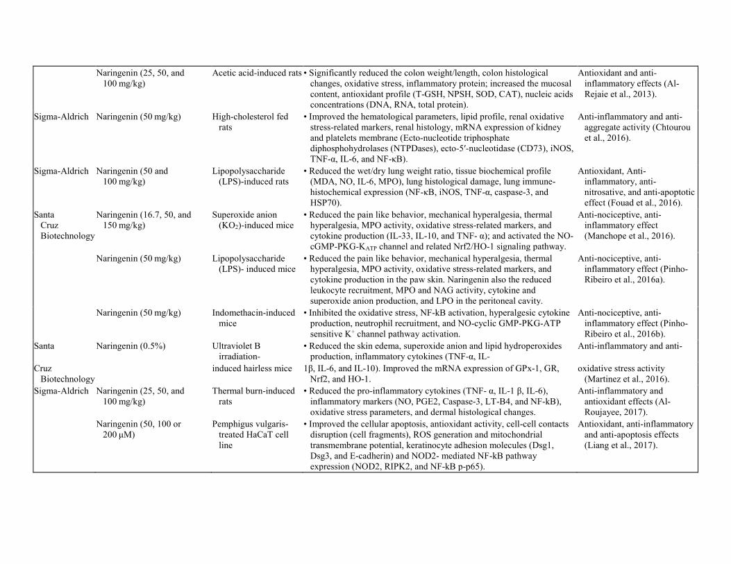
Fig. 2. Biological effects of naringenin. 3. Antioxidant and free radical scavenging activity of naringenin Oxidative stress is caused by an imbalance between oxidants and antioxidants. Overproduction of free radicals and peroxides can damage cell components such as lipid, protein, and DNA, etc. (Chen, Zhuang, Li, Shen, & Zheng, 2013; Zhang et al., 2017; Zhang, Liu, et al., 2017; Li, Bao, & Chen, 2018). Naringenin can potentially protect from oxidative damage via scavenging free radicals (Table 1).
Table 1. Antioxidant, Anti-hyperlipidemic and Anti-obesity effect of Naringenin. Antioxidant and free radical scavenging activity Source Compound/Extract and dose Model (in vitro/in vivo) Research Outcome Significance Zea mays Methanol (80): water (20) extract.
Dose- 100 mg/mL (contain naringenin- 148 mg/kg sample)
Albino male mice • Reduced the MDA level and improved the endogenous antioxidant enzyme (CAT, TPX, and SOD).
Antioxidant activity (Ramos-Escudero et al., 2012).
Xi'an Xiao Cao Botanical Development Co., Ltd
Naringenin - (50 mg/kg) Lead (Pb)-induced adult male rats
• Improved the biochemical parameters, oxidative stress and antioxidant enzyme (MDA, CAT, SOD, GSH, and GPX).
Xeno-protective effect (Wang et al., 2012).
Sigma-Aldrich Naringenin - (20 or 50 mg/kg) Arsenic (As)-induced adult male rats
• Improved the serum biomarkers and tissues oxidative stress/antioxidant enzymes in a dose-dependent manner and restored the tissues histology.
Xeno-protective effect (Mershiba et al., 2013).
Sigma-Aldrich Naringenin - (5 or 10 mg/kg) Arsenic (AS)-induced swiss albino mice
• Significantly normalized the body weights, organ weights, hematological and serum biochemical profile, tissues oxidative stress/antioxidant parameters, and DNA fragmentation.
Xeno-protective effect (Roy et al., 2014).
Citrus aurantium L.
Ethanolic extract - 100 and 300 mg/kg (contain naringenin)
Chromium (VI)-induced female Wistar rats
• Alleviated the oxidative stress/antioxidant enzyme profile of lung tissue and reformed the tissue histology.
Xeno-protective effect (Soudani et al., 2013).
Sigma-Aldrich Naringenin (4 and 8 mg/kg) Cadmium (Cd)-induced swiss albino mice
• Significantly improved the body weights, organ weights, hematological and serum biochemical profile, tissue oxidative stress/antioxidant parameters.
Xeno-protective effect (Das et al., 2016)
Solanum lycopersicum
Naringenin (10 μM) UV light-induced human dermal fibroblasts (CCD-1064Sk) cell line
• Reduced the ROS formation, increased cell viability, carotenoid stability, and HO-1 expression.
Photo-protective effect (Fernandez-Garcia, 2014)
Santa Cruz Biotechnology
Naringenin (20, 30, 100 mg/kg) UVB irradiation-induced hairless mice
• Inhibited the skin edema, neutrophil recruitment, MMP-9 activity, pro-/anti-inflammatory cytokines, and oxidative stress markers (O2•− production and gp91phox expression).
Photo-protective and anti-inflammatory effect (Martinez et al., 2015).
Sigma-Aldrich Naringenin (100 mg/kg) Retinoic acid-induced bone loss rats
• Improved the bone weight coefficient, bone length and diameter, bone ash content, bone calcium and phosphorus content.
Anti-osteoporosis effect (Orsolic et al., 2014).
Sigma-Aldrich Naringenin (50 mg/kg) Doxorubicin-induced dalton's lymphoma ascites (DLA) tumor-bearing mouse
• Improved the blood functional markers, tissues antioxidant enzymes, histopathological changes, and tumor microenvironment.
Chemoprotective effect (Kathiresan et al., 2016).
Naringenin-Oxime (20 and
40 mg/kg) Cisplatin-induced Wistar
albino rats • Restored the biochemical parameters, oxidative
stress/antioxidant profile of serum and tissues; and reduced the DNA damage markers (8-OHdG level, comet formation).
Chemoprotective effect (Koyuncu et al., 2017).
Sigma-Aldrich Naringenin - (50 mg/kg) Gentamycin-induced adult male rats
• Enhanced the audiological measurement (DPOAE and ABR), antioxidant and biochemical profiles, and cochleae histological.
Anti-ototoxicity effect (Kocak et al., 2017).

Sigma-Aldrich Naringenin (100 mg/kg) Benzo [a]pyrene-induced male Wistar rats
• Reduced the oxidative stress markers, inflammatory markers, NF-kB and COX-2 expression.
• Improved the antioxidant enzymes activity and lung histological.
Antioxidant and anti-inflammatory effect (Ali et al., 2017).
Naringenin (50 mg/kg) H2O2-induced male Wistar rats
• Improved the testis weight, oxidative stress/antioxidant markers (MDA, GSH, GST), and testis histology.
Antioxidant activity (Sahin et al., 2017).
Sigma-Aldrich Naringenin (5–100 μM) Paraquat-induced human bronchial epithelial (BEAS-2B) cell line
• Reduced the intracellular ROS production, upregulated the antioxidant-related genes expression, induced the NRF2 expression and its target genes (HO-1 and NQO1).
Cytoprotective effect (Podder et al., 2014)
Naringenin (0.30 mM) Sodium tungstate-exposed
male Wistar rats • Improved the blood δ-aminolevulinic acid dehydratase (ALAD)
activity, oxidative stress and antioxidant markers (blood, liver and spleen tissue).
Xeno-protective effect (Sachdeva & Flora, 2014)
Anti-hyperlipidemic and anti-obesity effect Sigma-Aldrich Experimental diet (naringenin -
0.1%) Male Sprague-Dawley rats • Decreased the plasma and hepatic cholesterol content, fecal
neutral sterols, and inhibited the HMG-CoA reductase and ACAT activities.
Hypolipidemic effect (Lee et al., 1999).
Naringenin (1 and 3%) High fat fed –induced LDL
receptor null (Ldr (−/−)) and C57BL/6J mice
• Improved the hepatic fatty acid oxidation, insulin sensitivity, and glucose tolerance. Decreased the hepatic cholesterol, cholesterol ester synthesis, VLDL derived and endogenous fatty acids.
Hypolipidemic effect (Mulvihill et al., 2009)
Sigma-Aldrich Naringenin - 3% High-/low-cholesterol-induced male (Ldr (−/−)) mice
• Improved dyslipidemia, insulin resistance, inflammation associated with obesity and atherosclerosis disease.
Anti-hyperlipidemic and anti-inflammatory activity (Assini et al., 2013).
Naringenin doses - 0.003, 0.006, and 0.012% in experimental diet.
High fat diet-induced male long-evans hooded rats
• Reduced the plasma, liver, and adipose tissue lipid profile, • Upregulated the fatty acid oxidation-related genes expression
(PPAR-α, CPT-1 and UCP2).
Hypolipidemic and anti-adiposity effects (Cho et al., 2011).
Naringenin (10 mg/kg) High fat diet-induced obese mice
• Improved the serum and hepatic lipid metabolism, lipolysis and lipogenesis-related gene expression.
Anti-obesity effect (Zar Kalai et al., 2013).
Sigma-Aldrich Naringenin (6–50 g/mL) 3T3-L1 cell line • Decreased the lipid accumulation, and protein expression of adipocyte (aP2, ADPN, PPAR, STAT5A).
Anti-adipogenic effect (Richard et al., 2013).
Table 2. Antidiabetic and Anti-inflammatory effect of Naringenin.
Anti-hyperglycemic and antidiabetic effect Source Compound/Extract and dose Model (in vitro/in vivo) Research Outcome Significance Acalypha
wilkesiana Aqueous extract, doses-100,
200 and 300 mg/kg (naringenin-11.12%)
Alloxan-induced diabetic rats
• Reduced the hyperglycemia and ocular oxidative stress markers. • Improved the lipid profile, hepato-specific profile, electrolyte profile,
atherogenic index, and hematological markers.
Retino-/Cardio-protective effects (Ikewuchi et al., 2011).
Sigma-Aldrich Naringenin (50 mg/kg) Alloxan-induced diabetic mice
• Improved the body weight, blood hematological and immunological parameters, and peripheral lymphocytes DNA damage.
Anti-genotoxic effect (Orsolic et al., 2011).

Naringenin (5 μg) Glucose-induced
primary hepatocyte (Rats)
• Improved the cell viability, intracellular antioxidant profile. • Reduced the ROS generation, Cyt-c and AIF/Endo-G translocation,
caspase-9/-3 activation, Bcl-2 family genes expression, DNA damage and chromatin condensation.
Anti-apoptosis effect (Kapoor et al., 2013).
Sigma-Aldrich Naringenin (50 mg/kg) Streptozotocin-induced diabetes rats
• Alleviated the ROS generation, oxidative stress parameters, liver and kidney biomarkers, mitochondrial membrane dysfunction, and apoptotic proteins expression of liver tissue.
Anti-apoptosis effect (Kapoor & Kakkar, 2014).
Sigma-Aldrich naringenin (5 and 10 mg/kg) Streptozotocin-induced diabetes rats
• Reduced the biochemical and oxidative stress parameters, renal degradation, TGF-β1 and IL-1 upregulation, and kidney apoptosis.
Nephroprotective effect (Roy et al., 2016).
Sigma-Aldrich Naringenin (50 mg/kg) Alloxan-induced diabetic mice
• Restore the diabetics induced the hepatic and renal pathological changes.
Hepato-/reno-protective effect (Sirovina, Oršolić, Gregorović, & Končić, 2016)
Sigma-Aldrich Naringenin (50 mg/kg) Streptozotocin–nicotinamide-induced diabetic rats
• Significantly lowered the blood glucose and glycosylated hemoglobin; improved the serum insulin, plasma, and pancreatic tissue antioxidants enzyme; restore the pancreatic histology.
Anti-hyperglycemic and antioxidant activity (Annadurai et al., 2012).
SRL, India Naringenin (25, 50 mg/kg) High-fat diet/high fat emulsion with streptozotocin-induced T2D rats
• Improved the body weight and blood glucose, MDA, nitric oxide and reduced glutathione levels.
• Reduced the locomotor activity and inhibited the cholinesterase activity.
Neuro-protective effect (Rahigude et al., 2012).
Sigma-Aldrich Naringenin (25 and 50 mg/kg) Streptozotocin-induced diabetic rats
• Lessened the serum glucose, pro-inflammatory cytokines, nitric oxide production, oxidative stress marker (MDA); improved the insulin levels, antioxidant enzyme activity, insulin growth factor (IGF) and nerve growth factor (NGF) expression of the sciatic nerve, and sciatic nerve histology.
Neuro-protective effects (Al-Rejaie et al., 2015).
Naringenin (50 mg/kg) Streptozotocin-induced
diabetic rats • Ameliorated the hyperglycemia, oxidative stress/antioxidant markers,
apoptosis markers (Bax/Bcl2, caspase-3), and neuroprotective factors (Brain-derived neurotrophic factor, tropomyosin-related kinase B, and synaptophysin).
Retino-protective effect (Al-Dosari et al., 2017).
Naringenin (50 or 100 mg/kg) High-fat diet
/streptozotocin-induced diabetic rats
• Ameliorated the glucose and lipid metabolism, insulin resistance, oxidative stress and inflammation markers, and thoracic aorta's pathology.
Anti-vasculopathy effect (Ren, et al., 2016).
Cola nitida Naringenin content- 3.6% In-vitro • Inhibited the α-amylase and α-glucosidase enzyme activity and reduced the pancreatic MDA level.
In-vitro anti-diabetic activity (Oboh et al., 2014).
Naringenin (50 mg/kg) Pancreatic endoderm derived from human embryonic stem cells (hESCs)
• Improved the body weight, blood glucose, and insulin level, oxidative stress/antioxidant markers, reproductive outcome.
Anti-diabetic activity (Xing et al., 2016).
Anti-inflammatory effect
Naringenin (25, 50, and
100 mg/kg) Acetic acid-induced rats • Significantly reduced the colon weight/length, colon histological
changes, oxidative stress, inflammatory protein; increased the mucosal content, antioxidant profile (T-GSH, NPSH, SOD, CAT), nucleic acids concentrations (DNA, RNA, total protein).
Antioxidant and anti-inflammatory effects (Al-Rejaie et al., 2013).
Sigma-Aldrich Naringenin (50 mg/kg) High-cholesterol fed rats
• Improved the hematological parameters, lipid profile, renal oxidative stress-related markers, renal histology, mRNA expression of kidney and platelets membrane (Ecto-nucleotide triphosphate diphosphohydrolases (NTPDases), ecto-5′-nucleotidase (CD73), iNOS, TNF-α, IL-6, and NF-κB).
Anti-inflammatory and anti-aggregate activity (Chtourou et al., 2016).
Sigma-Aldrich Naringenin (50 and 100 mg/kg)
Lipopolysaccharide (LPS)-induced rats
• Reduced the wet/dry lung weight ratio, tissue biochemical profile (MDA, NO, IL-6, MPO), lung histological damage, lung immune-histochemical expression (NF-κB, iNOS, TNF-α, caspase-3, and HSP70).
Antioxidant, Anti-inflammatory, anti-nitrosative, and anti-apoptotic effect (Fouad et al., 2016).
Santa Cruz Biotechnology
Naringenin (16.7, 50, and 150 mg/kg)
Superoxide anion (KO2)-induced mice
• Reduced the pain like behavior, mechanical hyperalgesia, thermal hyperalgesia, MPO activity, oxidative stress-related markers, and cytokine production (IL-33, IL-10, and TNF- α); and activated the NO-cGMP-PKG-KATP channel and related Nrf2/HO-1 signaling pathway.
Anti-nociceptive, anti-inflammatory effect (Manchope et al., 2016).
Naringenin (50 mg/kg) Lipopolysaccharide
(LPS)- induced mice • Reduced the pain like behavior, mechanical hyperalgesia, thermal
hyperalgesia, MPO activity, oxidative stress-related markers, and cytokine production in the paw skin. Naringenin also the reduced leukocyte recruitment, MPO and NAG activity, cytokine and superoxide anion production, and LPO in the peritoneal cavity.
Anti-nociceptive, anti-inflammatory effect (Pinho-Ribeiro et al., 2016a).
Naringenin (50 mg/kg) Indomethacin-induced
mice • Inhibited the oxidative stress, NF-kB activation, hyperalgesic cytokine
production, neutrophil recruitment, and NO-cyclic GMP-PKG-ATP sensitive K+ channel pathway activation.
Anti-nociceptive, anti-inflammatory effect (Pinho-Ribeiro et al., 2016b).
Santa Naringenin (0.5%) Ultraviolet B irradiation-
• Reduced the skin edema, superoxide anion and lipid hydroperoxides production, inflammatory cytokines (TNF-α, IL-
Anti-inflammatory and anti-
Cruz Biotechnology
induced hairless mice 1β, IL-6, and IL-10). Improved the mRNA expression of GPx-1, GR,
Nrf2, and HO-1. oxidative stress activity
(Martinez et al., 2016). Sigma-Aldrich Naringenin (25, 50, and
100 mg/kg) Thermal burn-induced
rats • Reduced the pro-inflammatory cytokines (TNF- α, IL-1 β, IL-6),
inflammatory markers (NO, PGE2, Caspase-3, LT-B4, and NF-kB), oxidative stress parameters, and dermal histological changes.
Anti-inflammatory and antioxidant effects (Al-Roujayee, 2017).
Naringenin (50, 100 or 200 μM)
Pemphigus vulgaris-treated HaCaT cell line
• Improved the cellular apoptosis, antioxidant activity, cell-cell contacts disruption (cell fragments), ROS generation and mitochondrial transmembrane potential, keratinocyte adhesion molecules (Dsg1, Dsg3, and E-cadherin) and NOD2- mediated NF-kB pathway expression (NOD2, RIPK2, and NF-kB p-p65).
Antioxidant, anti-inflammatory and anti-apoptosis effects (Liang et al., 2017).

As one of the xenobiotic metals, lead (Pb) can be detectable in all biological systems. Exposure to lead (Pb) also causes cellular oxidative damage of various vital organs, such as liver, kidney, heart, and others. Naringenin was reported to protect significantly (P < 0.05) against lead (Pb)-induced oxidative stress compared to control group as evaluated by biochemical, oxidative stress, and antioxidative markers in the serum, liver, and kidney (Wang et al., 2012). Mershiba, Dassprakash, and Saraswathy (2013) reported that treatment of naringenin (20 or 50 mg/kg/day) for 28 days significantly protects against arsenic (As)-induced oxidative damage in rat liver and kidney via exerting antioxidant effects and radical quenching action. Naringenin treatments also significantly improved the serum and antioxidant enzymes biomarkers in a dose-dependent manner; and restored the tissues pathological changes (Mershiba et al., 2013). Another study also revealed the protective effect of naringenin (5 or 10 mg/kg/day) against arsenic (As)-induced oxidative damage in mice model (Roy et al., 2014). On the other hand, the ethanol extract of Citrus aurantium L. at doses of 100 and 300 mg/kg/day (naringenin as the main component of citrus fruit) showed the anti-oxidative effects against chromium (VI)-induced lung dysfunction. The extract significantly alleviated the oxidative stress (MDA and protein carbonyl levels) and antioxidant (GSH, sulfhydryl groups, NPSH, vitamin C, vitamin E, Na+ K+ ATPase, CAT, GPx, and SOD) profile of lung tissue, and reformed the tissue histology (Soudani et al., 2013). Naringenin alleviates the cadmium (Cd)-induced hepatic and renal toxicity in mice (Das, Roy, Das, Bhattacharya, & Haldar, 2016). Tungsten (W) is a heavy metal and has been widely used in various household products and instruments. Human beings can also easily be exposed to this metal by the environment. Treatment with naringenin was found to attenuate the oxidative stress and antioxidant markers of blood, liver, and spleen in rats exposed to sodium tungstate (3 months, 100 ppm in drinking water) (Sachdeva & Flora, 2014). Naringenin reduced the sodium tungstate-induced oxidative stress through exerting metal chelating activity. Methanol: water (80:20) extract of Zea mays (naringenin-148 mg/kg) exhibited in vitro antioxidant activity. Mice treated with such extracts also showed reduced malondialdehyde (MDA) levels and improved endogenous enzymes activities (Catalase (CAT), total peroxidase (TPX), superoxide dismutase (SOD)) in the liver, kidney, and brain (Ramos-Escudero, Muñoz, Alvarado-Ortíz, Alvarado, & Yánez, 2012). It is recommended to isolate the naringenin compound from purple corn extracts to evaluate the specific antioxidant properties. Advanced glycation endproducts (AGEs) produces the cellular oxidative damage and plays causative roles in the development of different chronic diseases. Naringenin exerted anti-glycation activity by inhibiting total fluorescent AGEs and non-fluorescent carboxymethyl lysine (CML) formation (Zhang, Hu, Chen, & Wang, 2014). As a result, naringenin can reduce the thermal protein glycation, which could be a promising way to alleviate the oxidative stress. UV light exposure to the skin causes aging or photocarcinogenesis by the formation of reactive oxygen species (ROS) known as photooxidative stress. Solanum lycopersicum (tomato) contains lycopene, alpha-tocopherol, naringenin and other compounds. Co-incubation of lycopene (1 μM) and naringenin (10 μM) exhibited a protective effect against UV light-induced human dermal fibroblasts cell (HDF) oxidative damage. It was evaluated by a decrease in ROS formation and an increase in cell viability, carotenoid stability, and HO-1 expression (Fernandez-Garcia, 2014). It is indicated that naringenin could prevent oxidative degradation of carotenoids via inducing HO-1 expression. Also, treatment with naringenin (through intraperitoneal injection (i.p.), 20, 30, and 100 mg/kg) protected the hairless skin of mice from ultraviolet B (UVB)

irradiation-induced inflammation and oxidative damage. This treatment suppressed skin edema, neutrophil recruitment, MMP-9 activity, pro-inflammatory cytokines, and oxidative stress markers (O2
•− production and gp91phox expression) compared to a non-irradiated control group (Martinez et al., 2015). The compound reduced the skin-damage by enhancing free radicals scavenging activity, ameliorating endogenous antioxidants depletion, and inhibiting production of gp91phox-dependent O2
•− and its derivatives. In a paraquat-induced human bronchial epithelial (BEAS-2B) cell model, naringenin (5–100 μM) showed a cytoprotective effect against paraquat-induced intracellular ROS production. Naringenin upregulated the antioxidant-related gene expression (GPX2, GPX3, GPX5, and GPX7), and activated expression of Nrf2 and its targeted gene (HO-1 and NQO1) (Podder, Song, & Kim, 2014). High dose of retinoic acid play an essential role in inducing osteoporosis through oxidative stress. Naringenin (100 mg/kg) treatment significantly alleviated the oxidative damage and bone mineral contents induced by retinoic acid (80 mg/kg) (Orsolic et al., 2014). In Dalton's Lymphoma Ascites (DLA) tumor-bearing mouse model, treatment with naringenin (50 mg/kg/day) significantly attenuated doxorubicin-induced toxicity and hypoxic condition. Here, the treatment improved the blood functional markers, antioxidant enzymes in tissues (kidney, heart, lung, liver, spleen, and tumor), histopathological changes, and tumor-microenvironment as compared to a doxorubicin-treated group (Kathiresan et al., 2016). During doxorubicin chemotherapy, naringenin supplement prevented the doxorubicin-induced free radical generation and maintained antioxidant levels in the tumor-microenvironment. As a newly derived compound from naringenin, naringenin-oxime exhibited the greater anticancer potential than naringenin (Kocyigit, Koyuncu, Dikilitas, Bahadori, & Turkkan, 2016). Moreover, co-treatment with cisplatin (7 mg/kg) and naringenin-oxime (20 and 40 mg/kg) significantly reduced the cisplatin-induced adverse effects such as nephrotoxicity, neurotoxicity, hepatotoxicity, and genotoxicity. The co-treatment modulated the altered oxidative stress, antioxidant, genotoxic markers in serum and organs (Koyuncu, Kocyigit, Gonel, Arslan, & Durgun, 2017). Pretreatment of naringenin-oxime may protect from cisplatin-induced oxidative damage of DNA. Gentamicin is an aminoglycoside antibiotic against Gram-negative bacteria. However, it has potential side-effect, such as ototoxicity. Co-treatment with 120 mg/kg (i.p.) gentamicin and 50 mg/kg (p.o.) naringenin significantly minimized the gentamycin-induced ototoxicity compared to the group given gentamycin alone. The ototoxicity was assessed by distortion product otoacoustic emission (DPOAE) and auditory brainstem response (ABR) measurements, oxidative and antioxidant parameters, histology, and apoptosis (TUNEL assay) of cochleae (Kocak et al., 2017). Naringenin can suppress the gentamicin-induced ototoxicity via scavenging ROS, which are accumulated in the cochlea, and cause the cellular apoptosis and cell death. Benzo [a]pyrene is a potent mutagen and carcinogen from incompletely burned organic matters. Naringenin (100 mg/kg BW) enhanced the antioxidant and anti-inflammatory effects against benzo [a]pyrene-induced oxidative stress and pulmonary toxicity. The antioxidant and anti-inflammatory activity was determined by antioxidant enzyme activity markers, lung injury markers, lung histology, and inflammatory markers (NF-kB and COX-2) (Ali et al., 2017). Here, naringenin lowered the ROS-mediated oxidative damage and inflammation via exerting free radical scavenging activities and modulating NF-kB signaling pathway. In H2O2-induced testicular dysfunction rats, naringenin (50 mg/kg) significantly modulated the testis weight and oxidative stress/antioxidant markers (MDA, GSH, GST), when compared to a H2O2-treated control group. Naringenin supplement alleviates H2O2-induced seminiferous tubules

disorganization, germ cells detachment from the seminiferous epithelium and basal membrane, sloughing of germ cells as well as vacuolization in seminiferous tubules in the testis. (Sahin, Ozkaya, Cuce, Uckun, & Yologlu, 2017). 4. Anti-hyperlipidemic and anti-obesity effect of naringenin Hyperlipidemia is considered the prime symptom of obesity and associated metabolic diseases (Wu, Tang, et al., 2013; Wu, Yu, et al., 2013). Naringenin can effectively ameliorate the dyslipidemic condition and obesity of different biological models (Table 1). 3-Hydroxy-3-methylglutaryl coenzyme A (HMG-CoA) reductase increases the intracellular cholesterol synthesis, whereas the Acyl-coenzyme A: cholesterol O-acyltransferase (ACAT) enzyme catalyzes the cholesterol esterification leading to atherosclerosis disease. Rats fed with experimental diet (containing 0.1% naringenin) for six months significantly lowered the plasma and hepatic cholesterol content, fecal neutral sterols, and inhibited the HMG-CoA reductase and ACAT activities when compared to a control group (received experimental diet without naringenin supplement) (Lee et al., 1999). Naringenin supplementation at 1% and 3% in a Western diet (high-fat diet) for 4 and 30 weeks respectively significantly alleviated dyslipidemia, insulin resistance, glucose intolerance, and obesity compared to the western diet fed groups in LDL receptor knockout (Ldr (−/−)) mice and C57BL/6J mice (Mulvihill et al., 2009). Moreover, a supplement of 3% naringenin in the diet to (Ldr (−/−)) mice significantly prevented cholesterol-induced dyslipidemia, insulin resistance, inflammation associated with the obesity and atherosclerosis disease as compared to control groups (Assini et al., 2013). Naringenin can increase hepatic fatty acid oxidation through PPARs, prevent SREBPs-mediated lipogenesis in both liver and muscle, decrease hepatic cholesterol and cholesterol ester synthesis, reduce both VLDL-derived and endogenously synthesized fatty acids. Cho, Kim, Andrade, Burgess, and Kim (2011) reported that experimental diet treatments (high-fat diet with naringenin - 0.003, 0.006, and 0.012%) to normal rats led to maintenance of normal cholesterol levels. An experimental diet containing naringenin reduced the plasma, liver, and adipose tissue lipid profile, and modulated the fatty acid oxidation-related gene expression as compared to the control group (received only high fat diet) (Cho et al., 2011). Naringenin may reduce the hyperlipidemia and obesity via activating the PPAR-α transcription factor and upregulating fatty acid oxidation-related genes such as CPT-1 and UCP2. Zar Kalai et al. (2013) demonstrated that naringenin (10 mg/kg) exerted anti-obesity effects against high fat diet-induced obese mice as evaluated by enhancing the serum and hepatic lipid metabolism, lipolysis, and lipogenesis-related gene expression, when compared to high-fat diet-fed group (Zar Kalai et al., 2013). Furthermore, in 3T3-L1 cell line, naringenin (6–50 g/mL) inhibited the adipogenesis on murine as evaluated by reduced lipid accumulation and upregulated adipocyte protein expression (aP2, ADPN, PPAR, STAT5A) when compared to a control group (Richard, Amini-Vaughan, Ribnicky, & Stephens, 2013). 5. Antidiabetic effect of naringenin Diabetes is a noncommunicable metabolic disorder which is characterized by sustained hyperglycemia associated with either insulin resistance or absence of insulin production (Gowd, Jia, & Chen, 2017). Naringenin can improve hyperglycemia by reducing insulin resistance and exerting antioxidant activity (Table 2). Acalypha wilkesiana (naringenin-11.12%) is a Southern

Nigerian plant and has been widely used to treat diabetes mellitus. The aqueous extract of Acalypha wilkesiana leaves (doses-100, 200 and 300 mg/kg) treatments for 10 days to alloxan-induced diabetic rats exhibited antidiabetic activity. The treatment significantly (P < 0.05) modulated the plasma markers (glycemia, lipid profile, hepato-specific profile, electrolyte profile, atherogenic index, and hematology) and ocular oxidative stress markers as compared to diabetic control group (Ikewuchi, Onyeike, Uwakwe, & Ikewuchi, 2011). Cola nitida seed extract (3.6% naringenin content) inhibited the carbohydrate-digesting enzyme activities (α-amylase EC50 = 0.34 mg/mL and α-glucosidase EC50 = 0.32 mg/mL). The extract also inhibited the Fe2+-induced lipid peroxidation (MDA) in rat pancreas in a dose-dependent manner, indicating its anti-diabetic activity (Oboh, Nwokocha, Akinyemi, & Ademiluyi, 2014). Hyperglycemia is associated with the production of ROS, which is related to cellular genotoxicity in biological systems. In alloxan (75 mg/kg, i. v.)-induced diabetic mice, naringenin (50 mg/kg, i. p., 7 days) reduced the peripheral lymphocytes DNA damage as evaluated by comet assay. Treatment with naringenin also significantly reduced the body weight, blood hematological and immunological parameters as compared to diabetic mice (Orsolic et al., 2011). Naringenin showed a protective effect against diabetes, possibly by ameliorating oxidative stress via improving antioxidant status. Kapoor, Rizvi, and Kakkar (2013) reported that pretreatment with naringenin (5 μg) protected against high-glucose (40 mM)-induced primary hepatocyte apoptosis as confirmed by improved cell viability (MTT assay), ROS generation, and intracellular antioxidant profile (SOD, CAT, GPx). They also found that naringenin improved the mitochondrial membrane potential (Cyt-c and AIF/Endo-G translocation), and suppressed the caspase-9/3 activation, Bcl-2 family gene expression, intra-nucleosomal DNA damage and chromatin condensation (Kapoor et al., 2013). These results suggested that naringenin may alleviate the hepatic apoptosis of streptozotocin (STZ)-induced diabetes model. In a Wistar rat model, co- and post-treatment with naringenin (50 mg/kg) was also found to alleviate intracellular ROS generation, oxidative stress parameters, liver and kidney biomarkers. The treatment further ameliorated the mitochondrial membrane dysfunction and expression of genes encoding apoptotic proteins (Bax/Bcl-2, Caspase-3/9, AIF/Endo-G) (Kapoor & Kakkar, 2014). As a result, naringenin can prevent high glucose-induced apoptosis via scavenging ROS and modulate mitochondria-mediated apoptotic pathway. Furthermore, naringenin (5 and 10 mg/kg) protected from STZ-induced renal impairment. Treatment with naringenin for 10 weeks also significantly suppressed the biochemical and oxidative stress parameters, renal degradation, TGF-β1/IL-1 upregulation, and kidney apoptosis (TUNEL assay) (Roy, Ahmed, Banerjee, & Saha, 2016). Here, naringenin lowered the hyperglycemia-mediated oxidative damage, cytokines expression, apoptotic events, and thereby alleviated the structural alterations of kidney such as glomerulosclerosis, glomerular basement membrane thickening, proximal and convoluted tubules changes, etc. Sirovina, Orsolic, Gregorovic, and Koncic (2016) proved the hepato- and reno-protective effect of naringenin. Naringenin (50 mg/kg) ameliorated the pathological changes of the liver such as unequal vacuolated, higher vacuolated around central veins than Kiernan's spaces, lymphocyte infiltrations. The treatment also alleviated the kidney architectural changes, e.g. renal corpuscles and tubules changes, narrow bowman's spaces, parietal layer's thickness, and tubule epithelium damage, etc. of diabetic mice compared to normal mice (Sirovina, Oršolić, Gregorović, & Končić, 2016).

STZ and nicotinamide (NA)-induced diabetic model exerts the East-Asian non-obese type II diabetes (T2D) patients-like-phenotype (Nakamura et al., 2006). In single-dose STZ (50 mg/kg) and NA (110 mg/kg)-induced diabetic rats, naringenin (50 mg/kg) treatment ameliorated hyperglycemia and oxidative stress (Annadurai et al., 2012). Naringenin can reduce the diabetes via possibly protecting pancreatic islets (preserve the remaining β-cell mass and reduce the pancreatic islets vacuolization) from ROS-mediated oxidative stress. This compound also stimulated the pancreatic β-cells to produce and secrete extra insulin to maintain glucose homeostasis. Rahigude, Bhutada, Kaulaskar, Aswar, and Otari (2012) reported the neuroprotective effect of naringenin against high-fat diet (HFD)/high-fat emulsion (HFE) with STZ (single dose, 35 mg/kg)-induced T2D rats. Naringenin (25, 50 mg/kg) treatments modulated the body weight, blood glucose, cognitive behavior dysfunction (reduce locomotor activity and inhibit cholinesterase activity), and biochemical changes (decrease MDA, NO, and GSH levels) of diabetic rats (Rahigude et al., 2012). Naringenin can modulate the intracellular high-glucose-induced glucotoxicity in neuron via showing antioxidant activity, which can inhibit the cholinesterase activity-induced memory dysfunction in diabetic condition. In addition, Al-Rejaie et al. (2015) reported that naringenin (25 and 50 mg/kg, 2 weeks) ameliorated STZ (60 mg/kg)-induced diabetic neuropathy by lessening the serum glucose, pro-inflammatory cytokines (TNF-α, IL-1β, IL-6), nitric oxide (NO) production, and oxidative stress markers (MDA). Naringenin also improved the insulin levels, antioxidant enzyme activity (GSH, SOD, CAT, GPx, and GR), insulin growth factor (IGF) and nerve growth factor (NGF) expression of sciatic nerve, and sciatic nerve histology (Al-Rejaie et al., 2015). This study suggested that naringenin showed the neuroprotective effect through increasing the activity of antioxidant enzymes of sciatic nerves. The reason is that the sciatic nerve cells might be more susceptible to damage by high-glucose-induced oxidative stress. Retinal damage is one of the most common microvascular complications of diabetes induced by oxidative stress. Naringenin (50 mg/kg) protected from STZ-induced retina damage by lowering hyperglycemia, oxidative stress markers, and apoptosis markers (Bax/Bcl2, caspase-3). This compound also lowered the neuroprotective factors such as brain-derived neurotrophic factor, tropomyosin-related kinase B and synaptophysin as compared to a diabetic group (Al-Dosari et al., 2017). Naringenin may limit neurodegeneration by providing neurotrophic support to protect from retinal damage in diabetic retinopathy condition by reducing oxidative stress-induced apoptosis. Vascular endothelial dysfunction is the common complication of T2D associated with NO production and free fatty acids (FFA) generation. In HFD/STZ-induced diabetic rats and palmitic acid (PA)-induced human umbilical vein endothelial cells (HUVECs), naringenin treatments (50 or 100 mg/kg for 6 weeks and 30 μM) minimized the vascular dysfunction. Naringenin significantly alleviated the blood glucose and insulin levels, oxidative stress/antioxidant markers, intercellular adhesion molecule-1 (ICAM-1), thoracic aorta's pathology, insulin resistance (IR), and inhibited the in-vitro NF-κB and ICAM-1 mRNA expression (Ren et al., 2016). As a result, naringenin can modulate vascular endothelial dysfunctions in diabetes condition at least in part by suppressing oxidative stress and inflammation. Naringenin itself acts as an enhancer for insulin-secreting pancreatic endoderm (derived from human embryonic stem cells, hESCs) to reduce gestational diabetes. Naringenin treatments to pancreatic endoderm transplanted gestational diabetic mice increased the body weight, modulated the high blood glucose and low insulin levels, and improved the antioxidant status and reproductive outcome compared to the control group (Xing, Yang, & Wu, 2016). 6. Anti-inflammatory activity of naringenin

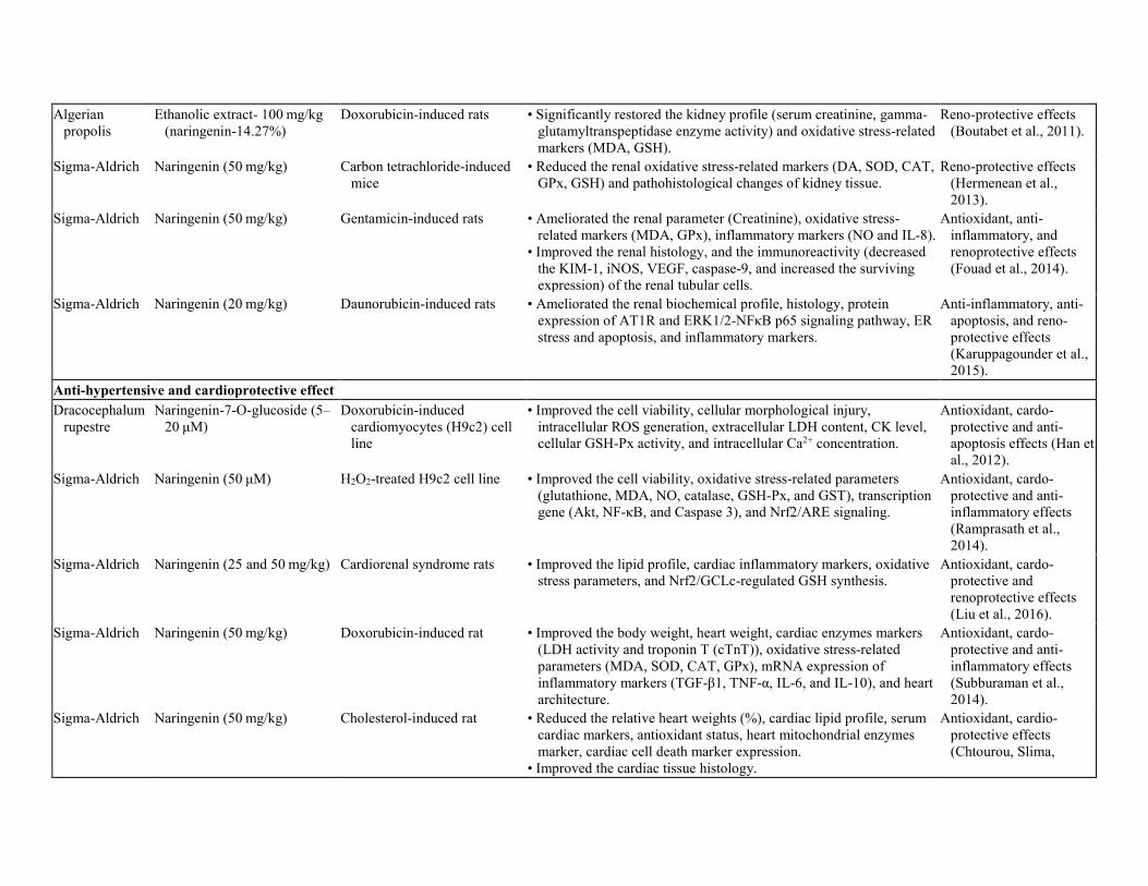
Inflammation is a complex biological response directly connected with oxidative stress, obesity, and diabetes; which can be ameliorated by naringenin (Table 2). In acetic acid- (4%)-induced ulcerative colitis (a subtype of inflammatory bowel disease, IBD), different doses of naringenin (25, 50, and 100 mg/kg) significantly lowered the colon weight/length, colon histological changes such as colitis with superficial erosions, stromal edema, inflammatory cells infiltration, and ulcerative mucosa. Naringenin also reduced the oxidative stress marker (MDA) and inflammatory protein (TNF-α, IL-1β, IL-6, PGE2, and NO); increased the mucosal content, antioxidant profile (T-GSH, NPSH, SOD, and CAT), total protein level, nucleic acid concentration (DNA and RNA) as compared to an acetic acid treated group (Al-Rejaie et al., 2013). There are several mechanisms which might explain anti-inflammatory activities of naringenin including NF-kB and activator protein-1 signals inhibition and increased phosphorylation of ERK 5 and P38 MAPK. Naringenin cured the altered kidney and platelet injury of high cholesterol-fed rats showing an anti-inflammatory and anti-aggregate activity. Co-treatments with cholesterol (10 gm/kg) and naringenin (50 mg/kg) for 3 months resolved the hematological parameters, lipid profile, renal oxidative stress-related markers, and renal histology. The co-treatment also resolved the mRNA expression of kidney and platelets membrane such as Ecto-nucleotide triphosphate diphosphohydrolases (NTPDases), ecto-5′-nucleotidase (CD73), iNOS, TNF-α, IL-6, and NF-kB as compared to high-cholesterol-induced group (Chtourou et al., 2016). Lipopolysaccharide (LPS), an endotoxin from the outer membrane of the Gram-negative bacterial cell wall has been widely used for inducing inflammation. Naringenin (50 and 100 mg/kg) exhibited anti-inflammatory activity against LPS-induced lung injury by decreasing wet/dry lung weight ratio and tissue biochemical profile (MDA, NO, IL-6, and MPO). Naringenin also lowered the lung histological damage (tissue damage, alveolar wall thickening, interstitial edema, and inflammatory cell infiltration) and lung immunohistochemical expression (NF-κB, iNOS, TNF-α, and caspase-3) (Fouad, Albuali, & Jresat, 2016). Naringenin exerted protective effect against acute lung injury via upregulating heat shock protein-70 (HSP70), which significantly reduced the inflammatory biomarkers (blocked the NF-κB pathway) and impaired the apoptotic pathway (caspases). Another study reported that naringenin lessened superoxide anion (KO2)-induced inflammatory pain in mice model via modulating pain-like-behavior, mechanical hyperalgesia, and thermal hyperalgesia. The treatment also reduced the MPO activity, oxidative stress-related markers, and cytokine production (IL-33, IL-10, and TNF-α). In addition, naringenin activated the NO-cGMP-PKG-KATP channel and related Nrf2/HO-1 signaling pathway (Manchope et al., 2016). Naringenin also reduced the LPS-induced inflammatory pain and leukocyte recruitment (Pinho-Ribeiro et al., 2016a). This study further showed that naringenin (50 mg/kg) treatment lowered the acute pain-behaviors and mechanical stimuli (such as carrageenan, capsaicin, and PGE2)-induced inflammation (Pinho-Ribeiro et al., 2016b). In UVB irradiation-induced skin damage, naringenin containing tropical formulation treatment significantly lessened the skin edema, superoxide anion and lipid hydroperoxides production, and inflammatory cytokines (TNF-α, IL-1β, IL-6, and IL-10). This formulation improved the mRNA expression of antioxidant markers such as GPx-1, GR, Nrf2, and HO-1 as compared to an UVB-untreated group (Martinez et al., 2016). Naringenin treatments (25, 50 and 100 mg/kg) healed the thermal burn (10 sec immersion in 90 °C water)-induced skin inflammation. Different doses of compound reversed the pro-inflammatory cytokines (TNF- α, IL-1β, and IL-6), inflammatory markers (NO, PGE2, caspase-3, LT-B4, and NF-kB), oxidative stress parameters, and dermal histological changes (Al-

Roujayee, 2017). Naringenin may contribute to promote tissue regeneration via exhibiting antioxidant activity (Nrf2/HO-1 pathway) and inhibiting inflammation (NF-kB pathway). In pemphigus vulgaris-treated human keratinocyte cell line (HaCaT), naringenin (50, 100 or 200 μM) reduced the cellular damage via modulating cellular apoptosis, antioxidant activity, and cell-cell contacts disruption (cell fragments). Naringenin further modulated the ROS generation and mitochondrial transmembrane potential, expression of keratinocyte adhesion molecules (Dsg1, Dsg3, and E-cadherin) and NOD2-mediated NF-kB pathway (NOD2, RIPK2, and NF-kB p-p65) (Liang et al., 2017). 7. Hepato- and reno-protective effect of naringenin Naringenin showed hepato- and reno-protective effect via exhibiting antioxidant and free radical scavenging activity against oxidative stress induced by different toxicants such as chemicals, drugs, heavy metals, etc. (Table 3). Oxytetracycline is an antibiotic that produces severe hepatic steatosis as an adverse effect. Oxytetracycline administration (i.p., 200 mg/kg for 15 days) significantly increased the hepato-specific biochemical profile such as aspartate transaminase (AST), alanine transaminase (ALT), alkaline phosphatase (ALP), lactate dehydrogenase (LDH), and bilirubin. Oxytetracycline drug also improved the oxidative stress profile such as thiobarbituric acid reactive substances (TBARS) and lipid hydroperoxides, reduced the antioxidant profile (Vit-C and -E, GSH, SOD, CAT, and GPx), and altered the hepatic histology. However, oxytetracycline-induced hepatotoxicity was ameliorated by naringenin (50 mg/kg) treatment by reducing lipid peroxidation and enhancing cellular antioxidant defense system (Pari & Gnanasoundari, 2006). Jayaraman, Veerappan, and Namasivayam (2009) also reported the hepatoprotective effect of naringenin against ethanol (20%, 6 g/kg)-induced hepatotoxicity (Jayaraman et al., 2009). Carbon tetrachloride (CCl4) is widely used to induce hepatic damage of animal model to evaluate the in vivo hepatoprotective effect of new compound or agent. In CCl4-induced acute hepatic failure, naringenin (100 mg/kg) lessened the hepatic biochemical parameters, oxidative stress markers, apoptosis markers (caspase-3, -8, and -9) (Yen, Wu, Lin, Cham, & Lin, 2009). Naringenin treatments further lowered the pro-inflammatory protein (iNOS, COX-2, and TNF-α) via upregulating the anti-oxidative defense protein (HO-1 and Nrf-2) (Esmaeili & Alilou, 2014). Accumulation of heavy metals (such as cadmium (Cd), arsenic (As), lead (Pb), etc.) can damage body organs via cellular oxidative damage by ROS. Naringenin (50 mg/kg) protected against cadmium (Cd)-/arsenic (As)-/lead (Pb)- induced hepatotoxicity via exerting antioxidant scavenging activity (Jain, Yadav, Bozhkov, Padalko, & Flora, 2011; Ozkaya, Sahin, Dag, & Ozkaraca, 2016; Renugadevi & Prabu, 2010). In old-aged rats, naringenin treatment improved the hepatic health, antioxidant status, and membrane phospholipid compositions (n-3 PUFA and n-6/n-3 PUFA ratio) in the liver tissue (Miler et al., 2016). As a result, depletion of n-3 PUFA content and enhancement of n-6/n-3 PUFA ratio is associated with age-related liver diseases, which can be revised by naringenin. Hypercholesterolemia produces free fatty acids and induces lipotoxicity such as hepatic fibrosis or inflammation. Naringenin (50 mg/kg) significantly lowered cholesterol-induced hepatic inflammation via decreasing plasma lipid profile, oxidative stress profile, inflammatory markers (iNOS, Emr1, TNF-α, IL-1 β, IL-6, and NF-κB). This compound further lessened the ECM dysregulation such as matrix metalloproteinases (MMP) −2/-9 expression and pro MMP-9/-2 activities, DNA fragmentation, and histological damages (Chtourou, Fetoui, Jemai, et al., 2015). Here, naringenin markedly lowered the lipid and protein oxidations, improved antioxidant status,

and suppressed ECM degradation by modulating the levels of necrotic inflammation. Simvastatin (SV) is used as a hypocholesterolemic drug that can cause hepatic damage. Co-treatment with SV (20 and 40 mg/kg/day) and naringenin (50 mg/kg/day) exerted hepatoprotective effect via significantly modifying liver functions, oxidative stress markers (MDA, GSH, SOD, GST, CAT), DNA fragmentation, and histopathological changes compared to vehicle control and SV control (Motawi, Teleb, El-Boghdady, & Ibrahim, 2014). Thus, concurrent treatment with SV and naringenin can manage the SV-induced hepatotoxicity, which could be an effective treatment for hypercholesterolemia patients. Isoniazid and rifampicin are the anti-tuberculosis drugs, and both can exert the hepatotoxic effect via generating ROS causing the mitochondrial permeability and hepatocyte death. Naringenin (50 and 100 mg/kg) protected from drugs (isoniazid and rifampicin)-induced oxidative stress and hepatic histological damage via possessing antioxidant and anti-apoptotic activities (inhibited the TUNEL positive cell, caspase-3, Bax/Bcl2 pathway) (Wang, Fan, Zhang, Nie, & Li, 2016). Ethanolic extract of Algerian propolis- 100 mg/kg (contained 14.27% naringenin) significantly minimized the doxorubicin drug (10 mg/kg)-induced kidney toxicity. This extract significantly restored the kidney profile (serum creatinine, γ-glutamyltranspeptidase enzyme activity) as well as oxidative stress-related markers (MDA and GSH) (Boutabet, Kebsa, Alyane, & Lahouel, 2011). This extract reduced the kidney toxicity either by increasing the rate of GSH or by scavenging free radicals. Hermenean et al. (2013) explained that naringenin protected (50 mg/kg) against CCl4-induced renal dysfunction. Naringenin treatments for seven days significantly suppressed the renal oxidative stress-related markers (DA, SOD, CAT, GPx, and GSH). Naringenin treatment significantly reformed the pathohistological changes of kidney tissue such as tubular renal epithelial vacuolization, glomerular atrophy, medulla area necrosis, collagen fibers around renal corpuscles and tubules, lipids proliferation in renal parenchyma, TGF-β1 distribution in glomeruli and tubular areas, and T-cell immunoglobulin (TIM-1) distribution in cortical area (Hermenean et al., 2013). Nephroprotective effect of naringenin was also proved by Fouad, Albuali, Zahran, and Gomaa (2014). They suggested that naringenin protects against the gentamycin-induced kidney toxicity as well as exhibiting antioxidant and anti-inflammatory activities. The results revealed that naringenin treatments alleviated the renal parameter (creatinine), oxidative stress-related markers (MDA and GPx), inflammatory markers (NO and IL-8). They also observed that naringenin improved the renal architectural damages such as tubular necrosis, tubular dilatation, vacuolization, epithelial lining desquamation, cast formation in tubular lumen; and decreased the immunoreactivity (KIM-1, iNOS, VEGF, and caspase-9) of the renal tubular cells (Fouad et al., 2014). In daunorubicin drug-induced genotoxicity, naringenin ameliorated the kidney biochemical profile, renal histology, protein expression of AT1R and ERK1/2-NFκB p65 signaling pathway (p67phox, ET1, ETAR, p-ERK1/2, p-NFκB p65, IFNγ, IL-6, TNFR1, and COX2). Naringenin also ameliorated the ER stress and apoptosis (GRP78, CHOP, cleaved caspase-7, and caspase-12 levels), and inflammatory marker (PPARγ) (Karuppagounder et al., 2015). Based on their research, naringenin can improve the renal function of nephrotoxic rats via attenuating AT1R, ERK1/2-NFκB p65 signaling pathway.

Table 3. Organo-protective effects of Naringenin. Hepato- and reno-protective effect Source Compound/Extract and dose Model (in vitro/in vivo) Research Outcome Significance Sigma-Aldrich Naringenin (50 mg/kg) Oxytetracycline-induce rats • Ameliorated the hepato-specific biochemical profile (AST, ALT,
ALP, LDH), bilirubin), oxidative stress profile (TBARS, lipid hydroperoxides), antioxidant profile (Vit-C and -E, GSH, SOD, CAT, GPx), and histological damage.
Hepatoprotective effect (Pari & Gnanasoundari, 2006).
Sigma-Aldrich Naringenin (50 mg/kg) Ethanol-induced hepatotoxicity • Significantly improved the biochemical parameters, oxidative stress/antioxidant markers, and histopathological changes.
Hepatoprotective effect (Jayaraman et al., 2009).
Sigma-Aldrich Naringenin (100 mg/kg) Carbon tetrachloride-induced rats
• Reduced the hepatic biochemical parameters, oxidative stress markers, apoptosis markers (caspase-3, -8, and -9).
Hepatoprotective effect (Yen et al., 2009).
Sigma-Aldrich Naringenin (50 mg/kg) Carbon tetrachloride-induced rats
• Reduced the hepatic biochemical parameters, oxidative stress markers, pro-inflammatory protein (iNOS, COX-2, and TNF-α).
• Upregulated the anti-oxidative defense protein (HO-1 and Nrf-2).
Anti-inflammatory and anti-hepatotoxic effect (Esmaeili & Alilou, 2014).
Sigma-Aldrich Naringenin (50 mg/kg) Cadmium (Cd)-induced rats • Ameliorated the biochemical profile (AST, ALT, ALP, LDH, GGT, and TB), oxidative stress profile (TBARS, lipid hydroperoxides), antioxidant profile (Vit-C and -E, GSH, SOD, CAT, GPx), and histological damage.
Hepatoprotective effect (Renugadevi & Prabu, 2010).
Sigma-Aldrich Naringenin (50 mg/kg) Arsenic (As)-induced rats • Increased the antioxidant profile (GSH, SOD, and CAT) and decreased the oxidative stress profile (GST and TBARS).
Hepatoprotective effect (Jain et al., 2011)
Merck KGaA Naringenin (50 mg/kg) Lead (Pb)-induced rats • Increased the antioxidant profile and decreased the oxidative stress profile.
Hepatoprotective effect (Ozkaya et al., 2016)
Sigma-Aldrich Naringenin (15 mg/kg) Old-aged Wistar rats • Improved the hepatic antioxidant status (SOD 1/2, GPx, GR, and GSH), membrane phospholipid composition (of n-3 PUFA and n-6/n-3 PUFA ratio).
• Significantly decreased the TBARS without affecting serum biochemical (AST, ALT), and liver histology.
Hepatoprotective effect (Miler et al., 2016).
Sigma-Aldrich Naringenin (50 mg/kg) Cholesterol-induced rats • Lowered the plasma lipid profile, oxidative stress profile, inflammatory markers (iNOS, Emr1, TNF-α, IL-1 β, IL-6, and NF-κB), ECM dysregulation (matrix metalloproteinases (MMP) −2/-9 expression and proMMP-9/-2 activities), DNA fragmentation, and histological damage.
Hypolipidemic, anti-inflammatory and anti-hepatotoxic effect (Chtourou, Fetoui, et al., 2015).
Alfa Aesar Naringenin (50 mg/kg) Adult female Wistar albino rats • Significantly improved the liver function, oxidative stress markers, DNA fragmentation, and histopathological changes.
Hepatoprotective effect (Motawi et al., 2014)
Sigma-Aldrich Naringenin (50 and 100 mg/kg) Drugs (Isoniazid and rifampicin)-induced mice
• Improved the serum biochemical (AST, ALT), liver histology, oxidative stress/anti-oxidant parameters (MDA, GSH, SOD), and hepatocyte apoptosis (TUNEL assay).
Hepatoprotective effect (Wang et al., 2016).
Algerian propolis
Ethanolic extract- 100 mg/kg (naringenin-14.27%)
Doxorubicin-induced rats • Significantly restored the kidney profile (serum creatinine, gamma-glutamyltranspeptidase enzyme activity) and oxidative stress-related markers (MDA, GSH).
Reno-protective effects (Boutabet et al., 2011).
Sigma-Aldrich Naringenin (50 mg/kg) Carbon tetrachloride-induced mice
• Reduced the renal oxidative stress-related markers (DA, SOD, CAT, GPx, GSH) and pathohistological changes of kidney tissue.
Reno-protective effects (Hermenean et al., 2013).
Sigma-Aldrich Naringenin (50 mg/kg) Gentamicin-induced rats • Ameliorated the renal parameter (Creatinine), oxidative stress-related markers (MDA, GPx), inflammatory markers (NO and IL-8).
• Improved the renal histology, and the immunoreactivity (decreased the KIM-1, iNOS, VEGF, caspase-9, and increased the surviving expression) of the renal tubular cells.
Antioxidant, anti-inflammatory, and renoprotective effects (Fouad et al., 2014).
Sigma-Aldrich Naringenin (20 mg/kg) Daunorubicin-induced rats • Ameliorated the renal biochemical profile, histology, protein expression of AT1R and ERK1/2-NFκB p65 signaling pathway, ER stress and apoptosis, and inflammatory markers.
Anti-inflammatory, anti-apoptosis, and reno-protective effects (Karuppagounder et al., 2015).
Anti-hypertensive and cardioprotective effect Dracocephalum
rupestre Naringenin-7-O-glucoside (5–
20 μM) Doxorubicin-induced
cardiomyocytes (H9c2) cell line
• Improved the cell viability, cellular morphological injury, intracellular ROS generation, extracellular LDH content, CK level, cellular GSH-Px activity, and intracellular Ca2+ concentration.
Antioxidant, cardo-protective and anti-apoptosis effects (Han et al., 2012).
Sigma-Aldrich Naringenin (50 μM) H2O2-treated H9c2 cell line • Improved the cell viability, oxidative stress-related parameters (glutathione, MDA, NO, catalase, GSH-Px, and GST), transcription gene (Akt, NF-κB, and Caspase 3), and Nrf2/ARE signaling.
Antioxidant, cardo-protective and anti-inflammatory effects (Ramprasath et al., 2014).
Sigma-Aldrich Naringenin (25 and 50 mg/kg) Cardiorenal syndrome rats • Improved the lipid profile, cardiac inflammatory markers, oxidative stress parameters, and Nrf2/GCLc-regulated GSH synthesis.
Antioxidant, cardo-protective and renoprotective effects (Liu et al., 2016).
Sigma-Aldrich Naringenin (50 mg/kg) Doxorubicin-induced rat • Improved the body weight, heart weight, cardiac enzymes markers (LDH activity and troponin T (cTnT)), oxidative stress-related parameters (MDA, SOD, CAT, GPx), mRNA expression of inflammatory markers (TGF-β1, TNF-α, IL-6, and IL-10), and heart architecture.
Antioxidant, cardo-protective and anti-inflammatory effects (Subburaman et al., 2014).
Sigma-Aldrich Naringenin (50 mg/kg) Cholesterol-induced rat • Reduced the relative heart weights (%), cardiac lipid profile, serum cardiac markers, antioxidant status, heart mitochondrial enzymes marker, cardiac cell death marker expression.
• Improved the cardiac tissue histology.
Antioxidant, cardio-protective effects (Chtourou, Slima,

Makni, Gdoura, & Fetoui, 2015).
Sigma-Aldrich Naringenin (40 μM) H2O2-induced H9c2 cell line • Modulated the ROS levels, DNA damage, the mitochondrial potassium channels, mitochondrial oxidative metabolism activity, estradiol level, and estrogen-related pathways (ER and VDR).
Anti-aging and cardio-protective effects (Da Pozzo et al., 2017).
Sigma-Aldrich Naringenin (50 mg/kg) Monocrotaline-induced pulmonary hypertensive rats.
• Improved the body, heart and lung weights, heart rate, blood pressure, electrocardiography, echocardiography.
• Ameliorated the oxidative stress-related markers, inflammatory markers, nitric oxide synthase, total nitrate/nitrite (NOx) level, apoptosis markers, tissues histological changes.
Anti-oxidant, anti-inflammation, hypotensive, and anti-apoptosis effect (Ahmed et al., 2014).
Schinus terebinthifolius
Methanolic extract (30 mg/kg) (contain naringenin)
Wistar rats and albino male mice
• Reduced blood pressure (Systolic, median, and diastolic), and relax the vasoconstriction
Anti-oxidant and antihypertensive effect (de Lima Gloria et al., 2017).
Neuroprotective effect Sisco research
Lab, India Naringenin (25 and 50 mg/kg) Chronic construction injury-
induced rats • Attenuated the pain induced physical and behavioral changes
(thermal hyperalgesia, cold allodynia, mechanical hyperalgesia, and foot deformity), oxidative stress markers (MDA, GSH, NO).
Antioxidant and anti-neuropathic pain effects (Kaulaskar et al., 2012).
Sigma-Aldrich Naringenin (50 mg/kg) Intracerebroventricular- streptozotocin-induced rats
• Improved the behavior change, spatial learning and memory, choline acetyltransferase (ChAT) expression, and hippocampus histology.
• Decrease the non-enzymatic parameters (4-HNE, MDA, TBARS, H2O2, PC, and GSH), increase the enzymatic activity (GPx, GR, GST, SOD, CAT, and Na+/K + - ATPase).
Neuroprotective effect (Khan et al., 2012).
Sigma-Aldrich Naringenin (100 mg/kg) Beta-amyloid (Aβ)-induced rats
• Improved the behavioral change, hippocampus oxidative stress markers (MDA, nitrate level, and SOD) and apoptosis (DNA fragmentation).
Neuroprotective effect (Ghofrani et al., 2015).
Sigma-Aldrich Naringenin (10 mg/kg) Hypoxia-induced swiss albino mice
• Ameliorated the behavioral changes (poking and rearing behavior), neurological damage by altering the hypoxic brain, protein, and immunoexpression of the cortex, stratum radiatum and hippocampus (VEGF, HIF1α, Hsp70, Hsp90, CHIP panel, caspase-3, and ubiquitination).
Neuroprotective effect (Sarkar et al., 2012).
Sigma-Aldrich Naringenin (100 μM) Carbaryl-induced mouse neuroblastoma cell line (Neuro 2A)
• Improved the cell viability, ROS generation, mitochondrial membrane potential, apoptotic genes expression (Bax/Bcl2, caspase-3).
Neuroprotective effect (Muthaiah et al., 2013).
Sigma-Aldrich Naringenin (50 mg/kg) MCAO-induce focal cerebral ischemia rats
• Improved the behavioral changes (neurological deficits, motor function, grip performance, and somatosensory deficit), upregulate the antioxidant profile, decreases the infarct size, lowered inflammatory parameters (MPO, NO, and cytokines), and improved
Anti-oxidant, anti-inflammatory and neuroprotective effect ((Raza et al., 2013).

brain tissue protein and immunoexpression (SOD, NF-kB, iNOS, COX-2, and Iba-1).
Sigma-Aldrich Naringenin (50 mg/kg) Iron-induced cerebral cortex neurotoxic rats
• Suppressed the oxidative stress-related markers, antioxidant enzyme activities, Na+/K + - ATPase and ChAT activity, DNA fragmentation, and cerebral cortex histology.
Anti-oxidant and neuro- protective effect (Chtourou et al., 2014).
Sigma-Aldrich Naringenin (50 mg/kg) Iron-induced hippocampus neurotoxic rats
• Suppressed the behavioral changes (anxiety-like behavior, locomotor activity and exploratory behavior, anxiolytic-like behavior), oxidative stress-related markers, antioxidant enzyme activities, ectonucleotidase and ChAT activity.
Anti-oxidant and neuro-protective effect (Chtourou, Slima, Gdoura, & Fetoui, 2015)
Sigma-Aldrich Naringenin (0.30 mM) Tungsten (W)-induced rats • Improved the oxidative stress-related parameters (ROS level, MDA, GSH, GSSG, GPx), biogenic amines (dopamine, norepinephrine, and 5-hydroxytryptamine), brain AChE and MAO activity.
Anti-oxidant and neuroprotective effect (Sachdeva et al., 2015).
Naringenin (70 mg/kg for in vivo) and (40 and 80 μM for in vitro)
6-hydroxydopamine (6-OHDA)-induced human neuroblastoma cells line (SH-SY5Y) and mice model
• Improved the cell viability, LDH release, ROS production and GSH level, Nrf2/ARE and JNK/p38 pathway, immunoreactivity and protein expression of tyrosine hydroxylase (striatum and SNc section of the brain).
Anti-oxidant, anti-apoptosis, and neuro-protective effect (Lou et al., 2014).
Naringenin Oxygen and glucose deprivation-cortical neuron cells and neonatal Sprague-Dawley rats
• Improved the neuron cell proliferation, oxidative stress-related profile (ROS, MDA, and SOD), mitochondrial dysfunction, cerebral edema and neurological defects, Nrf2 protein regulation and translation, apoptosis markers (caspase −3/-9, Bax/Bcl2, and DNA fragmentation).
Anti-oxidant, anti-apoptosis, and neuroprotective effect (Wang et al., 2017).
Naringenin (50 mg/kg) d-galactose-induced mice • Improved the body weight and organ index, lessen the behavioral
changes (locomotion, spatial learning, and memory performance), restored the brain histopathological, reduced the neuronal loss and apoptosis (TUNEL and Nissl staining of hippocampus and cortex), oxidative stress-related markers, and PI3K/Akt/Nrf2 pathway.
Anti-oxidant, anti-apoptosis, and neuro-protective effect (Zhang et al., 2017; Zhang, Liu, et al., 2017).

8. Antihypertensive and cardioprotective effect of naringenin Naringenin exhibits well-verified antihypertensive and cardioprotective effects (Table 3). In doxorubicin drug-induced cardiomyocytes (H9c2), naringenin-7-O-glucoside improved the cell viability and cellular glutathione peroxidase (GSH-Px) activity. Naringenin-7-O-glucoside further lessened the cellular morphological injury, intracellular ROS generation and Ca2+ concentration, extracellular lactate dehydrogenase (LDH) content, and creatinine kinase (CK) levels (Han et al., 2012). According to their results, naringenin protected from cardiotoxicity by inhibiting cell death, increasing antioxidant activity, decreasing cellular calcium-overload, and increasing mitochondrial function. Subburaman, Ganesan, and Ramachandran (2014) also explained the protective effect against doxorubicin-induced cardiotoxicity in the rat model. They found out that naringenin (50 mg/kg) treatments significantly modulated the body weight, heart weight, cardiac enzymes markers (LDH activity and Troponin T (cTnT)), oxidative stress-related parameters (MDA, SOD, CAT, and GPx). Naringenin treatment further revised the mRNA expression of inflammatory markers (TGF-β1, TNF-α, IL-6, and IL-10), and cardiac tissue architectural damage such as muscle fibers separation, inflammatory infiltration, myocardial tissue degeneration, and myofibrillar loss, etc. as compared to a doxorubicin-treated group (Subburaman et al., 2014). Also, naringenin reduced the H2O2-treated H9c2 damage by reversing cell viability, oxidative stress-related parameters (glutathione, MDA, NO, catalase, GSH-Px, and GST). Naringenin modulated the gene transcription (Akt, NF-κB, and caspase 3), and Nrf2/ARE signaling-mediated cellular defense system compared to a H2O2-treated group (Ramprasath et al., 2014). Authors found that naringenin showed a cardioprotective effect against oxidative stress-induced inflammatory stimuli by upregulating Nrf2/ARE and Akt signaling pathway, and downregulating NF-kB and caspase 3-dependent apoptosis pathway. Da Pozzo et al. (2017) also proved that naringenin affected H2O2-induced H9c2 cells senescence via modulating the ROS levels, DNA damage. Naringenin also modulated the mitochondrial potassium channels, mitochondrial oxidative metabolism activity, estradiol level and estrogen-related pathways (ER and VDR) (Da Pozzo et al., 2017). Besides in cardiac fibroblasts, naringenin improved the cardiac function via upregulating the Nrf2 and/or γ-glutamylcysteine ligase (GCLc) expression-mediated antioxidant effect (Liu, An, & Gao, 2016). Furthermore, naringenin inhibited the cholesterol-induced cardiotoxicity in rats, by altering the relative heart weights (%), cardiac lipid profile (TC, TG, FFA, and phospholipids), serum cardiac markers (CK and LDH), and antioxidant status (CAT, GPx, SOD, GSH, NPSH, Vit C, and Vit E). Naringenin treatment improved the heart mitochondrial enzymes markers (NADH dehydrogenase, succinate-cytochrome c oxidoreductase, cytochrome c reductase, and ATP synthase), reduced the cardiac cell death markers (TNF-α, Receptor interacting protein 3 (RIP3), and DNA fragmentation), and cardiac tissue histological damage such as disorganized myocardial fibers as compared to a disease control group (Chtourou, Slima, Makni, et al., 2015). Naringenin reduced the cholesterol-induced cardiotoxicity, at least in part, by ameliorating RIP3-TNF-α mediated necroptosis pathway. In monocrotaline (60 mg/kg)-induced pulmonary hypertensive rats, co-treatments with l-arginine (500 mg/kg) and naringenin (50 mg/kg) modified the body, heart, and lung weights, heart rate, blood pressure, electrocardiography, echocardiography. It also improved the oxidative stress-related markers (MDA and GSH), inflammatory markers (MPO and TNF-α), nitric oxide synthase (iNOS and eNOS), total nitrate/nitrite (NOx) level, apoptosis markers (TGF-β and

caspase-3), and histological changes of the right ventricle and lung tissues (Ahmed, Obaid, Zaki, & Agha, 2014). Therefore, this co-administration provided the additional benefits via improving antioxidant activity, preserving NO beneficial effects, stimulating eNOS expression, and inhibiting iNOS overexpression, and its associated inflammatory and apoptosis cascades. Methanolic extract of Schinus terebinthifolius (30 mg/kg) (containing naringenin) showed the antihypertensive effect as evaluated by improved in vitro antioxidant activity, reduced blood pressure (systolic, median, and diastolic), and vasorelaxant effect (measured by rotarod test) (de Lima Gloria et al., 2017). 9. Neuroprotective effect of naringenin Metabolic syndromes are the major risk factors for neurodegeneration and congestive dysfunction, which can be suppressed by naringenin (Table 3). Naringenin (25 and 50 mg/kg) showed protective effect against chronic construction injury (CCI)-induced neuropathic pain in the rat model by attenuating the pain-induced physical and behavioural changes (thermal hyperalgesia, cold allodynia, mechanical hyperalgesia, and foot deformity) as well as oxidative stress markers (MDA, GSH, and NO) (Kaulaskar, Bhutada, Rahigude, Jain, & Harle, 2012). Authors suggested that naringenin lowered the neuropathic pain by inhibition of nitric oxide pathway and amelioration of oxidative stress. Intracerebroventricular-streptozotocin (ICV-STZ) (3 mg/kg; 5 μl per site) injection to rat significantly altered the normal behavior (spatial learning and memory dysfunction), altered the non-enzymatic parameters in the hippocampus (4-HNE, MDA, TBARS, H2O2, PC, and GSH). ICV-STZ also reduced the enzymatic profiles in the hippocampus (GPx, GR, GST, SOD, CAT, and Na+ K+- ATPase), decreased choline acetyltransferase (ChAT) expression, and transformed the hippocampus histology such as shrunken neuron, dense/hyper chromatic nuclei, and clear space between neuron. All abnormalities were reversed by naringenin pre-treatment (50 mg/kg) (Khan et al., 2012). Neuroprotective effect of naringenin against beta-amyloid (Aβ)-induced learning, and memory dysfunction rats were further evaluated by improved behavioral change, hippocampus oxidative stress-related markers (MDA, nitrate level, and SOD) and apoptosis (DNA fragmentation) (Ghofrani et al., 2015). Their findings suggest that naringenin pretreatment attenuated the Aβ-induced Alzheimer's disease via migrating lipid peroxidation and apoptosis, and its beneficial effect was possibly mediated through the estrogenic pathway. In an iron-induced rat model, naringenin lowered the cerebral cortex and hippocampus neurotoxicity by modifying behavioral changes such as anxiety-like behavior, locomotor activity, and exploratory behavior. Naringenin further improved the oxidative stress-related markers, antioxidant enzyme activities, mitochondrial enzyme markers, Na+/K+- ATPase, ChAT, and ectonucleotidase activity, DNA fragmentation, and cerebral cortex histology (Chtourou, Fetoui, & Gdoura, 2014; Chtourou, Slima, Gdoura, et al., 2015). Naringenin can modulate cholinergic neurotransmission resting membrane potential of neurons via improving antioxidant defense system and preserving the cellular integrity. These effects can improve the iron overload-induced anxiogenic-like behavior, possibly by altering AChE and E-NTPDases activities. Exposure of tungsten (W) altered the normal neurological function by producing the neuro-oxidative damage. Naringenin (0.30 mM) treatment to tungsten (W) exposure rat, significantly ameliorated the oxidative stress-related parameters (ROS, MDA, GSH, GSSG, and GPx), biogenic amines (dopamine, norepinephrine, and 5-hydroxytryptamine), brain AChE, and MAO activity

(Sachdeva, Pant, Kushwaha, Bhargava, & Flora, 2015). According to the authors, naringenin exerted a neuroprotective effect by reducing sodium tungstate-induced oxidative stress, which leads to neurotransmitter levels alteration in different brain regions. Hypoxia produced the behavioral changes (poking and rearing behavior) and neurological damage by altering the hypoxic brain (neurons degeneration in the cortex, stratum radiatum, and hippocampus region). Hypoxia also altered the protein and immunoexpression of cortex, stratum radiatum, and hippocampus (VEGF, HIF1α, Hsp70, Hsp90, CHIP panel, caspase-3, and ubiquitination) compared to normoxia group, which is improved by naringenin (10 mg/kg) treatments (Sarkar, Angeline, Anand, Ambasta, & Kumar, 2012). Naringenin has the potential to reverse the hypobaric hypoxia-mediated physiological impairments such as misfolded proteins accumulation in the cellular environment, increased oxidative stress, and induction of apoptosis. All these biological disturbances effect leads to neuronal cell death and neurodegeneration. Wang et al. (2017) unveiled that naringenin could protect against oxygen-glucose-deprivation (OGD)-induced ischemic stroke. The results showed that naringenin treatment alleviated the neuron cell proliferation, biochemical indices (ROS, MDA, and SOD), mitochondrial dysfunction, cerebral oedema, and neurological defects. Naringenin also regulated the localization of the Nrf2 protein and reduced the apoptosis markers (caspase −3/-9, Bax/Bcl2, and DNA fragmentation) (Wang et al., 2017). Naringenin can modulate the ischemic cerebrovascular diseases by lowering apoptosis and oxidative stress through upregulating Nrf2 signaling pathway. Combination of naringenin and lipoic acid (VNAL-100 mg/kg) also exerted the synergistic effect against OGD-induced hypoxia and reoxygenation of neuronal culture as evaluated by LDH, SOD and intracellular antioxidant capacity (Saleh, Saleh, Connell, & Song, 2017). In mouse neuroblastoma cell line (Neuro 2A), naringenin (100 μM) reduced the carbaryl-induced neurotoxicity via improving cell viability, ROS generation, mitochondrial membrane potential, expression of genes involved in apoptosis (Bax/Bcl2 and caspase-3) (Muthaiah, Venkitasamy, Michael, Chandrasekar, & Venkatachalam, 2013). Based on their results from MTT and FACS analysis, naringenin could improve the cell survival through maintaining the mitochondrial membrane potential, upregulating anti-apoptotic gene Bcl-2, and downregulating pro-apoptotic genes (Bax and caspase-3). Naringenin (50 mg/kg) attenuated the ischemic stroke in rat model via improving behavioral changes such as neurological deficits, motor function, grip performance, and somatosensory deficits. The treatment significantly modulated the altered antioxidant profile, infarct size, and inflammatory parameters (MPO, NO, and cytokines). Also, naringenin modified the protein and immuno expression of brain tissue (SOD, NF-kB, iNOS, COX-2, and Iba-1) (Raza et al., 2013). According to their study, pretreatment of naringenin can reduce cerebral ischemia/reperfusion injury by attributing anti-oxidant, free radical-scavenging, and anti-inflammatory activities. These effects are more likely to be associated with the downregulation of NF-kB signaling pathway. In 6-hydroxydopamine (6-OHDA)-induced Parkinson's disease (PD) both in human neuroblastoma SH-SY5Y cells line and in mice model, naringenin compound improved the cell viability, LDH release, ROS production, GSH level, Nrf2/ARE pathway, and apoptotic signaling pathway (JNK/p38). Naringenin also improved the immunoreactivity and protein expression of tyrosine hydroxylase (striatum and SNc section of the brain) (Lou et al., 2014). Naringenin resolved the 6-OHDA-induced Parkinson's disease via activating Nrf2/ARE pathway and inhibiting apoptotic signaling pathway (JNK/p38). Naringenin

showed an anti-aging effect against d-galactose-induced behavioral dysfunction and neurological deficits via modifying body weight and organ index. Naringenin also modified the behavioral changes (locomotion, spatial learning, and memory performance), brain histopathological changes, neuronal loss and apoptosis (TUNEL and Nissl staining of hippocampus and cortex). This compound reduced the oxidative stress-related markers and improved the endogenous antioxidant defense system by activating PI-3K/Akt/Nrf2 pathways (Zhang et al., 2017; Zhang, Liu, et al., 2017).
Fig. 3. Major steps of naringenin production in plants. PAL, Phenyl-alanine ammonia lyase; TAL, Tyrosine ammonia lyase; C4H, Cinnamate 4-hydroxylase; 4CL, 4-coumarate: CoA ligase; CHS, Chalcone synthase; CHI, Chalcone isomerase. 10. Potential sources of naringenin

Naringenin production has become an attractive target because of its diverse biological functions in plants and beneficial effects on human. Normally, naringenin is obtained from citrus fruit using the extraction and purification method. Generally, in plants, naringenin and its derived flavonoid compounds are synthesized from the phenylpropanoid pathway. Except for some flavonoids, all the compounds can be biosynthesized from a common chalcone unit. Malonyl-CoA and 4-coumaroyl-CoA serve as precursor molecules for naringenin biosynthesis. 4-coumaroyl-CoA is produced by the enzymatic conversion of l-tyrosine or phenylalanine to 4-coumaric acid via tyrosine ammonia lyase (TAL)/phenylalanine ammonia lyase (PAL) followed by 4-coumarate: CoA ligase (4CL). The other precursor malonyl-CoA is originated from carbohydrate metabolism pathway. In details, TAL and PAL convert tyrosine and phenylalanine to the phenylpropanoids p-coumaric acid and cinnamic acid, respectively. Another additional enzyme named cinnamate 4-hydroxylase (C4H) further converts the cinnamic acid to p-coumaric acid. After that, 4CL converts p-coumaric acid into 4-coumaroyl-CoA by using one molecule of ATP and one molecule of coenzyme-A (CoA). One molecule of activated 4-coumaroyl-CoA is condensed with three malonyl-CoA by chalcone synthase (CHS) and formed the unstable yellow naringenin chalcone. The resulting unstable naringenin chalcone is converted into stable cyclic naringenin by the chalcone isomerase (CHI) (Fig. 3) (Lewinsohn, Britsch, Mazur, & Gressel, 1989; Martens & Mithöfer, 2005; Wu et al., 2014). Moreover, the addition of a sugar moiety with the naringenin aglycones sequentially converts the naringenin aglycones into the naringenin glycosides (Fig. 1) (Lewinsohn et al., 1989). Distribution of naringenin in different parts of fruit depends on the concentrations of biosynthesis enzymes. For example, in the Solanum lycopersicum peel, the level of CHS and flavanone 3-hydroxylase gene expression is higher than the CHI, indicating the accumulation of high amount of naringenin chalcone (Iijima, Suda, Suzuki, Aoki, & Shibata, 2008). Isolation of naringenin from natural sources for biological research and commercial applications is insufficient and has been engaged with various difficulties, such as low yield value, expensive isolation method, time-consuming processing, seasonal availability, and region-specific availability, etc. For example, Luengo and team (2013) applied pulsed-electric field treatment for extraction of polyphenol from orange peel. Even though their approaches improved the production of naringenin from 1 to 3.1 mg/100 g of fresh orange peels (Luengo, Álvarez, & Raso, 2013), the yield still shows limited output, when it is compared with microbial-assisted production of naringenin (overall yield approximately 474.2 mg/L) (Xu, Ranganathan, Fowler, Maranas, & Koffas, 2011). On the other hand, naringenin can be synthesized through a chemical process. Acylation of 1,3,5-trimethoxybenzene with p-methoxycinnamic acid yielded 29% naringenin (Wang et al., 2015). The alternative method used in the same study exhibited the naringenin yield is of 45–60%. The process includes the conversion of 3,5-dimethoxyphenol into 2-hydroxy-4,6-dimethoxyacetophenone followed by condensation with anisaldehyde to produce 2′-hydroxy-4,4′,6′-trimethoxychalcone. This chalcone is then cyclized and demethylated with aqueous HCl and pyridine hydrochloride to form naringenin (overall yield 45%). Also, condensation of 2-hydroxy-4,6-dimethoxyacetophenone with anisaldehyde can produce 4′,5,7-trimethoxyflavanone, which was then converted into naringenin with 60% yield (Wang et al., 2015). However, these approaches are troubled with high-cost, complex reaction cascades, toxic chemicals, and complex purification processes. As a result, alternative routes leading to naringenin e.g., microbial biosynthesis are important to maximize the production rate, minimize

the cost and time, and to improve product safety as well as quality (Xie, Su, Sun, Zheng, & Chen, 2018). This is because microorganisms and plants share a similar primary metabolism whereas the microorganisms naturally produce the precursors, such as phenylalanine, tyrosine, malonyl-CoA, etc. These precursor molecules are required to produce naringenin as well as other polyphenols using microorganism platforms. For this reason, various strain and process development for microbial naringenin production has become a recent interesting and attractive challenge in metabolic engineering (Milke, Aschenbrenner, Marienhagen, & Kallscheuer, 2018).
Fig. 4. Schematic overview of naringenin against metabolic diseases. 11. Overview and future directions The monotherapy of conventional drug is a challenge to treat various diseases related to metabolic syndrome. Clinical trial study of Vit-E treatment indicated an increase in the incidence of cardiac failure (Investigators, 1999; Lonn et al., 2005). In addition, Vit-C and -E treatments exerted cardiovascular side-effects (Sesso et al., 2008). These limitations of well-known antioxidant molecules create the new possibilities for investigating other bioactive compounds for the treatment of metabolic diseases. The studies discussed in this review showed that naringenin exhibited the potential antioxidant, antihyperlipidemic, anti-obesity, anti-hyperglycemic, anti-diabetic, anti-inflammatory, anti-hypertensive, cardioprotective, hepatoprotective, renoprotective, and neuroprotective activities (Fig. 4). Dietary naringenin and

its derivatives exert effects by targeting various cellular signaling pathways including its antioxidant activity by scavenging free radicals, inducing antioxidant enzymes and targeting on PI3K/Akt/Nrf2, NRf2/ARE, NF-kB, MAPK, HMG-CoA reductase, PPAR and NO-cGMP-PKG-KATP channels. It is important to note that most of the studies have been conducted in either cell type models or animal models (rat and mice). Therefore, further clinical study and trial are required to illustrate the protective effects of naringenin against metabolic syndromes in human. However, such studies should consider the appropriate doses to be used to achieve its protective effects. People may increase the citrus fruit consumption as a source of dietary supplement since naringenin is mostly obtained from the citrus fruit. Due to low yield value, time-consuming extraction, and the high cost of purification steps, the alternative process of naringenin production is needed. Consecutive research on naringenin biosynthesis in Saccharomyces cerevisiae, Streptomyces clavuligerus, and E. coli reported production of pure naringenin with high yields from glucose (Milke et al., 2018). In conclusion, naringenin may be a suitable alternative agent for the treatment of metabolic syndromes (Fig. 4), and its production in microorganism is one of the suitable alternative methods for achieving high-yield. Abbreviations ACAT, Acyl coenzyme A: cholesterol O-acyltransferase; ACE inhibitor, Angiotensin-converting-enzyme inhibitor; ADPN, Adiponutrin; STAT5A, Signal transducer and activator of transcription 5A; AGEs, Advanced glycation endproducts; AIF, Apoptosis-inducing factor; ALP, Alkaline phosphatase; ALT, Alanine transaminase; aP2, Adipocyte protein 2; AST, Aspartate transaminase; Bax, Bcl-2-associated X protein; Bcl-2, B-cell lymphoma 2; CAT, Catalase; CCl4, Carbon tetrachloride; CFA, complete Freund's adjuvant; CML, Carboxymethyllysine; COX-2, Cyclooxygenase-2; CPT-1, Carnitine palmitoyltransferase I; Cyt-c, Cytochrome-c; DA, Dopamine; Dsg, Desmoglein; ENDO-G, Endonuclease-G; eNOS, endothelial nitric oxide synthase; FFAs, Free fatty acids; FMD, Flow-mediated dilatation; GPx, Glutathione peroxidase; GR, Glutathione reductase; GSH, Reduced glutathione; GST, Glutathione S-transferase; HFD, High fat diet; HFE, High fat emulsion; HMG-CoA, 3-hydroxy-3-methylglutaryl coenzyme A; HO-1, Heme oxygenase-1; HPLC, High-performance liquid chromatography; HSP70,70 kilodalton heat shock proteins; ICAM-1, Intercellular adhesion molecule-1; IFNγ, Interferon gamma; IGF, Insulin growth factor; IL-1, Interleukin-1; IL-1β, Interleukin-1 beta; IL-6, Interleukin-6; iNOS, inducible nitric oxide synthase; IR, Insulin receptor; IBD, Inflammatory bowel disease; LDH, Lactate dehydrogenase; LDL, Low-density lipoprotein; LT-B4, Leukotriene B4; MDA, Malondialdehyde; MetS, Metabolic syndrome; MMP-9; Matrix metallopeptidase 9; MPO, Myeloperoxidase; NA, nicotinamide; Na + K + ATPase, Sodium-potassium adenosine triphosphatase; NCEP-ATPIII, National Cholesterol Education Program-Adult Treatment Panel III; NF-κB, Nuclear factor-κB; NGF, Nerve growth factor; NO, nitric oxide; NOD2, Nucleotide-binding oligomerization domain-containing protein 2; NPSH, Non-protein sulfhydryl; NQO1, NAD(P)H:quinone oxidoreductase-1; NRF2, Nuclear factor erythroid 2 (NFE2) related factor 2; O2•−, Superoxide anion; PBQ, Phenyl-p-benzoquinone; PC, Protein carbonyl; PGE2, Prostaglandin E2; PPAR-α, Peroxisome proliferator-activated receptor alpha; RIPK2, Receptor-interacting serine/threonine-protein kinase 2; ROS, Reactive oxygen species; SOD, Superoxide dismutase;

STZ, Streptozotocin; T2D, type 2 diabetes; TGF-β1, Transforming growth factor beta 1; TIM-1, T-cell immunoglobulin, TNF-α, Tumor necrosis factor alpha; TPX, Total peroxidase; TUNEL, Terminal deoxynucleotidyl transferase dUTP nick end labeling; UCP2, Mitochondrial uncoupling protein 2. Conflicts of interest: The authors declare that there are no conflicts of interest. Acknowledgments: This work was supported by Grants from Zhejiang Provincial Natural Science Foundation of China (LR18C200002), National Natural Science Foundation of China (U1703105), and the Fundamental Research Funds for the Central Universities (2017QNA6006). References Agus, S., Achmadi, S. S., & Mubarik, N. R. (2017). Antibacterial activity of naringenin-rich fraction of pigeon pea leaves toward Salmonella thypi. Asian Pacific Journal of Tropical Biomedicine, 7, 725–728. Ahmed, L. A., Obaid, A. A., Zaki, H. F., & Agha, A. M. (2014). Naringenin adds to the protective effect of L-arginine in monocrotaline-induced pulmonary hypertension in rats: Favorable modulation of oxidative stress, inflammation and nitric oxide. European Journal of Pharmaceutical Sciences, 62, 161–170. Aksay, E., Yanturali, S., BAYRAM, B., Hocaoğlu, N., & Kiyan, S. (2007). A rare side effect of metformin: Metformin-induced hepatotoxicity. Turkish Journal of Medical Sciences, 37, 173–175. Al-Dosari, D. I., Ahmed, M. M., Al-Rejaie, S. S., Alhomida, A. S., & Ola, M. S. (2017). Flavonoid naringenin attenuates oxidative stress, apoptosis and improves neurotrophic effects in the diabetic rat retina. Nutrients, 9, 1161. Al-Rejaie, S. S., Abuohashish, H. M., Al-Enazi, M. M., Al-Assaf, A. H., Parmar, M. Y., & Ahmed, M. M. (2013). Protective effect of naringenin on acetic acid-induced ulcerative colitis in rats. World Journal of Gastroenterology, 19, 5633–5644. Al-Rejaie, S. S., Aleisa, A. M., Abuohashish, H. M., Parmar, M. Y., Ola, M. S., Al-Hosaini, A. A., et al. (2015). Naringenin neutralises oxidative stress and nerve growth factor discrepancy in experimental diabetic neuropathy. Neurological Research, 37, 924–933. Al-Roujayee, A. S. (2017). Naringenin improves the healing process of thermally-induced skin damage in rats. Journal of International Medical Research, 45, 570–582. Ali, R., Shahid, A., Ali, N., Hasan, S. K., Majed, F., & Sultana, S. (2017). Amelioration of benzo[a]pyrene-induced oxidative stress and pulmonary toxicity by naringenin in wistar rats: A plausible role of COX-2 and NF-kB. Human & Experimental Toxicology, 36, 349–364.

Annadurai, T., Muralidharan, A. R., Joseph, T., Hsu, M. J., Thomas, P. A., & Geraldine, P. (2012). Antihyperglycemic and antioxidant effects of a flavanone, naringenin, in streptozotocin-nicotinamide-induced experimental diabetic rats. Journal of Physiology & Biochemistry, 68, 307–318. Annadurai, T., Thomas, P. A., & Geraldine, P. (2013). Ameliorative effect of naringenin on hyperglycemia-mediated inflammation in hepatic and pancreatic tissues of Wistar rats with streptozotocin- nicotinamide-induced experimental diabetes mellitus. Free Radical Research, 47, 793–803. Assini, J. M., Mulvihill, E. E., Sutherland, B. G., Telford, D. E., Sawyez, C. G., Felder, S. L., et al. (2013). Naringenin prevents cholesterol-induced systemic inflammation, metabolic dysregulation, and atherosclerosis in Ldlr(-)/(-) mice. Journal of Lipid Research, 54, 711–724. Ayoub, H. M., McDonald, M. R., Sullivan, J. A., Tsao, R., Platt, M., Simpson, J., et al. (2017). The effect of anthocyanin-rich purple vegetable diets on metabolic syndrome in obese Zucker rats. Journal of Medicinal Food, 20, 1240–1249. Bao, T., Xu, Y., Gowd, V., Zhao, J., Xie, J., Liang, W., et al. (2016). Systematic study on phytochemicals and antioxidant activity of some new and common mulberry cultivars in China. Journal of Functional Foods, 25, 537–547. Beltran-Sanchez, H., Harhay, M. O., Harhay, M. M., & McElligott, S. (2013). Prevalence and trends of Metabolic Syndrome in the adult US population, 1999–2010. Journal of the American College of Cardiology, 62, 697–703. Bourian, M., Runkel, M., Krisp, A., Tegtmeier, M., Freudenstein, J., & Legrum, W. (1999). Naringenin and interindividual variability in interaction of coumarin with grapefruit juice. Experimental & Toxicologic Pathology, 51, 289–293. Boutabet, K., Kebsa, W., Alyane, M., & Lahouel, M. (2011). Polyphenolic fraction of Algerian propolis protects rat kidney against acute oxidative stress induced by doxorubicin. Indian Journal of Nephrology, 21, 101–106. Bredsdorff, L., Nielsen, I. L., Rasmussen, S. E., Cornett, C., Barron, D., Bouisset, F., et al. (2010). Absorption, conjugation and excretion of the flavanones, naringenin and hesperetin from alpha-rhamnosidase-treated orange juice in human subjects. British Journal of Nutrition, 103, 1602–1609. Brevik, A., Rasmussen, S. E., Drevon, C. A., & Andersen, L. F. (2004). Urinary excretion of flavonoids reflects even small changes in the dietary intake of fruits and vegetables. Cancer Epidemiology and Prevention Biomarkers, 13, 843–849.

Camerino, G. M., Musumeci, O., Conte, E., Musaraj, K., Fonzino, A., Barca, E., et al. (2017). Risk of myopathy in patients in therapy with Statins: Identification of biological markers in a pilot study. Frontiers in Pharmacology, 8, 500. Chen, W., Feng, L., Shen, Y., Su, H., Li, Y., Zhuang, J., et al. (2013a). Myricitrin inhibits acrylamide-mediated cytotoxicity in human Caco-2 cells by preventing oxidative stress. BioMed Research International, 2013, 724183. Chen, W., Shen, Y., Su, H., & Zheng, X. (2014). Hispidin derived from Phellinus linteus affords protection against acrylamide-induced oxidative stress in Caco-2 cells. Chemico-Biological Interactions, 219, 83–89. Chen, W., Zhuang, J., Li, Y., Shen, Y., & Zheng, X. (2013b). Myricitrin protects against peroxynitrite-mediated DNA damage and cytotoxicity in astrocytes. Food Chemistry, 141, 927–933. Chiang, H. M., Lin, J. W., Hsiao, P. L., Tsai, S. Y., & Wen, K. C. (2011). Hydrolysates of citrus plants stimulate melanogenesis protecting against UV-induced dermal damage. Phytotherapy Research, 25, 569–576. Chiva-Blanch, G., & Badimon, L. (2017). Effects of polyphenol intake on metabolic Syndrome: Current evidences from human trials. Oxidative Medicine and Cellular Longevity, 2017, 5812401. Cho, K. W., Kim, Y. O., Andrade, J. E., Burgess, J. R., & Kim, Y. C. (2011). Dietary naringenin increases hepatic peroxisome proliferators-activated receptor alpha protein expression and decreases plasma triglyceride and adiposity in rats. European Journal of Nutrition, 50, 81–88. Choudhury, R., Chowrimootoo, G., Srai, K., Debnam, E., & Rice-Evans, C. A. (1999). Interactions of the flavonoid naringenin in the gastrointestinal tract and the influence of glycosylation. Biochemical and Biophysical Research Communications, 265, 410–415. Chtourou, Y., Fetoui, H., & Gdoura, R. (2014). Protective effects of naringenin on iron-overload- induced cerebral cortex neurotoxicity correlated with oxidative stress. Biological Trace Element Research, 158, 376–383. Chtourou, Y., Fetoui, H., Jemai, R., Ben Slima, A., Makni, M., & Gdoura, R. (2015a). Naringenin reduces cholesterol-induced hepatic inflammation in rats by modulating matrix metalloproteinases-2, 9 via inhibition of nuclear factor kappaB pathway. European Journal of Pharmacology, 746, 96–105. Chtourou, Y., Kamoun, Z., Zarrouk, W., Kebieche, M., Kallel, C., Gdoura, R., et al. (2016). Naringenin ameliorates renal and platelet purinergic signalling alterations in high-cholesterol fed rats through the suppression of ROS and NF-kB signaling pathways. Food & Function, 7, 183–193.

Chtourou, Y., Slima, A. B., Gdoura, R., & Fetoui, H. (2015b). Naringenin mitigates iron-induced anxiety-like behavioral impairment, mitochondrial dysfunctions, ectonucleotidases and acetylcholinesterase alteration activities in rat Hippocampus. Neurochemical Research, 40, 1563–1575. Chtourou, Y., Slima, A. B., Makni, M., Gdoura, R., & Fetoui, H. (2015c). Naringenin protects cardiac hypercholesterolemia-induced oxidative stress and subsequent necroptosis in rats. Pharmacological Reports, 67, 1090–1097. Da Pozzo, E., Costa, B., Cavallini, C., Testai, L., Martelli, A., Calderone, V., et al. (2017). The citrus flavanone naringenin protects myocardial cells against age-associated damage. Oxidative Medicine and Cellular Longevity, 2017, 9536148. Das, A., Roy, A., Das, R., Bhattacharya, S., & Haldar, P. K. (2016). Naringenin alleviates cadmium-induced toxicity through the abrogation of oxidative stress in swiss albino mice. Journal of Environmental Pathology, Toxicology and Oncology, 35, 161–169. Dou, W., Zhang, J., Sun, A., Zhang, E., Ding, L., Mukherjee, S., et al. (2013). Protective effect of naringenin against experimental colitis via suppression of Toll-like receptor 4/NF-kB signalling. British Journal of Nutrition, 110, 599–608. Erlund, I. (2004). Review of the flavonoids quercetin, hesperetin, and naringenin. Dietary sources, bioactivities, bioavailability, and epidemiology. Nutrition Research, 24, 851–874. Erlund, I., Silaste, M., Alfthan, G., Rantala, M., Kesdniemi, Y., & Aro, A. (2002). Plasma concentrations of the flavonoids hesperetin, naringenin and quercetin in human subjects following their habitual diets, and diets high or low in fruit and vegetables. European Journal of Clinical Nutrition, 56, 891–899. Esaki, S., Nishiyama, K., Sugiyama, N., Nakajima, R., Takao, Y., & Kamiya, S. (1994). Preparation and taste of certain glycosides of flavanones and of dihydrochalcones. Bioscience Biotechnology and Biochemistry, 58, 1479–1485. Esmaeili, M. A., & Alilou, M. (2014). Naringenin attenuates CCl4-induced hepatic inflammation by the activation of an Nrf2-mediated pathway in rats. Clinical and Experimental Pharmacology and Physiology, 41, 416–422. Felgines, C., Texier, O., Morand, C., Manach, C., Scalbert, A., Régerat, F., et al. (2000). Bioavailability of the flavanone naringenin and its glycosides in rats. American Journal of Physiology - Gastrointestinal and Liver Physiology, 279, 1148–1154. Fernandez-Garcia, E. (2014). Photoprotection of human dermal fibroblasts against ultraviolet light by antioxidant combinations present in tomato. Food & Function, 5, 285–290.

Fouad, A. A., Albuali, W. H., & Jresat, I. (2016). Protective effect of naringenin against lipopolysaccharide-induced acute lung injury in rats. Pharmacology, 97, 224–232. Fouad, A. A., Albuali, W. H., Zahran, A., & Gomaa, W. (2014). Protective effect of naringenin against gentamicin-induced nephrotoxicity in rats. Environmental Toxicology and Pharmacology, 38, 420–429. Fuhr, U., & Kummert, A. L. (1995). The fate of naringin in humans: A key to grapefruit juice-drug interactions? Clinical Pharmacology & Therapeutics, 58, 365–373. Ghofrani, S., Joghataei, M. T., Mohseni, S., Baluchnejadmojarad, T., Bagheri, M., Khamse, S., et al. (2015). Naringenin improves learning and memory in an Alzheimer's disease rat model: Insights into the underlying mechanisms. European Journal of Pharmacology, 764, 195–201. Gonzales, G. B., Smagghe, G., Wittevrongel, J., Huynh, N. T., Van Camp, J., & Raes, K. (2016). Metabolism of Quercetin and Naringenin by Food-Grade Fungal Inoculum, Rhizopus azygosporus Yuan et Jong (ATCC 48108). Journal of Agricultural and Food Chemistry, 64, 9263–9267. Gowd, V., Jia, Z., & Chen, W. (2017). Anthocyanins as promising molecules and dietary bioactive components against diabetes – a review of recent advances. Trends in Food Science & Technology, 68, 1–13. Habauzit, V., Verny, M. A., Milenkovic, D., Barber-Chamoux, N., Mazur, A., Dubray, C., et al. (2015). Flavanones protect from arterial stiffness in postmenopausal women consuming grapefruit juice for 6 mo: A randomized, controlled, crossover trial. American Journal of Clinical Nutrition, 102, 66–74. Han, X. Z., Gao, S., Cheng, Y. N., Sun, Y. Z., Liu, W., Tang, L. L., et al. (2012). Protective effect of naringenin-7-O-glucoside against oxidative stress induced by doxorubicin in H9c2 cardiomyocytes. BioScience Trends, 6, 19–25. Han, Y., Huang, K., Liu, Y., Jiao, T., Ma, G., Qian, Y., et al. (2017). Functional analysis of two flavanone-3-hydroxylase genes from camellia sinensis: A critical role in flavonoid accumulation. Genes, 8, 300. Hermenean, A., Ardelean, A., Stan, M., Herman, H., Mihali, C. V., Costache, M., et al. (2013). Protective effects of naringenin on carbon tetrachloride-induced acute nephrotoxicity in mouse kidney. Chemico-Biological Interactions, 205, 138–147. Hu, D., Xu, Y., Xie, J., Sun, C., Zheng, X., & Chen, W. (2018). Systematic evaluation of phenolic compounds and protective capacity of a new mulberry cultivar J33 against palmitic acid-induced lipotoxicity using a simulated digestion method. Food Chemistry, 258, 43–50.

Igual, M., Garcia-Martinez, E., Camacho, M., & Martínez-Navarrete, N. (2013). Jam processing and storage effects on β-carotene and flavonoids content in grapefruit. Journal of Functional Foods, 5, 736–744. Iijima, Y., Suda, K., Suzuki, T., Aoki, K., & Shibata, D. (2008). Metabolite profiling of chalcones and flavanones in tomato fruit. Journal of the Japanese Society for Horticultural Science, 77, 94–102. Ikewuchi, J. C., Onyeike, E. N., Uwakwe, A. A., & Ikewuchi, C. C. (2011). Effect of aqueous extract of the leaves of Acalypha wilkesiana 'Godseffiana' Muell Arg (Euphorbiaceae) on the hematology, plasma biochemistry and ocular indices of oxidative stress in alloxan induced diabetic rats. Journal of Ethnopharmacology, 137, 1415–1424. Investigators, G.-P. (1999). Dietary supplementation with n-3 polyunsaturated fatty acids and vitamin E after myocardial infarction: Results of the GISSI-prevenzione trial. The Lancet, 354, 447–455. Ishii, K., Furuta, T., & Kasuya, Y. (1997). Determination of naringin and naringenin in human urine by high-performance liquid chromatography utilizing solid-phase extraction. Journal of Chromatography B: Biomedical Sciences and Applications, 704, 299–305. Issa, I. A., & Hussen Bule, M. (2015). Hypoglycemic effect of aqueous and methanolic extract of artemisia afra on alloxan induced diabetic swiss albino mice. Evidence-based Complementary and Alternative Medicine, 2015, 752486. Jain, A., Yadav, A., Bozhkov, A. I., Padalko, V. I., & Flora, S. J. (2011). Therapeutic efficacy of silymarin and naringenin in reducing arsenic-induced hepatic damage in young rats. Ecotoxicology and Environmental Safety, 74, 607–614. Jayaraman, J., Veerappan, M., & Namasivayam, N. (2009). Potential beneficial effect of naringenin on lipid peroxidation and antioxidant status in rats with ethanol-induced hepatotoxicity. Journal of Pharmacy and Pharmacology, 61, 1383–1390. Kapoor, R., & Kakkar, P. (2014). Naringenin accords hepatoprotection from streptozotocin induced diabetes in vivo by modulating mitochondrial dysfunction and apoptotic signaling cascade. Toxicology Reports, 1, 569–581. Kapoor, R., Rizvi, F., & Kakkar, P. (2013). Naringenin prevents high glucose-induced mitochondria-mediated apoptosis involving AIF, Endo-G and caspases. Apoptosis, 18, 9–27. Karuppagounder, V., Arumugam, S., Thandavarayan, R. A., Pitchaimani, V., Sreedhar, R., Afrin, R., et al. (2015). Naringenin ameliorates daunorubicin induced nephrotoxicity by mitigating AT1R, ERK1/2-NFkappaB p65 mediated inflammation. International Immunopharmacology, 28, 154–159.

Kathiresan, V., Subburaman, S., Krishna, A. V., Natarajan, M., Rathinasamy, G., Ganesan, K., et al. (2016). Naringenin ameliorates doxorubicin toxicity and hypoxic condition in Dalton's lymphoma Ascites tumor mouse Model: Evidence from electron paramagnetic resonance imaging. Journal of Environmental Pathology, Toxicology and Oncology, 35, 249–262. Kaulaskar, S., Bhutada, P., Rahigude, A., Jain, D., & Harle, U. (2012). Effects of naringenin on allodynia and hyperalgesia in rats with chronic constriction injury-induced neuropathic pain. Journal of Chinese Integrative Medicine, 10, 1482–1489. Kaur, J. (2014). A comprehensive review on metabolic syndrome. Cardiology Research and Practice, 2014, 943162. Khan, M. B., Khan, M. M., Khan, A., Ahmed, M. E., Ishrat, T., Tabassum, R., et al. (2012). Naringenin ameliorates Alzheimer's disease (AD)-type neurodegeneration with cognitive impairment (AD-TNDCI) caused by the intracerebroventricular-streptozotocin in rat model. Neurochemistry International, 61, 1081–1093. Kocak, I., Sarac, S., Aydogan, E., Senturk, E., Akakin, D., Koroglu, K., et al. (2017). Evaluation of the possible protective role of naringenin on gentamicin-induced ototoxicity: A preliminary study. International Journal of Pediatric Otorhinolaryngology, 100, 247–253. Kocyigit, A., Koyuncu, I., Dikilitas, M., Bahadori, F., & Turkkan, B. (2016). Cytotoxic, genotoxic and apoptotic effects of naringenin-oxime relative to naringenin on normal and cancer cell lines. Asian Pacific Journal of Tropical Biomedicine, 6, 872–880. Koskinen, J., Magnussen, C. G., Sinaiko, A., Woo, J., Urbina, E., Jacobs, D. R., et al. (2017). Childhood age and associations between childhood metabolic syndrome and adult risk for metabolic syndrome, type 2 diabetes mellitus and carotid intima media Thickness: The international childhood cardiovascular cohort consortium. Journal of the American Heart Association, 6, 5632. Koyuncu, I., Kocyigit, A., Gonel, A., Arslan, E., & Durgun, M. (2017). The protective effect of naringenin-oxime on cisplatin-induced toxicity in rats. Biochemistry Research International, 2017, 9478958. Krauze-Baranowska, M., Poblocka-Olech, L., Glod, D., Wiwart, M., Zielinski, J., & Migas, P. (2013). HPLC of flavanones and chalcones in different species and clones of Salix. Acta Poloniae Pharmaceutica, 70, 27–34. Kumar, A., Dogra, S., & Prakash, A. (2010). Protective effect of naringin, a citrus flavonoid, against colchicine-induced cognitive dysfunction and oxidative damage in rats. Journal of Medicinal Food, 13, 976–984. Lee, S. H., Park, Y. B., Bae, K. H., Bok, S. H., Kwon, Y. K., Lee, E. S., et al. (1999). Cholesterol-lowering activity of naringenin via inhibition of 3-hydroxy-3-methylglutaryl

coenzyme A reductase and acyl coenzyme A:cholesterol acyltransferase in rats. Annals of Nutrition & Metabolism, 43, 173–180. Lee, Y. S., & Reidenberg, M. M. (1998). A method for measuring naringenin in biological fluids and its disposition from grapefruit juice by man. Pharmacology, 56, 314–317. Lentini, A., Forni, C., Provenzano, B., & Beninati, S. (2007). Enhancement of transglutaminase activity and polyamine depletion in B16-F10 melanoma cells by flavonoids naringenin and hesperitin correlate to reduction of the in vivo metastatic potential. Amino Acids, 32, 95–100. Lewinsohn, E., Britsch, L., Mazur, Y., & Gressel, J. (1989). Flavanone glycoside biosynthesis in citrus chalcone synthase, UDP-glucose: Flavanone-7-O-glucosyl-transferase and-rhamnosyl-transferase activities in cell-free extracts. Plant Physiology, 91, 1323–1328. Liang, J., Halipu, Y., Hu, F., Yakeya, B., Chen, W., Zhang, H., et al. (2017). Naringenin protects keratinocytes from oxidative stress injury via inhibition of the NOD2-mediated NF-κB pathway in pemphigus vulgaris. Biomedicine & Pharmacotherapy, 92, 796–801. Li, Y., Bao, T., & Chen, W. (2018). Comparison of the protective effect of black and white mulberry against ethyl carbamate-induced cytotoxicity and oxidative damage. Food Chemistry, 243, 65–73. de Lima Gloria, L., Barreto de Souza Arantes, M., Menezes de Faria Pereira, S., de Souza Vieira, G., Xavier Martins, C., Ribeiro de Carvalho Junior, A., et al. (2017). Phenolic compounds present Schinus terebinthifolius raddi influence the lowering of blood pressure in rats. Molecules, 22, 1792. Lin, S.-P., Hou, Y.-C., Tsai, S.-Y., Wang, M.-J., & Chao, P.-D. L. (2014). Tissue distribution of naringenin conjugated metabolites following repeated dosing of naringin to rats. Biomedicine, 4, 16. Liu, Y., An, W., & Gao, A. (2016). Protective effects of naringenin in cardiorenal syndrome. Journal of Surgical Research, 203, 416–423. Lonn, E., Bosch, J., Yusuf, S., Sheridan, P., Pogue, J., Arnold, J. M., et al. (2005). Effects of long-term vitamin E supplementation on cardiovascular events and cancer: A randomized controlled trial. Journal of the American Medical Association, 293, 1338–1347. Lou, H., Jing, X., Wei, X., Shi, H., Ren, D., & Zhang, X. (2014). Naringenin protects against 6-OHDA-induced neurotoxicity via activation of the Nrf2/ARE signaling pathway. Neuropharmacology, 79, 380–388. Luengo, E., Álvarez, I., & Raso, J. (2013). Improving the pressing extraction of polyphenols of orange peel by pulsed electric fields. Innovative Food Science & Emerging Technologies, 17, 79–84.

Manach, C., Morand, C., Gil-Izquierdo, A., Bouteloup-Demange, C., & Remesy, C. (2003). Bioavailability in humans of the flavanones hesperidin and narirutin after the ingestion of two doses of orange juice. European Journal of Clinical Nutrition, 57, 235–242. Manchope, M. F., Calixto-Campos, C., Coelho-Silva, L., Zarpelon, A. C., Pinho-Ribeiro, F. A., Georgetti, S. R., et al. (2016). Naringenin inhibits superoxide anion-induced inflammatory Pain: Role of oxidative stress, cytokines, Nrf-2 and the NO-cGMP-PKGKATP channel signaling pathway. PLoS One, 11, 153015. Martens, S., & Mithöfer, A. (2005). Flavones and flavone synthases. Phytochemistry, 66, 2399–2407. Martin, K., & Appel, C. (2010). Polyphenols as dietary supplements: A double-edged sword. Nutrition and Dietary Supplements, 2, 1–12. Martinez, R. M., Pinho-Ribeiro, F. A., Steffen, V. S., Caviglione, C. V., Vignoli, J. A., Barbosa, D. S., et al. (2015). Naringenin inhibits UVB irradiation-induced inflammation and oxidative stress in the skin of hairless mice. Journal of Natural Products, 78, 1647–1655. Martinez, R. M., Pinho-Ribeiro, F. A., Steffen, V. S., Silva, T. C., Caviglione, C. V., Bottura, C., et al. (2016). Topical formulation containing Naringenin: Efficacy against ultraviolet B irradiation-induced skin inflammation and oxidative stress in mice. PLoS One, 11, 146296. Mennen, L., Sapinho, D., Ito, H., Galan, P., Hercberg, S., & Scalbert, A. (2008). Urinary excretion of 13 dietary flavonoids and phenolic acids in free-living healthy subjects-variability and possible use as biomarkers of polyphenol intake. European Journal of Clinical Nutrition, 62, 519–525. Mershiba, S. D., Dassprakash, M. V., & Saraswathy, S. D. (2013). Protective effect of naringenin on hepatic and renal dysfunction and oxidative stress in arsenic intoxicated rats. Molecular Biology Reports, 40, 3681–3691. Micek, A., Grosso, G., Polak, M., Kozakiewicz, K., Tykarski, A., Puch Walczak, A., et al. (2017). Association between tea and coffee consumption and prevalence of metabolic syndrome in Poland - results from the WOBASZ II study (2013–2014). International Journal of Food Sciences & Nutrition, 69, 358–368. Miler, M., Zivanovic, J., Ajdzanovic, V., Orescanin-Dusic, Z., Milenkovic, D., Konic-Ristic, A., et al. (2016). Citrus flavanones naringenin and hesperetin improve antioxidant status and membrane lipid compositions in the liver of old-aged Wistar rats. Experimental Gerontology, 84, 49–60.

Milke, L., Aschenbrenner, J., Marienhagen, J., & Kallscheuer, N. (2018). Production of plant-derived polyphenols in microorganisms: Current state and perspectives. Applied Microbiology and Biotechnology, 102, 1575–1585. Motawi, T. K., Teleb, Z. A., El-Boghdady, N. A., & Ibrahim, S. A. (2014). Effect of simvastatin and naringenin coadministration on rat liver DNA fragmentation and cytochrome P450 activity: An in vivo and in vitro study. Journal of Physiology & Biochemistry, 70, 225–237. Mozumdar, A., & Liguori, G. (2011). Persistent increase of prevalence of metabolic syndrome among U.S. Adults: NHANES III to NHANES 1999–2006. Diabetes Care, 34, 216–219. Mulvihill, E. E., Allister, E. M., Sutherland, B. G., Telford, D. E., Sawyez, C. G., Edwards, J. Y., et al. (2009). Naringenin prevents dyslipidemia, apolipoprotein B overproduction, and hyperinsulinemia in LDL receptor-null mice with diet-induced insulin resistance. Diabetes, 58, 2198–2210. Muthaiah, V. P., Venkitasamy, L., Michael, F. M., Chandrasekar, K., & Venkatachalam, S. (2013). Neuroprotective role of naringenin on carbaryl induced neurotoxicity in mouse neuroblastoma cells. Journal of Pharmacology and Pharmacotherapeutics, 4, 192–197. Nakamura, T., Terajima, T., Ogata, T., Ueno, K., Hashimoto, N., Ono, K., et al. (2006). Establishment and pathophysiological characterization of type 2 diabetic mouse model produced by streptozotocin and nicotinamide. Biological and Pharmaceutical Bulletin, 29, 1167–1174. Niyogi, M. (2016). Polyphenols in a vegetarian diet. American Journal of Clinical Nutrition, 6, 113–116. Oboh, G., Nwokocha, K. E., Akinyemi, A. J., & Ademiluyi, A. O. (2014). Inhibitory effect of polyphenolic-rich extract from Cola nitida (Kolanut) seed on key enzyme linked to type 2 diabetes and Fe2+ induced lipid peroxidation in rat pancreas in vitro. Asian Pacific Journal of Tropical Biomedicine, 4, 405–412. Orsolic, N., Gajski, G., Garaj-Vrhovac, V., Dikic, D., Prskalo, Z. S., & Sirovina, D. (2011). DNA-protective effects of quercetin or naringenin in alloxan-induced diabetic mice. European Journal of Pharmacology, 656, 110–118. Orsolic, N., Goluza, E., Dikic, D., Lisicic, D., Sasilo, K., Rodak, E., et al. (2014). Role of flavonoids on oxidative stress and mineral contents in the retinoic acid-induced bone loss model of rat. European Journal of Nutrition, 53, 1217–1227. Ozkaya, A., Sahin, Z., Dag, U., & Ozkaraca, M. (2016). Effects of naringenin on oxidative stress and histopathological changes in the liver of lead acetate administered rats. Journal of Biochemical and Molecular Toxicology, 30, 243–248.

Palmer, B. F. (2004). Managing hyperkalemia caused by inhibitors of the renin-angiotensin-aldosterone system. New England Journal of Medicine, 351, 585–592. Pari, L., & Gnanasoundari, M. (2006). Influence of naringenin on oxytetracycline mediated oxidative damage in rat liver. Basic and Clinical Pharmacology and Toxicology, 98, 456–461. Parmar, N. S. (1983). The gastric anti-ulcer activity of naringenin, a specific histidine decarboxylase inhibitor. International Journal of Tissue Reactions, 5, 415–420. Peng, H., Cheng, F., Huang, Y., Chen, C., & Tsai, T. (1998). Determination of naringenin and its glucuronide conjugate in rat plasma and brain tissue by high-performance liquid chromatography. Journal of Chromatography B: Biomedical Sciences and Applications, 714, 369–374. Pereira-Caro, G., Polyviou, T., Ludwig, I. A., Nastase, A. M., Moreno-Rojas, J. M., Garcia, A. L., et al. (2017). Bioavailability of orange juice (poly) phenols: The impact of short-term cessation of training by male endurance athletes. American Journal of Clinical Nutrition, 106, 791–800. Pinho-Ribeiro, F. A., Zarpelon, A. C., Fattori, V., Manchope, M. F., Mizokami, S. S., Casagrande, R., et al. (2016b). Naringenin reduces inflammatory pain in mice. Neuropharmacology, 105, 508–519. Pinho-Ribeiro, F. A., Zarpelon, A. C., Mizokami, S. S., Borghi, S. M., Bordignon, J., Silva, R. L., et al. (2016a). The citrus flavonone naringenin reduces lipopolysaccharide-induced inflammatory pain and leukocyte recruitment by inhibiting NF-kB activation. Journal of Nutritional Biochemistry, 33, 8–14. Podder, B., Song, H. Y., & Kim, Y. S. (2014). Naringenin exerts cytoprotective effect against paraquat-induced toxicity in human bronchial epithelial BEAS-2B cells through NRF2 activation. Journal of Microbiology and Biotechnology, 24, 605–613. Rahigude, A., Bhutada, P., Kaulaskar, S., Aswar, M., & Otari, K. (2012). Participation of antioxidant and cholinergic system in protective effect of naringenin against type-2 diabetes-induced memory dysfunction in rats. Neuroscience, 226, 62–72. Ramos-Escudero, F., Muñoz, A. M., Alvarado-Ortíz, C., Alvarado, A., & Yánez, J. A. (2012). Purple corn (Zea mays L.) phenolic compounds profile and its assessment as an agent against oxidative stress in isolated mouse organs. Journal of Medicinal Food, 15, 206–215. Ramprasath, T., Senthamizharasi, M., Vasudevan, V., Sasikumar, S., Yuvaraj, S., & Selvam, G. S. (2014). Naringenin confers protection against oxidative stress through upregulation of Nrf2 target genes in cardiomyoblast cells. Journal of Physiology & Biochemistry, 70, 407–415.

Ranasinghe, P., Mathangasinghe, Y., Jayawardena, R., Hills, A. P., & Misra, A. (2017). Prevalence and trends of metabolic syndrome among adults in the asia-pacific region: A systematic review. BMC Public Health, 17, 101. Raza, S. S., Khan, M. M., Ahmad, A., Ashafaq, M., Islam, F., Wagner, A. P., et al. (2013). Neuroprotective effect of naringenin is mediated through suppression of NF-kB signaling pathway in experimental stroke. Neuroscience, 230, 157–171. Rendeiro, C., Dong, H., Saunders, C., Harkness, L., Blaze, M., Hou, Y., et al. (2016). Flavanone-rich citrus beverages counteract the transient decline in postprandial endothelial function in humans: A randomised, controlled, double-masked, cross-over intervention study. British Journal of Nutrition, 116, 1999–2010. Ren, B., Qin, W., Wu, F., Wang, S., Pan, C., Wang, L., et al. (2016). Apigenin and naringenin regulate glucose and lipid metabolism, and ameliorate vascular dysfunction in type 2 diabetic rats. European Journal of Pharmacology, 773, 13–23. Renugadevi, J., & Prabu, S. M. (2009). Naringenin protects against cadmium-induced oxidative renal dysfunction in rats. Toxicology, 256, 128–134. Renugadevi, J., & Prabu, S. M. (2010). Cadmium-induced hepatotoxicity in rats and the protective effect of naringenin. Experimental & Toxicologic Pathology, 62, 171–181. Richard, A. J., Amini-Vaughan, Z., Ribnicky, D. M., & Stephens, J. M. (2013). Naringenin inhibits adipogenesis and reduces insulin sensitivity and adiponectin expression in adipocytes. Evidence-based Complementary and Alternative Medicine, 2013, 549750. Roy, S., Ahmed, F., Banerjee, S., & Saha, U. (2016). Naringenin ameliorates streptozotocin-induced diabetic rat renal impairment by downregulation of TGF-β1 and IL-1 via modulation of oxidative stress correlates with decreased apoptotic events. Pharmaceutical Biology, 54, 1616–1627. Roy, A., Das, A., Das, R., Haldar, S., Bhattacharya, S., & Haldar, P. K. (2014). Naringenin, a citrus flavonoid, ameliorates arsenic-induced toxicity in Swiss albino mice. Journal of Environmental Pathology, Toxicology and Oncology, 33, 195–204. Sachdeva, S., & Flora, S. J. (2014). Efficacy of some antioxidants supplementation in reducing oxidative stress post sodium tungstate exposure in male wistar rats. Journal of Trace Elements in Medicine & Biology, 28, 233–239. Sachdeva, S., Pant, S. C., Kushwaha, P., Bhargava, R., & Flora, S. J. (2015). Sodium tungstate induced neurological alterations in rat brain regions and their response to antioxidants. Food and Chemical Toxicology, 82, 64–71.

Sahin, Z., Ozkaya, A., Cuce, G., Uckun, M., & Yologlu, E. (2017). Investigation of the effect of naringenin on oxidative stress-related alterations in testis of hydrogen peroxide-administered rats. Journal of Biochemical and Molecular Toxicology, 31. Saleh, T. M., Saleh, M. C., Connell, B. J., & Song, Y. H. (2017). A co-drug conjugate of naringenin and lipoic acid mediates neuroprotection in a rat model of oxidative stress. Clinical and Experimental Pharmacology and Physiology, 44, 1008–1016. Sarkar, A., Angeline, M. S., Anand, K., Ambasta, R. K., & Kumar, P. (2012). Naringenin and quercetin reverse the effect of hypobaric hypoxia and elicit neuroprotective response in the murine model. Brain Research, 24, 59–70. Serra, H., Mendes, T., Bronze, M., & Simplício, A. L. (2008). Prediction of intestinal absorption and metabolism of pharmacologically active flavones and flavanones. Bioorganic & Medicinal Chemistry, 16, 4009–4018. Sesso, H. D., Buring, J. E., Christen, W. G., Kurth, T., Belanger, C., MacFadyen, J., et al. (2008). Vitamins E and C in the prevention of cardiovascular disease in men: The physicians' health study II randomized controlled trial. Journal of the American Medical Association, 300, 2123–2133. Shimizu, T., Lin, F., Hasegawa, M., Okada, K., Nojiri, H., & Yamane, H. (2012). Purification and identification of naringenin 7-O-methyltransferase, a key enzyme in biosynthesis of flavonoid phytoalexin sakuranetin in rice. Journal of Biological Chemistry, 287, 19315–19325. Silberberg, M., Gil-Izquierdo, A., Combaret, L., Remesy, C., Scalbert, A., & Morand, C. (2006). Flavanone metabolism in healthy and tumor-bearing rats. Biomedicine & Pharmacotherapy, 60, 529–535. Sirovina, D., Orsolic, N., Gregorovic, G., & Koncic, M. Z. (2016). Naringenin ameliorates pathological changes in liver and kidney of diabetic mice: A preliminary study. Archives of Industrial Hygiene and Toxicology, 67, 19–24. Song, P., Yu, J., Chang, X., Wang, M., & An, L. (2017). Prevalence and correlates of metabolic syndrome in Chinese Children: The China health and nutrition survey. Nutrients, 9, 79. Soudani, N., Rafrafi, M., Ben Amara, I., Hakim, A., Troudi, A., Zeghal, K. M., et al. (2013). Oxidative stress-related lung dysfunction by chromium (VI): Alleviation by citrus aurantium L. Journal of Physiology & Biochemistry, 69, 239–253. Subburaman, S., Ganesan, K., & Ramachandran, M. (2014). Protective role of naringenin against doxorubicin-induced cardiotoxicity in a rat model: Histopathology and mRNA expression profile studies. Journal of Environmental Pathology, Toxicology and Oncology, 33, 363–376.

Su, H. M., Feng, L. N., Zheng, X. D., & Chen, W. (2016). Myricetin protects against diet-induced obesity and ameliorates oxidative stress in C57BL/6 mice. Journal of Zhejiang University - Science B, 17, 437–446. Tsai, T.-H. (2002). Determination of naringin in rat blood, brain, liver, and bile using microdialysis and its interaction with cyclosporin a, a p-glycoprotein modulator. Journal of Agricultural and Food Chemistry, 50, 6669–6674. Tu, Y., Liu, F., Guo, D., Fan, L., Zhu, Z., Xue, Y., et al. (2016). Molecular characterization of flavanone 3-hydroxylase gene and flavonoid accumulation in two chemotyped safflower lines in response to methyl jasmonate stimulation. BMC Plant Biology, 16, 016–0813. Tundis, R., Loizzo, M. R., Menichini, F., Bonesi, M., & Colica, C. (2011). In vitro cytotoxic activity of extracts and isolated constituents of Salvia leriifolia Benth. against a panel of human cancer cell lines. Chemistry and Biodiversity, 8, 1152–1162. Van Hul, M., Geurts, L., Plovier, H., Druart, C., Everard, A., Stahlman, M., et al. (2017). Reduced obesity, diabetes and steatosis upon cinnamon and grape pomace are associated with changes in gut microbiota and markers of gut barrier. American Journal of Physiology. Endocrinology and Metabolism, 314, 334–352. Wang, M.-J., Chao, P.-D. L., Hou, Y.-C., & Hsiu, S.-L. (2006). Pharmacokinetics and conjugation metabolism of naringin and naringenin in rats after single dose and multiple dose administrations. Journal of Food and Drug Analysis, 14. Wang, K., Chen, Z., Huang, J., Huang, L., Luo, N., Liang, X., et al. (2017). Naringenin prevents ischaemic stroke damage via anti-apoptotic and anti-oxidant effects. Clinical and Experimental Pharmacology and Physiology, 44, 862–871. Wang, C., Fan, R. Q., Zhang, Y. X., Nie, H., & Li, K. (2016). Naringenin protects against isoniazid- and rifampicin-induced apoptosis in hepatic injury. World Journal of Gastroenterology, 22, 9775–9783. Wang, J., Yang, Z., Lin, L., Zhao, Z., Liu, Z., & Liu, X. (2012). Protective effect of naringenin against lead-induced oxidative stress in rats. Biological Trace Element Research, 146, 354–359. Wang, Q., Yang, J., Zhang, X.-M., Zhou, L., Liao, X.-L., & Yang, B. (2015). Practical synthesis of naringenin. Journal of Chemical Research, 39, 455–457. Wu, T., Tang, Q., Gao, Z., Yu, Z., Song, H., Zheng, X., et al. (2013a). Blueberry and mulberry juice prevent obesity development in C57BL/6 mice. PLoS One, 8, 77585. Wu, T., Tang, Q., Yu, Z., Gao, Z., Hu, H., Chen, W., et al. (2014b). Inhibitory effects of sweet cherry anthocyanins on the obesity development in C57BL/6 mice. International Journal of Food Sciences & Nutrition, 65, 351–359.

Wu, T., Yu, Z., Tang, Q., Song, H., Gao, Z., Chen, W., et al. (2013b). Honeysuckle anthocyanin supplementation prevents diet-induced obesity in C57BL/6 mice. Food & Function, 4, 1654–1661. Wu, J., Zhou, T., Du, G., Zhou, J., & Chen, J. (2014a). Modular optimization of heterologous pathways for de novo synthesis of (2S)-naringenin in Escherichia coli. PLoS One, 9, 101492. Xie, L., Su, H., Sun, C., Zheng, X., & Chen, W. (2018). Recent advances in understanding the anti-obesity activity of anthocyanins and their biosynthesis in microorganisms. Trends in Food Science & Technology, 72, 13–24. Xi, B., He, D., Hu, Y., & Zhou, D. (2013). Prevalence of metabolic syndrome and its influencing factors among the Chinese adults: The China Health and Nutrition Survey in 2009. Preventive Medicine, 57, 867–871. Xing, B.-h, Yang, F.-z., & Wu, X.-h. (2016). Naringenin enhances the efficacy of human embryonic stem cell-derived pancreatic endoderm in treating gestational diabetes mellitus mice. Journal of Pharmacological Sciences, 131, 93–100. Xu, P., Ranganathan, S., Fowler, Z. L., Maranas, C. D., & Koffas, M. A. G. (2011). Genome-scale metabolic network modeling results in minimal interventions that cooperatively force carbon flux towards malonyl-CoA. Metabolic Engineering, 13, 578–587. Yanez, J. A., Andrews, P. K., & Davies, N. M. (2007). Methods of analysis and separation of chiral flavonoids. Journal of Chromatography B, 848, 159–181. Yen, F.-L., Wu, T.-H., Lin, L.-T., Cham, T.-M., & Lin, C.-C. (2009). Naringenin-loaded nanoparticles improve the physicochemical properties and the hepatoprotective effects of naringenin in orally-administered rats with CCl4-induced acute liver failure. Pharmaceutical Research, 26, 893–902. Zamora-Ros, R., Rabassa, M., Cherubini, A., Urpi-Sarda, M., Bandinelli, S., Ferrucci, L., et al. (2013). High concentrations of a urinary biomarker of polyphenol intake are associated with decreased mortality in older adults. Journal of Nutrition, 143, 1445–1450. Zar Kalai, F., Han, J., Ksouri, R., El Omri, A., Abdelly, C., & Isoda, H. (2013). Anti-obesity effects of an edible halophyte nitraria retusa forssk in 3t3-L1 preadipocyte differentiation and in C57B6J/L mice fed a high fat diet-induced obesity. Evidence-based Complementary and Alternative Medicine, 2013, 368658. Zeng, X., Su, W., Bai, Y., Chen, T., Yan, Z., Wang, J., et al. (2017). Urinary metabolite profiling of flavonoids in Chinese volunteers after consumption of orange juice by UFLC-Q-TOF-MS/MS. Journal of Chromatography B, 1061–1062, 79–88.

Zhang, X., Hu, S., Chen, F., & Wang, M. (2014). Treatment of proteins with dietary polyphenols lowers the formation of AGEs and AGE-induced toxicity. Food & Function, 5, 2656–2661. Zhang, P., Lin, R., Yang, G., Zhang, J., Zhou, L., & Liu, T. (2013). Solubility of naringenin in ethanol and water mixtures. Journal of Chemical & Engineering Data, 58, 2402–2404. Zhang, Y., Liu, B., Chen, X., Zhang, N., Li, G., Zhang, L. H., et al. (2017a). Naringenin ameliorates behavioral dysfunction and neurological deficits in a d-galactose-induced aging mouse model through activation of PI3K/akt/nrf2 pathway. Rejuvenation Research, 20, 462–472. Zhang, L., Xu, Y., Li, Y., Bao, T., Gowd, V., & Chen, W. (2017b). Protective property of mulberry digest against oxidative stress - a potential approach to ameliorate dietary acrylamide-induced cytotoxicity. Food Chemistry, 230, 306–315.