A primer on shark reproduction for aquarists · Castro: A primer on shark reproduction for...
18
Reproduction of Marine Life, Birth of New Life! Investigating the Mysteries of Reproduction Castro: A primer on shark reproduction for aquarists 52 A primer on shark reproduction for aquarists Jose I. Castro NOAA, Southeast Fisheries Science Center, 75 Virginia Beach Drive, Miami, Florida 33149, USA Contact e-mail: [email protected] Introduction Aquariums are one of the most popular and profitable public entertainment enterprises, and sharks are invariably one of their most popular and prized exhibits. Unfortunately, due to a variety or reasons, sharks are among the most difficult species to maintain in captivity. Only a few species can be maintained for long periods and even fewer reproduce in captivity. Some small catsharks have been maintained in aquaria for many successive generations, and a few large species, such as the sand tiger shark and the nurse shark can survive in captivity for decades. The larger species are generally difficult to maintain in captivity, and most survive in present day aquariums only for short periods of days or weeks, or at best a few months. The goal of most aquarists, and the final test of successful aquarium husbandry, is to provide a captive environment where a species can attain its full life span and reproduce successfully, engendering successive captive generations. The purpose of this paper is to provide a primer on shark reproduction for aquarists, to help in understanding the reproductive processes of the sharks they keep and to provide a framework for aquarists to make observations that will contribute to our understanding of the reproductive biology of sharks. Because of the difficulties of studying and observing sharks in the natural environment, much can be learned in the aquarium, and aquarists can contribute significantly to our knowledge of sharks when their observations are systematically recorded and subsequently published. In the course of their long evolution, sharks evolved different reproductive adaptations that enhance the survival of their offspring. The first of these adaptations is internal fertilization. In animals with internal fertilization, the male transfers its sperm to the female, and fertilization occurs inside the female's body, so it is said to be internal. In male sharks, the pelvic fins are modified into stout, tube-like copulatory organs known as claspers, which are inserted into the female during copulation to transfer sperm. The clasper tip is armed with hooks or claws and it expands when inserted into the female, effectively attaching the male, hence the name "claspers". All sharks have internal fertilization, and their embryos spend their most vulnerable stages protected inside their mother’s womb, and thus their survival is enhanced. By contrast, fishes with external fertilization cast hundreds of thousand of gametes into the waters where they suffer heavy mortality. In addition to internal fertilization, sharks evolved diverse reproductive modes of nourishing their young to relatively large size. Most sharks produce small numbers of relatively large young that hatch or are born fully developed, looking like miniature copies of the adults. These young may reach as much as 25-30 % of the length of the mother. By being born at a relatively large size, the young sharks encounter fewer predators and have more available prey, thus enhancing their chances of survival. The production large
Transcript of A primer on shark reproduction for aquarists · Castro: A primer on shark reproduction for...

Reproduction of Marine Life, Birth of New Life! Investigating the Mysteries of Reproduction
Castro: A primer on shark reproduction for aquarists 52
A primer on shark reproduction for aquarists
Jose I. Castro
NOAA, Southeast Fisheries Science Center, 75 Virginia Beach Drive, Miami, Florida 33149, USA
Contact e-mail: [email protected]
Introduction
Aquariums are one of the most popular and profitable public entertainment enterprises, and sharks are
invariably one of their most popular and prized exhibits. Unfortunately, due to a variety or reasons, sharks
are among the most difficult species to maintain in captivity. Only a few species can be maintained for long
periods and even fewer reproduce in captivity. Some small catsharks have been maintained in aquaria for
many successive generations, and a few large species, such as the sand tiger shark and the nurse shark can
survive in captivity for decades. The larger species are generally difficult to maintain in captivity, and most
survive in present day aquariums only for short periods of days or weeks, or at best a few months.
The goal of most aquarists, and the final test of successful aquarium husbandry, is to provide a captive
environment where a species can attain its full life span and reproduce successfully, engendering successive
captive generations. The purpose of this paper is to provide a primer on shark reproduction for aquarists, to
help in understanding the reproductive processes of the sharks they keep and to provide a framework for
aquarists to make observations that will contribute to our understanding of the reproductive biology of
sharks. Because of the difficulties of studying and observing sharks in the natural environment, much can
be learned in the aquarium, and aquarists can contribute significantly to our knowledge of sharks when
their observations are systematically recorded and subsequently published.
In the course of their long evolution, sharks evolved different reproductive adaptations that enhance the
survival of their offspring. The first of these adaptations is internal fertilization.
In animals with internal fertilization, the male transfers its sperm to the female, and fertilization occurs
inside the female's body, so it is said to be internal. In male sharks, the pelvic fins are modified into stout,
tube-like copulatory organs known as claspers, which are inserted into the female during copulation to
transfer sperm. The clasper tip is armed with hooks or claws and it expands when inserted into the female,
effectively attaching the male, hence the name "claspers". All sharks have internal fertilization, and their
embryos spend their most vulnerable stages protected inside their mother’s womb, and thus their survival is
enhanced. By contrast, fishes with external fertilization cast hundreds of thousand of gametes into the
waters where they suffer heavy mortality.
In addition to internal fertilization, sharks evolved diverse reproductive modes of nourishing their young to
relatively large size. Most sharks produce small numbers of relatively large young that hatch or are born
fully developed, looking like miniature copies of the adults. These young may reach as much as 25-30 % of
the length of the mother. By being born at a relatively large size, the young sharks encounter fewer
predators and have more available prey, thus enhancing their chances of survival. The production large

Reproduction of Marine Life, Birth of New Life! Investigating the Mysteries of Reproduction
Castro: A primer on shark reproduction for aquarists 53
young requires that large amounts of nutrients be available to nurture the embryo to large size, and sharks
have evolved various modes of transferring nutrients to the young. These will be discussed below.
Another adaptation of sharks which enhances the survival of their young is the use of nursery areas. These
are specific areas of high productivity where the females travel to give birth and where the young
encounter few predators and an abundance of small prey (Castro, 1993).
The reproductive modes of sharks have been traditionally grouped into two major categories: oviparity
(egg laying) and viviparity (giving birth to live young). In turn, viviparous sharks can be divided into
several groups, depending on their mode of nourishing their embryos to large size. The different forms of
viviparity have been the subject of various classifications (For reviews see Wourms, 1977; Hamlett and
Koob, 1999; Conrath and Musick, 2012). None of these is completely satisfactory because there are so
many adaptations and because we know so little about the maternal-embryonic processes of sharks. It is not
the author’s purpose here to propose a new classification of the modes of reproduction in sharks, nor is it to
demonstrate all the variations of viviparity in sharks. The purpose of this paper is to provide a simple
primer to be used by aquarists and others to understand some of the reproductive processes of some the
sharks they encounter in their work or investigations. Much remains to be learned about the reproductive
processes of sharks and aquarists can contribute much to that endeavor.
Oviparity
The most primitive mode of reproduction in sharks is oviparity or egg laying. This is a modified oviparity,
different to that of bony fishes. Oviparous sharks lay large eggs that contain sufficient yolk to nourish the
embryo through development, and allow it to emerge as a fully developed miniature shark. By contrast, the
eggs of most bony fishes are relatively minute and contain only a small amount of yolk. The small amount
of nutrients in the egg can only support the developing embryo for a short time, and thus the embryo
hatches out as a larva that usually bears little resemblance to the adult. Both eggs and larvae are highly
vulnerable to biotic factors and predators for prolonged periods of time and consequently suffer heavy
mortality. Oviparity in sharks is primarily confined to three families: the Scyliorhinidae, the Orectolobidae,
and the Heterodontidae.
The evolution of shark oviparity and the production of a small young that hatch at a relatively large size has
contributed to the evolutionary success of sharks. The eggs of oviparous sharks are enclosed in leathery
cases that are deposited on the bottom without any further contact with the parent. These egg cases are
usually rectangular of conical and have adhesive tendrils used for attachment to bottom structures (Figure
1). The embryos hatch out after some months or a year, depending on water temperature. The hatchlings of
oviparous sharks are very small in comparison to the mother's size, because the embryos have only that
amount of yolk in the egg case to nurture them through development. The chain dogfish (Scyliorhinus
retifer), a species common in the continental slope of eastern North America, is an example of an oviparous
shark that has been studied in the aquarium (Castro et al., 1988), and its reproductive biology will be
briefly summarized here.

Reproduction of Marine Life, Birth of New Life! Investigating the Mysteries of Reproduction
Castro: A primer on shark reproduction for aquarists 54
Courtship begins as male and a female swim together almost constantly, often in slow, tight circles near the
bottom, with the male often biting the female. When the sharks are ready to mate, the male bites the female
near the tail and proceeds to move it bite forward. The female may struggle at first but soon becomes
listless. The male moves its bite anteriorly along the flanks and eventually grabs the pectoral fin or the
axilla. When firmly attached to the pectoral fin or area, the male then curls it body around the female,
inserts a clasper and copulation is accomplished. After a short while the two sharks swim apart. In the
natural environment as well as in the aquarium, chain dogfish females use vertical bottom structures to
attach their eggs. When a female is ready to lay her eggs, the long adhesive tendrils at each corner of the
egg case protrude through the cloaca and trail behind the shark. The female then locates a vertical structure
(a coral, a sponge, or any solid structure) and begins to swim tight circles around the structure, causing the
trailing tendrils to adhere to the structure (Figure 2). Once the tendrils are firmly attached to the structure,
the female speeds up her circling, and this causes the egg case to be physically pulled from the cloaca.
Females usually ovulate their oocytes in pairs. The interval between successive pairs is about two weeks.
The interval between the deposition of the first egg case of a pair and the second ranges from a few minutes
to a few days, with the longest interval observed being 8 days. Each female produces egg cases of
consistent size and coloration, thus an aquarist can usually determine which female may have produce a
Figure 1. Chain dogfish egg case. Note adhesive
tendrils on each corner of the egg case.
Figure 2. Chain dogfish females attaching egg
cases to upright structure.

Reproduction of Marine Life, Birth of New Life! Investigating the Mysteries of Reproduction
Castro: A primer on shark reproduction for aquarists 55
given egg cases in a tank of several females. In the wild, structures suitable for egg attachment may be
scarce, and many females in one area may use the same structure, creating large masses of egg cases with
embryos in many different stages. Whether this is done because of the scarcity of suitable structures or
whether the eggs derive some protection from the clustering is unknown. The egg cases have small slits
that open enzymatically after the embryo reaches a given size (Figure 3). These slits serve to aerate the egg
case, and the embryo constantly fans its tail for this purpose. Embryo development is temperature
dependent and in the case of the chain dogfish average development time was 256 days at water
temperatures of 11.7–12.8ºC, and hatching occurred when embryos reached 10–11 cm. These patterns
outlined above may only apply to the genus Scyliorhinus. Other genera of oviparous sharks may be quite
different. Because so many species of catsharks and other small oviparous sharks are maintained in aquaria,
aquarists may make useful observations concerning their reproductive biology.
Viviparity
Viviparity is the retention of the fertilized eggs within the body of the female for a prolonged period of
development and often for the duration of gestation, so that the young can be born fully developed. In the
animal kingdom, viviparity has evolved separately over the eons in diverse phyla ranging from insects to
elasmobranchs and mammals. Viviparity is a common solution to the problems of embryo survival, and it
has been achieved through diverse adaptations in different phyla. In sharks, viviparity has evolved in
different lineages and it occurs in most families of sharks (or in 26 out of 29 families, depending on how
families are defined). The advantages of viviparity over oviparity are many. In viviparous animals, the
embryos spend their most vulnerable developmental stages inside the body of the female and their
vulnerability to predators is reduced. The embryos can be nourished to a large birth size with the
concomitant advantages of being born large mentioned above, and the females can select the most
propitious environment for their young to be born (nursery areas) further enhancing their survival.
Viviparity in sharks has been traditionally divided into two categories: aplacental viviparity and placental
viviparity. Aplacental viviparity is a heterogeneous category that includes sharks of different lineages and
includes all the reproductive variations of viviparity except for placental viviparity. By contrast, the term
Figure 3. Chain dogfish egg case with embryo. Egg
case slits are open just anterior to embryo’s head.

Reproduction of Marine Life, Birth of New Life! Investigating the Mysteries of Reproduction
Castro: A primer on shark reproduction for aquarists 56
placental viviparity is very specific and confined to those cases where a placental connection was formed
between mother and offspring, as in sharks of the genera Carcharhinus and Sphyrna.
The term ‘ovoviviparity” was used in the past to describe viviparous animals where the female retained the
fertilized eggs until hatching and birth, but passed no further nutrients to the embryo. The problem with the
term or concept is that it is too ambiguous or imprecise, and today we know enough about the
maternal-embryonic processes of sharks to use more accurate terms. Many of the sharks that were once
called ovoviviparous at one time now are known to be oophagous (see below) or are nourished in some
other way.
Yolk sac viviparity
This group includes sharks of different lineages where the young are solely nourished by yolk stored in the
egg or yolk sac (Figure 4). The spiny dogfish (Squalus acanthias) is an example of this mode of
reproduction, and it is likely that most of the squaloid sharks share this type of nutrition and reproductive
cycle. Gilbert (1959) demonstrated that the embryos of the spiny dogfish can be surgically removed from
the mother long before term and, if placed in bacteria-free environments (aquariums, finger bowls, or
plastic tubes) and provided flowing filtered sea water, they will develop normally to term. This experiment
demonstrates that all the nutrients necessary for embryonic development are present in the fertilized egg
and that the mother does not contribute additional nutrients during gestation.
When compared to oophagous or placental species, species nourishing their embryos by yolk-sac viviparity
produce relatively large eggs, while their new-born are relatively small (Figure 5). Their eggs are large
because they must contain all the nutrients necessary to bring the embryo to term; the embryos are small
because the amount of nutrients available is small, being limited to what can be stored in the egg. The
reproductive cycle of the spiny dogfish is biennial and involves a gestation lasting about 22 months
(Ketchen 1972, Jones and Geen, 1977) concurrent with vitellogenesis (Castro, 2009)
Figure 4. Spiny dogfish embryo near mid-term.

Reproduction of Marine Life, Birth of New Life! Investigating the Mysteries of Reproduction
Castro: A primer on shark reproduction for aquarists 57
Serial yolk sac viviparity
Two species, the nurse shark (Ginglymostoma cirratum) and the whale shark (Rhincodon typus) exhibit a
type of yolk sac viviparity where the embryos are in considerably different stages of development for most
of gestation. Both of these species produce large broods. Nurse shark broods can reach 52 young (Castro,
2000), while those of the whale shark may exceed 300 young (Joung et al., 1996). Because of the large
broods and because usually oocytes are ovulated in pairs at discrete intervals, ovulation is a prolonged
process probably lasting at least 2–3 weeks in the nurse shark and much longer in the whale shark. Thus the
first pairs of oocytes to be ovulated and fertilized may be developmentally several weeks more advanced
than the last pairs to be fertilized. In these species one can find that females in mid-gestation carry embryos
still inside the egg cases, while other older embryos have consumed their yolk sac and appear ready for
birth. Thus, I have chosen to call this mode of reproduction ‘serial yolk sac viviparity’
The nurse shark is one of the most commonly displayed sharks in aquariums of the tropical Americas.
Although it is a hardy shark that survives in captivity for many years, it seldom reproduces in captivity,
probably due to inadequate or too small aquariums. The embryos feed solely on nutrients stored in the
yolk-sac (Castro, 2000). For the first 12–14 weeks of gestation, the embryos are enclosed in their horny egg
cases, hatching when they reach a length of 218–233 mm (Figure 6). Because of their different ages,
Figure 5. Spiny dogfish reproductive tract near
mid-term. Note the embryos ready for birth and large
oocytes ready for ovulation.
Figure 6. Nurse shark embryo at hatching.
Reproduction of Marine Life, Birth of New Life! Investigating the Mysteries of Reproduction
Castro: A primer on shark reproduction for aquarists 58
embryos in a brood are at different developmental stages through the first months of gestation (Figure 7).
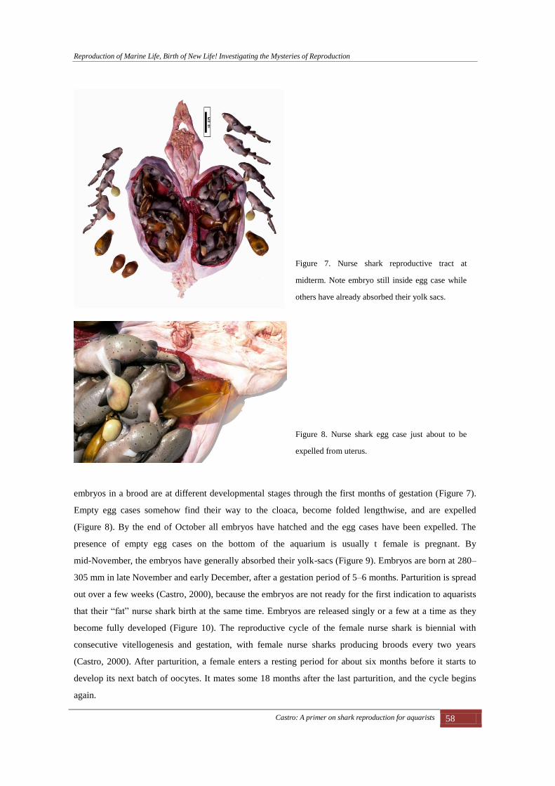
Empty egg cases somehow find their way to the cloaca, become folded lengthwise, and are expelled
(Figure 8). By the end of October all embryos have hatched and the egg cases have been expelled. The
presence of empty egg cases on the bottom of the aquarium is usually t female is pregnant. By
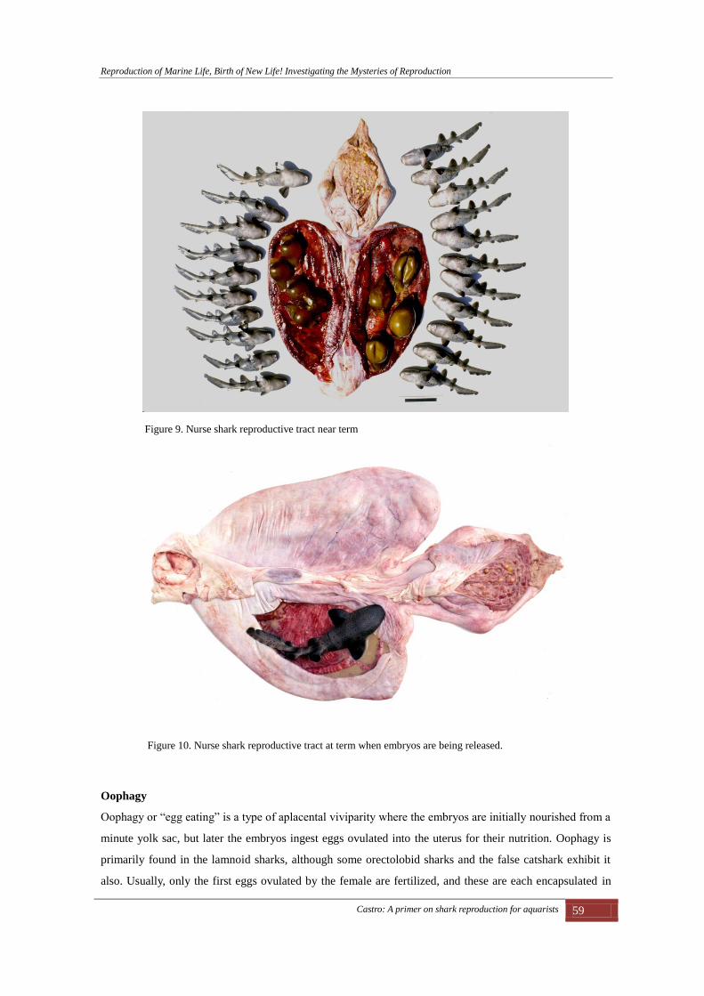
mid-November, the embryos have generally absorbed their yolk-sacs (Figure 9). Embryos are born at 280–
305 mm in late November and early December, after a gestation period of 5–6 months. Parturition is spread
out over a few weeks (Castro, 2000), because the embryos are not ready for the first indication to aquarists
that their “fat” nurse shark birth at the same time. Embryos are released singly or a few at a time as they
become fully developed (Figure 10). The reproductive cycle of the female nurse shark is biennial with
consecutive vitellogenesis and gestation, with female nurse sharks producing broods every two years
(Castro, 2000). After parturition, a female enters a resting period for about six months before it starts to
develop its next batch of oocytes. It mates some 18 months after the last parturition, and the cycle begins
again.
Figure 7. Nurse shark reproductive tract at
midterm. Note embryo still inside egg case while
others have already absorbed their yolk sacs.
Figure 8. Nurse shark egg case just about to be
expelled from uterus.
Reproduction of Marine Life, Birth of New Life! Investigating the Mysteries of Reproduction
Castro: A primer on shark reproduction for aquarists 59
Oophagy
Oophagy or “egg eating” is a type of aplacental viviparity where the embryos are initially nourished from a
minute yolk sac, but later the embryos ingest eggs ovulated into the uterus for their nutrition. Oophagy is
primarily found in the lamnoid sharks, although some orectolobid sharks and the false catshark exhibit it
also. Usually, only the first eggs ovulated by the female are fertilized, and these are each encapsulated in
Figure 9. Nurse shark reproductive tract near term
Figure 10. Nurse shark reproductive tract at term when embryos are being released.

Reproduction of Marine Life, Birth of New Life! Investigating the Mysteries of Reproduction
Castro: A primer on shark reproduction for aquarists 60
individual egg cases. The number of fertilized eggs going into each uterus is variable, depending on the
species, and can range from one to a dozen. Subsequent eggs are usually unfertilized and are used solely to
feed the embryos, each egg case containing many eggs. Apparently, oophagy is a very efficient way of
quickly nourishing embryos to large size. There are numerous patterns of oophagy in sharks, and a few
examples will follow.
The sand tiger shark (Carcharias taurus) is a popular aquarium sharks because it survives for decades in
captivity, where its fierce appearance and large size make it a favorite with the public The sand tiger shark
has a peculiar type of oophagy that has been termed adelphophagy (Wourms, 1977) or sibling cannibalism.
Figure 11. Sand tiger shark 5-cm embryo with
precocious dentition.
Figure 12. Sand tiger shark uterus with 10-cm
embryo that has killed smaller embryos. The
embryos swim in a sea of egg cases.
Figure 13. Sand tiger shark 30-cm embryo and
smaller dead embryos.

Reproduction of Marine Life, Birth of New Life! Investigating the Mysteries of Reproduction
Castro: A primer on shark reproduction for aquarists 61
Springer (1948) was the first to note sand tiger oophagy, and the process was described by Gilmore and
Dodrill (1983). In this species, when the largest embryo reaches 5–7 cm, its head is greatly developed and
it develops a precocious dentition (Figure 11). The largest embryo then seeks and kills all the smaller
embryos in the uterus (Figures 12 & 13). Later it will consume them along the large supply of egg cases
that has accumulated in the uterus. Like in the shortfin mako, most of the yolky oocytes in the huge ovary
are ovulated early in gestation and for a few weeks the embryo swims in a sea of egg cases. By the time the
embryo reaches 50 cm it will have ingested all the egg cases and has developed a huge yolk stomach. The
ovary will have released most of its ripe oocytes in the first half of gestation (Figure 14). The embryos will
have consumed their expanded yolk stomachs by the time of birth.
In the shortfin mako (Isurus oxyrinchus) multiple embryos (sometimes a dozen on more) coexist in the
uterus. In the shortfin mako, the ovary ovulates most of its ripe oocytes in the first few months of gestation.
The embryos ingest a large number of egg cases in early gestation and acquire huge distended yolk
stomachs (Figure 15). Normally up to ten embryos coexist in each uterus (Figure 16). However, in certain
occasions, when there are runts or dead embryos, the larger and normal embryos may turn cannibalistic and
consume other embryos. (Joung and Hsu, 2005).
In the pelagic thresher (Alopias pelagicus) only the first two eggs are fertilized (Figure 17) and each is
enclosed in its own egg case. Subsequent eggs are not fertilized (Castro 2009). These fertilized eggs, one
cm in diameter, are larger than subsequent eggs. One egg case containing a fertilized egg goes to each
Figure 14. Sand tiger shark embryos at about two
thirds of development. Note that the ovary has
released all the ripe oocytes and the egg cases have
been ingested by the embryos.
Figure 15. Shortfin mako 30-cm embryo with large
yolk stomach.

Reproduction of Marine Life, Birth of New Life! Investigating the Mysteries of Reproduction
Castro: A primer on shark reproduction for aquarists 62
uterus. Subsequent eggs are smaller and 10–20 are packed into each egg case (Figure 18). In this species,
the ovary ovulates eggs continuously throughout gestation. Thus the embryos have a continuous supply of
yolk and do not acquire the distended yolk stomachs of other oophagous species (Figure 19). The bigeye
thresher (Alopias superciliosus) has a similar pattern of oophagy (Figure 20).
Figure 16. Shortfin mako brood at midterm.
Figure 17. Pelagic thresher egg cases. The upper
two egg cases are the first to be ovulated and each
contains one fertilized egg. The following egg
cases (lower two cases) are just feeding egg cases.
Figure 18. Pelagic thresher egg cases packed with
eggs.

Reproduction of Marine Life, Birth of New Life! Investigating the Mysteries of Reproduction
Castro: A primer on shark reproduction for aquarists 63
The tawny nurse shark, Nebrius ferrugineus (= concolor), is a common shark of the Pacific Ocean. It has
been displayed in the Okinawa Churaumi Aquarium and other aquariums throughout the Pacific area.
Despite its abundance and being kept in captivity, little is known of its reproductive processes. Teshima et
al. (1995) demonstrated that the embryos of this orectoloboid shark are oophagous. Its birth size is not
known with certainty. Much could be learned about this interesting shark in captivity.
Figure 19. Pelagic thresher embryos at about two months of gestation. Note that the embryos do not
acquire distended yolk stomachs and the ovary continues to ovulate eggs during gestation.
Figure 20. Bigeye thresher, panoramic view of reproductive tract with two embryos and ovulated egg cases.

Reproduction of Marine Life, Birth of New Life! Investigating the Mysteries of Reproduction
Castro: A primer on shark reproduction for aquarists 64
Histotrophic viviparity
Another aplacental viviparous shark, the tiger shark (Galeocerdo cuvier), produces large young reaching 85
cm at birth (Castro, 2011) that are apparently nourished primarily by histotrophe (uterine milk). There is
little published information on the reproductive processes and development of the tiger shark. Sarangdhar
(1943) described 52-cm embryos removed from a female caught off Sassoon Dock, India in 1942. These
embryos had large yolk-sacs with a considerable amount of yolk left in them, and were enclosed in
‘water-filled’ sacs formed in the shell membrane (Figure 21). The absence of any folding in their distal
walls and the embryos being free inside the membranous egg case indicate that a placental connection does
not form in the tiger shark (Sarangdhar, 1943). Castro (1983: 23) wrote that term embryos had everted
stomachs protruding slightly from the mouth, and that it was possible that the abundant fluid may have
nutritive properties (“uterine milk”) and may be imbibed by the embryos through their everted stomachs.
Current work by the author, A. B. Bodine, and Keiichi Sato of the Okinawa Churaumi Aquarium has
revealed that the embryos gain considerable weight during development. The uterine fluid is currently
being analyzed to investigate its nutritive properties. The reproductive cycle of the tiger shark is the subject
of debate. Whitney and Crow 2007 proposed a 16-month development for tiger sharks off Hawaii, while
Castro (2009) proposed a 12-month gestation period for North American tiger sharks.
Placental viviparity.
Placental viviparity is the most advanced mode of reproduction in sharks. The embryos of viviparous
sharks are initially dependent on yolk stored in the yolk sac (Figure 22), just like other sharks, but they are
later nourished by the mother through a placental connection. Because the embryos spend their entire
developmental time inside their mother’s body, the egg case of placental sharks is thin and diaphanous. In
Figure 21. Tiger shark embryo in fluid-filled egg
case.

Reproduction of Marine Life, Birth of New Life! Investigating the Mysteries of Reproduction
Castro: A primer on shark reproduction for aquarists 65
Figure 22. Atlantic sharpnose shark, 12-mm
embryo inside egg case, during yolk-dependent
stage.
Figure 23. 80-mm Atlantic sharpnose embryo has
consumed all the contents of the yolk sac.stage.
Figure 24. Atlantic sharpnose shark with flaccid
yolk sac implanting on uterine wall.
Figure 25. Atlantic sharpnose shark. Where the
yolk sac comes in contact with the uterine wall, the
placenta is formed.

Reproduction of Marine Life, Birth of New Life! Investigating the Mysteries of Reproduction
Castro: A primer on shark reproduction for aquarists 66
placental sharks, the contents of the yolk sac are usually consumed in the first few weeks of gestation
(Figure 23). In these sharks the yolk sac enlarges as it contents are consumed, and develops as a long yolk
stalk that bears a reduced, flaccid yolk sac at its end (Figure 24). Where the yolk sac comes in contact with
the uterine wall, it becomes modified into the yolk sac placenta (Figure 25). In the placenta, the tissues of
mother and offspring come into intimate contact, and nutrients are passed on to the embryo. Thus the
embryo has an almost unlimited supply of nutrients that are shunted to it directly from the mother’s blood
stream. Placental viviparity is confined to the most advanced groups of sharks: the smoothhounds, the
requiem sharks and the hammerhead sharks. In some viviparous sharks, such as the sharpnose sharks and
most of the hammerheads (Figure 26), the umbilical cords are covered with thread or finger-like structures
known as "appendiculae." The function of these structures is not clear but it has been postulated that they
may serve to absorb histotrophe (uterine milk) produced by the uterine lining.
The patterns described above are only a few of the ones studied. It is certain that many more types exist in
this group of ancient fishes, and that there are transitional forms as well. These modes of reproduction are
modified by complex biennial or longer reproductive cycles. See Castro (2009) for descriptions of some of
these cycles.
In the past, the aquarium community in general contributed little to the knowledge of sharks because few
observations were published. Often valuable and interesting observations were forgotten or lost when
personnel retired or died. The pattern of neglect is slowly changing, with better education of aquarium
personnel, more enlightened leaders, better record keeping, the availability of PIT tags, etc. In 2004 the
aquarium community made its first attempt at collecting and publishing observations on elasmobranchs
Figure 26. Scalloped hammerhead embryo. Note the umbilical cord covered with appendiculae.

Reproduction of Marine Life, Birth of New Life! Investigating the Mysteries of Reproduction
Castro: A primer on shark reproduction for aquarists 67
with the publication of the 589-page The Elasmobranch Husbandry Manual (Smith et al., 2004). This fine
publication may serve as a guide for future contributions from the aquarium community.
The following topics are suggested for observation and recording:
Oviparous sharks
1. Record any reproductive behavior (courtship, mating, etc). Few species of sharks have been seen mating,
so these observations are often valuable and publishable.
2. A description of temperature and light regimes. A temperature log will be necessary to accompany the
determination of incubation time.
3. Egg laying behavior, type of substrate used for egg case attachment.
4. In many cases, the egg cases of a given female may bear distinctive markings and it may be possible to
attribute egg cases to a given female. Egg cases should be labeled and kept in separate tanks during their
developmental period.
5. Record the egg laying rate or periodicity of egg case laying.
6. Record the length of development the time to hatching.
7. Little is known about the length of the reproductive cycle and its periodicity in oviparous sharks and
much could be learned by keeping track of egg laying patterns over several years. Because many sharks
have biennial reproductive cycles, a female could lay eggs only every other year. Such observations would
be most interesting. Whether the constant light and temperature regimes affect the endogenous
reproductive cycles of the animals, or whether the endogenous cycles are maintained despite the constant
conditions will have to be determined empirically or by comparison to wild-caught specimens.
Viviparous sharks
Obviously, observations on the development of viviparous sharks are difficult to carry out in aquariums, as
normally they can only be made by dissection of dead specimens. Observations using methods such as
ultrasound usually lack the necessary precision to be useful. Ultrasound has been used to confirm that a
shark is pregnant, in most cases a foreknown fact. Sampling of reproductive hormone titers may be feasible
under some conditions. Little is known about this subject and much could be learned from aquarium sharks.
Injecting newborn sharks with tetracycline may reveal growth rates at a later time, although these growth
rates may differ significantly from growth rates in the wild.
Records of aquarist’s observations should be permanently maintained at the aquarium. Whenever possible
individual sharks should be marked with unobtrusive PIT tags, so that their identity and date of arrival at
the aquarium can be determined years later, if it is questionable at later times. The record should be part of
the aquarist’s duties and should be the property of the aquarium, so that it will remain available even after
aquarists leave. However, aquarists should copy their observations so that a second copy of the record is
available for future publication if the aquarium loses or does not publish the observations.

Reproduction of Marine Life, Birth of New Life! Investigating the Mysteries of Reproduction
Castro: A primer on shark reproduction for aquarists 68
Literature Cited
Castro, J. I. 1983. The Sharks of North American Waters. College Station: Texas A&M University Press.
180 pp.
Castro, J. I. 1993. The shark nursery of Bulls Bay, South Carolina, with a review of the shark nurseries of
the southeastern coast of the United States. Environmental Biology of Fishes 38:37-48.
Castro, J. I. 2000. The biology of the nurse shark, Ginglymostoma cirratum, off the Florida east coast and
the Bahama Islands. Environmental Biology of Fishes 58:1-22.
Castro, J. I. 2009. Observations on the reproductive cycles of some viviparous North American sharks.
Aqua: 15 (4):205-222
Castro, J. I. 2011. The Sharks of North America. New York: Oxford University Press. 613 pp.
Conrath, C. L., and J. A. Musick. 2012. Reproductive biology of elasmobranchs. Pp. 291-311. In Biology
of sharks and their relatives. Second edition. J. C. Carrier, J. A. Musick, and M. R. Heithaus, editors.
New York: CRC Press.
Gilbert, P. W. 1959. The ability of yolk-sac stage dogfish pups to survive outside the uterus. The Bulletin,
Mount Desert Island Biological Laboratory 1959: 68.
Gilmore, R. G., J. W. Dodrill, and P. A. Linley. 1983. Reproduction and embryonic development of the
sand tiger shark, Odontaspis taurus (Rafinesque). Fishery Bulletin 81 (2):201-225.
Hamlett, W. C., and T. J. Koob. 1999. Female reproductive system. Pp.398-443. In Sharks, skates, and rays.
The biology of elasmobranch fishes. W. C. Hamlett, editor. Baltimore: The Johns Hopkins University
Press.
Jones, B. C., and G. H. Geen. 1977. Reproduction and embryonic development of spiny dogfish Squalus
acanthias in the Strait of Georgia, British Columbia. Journal of the Fisheries Research Board of Canada
34 (9):1286-1292.
Joung, S.-J., and H.-H. Hsu. 2005. Reproduction and embryonic development of the shortfin mako, Isurus
oxyrinchus Rafinesque 1810, in the northwestern Pacific. Zoological Studies 44 (4): 487–496.
Joung, S-J., C-T. Chen, E. Clark, S. Uchida, and W. Y. P. Huang. 1996. The whale shark, Rhincodon typus,
is a livebearer: 300 embryos found in one ‘megamamma’ supreme. Environmental Biology of Fishes
46:219-223.
Ketchen, K. S. 1972. Size at maturity, fecundity, and embryonic growth of the spiny dogfish (Squalus
acanthias) in British Columbia waters. Journal of the Fisheries Research Board of Canada 29 (12):
1717-1723.
Sarangdhar, P. N. 1943. Tiger shark - Galeocerdo tigrinus - Muller and Henle. Feeding and breeding habits.
Journal of the Bombay Natural History Society 44:101-110.
Smith, M., D. Warmolts, D. Thoney, and R. Hueter (Editors). 2004. The Elasmobranch Husbandry Manual:
Captive care of sharks, rays, and their relatives. Columbus, Ohio. Special Publication of the Ohio
Biological Survey. 589 pp.
Springer, S. 1948. Oviphagous embryos of the sand shark, Carcharias taurus. Copeia 1948 (3): 153-157.

Reproduction of Marine Life, Birth of New Life! Investigating the Mysteries of Reproduction
Castro: A primer on shark reproduction for aquarists 69
Teshima, K., Y. Kamei, M. Toda, and S. Uchida. 1995. Reproductive mode of the tawny nurse shark taken
from the Yaeyama Islands, Okinawa, Japan, with comments on individuals lacking the second dorsal fin.
Bulletin of the Sekai National Fisheries Institute, No. 731-12.
Whitney, N. M. and G. L. Crow. 2007. Reproductive biology of the tiger shark (Galeocerdo cuvier) in
Hawaii. Marine Biology 151:63-70.
Wourms, J.P. 1977. Reproduction and development in chondrichthyan fishes. American Zoologist 17 (2);
379-410.