A polycycstin-type transient receptor potential (Trp...
47
© 2016. Published by The Company of Biologists Ltd. This is an Open Access article distributed under the terms of the Creative Commons Attribution License (http://creativecommons.org/licenses/by/3.0), which permits unrestricted use, distribution and reproduction in any medium provided that the original work is properly attributed. A polycycstin-type transient receptor potential (Trp) channel that is activated by ATP David Traynor & Robert R. Kay MRC Laboratory of Molecular Biology, Francis Crick Avenue, Cambridge, CB1 0QH, UK Correspondence to DT: [email protected] Key words: Trp channel, polycystin-2, purinergic signalling, ATP, DIF, Dictyostelium Summary statement We show that a Trp channel related to the mammalian polycystin channel, rather than a P2X receptor, is responsible for the purinergic stimulation of cytosolic calcium levels in Dictyostelium cells. Biology Open • Advance article by guest on September 11, 2018 http://bio.biologists.org/ Downloaded from
Transcript of A polycycstin-type transient receptor potential (Trp...

© 2016. Published by The Company of Biologists Ltd. This is an Open Access article distributed under the terms of the Creative Commons Attribution License
(http://creativecommons.org/licenses/by/3.0), which permits unrestricted use, distribution and reproduction in any medium provided that the original work is properly attributed.
A polycycstin-type transient receptor potential (Trp) channel
that is activated by ATP
David Traynor & Robert R. Kay
MRC Laboratory of Molecular Biology,
Francis Crick Avenue,
Cambridge, CB1 0QH, UK
Correspondence to DT: [email protected]
Key words: Trp channel, polycystin-2, purinergic signalling, ATP, DIF, Dictyostelium
Summary statement
We show that a Trp channel related to the mammalian polycystin channel, rather than a P2X
receptor, is responsible for the purinergic stimulation of cytosolic calcium levels in
Dictyostelium cells.
Bio
logy
Ope
n •
Adv
ance
art
icle
by guest on September 11, 2018http://bio.biologists.org/Downloaded from

Abstract
ATP and ADP are ancient extra-cellular signalling molecules that in Dictyostelium amoebae
cause rapid, transient increases in cytosolic calcium due to an influx through the plasma
membrane. This response is independent of hetero-trimeric G-proteins, the putative IP3
receptor IplA and all P2X channels. We show, unexpectedly, that it is abolished in mutants of
the polycystin-type transient receptor potential channel, TrpP. Responses to the
chemoattractants cyclic-AMP and folic acid are unaffected in TrpP mutants. We report that the
DIF morphogens, cyclic-di-GMP, GABA, glutamate and adenosine all induce strong cytoplasmic
calcium responses, likewise independently of TrpP. Thus TrpP is dedicated to purinergic
signalling. ATP treatment causes cell blebbing within seconds but this does not require TrpP,
implicating a separate purinergic receptor. We could detect no effect of ATP on chemotaxis
and TrpP mutants grow, chemotax and develop almost normally in standard conditions. No
gating ligand is known for the human homologue of TrpP, polycystin-2, which causes polycystic
kidney disease. Our results now show that TrpP mediates purinergic signalling in Dictyostelium
and is directly or indirectly gated by ATP.
Bio
logy
Ope
n •
Adv
ance
art
icle
by guest on September 11, 2018http://bio.biologists.org/Downloaded from

Introduction
ATP and other purines are ancient signalling molecules used widely in animals as neuro-
transmitters and also by protozoa and plants for diverse purposes (Burnstock and Verkhratsky,
2009). The signalling pathway typically consists of release of ATP, its detection by cell surface
receptors with consequent signal transduction, and destruction of the signal by ecto-ATPases.
Two types of ATP receptor are known in mammalian cells: P2X receptors are gated ion
channels, which generally allow calcium into the cell and P2Y receptors are G-protein coupled
receptors (GPCRs) (Burnstock, 2007).
Calcium signalling also has ancient origins and it is likely that ancestral single-celled eukaryotes
were able to produce Ca2+ gradients across their plasma membrane using calcium pumps and
transporters, and activate calcium entry into the cytoplasm through regulated channels in the
plasma membrane and the membranes of internal vesicular stores of Ca2+ ions. Changes in free
Ca2+ ion concentration could then alter the activity of sensitive proteins and processes in the
cytoplasm. Present day microbes use calcium signalling in a wide variety of ways and have
recognizable homologues in their genomes to many components of calcium signalling found in
mammalian cells (Martinac et al., 2008; Collins and Meyer, 2011; Plattner and Verkhratsky,
2015). These ancient signalling processes can be combined so that ATP causes a cytoplasmic
calcium increase.
The social amoeba Dictyostelium discoideum grows on bacteria or in liquid media as separate
cells (Kessin, 2001). These cells respond to starvation by aggregating together by chemotaxis,
to form a multicellular mass and ultimately a stalked fruiting body carrying a mass of spores at
its top. In the growth phase the cells are chemotactic to folic acid, which guides them to
bacteria, and after starvation they become chemotactic to cyclic-AMP, which is released
periodically from aggregation centres, to which it attracts the amoebae. Both folic acid and
cyclic-AMP are detected through G-protein coupled receptors (Klein et al., 1988; Pan et al.,
2016) and set off a variety of intra-cellular signalling responses, including an influx of Ca2+. The
coordinated movement and differentiation of amoebae into stalk cells and spores during
development is controlled by small molecule signalling, including by cyclic-AMP, the polyketides
DIF and MPBD (Morris et al., 1987; Saito et al., 2006), cyclic-di-GMP (Chen and Schaap, 2012)
and GABA/glutamate (Taniura et al., 2006; Anjard and Loomis, 2006).
Bio
logy
Ope
n •
Adv
ance
art
icle
by guest on September 11, 2018http://bio.biologists.org/Downloaded from
Dictyostelium cells also respond strongly to extracellular ATP and ADP, which both cause
an immediate and transient increase in cytosolic Ca2+ due to an influx through the plasma
membrane (Ludlow et al., 2008; Ludlow et al., 2009). Dictyostelium cells can also release ATP
into the medium in micro-molar concentrations (Sivaramakrishnan and Fountain, 2015) and
have an ecto-ATPase activity, which degrades ATP (Parish and Weibel, 1980), suggesting that
they have a complete set of purinergic signalling components. However, the receptor
responsible for the calcium influx in response to ATP is currently unknown.
The most obvious candidate for this ATP receptor is one or more of the five P2X receptors
encoded in the genome, four of which have been shown to be ATP-gated calcium channels in
heterologous expression experiments (Fountain et al., 2007; Ludlow et al., 2009; Baines et al.,
2013). However, these receptors are largely expressed on the intracellular membranes of the
contractile vacuole and have a role in its discharge (Fountain et al., 2007; Ludlow et al., 2009;
Sivaramakrishnan and Fountain, 2012; Parkinson et al., 2014). Crucially, a mutant with all five
P2X receptors knocked out still retains its calcium response to ATP (Ludlow et al., 2009). The
Dictyostelium genome only carries a limited set of candidate Ca2+ signalling proteins (Eichinger
et al., 2005; Wilczynska et al., 2005), which include two transient receptor potential (Trp)
channels (Clapham, 2003; Hardie, 2007). The nearest human homologues of these
Dictyostelium proteins are mucolipin and polycistin-2, which are named after the corresponding
genetic diseases (Lima et al., 2012; Lima et al., 2014). There is also a two-pore channel and an
IP3-like receptor, IplA (Traynor et al., 2000) and two potential stretch-operated channels: MscS
is homologous to the bacterial small conductance mechanosensitive channel (Martinac et al.,
2008) and a homologue of the eukaryotic Piezo mechanosensitive channel (Coste et al., 2010).
The role of extra-cellular ATP signalling in the Dictyostelium life-cycle is not yet clear. ATP has
been reported to affect various processes, including cellular aggregation, possibly by enhancing
cyclic-AMP signalling (Mato and Konijn, 1975; Perekalin, 1977), and recovery from hypo-
osmotic stress (Sivaramakrishnan and Fountain, 2015).
We sought to identify the channel mediating the purinergic response of Dictyostelium
cells by knocking out candidate calcium channels and assessing the response of the mutant cells
to ATP using a reporter for cytoplasmic calcium. In this way we show that the polycystin-type
Trp channel, TrpP is essential for the response, and either is the ATP receptor, or closely
coupled to it. We also show for the first time that a number of endogenous effector molecules
Bio
logy
Ope
n •
Adv
ance
art
icle
by guest on September 11, 2018http://bio.biologists.org/Downloaded from
including DIF, GABA and cyclic-di-GMP trigger calcium signals, and that these responses are
independent of TrpP.
Results
Visualising calcium signalling using the cameleon FRET reporter
In order to characterise the Ca2+ signalling triggered by ATP, we first set up a convenient assay
to measure changes in cytosolic calcium concentration. The cameleon series of FRET-based,
genetically encoded calcium reporters do not require loading into cells, nor do they require an
added cofactor, unlike aequorin (Nagai et al., 2004; Horikawa et al., 2010). We initially
characterized the well-studied response of starved, developing (aggregation-competent) cells
to the chemoattractant cyclic-AMP (Abe et al., 1988; Milne and Coukell, 1991; Nebl and Fisher,
1997; Nebl et al., 2002) (Fig. 1D). We found that the higher affinity chameleon YC2.60 (Kd 95
nM) was not saturated by a maximal dose of cyclic-AMP or ATP and could robustly detect sub-
maximal doses, unlike the lower affinity YC3.60 (Kd 215 nM) and thus was ideal for our
purposes (Fig. S1). Using this reporter, we found that stimulation with a uniform concentration
of cyclic-AMP causes a transient increase in cytosolic Ca2+ levels after a delay of 6.0 ± 1.0 sec
(n=7), with a peak at 22.1 ± 4.3 sec (n=7) and return to baseline by 58.1 ± 3.2 sec (n=7) (Fig. 1D).
Half-maximal response is somewhat variable at 87 ± 71 nM (n=4) cyclic-AMP (Fig. S2).
Cyclic-AMP signalling is mediated through a family of G-protein coupled receptors, principally
cAR1 (Klein et al., 1988). These receptors appear during development and are only expressed
at low levels in growing cells, which accordingly show little or no response to cyclic-AMP.
However vegetative cells do respond to folic acid (Nebl et al., 2002), which is also a
chemoattractant and is detected by a G-protein coupled receptor (Pan et al., 2016): as
expected, folate induces a delayed Ca2+ response, similar to that produced by cyclic-AMP (Fig.
1C).
Bio
logy
Ope
n •
Adv
ance
art
icle
by guest on September 11, 2018http://bio.biologists.org/Downloaded from

TrpP mediates the calcium response to ATP
We found that ATP and ADP robustly evoke transient increases in cytosolic Ca2+ from both
vegetative and aggregation-competent cells (Fig. 1A,B; for a response to ADP, see Fig. 3),
confirming previous work (Ludlow et al., 2008; Ludlow et al., 2009). There is a minimal delay of
around 1 second (1.3±0.2 sec n=7) before onset of the response to ATP, which is similar to the
mixing time, and a mean rise time of 11.0 ± 1.4 sec (n=7) from baseline to peak response. The
kinetics of this purinergic response clearly differ from those to folic acid and cyclic-AMP, having
a much shorter lag before onset. The half-maximal response was at 1.1 ± 0.4 µM (n=5) for ATP
and 1.6 ± 1.5 µM (n=3) for ADP in aggregation-competent cells (Fig. S2). These values were
obtained only from those cells producing a response, which was more than 90% at saturating
doses, but fell off at lower ATP concentrations (Fig. S2).
Genetically, the response to ATP does not depend on any of the P2X receptors encoded in the
Dictyostelium genome (Ludlow et al., 2009). To try and establish the signalling route used by
ATP we therefore examined the effect of mutating other signalling proteins. First, we tested
dependence on hetero-trimeric G-proteins using a null mutant in the only Gß subunit encoded
in the genome (Wu et al., 1995) and found that the response to ATP was unaffected (Fig. 2). In
contrast the response to folate was abolished. Similarly, the response in a mutant of IplA, the
homologue of the endoplasmic reticulum channel activated by IP3, was also intact (Traynor et
al., 2000). Thus the purinergic response does not appear to require either hetero-trimeric G-
proteins or IplA (Ludlow et al., 2008), in both cases differentiating it from the GPCR-mediated
responses to cyclic-AMP and folate.
We therefore investigated other candidate calcium signalling proteins and made null mutants in
three potential channels: the mechano-sensitive channel homologue, MscS (dictyBase
DDB_G0277253; http://dictybase.org) and two Trp channels, one a mucolipin homologue, MclN
(dictyBase DDB_G0291275) (Lima et al., 2012), the other a polycistin-2 homologue, which we
call TrpP (gene, trpP; the protein is also known as PKD2; dictyBase DDB_G0272999) (Lima et al.,
2014). In case of redundancy, we made a triple mutant lacking all three proteins and in our
initial experiments used the uptake of 45Ca2+ to measure the response. To our surprise, we
found that the fast responses to ATP and ADP are essentially abolished in this triple mutant (Fig.
S3A). Testing the single mutants individually showed that the rapid ATP and ADP responses are
abolished in the TrpP null mutant (Fig. 3, Fig. S3B), but unaffected in the other mutants (Fig.
S3C,D).
Bio
logy
Ope
n •
Adv
ance
art
icle
by guest on September 11, 2018http://bio.biologists.org/Downloaded from

The abolition of responses to ATP and ADP in trpP- mutant cells was confirmed using the
cameleon FRET reporter in both vegetative and aggregation-competent cells (Fig. 3, Fig. S4).
Responsiveness could be restored by expressing TrpP under the control of its own promoter,
demonstrating that the phenotype is due to the loss of TrpP and not a secondary mutation
introduced elsewhere in the genome. In addition, a C-terminal fusion of TrpP to GFP is largely
localised to the plasma membrane in living cells, consistent with TrpP acting as a plasma
membrane channel (Fig. S5)
Thus we conclude that TrpP mediates the fast calcium responses to ATP and ADP and is likely
gated, directly or indirectly, by ATP and ADP.
Purinergic and chemoattractant Ca2+ signalling use genetically distinct pathways
We have shown above that purinergic signalling does not require either Gß or IplA, whereas
these proteins are required for chemoattractant signalling by folic acid and cyclic-AMP
(although there are contradictory reports on the importance of Gß for cyclic-AMP signalling
(Milne and Devreotes, 1993; Nebl et al., 2002; Ludlow et al., 2008)). Both folate and cyclic-AMP
calcium signalling depend on an influx of extracellular calcium through the plasma membrane,
but the channel responsible has not been identified (Nebl and Fisher, 1997). In principle this
channel could be TrpP, to which these ligands might couple indirectly through their respective
GPCRs. However, we found that the responses to both cyclic-AMP and folate remain intact in
TrpP null cells, showing that TrpP is not their influx channel (Fig. 3G; Fig. S4E). Thus the
pathways of chemoattractant and purinergic signalling are genetically distinct.
We also noticed that TrpP null cells occasionally show a small, delayed response to ATP, which
can also be elicited by buffer alone (Fig. S6 and Table S1). A similar delayed response to ATP is
seen in wild-type cells treated with Zn2+ to inhibit the primary response (Ludlow et al., 2008).
This response might be due to a stretch-operated channel, which is activated by the physical
stresses of mixing in ligand or buffer. Since the timing of the delayed responses overlaps with
those depending on IplA, we created a double trpP-/iplA- mutant, and found that the response
is completely abolished (Fig. S6). Indeed this double mutant lacks a calcium response to all
ligands tested (illustrated for ATP and cyclic-AMP in Fig. S6) but remains sensitive to the
calmodulin inhibitor, calmidazolium which is known elevate cytosolic Ca2+ levels by Ca2+ release
from stores (Schlatterer and Schaloske, 1996).
Bio
logy
Ope
n •
Adv
ance
art
icle
by guest on September 11, 2018http://bio.biologists.org/Downloaded from

Ca2+ signalling is induced by DIF, cyclic-di-GMP and GABA independently of TrpP
The differentiation and behaviour of Dictyostelium cells during multicellular development is
controlled by a number of small signalling molecules, in addition to cyclic-AMP, but whether
these molecules cause rapid changes in cytosolic Ca2+ levels is not known. To determine the
scope of calcium signalling mediated by TrpP, we tested whether these endogenous signals
could trigger Ca2+ signals, and if so, whether the response depends on TrpP, or not.
The DIFs are a family of chlorinated polyketides that induce stalk cell differentiation during
development (Morris et al., 1987; Masento et al., 1988), particularly those of the fruiting body
basal disc (Saito et al., 2008). DIF has rapid effects on protein phosphorylation and
transcription (Sugden et al., 2015; Williams et al., 1987) but its receptor is unknown.
We found that DIF-1, the major species, causes a robust increase in cytosolic Ca2+ in
aggregation-competent cells after a delay of about 5.5±1.2 (n=7) seconds, reaching a peak at
around 15.5±2.1 (n=7) seconds (Fig. 1F). Half-maximal response is at around 20 nM (Fig. S1;
EC50 23.3 ± 3.7 nM; n=3), which is well within the range inducing cell differentiation and below
the estimated physiological concentration (Kay, 1998). Vegetative cells give similar but weaker
responses, consistent with the effects of DIF-1 early in development (Fig. 1E) (Wurster and Kay,
1990; Fukuzawa et al., 2001).
DIF-2 has 40% of the activity of DIF-1 in a cell differentiation assay but was equipotent in
causing a Ca2+ response in aggregation-competent cells (24.8 ± 13.7 nM, n=3; Fig. S1), whereas
the less potent analogue, DIF-3, only gives a weak response at ≥1 µM, as does an analogue
lacking the methyl group (des-methyl DIF-1; not shown). The unmodified polyketide (THPH)
from which DIF-1 is synthesised (Kay, 1998) produces no response up to 10 µM.
The response to DIF is totally dependent on IplA in both vegetative and aggregation competent
cells but is maintained in Gß null cells, suggesting that it does not depend on a heterotrimeric
G-protein (Fig. 2). The response also remains essentially unchanged in TrpP null cells and so
does not depend on TrpP (Fig. 3).
A second polyketide, MPBD, is important both during early development and for proper
spore maturation (Saito et al., 2006; Narita et al., 2014). It failed to evoke a calcium response
from vegetative and aggregation-competent cells at 1 µM (not shown).
We found that cyclic-di-GMP, which is required for stalk cell maturation (Chen and Schaap,
2012; Song et al., 2015), caused a delayed Ca2+ response from aggregation-competent cells at
125 µM (Fig. 4). Dictyostelium has a GABA signalling system, which is active during
Bio
logy
Ope
n •
Adv
ance
art
icle
by guest on September 11, 2018http://bio.biologists.org/Downloaded from

development (Taniura et al., 2006; Anjard and Loomis, 2006; Wu and Janetopoulos, 2013) and
we found that GABA and L-glutamate at 250 µM both evoke a cytoplasmic calcium signal from
aggregation-competent cells, although this was erratic. Finally, adenosine, which affects
prestalk and prespore patterning in development (Schaap and Wang, 1986) also causes a
calcium response. The calcium responses to GABA and L-glutamate are independent of TrpP,
remaining intact in the null mutant, but do depend on IplA (Fig. S7).
ATP causes cell blebbing
We sought to establish the role of purinergic signalling in Dictyostelium biology by
examining, first cellular responses to ATP, and then the TrpP mutant phenotype.
Early reports suggested that extracellular ATP stimulates aggregation centre formation in small
drop assays of starving cells (Mato and Konijn, 1975; Perekalin, 1977), but we could not
reproduce this effect using up to 1 mM ATP (not shown). We investigated the effects of ATP on
cell motility in detail. ATP was not a chemoattractant for aggregation-competent cells at a
range of concentrations, and a uniform concentration of ATP did not enhance or inhibit
chemotaxis to cyclic-AMP (Table S2). Neither was there a significant effect of a uniform
concentration of ATP on the speed of chemotaxing cells or on the random movement of either
vegetative or starving cells (Table S3).
In the course of the FRET measurements of calcium, we noticed that ATP addition causes cells
to bleb vigorously (Fig. 5A). Blebs occur when the plasma membrane becomes detached from
the underlying F-actin cortex, and is driven out by fluid pressure. Dictyostelium cells can move
using blebs instead of pseudopods (Yoshida and Soldati, 2006; Zatulovskiy et al., 2014; Tyson et
al., 2014) and blebbing is also induced by cyclic-AMP.
Morphologically, blebs induced by ATP resemble those induced by cyclic-AMP, with their
characteristic rapid expansion, smooth curvature and residual scar of F-actin, representing the
former cortex. A new cortex is then rapidly rebuilt on the exposed membrane of the bleb.
However, ATP induces blebbing much faster than the published timing for cyclic-AMP
(Langridge and Kay, 2006), with a delay of only 3-7 seconds compared to around 25 seconds for
cyclic-AMP (Fig. 5B). ATP-induced blebbing requires myosin-II and is abolished in null mutants
of either the myosin-II heavy chain or the essential light chain (Fig. S8), as is cyclic-AMP-induced
blebbing (Zatulovskiy et al., 2014).
The signalling and other events triggered by ATP and cyclic-AMP differ significantly: ATP does
not cause the transient actin polymerization characteristic of the period before blebbing starts
Bio
logy
Ope
n •
Adv
ance
art
icle
by guest on September 11, 2018http://bio.biologists.org/Downloaded from

in cells stimulated with cyclic-AMP (Langridge and Kay, 2006) (Fig. 5C). Nor does ATP stimulate
PIP3 production, as measured in live cells by recruitment of the PH-CRAC reporter to the
plasma membrane (Parent et al., 1998), or activate the MAP kinase ErkB or the AKT homologue
PKB, as detected in Western blots for the phosphorylated form of the protein kinase (data not
shown).
TrpP mutant phenotype
As an alternative approach to establish the role of purinergic signalling we examined the
phenotype of TrpP null cells in detail. A TrpP mutant made by insertional mutagenesis was
reported to have a growth defect in HL5 liquid medium (Waheed et al., 2014). We tested the
growth of six independent knock-out clones, shaken in HL5 liquid medium: two had modest
defects, possibly due to secondary mutations introduced during transformation, but the other
four were statistically indistinguishable from wild-type, suggesting that TrpP is not required in
any way for axenic growth (Table S2). FITC dextran uptake as a measure of fluid uptake, and
phagocytosis of yeast were also indistinguishable from wild-type cells measured in a clone with
normal growth (data not shown).
The TrpP gene is expressed at only low levels in growing cells, but the mRNA increases strongly
during early development, and then again during later development, suggesting that its main
role may lie in development (Fig. 6B; Parikh et al., 2010). However, overall development of
TrpP mutant cells is virtually indistinguishable from wild-type: the timing of different stages and
the size of the fruiting bodies are the same, and the only difference we noticed is that the
mutant fruiting bodies tended to collapse more frequently (Fig. 6A).
TrpP null cells had normal chemotactic parameters in both steep and shallow cyclic-AMP
gradients in paired comparisons with wild-type cells (cells were placed at different parts of the
same chemotaxis chamber; Tables S2 and S3) and moved with only slightly reduced speed and
chemotactic efficiency under an agarose overlay, which provides mechanical resistance and
causes cells to move using blebs (Table S2) (Zatulovskiy et al., 2014).
Surprisingly, blebbing of TrpP null cells is normal in response to ATP (Fig. S8). This suggests that
blebbing is mediated through a further, unidentified ATP receptor.
It has been reported that a TrpP mutant made in the DH1 background is defective in rheotaxis –
the movement of cells orientated by liquid flow (Lima et al., 2014). We tested TrpP mutants
made in our Ax2 background and found that they are still capable of efficient rheotaxis (Fig. S9).
Bio
logy
Ope
n •
Adv
ance
art
icle
by guest on September 11, 2018http://bio.biologists.org/Downloaded from

We conclude that ATP-stimulated Ca2+ signalling through TrpP can only have a subtle or
redundant effect on growth and development in standard laboratory conditions.
Discussion
ATP and ADP cause a rapid and transient increase in cytosolic calcium levels in Dictyostelium
cells (Ludlow et al., 2008; Ludlow et al., 2009). The major advance described in this paper is the
discovery that this response is mediated by the Trp channel TrpP, since in TrpP null mutants the
fast calcium response is totally abolished, yet can be restored when the protein is re-expressed.
Conversely this purinergic response is independent the Gß subunit of heterotrimeric G-proteins
and of IplA, the Dictyostelium homologue of the IP3-activated calcium release channel.
Although this genetic evidence is clear, direct gating by ATP has not yet been demonstrated by
electrophysiology with heterologously expressed TrpP, and so there remains a formal possibility
that gating is indirectly mediated by another protein. However, the specific requirement for
TrpP in ATP responses, and the lack of effect of TrpP mutation on chemoattractant, DIF, cyclic-
di-GMP or GABA signalling argues that, as a minimum, TrpP is likely to be dedicated to
purinergic signalling.
The purinergic calcium response is significantly different to the responses evoked by the
chemoattractants folic acid and cyclic-AMP, which are mediated by GPCRs. It seems that at
least two basic modes of calcium signalling can be distinguished in Dictyostelium: ‘GPCR-
dependent’ signalling (cyclic-AMP and folic acid) whose onset is delayed for 5-10 seconds after
the stimulus, and which depend on IplA and at least partially on Gß; and ‘purinergic’ signalling
(ATP and ADP) which has a rapid onset of less than 1 second, is independent of Gß and IplA, but
depends on TrpP.
We also show for the first time that the stalk cell-inducing morphogen DIF (Morris et al., 1987),
whose receptor is unknown, causes a fast, transient calcium response in the physiological
concentration range. The characteristics of this response – delayed and IplA-dependent – are
more consistent with the G-protein-dependent mode of signalling, but surprisingly we find that
this response is largely independent of Gß. Although not studied in detail we also found
Bio
logy
Ope
n •
Adv
ance
art
icle
by guest on September 11, 2018http://bio.biologists.org/Downloaded from

delayed calcium responses to di-cyclic-GMP (Chen and Schaap, 2012), L-glutamate and GABA
(Anjard and Loomis, 2006) but there was no response to the polyketide MPBD (Saito et al.,
2006).
TrpP is well conserved between dictyostelid species (Sucgang et al., 2011; Heidel et al., 2011;
Urushihara et al., 2015) arguing that purinergic signally must also have a conserved role. We
found that ATP is not a chemoattractant for aggregation-competent cells and does not
modulate chemotaxis to cyclic-AMP, nor could we detect a chemotactic defect in TrpP null
mutants. Growth of TrpP null cells in liquid medium was also normal, contrary to a recent
report (Waheed et al., 2014) and development only slightly perturbed. It has been reported
that TrpP is required for rheotaxis (Lima et al., 2014) but TrpP mutants made in our laboratory
strain showed no such defect. Such discrepancies are not unknown in the Dictyostelium
literature, and are most likely accounted for by genetic background effects, or secondary
mutations introduced during gene knock-out (Bloomfield et al., 2008; Schilde et al., 2004; Pollitt
et al., 2006; Sivaramakrishnan and Fountain, 2013).
The one clear effect we can detect of adding ATP or ADP to cells is to induce almost immediate
blebbing. Blebs form where the plasma membrane detaches from the underlying cortex and is
driven outwards by fluid pressure, and blebs are being increasingly recognised as an alternative
to pseudopods to drive cell motility, particularly when cells face mechanical resistance (Yoshida
and Soldati, 2006; Zatulovskiy et al., 2014; Tyson et al., 2014). Blebbing induced by ATP differs
in interesting ways from that induced by the chemoattractant cyclic-AMP (Langridge and Kay,
2006): in particular, it starts more quickly, there is no global polymerisation of actin, and
neither PI3-kinase nor the MAP kinase, ErkB, are activated. Surprisingly, blebbing induced by
ATP does not depend on TrpP, implying that another purinergic receptor must be responsible.
TrpP is homologous to the polycystin-2 or TRPP class of vertebrate Trp channels, which are also
found in non-metazoan organisms (Venkatachalam and Montell, 2007) and include the human
PKD2 protein (Mochizuki et al., 1996; Wu et al., 1998). PKD2 has been studied intensively as a
cause of the severe genetic disorder autosomal dominant polycystic kidney disease, in which
fluid-filled cysts grow within the kidney, and eventually disrupt its function (Chapin and Caplan,
2010). PKD2 cooperates with a large extracellular protein, PKD1, and together they can form
plasma membrane cation channels of high calcium permeability (Hanaoka et al., 2000;
Gonzalez-Perrett et al., 2001; Yu et al., 2009). However, we can detect no clear homologue of
Bio
logy
Ope
n •
Adv
ance
art
icle
by guest on September 11, 2018http://bio.biologists.org/Downloaded from

PKD1 in the Dictyostelium genome (Eichinger et al., 2005), and if one exists, it must be very
divergent.
To our knowledge, no gating agonist has been reported for PKD2. In electrophysiological
experiments it has been suggested to have an appreciable intrinsic conductance (Gonzalez-
Perrett et al., 2001), although this is disputed (Yu et al., 2009), and there is also the possibility
of mechanical gating. Our results showing that the primary response to extracellular ATP in
Dictyostelium is mediated by a PKD2 homologue, is therefore both surprising and promising,
raising the possibility that gating by ATP may be a more widespread feature of these channels.
Materials and methods
Cell cultivation, development, transfection and selection
Ax2 (Kay), with minimal chromosomal duplications (Bloomfield et al., 2008) was used as
parental stock; strains are listed in Table S5, and were renewed from frozen stocks every
month. Cell procedures were at 22oC, unless otherwise stated. Cells were grown in HL5 with
glucose (Formedium), plus 200 µg/ml dihydrostreptomycin, either in shaken suspension at 180
rpm, or in tissue culture dishes (Hirst et al., 2015). Development was initiated by washing cells
free of growth medium in KK2C (16.5 mM KH2PO4, 3.9 mM K2HPO4, 2 mM MgSO4, 0.1 mM CaCl2
pH 6.1) and settling 1x10⁸ cells from 4 ml onto 30 ml of 1.8% Oxoid L28 agar/KK2C in a 9 cm
diameter petri dish. After 10 min, excess buffer was aspirated off. Submerged development
was observed with 2x10⁶ cells under 2 ml of KK2C in 3.5 cm tissue culture dishes.
Total RNA was extracted (RNeasy kit, Qiagen) from 5x10⁷ - 1x10⁸ developing cells and cDNA
synthesised from 10.5 µg RNA for each timepoint (SuperScript™First-Strand Synthesis System
for RT-PCR, Life Technologies) using oligo(dT)12-18 for semi-quantitative PCR or a 1:1 mixture of
random hexamers/oligo(dT)12-18 for cDNA cloning. Standard curves were established with cDNA
dilutions (1:10 to 1:20000) for each primer pair. The PCR reaction contained in 50 µl: 50 pmole
of each primer, cDNA, 2 mM MgCl2, 200 µM dNTPs and 2.5 units Taq polymerase; PCR was run
for 25 cycles.
Bio
logy
Ope
n •
Adv
ance
art
icle
by guest on September 11, 2018http://bio.biologists.org/Downloaded from

Cells were transformed by electroporating 17.5 µg of gene disruption cassette, freed of plasmid
backbone by restriction digest, or 30 µg of supercoiled plasmid DNA into 4x10⁶ cells (Pang et al.,
1999; Hirst et al., 2015). Over-expression cell lines were selected and maintained in tissue
culture dishes with HL5 plus 20-40 µg/ml G418, whereas trpP knockout clones were isolated by
plating 60-240 cells/well of 96 well plates with 200 µl HL5 plus 10 µg/ml blasticidin S
(Invivogen). DNA was extracted from confluent wells after 10-14 days (Quick-gDNA MiniPrep,
Zymo Research) and screened using primers PC2S26 and PC2S27 (primer sequences are given in
Table S6) located outside the disruption cassette. Knockout clones were distinguished by the
size of the PCR product and the presence of unique restriction sites introduced into the locus by
the disruption cassette (Hirst et al., 2015).
Plasmids
Primer sequences are given in Table S6. To construct the trpP knockout vector (pDT27) the 5’
homology was amplified using oligos PC2KO1 plus PC2KO2 and ligated into the ApaI site of
pLPBLP (Faix et al., 2004) and the 3’ homology amplified using oligos PC2KO3 plus PC2KO4 and
ligated as a NotI/SacII fragment into the corresponding sites of the vector containing the 5’
homology. The trpP disruption cassette was liberated from pDT27 by digestion with KpnI and
SacII prior to transfection of the cells. The trpP CDS (without a stop codon) was amplified by
RT-PCR cDNA using oligos PCL5 plus PCL3 and ligated into the BamHI/XhoI sites of pDT29
creating pDT33 with an in frame C-terminal fusion of GFP(S65T). The plasmid pDXA-3C∆ was
made by digesting pDXA-3C with KpnI and SacI to remove the start codon from the A15 leader
in the MCS (Manstein et al., 1995). GFP(S65T) was amplified using oligos RAGFP9 plus
RAGFP10, then ligated into the XhoI site of pDXA-3C∆ giving pDT29. To construct trpP driven by
its own promoter, a silent restriction site was introduced into pDT33 using mutagenic primers
PC2S47 and PC2S48, changing +54A of the trpP CDS to +54T, giving a unique HindIII site.
Primers PC2S48 and PC2S37 were used to amplify 940 bp upstream and part of the first exon of
trpP from genomic DNA. The PCR product, digested with SalI and HindIII, was ligated into the
same sites within the mutated pDT33 thus exchanging the A15 for the trpP promoter, giving
pDT42. Partial digestion of pDT42 with SalI/XbaI removed the intact trpP CDS with its own
promoter, which was ligated into XhoI/SpeI sites of pDM304 (Veltman et al., 2009) giving
pDT41. The cameleons YC2.60 and YC3.60 (with Aequorea victoria codons) were removed from
their pBIG vectors by partial digestion with BamHI/SacI and ligated into the same sites in
Bio
logy
Ope
n •
Adv
ance
art
icle
by guest on September 11, 2018http://bio.biologists.org/Downloaded from

pET28a (Horikawa et al., 2010), providing a template to amplify both cameleons using oligos
YC367 plus YC368. The PCR products were ligated into the BamHI/SpeI sites of the shuttle
vector pDM344 (Veltman et al., 2009), removed as NgoMIV fragments and ligated into the
corresponding site in pDT41 giving pDT48 (YC3.60) and pDT50 (YC2.60). Finally, pDT48 was
used as the template to amplify the trpP CDS plus promotor using oligos PC2S69 plus PC2S70,
with the product ligated into the XhoI/BglII sites of pDM323 (Veltman et al., 2009) giving
pDT68.
Microscopy
Vegetative cells were harvested from tissue culture plates, washed 3 times in HKC buffer (10
mM HEPES, 10 mM KCl, 250 µm CaCl2, pH 6.8) by centrifugation (300 x g for 2 min) and
resuspended at 106/ml in HKC. Cells were plated at 105 cells/cm2 in 8 well Lab-Tek™ chambered
coverslips with 300 µl HKC/well. Coverslips were incubated in a moist atmosphere for up to 1
hr before use. Aggregation-competent cells were prepared by pulsing with cyclic-AMP for 3.5-
5.5 hr after 1 hr starvation in shaking suspension (Traynor and Kay, 2007), or by plating 106
washed cells per 35 mm tissue culture dish in 2 ml of HKC, incubating at 22oC for 1 hr, then 15oC
overnight (15-17 hr), before returning to 22oC for at least 1 hr, until they become elongated.
Cells were harvested in fresh MKC by pipetting up and down and transferred to a chambered
coverslip, where they normally formed long streams and aggregated after 2-4 hr at 22oC.
Confocal images were obtained using a Zeiss LSM 710 or 780 microscope with Zen 2010
software. Dunn chamber and micropipette chemotaxis assays and under-agarose motility
assays were as described (Fets et al., 2014; Zatulovskiy et al., 2014). Images were analysed with
ImageJ, Fiji (Schindelin et al., 2012) and Excel software.
Calcium imaging
Vegetative or aggregation-competent cells were challenged with effectors added as a 100 µl
bolus at 4x final concentration to individual wells of a chambered coverslip. Mixing time was
less than a second. Time-lapse images were collected on a Zeiss Axiovert 200 inverted
microscope using a 40x C-Apochromat W Corr M27 lens (NA 1.2) and a Cascade II 512 EMCCD
camera (Photometrics) controlled by Metamorph software (Molecular Devices). Cells were
illuminated with a Lambda LS light source (Sutter Instruments) containing a 175W xenon bulb
through a neutral density filter (Chroma Technology Corp, ND 2.0 A) with 1% transmittance and
an excitation filter (ET436/20x, Chroma Technology Corp) that ensured only CFP was
illuminated. The CFP and YFP emission light was separated using a beam splitter (Optical
Bio
logy
Ope
n •
Adv
ance
art
icle
by guest on September 11, 2018http://bio.biologists.org/Downloaded from

Insights, Dual View, filters D480/30, D535/40 and 505dexr dichromatic mirror). Exposure times
were 100-250 msec and images captured at 3-7 per sec with a binning of 2 unless otherwise
stated. Images were analysed using Jmalyse (Kerr and Schafer, 2006), Volocity (Perkin Elmer)
and ImageJ. For each image the CFP (C) and YFP (Y) average fluorescence intensities, obtained
by subtracting the background intensities (Ybkg and Cbkg) from the measured intensities (Ymeas
and Cmeas) and ratios (R) were corrected for bleed through from the CFP channel into the YFP
(measured at 0.683) using equation (1) in the Microsoft Excel software package.
(1)
𝑅 = [𝑌 (𝑌𝑚𝑒𝑎𝑠 − 𝑌𝑏𝑘𝑔)
𝐶 (𝐶𝑚𝑒𝑎𝑠 − 𝐶𝑏𝑘𝑔)− 0.683]
Response kinetics and peak areas were determined using GraphPad prism.
Rheotaxis
Sheer stress was created by a flow of KK2C driven by hydrostatic pressure in an Ibidi µ-Slide I 0.2
flow chamber (tissue culture treated, luer): 6.4 and 8.7 ml/min produced pressures of 3 and 4.5
Pa according to the manufacturers lookup table
(http://ibidi.com/fileadmin/support/application_notes/AN11_Shear). Cells in the chamber
were filmed (1 frame/sec; binning of 2) using a Zeiss Axiovert S100 inverted microscope with
motorised stage (Prior) and an ORCA-ER camera (Hammamatsu) controlled by µManager
(Edelstein et al., 2014) software. ImageJ (Schneider et al., 2012) was used to analyse the
movies.
Bio
logy
Ope
n •
Adv
ance
art
icle
by guest on September 11, 2018http://bio.biologists.org/Downloaded from

Acknowledgements
We should like to thank Mario de Bono for use of the calcium-imaging microscope. The pBIG
YC2.60 and 3.60 plasmids were obtained from Kazuki Horikawa and Takeharu Nagai. The Gß
null strain was courtesy of Peter Devreotes and Jane Borelis. This work was supported by the
Medical Research Council (MRC file reference number U105115237). No competing interests
are declared.
Bio
logy
Ope
n •
Adv
ance
art
icle
by guest on September 11, 2018http://bio.biologists.org/Downloaded from

Figures
Bio
logy
Ope
n •
Adv
ance
art
icle
by guest on September 11, 2018http://bio.biologists.org/Downloaded from

Fig. 1. Ligand induced calcium signalling in Dictyostelium Ax2 cells. Changes in cytosolic
calcium, [Ca2+]c, in response to different ligands added at the indicated final concentrations:
(A,B) 30 µM ATP; (C) 100 µM folate; (D) 1 µM cyclic-AMP; (E,F) 100 nM DIF-1; (G,H) 100 nM DIF-
2. Vegetative cells (A,C,E,G) or aggregation-competent cells (B,D,F,H) were used. Cells
expressing the cameleon YC2.60 FRET reporter for [Ca2+]c were stimulated with ligand and the
ratiometric changes in fluorescence measured, with each panel showing the mean ratio ± SEM
(grey bars) of 6-20 cells. The data is representative of at least 4 independent experiments. The
arrow indicates when the compound was added.
Bio
logy
Ope
n •
Adv
ance
art
icle
by guest on September 11, 2018http://bio.biologists.org/Downloaded from
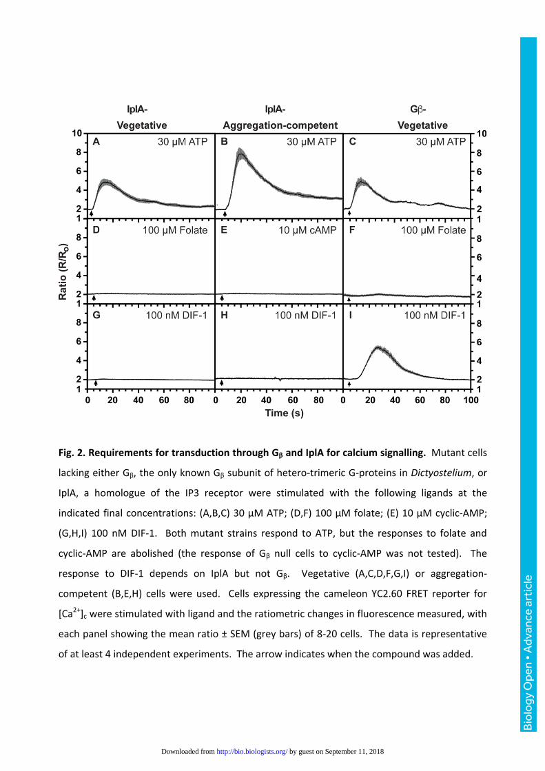
Fig. 2. Requirements for transduction through Gβ and IplA for calcium signalling. Mutant cells
lacking either Gβ, the only known Gβ subunit of hetero-trimeric G-proteins in Dictyostelium, or
IplA, a homologue of the IP3 receptor were stimulated with the following ligands at the
indicated final concentrations: (A,B,C) 30 µM ATP; (D,F) 100 µM folate; (E) 10 µM cyclic-AMP;
(G,H,I) 100 nM DIF-1. Both mutant strains respond to ATP, but the responses to folate and
cyclic-AMP are abolished (the response of Gβ null cells to cyclic-AMP was not tested). The
response to DIF-1 depends on IplA but not Gβ. Vegetative (A,C,D,F,G,I) or aggregation-
competent (B,E,H) cells were used. Cells expressing the cameleon YC2.60 FRET reporter for
[Ca2+]c were stimulated with ligand and the ratiometric changes in fluorescence measured, with
each panel showing the mean ratio ± SEM (grey bars) of 8-20 cells. The data is representative
of at least 4 independent experiments. The arrow indicates when the compound was added.
Bio
logy
Ope
n •
Adv
ance
art
icle
by guest on September 11, 2018http://bio.biologists.org/Downloaded from
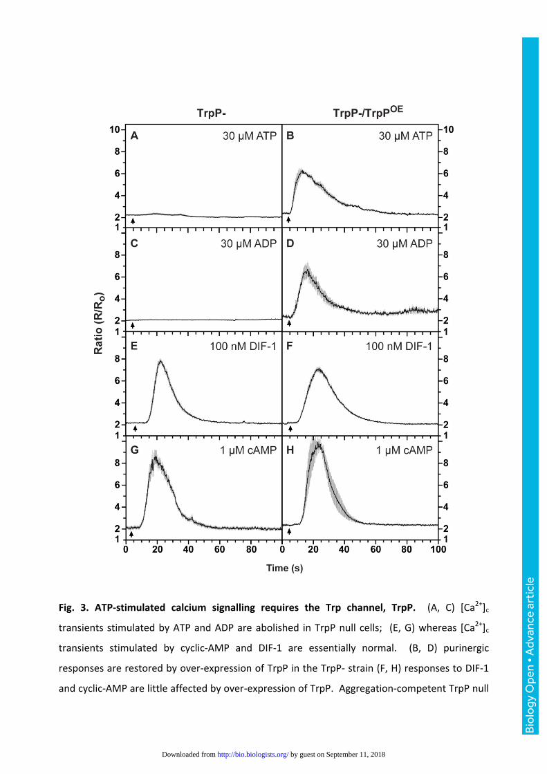
Fig. 3. ATP-stimulated calcium signalling requires the Trp channel, TrpP. (A, C) [Ca2+]c
transients stimulated by ATP and ADP are abolished in TrpP null cells; (E, G) whereas [Ca2+]c
transients stimulated by cyclic-AMP and DIF-1 are essentially normal. (B, D) purinergic
responses are restored by over-expression of TrpP in the TrpP- strain (F, H) responses to DIF-1
and cyclic-AMP are little affected by over-expression of TrpP. Aggregation-competent TrpP null
Bio
logy
Ope
n •
Adv
ance
art
icle
by guest on September 11, 2018http://bio.biologists.org/Downloaded from

cells expressing the [Ca2+]c reporter cameleon YC2.60 were used, with as indicated, a plasmid
for expression of TrpP (pDT50). Changes in the fluorescence ratio with time are presented with
each panel showing the mean ratio ± SEM (grey bars) of 6-15 cells. The data is representative of
at least 3 independent experiments. The arrow indicates when the compound was added.
Bio
logy
Ope
n •
Adv
ance
art
icle
by guest on September 11, 2018http://bio.biologists.org/Downloaded from

Fig. 4. Calcium responses to adenosine, c-di-GMP, L-glutamate and GABA obtained using
aggregation-competent cells. Changes in cytosolic calcium, [Ca2+]c, in response to the ligands
added at the following final concentrations: (A) 100 µM adenosine; (B) 500 µM L-glutamate; (C)
1 mM GABA; (D) 125 µM c-di-GMP. Cells expressing the cameleon YC2.60 FRET reporter for
[Ca2+]c were stimulated with ligand and the ratiometric changes in fluorescence measured, with
each panel showing the mean ratio ± SEM (grey bars) of at least 6 cells in each of 3
experiments. The arrow indicates when the compound was added.
Bio
logy
Ope
n •
Adv
ance
art
icle
by guest on September 11, 2018http://bio.biologists.org/Downloaded from
Bio
logy
Ope
n •
Adv
ance
art
icle
by guest on September 11, 2018http://bio.biologists.org/Downloaded from

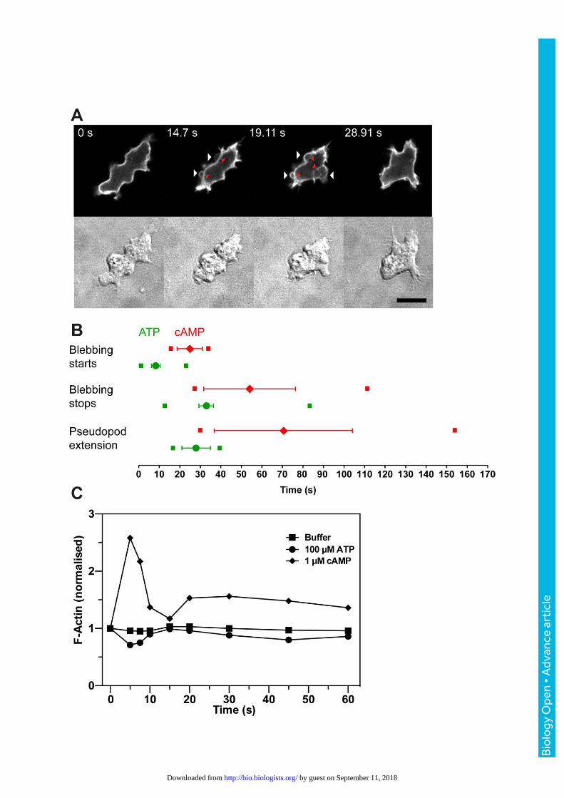
Fig. 5. ATP addition induces cellular blebbing. (A) Ax2 cell stimulated with 30 µM ATP at t0 and
expressing the F-actin reporter GFP-ABP120 observed by fluorescence confocal and DIC
microscopy. The cell starts to bleb after 14 seconds (arrows) and stops by 29 seconds. When
first formed, the blebs are almost devoid of F-actin but leave an F-actin scar behind (red arrow
heads). The bar is 10 µm. (B) Comparison of the timings of ATP and cyclic-AMP induced bleb
formation (cyclic-AMP data reproduced from Langridge and Kay, 2006). The mean ± SEM for
onset of each of the events are indicated for ATP (green) and cyclic-AMP (red). The small
squares indicate the times of earliest onset and cessation of each event for the cells (n=31) in
this data set. (C) Actin polymerization in cells in response to 100 µM ATP, 1 µM cyclic-AMP or
buffer only. The data shown is representative of three experiments, but the slight F-actin de-
polymerization seen in this example did not repeat.
Bio
logy
Ope
n •
Adv
ance
art
icle
by guest on September 11, 2018http://bio.biologists.org/Downloaded from

Fig. 6. Development of wild-type and TrpP null cells on agar and developmental expression of
TrpP mRNA. (A) Development of Ax2 parental cells (panels A,D,G); TrpP null cells (B,E,H) strain
HM1531; and TrpP null cells, strain HM1532 (panels C,F,I). All strains develop with nearly
Bio
logy
Ope
n •
Adv
ance
art
icle
by guest on September 11, 2018http://bio.biologists.org/Downloaded from

identical timing; the time points shown are: 14 hr – slugs; 19 hr - early culminants; 23 hr –
fruiting bodies. The white bar is 0.5 mm. (B) Developmental regulation of TrpP mRNA in
comparison to standard markers as determined by RT-PCR.
Bio
logy
Ope
n •
Adv
ance
art
icle
by guest on September 11, 2018http://bio.biologists.org/Downloaded from

References
Abe, T., Maeda, Y. and Iijima, T. (1988). Transient increase of the intracellular Ca2+ concentration during chemotactic signal transduction in Dictyostelium discoideum cells. Differentiation, 39, 90-96. Anjard, C. and Loomis, W. F. (2006). GABA induces terminal differentiation of Dictyostelium through a GABAB receptor. Development 133, 2253-2261. Baines, A., Parkinson, K., Sim, J. A., Bragg, L., Thompson, C. R. and North, R. A. (2013). Functional properties of five Dictyostelium discoideum P2X receptors. J. Biol. Chem. 288, 20992-21000. Bloomfield, G., Tanaka, Y., Skelton, J., Ivens, A. and Kay, R. R. (2008). Widespread duplications in the genomes of laboratory stocks of Dictyostelium discoideum. Genome Biol. 9, R75. Burnstock, G. (2007). Purine and pyrimidine receptors. Cell. Mol. Life Sci. 64, 1471-1483. Burnstock, G. and Verkhratsky, A. (2009). Evolutionary origins of the purinergic signalling system. Acta Physiol. 195, 415-447. Chapin, H. C. and Caplan, M. J. (2010). The cell biology of polycystic kidney disease. J. Cell. Biol. 191, 701-710. Chen, Z. H. and Schaap, P. (2012). The prokaryote messenger c-di-GMP triggers stalk cell differentiation in Dictyostelium. Nature 488, 680-683. Clapham, D. E. (2003). TRP channels as cellular sensors. Nature 426, 517-524. Collins, S. R. and Meyer, T. (2011). Evolutionary origins of STIM1 and STIM2 within ancient Ca2+ signaling systems. Trends Cell Biol. 21, 202-211. Coste, B., Mathur, J., Schmidt, M., Earley, T. J., Ranade, S., Petrus, M. J., Dubin, A. E. and Patapoutian, A. (2010). Piezo1 and Piezo2 are essential components of distinct mechanically activated cation channels. Science 330, 55-60. Edelstein, A. D., Tsuchida, M. A., Amodaj, N., Pinkard, H., Vale, R. D. and Stuurman, N. (2014). Advanced methods of microscope control using muManager software. J Biol Methods 1. Eichinger, L., Pachebat, J. A., Glockner, G., Rajandream, M. A., Sucgang, R., Berriman, M., Song, J., Olsen, R., Szafranski, K., Xu, Q. et al. (2005). The genome of the social amoeba Dictyostelium discoideum. Nature 435, 43-57. Faix, J., Kreppel, L., Shaulsky, G., Schleicher, M. and Kimmel, A. R. (2004). A rapid and efficient method to generate multiple gene disruptions in Dictyostelium discoideum using a single selectable marker and the Cre-loxP system. Nucleic Acids Res. 32, e143. Fets, L., Nichols, J. M. and Kay, R. R. (2014). A PIP5 kinase essential for efficient chemotactic signaling. Curr. Biol. 24, 415-421. Fountain, S. J., Parkinson, K., Young, M. T., Cao, L., Thompson, C. R. and North, R. A. (2007). An intracellular P2X receptor required for osmoregulation in Dictyostelium discoideum. Nature 448, 200-203. Fukuzawa, M., Araki, T., Adrian, I. and Williams, J. G. (2001). Tyrosine phosphorylation-independent nuclear translocation of a Dictyostelium STAT in response to DIF signaling. Mol. Cell 7, 779-788. Gonzalez-Perrett, S., Kim, K., Ibarra, C., Damiano, A. E., Zotta, E., Batelli, M., Harris, P. C., Reisin, I. L., Arnaout, M. A. and Cantiello, H. F. (2001). Polycystin-2, the protein mutated in autosomal dominant polycystic kidney disease (ADPKD), is a Ca2+-permeable nonselective cation channel. Proc. Natl. Acad. Sci. USA 98, 1182-1187.
Bio
logy
Ope
n •
Adv
ance
art
icle
by guest on September 11, 2018http://bio.biologists.org/Downloaded from

Hanaoka, K., Qian, F., Boletta, A., Bhunia, A. K., Piontek, K., Tsiokas, L., Sukhatme, V. P., Guggino, W. B. and Germino, G. G. (2000). Co-assembly of polycystin-1 and -2 produces unique cation-permeable currents. Nature 408, 990-994. Hardie, R. C. (2007). TRP channels and lipids: from Drosophila to mammalian physiology. J. Physiol. 578, 9-24. Heidel, A. J., Lawal, H. M., Felder, M., Schilde, C., Helps, N. R., Tunggal, B., Rivero, F., John, U., Schleicher, M., Eichinger, L. et al. (2011). Phylogeny-wide analysis of social amoeba genomes highlights ancient origins for complex intercellular communication. Genome Res. 21, 1882-1891. Hirst, J., Kay, R. R. and Traynor, D. (2015). Dictyostelium cultivation, transfection, microscopy and fractionation. Bio. Protoc. 5. Horikawa, K., Yamada, Y., Matsuda, T., Kobayashi, K., Hashimoto, M., Matsu-ura, T., Miyawaki, A., Michikawa, T., Mikoshiba, K. and Nagai, T. (2010). Spontaneous network activity visualized by ultrasensitive Ca(2+) indicators, yellow Cameleon-Nano. Nat. Methods 7, 729-732. Kay, R. R. (1998). The biosynthesis of differentiation-inducing factor, a chlorinated signal molecule regulating Dictyostelium development. J. Biol. Chem. 273, 2669-2675. Kerr, R. A. and Schafer, W. R. (2006). Intracellular Ca2+ imaging in C. elegans. Method. Mol. Biol. 351, 253-264. Kessin, R. H. (2001). Dictyostelium. Cambridge: Cambridge University Press. Klein, P. S., Sun, T. J., Saxe III, C. L., Kimmel, A. R., Johnson, R. L. and Devreotes, P. N. (1988). A chemoattractant receptor controls development in Dictyostelium discoideum. Science 241, 1467-1472. Kosaka, C. and Pears, C. J. (1997). Chemoattractants induce tyrosine phosphorylation of ERK2 in Dictyostelium discoideum by diverse signalling pathways. Biochem. J. 324 ( Pt 1), 347-352. Langridge, P. D. and Kay, R. R. (2006). Blebbing of Dictyostelium cells in response to chemoattractant. Exp. Cell Res. 312, 2009-2017. Lima, W. C., Leuba, F., Soldati, T. and Cosson, P. (2012). Mucolipin controls lysosome exocytosis in Dictyostelium. J. Cell Sci. 125, 2315-2322. Lima, W. C., Vinet, A., Pieters, J. and Cosson, P. (2014). Role of PKD2 in rheotaxis in Dictyostelium. PloS one 9, e88682. Ludlow, M. J., Durai, L. and Ennion, S. J. (2009). Functional characterization of intracellular Dictyostelium discoideum P2X receptors. J. Biol. Chem. 284, 35227-35239. Ludlow, M. J., Traynor, D., Fisher, P. R. and Ennion, S. J. (2008). Purinergic-mediated Ca2+ influx in Dictyostelium discoideum. Cell Calcium 44, 567-579. Manstein, D. J., Schuster, H. P., Morandini, P. and Hunt, D. M. (1995). Cloning vectors for the production of proteins in Dictyostelium discoideum. Gene 162, 129-134. Martinac, B., Saimi, Y. and Kung, C. (2008). Ion channels in microbes. Physiol. Rev. 88, 1449-1490. Masento, M. S., Morris, H. R., Taylor, G. W., Johnson, S. J., Skapski, A. C. and Kay, R. R. (1988). Differentiation-inducing factor from the slime mould Dictyostelium discoideum and its analogues. Biochem. J. 256, 23-28. Mato, J. M. and Konijn, T. M. (1975). Enhanced cell aggregation in Dictyostelium discoideum by ATP activation of cyclic AMP receptors. Dev. Biol. 47, 233-235. Meili, R., Ellsworth, C., Lee, S., Reddy, T. B. K., Ma, H. and Firtel, R. A. (1999). Chemoattractant-mediated transient activation and membrane localization of Akt/PKB is required for efficient chemotaxis to cAMP in Dictyostelium. EMBO J. 18, 2092-2105.
Bio
logy
Ope
n •
Adv
ance
art
icle
by guest on September 11, 2018http://bio.biologists.org/Downloaded from

Milne, J. L. and Coukell, M. B. (1991). A Ca2+ transport system associated with the plasma membrane of Dictyostelium discoideum is activated by different chemoattractant receptors. J. Cell Biol. 112, 103-110. Milne, J. L. and Devreotes, P. N. (1993). The surface cyclic AMP receptors, cAR1, cAR2, and cAR3, promote Ca-2+ influx in Dictyostelium discoideum by a G-alpha-2-independent mechanism. Mol. Biol. Cell 4, 283-292. Mochizuki, T., Wu, G., Hayashi, T., Xenophontos, S. L., Veldhuisen, B., Saris, J. J., Reynolds, D. M., Cai, Y., Gabow, P. A., Pierides, A. et al. (1996). PKD2, a gene for polycystic kidney disease that encodes an integral membrane protein. Science 272, 1339-42. Morris, H. R., Taylor, G. W., Masento, M. S., Jermyn, K. A. and Kay, R. R. (1987). Chemical structure of the morphogen differentiation inducing factor from Dictyostelium discoideum. Nature 328, 811-814. Nagai, T., Yamada, S., Tominaga, T., Ichikawa, M. and Miyawaki, A. (2004). Expanded dynamic range of fluorescent indicators for Ca(2+) by circularly permuted yellow fluorescent proteins. Proc. Natl. Acad. Sci. USA 101, 10554-10559. Narita, T. B., Chen, Z. H., Schaap, P. and Saito, T. (2014). The hybrid type polyketide synthase SteelyA is required for cAMP signalling in early Dictyostelium development. PloS one 9, e106634. Nebl, T. and Fisher, P. R. (1997). Intracellular Ca2+ signals in Dictyostelium chemotaxis are mediated exclusively by Ca2+ influx. J. Cell Sci. 110, 2845-2853. Nebl, T., Kotsifas, M., Schaap, P. and Fisher, P. R. (2002). Multiple signalling pathways connect chemoattractant receptors and calcium channels in Dictyostelium. J. Muscle Res. Cell Motil. 23, 853-865. Pan, M., Xu, X., Chen, Y. and Jin, T. (2016). Identification of a chemoattractant G-protein-coupled receptor for folic acid that controls both chemotaxis and phagocytosis. Dev. Cell 36, 428-439. Pang, K. M., Lynes, M. A. and Knecht, D. A. (1999). Variables controlling the expression level of exogenous genes in Dictyostelium. Plasmid 41, 187-197. Parent, C. A., Blacklock, B. J., Froelich, W. M., Murphy, D. B. and Devreotes, P. N. (1998). G Protein signaling events are activated at the leading edge of chemotactic cells. Cell 95, 81-91. Parikh, A., Miranda, E. R., Katoh-Kurasawa, M., Fuller, D., Rot, G., Zagar, L., Curk, T., Sucgang, R., Chen, R., Zupan, B. et al. (2010). Conserved developmental transcriptomes in evolutionarily divergent species. Genome Biol. 11, R35. Parish, R. W. and Weibel, M. (1980). Extracellular ATP, ecto-ATPase and calcium influx in Dictyostelium discoideum cells. FEBS Lett. 118, 263-266. Parkinson, K., Baines, A. E., Keller, T., Gruenheit, N., Bragg, L., North, R. A. and Thompson, C. R. (2014). Calcium-dependent regulation of Rab activation and vesicle fusion by an intracellular P2X ion channel. Nature Cell Biol. 16, 87-98. Perekalin, D. (1977). The influence of light and different ATP concentrations on cell aggregation in cyclic AMP sensitive and insensitive species of the cellular slime molds. Arch. Microbiol. 115, 333-337. Plattner, H. and Verkhratsky, A. (2015). The ancient roots of calcium signalling evolutionary tree. Cell Calcium 57, 123-132. Pollitt, A. Y., Blagg, S. L., Ibarra, N. and Insall, R. H. (2006). Cell motility and SCAR localisation in axenically growing Dictyostelium cells. Eur. J. Cell Biol. 85, 1091-1098. Saito, T., Kato, A. and Kay, R. R. (2008). DIF-1 induces the basal disc of the Dictyostelium fruiting body. Dev. Biol. 317, 444-453.
Bio
logy
Ope
n •
Adv
ance
art
icle
by guest on September 11, 2018http://bio.biologists.org/Downloaded from

Saito, T., Taylor, G. W., Yang, J. C., Neuhaus, D., Stetsenko, D., Kato, A. and Kay, R. R. (2006). Identification of new differentiation inducing factors from Dictyostelium discoideum. Biochim. Biophys. Acta 1760, 754-761. Schaap, P. and Wang, M. (1986). Interactions between adenosine and oscillatory cAMP signaling regulate size and pattern in Dictyostelium. Cell 45, 137-144. Schilde, C., Araki, T., Williams, H., Harwood, A. and Williams, J. G. (2004). GSK3 is a multifunctional regulator of Dictyostelium development. Development 131, 4555-4565. Schindelin, J., Arganda-Carreras, I., Frise, E., Kaynig, V., Longair, M., Pietzsch, T., Preibisch, S., Rueden, C., Saalfeld, S., Schmid, B. et al. (2012). Fiji: an open-source platform for biological-image analysis. Nat Methods 9, 676-82. Schlatterer, C. and Schaloske, R. (1996). Calmidazolium leads to an increase in the cytosolic Ca2+ concentration in Dictyostelium discoideum by induction of Ca2+ release from intracellular stores and influx of extracellular Ca2+. Biochem. J. 313, 661-667. Schneider, C. A., Rasband, W. S. and Eliceiri, K. W. (2012). NIH Image to ImageJ: 25 years of image analysis. Nat Methods 9, 671-5. Sivaramakrishnan, V. and Fountain, S. J. (2012). A mechanism of intracellular P2X receptor activation. J. Biol. Chem. 287, 28315-28326. Sivaramakrishnan, V. and Fountain, S. J. (2013). Intracellular P2X receptors as novel calcium release channels and modulators of osmoregulation in Dictyostelium: a comparison of two common laboratory strains. Channels 7, 43-46. Sivaramakrishnan, V. and Fountain, S. J. (2015). Evidence for extracellular ATP as a stress signal in a single-celled organism. Eukaryot. Cell 14, 775-782. Song, Y., Luciani, M. F., Giusti, C. and Golstein, P. (2015). c-di-GMP induction of Dictyostelium cell death requires the polyketide DIF-1. Mol. Biol. Cell 26, 651-658. Sucgang, R., Kuo, A., Tian, X., Salerno, W., Parikh, A., Feasley, C. L., Dalin, E., Tu, H., Huang, E., Barry, K. et al. (2011). Comparative genomics of the social amoebae Dictyostelium discoideum and Dictyostelium purpureum. Genome Biol. 12, R20. Sugden, C., Urbaniak, M. D., Araki, T. and Williams, J. G. (2015). The Dictyostelium prestalk inducer differentiation-inducing factor-1 (DIF-1) triggers unexpectedly complex global phosphorylation changes. Mol. Biol. Cell 26, 805-820. Taniura, H., Sanada, N., Kuramoto, N. and Yoneda, Y. (2006). A metabotropic glutamate receptor family gene in Dictyostelium discoideum. J. Biol. Chem. 281, 12336-12343. Traynor, D. and Kay, R. R. (2007). Possible roles of the endocytic cycle in cell motility. J. Cell Sci. 120, 2318-2327. Traynor, D., Milne, J. L., Insall, R. H. and Kay, R. R. (2000). Ca(2+) signalling is not required for chemotaxis in Dictyostelium. EMBO J. 19, 4846-4854. Tyson, R. A., Zatulovskiy, E., Kay, R. R. and Bretschneider, T. (2014). How blebs and pseudopods cooperate during chemotaxis. Proc. Natl. Acad. Sci. USA 111, 11703-11708. Urushihara, H., Kuwayama, H., Fukuhara, K., Itoh, T., Kagoshima, H., Shin, I. T., Toyoda, A., Ohishi, K., Taniguchi, T., Noguchi, H. et al. (2015). Comparative genome and transcriptome analyses of the social amoeba Acytostelium subglobosum that accomplishes multicellular development without germ-soma differentiation. BMC Genomics 16, 80. Veltman, D. M., Akar, G., Bosgraaf, L. and Van Haastert, P. J. (2009). A new set of small, extrachromosomal expression vectors for Dictyostelium discoideum. Plasmid 61, 110-118. Venkatachalam, K. and Montell, C. (2007). TRP channels. Ann. Rev. Biochem. 76, 387-417.
Bio
logy
Ope
n •
Adv
ance
art
icle
by guest on September 11, 2018http://bio.biologists.org/Downloaded from
Waheed, A., Ludtmann, M. H., Pakes, N., Robery, S., Kuspa, A., Dinh, C., Baines, D., Williams, R. S. and Carew, M. A. (2014). Naringenin inhibits the growth of Dictyostelium and MDCK-derived cysts in a TRPP2 (polycystin-2)-dependent manner. Br. J. Pharmacol. 171, 2659-2670. Wilczynska, Z., Happle, K., Muller-Taubenberger, A., Schlatterer, C., Malchow, D. and Fisher, P. R. (2005). Release of Ca2+ from the endoplasmic reticulum contributes to Ca2+ signaling in Dictyostelium discoideum. Eukaryot. Cell 4, 1513-1525. Williams, J. G., Ceccarelli, A., McRobbie, S., Mahbubani, H., Kay, R. R., Early, A., Berks, M. and Jermyn, K. A. (1987). Direct induction of Dictyostelium prestalk gene expression by DIF provides evidence that DIF is a morphogen. Cell 49, 185-192. Wu, G., D'Agati, V., Cai, Y., Markowitz, G., Park, J. H., Reynolds, D. M., Maeda, Y., Le, T. C., Hou, H., Jr., Kucherlapati, R. et al. (1998). Somatic inactivation of Pkd2 results in polycystic kidney disease. Cell 93, 177-88. Wu, L. J., Valkema, R., van Haastert, P. J. M. and Devreotes, P. N. (1995). The G protein beta subunit is essential for multiple responses to chemoattractants in Dictyostelium. J. Cell Biol. 129, 1667-1675. Wu, Y. and Janetopoulos, C. (2013). Systematic analysis of gamma-aminobutyric acid (GABA) metabolism and function in the social amoeba Dictyostelium discoideum. J. Biol. Chem. 288, 15280-15290. Wurster, B. and Kay, R. R. (1990). New roles for DIF? Effects on early development in Dictyostelium. Dev. Biol. 140, 189-195. Yoshida, K. and Soldati, T. (2006). Dissection of amoeboid movement into two mechanically distinct modes. J. Cell Sci. 119, 3833-3844. Yu, Y., Ulbrich, M. H., Li, M. H., Buraei, Z., Chen, X. Z., Ong, A. C., Tong, L., Isacoff, E. Y. and Yang, J. (2009). Structural and molecular basis of the assembly of the TRPP2/PKD1 complex. Proc. Natl. Acad. Sci. USA 106, 11558-11563. Zatulovskiy, E., Tyson, R., Bretschneider, T. and Kay, R. R. (2014). Bleb-driven chemotaxis of Dictyostelium cells. J. Cell Biol. 204, 1027-1044.
Bio
logy
Ope
n •
Adv
ance
art
icle
by guest on September 11, 2018http://bio.biologists.org/Downloaded from

2
4
6
8
10
2
4
6
8
10
2
4
6
8
10
2
4
6
8
10
1
2
4
6
8
10
2
4
6
8
10
2
4
6
8
10
2
4
6
8
10
1
20 40 600 20 40 600
YC2.60 YC3.60
100 µM ATP 100 µM ATP
10 µM ATP 10 µM ATP
1 µM ATP 1 µM ATP
0.5 µM ATP 0.5 µM ATP
A B
C D
E F
G H
Time (s) Time (s)
Rati
o (
R/R
o)
Fig.S1.InDictyosteliumcells,cameleonYC2.60ismoresuitedtomeasureATPmediatedchangesincytosolicCa2+thanYC3.60(A)FRETmeasurementsofchangesincytosoliccalciumwithYC2.60(A,C,EandG)showthatitgenerateslargerchangesinraCoandismoresensiCvetosub-maximaldoesofATPcomparedtoYC3.60(B,D,FandH).ThemeanandS.E.M.oftheraCofromupto10aggregaCon-competentAx2cellsareploQedineachpanelandtheresultsarerepresentaCveofatleast3independentexperiments.WhenachangeinraCowasobserved>95%ofthecellsrespondedwhereasinpanels(F)and(H)noneofthecellsresponded.
Supplementary Material
Supplementary figures
Biology Open (2017): doi:10.1242/bio.020685: Supplementary information
Bio
logy
Ope
n •
Sup
plem
enta
ry in
form
atio
n
by guest on September 11, 2018http://bio.biologists.org/Downloaded from

-8 -7 -6 -5 -4 -3
0
50
100
Nor
mal
ised
Res
pons
e
-8 -7 -6 -5 -4 -3
0
50
100N
orm
alis
ed R
espo
nse
ATP (M)
-11 -10 -9 -8 -7 -6 -5 -4
0
50
100
cAMP (M)
Nor
mal
ised
Res
pons
e
ADP (M)
-10 -9 -8 -7 -6
0
50
100
150
DIF-1 (M)
Nor
mal
ised
Res
pons
e
-10 -9 -8 -7 -6
0
50
100
150
DIF-2 (M)
Nor
mal
ised
Res
pons
e
Fig.S2.Dose-responsecurvesforATP,ADP,DIF-1,DIF-2andcyclic-AMPobtainedusingaggregaJoncompetentcells.ThepercentageofcellsrespondingateachconcentraConwhereasfollows.ATP100µM94%,30µM92%,10µM83%,5µM87%,2.5µM77%,1µM84%,500nM68%,250nM63%,125nM17%,62.5nM5%and31.25nM0%.ADP1mM84%,100µM84%,30µM95%,10µM96%,5µM77%,2.5µM93%,1µM89%,500nM72%,250nM5%,125nM6%and62.5nM11%.cAMP100µM93%,10µM100%,1µM100%,500nM82%,250nM97%,100nM89%,10nM91%,1nM48%,100pM26%,50pM20%and10pM0%.DIF21µM93%,500nM97%,250nM69%,100nM98%,50nM90%,25nM74%,10nM56%,1nM50%,500pM33%and100pM47%.DIF1250nM74%,100nM78%,50nM57%,25nM71%,10nM32%,5nM33%,1nM22%,500pM50%and100pM0%.DIF21µM93%,500nM97%,250nM69%,100nM98%,50nM90%,25nM74%,10nM56%,1nM50%,500pM33%and100pM47%.
Biology Open (2017): doi:10.1242/bio.020685: Supplementary information
Bio
logy
Ope
n •
Sup
plem
enta
ry in
form
atio
n
by guest on September 11, 2018http://bio.biologists.org/Downloaded from
0
2
4
6Ax2MscS-/MclN-/TrpP-
A
0 20 40 60 80 100 120
0
2
4
6Ax2TrpP-
Stim
ulat
ed C
a2+
upta
ke(n
mol
es/m
g pr
otei
n)St
imul
ated
Ca2
+ up
take
(nm
oles
/mg
prot
ein)
Stim
ulat
ed C
a2+
upta
ke(n
mol
es/m
g pr
otei
n)
Time (s)
B
1 µM ATP
30 µM
ATP
30 µM
ADP
10 µM
cAMP
AxM
2MclN clN Ax
M2
MclN clN AxM
2MclN clN
0
2
4
6
A Mx2 sc Ms
S cS A Mx2 sc Ms
S cS A Mx2 sc Ms
S cS0
2
4
610 µM ATP10 µM ADP10 µM cAMP
DC
Biology Open (2017): doi:10.1242/bio.020685: Supplementary information
Bio
logy
Ope
n •
Sup
plem
enta
ry in
form
atio
n
by guest on September 11, 2018http://bio.biologists.org/Downloaded from

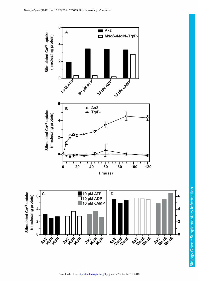
Fig.S3.IdenJficaJonofTrpPasthemediatorofATPsJmulatedcalciumuptakeusing45Ca2+.(A)AmscS-/mclN-/trpP-triplemutantwastestedforATP,ADPandcyclic-AMPsCmulatedcalciumuptakefor1minute.ThemutantandtheAx2parenthadsimilaruptakesofCa2+sCmulatedbycyclic-AMP,butthemutantrespondedmuchlesswelltoATPandADPthanAx2cells.(B)ThedefectinATP-sCmulatedCa2+
uptakeistheresultofablaConoftrpP.TimecourseofATP(30µM)sCmulatedCa2+
uptakeinAx2(opencircles)andtrpP-nullcells(solidcircles).ThemclA-andmscS-nullcellsaccumulateCa2+tosimilarlevelswhensCmulatedwithATP,ADPandcyclic-AMPcomparedtoAx2(C,D).TwoindependentmclN-clones(C)andmscS-clones(D)areshown.Uptakewasmeasuredfor1minutea_ersCmulaCon.AllexperimentsusedaggregaCon-competentcellsandarerepresentaCve.Thebufferusedtomeasurecalciumuptakewasspikedwith45Ca2+anduptakemeasuredessenCallyasdescribedinTraynoretal.,(2000).
Biology Open (2017): doi:10.1242/bio.020685: Supplementary information
Bio
logy
Ope
n •
Sup
plem
enta
ry in
form
atio
n
by guest on September 11, 2018http://bio.biologists.org/Downloaded from
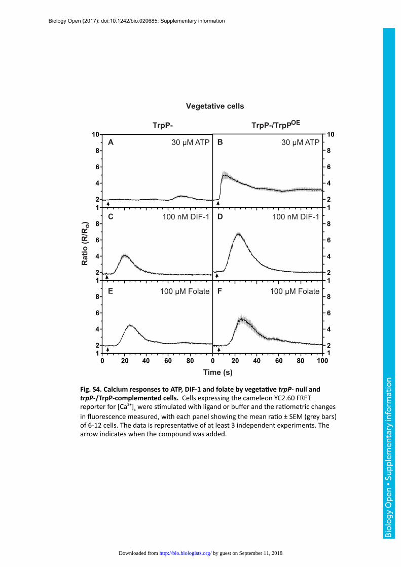
Fig.S4.CalciumresponsestoATP,DIF-1andfolatebyvegetaJvetrpP-nullandtrpP-/TrpP-complementedcells.CellsexpressingthecameleonYC2.60FRETreporterfor[Ca2+]cweresCmulatedwithligandorbufferandtheraCometricchangesinfluorescencemeasured,witheachpanelshowingthemeanraCo±SEM(greybars)of6-12cells.ThedataisrepresentaCveofatleast3independentexperiments.Thearrowindicateswhenthecompoundwasadded.
2
4
6
8
2
1
4
6
8
10
2
4
6
8
1
2
4
6
8
10
2
4
6
8
1
2
4
6
8
1
1 1
Time (s)
0 20 40 60 800 20 40 60 80 100
30 µM ATP
100 nM DIF-1 100 nM DIF-1
30 µM ATP
100 µM Folate
TrpP- TrpP-/TrpPOE
A B
C D
E F100 µM Folate
Rati
o (
R/R
o)
Vegetative cells
Biology Open (2017): doi:10.1242/bio.020685: Supplementary information
Bio
logy
Ope
n •
Sup
plem
enta
ry in
form
atio
n
by guest on September 11, 2018http://bio.biologists.org/Downloaded from

Fig.S5.LocalizaJonofTrpPproteininvegetaJvecells.ExpressionofaTrpP-GFPfusionproteindrivenbythetrpPpromoterintrpP-cells.Atypicalcellisshown.Thetoppanelsarefluorescenceimagesfrom3differentCmesandtheboQompanelsarethecorrespondingDICimages.TrpP-GFP(plasmidpDT68)ispresentconCnuouslyontheplasmamembrane,thoughcytoplasmicpunctaarealsoevident.
5 µm
Biology Open (2017): doi:10.1242/bio.020685: Supplementary information
Bio
logy
Ope
n •
Sup
plem
enta
ry in
form
atio
n
by guest on September 11, 2018http://bio.biologists.org/Downloaded from

2
4
6
8
10
0 20 40 60 80 100
2
4
6
8
10
2
4
6
8
10
2
4
6
8
102
4
6
8
102
4
6
8
1030 µM ATP 30 µM ATP
30 µM ADP 10 µM cAMP
Buffer 5 µM Calmidazolium
10 20 40 60 80
Time (s)
1
A B
C D
E F
TrpP-R
atio
(R/R
o)TrpP-/IplA-
Fig.S6.AdelayedcalciumresponseremainsintrpP-cellsanddependsonIplA.AddiConofATP,ADPorbufferalonecansomeCmeselicitadelayedincreasein[Ca2+]cintrpP-cells,whichdependsonIplA.(A,C,E)residualresponsesintrpP-nullcellstoATP,ADPortobufferalone;(B,D)aboliConofresponsestoATPandADPintrpP-/iplA-doublenullcells;(F)controlshowingthattrpP-/iplA-doublenullcellscansCllrespondtothecalmodulinantagonistcalmidazolium(5µM).ParentalAx2cellsrespondsimilarlytocalmidazolium.CellsexpressingthecameleonYC2.60FRETreporterfor[Ca2+]cweresCmulatedwithligandorbufferandtheraCometricchangesinfluorescencemeasured,witheachpanelshowingthemeanraCo±SEM(greybars)of6-12cells.ThedataisrepresentaCveofatleast3independentexperiments.Thearrowindicateswhenthecompoundwasadded.
Biology Open (2017): doi:10.1242/bio.020685: Supplementary information
Bio
logy
Ope
n •
Sup
plem
enta
ry in
form
atio
n
by guest on September 11, 2018http://bio.biologists.org/Downloaded from

2
4
6
8
10
2
4
6
8
10
2
4
6
8
10
100 1
0 20 40 60 80
Time (s)
Rat
io (R
/Ro)
30 µM ATPA
B
C
1 mM GABA
1 mM L-Glu
IplA-
Fig.S7.GABAandL-Glutamateinduced[Ca2+]cresponsesaredependentonIplA.(A)iplA-nullcellsproducethetypicalrapidresponseto30µMATP,whereas(B)1mMGABAand,(C)1mML-Glutamatefailtoevokeanyresponse.ThearrowsindicatewhenthecompoundwasaddedtoaggregaCon-competentiplA-cells.Allthecellsanalysed(7)in(A)respondedwhereasnoneofthecells(6and7)respondedin(B)or(C).TheresultsarerepresentaCveof3experiments.
Biology Open (2017): doi:10.1242/bio.020685: Supplementary information
Bio
logy
Ope
n •
Sup
plem
enta
ry in
form
atio
n
by guest on September 11, 2018http://bio.biologists.org/Downloaded from

-0.58 s
6.96 s
13.34 s
16.82 s
35.96 s
64.38 s
∆ ∆
TrpP- MlcE-
-0.60 s
6.00s
24.00 s
36.00 s
66.00 s
12.00s
A B
Fig.S8.ATPinducesblebbingintrpP-cellsandisdependentonthemyosinessenJallightchain,MlcE.(A)ConfocalflourescenceandDICimagesofatrpP-cellsCmulatedwith30µMATPatt0.ThecellexpressestheF-acCnreporterGFP-ABP120.BlebsaremarkedwithwhitearrowheadswhereasacCnscarsareindicatedbyredones.Thefirstinfocusblebisobserveda_er~14secondsandthelastblebsareformedat~36secondspostsCmulaCon.(B)ConfocalDICimagesofmlcE-nullcellssCmulatedwith100µMATPasin(A).Therearenoobservableblebsevena_er66secondsofsCmulaCon.AllcellsusedareaggregaCon-competent.Timesandthescalebars(10µm)areindicatedinwhite.
Biology Open (2017): doi:10.1242/bio.020685: Supplementary information
Bio
logy
Ope
n •
Sup
plem
enta
ry in
form
atio
n
by guest on September 11, 2018http://bio.biologists.org/Downloaded from

120
80
100
60
40
20
0
050100 50 100
-20
-60
-40
-80
-100
-120
y (µ
m)
x (µm)
120
80
100
60
40
20
0
050100 50 100
-20
-60
-40
-80
-100
-120
y (µ
m)
x (µm)
120
80
100
60
40
20
0
050100 50 100
-20
-60
-40
-80
-100
-120
y (µ
m)
x (µm)
120
80
100
60
40
20
0
050100 50 100
-20
-60
-40
-80
-100
-120
yµm
)(
x (µm)
Ax2 No flow
Ax2 Flow
4.20 ± 0.28
5.70 ± 0.52
0.18 ± 0.02 (n=33)
0.44 ± 0.06 (n=33)
TrpP- No flow
TrpP- Flow
4.28 ± 0.20
4.68 ± 0.23
0.20 ± 0.02 (n=28)
0.51 ± 0.05 (n=28)
Velocity (µm min-1) ± s.e.m Directionality
A Ax2 B Ax2
C TrpP- D TrpP-
No Flow Flow
No Flow Flow
Fig.S9.RheotaxisofvegetaCveparentalAx2andmutanttrpP-nullcellsexposedtoasheerstressof3Pa.Cellstracksandmovementparametersaregiven.
Biology Open (2017): doi:10.1242/bio.020685: Supplementary information
Bio
logy
Ope
n •
Sup
plem
enta
ry in
form
atio
n
by guest on September 11, 2018http://bio.biologists.org/Downloaded from

Supplementarytables
TableS1.Theresponseofindividualcellsto30µMATPorbuffer
Fastresponse NoResponse LateResponse
Ax2+30µMATP 115 5 5Ax2+Buffer 0 55 5TrpP-+30µMATP 0 95 21TrpP-+Buffer 0 88 10
Calcium responses to ATP or buffer alone in individual cells were detected byimaging with the cameleonYC2.60FRETreporter. Most Ax2 cells respond rapidly toATP as shown in Fig. 1 of the main text (‘fast response’) but TrpP- cells neverrespond in this way. However, both Ax2 and TrpP- cells more occasionally produce adelayed response to ATP or to buffer (‘late response’) and some do not respond at all(‘no response’) as tabulated here. The frequency of the late response in TrpP- cellswas not statistically different with or without ATP (the proportion of responding cellswas used to generate a Z-score with a two-tailed hypothesis giving a p value of0.101). On this basis it is not necessary to invoke the existence of a further ATPreceptor to explain these observations.
Biology Open (2017): doi:10.1242/bio.020685: Supplementary information
Bio
logy
Ope
n •
Sup
plem
enta
ry in
form
atio
n
by guest on September 11, 2018http://bio.biologists.org/Downloaded from
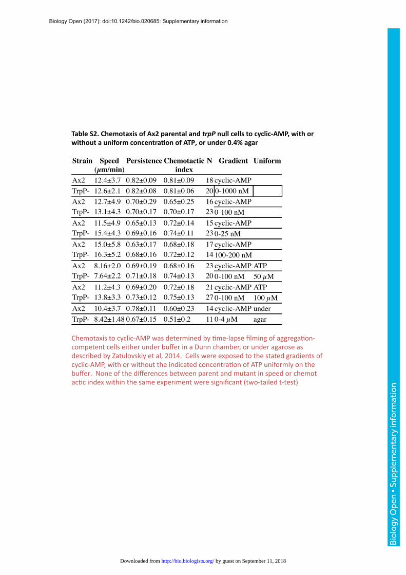
TableS2.ChemotaxisofAx2parentalandtrpPnullcellstocyclic-AMP,withorwithoutauniformconcentraJonofATP,orunder0.4%agar
Strain Speed(µm/min)
Persistence Chemotacticindex
N Gradient Uniform
18 cyclic-AMP20 0-1000 nM
Ax2 12.4±3.7 0.82±0.09 0.81±0.09TrpP- 12.6±2.1 0.82±0.08 0.81±0.06Ax2 12.7±4.9 0.70±0.29 0.65±0.25TrpP- 13.1±4.3 0.70±0.17 0.70±0.17Ax2 11.5±4.9 0.65±0.13 0.72±0.14TrpP- 15.4±4.3 0.69±0.16 0.74±0.11Ax2 15.0±5.8 0.63±0.17 0.68±0.18TrpP- 16.3±5.2 0.68±0.16 0.72±0.12Ax2 8.16±2.0 0.69±0.19 0.68±0.16TrpP- 7.64±2.2 0.71±0.18 0.74±0.13Ax2 11.2±4.3 0.69±0.20 0.72±0.18TrpP- 13.8±3.3 0.73±0.12 0.75±0.13Ax2 10.4±3.7 0.78±0.11 0.60±0.23TrpP- 8.42±1.48 0.67±0.15 0.51±0.2
16 cyclic-AMP 23 0-100 nM 15 cyclic-AMP 23 0-25 nM17 cyclic-AMP 14 100-200 nM 23 cyclic-AMP ATP20 0-100 nM 50 µM21 cyclic-AMP ATP27 0-100 nM 100 µM14 cyclic-AMP under11 0-4 µM agar
Chemotaxistocyclic-AMPwasdeterminedbyCme-lapsefilmingofaggregaCon- competentcellseitherunderbufferinaDunnchamber,orunderagaroseas describedbyZatulovskiyetal,2014.Cellswereexposedtothestatedgradientsof cyclic-AMP,withorwithouttheindicatedconcentraConofATPuniformlyonthe buffer.NoneofthedifferencesbetweenparentandmutantinspeedorchemotacCc indexwithinthesameexperimentweresignificant(two-tailedt-test)
Biology Open (2017): doi:10.1242/bio.020685: Supplementary information
Bio
logy
Ope
n •
Sup
plem
enta
ry in
form
atio
n
by guest on September 11, 2018http://bio.biologists.org/Downloaded from

No. Strain Genotype Stage AddiJon Speed(µm/min)
Persistence N
1 Ax2 wild-type veg 5.71±2.3 0.42 262 HM1531 trpP- veg 6.12±1.7 0.70 273 Ax2 wild-type veg 5.36±2.0 0.52 234 HM1532 trpP- veg 5.17±2.4 0.59 245 Ax2 wild-type veg ATP 5.29±1.6 0.51 276 HM1531 trpP- veg 100µM 5.51±2.1 0.52 267 Ax2 wild-type aggcomp 10.9±2.9 0.53 258 HM1531 trpP- aggcomp 9.78±4.5 0.44 229 Ax2 wild-type aggcomp 9.47±4.6 0.40 2410 HM1532 trpP- aggcomp 7.59±4.9 0.35 2211 Ax2 wild-type aggcomp ATP 11.7±3.6 0.46 2612 HM1531 trpP- aggcomp 100µM 8.64±3.5 0.33 2613 Ax2 wild-type aggcomp ATP 11.3±3.7 0.43 3114 HM1532 trpP- aggcomp 100µM 7.56±3 0.27 28
Table S3.RandommovementparametersofAx2parentalandtrpP-cellswithorwithouta uniformconcentraJonofATP
Randomcellmovement(intheabsenceofchemoaQractantgradients)wasdeterminedfromCme-lapsemoviesofvegetaCveoraggregaCon-competent(vegoraggcomp)cellseitherwithorwithoutauniformconcentraConof100µMATPinthemedium.TwoindependenttrpP-cloneswereused.Thelackofdifferenceinspeedbetweenwild-typeandmutantcomesfromcomparisonsinthesameexperiment(egcomparerows1&2)noneofwhichwerestaCsCcallysignificant(two-tailedt-test).Whilethe(lackof)effectofATPonthespeedofthewild-typeAx2comesfromcomparisonswithandwithoutATP(comparerows1&5;2&6;7&11;9&13),noneofwhichweresignificant.
TableS4.GrowthofparentalAx2andtrpP-nullcellsinliquidmedium
Strain GenotypeMGT(hr) SEM NAx2 wild-type 9.49 0.23 12
21.4 3.0 69.18 0.68 914.0 0.17 38.46 0.09 38.86 0.65 6
HM1531 trpP-HM1532 trpP-HM1578 trpP-HM1579 trpP-HM1822 trpP-HM1823 trpP- 8.94 0.61 6
CellsweregrowninHL5mediumat220Cwithshakingat180rpmandtheirnumbersmeasuredtwicedailyusingaCoulterparCclecounter.ThemeangeneraConCme(MGT)wasdeterminedduringlogphase.
Biology Open (2017): doi:10.1242/bio.020685: Supplementary information
Bio
logy
Ope
n •
Sup
plem
enta
ry in
form
atio
n
by guest on September 11, 2018http://bio.biologists.org/Downloaded from

TableS5.Strainsusedinthiswork
Strain Genotype Parent Source
Ax2(Kay) Wild-typeHM1486 iplA- Ax2(Kay) this work
HM1691 gpbA- Ax2(Kay) P. Devreotes/J.Borleis
HM1317 mscS- Ax2(Kay) thiswork
HM1449 mclN- Ax2(Kay) thiswork
HM1492 mscS-,mclN-,trpP-
Ax2(Kay) thiswork
HM1493 mscS-,mclN-,trpP-
Ax2(Kay) thiswork
HM1531 trpP- Ax2(Kay) thiswork
HM1532 trpP- Ax2(Kay) thiswork
HM1578 trpP- Ax2(Kay) thiswork
HM1579 trpP- Ax2(Kay) thiswork
HM1822 trpP- Ax2(Kay) thiswork
HM1823 trpP- Ax2(Kay) thiswork
HM1578 trpP-,iplA- HM1531 thiswork
HM1823 trpP-
HM1578 trpP-,iplA-
HM1579 trpP-,iplA-
Ax2(Kay) thiswork
HM1531 thiswork
HM1531 thiswork
Biology Open (2017): doi:10.1242/bio.020685: Supplementary information
Bio
logy
Ope
n •
Sup
plem
enta
ry in
form
atio
n
by guest on September 11, 2018http://bio.biologists.org/Downloaded from

TableS6.Primersused
PC2S26 GGTGAAGGTGATGCTGAGACTGATGATGPC2S27 CTTGTAAATTGTAAAGAACAATGTTGAC-3’PC2KO1 TAAGGGCCCAATGAATAGGTTGGGAGTGATAATACTGGATCPC2KO2 CTAGGGCCCTAACAGCTTCAAGTCTTTGAGAGAAAACTGTCPC2KO3 GTAGCGGCCGCAATGGTAGTATAAATGGTAGTAATCATATCPC2KO4 GTAGCGGCCGCAATGGTAGTATAAATGGTAGTAATCATATCPCL5 AAAGGATCCAAAGGATGAATACATTAAAGAGGACAGTTTTCTCTCPCL3 TTACTCGAGTAAAGGTGTATTAGTACCACCAGATAAATTGATATGRAGFP9 AATCTCGAGATGAGTAAAGGAGAAGAACTTTTCACTGGAGRAGFP10 AGAGTCGACTTATTTGTATAGTTCATCCATGCCATGTGTAATCPC2S47 GACTTGAAGCTGTTAAAGCTTTAGAGAATAAAACCAGPC2S48 CTGGTTTTATTCTCTAAAGCTTTAACAGCTTCAAGTCPC2S37 TTTGTAGACGACGAACTAGAATGATTACATTC YC367YC367 GGCTAGCATGACTGGTGGACAGCAAATGGGTCGCAATGGYC368 GTGCGGCCGCAAGCTTGTCGACGACTAGTTTATTCAATGTTGTGTCPC2S69 AAACTCGAGGACGAACTAGAATGATTACATTCACCPC2S70 CATGGATCCTGAAGGTGTATTAGTACCACCAGATAAATTGcAR1F CACCTATTTGAGTGTATCCCcAR1R CAGTCTGTTTTTCTTCATCAGATGecmAF CATCATGTTGTGATGGTGTTTGTACCTCecmAR CAGAGATTAAATTAGAACCACTTGTGATAACpspAF CCAGTTTGTGCTTCAGTAGATGTCpspAR GCAACAACAGTTGAAGCAGAACCrnlAF GTACTGTGAAGGAAAGATGAAAAGGrnlAR CCTATGGACCTTAGCGCTCspiAF GAAGAAGATATTGCAATTCGTGspiAR CCAGCATTTTGAATTTCACCtrpPF CTAGATGGGAGAGAACAATGtrpPR GTGATTGTGGCATACCAGTACTACC
Restriction endonuclease sites are in bold text.
Biology Open (2017): doi:10.1242/bio.020685: Supplementary information
Bio
logy
Ope
n •
Sup
plem
enta
ry in
form
atio
n
by guest on September 11, 2018http://bio.biologists.org/Downloaded from