A pharmacognostic study of the root of Heracleum ...
84
AN ABSTRACT OF THE THESIS OF Eugene Carlton Lee for the M. S. in Pharmacognosy (Name) (Degree) (Major) Date thesis is presented Ss.- i/- 6s Title A PHARMACOGNOSTIC STUDY OF THE ROOT OF HERACLEUM MANTEGAZZIJ NUM SOMIER ET LEVIER Abstract approved Major rofe 'sor A pharmacognostic study was made of the root of Heracleum mantegazzianum, an umbelliferous species which is of value as a source of natural coumarins. Three groups of seeds were tested to determine the presence of a germination requirement for moist cold and the possible value of gibberellic acid in obviating this requirement. The first group of seeds was cold- treated for 74 days at a range of from 2 to 5oC. The second group of seeds was soaked for 20 hours in a solution con- taining 100 parts per million of gibberellic acid. The third group of seeds, used as controls, was soaked in water. When planted and maintained under greenhouse conditions only the cold- treated seeds germinated. The germination rate was 10.3%. Both seeds treated with gibberellic acid and the controls failed to germinate, showing that a cold requirement does exist and that treatment with gibber - ellic acid will not substitute for the cold requirement. A fourth
Transcript of A pharmacognostic study of the root of Heracleum ...

AN ABSTRACT OF THE THESIS OF
Eugene Carlton Lee for the M. S. in Pharmacognosy (Name) (Degree) (Major)
Date thesis is presented Ss.- i/- 6s Title A PHARMACOGNOSTIC STUDY OF THE ROOT OF
HERACLEUM MANTEGAZZIJ NUM SOMIER ET LEVIER
Abstract approved Major rofe 'sor
A pharmacognostic study was made of the root of Heracleum
mantegazzianum, an umbelliferous species which is of value as a
source of natural coumarins.
Three groups of seeds were tested to determine the presence
of a germination requirement for moist cold and the possible value
of gibberellic acid in obviating this requirement. The first group of
seeds was cold- treated for 74 days at a range of from 2 to 5oC. The
second group of seeds was soaked for 20 hours in a solution con-
taining 100 parts per million of gibberellic acid. The third group
of seeds, used as controls, was soaked in water. When planted and
maintained under greenhouse conditions only the cold- treated seeds
germinated. The germination rate was 10.3%. Both seeds treated
with gibberellic acid and the controls failed to germinate, showing
that a cold requirement does exist and that treatment with gibber -
ellic acid will not substitute for the cold requirement. A fourth

group of seeds, cold- treated for 294 days, germinated at a rate of
approximately 55%, showing that germination is proportional to total
days of cold treatment.
Sequential selective solvent extraction with a series of in-
creasing polarity gave the following fractions as average percentages
of dry weight: petroleum ether, 2.4; ether, anhydrous, 2.5; ether
U. S. P. , 2. 0 alcohol 95%, 8. 5; water, 19.4. Copious foaming of
the aqueous fraction showed the possible presence of saponins.
The description and histology of the whole root as well as the
character of the powdered root was recorded. Among diagnostic
characters of the powdered root are its blue coloration when mois-
tened with iodine water, a slight yellow fluorescence under ultra-
violet light turning to luminescence for several seconds following
extinction of the light, and the presence of numerous clustered
starch grains which gelatinize rapidly in chloral hydrate solution.
Thin -layer chromatography of the ether extract of the air -
dried root on Silica Gel G showed seven principal spots fluorescing
under ultraviolet light. Rf values and colors corresponded to those
of six standard coumarins. The following coumarins are presumed
to be present: isobergapten, pimpinellin, bergapten, isopimpinellin,
sphondin and umbelliferone. The seventh spot was not identified.
The Rf values in two solvent systems and the fluorescent colors
under ultraviolet light are reported for the extract spots as well as

for six coumarins.
Studies were also made to determine the relative stability of
coumarins to heat. Six standard coumarins as well as the extract of
the air -dried root were spotted individually on Silica Gel G plates
and developed two -dimensionally. Following development in the
first direction and prior to development in the second direction the
plates were exposed to a temperature of 65°C. for 30 minutes in an
oven. With the exception of sphondin the chromatograms of the
individual coumarins showed from one to two additional spots. This
indicates that coumarins as a group are subject to modification at a
temperature of 65oC.
Regular two- dimensional chromatography of the ether extract
of the root material which had been dried at 38oC. gave spots for
only five coumarins. It is concluded that the roots should be dried
only at the normal air temperatures.

A PHARMACOGNOSTIC STUDY OF THE ROOT OF HERACLEUM MANTEGAZZIANUM SOMIER ET LEVIER
by
EUGENE CARLTON LEE
A THESIS
submitted to
OREGON STATE UNIVERSITY
in partial fulfillment of the requirements for the
degree of
MASTER OF SCIENCE
June 1965
APPROVED:
Professor of Pharmacognosy
In Charge of Major
Head of Department of Pharmacognosy
Dean of'Graduate School
Date thesis is presented / /` Typed by Eula Weathers

ACKNOWLEDGEMENT
The author expresses his appreciation to Dr. Leo A. Sciuchetti,
Professor of Pharmacognosy, for his counsel and guidance during
the course of the research and the preparation of this paper.
The author also acknowledges the valued advice and interest of
Dr. Philip Catalfomo, Assistant Professor of Pharmacognosy.
Thanks go also to Dr. David French of Reed College, Portland,
Oregon, whose original interest in this plant and whose generous
provision of seeds initiated this research; to Dr. Stewart A. Brown
of Trent University, Peterborough, Ontario, Canada; and to Dr.
Michiichi Fujita of the Tokyo College of Pharmacy, Tokyo, Japan,
who freely provided coumarins for use as standards.
Special thanks are extended to my wife, Martine, whose as-
sistance and encouragement made this work possible.
TABLE OF CONTENTS
Chapter Page
I INTRODUCTION 1
II EXPERIMENTAL 17
Germination of Seeds . . . . . 18 Propagation of Plants 20
Harvest 26
Histological Study 27
Selective Solvent Extraction 37
Thin -Layer Chromatography 41
III SUMMARY AND CONCLUSIONS 66
BIBLIOGRAPHY 69
Figure
1
LIST OF FIGURES
Page
Heracleum mantegazzianum growing in garden of Dr. David French, Portland, Oregon 16
2 Germinating seedling, showing: A. early stage; B. later stage with linear cotyledons emerging from "husk" 22
3 Contrast in growth attained by two plants of different groups at harvest 25
4 Transverse section of stele of root 30
5 Longitudinal section of stele of root 31
6 Histological features of powdered root 35
7 Effect of load on Rf values of sphondin and isopimpinellin - 48
8 Effect of load on Rf values of isobergapten and umbelliferone - 49
9 Chromatogram of extract of air -dried root. 53
10 Chromatogram of mixed standard coumarins . 54
11 Chromatogram of mixed standard coumarins and root extract 55
12 Chromatogram of extract of oven -dried root . 56
13 Artifacts noted from heating of isopimpinellin on a Silica Gel G plate 58
14 Artifacts noted from heating of isobergapten on a Silica Gel G plate 59
15 Artifacts noted from heating of bergapten on a Silica Gel G plate.. 60
.
.

16 Artifacts noted from heating of pimpinellin on a Silica Gel G plate 62
17 Artifacts noted from heating of sphondin on a Silica Gel G plate 63
18 Artifacts noted from heating of imperatorin on a Silica Gel G plate 64
19 Artifacts noted from heating of air -dried root extract on a Silica Gel G plate 65

LIST OF TABLES
Table Page
I REPRESENTATIVE COUMARINS . 6
II DISTRIBUTION OF COUMARINS IN HERACLEUM SPECIES 9
III GROWTH OF PLANTS AT HARVEST 24
IV DEHYDRATION SCHEDULE 28
V EXTRACTION PERIODS USED WITH SOLVENTS . 38
VI SELECTIVE SOLVENT EXTRACTION 39
VII FLUORESCENT COLORS OF THE STANDARD COUMARINS TO LONG -WAVE ULTRAVIOLET LIGHT 50
VIII Rf VALUES OF STANDARD COUMARINS AND PRINCIPAL SPOTS FROM ROOT EXTRACT . 52

A PHARMACOGNOSTIC STUDY OF THE ROOT OF HERACLEUM MANTEGAZZIANUM SONIIER ET LEVIER
I. INTRODUCTION
Renewed interest in the plant kingdom as a source of new drugs
has led to various studies among genera of the Umbelliferae. Con-
siderable attention has recently been focused upon species of the
genus Heracleum.
Heracleum is a boreal genus of wide distribution consisting of
about 60 species. The greatest distribution is to be found in China,
Siberia and montane India, with the range extending southward to
Ethiopia and eastward to Japan. Elsewhere the species are found
chiefly throughout the mountains of southern Europe, in Spain,
Greece, Sicily, throughout the Caucasus and parts of Asia Minor
(9, P. 239) .
The most common European species is Heracleum sphondylium
L. , extending from Spain through most of central and northern Eu-
rope to the western boundaries of the Orient and through Siberia to
Kamchatka (9, p. 240). Heracleum sibiricum L. occupies a range in
Europe and northern Asia (22, p. 1131). Heracleum mantegazzianum'
Som. et Lev. was originally a species in the region of the Caucasus.
'Named after Paolo Mantegazzi, an Italian natural scientist and ethnographer (1848 --1922) (21, p. 1422).

2
It has since been introduced into England and become naturalized in
waste areas, particularly near rivers (7, p. 674).
Only one species, Heracleum lanatum Michx. is native to
North America. It is also found in Siberia and the Kurile Islands
(21, p. 535). Hultén (24, p. 252) found it common on all islands of
the Aleutian chain, agreeing completely with the Kamchatka speci-
mens except that the Aleutian plants were of a more depressed
growth.
The Heracleums2 are large, sturdy, pubescent biennials or
perennials having large, ternately or pinnately- compound leaves
with broad, sheathing petioles generally conspicuously inflated (31,
p. 268). The inflorescence is a large, flat- topped compound umbel
with small flowers which are white (yellow) or tinged with red or
green. The outer flowers of at least the marginal umbellets are
usually irregular and larger than the inner and often bilobate (22,
p. 535). The involucre may be present or lacking, with the involucel
composed of numerous linear bractlets.
The fruits are flattened dorsally, and broadly ovate, ovate or
orbicular, with thin lateral wings (37, p. 580). These character-
istic dicarpellate fruits are termed "cremocarps. " At maturity
they separate into two halves, each of which is known as a
2From the name for Hercules (31, p. 268).

3
"rn,ricarp . " The plane of separation is referred to as the ' corn -
missure" (43, p. 111).
Heracleum sphondylium L. (European Cow Parsnip, Hogweed,
Eltrot) has been used in Europe as a vegetable. The young shoots
and leaves were boiled and eaten while newly- sprouted shoots were
considered to rival asparagus in flavor. The stalks stripped of their
rind have been used as food in some r_,._°t,- of Asiatic Russia (11,
p. 300).
In North America Heracleum lanatum L. (Cow Parsnip, Mas-
terwort) has not generally been recognized by caucasians as a food
plant although it has been used by the Indians. The northern Indians
ate the peeled stalks, raw or cooked, while the Meswaki ate the
cooked root (11, p. 298). Gunther (17, p. 42) states that the plants
were also eaten by the Makah, the Chinook and the Quinault. Haskin
(20, p. 235) states that all tribes from Alaska to California used the
fresh stems.
Medicinally, Stuhr (52, p. 137) indicates that the roots and
leaves are acrid, irritant and poisonous, yet reputed to have car-
minative, stimulant and antidyspeptic properties. The National
Standard Dispensatory of 1908 (18, p. 1664) stated that the plant has
been used with alleged value in epilepsy, apparently depending on
gastro -intestinal irritation, and that it was thought to correct dys-
peptic disorders. Train et al. (1, p. 57) enumerated various
4
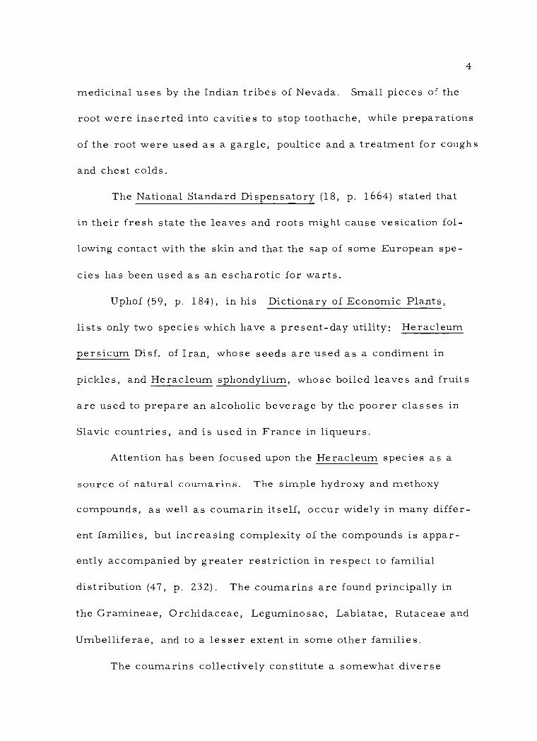
medicinal uses by the Indian tribes of Nevada. Small pieces of the
root were inserted into cavities to stop toothache, while preparations
of the root were used as a gargle, poultice and a treatment for coughs
and chest colds.
The National Standard Dispensatory (18, p. 1664) stated that
in their fresh state the leaves and roots might cause vesication fol-
lowing contact with the skin and that the sap of some European spe-
cies has been used as an escharotic for warts.
Uphof (59, p. 184), in his Dictionary of Economic Plants,
lists only two species which have a present -day utility: Heracleum
persicum Disf. of Iran, whose seeds are used as a condiment in
pickles, and Heracleum sphondylium, whose boiled leaves and fruits
are used to prepare an alcoholic beverage by the poorer classes in
Slavic countries, and is used in France in liqueurs.
Attention has been focused upon the Heracleum species as a
source of natural coumarins. The simple hydroxy and methoxy
compounds, as well as coumarin itself, occur widely in many differ-
ent families, but increasing complexity of the compounds is appar-
ently accompanied by greater restriction in respect to familial
distribution (47, p. 232). The coumarins are found principally in
the Gramineae, Orchidaceae, Leguminosae, Labiatae, Rutaceae and
Umbelliferae, and to a lesser extent in some other families.
The coumarins collectively constitute a somewhat diverse

5
group and may be classified according to several systems. A
practical classification is outlined in Table 1. in the following text
the term "coumarins" will be used in a broad sense and will be in-
clusive of all categories unless qualified.
Although the coumarins constitute a large and significant class
of compounds, their pharmacology has not yet been fully determined.
Bose (5) has pointed out that they possess numerous and often unique
physiological actions. Recognition of the anticoagulant character of
the coumarin moiety has resulted in the development of such useful
and effective drugs such as bishydroxycoumarin, warfarin, couma-
chlor, acenocoumarol and cyclocoumarol (47, p. 247).
The estrogenic properties of compounds such as coumestrol
(VII) are well known and numerous related compounds have been
studied to determine their comparative estrogenic activity. The
unique dermal -photosensitizing action of the furanocoumarins has
been utilized in the treatment of viti igo a r. d leucoderma (47,
p. 254 --257).
Other coumarins have demonstrated some degree of antibiotic
activity. Novobiocin is a fungal metabolite of Streptomyces niveus.
Chartreusin (VIII), another antibiotic, has been isolated as a me-
tabolite of Streptomyces chartreusis (47, p. 258 -259). Other im-
portant properties of the coumarins are curare -like, sedative,
narcotic, analgesic and anthelmintic actions. These, as well as

TABLE I. REPRESENTATIVE COUMARINS
HO
HO
SUBSTITUTED COUMARINS
(I) Esculetin
H O
(II) 4-Hydroxy coumarin
FURANOCOUMARINS
(III) Psoralen (IV) Angelicin
o
rn
\ \ 0 o

TABLE I. REPRESENTATIVE COUMARINS
H
PYRANOCOUMARINS
CH3
( V) Xanthoxyletin ( VI) Seselin
COMPLEX COUMARINS
( VII) Coumestrol
Digitalose-Fucose -0
0
( VIII) Chartruesin
OH H3C
HO

8
other active properties, are summarized by Soine (47, p. 246).
Previous phytochemical studies have demonstrated the presence
of coumarins in a number of Heracleum species. The distribution
of coumarins throughout the species studied is presented in Table II.
A survey of the literature revealed that no work had been done to
show the presence of coumarins in Heracleum mantegazzianum al-
though other chemical aspects have been reported.
Piguleoskii and Kovaleva (38) have determined that the essen-
tial oil distilled with steam from the fruit contained no aldehydes
but unsaturated compounds which proved to be largely esters of
octyl alcohol and, to some extent, hexyl alcohol with acetic, butyric
and optically- inactive isovaleric acid. Piguleoskii (39) established
that the volatile oil of the leaves, studied by the Raman method,
contained ocimene (2, 6- dimethyl -1, 5, 7- octatriene). Ziegler and
Mittel (66) found that sucrose was the only sugar present in the
seive tube sap and Ziegler (64) showed the presence of uridine
disphosphate -glucose in the sap of the phloem. Ziegler (65) also
had studied the respiration and transport of substances in the iso-
lated conductive vessels of the petiole.
In anatomical studies, Troll (58) had isolated from the pit
cavity of the petioles folded vascular bundles which might be as long
as one meter when stretched out.
Seeds of the Umbelliferae have been noted for germination

TABLE II. DISTRIBUTION OF COUMARINS IN HERACLEUM SPECIES.
Name Structure
Umbeiliferone
Pimpinellin
HO
panac es
sibiricum lanaturn
var.
Species
nipponicum
(56)
( 55)
( 15)
References
(54)
( 54) ( 27, p. 534)
sphondylium (48) (25) (27, p. 560)
lanatum (14) (27, p. 560)
var asiaticum (34)
var nipponicum (54) (15)
panaces (54) (56)
sibiricum (55) (54) (28)
Is opimpinell in
CH3 0 3
o 0
CH3
sphondylium (48) (25) (27, p. 555)
concanense ( 2)
lanatum ( 14) (27, p. 556)
var. asiaticum ( 34)
var. nipponicum ( 53) ( 15)
panaces ( 54) ( 56)
sibiricum ( 54) ( 55) ( 28) ID
i
i
;
;
\ \ 0 0

TABLE II (Continued)
Name Structure Species References
Sphondin
Bergapten (Heraclin) (Majudin)
CH3 0
sphondylium
panaces
sibiricum lanatum
var. nipponicum
(48) (25) (27, p. 560)
( 56)
(55) (28)
( 14) (27, p. 560)
( 15)
CH 0 3
sphondylium
giganteum
nepalense panaces sibiricum concanense lanatum
var. asiaticum var. nipponicum
(25) (49) (27, p. 551)
( 54) ( 27, p. 551)
(27, p. 552) (3)
( 54) ( 56)
(55) (54) (28)
( 2)
(27, p. 552) ( 34) ( 15)
Isobergapten
s phondylium
panaces
sibiricum lanatum
var. asiaticum var. nipponicum
(48) ( 27, p. 560)
( 54) (56)
(55) (54) (28)
( 14) ( 27, p. 560)
( 34)
( 15)
o
0 ® 0
\ ® 0 0 0
CH 0 3
0 ® \
I
0

TABLE II. (Continued)
Name Structure Species References
Imperatorin
I0
\ lanatum var.
asiaticum (34)
I
Heraclenin
V 0 p candicans (45)
\pX
Heraclenol
OH OH
candicans (46)
O t
0
®
0'
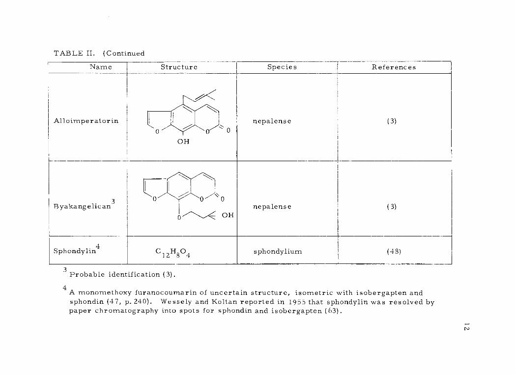
TABLE II, (Continued
Name Structure Species References
Alloimperatorin
t
OH 1 \ 0
nepalense (3)
Byakangelican3 Ò
0''------'<.
0 0 0
OH nepalense (3)
Sphondyïin4 C 12H804 sphondylium (48)
3 Probable identification (3) ,
4 A monomethoxy furanocoumarin of uncertain structure, isometric with isobergapten and sphondin (47, p. 240). Wessely and Koltan reported in 1955 that sphondylin was resolved by paper chromatography into spots for sphondin and isobergapten (63).
7
II ®

13
difficulties. Germination standards for the cultivated Umbelliferae
have been set much lower than those for the majority of other plants.
In some cases this is due to a lack of embryos in apparently viable
seeds. This problem appears to be universal, occurring in all
parts of the world and in all members of the Umbelliferae and, more-
over, appears to be correlated with the season of the year. There
is strong evidence that infestation by the Lygus bug, as well as
possibly other members of the Miridae, is a prime factor in the
destruction of the embryo in numerous species of the Umbelliferae
(41, p. 531 -538).
Certain unsaturated lactones appear to be widely distributed
in plants and to possess the power of inhibiting seed germination.
Among these compounds are coumarin and parasorbic acid (33,
p. 520). In spite of the frequent occurrence of other germination
inhibitors the compounds have been identified only in comparatively
few cases (60, p. 25).
The seeds of some species have a requirement for after -
ripening in moist cold. Stokes has shown that in the case of
Heracleum sphondylium there is an after - ripening requirement of
8 to 12 weeks of moist cold in order for the seeds to germinate (50).
There have been no reports in the literature for such a requirement
in the case of Heracleum mantegazzianum.
Attempts have been made to obviate the cold requirement in

14
the dormancy of certain seeds by chemical means. These efforts
have not been rewarded by signal success (8, p. 104). Considerable
interest has focused upon growth substances such as the gibberellins
in these attempts. Gibberellin treatment substituted completely for
the light requirement in the seeds of lettuce and several other spe-
cies, but it only partially substituted for the cold requirement in
experiments with the sweet cherry and peach. It did not totally re-
place the cold requirement (53, p. 380 -381). No reports were
found concerning the effect of gibberellins on the seed of Heracleum
mantegazzianum.
In view of the foregoing information it was decided to pursue
the following research objectives:
1. To conduct germination studies to ascertain the presence
of a cold requirement in the seeds and, further, to determine if
gibberellic acid treatment would substitute for such a cold require-
ment.
Z. To propagate plants under greenhouse conditions for the
purpose of harvesting root material for further pharmacognostic
study.
root.
3. To study the histological and diagnostic characters of the
4. To determine the type of components present in the roots
by performing a selective solvent extraction.

15
5. To test for the presence of coumarins in the root by the
use of thin -layer chromatography.
6. To determine if heat should be avoided in the drying of
the root.
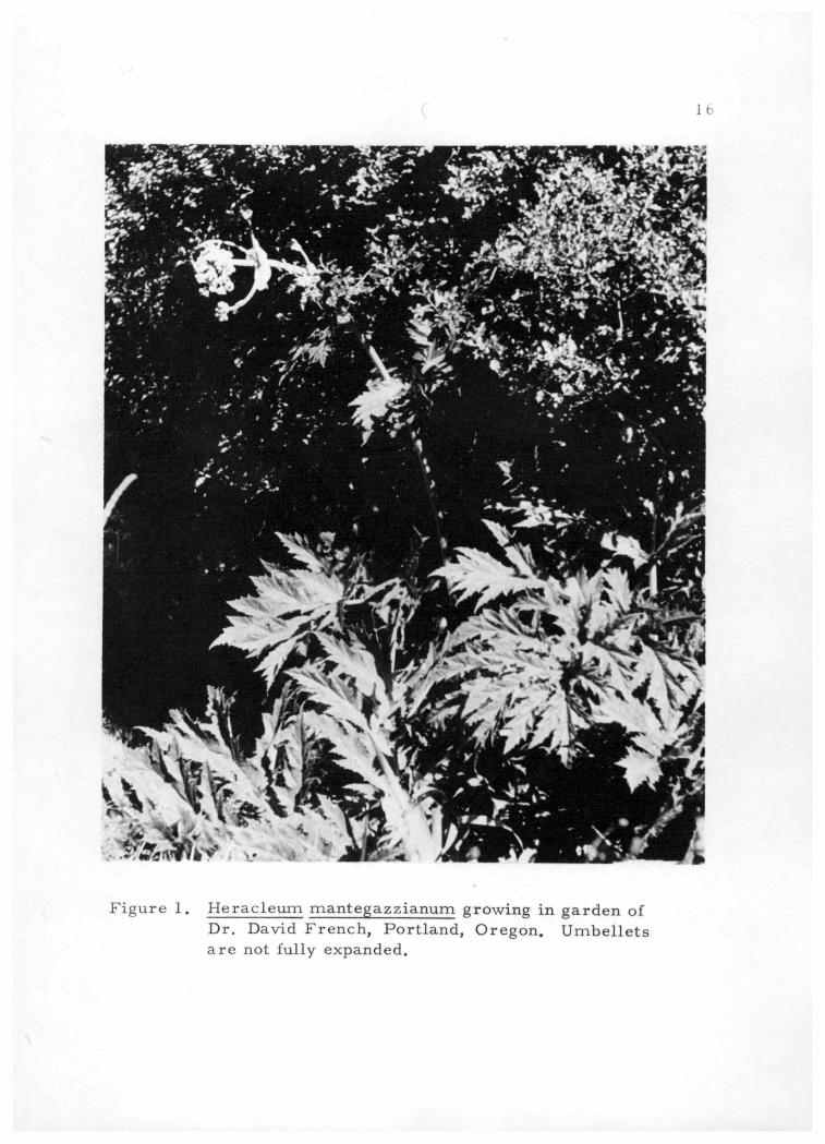
Figure 1. Heracleum mantegazzianum growing in garden of Dr. David French, Portland, Oregon. Umbellets are not fully expanded.
-%5" 1
` J' t - . . °
s ' . k - r, . «y k... i o . . % a a
16
I Y 1, t ' ̂y
,, t. p
.
:1 ..e - -
44 S ' .
!,
¿
I
°
,..° -
e -
. Y1' :
j
a
41
ZII
_' *??
. A'r
jr
r o .I
f3 .127:` A . '` ' f
4Al 1=
°
;yitda 1:- '_ r1
+ . .. 'r` .
x.-0' -
.
1
° -
i r
°
d
I° 1
í ' ;¡ o I ' , ~ :-` ', , i f °
-. *
. .
°
: í
- % J
.
' tir e 1k
A . .
v- + .
ô
:
° . .
y,
t r
. ' q,
, ,' . V
r -
r
. .`".wf41
F ri )
'. ,-y Ai Vol '; AO "t1
, 1 #Ir ' tf - i. A ,. p ..Y,
'
- 1
` ,0 6-
- r.- I , \' 11 n :,
. A.-

17
II, EXPERIMENTAL
All seeds used in this study were received in the fall of 1962
from Dr. David French, Professor of Anthropology at Reed College,
Portland, Oregon. Dr. French had originally received seeds
provided by Dr. C. Leo Hitchcock from his Seattle garden in 1958
and which Dr. Hitchcock had stated to be those of Heracleum
mantegazzianum. These seeds had been planted by Dr. French in
his garden at 3549 S. E. Woodstock, Portland, Oregon, in 1959. In
1960 the plants produced basal leaves only, but the following year
they produced stalks which flowered and fruited. The plants were
about ten feet tall. The seeds used in the research were harvested
from one of these plants and are represented by lot no. 2122D.
In 1962 Dr. French submitted some of these plant materials
to Dr. Lincoln Constance, a specialist in the Umbelliferae, at the
University of California at Berkeley for confirmation. Documentary
evidence of his verification is in the form of a letter from Dr. French
to the author in which he states:
Lincoln Constance at Berkeley, one of the world's foremost authorities on the Umbelliferae has now positively identified 2122C (in flower) as Heracleum mantegazzianum Somm. & Lev. Specimens from such numbers as 2122D are from the same plants, and the identification can be extended to them as duplicates.
Dr. French has deposited a voucher specimen of this plant in
the Oregon State University herbarium as well as vouchers for the

18
chromosome count (n -11) in the herbarium of the University of
California at Berkeley.
Germination of Seeds
Group 1 (Cold Treatment)
On March 29, 1963 a total of 60 seeds were wrapped in cloth,
moistened with tap water and placed in a refrigerator at a tempera-
ture range of from 0 to 2 C. 0 Four days later, since ice crystals
formed on top of the cloth, the temperature was raised to a range of
from 2 to 5oC. On June 11, after 74 days of cold, they were re-
moved and soaked in several changes of distilled water. Small and
depauperate seeds were removed and the remaining 39 seeds were
designated as Group 1.
Group 2 (G. A. Treatment)
Thirty-nine dry seeds, selected for size and ample endosperm,
were placed in a beaker and soaked for 20 hours in a solution con-
taming 100 p. p.m. of gibberellic acid5 in distilled water.
seeds were designated as Group 2.
These
5 Gibberellic acid, 88. 9% pure. Supplied through the courtesy of Dr. Edwin F. Alder, Agricultural Research Center, Eli Lilly &
Co. , Greenfield, Ind.

19 Group 3 (Controls)
Thirty -nine dry seeds, selected for size and ample endosperm,
were placed in a beaker and soaked for 20 hours in several changes
of distilled water. These seeds were designated as Group 3 and in-
tended for use as controls.
Germination
Each group of seeds was individually planted in a flat contain-
ing a mixture of one part sand and two parts sandy loam with 50
grams of complete fertilizer. 6 The seeds were planted by being
placed in rows on top of the soil mixture and being covered lightly
with a thin layer of vermiculite.
The seeds were maintained under normal greenhouse care and
allowed to germinate at a temperature range of from 18 to 27 C. 0
for a period of 38 days. At the end of that time the results were
recorded.
In Group 1 (the cold- treated seeds) there was a total of three
seedlings. Four seeds had actually germinated, but one seedling
had failed on the 29th day, possibly due to "damping off. " Germina-
tion did not occur in Group 2 (gibberellic acid -treated) or in Group 3
(controls) .
6Organic Morcrop, Chas. Lilly Co. , Seattle, Washington. (Analysis: total nitrogen, 5%; available phosphate, 3 %; available potash, 2%.
20
Extended Cold- Treatment
On November 5, 1963, 1000 seeds, selected for good size and
ample endosperm, were placed in a litre of tap water and soaked for
a period of 36 hours. They were then removed and placed upon a
towel which was then formed into a roll, moistened with water, and
placed in a refrigerator at a temperature range of from 2 to 5°C.
After a total of 294 days the cloth roll was removed from the refrig-
erator and examined. Extensive germination had occurred and many
of the primary rootlets had penetrated the fabric. This resulted in
considerable damage to the seedlings during the opening of the roll
and made it impossible to determine exactly the total number of
germinated seeds. However, it was estimated that at least 550 seeds
had germinated.
Propagation of Plants
On July 26, 1963 the three seedlings obtained from the germi-
nation experiment of seed Group 1 were transplanted to ten inch peat
pots containing a mixture of one part of sand, and two parts of sandy
loam with ten grams of Organic Morcrop. These plants were re-
spectively designated as SF -1, SF -2 and SF -3.
Earlier, on June 28, six seedlings, obtained by cold -treating
and germinating seeds on moist blotting paper in Petri dishes, had

21
been transplanted to four inch peat pots with one part of sand and
two parts of sandy loam with 2.5 grams of Organic Morcrop per pot.
Two of the seedlings had failed. On July 26, the sides of the pots
containing the four surviving seedlings were slashed and the pots
were embedded in ten inch peat pots with the same soil mixture as
Group 1. These plants were designated respectively as PD -1, PD -2,
PD -3 and PD -4.
On June 15, a number of volunteer seedlings were obtained
from the Portland garden of Dr. David French. These were all de-
scendants of a single plant which had fruited in 1962 and had been
designated as French 2122D. At the time of collection the seedlings
varied from 13 cm. to 25 cm. in height. The seedlings were dug
from the soil, their roots moistened, and were then transported by
automobile to Corvallis. On the following day they were placed in
ten inch peat pots with a soil mixture identical to that previously
described and watered. On the next day it was observed that four
of the plants were very badly wilted so their leaves were removed
as well as part of the leaves of several other plants. The plants
were maintained in the greenhouse where the temperatures ranged
from 15°C. at night to 35°C. during the day. In order to discourage
infestation by "white fly" the under surfaces of the leaves were
A B
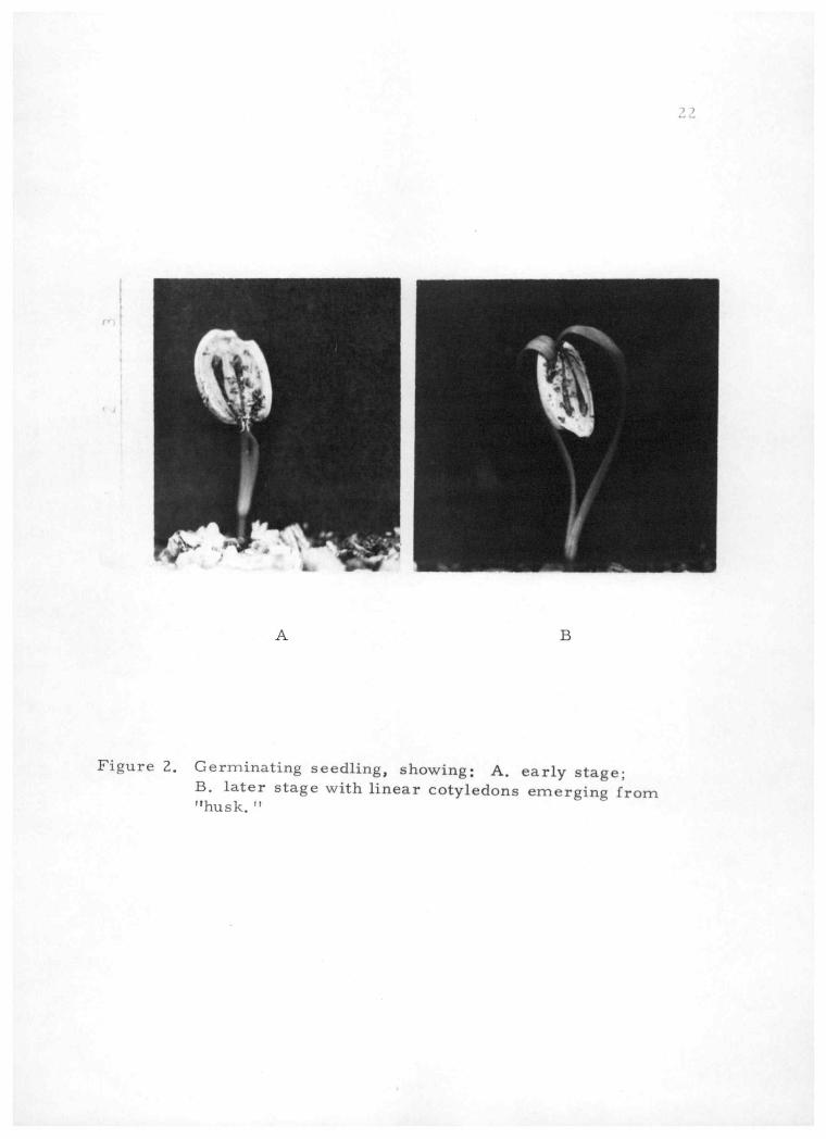
Figure 2. Germinating seedling, showing: A. early stage; B. later stage with linear cotyledons emerging from "husk. "
22
Jj' wI ' kI
- tt`.

23
sprayed twice weekly with Miller's Tetradane. 7
On August 15, it was observed that some foliar damage had
occurred. On some of the younger leaves the areas at the endings
of the veins in the apices and notches of the leaves displayed a drawn -
together, shriveled appearance. These areas later became desic-
cated and devitalized. It was also observed that a number of young,
emergent leaf buds had withered and died.
On the assumption that damage might have been caused by the
spray in combination with high daytime temperatures the greenhouse
temperatures were reduced to a range from 18oC. at night to 21oC.
during the day, and from August 16, "white fly" was controlled by
application of Ortho Rose Dust8 to the tops and bottoms of the leaves
with a dust gun. Although damage to tender leaves and buds was
reduced it was not totally eliminated.
On October 20, at time of harvest, the heights of the plants
were recorded. These data as well as the average height of each
plant group are shown in Table III.
7Miller's Tetradane (Insect Concentrate Spray) Ornamental and Rose Spray, Miller Products Co. , Portland, Oregon. (Con- taining Kelthane, Diazinon, Lindane and DDT)
8Ortho Rose Dust, California Spray - Chemical Corp. , Rich- mond, California. (Ingredients: Lindane 1 %, Phaltan 7. 5 %,
DDT 5 %, Sulfur 30 %, Inert ingredients 56. 5 %. )

24
TABLE III. GROWTH OF PLANTS AT HARVEST
Plant No. Days From Transplant
Height Per Average Height Plant Per Group (cm.) (cm. )
PD-1
PD-2
PD-3
PD-4
SF-1
SF-2
SF-3
TP-1
TP-2
TP-3
TP-4
TP-5
TP-6
TP-7
TP-8
TP-9
TP-10
TP-11
TP-12
TP-13
TP-14
92
92
92
92
92
92
92
132
132
132
132
132
132
132
132
132
132
132
132
132
132
36. 3
32.3
31.6
33. 7
30.0
33.7
40. 0
13. 2
24. 2
16. 0
16. 7
19. 3
31. 0
27. 8
21.0
10. 7
22. 5
26. 0
16. 0
14. 5
12.4
33. 5
34. 6
19.4

Plant PD -1 Height: 33 cm.
Plant TP -8 Height: 20 cm.
Figure 3. Contrast in growth attained by two plants of different groups at harvest.
25
.7g,54.4-
tviq 4 . '

26
Harvest
On October 20, plants PD -1 and PD -4 were set aside for
further study and the remainder of the plants of the three groups
were harvested. The petioles were first severed several inches
above the crowns and discarded together with the leaves. The roots
were removed from the pots by washing the soil away with water
from a hose in such a way as not to detach any small rootlets and to
remove last traces of soil. Extraneous matter such as twigs and
small bark fragments were removed by garbling. The remaining
portions of the petioles were cut away from the crowns and excess
moisture removed by blotting with a cloth towel. The roots were
then cut into small pieces and the larger segments split to facilitate
drying. The roots were immediately weighed on a Welch balance to
determine fresh weight.
The roots were then spread out on mesh screen drying racks
and placed in a forced -air drying oven at 38 0 C. for 50 -1/2 hours.
The roots were then removed from the drying oven, weighed to
determine their dry weight, placed in plastic bags with closures
and stored in clean, dry cans which were sealed with tape.

Histological Study
Preparation of Slides
27
On February 17, 1964 plants PD -1 and PD -4, previously re-
served, were harvested. These plants had been maintained outside
of the greenhouse under normal environmental conditions. A portion
of the root from one of the plants was prepared for killing and fixing
by being washed thoroughly with water and then sectioned into short
segments. The segments were then immersed in formalin- aceto-
alcohol solution9 of the following formula:
Formaldehyde
Glacial acetic acid
Ethanol 95%
5 ml.
5 ml.
90 ml.
The vial containing the root segments in the killing solution was
transferred to a refrigerator where it was stored for several weeks.
Upon removal from the killing solution the segments were
washed with two changes of 50% ethanol and then run through the
tertiary - butanol dehydration process according to the schedule of
Table IV and as outlined by Johansen (26, p. 130 -131) with modifica-
tions in regard to time.
9This killing reagent may be used with almost any plant material intended for anatomical study. Material may be kept in it almost indefinitely without appreciable damage (26, p. 41).
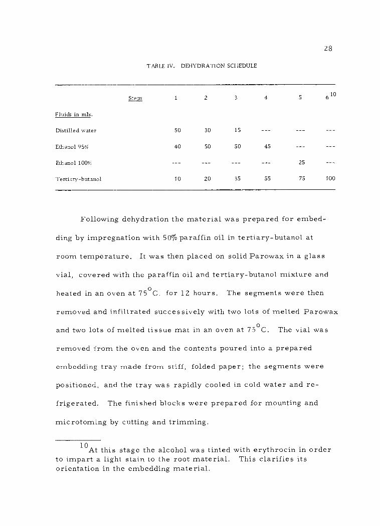
28
TABLE IV. DEHYDRATION SCHEDULE
Fluids in mis.
Steps 1 2
30
50
20
3
15
50
35
4
45
55
5
25
75
610
100
50
40
10
Distilled water
Ethanol 95%
Ethanol 100%
Tertiary -butanol
Following dehydration the material was prepared for embed-
ding by impregnation with 50% paraffin oil in tertiary - butanol at
room temperature. It was then placed on solid Parowax in a glass
vial, covered with the paraffin oil and tertiary - butanol mixture and
heated in an oven at 75oC. for 12 hours. The segments were then
removed and infiltrated successively with two lots of melted Parowax
and two lots of melted tissue mat in an oven at 75oC. The vial was
removed from the oven and the contents poured into a prepared
embedding tray made from stiff, folded paper; the segments were
positioned, and the tray was rapidly cooled in cold water and re-
frigerated. The finished blocks were prepared for mounting and
microtoming by cutting and trimming.
10At this stage the alcohol was tinted with erythrocin in order to impart a light stain to the root material. This clarifies its orientation in the embedding material.
- -- - --
- --

29 The microtoming, staining and completion of the slides was
done by Mr. Gerald Bogar, a recent graduate student in the botany
department, as hereafter briefly described. The trimmed blocks
were sectioned at ten microns on a rotary microtome, the ribbons
affixed to slides with Haupt's adhesive, and the sections stained with
1% safranin in water, destained with acid alcohol, and counterstained
with aniline -blue in clove oil and 100% alcohol. The stained sections
were cleared with clove oil, rinsed and washed with xylene and then
mounted in 60% H. S. R. mounting resin in toluene.
Photoni c rog raphy
Photomicrographs were prepared as follows: The slides were
placed upon the stage of a Bausch and Lomb binocular microscope
using an independent light source11 coupled with a Jefferson trans-
former. A 5X ocular was used in combination with l0X and 43X
objectives. The camera was a 35 mm. model XV Exacta with the
lens removed and equipped with an improvised adapter. No filters
were used.
Eastman Plus -X film was used and the optimum exposures de-
termined by experimentation. The negatives were developed with
Ethol, single mix, ultra fine grain developer and were fixed with
Eastman Kodak fixer. Enlargements were made on Eastman Koda-
bromide paper with an Omega type A2 enlarger. Interpretations of
the photomicrographs are shown in Figures 4 and 5.
"Bausch and Lomb lamp, type 31- 33 -77.

85X
Figure 4. Transverse section of stele of root. Explanation: Sp, secondary phloem with phloem parenchyma; Ca, cambium; Mx, metaxylem; Px, protoxylem; Xr, xylem ray; Lr, lateral root; Xp, xylem parenchyma.
., `34.-t..,
Q 1(; . 104 3 r; . -t] i#,'
; , a
101. 0 .3 , . . :_
r , ± '
. .` . : . l 160"6 .,. ` i . J. _.
i . . . _
.4.ist e ; . - .
,r; .,+` a+i '-~ o: i'' -. : . ,' . i91;;! 1 ; ,='/' . t. í-r,.' =''' :/ .¡;4116 ` A S .' '-Ï::i r r'o t4 ; ' :Ñ : . r0 SP.C'b :._ : :- ,.!r..`r. ..+ ;: w ,' .j.t - .._., r i.' MX ; +, ri °v
2 ` M yy[.. rr , }tiP'' 4r.
0 At ti ,' .A.4' .M Lit °02 - =.a .
30
Sp
Ca
Px
Al
+ ,r 4:,
'e - C 5 y". yt 3 0
11, ,: '~ .r. + .) !. c',. .,. .
Xr

Figure 5. Longitudinal section of stele of root. Explanation: Cp, collapsed protoxylem; Hv, helically plated vessel elements; Xp, xylem parenchyma.
r
'+ ya i
sr, i
1 4
t r r----- 'w , r-
F
1111r.
'k .{r.. I
31
-Cp
Ma MINI Ow
-Hv
- Xp

32
Histology
The primary tissues of the root originally constituted a triarch
protostele. The material under study shows substantial secondary
growth with subsequent loss of the cortex. Peripheral tissues con-
sist of four to seven layers of thin -walled, tabular cork cells
(phellem) and a cork cambium (phellogen). These are subtended by
a zone of pericycle from three to five cells in depth, contiguous to
a broad zone of secondary phloem which is radially traversed oppo-
site the protoxylem ridges by vascular rays from four to six cells
wide. Sieve tube strands of the secondary phloem are separated
from each other by parenchyma. A functional primary phloem is
not discernible. A cambial zone separates the mature xylem from
the secondary phloem. Lateral roots are very numerous and in
different stages of development. The presence of secretory canals
is observed in the secondary phloem. According to Metcalf and
Chalk such canals are characteristic of the Umbelliferae (32, p. 717).
Description of the Unground Root
The root after being cut into segments and dried appears
macroscopically as follows: split sections from larger diameters
of the root range from 1 to 4 cm. in length and from 0. 6 to
1.4 cm. across. The unsplit root segments range from 2 to 3. 5 cm.

33
in length and from one to about five mm. in diameter. The external
surface is weak brown to moderately yellowish- brown, slightly
annulated and somewhat wrinkled longitudinally. Hair -like second-
ary rootlets are frequently present. The ends of the segments often
show a spotty, orange -yellow coloration. The inner surfaces of the
segments are dull white with flecks of orange -yellow. The fracture
is short and the fractured surfaces are slightly irregular and porous.
The odor is characteristic and light, the taste insipid and bland.
Description of the Powdered Root
The root reduced to a number 40 powder is a light yellowish -
tan in color and slightly grainy when rubbed between the fingers.
The characteristic odor is more pronounced than in the unground
root and slightly acrid. When a small portion of the powder is
moistened with Wallis' iodine12 it turns blue.
Microscopic examination of the powder in a water mount re-
veals the presence of numerous single and clustered starch grains
in such quantity as to obscure other features. The starch grains
are ovoid to polyhedral, non - striated, with slight erosions radi-
ating from the hilum or ridge. The grains vary in size from
12One volume of weak solution of iodine B. P. mixed with nine volumes of water (61, p. 222).

34
1. 8 to 8. 7 microns in diameter and the larger clusters from 70 to
126 microns. The starch gelatinizes very slowly or hardly at all in
water. Water mounts examined under polarized light show the
starch grains to have a bright cross at total extinction.
Examination of mounts in chloral hydrate solution13 reveals
rapid gelatinization of the starch with rupture of parenchyma cells
containing large starch clusters. It would appear that many paren-
chyma cells are totally filled with starch. Parenchyma cells were
observed to range in length from 70 to 140 microns.
Clearing with chloral hydrate shows the presence of frag-
mented vessel elements with helical to helical - scalariform
pitting. When treated successively with phloroglucinol test solution 4
and concentrated hydrochloric acid the vessel elements are colored
red - a positive test for lignified tissue. Tests with Sudan III did
not reveal the presence of any oil. No crystals were observed.
Line drawings of the microscopic characters are shown in Figure 6.
Ultraviolet Tests
Many drugs exhibit fluorescence when the cut surface or the
13Fifty grams of chloral hydrate dissolved in 20 ml. of purified water (61, p. 220).
14 One gram of phloroglucinol dissolved in 100 ml. of 90%
alcohol (61, p. 222).

35
Figure 6. Histological features of powdered root, magnified about 100 times in chloral hydrate mount. Showing: P, parenchyma cells; S, gelatinizing starch grain clusters; V, vascular element gragments.
36
powder is exposed to ultraviolet radiation. This property provides
a useful tool for the routine investigation of crude drugs. In some
cases this method of examination will provide information which
cannot be obtained by any other means (62, p. 553). In order to
characterize the material under study, its reaction to irradiation
from an ultraviolet lamp15 was observed in a darkroom.
1. The intact external cork of split or cylindrical sections
showed no fluorescence. The inner portions displayed a light - yellow
fluorescence which was more intense and slightly tinged with blue on
the surfaces of fresh cuts and fractures. In addition, these inner
surfaces showed a definite luminescence which persisted for five
seconds following extinction of the light.
2. The powdered dried root showed a slight yellow fluores-
cence which luminesced for several seconds following extinction of
the light.
3. The powdered dried root, previously exhausted by ether
extraction in a Soxhlet extractor, showed a somewhat blue and
slightly more intense fluorescence while under irradiation but dis-
played no luminescence following extinction of the light.
4. A two -gram sample of powdered dried root was macerated
with 20 ml. of 95% ethanol for one hour and filtered. Five ml. of
15Mineralight, long -wave ultraviolet, model SL 3660 Ultra- violet Products, Inc. , South Pasadena, California.

37
the filtrate was placed in each of two test tubes and to one of these
one ml. of 28% ammonia was added. Both filtrates were then ob-
served under ultraviolet light. The filtrate with ammonia gave a
dark green fluorescence. A weak dark -blue fluorescence was noted
with the other filtrate. No luminescence occurred.
Selective Solvent Extraction
The root material, previously dried at 30oC. , was ground to
a no, 40 powder in a Wiley mill. Fibrous rootlets were excluded.
Five -gram and two -gram samples were extracted successively with
solvents of an increasing order of polarity. The technique employed
was a modification of the general method of Rosenthaler (42, p. 35-
39) . A Soxhlet extractor was used in performing the extractions
with the five -gram samples and a micro - Soxhlet extractor was
employed with the two -gram samples.
Each thimble with its contents was oven -dried following ex-
traction with a solvent and the amount of extractive was determined
by loss of weight. An oven temperature of 65oC. was used to re-
move the petroleum ether from the thimble and a temperature of
90oC. was employed for the other solvents.
In order to insure complete extraction with water following
treatment with that solvent on the Soxhlet, each thimble with its
contents was opened and macerated with distilled water in a beaker
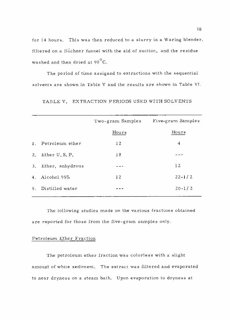
38
for 14 hours. This was then reduced to a slurry in a Waring blender,
filtered on a Büchner funnel with the aid of suction, and the residue
washed and then dried at 90oC.
The period of time assigned to extractions with the sequential
solvents are shown in Table V and the results are shown in Table VI.
TABLE V. EXTRACTION PERIODS USED WITH SOLVENTS
Two -gram Samples
Hours
Five -gram Samples
Hours
1. Petroleum ether 12 4
2. Ether U. S. P. 18 -
3. Ether, anhydrous - 12
4. Alcohol 95% 12 22-1/ 2
5. Distilled water - 20 -1 / 2
The following studies made on the various fractions obtained
are reported for those from the five -gram samples only.
Petroleum Ether Fraction
The petroleum ether fraction was colorless with a slight
amount of white sediment. The extract was filtered and evaporated
to near dryness on a steam bath. Upon evaporation to dryness at
-

TABLE VI. SELECTIVE SOLVENT EXTRACTION16
No. Weight (grams)
Petroleum17 Ether
Ether U. S. P.
Ether Anhyd.
Alcohol 95% Water
1 1.914 2.2 2.1 - -- 7.8
2 1.691 2.5 2.0 - -- 8.7
3 1.927 2.4 2.0 - -- 8.1
4 5.078 2.4 - 2.5 9.1 20. 7
5 5.087 2.6 - 2. 5 8. 5 18. 5
6 5.085 2.5 - 2. 5 8.4 18.9
7 5.094 2.5 - 2.5 8.9 20. 1
Averages 2.4 2.0 2.5 8.5 19.4
16Extractive fractions expressed as a percentage of the oven -dried weight. 17Petroleum ether (hexane) B. P. 68 to 69°C.
- --
- --
--
--
--
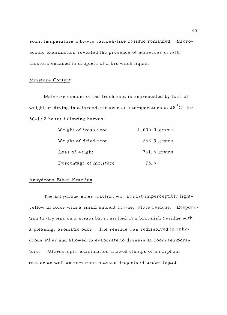
40
room temperature a brown varnish -like residue remained. Micro-
scopic examination revealed the presence of numerous crystal
clusters encased in droplets of a brownish liquid.
Moisture Content
Moisture content of the fresh root is represented by loss of
weight on drying in a forced -air oven at a temperature of 38°C. for
50-1/ 2 hours following harvest.
Weight of fresh root
Weight of dried root
Loss of weight
Percentage of moisture
Anhydrous Ether Fraction
1,030.3 grams
268.9 grams
761.4 grams
73.9
The anhydrous ether fraction was almost imperceptibly light -
yellow in color with a small amount of fine, white residue. Evapora-
tion to dryness on a steam bath resulted in a brownish residue with
a pleasing, aromatic odor. The residue was redissolved in anhy-
drous ether and allowed to evaporate to dryness at room tempera-
ture. Microscopic examination showed clumps of amorphous
matter as well as numerous massed droplets of brown liquid.

41
Alcohol Fraction
The alcohol fraction appeared light -yellow in transmitted light
and displayed a bluish opalescence with reflected light. A small
amount of light -brown sediment was present. Upon evaporation to
near -dryness on a steam bath a thick, brown, syrupy liquid re-
mained.
Aqueous Fraction
The aqueous fraction was brown in color with a small amount
of flocculent sediment. The copious foam produced by shaking the
liquid in a flask indicated the possible presence of saponins.
Thin -Layer Chromatography
In order to determine the presence of coumarins in the root
and to ascertain their identity a thin -layer chromatographic study
was made.
Standards
The following coumarins and furanocoumarins were employed

42
as standards. 18
1. Umbelliferone 5. Sphondin
2. Iscbergapten 6. Bergapten
3. Pimpinellin 7. Imperatorin
4. Isopimpinellin
Several of these compounds contained traces of impurities. This,
however, did not affect their value as standards.
The following amounts of the above materials were weighed on
a Mettler balance: umbelliferone, 1.5 mg. ; isobergapten, 3. 5 mg. ;
pimpinellin, 6. 9 mg. ; isopimpinellin, 6. 3 mg. ; sphondin, 5.7 mg. ;
bergapten, 2. 5 mg. and imperatorin, about O. 25 mg.
These quantities were transferred to individual10 ml. volu-
metric flasks and, with the exception of the umbelliferone and the
imperatorin, were filled to the mark with spectro grade chloroform.
To the flask containing the imperatorin only 2 ml. of chloroform
were added. The flask containing the umbelliferone was filled to the
mark with 95% ethenol due to the limited solubility of umbelliferone
18Obtained from the following sources: Comp. no. 1: British Drug Houses, the Ealing Corp. , 33 University Road, Cambridge, Mass. Comp no. 2: Dr. Michiichi Fujita, Tokyo College of Phar- macy, Institute of Pharmacognosy and Plant Chemistry, 4 -600 Kasiwagi, Shinjuku -ku, Tokyo, Japan. Comp. nos. 3, 4 and 5: Dr. Stewart A. Brown, Trent Univ. , Peterborough, Ontario, Canada (originally from Dr. Baerheim Svendsen, School of Phar- macy, Oslo -Blindern, Norway). Comp. nos. 6 and 7: Dr. Stewart A. Brown.
43
in chloroform.
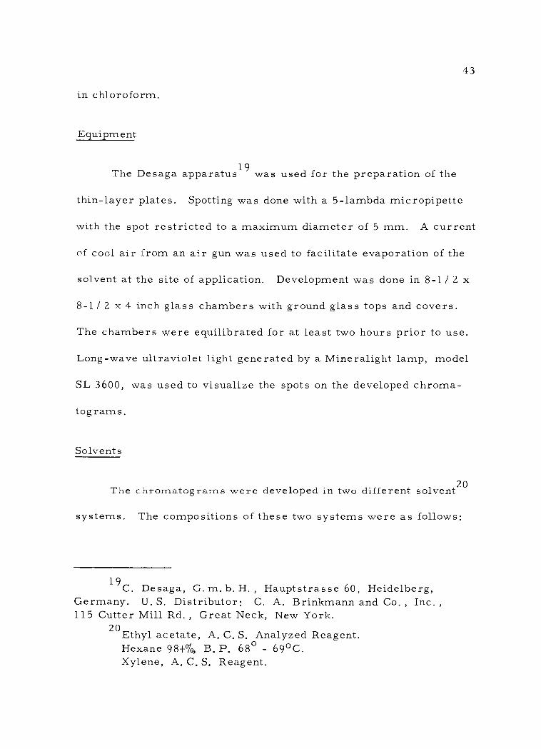
Equipment
The Desaga apparatus19 was used for the preparation of the
thin -layer plates. Spotting was done with a 5- lambda micropipette
with the spot restricted to a maximum diameter of 5 mm. A current
of cool air from an air gun was used to facilitate evaporation of the
solvent at the site of application. Development was done in 8-1/ 2 x
8 -1 / 2 x 4 inch glass chambers with ground glass tops and covers.
The chambers were equilibrated for at least two hours prior to use.
Long -wave ultraviolet light generated by a Mineralight lamp, model
SL 3600, was used to visualize the spots on the developed chroma-
tograms.
Solvents
The chromatograms were developed in two different solvent20
systems. The compositions of these two systems were as follows:
19C. Desaga, G. m. b. H. , Hauptstrasse 60, Heidelberg, Germany. U. S. Distributor: C. A. Brinkmann and Co. , Inc. ,
115 Cutter Mill Rd. , Great Neck, New York. 20Ethyl acetate, A. C. S. Analyzed Reagent.
Hexane 98 + %, B. P, 68° - 69 °C. Xylene, A. C. S. Reagent.

44
1. Ethyl acetate : Xylene 1: 1
2. Hexane : Ethyl acetate 2: 1
Preparation of Plates
Prior to use the glass plates were cleaned with Ajax scouring
powder, rinsed successively with tap water and distilled water, and
dried with a clean towel.
Thirty grams of Silica -Gel G (Acc. to Stahl) were placed in a
4 -inch glass mortar and 60 ml. of distilled water were added in one
portion. The mixture was carefully stirred with a stainless steel
spatula for 60 seconds and then triturated with a glass pestle for
30 seconds. The slurry was immediately poured into the cylinder
of the applicator positioned on the initial plate and set to produce a
layer 250 microns thick. The cylinder was rotated and as soon as
the slurry was seen to flow out the applicator was drawn smoothly
and rapidly from left to right across the glass plates.
The layer was allowed to set for ten minutes before the plates
were removed and placed in the drying rack. After approximately
an additional five minutes the drying rack was placed in an oven
preheated to 105°C. and the plates were dried for one hour. After
activation the plates were placed in a desiccator over Drierite until
cool and used at once or as required.

45
Preparation of the Aid -Dried Root Extract
On February 17, 1964 the remainder of the roots of the two
plants harvested as previously described on page 27 were thoroughly
washed, cut into segments, placed upon absorbent paper in wooden
flats and allowed to dry for several months in the air at normal
laboratory temperatures. The material was then reduced to a no. 20
powder in a Wiley mill. A 13. 35 -gram portion of the powder was
extracted with 100 ml. of ethyl ether in a Soxhlet apparatus. The
ether solution was light -yellow in color with a small amount of fine,
white sediment. The solution was filtered and the filtrate was
evaporated to near -dryness on a steam bath. It was then redissolved
with ether and transferred to a 10 ml. beaker. The solution was
again evaporated to near -dryness on a steam bath and the remaining
ether was allowed to evaporate at room temperature.
The residue consisted of a thick, brown liquid, somewhat
fluid when warm, almost immobile when cold, and had a light, aro-
matic odor characteristic of the seeds. The residue was washed
with 6 ml. of petroleum ether, in divided portions, to remove
possible lipid material and the remaining residue redissolved in a
mixture of ether and alcohol to a total volume of 10 ml. in a volu-
metric flask.

46
Preparation of Oven -Dried Extract
An extract from 13.35 grams of powdered, over -dried root
was prepared as described above and the extract redissolved in a
mixture of ether and alcohol to a volume of 10 ml.
Technique
Activated plates were removed from the desiccator as requir-
ed. Lateral and bottom edges of all thin layers were trimmed with
a plastic rule. Both square and rectangular plates were used for
the determination of Rf values of the standards.
Due to confluescence of the spots when the root extracts were
developed in one direction only these plates were developed in a
second direction at 90 degrees to the first before Rf values were
determined.
In development, the solvent was allowed to rise until all parts
of the front had reached the line. Plates in which the solvent front
was not substantially horizontal were rejected. Developed plates
were dried in a current of warm air from an air gun and imme-
diately examined under ultraviolet light. This is essential as the
intensity of the fluorescent spots decreases rapidly.
Prior to being developed in the second direction the plates
were dried for 15 minutes in a current of warm air from an air gun

47
and then cooled to room temperature.
Chromatography of Standards and Root Extracts
Effect of Load Upon Rf Values. In order to determine the
effect of variations in load upon the Rf values 5 -, 10 -, and 15-
lambda loads were spotted respectively from each of the following
solutions:
1. Sphondin
2. Isopimpinellin
3. Isobergapten
4. Umbelliferone
The behaviour of these compounds may be considered to be
characteristic of the coumarins as a group. The progressive loads
of each individual solution were spotted onto the same plate. The
chromatograms were developed with Ethyl acetate : Xylene 1 : 1
and the migrated spots were visualized with ultraviolet light. The
results shown in Figures 7 and 8 demonstrate that within practical
limits the size of the load has no significant effect upon Rf values.
Slight variations may be considered inconsequential.
Fluorescence to Ultraviolet Light. Long -wave ultraviolet
light was found to be an excellent tool for the visualization of
developed chromatograms. Color values of the fluorescent spots
of the respective coumarins are shown in Table VII. Due to the

48
Sphondin
5 X 10X 15X
Front
Isopimpinellin
5h 10X 15X
DOD o o G
o
Figure 7. Effect of load on Rf values of sphondin and isopimpinellin.

Is obe rgapten
5A 10A 15 X
Front
Umbellife rone
5 X 10X 15N
49
o O O
ODD C)
Figure 8. Effect of load on Rf values of isobergapten and umbellìferone.J

50
difficulty of circumscribing colors effectively the table presents this
author's evaluation together with their closest identity on the plates
of the standard color reference, Maerz and Paul's Dictionary of
Color (30).
TABLE VII. FLUORESCENT COLORS OF THE STANDARD COUMARINS TO LONG -WAVE ULTRAVIOLET LIGHT.
Coumarin Color Reference Plate
1. Isobergapten Yellow L -1 18
2. Pimpinellin Brown I -7 14
3. Imperatorin Yellow L -1 18
4, Bergapten Yellow L -1 18
5. Isopimpinellin Brown I -7 14
6. Sphondin Greenish -blue K -7 34
7. Umbelliferone21 Bright blue Variable
21It is to be noted that when umbelliferone was spotted alone in 5- lambda amounts the color was bright blue. When it was spotted mixed with the other coumarins or with root extract the color appeared dark blue (equivalent to L -12 on plate 39). This darker color was also observed when a substantially attenuated dilution was spotted.

51
Rf Values of the Standard Coumarins. The Rf values of the
standard coumarins as well as those of the principal spots of the
root extracts were determined. Rf values shown in Table VIII
represent an average of not less than three individual spots.
Chromatography of Air -Dried Extract. Two -dimensional
chromatography of the extract of air -dried root revealed seven
principal spots in the order of their decreasing Rf values. The
relative positions of these spots as well as their colors to ultraviolet
light are shown in Figure 9.
The standard coumarins, isobergapten, pimpinellin, bergap-
ten, isopimpinellin, sphondin and umbelliferone were spotted to-
gether. When developed two - dimensionally the chromatograms
show a pattern almost identical to that of the root extract with the
exception of spot no. 7. Colors of the spots to ultraviolet light
corresponded exactly. This is shown in Figure 10.
The standard coumarins and the air -dried root extract were
spotted together and developed as described above. The chroma-
togram showed no significant divergence from the patterns of the
standards when spotted together or from the root extract. This
chromatogram is shown in Figure 11.
Chromatography of the Oven -Dried Extract. Two - dimensional
chromatography of the extract of oven -dried root revealed five
principal spots. This chromatogram is shown in Figure 12.

TABLE VIII. Rf VALUES OF STANDARD COUMARINS AND PRINCIPAL SPOTS FROM ROOT EXTRACT
Solvent System
1
Solvent System
2
Spot no. 1 .73 .49
Isobergapten .74 . 53
Spot no. 2 .. 69 .43
Pimpinellin .72 .47
Spot no. 3 .63 .33
Be rgapten . 63 .38
Spot no. 4 . 59 . 26
Isopimpinellin . 60 . 28
Spot no. 5 . 53 . 23
Sphondin .55 .27
Spot no. 6 .43 .17
Umbelliferone .43 .17
Spot no. 7 . 20 .06
52

1
53
Solvent systems
(1) Ethyl acetate : Xylene 1:1
(2) Hexane : Ethyl acetate 2:1
Quantity spotted: 20
Front
e
Ob.
1. Yellow 2. Brown 3. Yellow 4. Brown 5. Greenish -blue 6. Dark blue 7. Bright blue
2
Figure 9. Chromatogram of extract of air -dried root.
o
W
--

Spot no. Standard 1 5 X Isobergapten 2 15 X Pimpinellin 3 10 % Bergapten 4 5 A Isopimpinellin 5 5 X Sphondin 6 5 A Umbelliferone
Solvent systems
(1) Ethyl acetate : Xylene 1 :1
(2) Hexane : Ethyl acetate 2:1
Front
1. Yellow 2. Brown 3. Yellow 4. Brown 5. Greenish blue 6. Dark blue
2
Figure 10. Chromatogram of mixed standard coumarins.
o
54
s

55
Quantities spotted
5 X Isobergapten 15N Pimpinellin 10 A Bergapten 5 A Isopimpinellin
5 ' Sphondin 20 A Root extract 5 N Umbelliferone
Solvent systems
(1) Ethyl acetate : Xylene 1:1
(2) Hexane : Ethyl acetate 2:1
Front
1. Yellow 2. Brown 3. Yellow 4. Brown 5. Greenish blue 6. Dark blue 7. Bright blue
2
Figure 11. Chromatogram of mixed standard coumarins and root extract.
o
W
1

56
Solvent systems
(1)
(2)
Ethyl acetate : Xylene 1 :1
Hexane : Ethyl acetate 2:1
Quantity spotted : 20 À
Front
1. Yellow 2. Brown 3. Yellow 4. Brown 5. Greenish -blue
2
Figure 12. Chromatogram of extract of oven -dried root.
C
Cy

57
Effect of Heat Upon Coumarins on Silica Gel G
In order to determine what effect heat would have upon cou-
marins the following experiments were conducted on coumarins in
situ in the silica gel of thin -layer plates. The standard coumarins,
isobergapten, bergapten, sphondin, umbelliferone, pimpinellin and
isopimpinellin were spotted individually onto thin -layer plates and
the plates then developed in the first direction. The plates were
then removed from the chamber and dried in a current of cool air
from an air gun. The plates were then placed in an oven and ex-
posed to a temperature of 65°C. for 30 minutes. The plates were
cooled to room temperature and immediately developed in the sec-
ond direction.
Examination showed that the umbelliferone was very slightly
affected and that sphondin was not affected at all by this temperature.
A study of other plates showed the presence of additional spots as
follows:
1. Figure 13. Isopimpinellin. One additional spot, station-
ary and fluorescing yellow under ultraviolet light.
2. Figure 14. Isobergapten. One additional spot, stationary
and fluorescing bright blue to ultraviolet light.
3. Figure 15. Bergapten. Two additional spots, one sta-
tionary and fluorescing bright blue, the other mobile and
fluorescing yellow to ultraviolet light.

1
1
58
ISOPIMPINELLIN
Brown oYellow
(B) Heated
.......,
(A) Unheated
........--....,....."--",..,
2
Figure 13. Artifacts noted from heating of isopimpinellin on a Silica Gel G plate.

59
O Bright blue
ISOBERGAPTEN
OYellow
(B) Heated
(A) Unheated
e
2
Figure 14. Artifacts noted from heating of isobergapten on a Silica Gel G plate.
1
1 s..____---.__------
..-
v---w-

60
1
B. blue OO Yellow
BERGAPTEN
Yellow
(B) Heated
(A) Unheated
2
Figure 15. Artifacts noted from heating of bergapten on a Silica Gel G plate.

61
4. Figure 16. Pimpinellin. One additional spot, stationary
and fluorescing bright blue under ultraviolet light.
5. Figure 17. Sphondin. Sphondin itself was not affected.
The additional bright blue spot was due to the conversion of a
mobile, brown -fluorescent secondary spot to a stationary
spot to a stationary spot fluorescing bright blue under ultra-
violet light.
6. Figure 18. Imperatorin. Two additional spots, one sta-
tionary and fluorescing light green, the other mobile and
fluorescing yellow under ultraviolet light.
Effect of Heat Upon Air -Dried Root Extract
Twenty lambdas of air -dried root extract were spotted onto a
thin -layer plate and the plate was treated and developed as described
above. The chromatogram is shown in Figure 19. Five new spots,
fluorescent, under ultraviolet light were observed.

62
PIMPINELLIN
Bright blue
O Brown
(B) Heated
(A) Unheated
2
Figure 16. Artifacts noted from heating of pimpinellin on a silica -gel plate.
J
a

63
Bright blue
o
SPHONDIN
Greenish -blue
(B) Heated
QGreenish blue
(B) Unheated
2
Figure 17. Artifacts noted from heating of sphondin on a Silica Gel G plate.
- ___...-1i

1
64
Light green
IMPERATORIN
Yellow Yellow
(B) Heated
0 Yellow
(A) Unheated
2
Figure 18. Artifacts noted from heating of imperatorin on Silica Gel G plate.
.." --
..___-_^

65
Solvent systems
(1) Ethyl acetate : Xylene 1 :1
(2) Hexane : Ethyl acetate 2 :1
Quantity spotted: 202\
Between first and second developments the plate was heated in an oven at 65° C. for 30 minutes.
Front
1. Yellow 2. Brown 3. Yellow 4. Brown 5. Greenish -blue 6. Dark blue 7. Bright blue 8. Bright blue 9. Bluish
10. Bright blue 11. Yellow 12. Yellowish
2
Figure 19. Artifacts noted from heating of air -dried root extract on a Silica Gel G plate.
o
W
2

66
III. SUMMARY AND CONCLUSIONS
1. Germination studies indicated that the group of seeds which
served as controls failed to show any germination. On the other
hand, the group of seeds which had been exposed to moist cold for
a period of 74 days showed a germination rate of 10. 3 %. The fact
that one of the four germinated seeds failed shortly after germina-
tion may possibly be ascribed to "damping off" or to an injury sus-
tained in watering. The remaining three seeds of the group con-
tinued to develop in a satisfactory manner. This shows conclusively
that the seeds of Heracleum mantegazzianum do have a requirement
for moist cold.
2. The group of seeds treated with a solution of 100 p. p. m.
of gibberellic acid failed to show any degree of germination. This
indicates that gibberellic acid, in the strength employed, will not
substitute for the cold requirement.
3. The group of seeds which was subjected to an extended
period of moist cold (294 days) had an estimated germination rate
of 55%. It was not possible to determine the rate exactly since many
roots had grown through the fabric and were broken when the roll
was opened. However, the considerable increase in the germination
clearly demonstrates that in this species the germination rate is
proportional to the total days of cold treatment. It is of interest to

67
note that in this group not only was the cold requirement satisfied
but the seeds actually germinated and maintained some growth at a
temperature range of from 2 to 5oC. This might be expected con-
sidering the original geographic distribution of the species.
4. The histology and diagnostic characters of the unground
as well as the powdered root has been described on pages 32 to 37.
5. A selective solvent extraction was done by a modification
of the general method of Rosenthaler, as previously described. The
results are recorded on pages 38 to 41.
6. Thin -layer chromatographic studies of the ether extract
of the air -dried root revealed seven principal spots under ultraviolet
light. The Rf values and fluorescence of these spots corresponded
to six of the standard coumarins. On this basis it was concluded
that isobergapten, pimpinellin, bergapten, isopimpinellin, sphondin
and umbelliferone are present. Imperatorin was not found to be
present. The nature of spot no. 7 was not determined. The Rf
values and fluorescence of six standard coumarins as well as the
seven principal spots of the root extract were reported for the
solvent systems used.
7. The effect of heat upon coumarins was investigated. The
effect of exposure to 65°C. for 30 minutes was observed on six
standard coumarins spotted individually and on air -dried root ex-
tract spotted on Silica Gel G plates. In the case of the individual

68
coumarins tested all chromatograms with the exception of that for
sphondin showed at least one additional spot. The chromatogram of
the root extract showed five additional spots.
8. The ether extract of the oven -dried root regularly chroma-
togramed on Silica Gel G gave spots for only five of the coumarins.
The spot for umbelliferone was absent. The oven temperature em-
ployed in drying the roots had been 38°C. It was concluded that in
order to preserve the character of the coumarins contained in the
original plant material only the normal air temperatures should be
employed in the drying process and that the use of supplemental heat
should be avoided.
69 BIBLIOGRAPHY
1. Archer, W. Andrew. Medicinal uses of plants by Indian tribes of Nevada, by Percy Train, James R. Henrichs and W. Andrew Archer. Rev, ed. with summary of pharmacological research by W. Andrew Archer. Beltsville, Md. , U.S. Dept. of Agri- culture, 1957. (Contributions toward a flora of Nevada. no. 45)
2. Banerjee, Ajik K. and P. K. Bose. Chemical investigation of Heracleum concanense. Annals of Biochemistry and Experi- mental medicine 19:181 -182. 1959. (Abstracted in Biological Abstracts 35: no. 23280. 1960)
3. Bhar, Chandranath. Crystalline components of the seed of Heracleum nepalense (N. O. Umbelliferae) I. Journal of the Indian Chemical Society 25: 139 -144. 1948. (Abstracted in Biological Abstracts 23:12640. 1949)
4. Bobbitt, James M. Thin -layer chromatography. New York, Reinhold, 1963. 208 p.
5. Bose, P. K. On some biochemical properties of natural coumarins. Journal of the Indian Chemical Society 36: 367 -375. 1958.
6. Chaterjee, Asima and Anima Choudhury. Chromatographic resolution of natural coumarins. Die Naturwissenshaften 42 :535 -536. 1955.
7. Clapham, A. R. , T. G. Tutin and E. F. Warburg. Flora of the British Isles. Cambridge, University Press, 1952. 1591 p.
8. Crocker, William and Lela V. Barton. Physiology of seeds. Waltham, Mass. , Chronica Botanica Company, 1957. 267 p.
9. Drude, O. Umbelliferae. In: A. Engler and K. Prantl's Die naturlichen Pflanzenfamilien, III teil, 8 abteilung. Leipzig, Wilhelm Engelman, 1898. p. 63 -250.
10. Eckerson, Sophia. A physiological and chemical study of after - ripening. The Botanical Gazette 55: 286 -299. 1913.
11. Fernald, Merritt Lyndon and Alfred Charles Kinsey. Edible wild plants of eastern North America. Cornwall -on- Hudson, N. Y. , Idlewild Press, 1943. 452 p.
70 12. Flemion, Florence, Richard M. Weed and Lawrence P. Miller.
Deposition of P32 into host tissue through the oral secretions of Lygus olineatus. Contributions from Boyce Thompson Institute 16:285 -294. 1951.
13. Flemion, Florence and June Olson. Lygus bugs in relation to seed production and occurrence of embryoless seeds in various Umbelliferous species. Contributions from Boyce Thompson Institute 16:39 -46. 1950.
14. Fujita, Michiichi and Tsutomu Furuyu. Furanocoumarins from the root of Heracleum lanatum. Yakugaku Zasshi 74:795. 1954. (Abstracted in Chemical Abstracts 49:4241b. 1955)
15. Fujita, Michiichi and Tsutomu Furuyu. Pharmacognostic study of crude drugs containing coumarins and their derivitives. I. Furanocoumarins from Heracleum species in Japan. Yakugaku Zasshi 76:535 -537. 1956. (Abstracted in Chemical Abstracts 50:12999i. 1956)
16. Gathercoal, Edmund N. and Elmer H. Wirth. Pharmacognosy. Philadelphia, Lea & Febiger, 1947. 756 p.
17. Gunther, Erna. Ethnobotany of western Washington. Seattle, University of Washington Press, 1945. 62 p. (University of Washington Publications in Anthropology, Vol. 10, no. 1)
18. Hare, Hobart Amory et al. National standard dispensatory. 2d ed. Philadelphia, Lea & Febiger, 1908. 2011 p.
19. Hartman, Hudson T. and Dale E. Kester. Plant propagation principles and practice. Englewood Cliffs, N. J. , Prentice - Hall, 1959. 559 p.
20. Haskin, Leslie L. Wild flowers of the Pacific coast. Portland, Ore. , Binfords and Mort, 1943. 407 p.
21. Hegi, Gustay. Illustrierte Flora von Mittel- Europa, mit besonderer Berucksichtigung von Deutschland, Oesterreich und der Schweiz. V. band, 2 teil. Munchen, J. F. Lehrmans Verlag, n. d. , p. 679 -1562.
22. Hitchcock, C. Leo and Arthur Cronquist. Part III, Saxi- fragaceae to Ericaceae, Vascular plants of the Pacific north- west. Seattle, 1961. 614 p. (University of Washington Publica- tions in Biology v. 17)

71
23. Hooker, J. D. Index kewensis. Vol. 1. Oxford, Clarendon Press, 1895. 1268 p.
24. Hultén, Eric. Flora of the Aleutian Islands and westernmost Alaska peninsula with notes on the flora of the Commander Islands. Stockholm, Bokforslag Aktiebolagel Thule, 1937. 397 p.
25. Jastrzebski, Marek. The roots of Heracleum sphondylium. Acta Poloniae Pharmaceutica 16: 215 -221. 1959. (Abstracted in Biological Abstracts 36: no. 61698. 1961)
26. Johansen, Donald Alexander. Plant microtechnique. New York, McGraw -Hill, 1940. 523 p.
27. Karrer, Walter. Konstitution und Vorkommen der organischen Pflanzenstoffe. Basle, Birkhauser Verlag, 1958. 1207 p.
28. Kolesnikiv, D. G., N. F. Kimissarenko and V. T. Chernabai. Coumarins from Heracleum sibiricum. Meditsinskaya Promyslennost SSSR 15 (6): 32 -35. 1961. (Abstracted in Chemical Abstracts 55: 27551e. 19 61)
29. Lawrence, George H. M. Taxonomy of vascular plants. New York, Macmillan, 1951. 823 p.
30. Maerz, A. and M. Rea Paul. A dictionary of color. 1st ed. New York, McGraw -Hill, 1930. 207 p.
31. Mathias, Mildred and Lincoln Constance. Umbelliferae. In: Le Roy Abrams' Illustrated flora of the Pacific States, Wash- ington and California. Vol. III. Palo Alto, Stanford University Press, 1951. p. 215 -283.
32. Metcalf, C. R. and L. Chalk. Anatomy of the dicotyledons. Oxford, Clarendon Press, 1959. 2 Vols.
33. Meyer, Bernard S., Donald B. Anderson and Richard H. Bohning. Introduction to plant physiology. Princeton, Van Nostrand, 1960. 541 p.
34. Mitsuhashi, Hiroshi et al. Studies on the constituents of Umbelliferae plants. IV. On the components of Heracleum lanatum var. asiaticum Hara. Yakugaku Zasshi 81: 464 -267. 1961
35. Muenscher, Walter Conrad. Poisonous plants of the United States. New York, McMillan, 1940. 267 p.
72
36. Munz, Philip A. and David C. Keck. A California flora. Berkeley, University of California Press, 1959. 1681 p.
37. Peck, Morton Eaton. A manual of the higher plants of Oregon. 2d ed. Portland, Ore. , Binfords and Mort, 1961. 936 p.
38. Pigulevskii, G. V. and V. I. Kovaleva. Chemical composition of the essential oil of Heracleum mantegazzianum. Zhurnal Prikladnoi Khimii 23:1320-1325. 1950. (Abstracted in Chemical Abstracts 46:63331o. 1952)
39. Pigulevskii, G. V. The Raman method in the chemistry of natural substances. Vestnik Leningradskogo Universiteta, Seriya Fiziki Khimii 5:115 -120. 1950. (Abstracted in Chemical Abstracts 49:4241d. 1955)
40. Roberts, H. F. Germination of seeds exposed to low tempera- tures. Nature 114:393. 1924
41. Robinson, Richard W. Seed germination problems in the Umbelliferae. The Botanical Review 20:531 -550. 1954.
42. Rosenthaler, L. The chemical investigation of plants. London, Bell and Sons, 1930. 197 p. (Authorized translation of the third improved and enlarged German edition)
43. Rusby, Henry Hurd. The morphology of plants. In: Henry H. Rusby and Smith Ely Jelliffe's Morphology and histology of plants. New York, the authors, 1899. p. 1 -243.
44. Scoggan, H. I. The flora of Biq and the Gaspé Peninsula, Quebec, Canada. Ottawa, 1950. 399 p. (National Museum of Canada, Bulletin no. 115. Biological series no. 39)
45. Sharma, Y. N. , A. Zaman and A. R. Kidwai. Chemical examination of Heracleum candicans. I. Isolation and structure of a new furocoumarin - heraclenin.
46. Sharma, Y. N. et al. Chemical examination of Heracleum candicans. II. Isolation and structure of a new furocoumarin, heraclenol. Die Naturwissenschaften 51:537. 1964.
47. Soine, Taito O. Naturally occurring coumarins and related physiological activities. Journal of Pharmaceutical Sciences 53:231 -264. 1964.

73
48. Spath, E. and A. F. J. Simon. Natural coumarins. XVI. Coumarins of the root of Heracleum sphondylium L. Monat- shefte für Chemie und verwandte Teile anderer Wissenshaften 67:344 -351. 1936.
49. Späth, Ernest and Sigwin Rashka. Heraclin. Berichte der Deutschen Chemisce Gesellshaft 67B:62 -63. 1934. (Abstracted in Chemical Abstracts 28:1684. 1934)
50. Stokes, Pearl. A physiological study of embryo development in Heracleum sphondylium L. I. The effect of temperature upon embryo development. Annals of Botany, n. s. 16:441 -447. 1952.
51. Stokes, Pearl. The stimulation of growth by low temperatures in embryos of Heracleum sphondylium L. Journal of Experi- mental Botany 4:222 -234. 1954.
52. Stuhr, Ernest T. Manual of Pacific coast drug plants. Lan- caster, Pa., Scientific Press, 1933. 189 p.
53. Stuart, Neil W. and Henry M. Cathey. Applied aspects of the gibberellins. In: Leonard Machlis and Winslow R. Briggs'
A Annual review of plant physiology. Vol. 12. Palo Alto, Annual Reviews Inc., 1961. p. 369 -394.
54. Svendsen, A. Baerheim and E. Ottestad. Cumarine der Fruchte von Heracleum panaces und Heracleum sibiricum L. Pharmaceutica Acta Helvetiae 32:457 -461. 1957.
55. Svendsen, A. Baerheim, E. Ottestad and M. Blyberg. Cumarine der Wurtzeln von Heracleum sibiricum L. Planta Medica 7:113 -117. 1959.
56. Svendsen, A. Baerheim and M. Blyberg. Cumarine der Wurtzeln von Heracleum panaces L. Pharmaceutica Acta Helvetiae 34:33 -36. 1959.
57. Trease, George Edward. A textbook of pharmacognosy. 6th ed. London, Balliere, Tindall and Cox, 1952. 821 p.
58. Troll, Wilhelm. Isolated vascular bundles in axis -like organs. Planta 36 (5/ 6) :402 -410. 1949. (Abstracted in Biological Abstracts 24: no. 4847. 1950)
74
59. Uphof, J. C. Th. Dictionary of economic plants. Weinheim, Germany, H. R. Engelmann (J. Cramer), 1959. 400 p.
60. Van Sumere, C. F. Germination inhibitors in plant material. In: J. B, Pridham's Phenolics in plants in health and disease. New York, Pergamon, 1960. p, 25 -34.
61. Wallis, T. E. Practical pharmacognosy. 5th ed. London, J. & A. Churchill, 1948. 230 p.
62. Wallis, T. E. Textbook of phar roacognosy. 4th ed. London, J. & A. Churchill, 1960. 640 p.
63. Wessely, F. and J. Koltan. Phc:tr:,_?`_:nerizacion of furano- coumarins, sphondylin. MonatsHe'tt, fur Chemie und verwandte Teile anderer Wissenshaften 86 :430 -431. 1955, (Abstracted in Chemical Abstracts 50 :11330i, 1956)
64. Ziegler, Hubert, Detection of uridinediphosphate glucose (DDPG) in the phloem of Heracleum mantegazzianum. Naturwissenschaften 47:140. 1960. (Abstracted in Chemical Abstracts 54:16563a. 1960)
65. Ziegler, Hubert. The respiration and transport in the isolated conductive vessels of the petiole of Heracleum mantegazzianum. Planta 51 :186 -200, 1958. (Abstracted in Chemical Abstracts 52:11187f. 1958)
66. Ziegler, Hubert and Tom E. Mittler. Uber den Zuckergehalt der Siebrahen -bzw Siebzellansafte von Heracleum mante - gazzianum und Picea abies (L) Karst. Zeitschrift für Naturforschung 14 b: 278 -281. 1959