Excimer laser refractive surgery versus phakic intraocular ...
A Patients’ Guide to
Excimer Laser Refractive Surgery
July 2011
Contents
1. Introduction 2. Understanding your refractive error 3. Changing the eye’s focus by surgery (refractive surgery) 4. Indications and contraindications to refractive surgery 5. Assessment for excimer laser refractive surgery 6. The day of surgery 7. The period after surgery 8. Results 9. Complications 10. Standards for laser refractive surgery 11. Glossary

Royal College of Ophthalmologists 2
1. Introduction Focusing (refractive) errors such as short-sightedness (myopia), astigmatism, and long-sightedness (hyperopia) are usually corrected by wearing spectacles or contact lenses. Over the years a number of surgical techniques have been used to treat refractive errors and reduce the need for glasses (Table 1.1). The most common treatment uses an excimer laser. The following information explains the different excimer techniques, their advantages and disadvantages and the various terms used. Its aim is to help you come to an informed decision about any prospective treatment. If you have any further questions, your ophthalmic surgeon who will be performing the treatment should answer them. There are other surgical techniques as well as using the excimer laser. These other techniques are summarised in Table 1. Some are much more commonly used than others. (Please see section 2 for an explanation of the focusing problems of the eye). Site of Treatment Technique Procedure Indications
PRK – Photo-Refractive Keratectomy LASEK – LASer Epithelial Keratomileusis / EpiLASIK
Excimer laser
LASIK – LASer In situ Keratomileusis
Low, mod & high: myopia astigmatism Low and moderate: hyperopia
Excimer laser – non-refractive
PTK – Photo-Therapeutic Keratectomy
Removing scarring and smoothing
Thermokeratoplasty (TK)
Holmium laser (LTK) Conductive Keratoplasty (CK)
Low hyperopia
RK – Radial Keratotomy
Low and moderate myopia
AK – Arcuate Keratotomy
Moderate and high astigmatism
Corneal techniques
Microsurgical techniques
ICR – IntraCorneal Rings
Low myopia
Cataract extraction & IOL
Clouding of the lens by cataract
Clear lens extraction & IOL / Refractive lens exchange
Lens techniques Microsurgical techniques
ICL (Intraocular Contact Lens or Phakic IOL)
Moderate and high myopia hyperopia
Table 1. Refractive surgery techniques (IOL = intraocular lens. Low = 0 - 3D (dioptres), Moderate = 3 to 6D, High = 6 to 10D, Extreme = more than 10D)
Royal College of Ophthalmologists 3
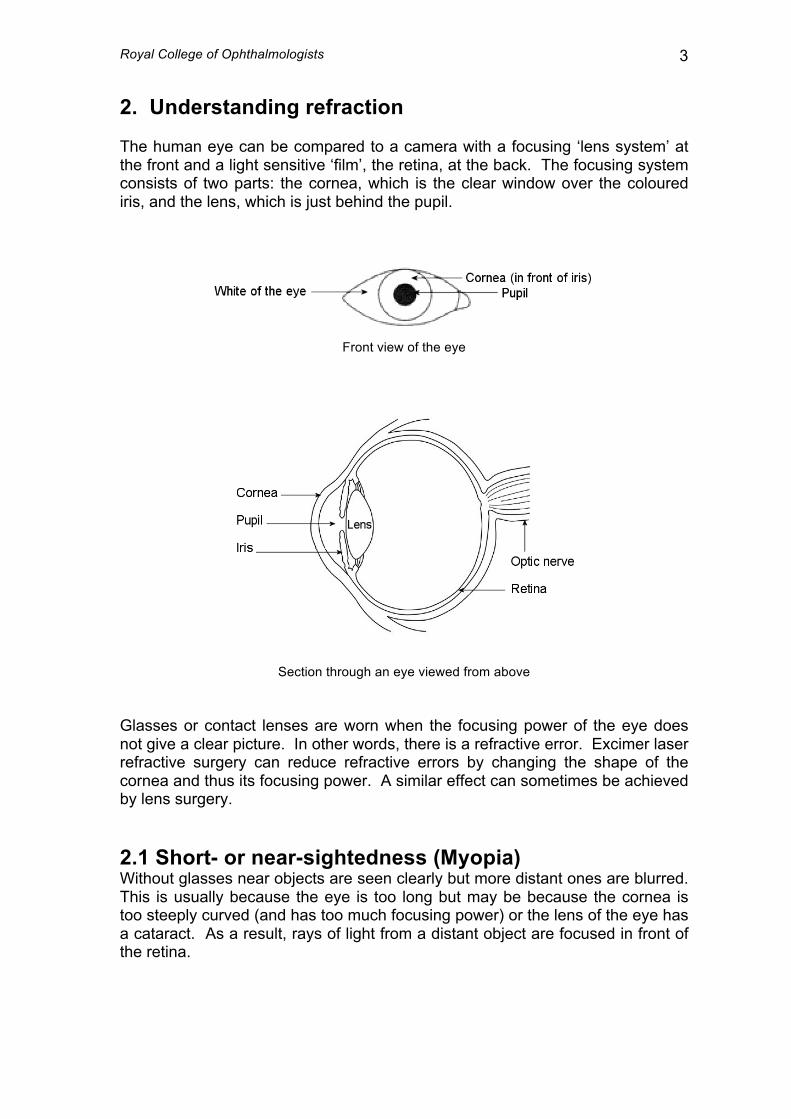
2. Understanding refraction The human eye can be compared to a camera with a focusing ‘lens system’ at the front and a light sensitive ‘film’, the retina, at the back. The focusing system consists of two parts: the cornea, which is the clear window over the coloured iris, and the lens, which is just behind the pupil.
Front view of the eye
Section through an eye viewed from above
Glasses or contact lenses are worn when the focusing power of the eye does not give a clear picture. In other words, there is a refractive error. Excimer laser refractive surgery can reduce refractive errors by changing the shape of the cornea and thus its focusing power. A similar effect can sometimes be achieved by lens surgery. 2.1 Short- or near-sightedness (Myopia) Without glasses near objects are seen clearly but more distant ones are blurred. This is usually because the eye is too long but may be because the cornea is too steeply curved (and has too much focusing power) or the lens of the eye has a cataract. As a result, rays of light from a distant object are focused in front of the retina.

Royal College of Ophthalmologists 4
2.2 Long- or far-sightedness (Hypermetropia/Hyperopia) Without glasses, distant objects are seen more clearly than near ones, although in more severe cases the vision may be blurred at all distances. In hypermetropia the eye is either too short and/or the cornea is relatively flat and as a result does not have sufficient focusing power. 2.3 Presbyopia The ability of the human lens (behind the pupil and iris) to accommodate (or focus on close objects, read etc) declines with age and almost everyone requires spectacles for reading in the fullness of time. However, short-sighted people often do not require glasses for reading when they reach this age as removing their distance glasses allows reading unaided. Presbyopia is a normal and inevitable aging process. It is not altered by laser refractive surgery and you will probably still need reading glasses in older age. 2.4 Astigmatism Astigmatism is usually due to the cornea being more steeply curved in one direction than the other - similar to the shape of a rugby ball rather than a football. Objects are seen stretched out in the direction of the astigmatism. The spectacle lens required to correct this is called an astigmatic, cylindrical, or toric lens. Astigmatism is measured by its magnitude or power and also its direction, or axis. In some cases excimer laser can be used to reduce astigmatism. 2.5 Strength of refractive errors Your refractive error or spectacle prescription consists of: The sphere (sph): The strength of the corrective lens required to correct myopia (-) or hypermetropia (+) measured in units called dioptres (D) and The cylinder (cyl): The strength of the corrective lens required to correct astigmatism measured in dioptres (D) at a particular angle, or axis. A typical spectacle prescription might be: RIGHT LEFT Sphere Cylinder Axis Prism Base Sphere Cylinder Axis Prism Base
-4.50 -2.50 120 Distance -3.50 -2.00 80 Near Or Right Eye -4.50 / -2.50 x 120 Left Eye -3.50 / -2.00 x 80 This is a short-sighted person with -4.50 dioptres (or D) of myopia in the right eye combined with –2.50 dioptres (or D) of astigmatism at axis 120 degrees. Sometimes this is written with the abbreviations ‘DS’, dioptres of sphere, and ‘DC’, dioptres of cylinder.
Royal College of Ophthalmologists 5
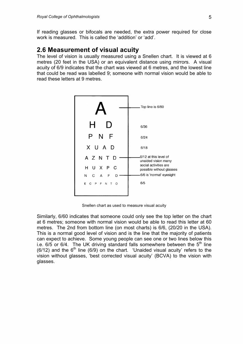
If reading glasses or bifocals are needed, the extra power required for close work is measured. This is called the ‘addition’ or ‘add’. 2.6 Measurement of visual acuity The level of vision is usually measured using a Snellen chart. It is viewed at 6 metres (20 feet in the USA) or an equivalent distance using mirrors. A visual acuity of 6/9 indicates that the chart was viewed at 6 metres, and the lowest line that could be read was labelled 9; someone with normal vision would be able to read these letters at 9 metres.
Snellen chart as used to measure visual acuity Similarly, 6/60 indicates that someone could only see the top letter on the chart at 6 metres; someone with normal vision would be able to read this letter at 60 metres. The 2nd from bottom line (on most charts) is 6/6, (20/20 in the USA). This is a normal good level of vision and is the line that the majority of patients can expect to achieve. Some young people can see one or two lines below this i.e. 6/5 or 6/4. The UK driving standard falls somewhere between the 5th line (6/12) and the 6th line (6/9) on the chart. ‘Unaided visual acuity’ refers to the vision without glasses, ‘best corrected visual acuity’ (BCVA) to the vision with glasses.

Royal College of Ophthalmologists 6
3. Changing the eye’s focus by surgery (refractive surgery) The cornea is the transparent surface of the eye in front of the coloured iris. The extent to which it is curved determines its focusing power. Excimer lasers reshape the cornea:
• In Short-sightedness (Myopia): the centre has to be flatter by removing more tissue from the centre than the edge
• In Long-sightedness (Hypermetropia): the centre has to be more curved by removing more tissue from the edge than the centre as in a ring doughnut shape
• In Astigmatism: the curve has to be evened out (ie: the cornea converted from a rugby ball shape to a football shape).
The benefit of corneal laser surgery is that instruments do not need to enter the eye itself, so damage or infection inside the eye is very rare. Corneal surgery can only correct short-sightedness up to -14D and long-sightedness up to +6D. Any surgery on the cornea can induce clouding or distortion, which may reduce visual acuity (see Complications, section 9). 3.1 Excimer Laser Techniques 3.1.1 Photorefractive keratectomy (PRK) PRK has been widely performed since the late 1980s. With the development of LASEK and LASIK, it is now mainly used for low refractive errors. Since little corneal tissue is removed, the remaining cornea is strong. The eye may be sore for about 48 hours after surgery. The healing process then continues for several months and can vary between patients. During this time the refraction slowly changes due to the healing process. There is usually a period of corneal haze which can cause blurring of vision and glare. In some patients (particularly those with higher refractive errors) these symptoms can persist to a greater or lesser extent. 3.1.2 Laser epithelial keratomilieusis (LASEK) LASEK is similar to PRK but the surface layer (epithelium) of the cornea is retained as a flap. A special soft contact lens is kept on the eye for 3-4 days to allow the surface to heal. The eye is much more comfortable than following PRK. Retaining the epithelium is thought to prevent later complications of haze and speed up healing 3.1.3 Laser in situ keratomilieusis (LASIK) LASIK has been widely performed since the mid 1990s. Most types of refractive error may be corrected with LASIK but it may not be suitable for extreme corrections as the procedure may make the cornea too thin and unstable. It differs from PRK as a cut is made across the cornea by either a special machine (microkeratome) or a special laser (femtosecond) to raise a flap of the cornea (like cutting the top off a boiled egg, and leaving the shell attached at one side).

Royal College of Ophthalmologists 7
The exposed surface is then sculpted in the same way using the excimer laser and the flap is replaced. This results in tissue being removed from the middle layers of the cornea (stroma). LASIK causes minimal pain, and vision tends to recover quickly. The wound healing process is less pronounced. However, the surgical technique is more involved, and if complications do occur, they may be more serious than after PRK. 3.1.4 EpiLASIK EpiLASIK is a new procedure using an epikeratome, a separator, that creates a thin flap of epithelium. The excimer laser beam is then applied under the epithelial flap. The epithelial flap is then replaced onto the cornea. 3.1.5 Wavefront There are natural irregularities (aberrations) of the structural components of the eye, which can cause light rays to focus incorrectly. Wavefront analysers can detect such aberrations. Laser treatment can also cause ocular aberrations. These aberrations have been reduced with newer lasers, e.g. the use of larger diameter treatment zones. Customised ablations calculated from preoperative wavefront analysis are becoming available but it remains to be seen whether customised ablations will make a significant difference to the average refractive surgery patient. A special technique called iris registration is sometimes used to make sure that the eye does not move during treatment.
3.1.6 Iris Registration The eyes of some patients rotate when they go from a sitting to a lying position Iris registration technology is similar to security eye scanners. This new technology maps points on the iris, and focuses the laser using those points thus adjusting for misalignment such as rotation.
3.1.6 Adjunctive Treatment In some patients treatments such as mitomycin may be used to control healing. 3.2 Thermokeratoplasty Applying a ring of small spots of heat in specific locations, using a holmium laser in Laser Thermokeratoplasty (LTK) or using an electrically heated wire in Conductive Keratoplasty, alters the front shape of the cornea by contraction. The coagulated tissue within the spot contracts resulting in steepening of the cornea. Thermokeratoplasty will only correct low degrees of long-sightedness and the effect of the surgery may gradually reduce over time resulting in the return of the long-sightedness.
Royal College of Ophthalmologists 8
3.3 Radial Keratotomy (RK) Radial keratotomy was one of the earlier methods of treating short sight. Shallow radial cuts were made into the cornea around the pupil. The technique has now been almost completely replaced by excimer laser surgery. 3.4 Monovision Monovision is a form of treatment for presbyopia in which the dominant eye is used for distance vision and the non-dominant eye for near vision. This situation can occur naturally and if created by laser treatment, it is tolerated by about 70% of patients. 4. Indications and Contraindications 4.1 Suitable for treatment
• Age 21 years or over • Stable prescription i.e. less than 0.5D change over the preceding 2-3
years • Healthy eyes • Good general health • People with reasonable expectations
4.2 Unsuitable for treatment
• Under 21 years of age • Prescription change of 0.5D or greater over the last 2-3 years. • Pregnancy / breast feeding • Significant keratoconus, cataract or glaucoma, herpes eye infection.
(These should be discussed further with your surgeon). • Patients on certain prescription drugs, such as oral steroids. • Excimer laser surgery may not be suitable for patients with medical
conditions such as diabetes, rheumatoid arthritis, systemic lupus erythematosus. This should be discussed with your surgeon
Royal College of Ophthalmologists 9
5. Initial patient assessment Much of the initial assessment may be undertaken by an optometrist (optician) but you should also be examined by an ophthalmologist who should be the person who will perform your surgery. This will also give you the opportunity to fully discuss any questions you may have with the operating surgeon. The purpose of the initial assessment is to establish whether there are any contraindications to surgery and also to decide which refractive surgical procedure may be suitable for you. Patients with an eye disease or certain medical conditions may not be suitable. To be considered for refractive surgery your spectacle prescription should be stable i.e. it should not have changed significantly in the past two years. You should bring previous written spectacle prescriptions (copies should be available from your optometrist/optician) to your initial assessment appointment so that any changes may be assessed. 5.1 Your first visit 1. There should be a discussion about why you want laser treatment and your
expectations of the result. 2. You will be asked questions about past eye problems (e.g. accidents or
injuries to your eye) and your general health. 3. Before the consultation, it is necessary to remove contact lenses for at least: soft 1 day gas permeable/ /hard 7 days
Sometimes longer is required. If you wear your contact lenses to your assessment appointment, it is not possible to measure the eye properly due to alteration in the shape of the surface. This usually reverses if you stop wearing the lenses and you may be advised to delay treatment until the shape of the cornea has returned to normal and remained stable for some time. You will also have to leave out your contact lenses for a length of time before the day of treatment.
4. Checking your unaided vision, your current spectacle prescription and how
well your eyes work together (binocular vision). 5. Long sighted (hypermetropic) patients usually have an additional test
involving eye drops that relax the focusing muscles of the eye and reveal any hidden additional refractive error. The drops may sting for a few seconds but are otherwise painless. They may blur your vision for up to 24 hours so you will need somebody to drive you home.

Royal College of Ophthalmologists 10
6. A computerised picture of the surface of the eye and ultrasound
measurements are taken to determine its shape and thickness. This will reveal any warpage (see 3 above) and a condition called keratoconus that causes the surface of the eye to become cone shaped. This test may show that you are not suitable for immediate laser treatment.
7. Assessment of the size of your pupil in low light conditions. People with very
large pupils may require a larger area of treatment to minimise the risk of side-effects at night (haloes, glare and ghosting).
8. Assessment of the structure of the eye using a special microscope,
measurement of the pressure in the eye and review of the back of the eye (the retina) to exclude cataracts and other eye problems. This may require drops to dilate the eye which can cause temporary blurred vision and light sensitivity, so you may not be able to drive for a few hours.
9. Finally you should have the opportunity to discuss any questions you may
have with the surgeon, who should outline all the options available to you and discuss the risks and benefits of each possible procedure.
Things to take with you: • A list of medicines you are on • Your current contact lenses, but do not wear them (see above) • Previous glasses prescriptions Things to remember/check before going to your appointment • You may be given eye drops that will cause blurred vision, so check with the
clinic whether you will be able to drive home after your appointment • You should have realistic expectations about the outcome of laser treatment.
If you have a minor or moderate refractive error you can expect very good results. People with poorer eyesight may still need glasses for some activities.
If your surgeon recommends 'LASIK' treatment (this involves cutting a flap or “trap door” on the surface of the eye), s/he must be sure the cornea is thick enough to allow a good outcome and avoid side effects.
Royal College of Ophthalmologists 11
6. The day of surgery PRK, LASEK and LASIK are performed on an outpatient basis in a clean room with air conditioning and control of the humidity and temperature. You should allow for a stay of at least one hour on the day of treatment. Options for treatment:
• One eye only in the first instance • Both eyes on the same day (bilateral simultaneous treatments).
Necessary preparations:
• Someone should accompany the patient on the day of treatment • Make-up on the day of treatment should be avoided • Sunglasses are helpful in the postoperative period
Procedure details:
• Local anaesthetic drops are placed in the eye being treated • Lie on an automatic reclining chair underneath the laser • The eye that is not being treated is covered to prevent distractions • Eye cleaned with antiseptic and tape placed over eyelashes (LASIK) • A special lid ‘clip’ is used to keep the eye open • Surface Preparation:
o Surface layer (epithelium) of the cornea is removed mechanically (PRK) or loosened with the aid of a dilute alcohol solution (LASEK)
o A flap is cut with a special mechanical cutter or a special laser after a suction ring is placed on the eye (painless but loss of vision for 20 seconds) (LASIK)
• Fixation on a flashing red or green light in the laser opening • Firing of the laser (average treatment time is 30 seconds). In operation
the laser makes a repetitive ‘tapping’ sound due to the pulse nature of excimer laser delivery. Although the process does not generate significant amounts of heat, you may be aware of a faint smell of burning
• Replacement of the surface epithelium (LASEK) and a contact lens placed on the eye (PRK and LASEK). Flap repositioned (LASIK)
• Instillation of antibiotic, anti-inflammatory and steroid eye drops • A plastic eye shield is applied and remains in place for up to 24 hours
If the flap in LASIK is judged to be unsatisfactory (e.g. a partial or damaged flap) the excimer laser treatment is not applied. The flap is then repositioned. A repeat treatment can normally be carried out 3 to 6 months later.
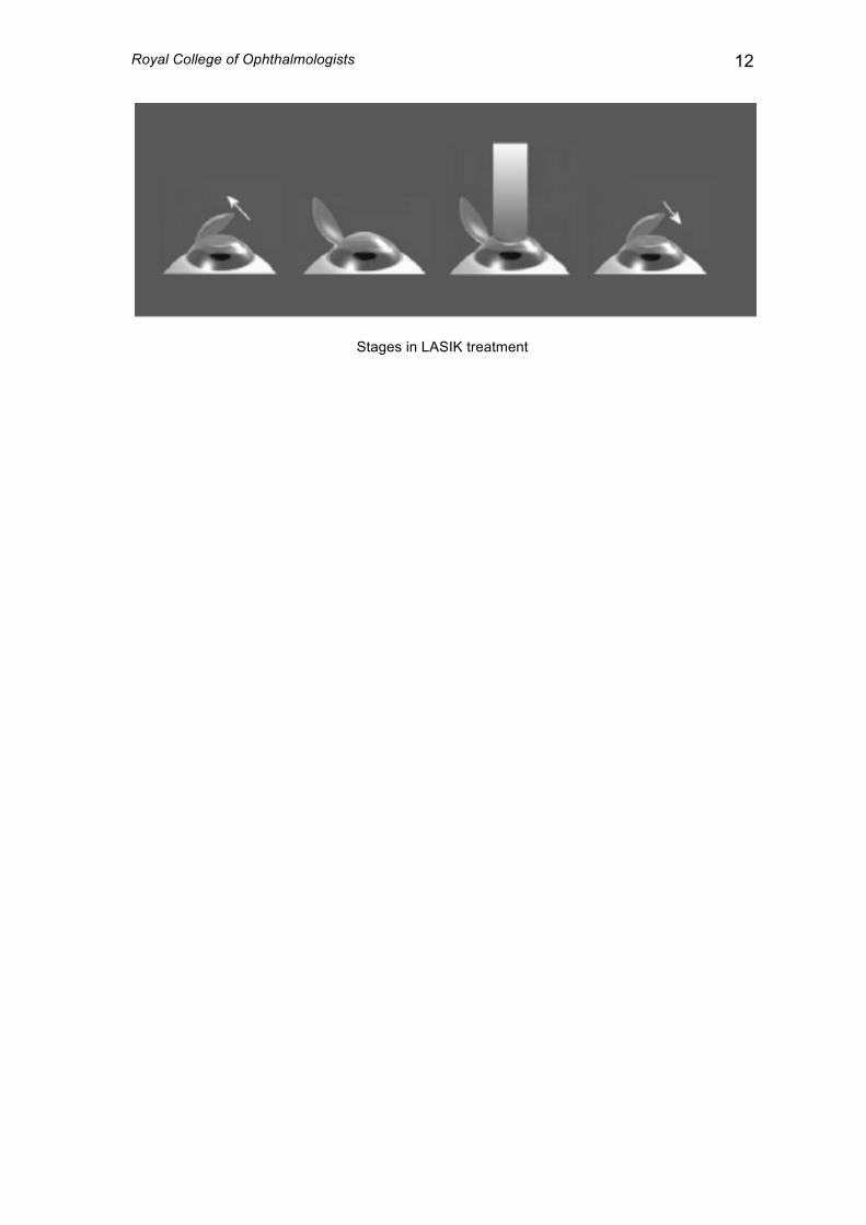
Royal College of Ophthalmologists 12
Stages in LASIK treatment

Royal College of Ophthalmologists 13
7. The period after surgery It is common to protect the eye at night time for the first week or so with a plastic shield. Antibiotic, anti-inflammatory (steroid) eye drops and artificial tears are prescribed for use after surgery. The dosage and duration varies with each technique. Patients generally use the antibiotic and anti-inflammatory eye drops for a week or so while the artificial tears may be used for a longer period (often up to six months in patients with dry eyes after LASIK). Most patients have a reasonably comfortable period after surgery and are back at work within a few days to a week. Depending on the vision correction attempted, driving may be unsafe for 1-2 weeks. Tinted glasses with ultraviolet protection are needed when out in the sun for the first three months. There are significant variations in the way patients recover from the three (PRK, LASIK & LASEK) laser techniques. The following table highlights these variations in cases where the surgery has been uneventful and free of complications.
Laser Technique
PRK LASIK LASEK
Pain Moderate to severe with significant patient variability
Minimal if any Mild for the first 24 hours or so Light sensitivity
Recovery of useful vision
2-4 weeks 1-2 days 3-6 days
Stability of Vision
Usually 1-3 months, can take 6 months to a year in some cases
Usually 1 week to 1 month, in some complicated cases can take 3 to 9 months
1 week to 1 month, in some complicated cases can take 3 to 9 months
Contact lens First 4-7 days Usually not required. Used in cases where the surface epithelium is scratched.
Used for the first 3-4 days.
Return to work Usually a week, can take longer
2-3 days 3-6 days

Royal College of Ophthalmologists 14
8. Results of laser refractive surgery In March 2006, the National Institute for Health and Clinical Excellence (NICE) produced Interventional Procedure Guidance (IPG) on ‘Photorefractive (laser) surgery for the correction of refractive errors’. This is available at www.nice.org.uk/IPG164publiinfo and the background research is available at www.nice.org.uk/ip320review This guidance compliments and expands some of the comments below. 8.1 Surface based photorefractive surgery (PRK, PARK, HPRK, HPARK AND LASEK) 8.1.1 Myopia (PRK & PARK) Studies between 1992 and 2004 show that for low to moderate degrees of myopia (-1.00 to -6.00D) the majority of patients achieve a very satisfactory result following surface based laser treatment (approximately 70% obtained a result within 0.5D and between 85% and 92% of patients obtained a result within 1.00D of the intended correction and see 6/6 unaided and all see 6/12 unaided). With higher corrections up to –10.00D, patients may have more visual side effects such as haloes. In this higher correction group, about 40% achieve 6/6 vision unaided and about 75% achieve 6/12 vision unaided. Correction of astigmatism is possible up to 4 dioptres and success is slightly less than those results for myopia without astigmatism. Reduction of the original laser correction can occur with time. Newer excimer lasers produce smoother treatment zones and more stable results so that fewer than 10% of eyes with low to moderate levels of myopia (-1.00 to -6.00D) will regress. 8.1.2 Longsightedness (HPRK & HPARK) Results for treatment of hyperopia with PRK show less accuracy (with approximately 60% within 0.5D and between 80% and 88% within 1.0D) and stability (compared to myopia results) with lessening of the effects after two years (regression). Correction of hyperopia up to 3.5D is likely to be achieved and no more than 6 dioptres can be corrected as the visual quality is greatly reduced if more is corrected. 8.1.3 Laser in situ epithelial keratomileusis (LASEK) LASEK as an excimer laser technique has only been regularly practised in the UK since 1999 and at this time published data on the results of treatment is limited. At present it would appear the refractive and visual results of LASEK appear broadly similar to the results of PRK with possible reduction in corneal haze. 8.2 Laser in situ keratomileusis (LASIK) LASIK has been practised in the UK since 1995. Initially it was used to treat the higher levels of myopia where it was thought likely that the result would be unsatisfactory using a surfaced based PRK treatment. In many centres in the UK, LASIK is being used as an alternative to PRK for corrections up to –12D.
Royal College of Ophthalmologists 15
The refractive and visual results for treatment of patients with myopia, hypermetropia and astigmatism with the LASIK procedure are broadly similar to the results of treatment with PRK after one year. No statistical difference has been found between the results of the two procedures in randomised clinical trials published in English between 1994 and 2004 in International Specialist Journals. Occasionally there may be a permanent reduction in best-corrected visual acuity (BCVA). There is a risk (between 2.7% and 4.8%) of loss of two or more lines of BCVA after LASIK. There is a higher risk of loss of two or more lines in treatments for hyperopia with even higher risks if treatment is more than 3.5D.
Royal College of Ophthalmologists 16
9. Complications of excimer laser surgery There is a wide range of the reported incidence of complications (from less than 1% up to 40%). Some complications, such as halo formation, were much more common with earlier laser machines using 3.5 to 4.5 mm diameter treatment zones than current lasers using 6.0 to 7.0 mm treatment zones. Since PRK, LASEK and LASIK utilise an excimer laser to produce the refractive correction many of the complications encountered are similar. 9.1 Miscellaneous side effects occurring equally in surface treatments and LASIK
• Minor over correction or under correction of refractive error. Since
all patients’ eyes differ in the rate and manner of healing, the computer predicted result might not achieve the expected correction leaving some patient’s over- or under- corrected. Some patients may be offered a second procedure (enhancement) but others may need spectacles or contact lenses for some tasks. Enhancement rates vary between 5% and 15%.
• Presbyopia is difficulty reading without glasses. This usually occurs
when people reach their early to mid 40’s. However, short-sighted people often do not require glasses for reading when they reach this age as removing their distance glasses allows reading unaided. A young myopic patient treated for distance vision will effectively become “normal sighted” for reading, but when a treated patient reaches 40-45 years of age, reading glasses will be required for near work. (See also Section 3.3 Monovision)
• Difference in refractive error between the two eyes (anisometropia)
may occur if only one eye is treated and patients may need to continue using glasses or contact lenses to balance the two eyes.
• Contact lens wear if needed may be more difficult following laser
treatment due to the changed shape of the cornea.
• Ptosis (drooping of the upper lid) may occur in the first few weeks following surface laser treatment but rarely persists.
• Eye sensitivity. The eye after surface laser treatment can be slightly
more sensitive to touch. Minor symptoms are relatively common in the first few months but severe symptoms are rare. These problems are usually self-limiting and persist in less than 1% of patients.
• Decreased night or low light vision is characterised by symptoms such
as glare, halos and starbursts seen around objects at night or in dim light conditions. Although visual acuity as measured on an eye chart may not
Royal College of Ophthalmologists 17
be affected, for some patients these symptoms can interfere with daily activities and in particular with driving at night. Reduced night vision is often temporary lasting one month to six weeks. A few patients however continue to experience these symptoms on a long-term basis. Research has shown that the incidence of these symptoms is caused by increased light scatter and induced irregularities (higher order aberrations) in the eye. Such problems are more common in those with particularly large pupils.
• Risk of Retinal Detachment. People with Myopia (short-sightedness)
have a greater risk of retinal detachment. It is important to remember that your retinal detachment risk remains after laser surgery.
9.2 Complications of surface based laser treatment (PRK, LASEK)
• Instability of the cornea following PRK or LASEK. The cornea is made
10%-20% thinner after surface based excimer laser treatment. The cornea is not significantly weakened and is therefore unlikely to be at risk from trauma.
• Vision reduction from all causes. The majority of scientific publications
regard a reduction in visual acuity by two or more lines on the Snellen letter chart as being significant. This may occur in: • Less than 4% of patients with low to moderate myopia (-0.5 to -6.00
D) • 10% or more patients with high or extreme myopia (over –6.00 D).
• Haze and scarring. All patients develop at least a mild degree of corneal
haze. The haze is worse during the first two to three months and in most cases will disappear within six to twelve months. Scientific publications indicate that the percentage of patients with haze and the degree of haze after laser treatment are related to the preoperative degree of myopia. With higher myopia it is more likely that haze will occur and will affect the vision. Haze may occur in: • 1-3% of patients with low to moderate myopia (less than –6.00D) • 5% or more patients with high or extreme myopia (over –6.00D).
• Infection. Since the surface of the cornea is removed during treatment it is possible that the eye may develop infection in the first week following laser treatment. Surface laser treatment infection is rare and occurs in less than 0.1% of treatments. (Severe infection can result in permanent corneal scarring, reduced vision and possible loss of the eye). Excimer laser treatment may result in reactivation of old herpes simplex virus corneal infection and the laser surgeon must be informed if a patient has had presumed herpes infection of the eye in the past.

Royal College of Ophthalmologists 18
9.3 Complications of LASIK
• Instability of the cornea following LASIK. Ectasia is the forward bulging of the centre of the cornea resulting in irregularity of the optical surface and poor quality vision. It results from weakening of the cornea due to removal of too much tissue. It is thought that 250 microns of untouched deep corneal tissue must remain to prevent ectasia following laser treatment. A LASIK procedure will create a corneal flap (usually between 120 and 150 microns depth) and the laser treatment is then carried out on the remaining corneal tissue. The untouched deep corneal tissue is, therefore, thinner following a LASIK procedure than a surface based laser treatment. Although the figure of 250 microns of untouched corneal tissue is generally taken as a safe level following LASIK treatment, there is relatively little scientific data to validate this figure, which is speculative, and still the subject of debate. A few studies have shown a small incidence of corneal ectasia (weakening of the cornea to produce permanently reduced vision and irregular astigmatism) in the range of 0% to 0.87% after a few years. However the majority of these eyes were cut too thin or had early signs of keratoconus.
• Corneal haze. Due to cutting of the cornea, there is less healing after
LASIK and a greatly reduced incidence of haze. • Reduction in visual quality from all causes. There is a risk (between
2.7% and 4.8%) of loss of two or more lines of BCVA after LASIK.
• Problems specifically related to the LASIK surgical procedure. Due to the creation of the hinged corneal flap in LASIK, complications related to the corneal flap are necessarily unique to the LASIK procedure. The following complications have generally been reported as occurring in between 0% and 4% of patients undergoing the LASIK procedure. All these complications may be associated with a permanent reduction of best-corrected vision. Other rare problems are:
• Incomplete cut of the corneal flap (usually remedied by a repeat
procedure after 2-3 months) • Loss or extensive damage to the corneal flap • Completely free corneal flap – which might require stitches to hold it in
place • Debris or fibres under the corneal flap which may cause inflammation • Epithelial ingrowth under the corneal flap • Wrinkling of the corneal flap • Retinal haemorrhage or retinal artery or venous occlusion • Penetration of the eye by the microkeratome and possible loss of the
eye because of haemorrhage or infection (very rare)
• Dry eye. The majority of patients complain of dry eye symptoms after LASIK because the surface nerves have been cut. (These nerves take six months to regrow). The use of artificial tears (lubricant drops) alleviate
Royal College of Ophthalmologists 19
the sensation of irritation. Occasionally a temporary punctal plug is placed in the opening of the tear duct to slow the tear drainage from the eye.

Royal College of Ophthalmologists 20
10. Standards for laser refractive surgery The Royal College of Ophthalmologists has published a set of standards (http//:www.rcophth.ac.uk) for refractive surgery which cover the qualifications and experience your surgeon should have, the information made available to you, the consent process, advertising, best clinical practices and the facilities in which you have the treatment. 10.1 Qualifications of the surgeon It is a legal requirement for all doctors to be on the medical register of the General Medical Council. Apart from this register The General Medical Council also maintains a specialist register. Specialist registration in Ophthalmology can only be obtained by doctors after 7 years rigorous training and examinations. It is a legal requirement for a doctor to be on the specialist register in order to work as a consultant in the NHS. The Royal College of Ophthalmologists recognises and accepts that specialist registration is not a legal requirement to perform laser eye surgery and currently many laser eye surgeons are not on the specialist register. Some refractive surgeons are on the specialist register have experience of a wide variety of eye diseases in addition to laser eye surgery. They regularly perform intra-ocular microsurgical procedures as well as other non laser refractive surgery operations. The Royal College of Ophthalmologists has introduced an assessment in laser refractive surgery and recommends that all refractive surgeons hold the Certificate, indicating that they have passed the assessment.

Royal College of Ophthalmologists 21
10.2 Preparing for Success: You will maximise your chances of success if you know the answers to the following questions after your research and pre surgery consultation with the surgeon.
1. Do you know enough about the full range of laser and non laser vision correction treatments? There is a range of laser and non laser vision correction treatments available. It is critical to choose the particular vision correction treatment which has the best chance of success and carries the least risk in your individual case. Laser eye surgery may not be the best treatment for you.
2. What is the level of vision you could realistically expect ?
After successful laser eye surgery you may not achieve perfect vision or the level of vision you achieve with you glasses or contact lenses. The level of vision achieved may also vary from one person to another. It is thus critical to know the level of vision you could realistically achieve.
3. Do the benefits outweigh the small risks of the treatment in your
individual case? You should be aware of the risks of the laser eye surgery in your individual case. Risks of surgery can vary from person to person. You should also be aware of rare sight threatening risks of the surgery.
4. Do you know enough about your surgeon?
Laser eye surgery is carried out by surgeons not clinics or companies. It is important to know your surgeon’s level of training and experience. Legally any doctor can perform laser eye surgery however The Royal College of Ophthalmologists recommends that refractive surgeons should
• Be fully trained Ophthalmologists on the specialist register of the General Medical Council.
• Have undergone additional specialist training in refractive surgery. • Surgeons hold the Certificate in Laser Refractive Surgery
5. Do you know why prices vary so much?
You may have come across a very wide range of advertised prices for laser eye surgery. Different prices may reflect different standards of care. The advertised price may be a starting price and the real price for your treatment may be very different. There may be differences in the costs of laser technology used as well as individual surgeon experience and training. You need to be aware of these factors when you compare prices and make an informed choice as to the level of care you deserve.

Royal College of Ophthalmologists 22
Glossary Antibiotics – Drugs used to prevent or treat infection by bacteria Antivirals - Drugs used to prevent or treat infection by a virus Astigmatism – Oval shape of the eye or cornea Conjunctiva – Loose sealing layer over the white sclera of the eye which keeps the eye moist and protected Dominant –Lead eye Excimer – A type of laser (EXCited dIMER) Epithelium – Surface layer of the cornea Endothelium – Cell layer on the inside of the cornea responsible for keeping the cornea dehydrated and clear Femtosecond – a special pulsed laser to cut a corneal flap Haloes – Ring of light around a light Haze – Loss of clarity of the cornea HPRK – Hyperopic PRK Hyperopia & hypermetropia – Longsightedness corrected using plus (+) lenses Keratectomy – Removal of corneal tissue Keratome – Knife/blade for cutting the cornea Keratomileusis – Making a flap of the cornea LASEK – LASer Epithelial Keratomileusis LASER – Light Amplification by the Stimulated Emission of Radiation LASIK – LASer In-situ Keratomileusis Microkeratome – Small mechanised blade for cutting the cornea Monovision – Use of each eye for different distances, usually dominant eye for distance vision and the non-dominant eye for near vision Myopia – Short-sightedness corrected using minus (-) lenses PARK – Photo-astigmatic Refractive Keratectomy Presbyopia – Loss of accommodation with age (near focus) PRK – Photo-refractive Keratectomy PTK – Photo-therapeutic Keratectomy (smoothing of an irregular surface)

Royal College of Ophthalmologists 23
Refraction – Assessment using lenses of the focus of the eye Retina – Light sensitive layer at the back of the eye containing rods and cones, analogous to the film in a camera Scatter – Spreading out of light inside the eye, resulting in a reduction in the quality of vision Speculum – Clip used to keep the eyes open during surgery Squint or Strabismus – Abnormal eye turn Steroids – Drugs used to reduce the inflammatory reaction that occurs during the healing process Stroma – The main bulk of the cornea 6/6 – normal level of vision, measured in metres. It is the same as 20/20 which is measured in feet. 6/12 – at this level of unaided vision many social activities are possible without glasses (equivalent to 20/40) 6/60 – sufficient vision to see the top letter on a vision testing chart (equivalent to 20/200)