A PATHOGENETIC APPROACH TO VACCINATION AGAINST...
145
A PATHOGENETIC APPROACH TO VACCINATION AGAINST PLEUROPNEUMONIA IN SWINE Ingrid Van Overbeke Thesis submitted in fulfillment of the requirements for the degree of Doctor of Veterinary Science (PhD), Ghent University, October, 2004 Promotor: Prof. Dr. F. Haesebrouck Copromotor: Prof. Dr. R. Ducatelle Faculty of Veterinary Medicine Department of Pathology, Bacteriology and Poultry diseases
Transcript of A PATHOGENETIC APPROACH TO VACCINATION AGAINST...
A PATHOGENETIC APPROACH TO VACCINATION AGAINST
PLEUROPNEUMONIA IN SWINE
Ingrid Van Overbeke
Thesis submitted in fulfillment of the requirements for the degree of Doctor of Veterinary Science (PhD), Ghent University, October, 2004
Promotor: Prof. Dr. F. Haesebrouck Copromotor: Prof. Dr. R. Ducatelle
Faculty of Veterinary Medicine
Department of Pathology, Bacteriology and Poultry diseases

Contents
CONTENTS
LIST OF ABBREVIATIONS 7
INTRODUCTION CONTAGIOUS PORCINE PLEUROPNEUMONIA: A REVIEW WITH EMPHASIS ON PATHOGENESIS AND DISEASE CONTROL 1. Etiology 11
2. Prevalence and epizootiology 11
3. Clinical signs and lesions 12
4. Pathogenesis 14
5. Role of virulence factors in pathogenesis and protection 15
6. Disease control with emphasis on vaccination 21
7. References 26
SCIENTIFIC AIMS 35
EXPERIMENTAL STUDIES CHAPTER 1 EVALUATION OF THE EFFICACY OF COMMERCIALLY
AVAILABLE VACCINES AGAINST PLEUROPNEUMONIA Effects of endobronchial challenge with Actinobacillus
pleuropneumoniae serotype 9 of pigs vaccinated with inactivated
vaccines containing the Apx toxins 41
Summary 42
Introduction 43
Materials and methods 43
Results 45
Discussion 50
References 51
Effects of endobronchial challenge with Actinobacillus
pleuropneumoniae serotype 9 of pigs vaccinated with a vaccine
containing Apx toxins and transferrin-binding proteins 53
Summary 54
Introduction 55
Materials and methods 55
Results 57
Discussion 60
References 62
3
Contents
CHAPTER 2 ADHESION OF ACTINOBACILLUS PLEUROPNEUMONIAE TO PORCINE ALVEOLAR EPITHELIAL CELLS IN VITRO AND IN
VIVO Characterization of the in vitro adhesion of Actinobacillus
pleuropneumoniae to alveolar epithelial cells 67
Summary 68
Introduction 69
Materials and methods 69
Results 73
Discussion 81
References 85
Effect of culture conditions of Actinobacillus pleuropneumoniae
serotype 2 and 9 strains on in vivo adhesion to alveoli of pigs 89
Summary 90
Introduction 91
Materials and methods 91
Results 93
Discussion 93
References 95
CHAPTER 3 EVALUATION OF THE EFFICACY OF A VACCINE CONTAINING CANDIDATE-ADHESINS Effect of endobronchial challenge with Actinobacillus
pleuropneumoniae serotype 10 of pigs vaccinated with bacterins
consisting of Actinobacillus pleuropneumoniae serotype 10 grown
under NAD-rich and NAD-restricted conditions 99
Summary 100
Introduction 101
Materials and methods 102
Results 105
Discussion 108
References 110
GENERAL DISCUSSION 113
SUMMARY 129 SAMENVATTING 133
4

Contents
DANKWOORD 137
CURRICULUM VITAE 141
5

List of abbreviations
LIST OF ABBREVIATIONS
™: trade mark
µl: microliter
Apx: Actinobacillus pleuropneumoniae exotoxin
PBSS: phosphate buffered salt solution
kDa: kiloDalton
cfu: colony forming units
mg: milligram
mm: millimeter
NAD: nicotinamide-adenine dinucleotide
nm: nanometer
OD: optical density
RTX: Repeat in ToXins
SDS-PAGE: sodiumdodecylsulphate polyacrilamide gel electrophoresis
SPF: specific pathogen free
UV: ultra violet light
7

List of abbreviations
8
Introduction
CONTAGIOUS PORCINE PLEUROPNEUMONIA: A REVIEW WITH EMPHASIS ON PATHOGENESIS AND
DISEASE CONTROL
1. ETIOLOGY 2. PREVALENCE AND EPIZOOTIOLOGY 3. CLINICAL SIGNS AND LESIONS 4. PATHOGENESIS 5. ROLE OF VIRULENCE FACTORS IN PATHOGENESIS AND
PROTECTION 6. DISEASE CONTROL WITH EMPHASIS ON VACCINATION 7. REFERENCES
9

Introduction
10
Introduction
1. ETIOLOGY Actinobacillus pleuropneumoniae (A. pleuropneumoniae) is an obligate parasite of the porcine
respiratory tract (Taylor, 1999). The bacterium is a small, Gram-negative capsulated rod with
typical coccobacillary morphology (Nicolet, 1992). Based on nicotinamide adenine
dinucleotide (NAD) requirements, A. pleuropneumoniae can be divided into 2 biotypes.
Biotype 1 strains are NAD-dependent whereas biotype 2 strains are NAD-independent. So
far, 15 serotypes have been described (Blackall et al., 2002) although serotypes 1 and 5 are
subdivided into 1a and 1b and 5a and 5b, respectively (Jolie et al., 1994; Nielsen, 1986;
Nielsen et al, 1997). All serotypes are haemolytic and produce a positive CAMP (Christie,
Atkins, Munch-Peterson) reaction with beta-haemolytic Staphylococcus aureus (Taylor,
1999). The incomplete haemolysin zone induced by the ß-toxin is converted in a complete
zone of haemolysis around the A. pleuropneumoniae colony. Four toxins are produced: ApxI,
II, III and IV (Dom et al., 1994a ; Frey et al., 1993 ; Frey et al., 1994 ; Jansen, 1994; Kamp et
al., 1991 ; Schaller et al., 1999). Serotyping is mainly based on capsular antigens.
Furthermore, the serotypes have different lipopolysaccharide (LPS) composition, except that
serotypes 1, 9 and 11, serotypes 3, 6 and 8 and serotypes 4 and 7 have common epitopes.
Although there is evidence that all serotypes of A. pleuropneumoniae can cause severe
disease and death in pigs, significant differences in virulence have been observed (Frey,
1995; Rogers et al., 1990; Rosendal et al., 1985). These variations may be partly attributed
to the production of different combinations of Apx toxins, with the most virulent serotypes
producing both Apx I and Apx II (Frey, 1995). Field observations and experimental infections
provide evidence that biotype 2 strains are less virulent than biotype 1 strains. Field
observations also indicate that biotype 1 serotype 1a, 1b, 5a, 5b, 9 and 10 strains are more
virulent than the other biotype 1 serotypes. This was, however, not confirmed under
experimental conditions (Dom and Haesebrouck, 1992a ; Jacobson et al., 1995).
2. PREVALENCE AND EPIZOOTIOLOGY Pleuropneumonia is a major problem in much of Europe, the USA, Canada and Eastern Asia.
Control measures may suppress clinical disease but reports from many countries suggest that
30-50% of all pigs are infected. In Belgium, the biotype 1-serotypes 2, 3, 5, 6, 7, 8, 9 and 11
strains and the biotype 2-serotype 2 strains are mostly found (Hommez et al., 1988; Hommez
et al., 1990).
A. pleuropneumoniae can be isolated from nasal cavities, tonsils, middle ear cavities and
lungs of infected pigs (Dom et al., 1994; Duff et al., 1996; Sidibe et al., 1993). The bacterium
is normally not considered as invasive, but there is one report of A. pleuropneumoniae being
recovered from osteomyelitis in pigs (Jensen et al., 1999). The bacterium is mainly
transmitted by direct contact between infected pigs or by aerosols. After clinical or subclinical
infections, pigs can become carriers of A. pleuropneumoniae. In such pigs, the infectious
11
Introduction
agent is located mainly in necrotic lung lesions and/or tonsils, less frequently in the nasal
cavities (Nicolet, 1992; Sidibé et al., 1993).
Transmission between herds occurs through the introduction of carriers to populations without
previous experience of the disease. A. pleuropneumoniae is a strict pathogen of the porcine
respiratory system, has a very short survival time in the environment and is very fragile and
sensitive to the usual disinfectants (Taylor, 1999). The bacterium can survive for a few days
in mucus or other organic material (Nicolet, 1992). In case of acute outbreaks of
pleuropneumonia, indirect transmission can occur via exudate on booths or clothing (Nicolet,
1992).
An increased incidence of pleuropneumonia is associated with stress situations such as
transports, stable changing, overcrowding and inappropiate housing (Nicolet, 1992). Another
trigger factor is infection with other respiratory pathogens. It was demonstrated that a
concomitant infection with Mycoplasma hyopneumoniae (Caruso and Ross, 1990; Yagihashi
et al., 1984) or with Aujeszky’s disease virus (Sakano et al., 1993) can worsen the symptoms
of pleuropneumonia. In contrast, a concomitant experimental infection with PRRSV had no
effect on clinical symptoms and lesions caused by A. pleuropneumoniae (Pol et al., 1997).
Sows from a chronically infected herd confer passive immunity to their offspring through
colostral antibodies (Nielsen, 1985). As the colostral antibody level declines, the piglets
become susceptible to infection. Where the infection is enzootic, the condition is mostly
found amongst pigs of 6-12 weeks of age.
3. CLINICAL SIGNS AND LESIONS The pace of disease can range from peracute to chronic depending on the serotype, the
immune status of the host, and the infection doses (Cruijsen et al., 1995; Hensel et al., 1993;
Rogers et al., 1990; Rosendal et al., 1985; Sebunya et al., 1983). Peracutely or acutely
diseased pigs may have some or all of the following clinical symptoms: high fever, increased
respiratory rate, coughing, sneezing, dyspnoea, anorexia, ataxia, vomiting, diarrhoea and
severe respiratory distress with cyanosis and presence of haemorrhagic foam on mouth
and/or nostrils (Ajito et al., 1996; Ligget et al., 1987; Rosendal et al., 1985; Taylor, 1999).
The subacute and chronic forms develop after the disappearance of acute signs. Recovering
animals may cough, and show respiratory distress particularly when disturbed. Exercise
intolerance may continue for days and affected animals may have reduced appetite, appear
gaunt and hairy, be depressed and show reduced rates of liveweight gain.
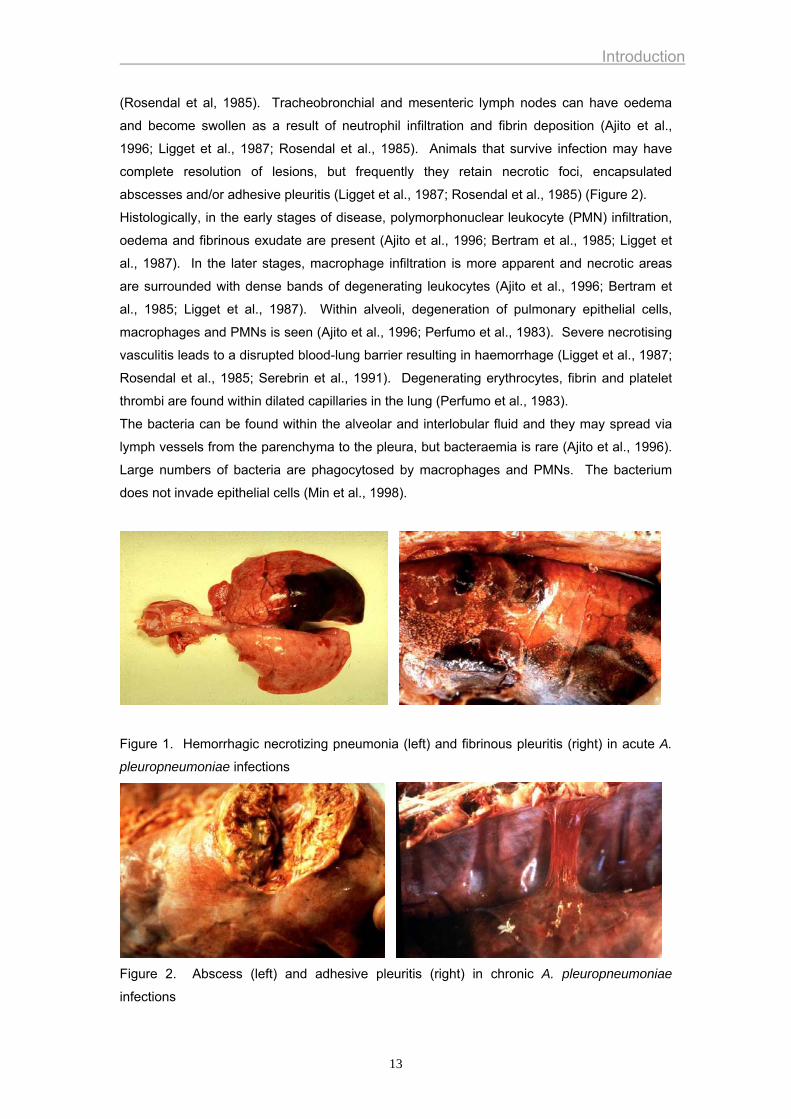
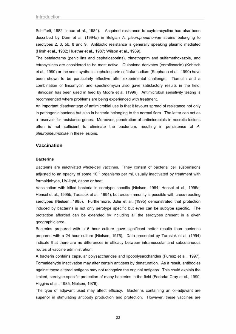
Lesions are mainly characterised by a hemorrhagic necrotizing pneumonia and fibrinous
pleuritis (Figure 1). The pneumonia is mostly bilateral, with involvement of the cardiac and
apical lobes, as well as at least part of the diaphragmatic lobes where pneumonic lesions are
often focal and well demarcated. In the peracute and acute form of the disease, pulmonary
lesions are characterised by severe oedema, inflammation, haemorrhage and necrosis (Ajito
et al., 1996; Bertram et al., 1985; Rosendal et al., 1985). The thoracic cavity is often filled
with bloody fluid and fibrin clots. Diffuse fibrinous pleuritis and pericarditis are also common
12
Introduction
(Rosendal et al, 1985). Tracheobronchial and mesenteric lymph nodes can have oedema
and become swollen as a result of neutrophil infiltration and fibrin deposition (Ajito et al.,
1996; Ligget et al., 1987; Rosendal et al., 1985). Animals that survive infection may have
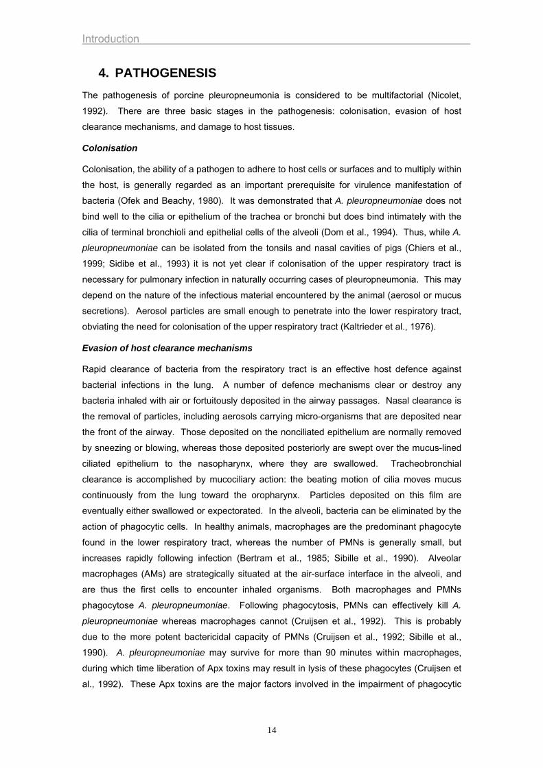
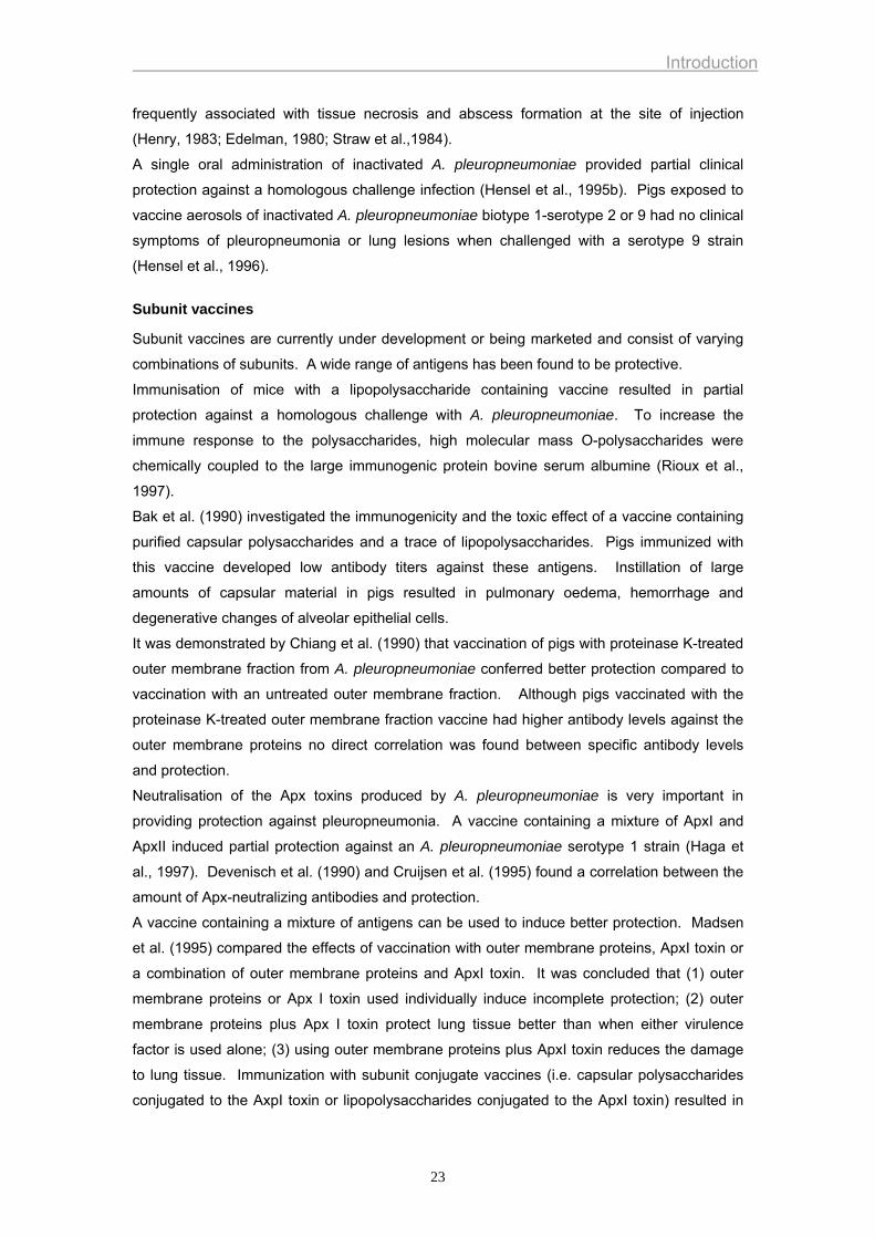
complete resolution of lesions, but frequently they retain necrotic foci, encapsulated
abscesses and/or adhesive pleuritis (Ligget et al., 1987; Rosendal et al., 1985) (Figure 2).
Histologically, in the early stages of disease, polymorphonuclear leukocyte (PMN) infiltration,
oedema and fibrinous exudate are present (Ajito et al., 1996; Bertram et al., 1985; Ligget et
al., 1987). In the later stages, macrophage infiltration is more apparent and necrotic areas
are surrounded with dense bands of degenerating leukocytes (Ajito et al., 1996; Bertram et
al., 1985; Ligget et al., 1987). Within alveoli, degeneration of pulmonary epithelial cells,
macrophages and PMNs is seen (Ajito et al., 1996; Perfumo et al., 1983). Severe necrotising
vasculitis leads to a disrupted blood-lung barrier resulting in haemorrhage (Ligget et al., 1987;
Rosendal et al., 1985; Serebrin et al., 1991). Degenerating erythrocytes, fibrin and platelet
thrombi are found within dilated capillaries in the lung (Perfumo et al., 1983).
The bacteria can be found within the alveolar and interlobular fluid and they may spread via
lymph vessels from the parenchyma to the pleura, but bacteraemia is rare (Ajito et al., 1996).
Large numbers of bacteria are phagocytosed by macrophages and PMNs. The bacterium
does not invade epithelial cells (Min et al., 1998).
Figure 1. Hemorrhagic necrotizing pneumonia (left) and fibrinous pleuritis (right) in acute A.
pleuropneumoniae infections
Figure 2. Abscess (left) and adhesive pleuritis (right) in chronic A. pleuropneumoniae
infections
13
Introduction
4. PATHOGENESIS The pathogenesis of porcine pleuropneumonia is considered to be multifactorial (Nicolet,
1992). There are three basic stages in the pathogenesis: colonisation, evasion of host
clearance mechanisms, and damage to host tissues.
Colonisation
Colonisation, the ability of a pathogen to adhere to host cells or surfaces and to multiply within
the host, is generally regarded as an important prerequisite for virulence manifestation of
bacteria (Ofek and Beachy, 1980). It was demonstrated that A. pleuropneumoniae does not
bind well to the cilia or epithelium of the trachea or bronchi but does bind intimately with the
cilia of terminal bronchioli and epithelial cells of the alveoli (Dom et al., 1994). Thus, while A.
pleuropneumoniae can be isolated from the tonsils and nasal cavities of pigs (Chiers et al.,
1999; Sidibe et al., 1993) it is not yet clear if colonisation of the upper respiratory tract is
necessary for pulmonary infection in naturally occurring cases of pleuropneumonia. This may
depend on the nature of the infectious material encountered by the animal (aerosol or mucus
secretions). Aerosol particles are small enough to penetrate into the lower respiratory tract,
obviating the need for colonisation of the upper respiratory tract (Kaltrieder et al., 1976).
Evasion of host clearance mechanisms
Rapid clearance of bacteria from the respiratory tract is an effective host defence against
bacterial infections in the lung. A number of defence mechanisms clear or destroy any
bacteria inhaled with air or fortuitously deposited in the airway passages. Nasal clearance is
the removal of particles, including aerosols carrying micro-organisms that are deposited near
the front of the airway. Those deposited on the nonciliated epithelium are normally removed
by sneezing or blowing, whereas those deposited posteriorly are swept over the mucus-lined
ciliated epithelium to the nasopharynx, where they are swallowed. Tracheobronchial
clearance is accomplished by mucociliary action: the beating motion of cilia moves mucus
continuously from the lung toward the oropharynx. Particles deposited on this film are
eventually either swallowed or expectorated. In the alveoli, bacteria can be eliminated by the
action of phagocytic cells. In healthy animals, macrophages are the predominant phagocyte
found in the lower respiratory tract, whereas the number of PMNs is generally small, but
increases rapidly following infection (Bertram et al., 1985; Sibille et al., 1990). Alveolar
macrophages (AMs) are strategically situated at the air-surface interface in the alveoli, and
are thus the first cells to encounter inhaled organisms. Both macrophages and PMNs
phagocytose A. pleuropneumoniae. Following phagocytosis, PMNs can effectively kill A.
pleuropneumoniae whereas macrophages cannot (Cruijsen et al., 1992). This is probably
due to the more potent bactericidal capacity of PMNs (Cruijsen et al., 1992; Sibille et al.,
1990). A. pleuropneumoniae may survive for more than 90 minutes within macrophages,
during which time liberation of Apx toxins may result in lysis of these phagocytes (Cruijsen et
al., 1992). These Apx toxins are the major factors involved in the impairment of phagocytic
14
Introduction
function of macrophages and PMNs. Furthermore, A. pleuropneumoniae produces several
factors which may contribute to its ability to survive within the macrophages: capsule and
lipopolysaccharides (Bilinski et al., 1991); copper-zinc superoxide dismutase (Langford et al.,
1996); stress proteins (Fuller et al., 2000); and ammonia (Bossé et al., 2000).
Damage to host tissues
Most of the pathological consequences of pleuropneumonia can be attributed to the Apx
toxins which exert cytotoxic effects on endothelial cells (Serebrin et al., 1991), macrophages
(Dom et al., 1992b), neutrophils (Dom et al., 1992a) and alveolar epithelial cells (Van de
Kerkhof et al., 1996). Activation of neutrophils, alveolar and intravasal macrophages, largely
due to Apx toxins and LPS, leads to release of toxic oxygen metabolites, as well as proteolytic
enzymes and various cytokines (Dom et al, 1992a; Dom et al., 1992b; Sibille et al., 1990;
Pabst, 1996; Udeze et al., 1987). LPS can enhance the effects of Apx toxins on phagocytes
(Fenwick, 1994).
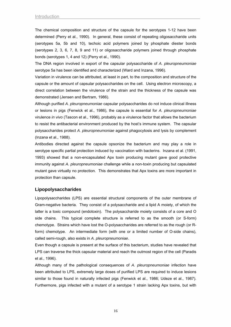
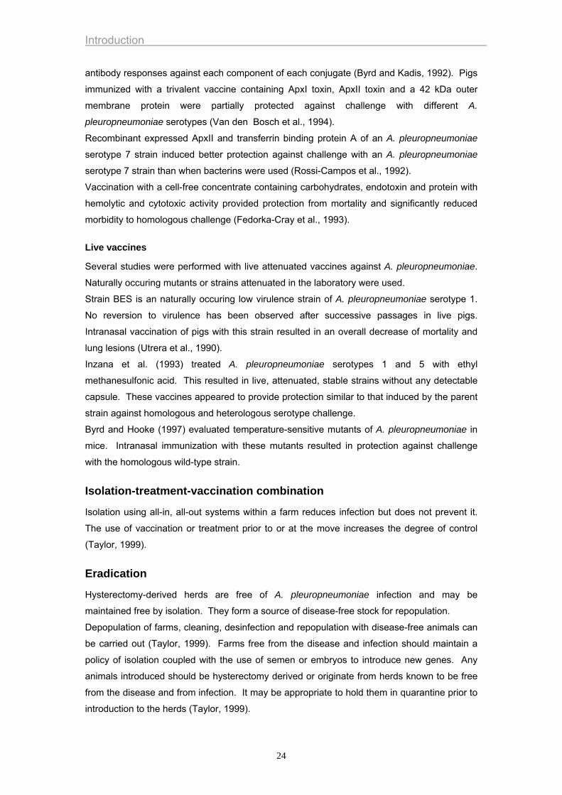
5. ROLE OF VIRULENCE FACTORS IN PATHOGENESIS AND PROTECTION
Different virulence factors have been described, including capsules, lipopolysaccharides,
outer membrane proteins, transferrin binding proteins, proteases, Apx toxins and adhesins
(Figure 3).
Figure 3. Virulence factors of A. pleuropneumoniae.
Capsule
Capsules are found in all strains of A. pleuropneumoniae. They mainly consist of derivatized
repeating oligosaccharides that determine serotype specificity (Beybon et al., 1993; Perry et
al., 1990). The capsule is responsible for the characteristic iridescence of the colony on a
clear medium.
LLiippooppoollyyssaacccchhaarriiddeess
TTrraannssffeerrrriinn bbiinnddiinngg pprrootteeiinnss
AAppxx ttooxxiinneess pprrootteeaasseess
OOuutteerr mmeemmbbrraannee pprrootteeiinnss
aaddhheessiinnss
ccaappssuullee
15
Introduction
The chemical composition and structure of the capsule for the serotypes 1-12 have been
determined (Perry et al., 1990). In general, these consist of repeating oligosaccharide units
(serotypes 5a, 5b and 10), techoic acid polymers joined by phosphate diester bonds
(serotypes 2, 3, 6, 7, 8, 9 and 11) or oligosaccharide polymers joined through phosphate
bonds (serotypes 1, 4 and 12) (Perry et al., 1990).
The DNA region involved in export of the capsular polysaccharide of A. pleuropneumoniae
serotype 5a has been identified and characterized (Ward and Inzana, 1996).
Variation in virulence can be attributed, at least in part, to the composition and structure of the
capsule or the amount of capsular polysaccharides on the cell. Using electron microscopy, a
direct correlation between the virulence of the strain and the thickness of the capsule was
demonstrated (Jensen and Bertram, 1986).
Although purified A. pleuropneumoniae capsular polysaccharides do not induce clinical illness
or lesions in pigs (Fenwick et al., 1986), the capsule is essential for A. pleuropneumoniae
virulence in vivo (Tascon et al., 1996), probably as a virulence factor that allows the bacterium
to resist the antibacterial environment produced by the host’s immune system. The capsular
polysaccharides protect A. pleuropneumoniae against phagocytosis and lysis by complement
(Inzana et al., 1988).
Antibodies directed against the capsule opsonize the bacterium and may play a role in
serotype specific partial protection induced by vaccination with bacterins. Inzana et al. (1991,
1993) showed that a non-encapsulated Apx toxin producing mutant gave good protective
immunity against A. pleuropneumoniae challenge while a non-toxin producing but capsulated
mutant gave virtually no protection. This demonstrates that Apx toxins are more important in
protection than capsule.
Lipopolysaccharides
Lipopolysaccharides (LPS) are essential structural components of the outer membrane of
Gram-negative bacteria. They consist of a polysaccharide and a lipid A moiety, of which the
latter is a toxic compound (endotoxin). The polysaccharide moiety consists of a core and O
side chains. This typical complete structure is referred to as the smooth (or S-form)
chemotype. Strains which have lost the O-polysaccharides are referred to as the rough (or R-
form) chemotype. An intermediate form (with one or a limited number of O-side chains),
called semi-rough, also exists in A. pleuropneumoniae.
Even though a capsule is present at the surface of this bacterium, studies have revealed that
LPS can traverse the thick capsular material and reach the outmost region of the cell (Paradis
et al., 1996).
Although many of the pathological consequences of A. pleuropneumoniae infection have
been attributed to LPS, extremely large doses of purified LPS are required to induce lesions
similar to those found in naturally infected pigs (Fenwick et al., 1986; Udeze et al., 1987).
Furthermore, pigs infected with a mutant of a serotype 1 strain lacking Apx toxins, but with
16
Introduction
normal LPS, do not develop clinical disease or lung lesions (Tascon et al., 1994). This
indicates that LPS is not responsible for the typical A. pleuropneumoniae lesions.
LPS activate the alternative complement cascade resulting in release of complement
components that attract and activate PMNs and macrophages and stimulate release of
inflammatory mediators, resulting in further PMN and platelet activation, vasodilation and
constriction of pulmonary airways (Bertram, 1988; Udeze et al., 1987).
LPS have also been implicated in adhesion of A. pleuropneumoniae to tracheal mucus,
tracheal and lung frozen sections (Paradis et al, 1994) and host glycosphingolipids (Abul-Mihl
et al., 1999).
Pigs immunised with LPS were only partially protected against challenge with the homologous
A. pleuropneumoniae serotype (Inzana, 1988), indicating that LPS may play a role in the
partial serotype specific protection that is induced by vaccination with bacterins.
Outer membrane proteins
Several proteins of the outer membrane of A. pleuropneumoniae are recognized by
convalescent sera. Furthermore, specific outer membrane proteins can be induced under
conditions of iron restriction or addition of maltose (Deneer and Potter, 1989a&b; Jansen,
1994). Although outer membrane protein profiles differ for most serotypes of A.
pleuropneumoniae (Rapp et al, 1986), it has been shown that isolates of all serotypes contain
several common outer membrane proteins, including the peptidoglycan-associated lipoprotein
PalA of 14 kDa (Frey et al., 1996), a 29/41-kDa heat-modifiable protein, a major protein that
varies from 32 to 42 kDa depending on the serotype and a 48-kDa protein (Cruz et al., 1996).
DNA sequence analysis of the gene encoding PalA revealed high similarity of the protein's
amino acid sequence to that of the E. coli peptidoglycan-associated lipoprotein PAL, to the
Haemophilus influenzae outer membrane protein P6 and to related proteins of several Gram-
negative bacteria. This gene is conserved and expressed in all A. pleuropneumoniae
serotypes and in A. lignieresii. A very similar gene is present in A. suis and A. equuli (Frey et
al., 1996).
PalA as well as proteins of 32K and 42K are immunodominant (Frey et al., 1996; Jansen,
1994; MacInnes and Rosendal, 1987). Immunisation with an outer membrane extract or a
crude outer membrane preparation conferred limited protection against challenge with A.
pleuropneumoniae (Beaudet et al, 1994; Jansen, 1994). Immunisation with recombinant
outer membrane lipoprotein Oml A (40 kDa), which is probably present in all serotypes of A.
pleuropneumoniae, protected pigs from death upon challenge with the homologous strain, but
lesions were found in the lungs and A. pleuropneumoniae was isolated from the lungs
(Gerlach et al, 1993). This indicates that antibodies against Oml A may contribute to, but are
not sufficient for protection of pigs against A. pleuropneumoniae infection (Jansen, 1994).
17
Introduction
Transferrin binding proteins
Iron is essential for bacterial growth. However, it is not readily available in the extracellular
environment of the host due to complexation by the host glycoproteins, transferrin and
lactoferrin. A. pleuropneumoniae expresses a number of factors that are involved in the
acquisition and uptake of iron. It is capable of utilising porcine transferrin, but not transferrin
from other animal species, as a sole source of iron (Niven et al., 1989). These receptors are
expressed under iron limited conditions. They consist of two distinct proteins. The transferrin
binding protein A has an approximate molecular mass of 60 kDa and appears to be a
lipoprotein anchored to the outer membrane by N-terminal fatty acid residues (Fuller et al.,
1998; Gerlach et al., 1992; Gonzalez et al., 1995). Transferrin binding protein B has an
approximate molecular mass of 100 kDa and likely forms a transmembrane channel for
transport of iron across the outer membrane (Gonzalez et al., 1995; Wilke et al., 1997). The
pathway of iron acquisition suggested by Kirby et al. (1995) involves binding and iron removal
from transferrin at the bacterial surface by the co-ordinate action of TbpA and TbpB followed
by transport of iron across the outer membrane via Tbp A and binding of iron by a periplasmic
binding protein.
Although both proteins are surface accessible and bind to the C-lobe of porcine transferrin,
there is evidence that an interaction between these proteins is required for optimal utilisation
of transferrin as a source of iron (Fuller et al., 1998; Gonzalez et al., 1995; Litt et al., 2000).
In addition, Gerlach et al. (1992) showed that TbpB is also capable to bind haemin but not
haemoglobin. This binding specificity has not been tested for TbpA.
Immunisation of pigs with the TbpB conferred limited protection against challenge with the
homologous strains (Gerlach et al., 1992 ; Rossi-Campos et al., 1992). This indicates that
Tbp proteins contribute to, but are not sufficient for protection of pigs against A.
pleuropneumoniae infection (Jansen, 1994).
A. pleuropneumoniae can also use haem compounds including free haem, haemin, haematin
and haemoglobin as a source of iron (Deneer and Potter, 1989). All serotypes of A.
pleuropneumoniae are capable of obtaining haem products via production of haemolysins
(Frey et al., 1993).
Proteases
All serotypes of A. pleuropneumoniae appear to secrete a high-molecular-mass protease
complex (>200 kDa) that degrades porcine gelatine, Ig A and haemoglobin (Garcia-Cuellar et
al., 2000; Negrete-Abascal et al., 1994). It has been suggested that the Ig A cleaving
proteases could facilitate the mucosal spread of A. pleuropneumoniae and that proteolytic
cleavage of haemoglobin could be a mechanism of iron acquisition. The exact role of these
proteases in the development of pathology has yet to be investigated.
18
Introduction
Apx toxins
Four Apx toxins have been described: two hemolytic exotoxins (ApxI and Apx II), one non-
hemolytic exotoxin (ApxIII) and one exotoxin that is only produced in vivo (ApxIV) (Dom et al.,
1994a ; Frey et al., 1993 ; Frey et al., 1994 ; Jansen, 1994; Kamp et al., 1991 ; Schaller et al.,
1999). They are toxic for porcine alveolar macrophages and neutrophils and belong to the
family of pore forming RTX-toxins, a group of protein toxins which is widely spread among
pathogenic Gram-negative bacteria (Frey et al, 1994).
ApxI is a strongly hemolytic and cytolytic protein with an apparent molecular mass of 105
kDa. It was previously named haemolysin I or cytolysin I and shows strong similarities to the
Escherichia coli α-haemolysin and to a lesser extent to the Mannheimia haemolytica
leukotoxin. It is produced by serotypes 1, 5, 9, 10 and 11 as well as by Actinobacillus suis
(Kamp et al., 1994).
ApxII is a weakly hemolytic and weakly cytotoxic exotoxin which was previously named
haemolysin II or cytolysin II. It has an apparent molecular mass of 103 kDa and is produced
by all serotypes, except serotype 10. It is also produced by Actinobacillus suis (Kamp et al.,
1994). Based on DNA derived amino acid sequence ApxII is closely related to the
Mannheimia haemolytica leukotoxin.
ApxIII is not hemolytic but strongly cytotoxic. It was previously named pleurotoxin. It is
produced by biotype1 serotype 2, 3, 4, 6 and 8 strains and has an apparent molecular mass
of 120 kDa.
In contrast to ApxI, ApxII and ApxIII cytotoxins, each of which is produced by some but not all
serotypes, the ApxIV toxin is produced by all serotypes (Cho and Chae, 2001; Schaller et al.,
1999). The apxIV gene product could not be detected in A. pleuropneumoniae cultures grown
under various conditions in vitro. Pigs experimentally infected with serotypes 1, 5 or 7
produced antibodies that reacted with the ApxIV toxin (Schaller et al., 1999), suggesting that
the apxIV gene product is induced in vivo. The apxIV gene was detected in degenerating
neutrophils and macrophages by Cho and Chae (2001). This suggests that ApxIV toxin may
lead to host-mediated tissue damage by the lysis of inflammatory cells. Further studies on
function and regulation of the apxIV gene in vivo are needed.
The structural toxin genes as well as the genes encoding activation and secretion of the Apx
toxins have been characterised (for a review, see Frey et al., 1994; Frey, 1995; Jansen,
1994).
It is now clear that Apx toxins play an important role in the pathogenesis of porcine
pleuropneumonia. They play a role in evasion of the hosts first line defence: at high
concentrations RTX toxins form pores in membranes of phagocytic and other target cells,
resulting in osmotic swelling and cell death. Most of the pathological consequences of
porcine pleuropneumonia can be attributed to the Apx I, II and III toxins: they are toxic for
endothelial cells (Serebrin et al., 1991) and provoke an oxidative burst in macrophages (Dom
et al., 1992c) and neutrophils (Dom et al., 1992b) resulting in excessive production of oxygen
radicals which can have deleterious effects on host cells. Moreover, purified recombinant Apx
19
Introduction
toxins were able to cause lesions upon endobronchial instillation and mutant strains which are
unable to produce Apx toxins did not induce lesions (Kamp et al., 1993; Stockhofe-Zurwieden
et al., 1996). Use of transposon mutagenesis (Tascon et al., 1994) and complementation
experiments (Reimer et al., 1995) also prove that Apx toxins are essential in the pathogenesis
of porcine pleuropneumonia.
It was demonstrated that A. pleuropneumoniae and its metabolites are able to kill type II
alveolar epithelial cells and that cytotoxicity is at least in part due to production of Apx toxins
(Van De Kerkhof et al., 1996). It is remarkable that rabbit sera against ApxI and ApxII were
not able to protect alveolar epithelial cells against the biotype 1 serotype 1 strain although
such sera were able to neutralize toxicity of culture supernatant. It has been shown that A.
pleuropneumoniae adheres to alveolar epithelial cells in vivo (see below). It was
demonstrated that this also occurs in vitro (Haesebrouck et al., 1996). This association may
mediate delivery of high concentrations of Apx toxins directly to the surface of the alveolar
epithelial cells, resulting in destruction of the target cells even in the presence of neutralizing
antibodies. It has been described that toxins produced by adhering bacteria are targeted more
efficiently and become relatively inaccessible to neutralization by toxin inhibitors (Ofek et al.,
1990).
The importance of Apx toxins in protective immunity against porcine pleuropneumonia was
demonstrated by immunisation with Apx toxins in combination with other bacterial
compounds. In these experiments the Apx toxins were essential vaccine components to
confer protection against challenge (Beaudet et al., 1994 ; Byrd and Kadis, 1992 ; Frey, 1995;
Van den Bosch et al., 1992 ; Jansen, 1994 ). However, it has also become clear that the Apx
toxins are not the only factors involved in protective immunity.
Adhesins
During the course of many infectious diseases, including pulmonary infections, bacteria
colonize body sites by engaging their surface-bound adhesins with cognate receptors
available on host cells. The recognition and attachment processes are therefore considered
to be the first steps in establishing bacteria at a given site. It is a prerequisite for colonization
and virulence manifestation of bacteria (Ofek and Beachy, 1980). Adherence enables
colonization to occur and allows the bacterium to exert its pathogenic effects.
It was shown that A. pleuropneumoniae adheres to the epithelium of the alveoli or the cilia of
the terminal bronchioli of experimentally infected pigs (Dom et al., 1994). It has also been
shown that A. pleuropneumoniae adheres to porcine tracheal rings maintained in culture
(Bélanger et al., 1990), to porcine frozen tracheal and lung sections (Jacques et al., 1991), to
erythrocytes from various animal species (Jacques et al., 1988), to tonsillar epithelial cells
(Chiers et al., 1999), to swine buccal epithelial cells (Hamer et al., 1999) and to type III swine-
lung collagen (Enriquez et al., 1999).
Several surface structures have been described in the family of Pasteurellaceae that are
involved in adhesion, including the capsule (Confer et al., 1995), fimbriae (Read et al., 1992),
20

Introduction
lipopolysaccharides (Bélanger et al., 1990) and outer membrane proteins (Confer et al.,
1995). Concerning A. pleuropneumoniae, only few adhesion factors have been described.
Lipopolysaccharides seem to be involved in the in vitro adhesion to porcine tracheal rings
(Bélanger et al., 1990) and mucus (Bélanger et al., 1992&1994). It was shown that the
capsule is not responsible for adherence to tracheal frozen sections (Jacques, 1999). Type 4
fimbriae have been demonstrated on A. pleuropneumoniae (Dom et al., 1994a; Utrera and
Pijoan, 1991; Zhang et al. 2000) but their role in adherence needs to be elucidated.
6. DISEASE CONTROL WITH EMPHASIS ON VACCINATION Disease control of pleuropneumonia may be accomplished in a number of different ways: (1)
antimicrobial treatment, (2) vaccination, (3) isolation-treatment-vaccination combination, and
(4) eradication. Besides these measures, control of environmental factors such as
temperature and ventilation and use of solid partitions between pens is essential.
Desinfection should be included in any control program; the organism is sensitive to a wide
range of commonly used desinfectants (Gutierrez et al., 1995).
Antimicrobial treatment
Antimicrobial therapy is efficient only in the initial phase of the disease, when it can reduce
mortality. For treatment of acutely affected animals antimicrobials should be applied in high
dosage and parenterally (subcutaneously or intramuscularly), as affected animals have
reduced food and water consumption. To ensure effective and durable blood concentrations,
repeated injections may be required. A combination of parenteral and peroral medication in a
recent outbreak often gives best results. The success of therapy depends mainly on early
detection of clinical signs and on rapid therapeutic intervention. In some outbreaks,
reapparance of the disease occurs a few weeks after cessation of peroral therapy. In these
cases, pulse medication is sometimes recommended i.e. medication periods (during 4 or 5
days) interrupted by periods during which animals are not treated (during 4 or 5 days). The
idea behind pulse medication is that during the non-treatment period animals build up
immunity.
Both lung damage and fibrous pleurisy may persist affecting the performance for the
remainder of the finishing period. Lesions may be colonised by organisms such as
Pasteurella spp. and these may persist after A. pleuropneumoniae has been eliminated.
A. pleuropneumoniae is particularly susceptible in vitro to penicillin, amoxicillin, ampicillin,
cephalosporin, tetracyclines, colistin, sulfonamide, combination of trimethoprim and
sulfamethoxazole, and gentamicin. Higher minimum inhibitory concentrations (MIC) values
are found for streptomycin, kanamycin, spectinomycin, spiramycin and lincomycin (Nicolet
and Schifferli, 1982; Gilbride and Rosendal, 1984; Nadeau et al., 1988; Inoue et al., 1984;
Hommez et al., 1988). Gilbride and Rosendal (1984) and Vaillancourt et al. (1988) described
that acquired resistance to ampicillin, streptomycin, sulfonamides and tetracyclines is frequent
in serotypes 1, 3, 5 and 7 but rare in other serotypes, particularly serotype 2 (Nicolet and
21
Introduction
Schifferli, 1982; Inoue et al., 1984). Acquired resistance to oxytetracycline has also been
described by Dom et al. (1994a) in Belgian A. pleuropneumoniae strains belonging to
serotypes 2, 3, 5b, 8 and 9. Antibiotic resistance is generally speaking plasmid mediated
(Hirsh et al., 1982; Huether et al., 1987; Wilson et al., 1989).
The betalactams (penicillins and cephalosporins), trimethoprim and sulfamethoxazole, and
tetracyclines are considered to be most active. Quinolone derivates (enrofloxacin) (Kobisch
et al., 1990) or the semi-synthetic cephalosporin ceftiofur sodium (Stephano et al., 1990) have
been shown to be particularly effective after experimental challenge. Tiamulin and a
combination of lincomycin and spectinomycin also gave satisfactory results in the field.
Tilmicosin has been used in feed by Moore et al. (1996). Antimicrobial sensitivity testing is
recommended where problems are being experienced with treatment.
An important disadvantage of antimicrobial use is that it favours spread of resistance not only
in pathogenic bacteria but also in bacteria belonging to the normal flora. The latter can act as
a reservoir for resistance genes. Moreover, penetration of antimicrobials in necrotic lesions
often is not sufficient to eliminate the bacterium, resulting in persistence of A.
pleuropneumoniae in these lesions.
Vaccination
Bacterins
Bacterins are inactivated whole-cell vaccines. They consist of bacterial cell suspensions
adjusted to an opacity of some 1010 organisms per ml, usually inactivated by treatment with
formaldehyde, UV-light, ozone or heat.
Vaccination with killed bacteria is serotype specific (Nielsen, 1984; Hensel et al., 1995a;
Hensel et al., 1995b; Tarasiuk et al., 1994), but cross-immunity is possible with cross-reacting
serotypes (Nielsen, 1985). Furthermore, Jolie et al. (1995) demonstrated that protection
induced by bacterins is not only serotype specific but even can be subtype specific. The
protection afforded can be extended by including all the serotypes present in a given
geographic area.
Bacterins prepared with a 6 hour culture gave significant better results than bacterins
prepared with a 24 hour culture (Nielsen, 1976). Data presented by Tarasiuk et al. (1994)
indicate that there are no differences in efficacy between intramuscular and subcutanuous
routes of vaccine administration.
A bacterin contains capsular polysaccharides and lipopolysaccharides (Furesz et al., 1997).
Formaldehyde inactivation may alter certain antigens by denaturation. As a result, antibodies
against these altered antigens may not recognize the original antigens. This could explain the
limited, serotype specific protection of many bacterins in the field (Fedorka-Cray et al., 1990;
Higgins et al., 1985; Nielsen, 1976).
The type of adjuvant used may affect efficacy. Bacterins containing an oil-adjuvant are
superior in stimulating antibody production and protection. However, these vaccines are
22
Introduction
frequently associated with tissue necrosis and abscess formation at the site of injection
(Henry, 1983; Edelman, 1980; Straw et al.,1984).
A single oral administration of inactivated A. pleuropneumoniae provided partial clinical
protection against a homologous challenge infection (Hensel et al., 1995b). Pigs exposed to
vaccine aerosols of inactivated A. pleuropneumoniae biotype 1-serotype 2 or 9 had no clinical
symptoms of pleuropneumonia or lung lesions when challenged with a serotype 9 strain
(Hensel et al., 1996).
Subunit vaccines
Subunit vaccines are currently under development or being marketed and consist of varying
combinations of subunits. A wide range of antigens has been found to be protective.
Immunisation of mice with a lipopolysaccharide containing vaccine resulted in partial
protection against a homologous challenge with A. pleuropneumoniae. To increase the
immune response to the polysaccharides, high molecular mass O-polysaccharides were
chemically coupled to the large immunogenic protein bovine serum albumine (Rioux et al.,
1997).
Bak et al. (1990) investigated the immunogenicity and the toxic effect of a vaccine containing
purified capsular polysaccharides and a trace of lipopolysaccharides. Pigs immunized with
this vaccine developed low antibody titers against these antigens. Instillation of large
amounts of capsular material in pigs resulted in pulmonary oedema, hemorrhage and
degenerative changes of alveolar epithelial cells.
It was demonstrated by Chiang et al. (1990) that vaccination of pigs with proteinase K-treated
outer membrane fraction from A. pleuropneumoniae conferred better protection compared to
vaccination with an untreated outer membrane fraction. Although pigs vaccinated with the
proteinase K-treated outer membrane fraction vaccine had higher antibody levels against the
outer membrane proteins no direct correlation was found between specific antibody levels
and protection.
Neutralisation of the Apx toxins produced by A. pleuropneumoniae is very important in
providing protection against pleuropneumonia. A vaccine containing a mixture of ApxI and
ApxII induced partial protection against an A. pleuropneumoniae serotype 1 strain (Haga et
al., 1997). Devenisch et al. (1990) and Cruijsen et al. (1995) found a correlation between the
amount of Apx-neutralizing antibodies and protection.
A vaccine containing a mixture of antigens can be used to induce better protection. Madsen
et al. (1995) compared the effects of vaccination with outer membrane proteins, ApxI toxin or
a combination of outer membrane proteins and ApxI toxin. It was concluded that (1) outer
membrane proteins or Apx I toxin used individually induce incomplete protection; (2) outer
membrane proteins plus Apx I toxin protect lung tissue better than when either virulence
factor is used alone; (3) using outer membrane proteins plus ApxI toxin reduces the damage
to lung tissue. Immunization with subunit conjugate vaccines (i.e. capsular polysaccharides
conjugated to the AxpI toxin or lipopolysaccharides conjugated to the ApxI toxin) resulted in
23
Introduction
antibody responses against each component of each conjugate (Byrd and Kadis, 1992). Pigs
immunized with a trivalent vaccine containing ApxI toxin, ApxII toxin and a 42 kDa outer
membrane protein were partially protected against challenge with different A.
pleuropneumoniae serotypes (Van den Bosch et al., 1994).
Recombinant expressed ApxII and transferrin binding protein A of an A. pleuropneumoniae
serotype 7 strain induced better protection against challenge with an A. pleuropneumoniae
serotype 7 strain than when bacterins were used (Rossi-Campos et al., 1992).
Vaccination with a cell-free concentrate containing carbohydrates, endotoxin and protein with
hemolytic and cytotoxic activity provided protection from mortality and significantly reduced
morbidity to homologous challenge (Fedorka-Cray et al., 1993).
Live vaccines
Several studies were performed with live attenuated vaccines against A. pleuropneumoniae.
Naturally occuring mutants or strains attenuated in the laboratory were used.
Strain BES is an naturally occuring low virulence strain of A. pleuropneumoniae serotype 1.
No reversion to virulence has been observed after successive passages in live pigs.
Intranasal vaccination of pigs with this strain resulted in an overall decrease of mortality and
lung lesions (Utrera et al., 1990).
Inzana et al. (1993) treated A. pleuropneumoniae serotypes 1 and 5 with ethyl
methanesulfonic acid. This resulted in live, attenuated, stable strains without any detectable
capsule. These vaccines appeared to provide protection similar to that induced by the parent
strain against homologous and heterologous serotype challenge.
Byrd and Hooke (1997) evaluated temperature-sensitive mutants of A. pleuropneumoniae in
mice. Intranasal immunization with these mutants resulted in protection against challenge
with the homologous wild-type strain.
Isolation-treatment-vaccination combination
Isolation using all-in, all-out systems within a farm reduces infection but does not prevent it.
The use of vaccination or treatment prior to or at the move increases the degree of control
(Taylor, 1999).
Eradication
Hysterectomy-derived herds are free of A. pleuropneumoniae infection and may be
maintained free by isolation. They form a source of disease-free stock for repopulation.
Depopulation of farms, cleaning, desinfection and repopulation with disease-free animals can
be carried out (Taylor, 1999). Farms free from the disease and infection should maintain a
policy of isolation coupled with the use of semen or embryos to introduce new genes. Any
animals introduced should be hysterectomy derived or originate from herds known to be free
from the disease and from infection. It may be appropriate to hold them in quarantine prior to
introduction to the herds (Taylor, 1999).
24

Introduction
Antimicrobial treatment alone has not been successful in eradication but combinations with
vaccination, partial depopulation and removal of serological and tonsillar carriers has been
successful (Taylor, 1999).
25

Introduction
7. REFERENCES Abul-Mihl, M., Paradis, S.E., Dubreuil, J.D., Jacques, M., 1999. Binding of
Actinobacillus pleuropneumoniae lipopolysaccharides to glycosphingolipids
evaluated by thin-layer chromatography. Infect. Immun. 67, 4983-4987.
Ajito, T., Haga, Y., Homma, S., Goryo, M., Okada, K., 1996. Immunohistological
evaluation on respiratory lesions of pigs intranasally inoculated with
Actinobacillus pleuropneumoniae serotype 1. J. Vet. Med. Sci. 58, 297-303.
Beaudet, R, McSween, G, Boulay, G, Rousseau, P, Bisaillon, JG, Descoteaux, JP,
Ruppanner, R., 1994. Protection of mice and swine against infection with
Actinobacillus pleuropneumoniae by vaccination. Vet. Microbiol. 39, 71-81.
Bélanger, M., Dubreuil, D., Harel, J., Girard, C., Jacques, M., 1990. Role of
lipopolysaccharides in adherence of Actinobacillus pleuropneumoniae to porcine
tracheal rings. Infect. Immun. 58, 3523-3530.
Bélanger, M., Rioux, S., Foiry, B., Jacques, M., 1992. Affinity for porcine respiratory
tract mucus is found in some isolates of Actinobacillus pleuropneumoniae. FEMS
Microbiol. Lett. 76, 119-125.
Bélanger, M., Dubreuil, D., Jacques, M., 1994. Proteins found within porcine respiratory
tract secretions bind lipopolysaccharides of Actinobacillus pleuropneumoniae.
Infect. Immun. 62, 868-873.
Bertram, T.A., 1985. Quantitative morphology of peracute pulmonary lesions in swine
induced by Actinobacillus pleuropneumoniae. Vet. Pathol. 22, 598-609.
Bertram, T.A., 1988. Pathobiology of acute pulmonary lesions in swine infected with
Haemophilus (Actinobacillus) pleuropneumoniae. Can. J. Vet. Res. 29, 574-577.
Beybon, L.M., Richards, J.C., Perry, M.B., 1993. Characterization of the Actinobacillus
pleuropneumoniae serotype K11/01 capsular antigen. Eur. J. Biochem. 214, 209-
214.
Bilinski, T., 1991. Oxygen toxicity and microbial evolution. Biosystems 24, 305-312.
Blackall, P.J., Klaasen, H.L.B.M, Van den Bosch, H., Kuhnert, P., Frey, J., 2002.
Proposal of a new serovar of Actinobacillus pleuropneumoniae: serovar 15. Vet.
Microbiol. 84, 47-52.
Bossé, J.T., MacInnes, J.I., 2000. Urease activity may contribute to the ability of
Actinobacillus pleuropneumoniae to establish infection. Can. J. Vet. Res. 64,
145-150.
Byrd, W., Kadis, S., 1992. Preparation, characterization and immunogenicity of
conjugate vaccines directed against Actinobacillus pleuropneumoniae virulence
determinants. Infect. Immun. 60, 3042-3051.
26
Introduction
Caruso, J.P., Ross, R.F., 1990. Effects of Mycoplasma hyopneumoniae and
Actinobacillus (Haemophilus) pleuropneumoniae infections on alveolar
macrophage functions in swine. Am. J. Vet. Res. 51, 227-231.
Chiers, K., Haesebrouck, F., Van Overbeke, I., Charlier, G., Ducatelle, R., 1999. Early
in vivo interactions of Actinobacillus pleuropneumoniae with tonsils of pigs. Vet.
Microbiol. 68, 301-306.
Cho, W.S., Chae, C., 2001. Expression of the apxIV gene in pigs naturally infected with
Actinobacillus pleuropneumoniae. J. Comp. Path. 125, 34-40.
Confer, A.W., Clinckenbeard, K., Murphy, G.L., 1995. Pathogenesis and virulence of
Pasteurella haemolytica in cattle: an analysis of current knowledge and future
approaches. In: Haemophilus, Actinobacillus and Pasteurella (Eds: W. Donachie,
F.A. Lainson and J.C. Hodgson). Proc. Haemophilus, Actinobacillus and
Pasteurella Conf., Edinburgh, Scotland, 51-62.
Cruijsen, T., Van Leengoed, L.A.M.G., Dekker-Nooren, T.C., Schoevers, J.H.,
Verheijden, J.H., 1992. Phagocytosis and killing of Actinobacillus
pleuropneumoniae by alveolar macrophages and polymorphonuclear leukocytes
isolated from pigs. Infect. Immun. 60, 4867-4871.
Cruijsen, T., Van Leengoed , L.A.M.G., Ham-Hoffies, M., Verheijden, J.H.M., 1995.
Convalescent pigs are protected completely against infection with a homologous
Actinobacillus pleuropneumoniae strain but incompletely against a heterologous-
serotype strain. Infect. Immun. 63, 2341-2343.
Cruz, W.T., Nedialkov, Y.A., Thacker, B.J., Mulks, M.H., 1996. Molecular
characterization of a common 48 kilodalton outer membrane protein of
Actinobacillus pleuropneumoniae. Infect. Immun. 64, 83-90.
Deneer, H.G., Potter, A.A., 1989a. Effect of iron restriction on the outer membrane
proteins in Actinobacillus pleuropneumoniae. Infect. Immun. 57, 798-804.
Deneer, H.G., Potter, A.A., 1989b. Identification of a maltose-induced major outer
membrane protein in Actinobacillus (Haemophilus) pleuropneumoniae. Microb.
Pathog. 6, 425-432.
Dom, P., Haesebrouck, F., 1992a. Comparative virulence of NAD-dependent and NAD-
independent Actinobacillus pleuropneumoniae strains. J. Vet. Med. Series B 39,
303-306.
Dom, P., Haesebrouck, F., Kamp, E., Smits, M.A., 1992b. Influence of Actinobacillus
pleuropneumoniae serotype 2 and its cytolysins on porcine neutrophil
chemiluminescence. Infect. Immun. 60, 4328-4334.
Dom, P., Haesebrouck, F., De Baetselier, P., 1992c. Stimulation and suppression of
the oxygenation activity of porcine pulmonary alveolar macrophages by
27
Introduction
Actinobacillus pleuropneumoniae and its metabolites. Am. J. Vet. Res. 53, 1113-
1118.
Dom, P., Haesebrouck, F., Kamp, E.M., Smits, M.A., 1994a. NAD-independent
Actinobacillus pleuropneumoniae strains: production of RTX toxins and
interactions with porcine phagocytes. Vet. Microbiol. 39, 205-218.
Dom, P., Haesebrouck, F., Ducatelle, R., Charlier, G., 1994b. In vivo association of
Actinobacillus pleuropneumoniae serotype 2 with the respiratory epithelium of
pigs. Infect. Immun. 62, 1262-1267.
Duff, J.P., Scott, J.W., Wilkes, M.K., Hunt, B., 1996. Otitis in a weaned pig: a new
pathological role for Actinobacillus (Haemophilus) pleuropneumoniae. Vet. Rec.
139, 561-563.
Enriquez, I., Guerrero, A.L., Serrano, J.J., Rosales, M.E., Hamer, R.C., Martinez, R.,
Godinez, D., de la Garza, M., 1999. Adhesion of Actinobacillus
pleuropneumoniae to type III swine lung collagen. Proc. Haemophilus,
Actinobacillus and Pasteurella Conf., Mabula, South-Africa, 52.
Fenwick, B.W., Osburn, B.I., 1986, Isolation and biologic characterization of two
lipopolysaccharides and a capsular enriched polysaccharide fraction isolated
from Actinobacillus pleuropneumoniae. Am. J. Vet. Med. Res. 47, 1433.
Fenwick, B.W., 1994. Lipopolysaccharides and capsules of the HAP group of bacteria.
In: Donachie, W., Lainson, F.A., Hodgson, J.C. (Eds.), Haemophilus,
Actinobacillus and Pasteurella. Plenum, New York and London, 75-87.
Frey, J., Bossé, J.T., Chang, Y.F., Cullen, J.M., Fenwick, B., Gerlach, G.F., Gygi, D.,
Haesebrouck, F., Inzana, T.J., Jansen, R., 1993. Actinobacillus
pleuropneumoniae RTX-toxins: uniform designation of haemolysins, cytolysins,
pleurotoxin and their genes. J. Gen. Microbiol. 139, 1723-1728.
Hamer, R.C., Enriquez, I., Godinez, D., Guerrero, A.L., Martinez, R., Talamas, P.,
Vaca, S., Garcia, C., de la Garza, M., 1999. Adhesion of Actinobacillus
pleuropneumoniae to swine buccal epithelial cells. Proc. Haemophilus,
Actinobacillus and Pasteurella Conf., Mabula, South-Africa, 51.
Frey, J., Kuhn, R., Nicolet, J., 1994. Association of the CAMP phenomenon in
Actinobacillus pleuropneumoniae with the RTX toxins ApxI, AxpII and ApxIII.
FEMS Microbiol. Lett. 124, 245-251.
Frey, J., Beck, M., Bosch, J.F. Van den, Segers, R.P.A.M., Nicolet, J., 1995.
Development of an efficient PCR method for toxin typing of Actinobacillus
pleuropneumoniae strains. Mol. Cell Probes 9, 277-282.
Frey, J., Kuhnert, P., Villiger, L., Nicolet, J., 1996. Cloning and characterization of an
Actinobacillus pleuropneumoniae outer membrane protein belonging to the
family of PAL lipoproteins. Res. Microbiol. 147, 351-361.
28
Introduction
Fuller, C.A., Yu, R., Irwin, S.W., 1998. Biochemical evidence for a conserved
interaction between bacterial transferrin binding protein A and transferrin binding
protein B. Microb. Pathog. 24, 75-87.
Fuller, T.E., Martin, S., Teel, J.F., Alaniz, G.R., Kennedy, M.J., Lowery, D.E., 2000.
Identification of Actinobacillus pleuropneumoniae virulence genes using
signature-tagged mutagenesis in a swine infection model. Microb. Pathog. 29,
39-51.
Garcia-Cuellar, C., Montanez, C., Tenorio, V., Reyes-Esparza, J., Duran, M.J.,
Negrete, E., Guerrero, A., de la Garza, M., 2000. A 24 kDa cloned zinc
metalloprotease from Actinobacillus pleuropneumoniae is common to all
serotypes and cleaves actin in vitro. Can. J. Vet. Res. 64, 88-95.
Gerlach, G.F., Anderson, C., Potter, A.A., Klashinsky, S., Wilson, P.J., 1992. Cloning
and expression of a transferrin binding protein from Actinobacillus
pleuropneumoniae. Infect. Immun. 60, 892-898.
Gerlach, G.F., Anderson, C., Klashinsky, S.,Rossi-Campos, A., Potter, A.A., Wilson,
P.J., 1993. Molecular characterisation of a protective outer membrane lipoprotein
(OmlA) from Actinobacillus pleuropneumoniae serotype 1. Infect. Immun. 61,
565-572.
Gonzalez, G.C., Yu, R.H., Rosteck, P.R., Schryvers, A.B., 1995. Sequence, genetic
analysis, and expression of Actinobacillus pleuropneumoniae transferrin receptor
genes. Microbiol. 141, 2405-2416.
Haesebrouck, F., Van de Kerkhof, A., Chiers, K., Ducatelle, R., 1996. Interactions of
Actinobacillus pleuropneumoniae with alveolar epithelial cells. Proc. 14th
Congress of the European Society of Veterinary Pathology, Ghent, Belgium, 87.
Hensel A., Windt, H., Stockhofe-Zurwieden, N., Lödding, N., Koch, W., Petzoldt, K.,
1993. A porcine aerosol infection model for studying dose dependent effects
caused by Actinobacillus pleuropneumoniae bacteria. J. Aerosol. Med. 6, 73-88.
Hommez, J., Devriese, L.A., Cassimon, P., Castryck, F., 1988. Serotypes and antibiotic
sensitivity of Actinobacillus (Haemophilus) pleuropneumoniae strains isolated in
Belgium. Vlaams Diergeneeskundig Tijdschrift 57, 46-52.
Hommez, J., Devriese, L.A., Castryck, F., Cassimon, P., 1990. Slide precipitation: a
simple method to type Actinobacillus (Haemophilus) pleuropneumoniae. Vet.
Microbiol. 24, 123-126.
Inzana, T.J., Ma, J., Workman, R.P., Gogolewski, P., Anderson, P., 1988. Virulence
properties and protective efficacy of the capsular polymer of Haemophilus
(Actinobacillus) pleuropneumoniae serotype 5. Infect. Immun. 56, 1880-1889.
29
Introduction
Inzana, T.J., Todd, J., Ma, J., Veit, P., 1991. Characterization of a non-hemolytic
mutant of Actinobacillus pleuropneumoniae serotype 5: role of the 110 kilodalton
hemolysin in virulence and immunoprotection. Microb. Pathogen. 10, 281-296.
Inzana, T.J., Todd, J., Veit, P., 1993. Safety, stability and efficacy of noncapsulated
mutants of Actinobacillus pleuropneumoniae for use in live vaccines. Infect.
Immun. 61, 1682-1686.
Jacobsen, M.J., Nielsen, J.P., 1995. Development and evaluation of a selective and
indicator medium for isolation of Actinobacillus pleuropneumoniae from tonsils.
Vet. Microbiol. 47, 191-197.
Jacques, M., Roy, G., Mittal, K.R., 1988. Hemagglutinating properties of Actinobacillus
pleuropneumoniae. Can. J. Microbiol. 34, 1046-1049.
Jacques, M., 1999. Adherence of mini-Tn10-generated, surface polysaccharides
mutants of Actinobacillus pleuropneumoniae serotype 1. Proc. Haemophilus,
Actinobacillus and Pasteurella Conf., Mabula, South-Africa, 23.
Jansen, R., 1994. The RTX toxins of Actinobacillus pleuropneumoniae. PhD thesis,
Utrecht, Netherlands.
Jansen, R., Briaire, J., Kamp, E.M., Gielkens, E.L.J., Smits, M.A., 1994. Genetic map
of the Actinobacillus pleuropneumoniae RTX toxin (apx) operons:
characterisation of the apxIII operons. Infect. Immun. 62, 4411-4418.
Jensen, A.E., Bertram, T.A., 1986. Morphological and biochemical comparison of
virulent and avirulent isolates of Actinobacillus pleuropneumoniae serotype 5.
Infect. Immun. 51, 419-424.
Jensen, T.K., Boye, M., Hagedorn-Olsen, T., Riising, H.J., Angen, O., 1999.
Actinobacillus pleuropneumoniae osteomyelitis in pigs demonstrated by
fluorescent in situ hybridization. Vet. Pathol. 36, 258-261.
Jolie, R.A.V., Mulks, M.H., Thacker, B.J., 1994. Antigenic differences within
Actinobacillus pleuropneumoniae serotype 1. Vet. Microbiol. 38, 329-349.
Kaltrieder, H.B., 1976. Initiation of immune responses in the lower respiratory tract with
rd cell antigens, in: C.H. Kirkpatrick, H.Y. Reynolds (Eds.), Immunologic and
infectious reactions in the lung, Marcel Dekker, Inc., New York, 1976, 73-97.
Kamp, E.M., Popma, J.K., Anakotta, J., Smits, M.A., 1991. Identification of hemolytic
and cytotoxic proteins of Actinobacillus pleuropneumoniae by use of monoclonal
antibodies. Infect. Immun. 59, 3079-3085.
Kamp, E.M., Pol, J.M.A., Stockhofe, N., Smits, M.A., 1993. The importance of ApxI and
ApxIII for pathogenicity of Actinobacillus pleuropneumoniae. Med. Microbiol.
Immunol. 182, 37, abstract.
30
Introduction
Kamp, E.M., Vermeulen, T.M.M., Smits, M., Haagsma, J., 1994. Production of Apx
toxins by field strains of Actinobacillus pleuropneumoniae and Actinobacillus
suis. Infect. Immun. 62, 4063-4065.
Kirby, S.D., Ogunnariwo, J.A., Schryvers, A.B., 1995. Receptor-mediated iron
acquisition from transferrin in the Pasteurellaceae. In: Donachie, W., Lainson,
F.A., Hodgson, J.C. (Eds.), Haemophilus, Actinobacillus and Pasteurella.
Plenum, New York and London, 115-127.
Langford, P.R., Loynds, B.M., Kroll, J.S., 1996. Cloning and molecular characterization
of Cu, Zn superoxide dismutase from Actinobacillus pleuropneumoniae. Infect.
Immun. 64, 5035-5041.
Ligget, A.D., Harrison, L.R., Farrell, R.L., 1987. Sequential study of lesions
development in experimental Actinobacillus pleuropneumoniae. Res. Vet. Sci 42,
204-212.
Litt, D.J., Palmer, H.M., Borriello, S.P., 2000. Neisseria meningitidis expressing
transferrin binding proteins of Actinobacillus pleuropneumoniae can utilize
porcine transferrin for growth. Infect. Immun. 68, 550-557.
MacInnes, J.I., Rosendal, S., 1987. Analysis of major antigens of Haemophilus
(Actinobacillus) pleuropneumoniae and related organisms. Infect. Immun. 55,
1626-1634.
Min, K., Chae, C., 1998. Detection and distribution of DNA of Actinobacillus
pleuropneumoniae in the lungs of naturally infected pigs by in-situ hybridization.
J. Comp. Pathol. 119, 169-175.
Negrete-Abascal, E., Tenorio, V.R., Serrano, J.J., Garcia, C., de la Garza, M., 1994.
Secreted proteases from Actinobacillus pleuropneumoniae serotype 1 degrade
porcine gelatin, hemoglobin and immunoglobulin A. Can. J. Vet. Res. 58, 83-86.
Nicolet, J., 1985. Bacteriology and epidemiology of Haemophilus pleuropneumoniae.
Proc. Am. Assoc. Swine Practitioners, 7-11.
Nicolet, J., 1992. Actinobacillus pleuropneumoniae. In: Diseases of swine, Leman,
A.D., Straw, B., Mengeling, W.L., D’Allaire, S., Taylor, D.J., eds., University
Press, Ames, Iowa State, 401-408.
Nielsen, R. 1985. Haemophilus pleuropneumoniae (Actinobacillus pleuropneumoniae)
serotypes 8, 3 and 6-Serological response and cross-immunity in pigs. Nord.
Vet. Med. 37, 217-227.
Nielsen, R., 1986. Serology of Haemophilus (Actinobacillus) pleuropneumoniae
serotype 5 strains: establishment of subtype A and B. Acta Vet. Scand. 27, 49-
58.
31
Introduction
Nielsen, R., Andresen, L.O., Plambeck, T., Nielsen, J.P., Krarup, L.T., Jorsal, S.E.
(1997). Serological characterization of Actinobacillus pleuropneumoniae biotype
2 strains isolated from pigs in two Danish herds. Vet. Microbiol. 54, 35-46.
Niven, D.F., Donga, J., Archibald, F.S., 1989. Responses of Actinobacillus
pleuropneumoniae to iron restriction: changes in the outer membrane protein
profile and the removal of iron from porcine transferrin. Mol. Microbiol. 3, 1083-
1089.
Ofek, I., Beachy, E.H., 1980. Bacterial adherence. Adv. Int. Med. 25, 505-532.
Pabst, R., 1996. The respiratory immune system of pigs. Vet. Immunol. Immunopathol.
54, 191-195.
Ofek, I., Zafriri, D., Goldhar, J., Eisenstein, B.I., 1990. Inability of toxin inhibitors to
neutralize enhanced toxicity caused by bacteria adherent to tissue culture cells.
Infect. Immun; 58, 3737-3742.
Paradis, S.E., Dubreuil, D., Rioux, S., Gottschalk, M., Jacques, M., 1994. High
molecular mass lipopolysaccharides are involved in Actinobacillus
pleuropneumoniae adherence to porcine respiratory tract cells. Infect. Immun.
62, 3311-3319.
Paradis, S.E., Dubreuil, D., Jacques, M., 1996. Examination of surface polysaccharides
of Actinobacillus plueropneumoniae serotype 1 grown under iron-restricted
conditions. FEMS Microbiol. Lett. 137, 201-206.
Perfumo, C.J., Rehbinder, C., Karlsson, K., 1983. Swine pleuropneumonia produced by
Actinobacillus pleuropneumoniae. III. An electron microscopic study. Zentralbl.
Veterinarmed. (B) 30, 678-684.
Perry, M.B., Altman, E., Brisson, J.R., Beynon, L.M., Richards, J.C., 1990. Structural
characteristics of the antigen capsular polysaccharides and lipopolysaccharides
involved in the serological classification of Actinobacillus (Haemophilus)
pleuropneumoniae strains. Serodiagn. Immunother. Infect. Dis. 4, 299-308.
Pol, J.M.A., Leengoed, L.A.M.G. Van; Stockhofe, N., Kok, G., Wensvoort, G., 1997.
Dual infections of PRRS/influenza or PRRS/Actinobacillus pleuropneumoniae in
the respiratory tract. Vet. Microbiol. 55, 259-264.
Rapp, V.J., Munson, R.S., Ross, R.F., 1986. Outer membrane proteins profiles of
Actinobacillus pleuropneumoniae. Infect. Immun. 52, 414-420.
Read, R., Rutman, A., Jeffrey, P., Lund, V., Brain, A., Moxon, R., Cole, P., Wilson, R.,
1992. Interaction of capsulated Haemopilus influenzae with human airway
mucosa in vitro. Infect. Immun. 60, 3244-3252.
Reimer, D., Frey, J., Jansen, R., Veit, H.P., Inzana, T.J., 1995. Molecular investigation
of the role of ApxI and ApxII in the virulence of Actinobacillus pleuropneumoniae
serotype 5. Microb. Pathog. 18, 197-209.
32
Introduction
Rogers, R.J., Eaves, L.E., Blackall, P.J., Truman, K.F., 1990. The comparative
pathogenicity of four serovars of Actinobacillus (Haemophilus)
pleuropneumoniae. Aust. Vet. J. 67, 9-12.
Rosendal, S., Boyd, D.A., Gilbride, K.A., 1985. Comparative virulence of porcine
Haemophilus bacteria. Can. J. Comp. Med. 49, 68-74.
Rossi-Campos, A., Anderson, C., Gerlach, G.F., Klashinsky, S., Potter, A.A., Wilson,
P.J., 1992. Immunisation of pigs against Actinobacillus pleuropneumoniae with
two recombinant protein preparations. Vaccine 10, 512-518.
Sakano, T., Shibata, I., Samegai, Y., Taneda, A., Okada, M., Irisawa, T., Sato, S.,
1993. Experimental pneumonia of pigs infected with Aujeszky's disease virus
and Actinobacillus pleuropneumoniae. J Vet. Med. Sci. 55, 575-579.
Schaller, A., Kuhn, R., Kuhnert, P., Nicolet, J., Anderson, T.J., MacInnes, J.I., Segers,
R.P., Frey, J., 1999. Characterization of apxIVA, a new RTX determinant of
Actinobacillus pleuropneumoniae. Microbiol. 145, 2105-2116.
Sebunya, T.N., Saunders, J.R., Osborne, A.D., 1983. Dose response relationship of
Actinobacillus pleuropneumoniae aerosols in pigs. Can. J. Comp. Med. 47, 54-
56.
Serebrin, S, Rosendal, S., Valdivieso-Garcia, A., Little, P.B., 1991. Endothelial
cytotoxicity of Actinobacillus pleuropneumoniae. Res. Vet. Sci. 50, 18-22.
Sibille, Y., Reynolds, H.Y., 1990. Macrophages and polymorphonuclear neutrophils in
lung defense and injury. Am. Rev. Respir. Dis. 141, 471-501.
Sidibé, M., Messier, S., Lariviere, S., Gottschalk, M., Mittal, K.R., 1993. Detection of
Actinobacillus pleuropneumoniae in the porcine upper respiratory tract as a
complement to serological tests. Can. J. Vet. Res. 57, 204-208.
Stockhofe-Zurwieden, N., Kamp, E.M., van Leengoed, L., Smits, M., 1996.
Pathogenicity of RTX toxin mutants of Actinobacillus pleuropneumoniae: results
of in vivo studies. Proc. International Pig Veterinary Society Congress, Bologna,
Italy, 189.
Tascon, R.I., Vazquez-Boland, J.A., Gutierrez-Martin, C.B., Rodriguez-Barbosa, I.,
Rodriguez-Ferri, E.F., 1994. The RTX haemolysins ApxI and ApxII are major
virulence factors of the swine pathogen Actinbacillus pleuropneumoniae:
evidence from mutational analysis. Mol. Microbiol. 14, 207-216.
Tascon, R.I., Vazquez-Boland, J.A., Gutierrez-Martin, C.B., Rodriguez-Barbosa, I.,
Rodriguez-Ferri, E.F., 1996. Virulence factors of the swine pathogen
Actinobacillus pleuropneumoniae. Microbiologia 12, 171-184.
Taylor, D.J., 1999. Actinobacillus pleuropneumoniae, in: Diseases of swine, B.E.
Shaw, S. D’Allaire, W.L. Mengeling, D.J. Taylor (Eds), , Blackwell Science,
Oxford, 343-354.
33
Introduction
Udeze, F.A., Latimer, R.W., Kadis, S., 1987. Role of Actinobacillus pleuropneumoniae
lipopolysaccharide endotoxin in the pathogenesis of porcine Actinobacillus
pleuropneumoniae. Am. J. Vet. Res. 48, 768-773.
Utrera, V., Pijoan, C., 1991. Fimbriae in Actinobacillus pleuropneumoniae strains
isolated from pig respiratory tracts. Vet. Rec. 128, 357-358.
Van de Kerkhof, A., Haesebrouck, F., Chiers, K., Ducatelle, R., Kamp, E.M., Smits,
M.A., 1996. Influence of Actinobacillus pleuropneumoniae and its metabolites on
porcine alveolar epithelial cells. Infect. Immun. 64, 3905-3907.
Van den Bosch, J.F., Jongenelen, I.M.C.A., Pubben, N.B., Van Vugt, F.G.A., Segers,
R.P.M.A., 1992. Protection induced by a trivalent Actinobacillus
pleuropneumoniae subunit vaccine. Proc. 12th International Pig Veterinary
Society Congress, The Hague, Netherlands, 194.
Ward, C.K., Inzana, T.J., 1996. Identification and characterization of a DNA region
involved in export of the capsular polysaccharide of Actinobacillus
pleuropneumoniae serotype 5a. Proc. 14th International Pig Veterinary Society
Congress, Italy, 187.
Wilke, M., Franz, B., Gerlach, G.F., 1997. Characterization of a large transferrin-binding
protein from Actinobacillus pleuropneumoniae serotype 7. Zentralbl.
Veterinarmed. (B) 44, 73-86.
Yagihashi, T., Nunoya, T., Mitui, T., Tajima, M., 1984. Effect of Mycoplasma
hyopneumoniae infection on the development of Haemophilus
pleuropneumoniae pneumonia in pigs. Jpn. J. Vet. Sci. 46, 705-713.
Zhang, Y.M., Tennent, J.M., Ingham, A., Beddome, G., Prideaux, C., Michalski, W.P.,
2000. Identification of type 4 fimbriae in Actinobacillus pleuropneumoniae. FEMS
Microbiol. Lett. 189, 15-18.
34

Scientific aims
SCIENTIFIC AIMS
A. pleuropneumoniae causes severe losses in the pig rearing industry. Although the disease
can be controlled by antimicrobial agents, the use of these products has several
disadvantages including induction of acquired resistance in pathogenic bacteria and bacteria
belonging to the normal flora of pigs. Therefore, prevention should be encouraged in the
control of the disease. Vaccines could be very useful to control porcine pleuropneumonia.
Rational design of effective antibacterial vaccines requires knowledge of the virulence factors
of the bacterium and the pathogenesis of the disease.
It has been shown that Apx toxins play an important role in the pathogenesis of porcine
pleuropneumonia. The first aim of this thesis was, therefore, to evaluate the efficacy of
vaccines mainly based on the inclusion of these toxins.
Inclusion of bacterial adhesins in subunit vaccines might be of value. Indeed, A.
pleuropneumoniae first adheres to alveolar epithelial cells before it causes lung lesions. The
hypothesis was that, once the bacteria have attached to their target cells, i.e. alveolar
epithelial cells, the Apx toxins are released directly onto the host cell. Hereby, neutralizing
antibodies have no opportunity to bind to the toxins and prevent their action. Therefore, in the
second part of this thesis, the purpose was to characterize the adhesion of A.
pleuropneumoniae to alveolar epithelial cells in vitro and in vivo.
In a final study it was determined whether pigs vaccinated with a bacterin consisting of
bacteria grown under conditions resulting in high in vitro adhesion, were better protected
against an A. pleuropneumoniae infection than pigs vaccinated with a bacterin consisting of
bacteria grown under conditions resulting in low in vitro adhesion.
35

Scientific aims
36
Experimental studies
EXPERIMENTAL STUDIES
CHAPTER 1. EVALUATION OF THE EFFICACY OF COMMERCIALLY AVAILABLE VACCINES AGAINST PLEUROPNEUMONIA
Effects of endobronchial challenge with Actinobacillus pleuropneumoniae serotype 9 of pigs vaccinated with inactivated vaccines containing the Apx toxins
Effect of endobronchial challenge with Actinobacillus pleuropneumoniae serotype 9 of pigs vaccinated with a vaccine containing Apx toxins and transferrin-binding proteins
CHAPTER 2. ADHESION OF ACTINOBACILLUS
PLEUROPNEUMONIAE TO PORCINE ALVEOLAR EPITHELIAL CELLS IN VITRO AND IN VIVO
Characterization of the in vitro adhesion of Actinobacillus pleuropneumoniae to alveolar epithelial cells
Effect of culture conditions of Actinobacillus pleuropneumoniae serotype 2 and 9 strains on in vivo adhesion to alveoli of pigs
CHAPTER 3. EVALUATION OF THE EFFICACY OF A VACCINE CONTAINING CANDIDATE-ADHESINS
Effect of endobronchial challenge with Actinobacillus pleuropneumoniae serotype 10 of pigs vaccinated with bacterins consisting of Actinobacillus pleuropneumoniae serotype 10 grown under NAD-rich and NAD-restricted conditions
37

Experimental studies
38
Experimental studies
CHAPTER 1
EVALUATION OF EFFICACY OF COMMERCIALLY AVAILABLE VACCINES AGAINST
PLEUROPNEUMONIA
39

Experimental studies
40
Experimental studies
Effects of endobronchial challenge with Actinobacillus pleuropneumoniae serotype 9 of pigs vaccinated with inactivated vaccines containing the Apx toxins
Koen Chiers1, Ingrid Van Overbeke1, Piet De Laender1, Richard Ducatelle1, Serge
Carel2, Freddy Haesebrouck1
1 Laboratory of Veterinary Bacteriology and Mycology and Laboratory of Veterinary
Pathology, Department of Pathology, Bacteriology and Poultry diseases, Faculty of
Veterinary Medicine, University of Ghent, Salisburylaan 133, B-9820 Merelbeke, Belgium
2 Biokema S. A., Chemin de la Chatanerie 2, 1023 Crissier - Lausanne, Switzerland
Veterinary Quarterly 20 (1998): 65-69.
41

Experimental studies
SUMMARY
The efficacy of two inactivated vaccines containing the Apx toxins of Actinobacillus
pleuropneumoniae (Hemopig™, Biokema, Lausanne, Switzerland and Porcilis™ App,
Intervet, Boxmeer, The Netherlands) was determined. Ten pigs were vaccinated twice with
Hemopig™ and eight pigs with Porcilis™ App. Ten control animals were injected twice with a
saline solution. Three weeks after the second vaccination, all pigs were endobronchially
inoculated with 105 colony-forming units (CFU) of an A. pleuropneumoniae serotype 9 strain.
Increased respiratory rate and/or fever were observed in all vaccinated and control pigs after
challenge. One pig of the Hemopig™ group and of the Porcilis™ App group died, whereas all
pigs of the control group survived the challenge. Surviving pigs were killed at 7 days after
challenge. The mean percentage of affected lung tissue was 34% in the control group, 16%
in the Hemopig™ group, and 17% in the Porcilis™ App group. A. pleuropneumoniae was
isolated from the lungs of all 10 control animals, from 7 of the 10 animals vaccinated with
Hemopig™ and from 5 of the 8 animals vaccinated with Porcilis™ App. The mean bacterial
titres of the caudal lung lobes were 1.4x10⁶ CFU/g in the control group, 1.7x10
3 CFU/g in the
Hemopig™ group, and 4.8x10³ CFU/g in the Porcilis™ App group. In both vaccinated groups
the mean number of days with dyspnoea, the mean number of days with fever, the mean
percentage of affected lung tissue, and the mean bacterial titre in the caudal lung lobes were
significantly lower than in the control group. Significant differences between the two
vaccinated groups were not observed. It was concluded that both vaccines induced partial
protection.
42
Experimental studies
INTRODUCTION
A. pleuropneumoniae is the causative agent of porcine pleuropneumonia which induces great
economic losses in the pig-rearing industry. It also causes severe animal suffering and
hampers animal welfare. Infected pigs may develop acute hæmorrhagic-necrotizing
pneumonia and fibrinous pleuritis or chronic localized lung lesions and adhesive pleuritis
(Nicolet, 1992). For control of porcine pleuropneumonia, improvement of housing conditions
and climate is essential. To control outbreaks of this disease, vaccination may also be useful.
Although whole-cell bacterins may reduce mortality after infection with the homologous
serotype, they generally do not confer protection against challenge with heterologous
serotypes (Fenwick and Henry, 1994). An explanation for the limited protection might be the
absence of secreted and certain bacteria-associated virulence factors in the bacterins. More
recently, vaccines containing the A. pleuropneumoniae-RTX-toxins (Apx toxins) have become
commercially available. Among the different serotypes of A. pleuropneumoniae, three of
these exotoxins have been described and each serotype produces either one or two of them
(Dom et al., 1994; Frey et al., 1993; Frey et al., 1994; Kamp et al., 1991). Specific pathogen-
free (SPF) pigs vaccinated twice with a vaccine containing the Apx toxins and a 42-kDa outer
membrane protein developed no or less severe clinical symptoms and lung lesions than non-
vaccinated controls after challenge with serotype 1, 2, and 9 strains (Kobisch and Van den
Bosch, 1992; Van den Bosch et al., 1992). Field trials carried out in France (Pommier et al.,
1996), the Netherlands (Valks et al., 1996), and Italy (Martelli et al., 1996) confirmed that
vaccination with this vaccine can result in reduction of clinical symptoms and lung lesions of
acute and chronic pleuropneumonia and improvement of production parameters (growth, feed
conversion, medication).
In the present study, the efficacy of two inactivated vaccines containing the Apx toxins of A.
pleuropneumoniae was evaluated, using a well-standardized challenge model which results in
the acute form of porcine pleuropneumonia (Dom and Haesebrouck, 1994; Haesebrouck et
al., 1996).
MATERIALS AND METHODS
Challenge strain
The A. pleuropneumoniae biotype 1-serotype 9 strain (reference nr 13261) was used (Smits
et al., 1991). Bacteria were grown for 6 hours (log phase of growth) on Columbia agar
(Columbia Agar Base, Lab M, Bury, Great Britain) supplemented with 3% horse serum, 0.03%
NAD (Sigma Chemical Co, St Louis, Mo, USA), and 5% yeast extract at 37°C in a humid
atmosphere with 5% CO2. Bacteria were harvested in phosphate-buffered saline solution
(PBSS, pH 7.3), centrifuged at 400 x g for 20 minutes, and suspended in RPMI 1640
supplemented with 10% non-essential amino acids, 10% glutamine, 10% fetal calf serum, and
1% sodium pyruvate. The suspension was checked for purity and the number of colony-
43
Experimental studies
forming units (CFU) was determined by plating tenfold dilutions on Columbia agar
supplemented with 3% horse serum, 0.03% NAD, and 5% yeast extract. Bacterial
suspensions were stored overnight at 4°C. The next day, they were used in the experiments.
Pigs
In these studies 28 pigs were used. All animals were obtained by using a medicated
segregated early weaning programme. They were weaned at 18 days of age and kept in
isolation until used in the experiments.
Vaccines
Two commercial subunit vaccines were used, Hemopig™ (Biokema S.A., Lausanne,
Switzerland) and Porcilis™ App (Intervet, Boxmeer, The Netherlands). The Hemopig™
vaccine contains the capsular antigens of an A. pleuropneumoniae serotype 2, 7, and 9
strain, their Apx toxins, and the Apx toxins of a serotype 1 strain. The Porcilis™ App vaccine
contains Apx I, II, and III toxins and a 42-kDa outer membrane protein (Van den Bosch et al.,
1992).
Experimental design
At the age of 19 and 22 weeks, 10 pigs were injected subcutaneously with 4 ml of the
Hemopig™ vaccine, 8 pigs were injected intramuscularly with 2 ml of the Porcilis™ App
vaccine, and 10 pigs were injected subcutaneously with 4 ml of a saline solution. Three
weeks after the second vaccination, all pigs were experimentally infected. The pigs were
anaesthetized with azaperone 2 mg/kg IM (Stresnil®, Janssen Pharmaceutica, Beerse,
Belgium) and thiopental 10 mg/kg IV (Pentothal®, Abott, Louvain-La-Neuve, Belgium). They
were inoculated endobronchially with 105
CFU of the A. pleuropneumoniae serotype 9 strain
in 5 ml inoculum (Dom et al., 1992; Haesebrouck et al., 1996). All pigs were examined
clinically. Pigs that died were autopsied immediately; those that survived the challenge were
killed 7 days after inoculation. At necropsy, the lungs were examined macroscopically and
the percentage of affected lung tissue was determined. For this purpose, a diagram was
used that divides the lung into 74 equal triangles (Hannan et al., 1982). The percentage of
affected lung tissue was determined by summation of the triangles showing pneumonia
and/or pleuritis divided by 74. Samples from lungs, tracheobronchial lymph nodes, tonsils,
liver, kidney, and spleen were taken for bacteriological examination. Sera were collected
before the first and the second vaccination and before the challenge.
Clinical examination
The pigs were examined for signs of pneumonia, characterized by increased respiratory rate
(>40 inspirations/min), dyspnoea, sneezing, coughing, and the presence of bloody foam on
mouth and/or nostrils. Rectal temperature was measured 1 day before inoculation, just
before inoculation, and during the 7 days after inoculation. Other indications of infection were
vomiting and a depressed appearance.
44
Experimental studies
Bacteriological examination
Twenty per cent (w/v) suspensions of the right and the left caudal lung lobes were made in
PBSS. The number of CFU was determined by plating tenfold dilutions of the suspensions on
Columbia agar supplemented with 3% equine serum, 0.03% NAD, and 5% yeast extract.
Samples were also grown on Columbia agar supplemented with 5% bovine blood with a
Staphylococcus intermedius streak. Twenty per cent (w/v) suspensions of tonsils were made
in PBSS and inoculated onto blood agar with S. intermedius. Suspected colonies were
subcultured on the same medium. Samples from lung lesions, tracheobronchial lymph nodes,
liver, kidney, and spleen were tested for the presence of A. pleuropneumoniae by making an
incision in these tissues and taking a sample with an inoculation loop. These samples were
also inoculated onto blood agar with S. intermedius.
Serology
All pig sera were tested for neutralizing antibodies against Apx I, Apx II, and Apx III, using a
bioassay based on neutral red uptake by viable pulmonary alveolar macrophages, as
described previously (Dom et al., 1994).
Statistical analysis
Statistical analysis was performed on the following variables: mortality, morbidity (i.e.
percentage of animals with dyspnoea and/or fever), percentage of animals with dyspnoea,
mean number of days during which dyspnoea was observed, percentage of animals with
fever, mean number of days during which fever was observed, mean percentage of affected
lung tissue, and logarithmic mean of bacterial titre in caudal lung lobes.
Fisher's Exact test was used to compare proportions between groups, and the means were
compared using the non-parametric Wilcoxon rank sum test.
RESULTS
Clinical examination
Disease signs were not observed before challenge and at the time of challenge. In the
control group (n = 10), dyspnoea was observed 1 day and 2 days after challenge in 7 and 5
animals, respectively. The mean number of days during which dyspnoea was observed was
1.2 (Table 1). In 3, 4, 4, 2, 6, and 2 pigs the respiratory rate was increased on days 1, 2, 3, 4,
5, and 6 after the challenge, respectively. Sneezing or coughing was observed in all pigs of
this group. All animals were depressed the first 3 days after the challenge. They were seen
resting on the sternum and it was difficult to force them to move. Seven animals were still
depressed 5 days after challenge. Fever (> 40˚C) was detected in 10, 6, 6, 4, 5, 2, and 1 pig
on days 1, 2, 3, 4, 5, 6, and 7, respectively. The mean number of days during which fever
was observed was 3.4 (Table 1).
45
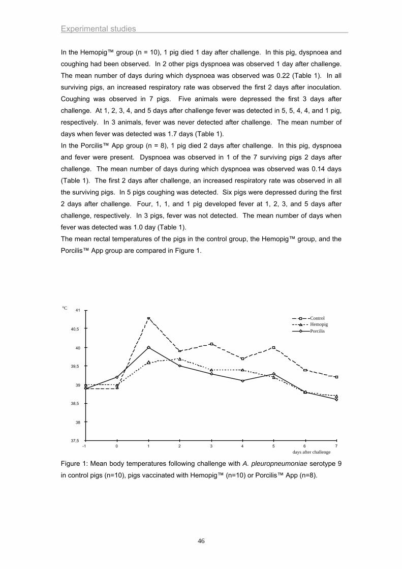
Experimental studies
In the Hemopig™ group (n = 10), 1 pig died 1 day after challenge. In this pig, dyspnoea and
coughing had been observed. In 2 other pigs dyspnoea was observed 1 day after challenge.
The mean number of days during which dyspnoea was observed was 0.22 (Table 1). In all
surviving pigs, an increased respiratory rate was observed the first 2 days after inoculation.
Coughing was observed in 7 pigs. Five animals were depressed the first 3 days after
challenge. At 1, 2, 3, 4, and 5 days after challenge fever was detected in 5, 5, 4, 4, and 1 pig,
respectively. In 3 animals, fever was never detected after challenge. The mean number of
days when fever was detected was 1.7 days (Table 1).
In the Porcilis™ App group (n = 8), 1 pig died 2 days after challenge. In this pig, dyspnoea
and fever were present. Dyspnoea was observed in 1 of the 7 surviving pigs 2 days after
challenge. The mean number of days during which dyspnoea was observed was 0.14 days
(Table 1). The first 2 days after challenge, an increased respiratory rate was observed in all
the surviving pigs. In 5 pigs coughing was detected. Six pigs were depressed during the first
2 days after challenge. Four, 1, 1, and 1 pig developed fever at 1, 2, 3, and 5 days after
challenge, respectively. In 3 pigs, fever was not detected. The mean number of days when
fever was detected was 1.0 day (Table 1).
The mean rectal temperatures of the pigs in the control group, the Hemopig™ group, and the
Porcilis™ App group are compared in Figure 1.
37,5
38
38,5
39
39,5
40
40,5
41
-1 0 1 2 3 4 5 6days after challenge
°C
7
ControlHemopigPorcilis
Figure 1: Mean body temperatures following challenge with A. pleuropneumoniae serotype 9
in control pigs (n=10), pigs vaccinated with Hemopig™ (n=10) or Porcilis™ App (n=8).
46

Experimental studies
Necropsy findings
At necropsy, a hæmorrhagic necrotizing pneumonia and fibrinous pleuritis were found in all
piglets of the control group. The percentages of affected lung tissue varied from 20% to 73%
(mean 34.4%; Table 1).
Lung lesions were not observed in 3 animals of the Hemopig™ group. In the pig that died,
58% of the lungs were affected. In the other animals that developed lung pathology, the
percentage of affected lung tissue varied from 2 to 56% (mean: 16.1%; Table 1). The lung
lesions were characterized by hæmorrhagic necrotizing pneumonia.
In the Porcilis™ App group, lung lesions were not observed in 3 animals. In the pig that died,
51% of the lung tissue was affected. In the other animals with lung lesions, the percentage of
affected lung tissue varied between 10% to 35% (mean: 16.9%; Table 1). Lesions were
similar to those described above for the Hemopig™ group.
Bacteriology
The results are summarized in tables 1 and 2. In the control pigs, A. pleuropneumoniae was
isolated from all lung lobes with lesions. The logarithmic mean bacterial titre of the caudal
lobes was 1.4x10⁶ CFU/g (Table 1). A. pleuropneumoniae was isolated from the tonsils of 3
pigs. The challenge strain could not be isolated from any of the other samples.
A. pleuropneumoniae was not isolated from the lungs of 3 animals vaccinated with
Hemopig™. The logarithmic mean number of CFU/g lung tissue of the caudal lung lobes was
2.2x10³. The strain could also be isolated from the tonsils, tracheobronchial lymph nodes,
and spleen of the pig that died. A. pleuropneumoniae was not isolated from the other
samples.
A. pleuropneumoniae was not isolated from the lungs of 3 animals vaccinated with Porcilis™
App. The logarithmic mean bacterial titre of the caudal lobes was 4.8x10³ CFU/g in this
group. A. pleuropneumoniae was isolated from the tonsils of the pig that died. A.
pleuropneumoniae was not isolated from other samples.
Serology
Antibodies against Apx I, Apx II, and Apx III were not detected in sera from pigs of the control
group and in sera from vaccinated pigs collected at the time of first and second vaccination.
Antibody titres against Apx I and II at the time of challenge of the vaccinated pigs are
presented in table 2. It can be seen that antibody titres against Apx I and II were either low or
absent. In none of the animals were antibodies against Apx III detected.
Statistical analysis
Significant differences (p <0.05) between the control group and both vaccinated groups were
found for the mean number of days during which dyspnoea was observed, the mean number
of days during which fever was detected, the mean percentage of affected lung tissue, and
47
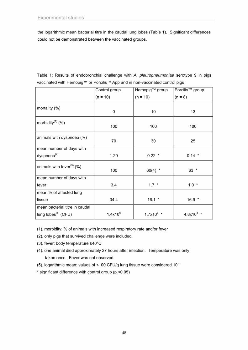
Experimental studies
the logarithmic mean bacterial titre in the caudal lung lobes (Table 1). Significant differences
could not be demonstrated between the vaccinated groups.
Table 1: Results of endobronchial challenge with A. pleuropneumoniae serotype 9 in pigs
vaccinated with Hemopig™ or Porcilis™ App and in non-vaccinated control pigs
Control group
(n = 10)
Hemopig™ group
(n = 10)
Porcilis™ group
(n = 8)
mortality (%)
0
10
13
morbidity(1) (%)
100
100
100
animals with dyspnoea (%)
70
30
25
mean number of days with
dyspnoea(2)
1.20
0.22 *
0.14 *
animals with fever(3) (%)
100
60(4) *
63 *
mean number of days with
fever
3.4
1.7 *
1.0 *
mean % of affected lung
tissue
34.4
16.1 *
16.9 *
mean bacterial titre in caudal
lung lobes(5) (CFU)
1.4x10⁶
1.7x103 *
4.8x103 *
(1). morbidity: % of animals with increased respiratory rate and/or fever
(2). only pigs that survived challenge were included
(3). fever: body temperature ≥40°C
(4). one animal died approximately 27 hours after infection. Temperature was only
taken once. Fever was not observed.
(5). logarithmic mean: values of <100 CFU/g lung tissue were considered 101
* significant difference with control group (p <0.05)
48

Experimental studies
49
Experimental studies
DISCUSSION
In the present study, an endobronchial challenge model was used. An advantage of this
method is that it delivers an exact number of bacteria to the lungs, allowing standardization of
experimental infections.
All control pigs developed acute disease and at necropsy, macroscopic pulmonary lesions
typical of porcine pleuropneumonia were observed. None of these animals died, indicating
that the challenge model was of planned severity.
The clinical signs of disease persisted for a shorter time in the vaccinated than in the control
animals. Furthermore, lesions were more severe and the mean bacterial titre in lung tissue
was higher in control animals, indicating partial protection in both vaccinated groups.
Although both vaccines contained Apx toxins, no or only low neutralizing antibody titres were
detected in vaccinated animals. The reason for this finding is not clear. Interference with
maternally derived antibodies can most probably be excluded, since the first vaccination was
carried out at 19 weeks of age and antibodies against Apx toxins were not detected at the
time of first vaccination. It is possible that the assay used here is of limited sensitivity, but in
previous studies with the same assay, high toxin-neutralizing titres were detected in pigs
infected with virulent A. pleuropneumoniae strains (Haesebrouck et al., 1996). In the latter
studies, animals were completely protected against challenge with the homologous serotype.
Further research is necessary to develop vaccines that induce high Apx toxin-neutralizing
antibody titres.
It has been shown that the Apx toxins are essential vaccine components to confer protection
against challenge with pathogenic A. pleuropneumoniae (Beaudet et al., 1994; Byrd et al.,
1992; Van den Bosch et al., 1992). Although both vaccines used in the present study
contained Apx toxins, they provided only partial protection against challenge carried out 3
weeks after the second vaccination. These results and the results of previous studies
indicate that other bacterial components may also play a role in protection (Haesebrouck et
al., 1996) and/or that higher titres are required for protection. It is possible that antibodies
against the Apx toxins decrease the severity of the disease and allow animals to recover
faster but do not prevent the initial infection. This might require antibodies against other
bacterial compounds.
In the present studies, the protection obtained with the Porcilis™ App vaccine was lower than
the protection described by Kobisch and Van den Bosch (1992) and Van den Bosch et al.
(1992). In the latter studies, SPF pigs were used whereas in the study described here,
conventional pigs were used. It is possible that protection induced in conventional pigs is
more variable than that induced in SPF pigs.
In only 5 of the 28 animals was the challenge strain isolated from tonsils. The isolation of A.
pleuropneumoniae from tonsils, however, is difficult. The media used for the isolation are
often overgrown by the abundant microflora present in the tonsils. This complicates visual
identification.
50
Experimental studies
REFERENCES
Beaudet, R., McSween, G., Boulay, G., Rousseau, P., Bisaillon, J.G., Descoteaux ,
J.P., Ruppanner, R., 1994. Protection of mice and swine against infection with
Actinobacillus pleuropneumoniae by vaccination. Vet. Microbiol. 39, 71-81.
Byrd, W., Harmon, B.G., Kadis, S., 1992. Protective efficacy of conjugate vaccines
against experimental challenge with porcine Actinobacillus pleuropneumoniae.
Vet Immunol. Immunopathol. 34, 307-24.
Dom, P., Haesebrouck F., 1992. Comparitive virulence of NAD dependent and NAD
independent Actinobacillus pleuropneumoniae strrains. J. Vet. Med. 39, 303-
306.
Dom, P., Haesebrouck, F., Kamp, E.M., Smits, M.A., 1994. NAD-independent
Actinobacillus pleuropneumoniae strains: production of RTX toxins and
interactions with porcine phagocytes. Vet. Microbiol. 39, 205-18.
Frey, J., Bosse, J.T., Chang, Y.F., Cullen, J.M., Fenwick, B., Gerlach, G.F., Gygi, D.,
Haesebrouck, F., Inzana, T.J., Jansen, R., Kamp, E.M., Macdonald, J.,
MacInnes, J.I., Mittal, K.R., Nicolet, J., Rycroft, A., Segers, R.P.A.M., Smits,
M.A., Stenbaek, E., Struck, D.K., Van den Bosch, J.F., Willson, P.J., Young, R.,
1993. Actinobacillus pleuropneumoniae-RTX-toxins: Uniform designation of
haemolysins, cytolysins, pleurotoxin and their genes. J. Gen. Microbiol. 139,
1723-1728.
Fenwick, B., Henry, S., 1994. Porcine pleuropneumonia. J. Am. Vet. Med. Assoc. 204,
1334-1340.
Frey, J., Beck, M., Nicolet, J., 1994. RTX-toxins of Actinobacillus pleuropneumoniae .
Zbl. Bakt. Suppl. 24, 322-332.
Haesebrouck, F., Van de Kerkhof, A., Dom, P., Chiers, K., Ducatelle, R., 1996. Cross-
protection between Actinobacillus pleuropneumoniae biotypes-serotypes in pigs.
Vet. Microbiol. 52, 277-284.
Hannan, P.C.T., Bhogal, B.S., Fish, J.P., 1982. Tylosin tartrate and tiamutilin effects on
experimental piglet pneumonia induced with pneumonic pig lung homogenate
containing mycoplasmas, bacteria and viruses. Res. Vet. Sci. 33, 76-88.
Kamp, E.M., Popma, J.K., Anakotta, J., Smits, M.A., 1991. Identification of hemolytic
and cytotoxic proteins of Actinobacillus pleuropneumoniae by use of monoclonal
antibodies. Infect. Immun. 59, 3079-3085.
Kobisch, M., Van Den Bosch, J.F., 1992. Efficacy of an Actinobacillus
pleuropneumoniae subunit vaccine. Proc. 12th International Pig Veterinary
Society Congress, The Hague, Netherlands, 216.
Martelli, P., Guadagnini, P.F., Foccoli, E., Ballarini, G., 1996. Efficacy of an
Actinobacillus pleuropneumoniae subunit vaccine in the control of
51
Experimental studies
pleuropneumonia: a field trial. Proc. 14th International Pig Veterinary Society
Congress,Bologna, Italy, 214.
Nicolet, J., 1992. Actinobacillus pleuropneumoniae. In: A.D. Leman, B. Straw, W.L.
Mengeling, S. D'Allaire and D.J. Taylor (Eds.). Diseases of swine. Ames, Iowa
State University Press, 401-8.
Pommier, P., Ridremont, B., Wessel-Robert, S., Keïta, A., 1996. Field study into
efficacy of a new Actinobacillus pleuropneumoniae subunit vaccine. Proc. 14th
International Pig Veterinary Society Congress,Bologna, Italy, 206.
Smits, M.A., Briaire, J., Jansen, R., Smith, H.E., Kamp, E.M., Gielkens, A.L.J., 1991.
Cytolysins of Actinobacillus pleuropneumoniae serotype 9. Infect. Immun. 59,
4497-4504.
Valks, M.M.H., Nell, T., Van den Bosch, J.F., 1996. A clinical field trial in finishing pigs
to evaluate the efficacy of a new APP subunit vaccine. Proc. 14th International
Pig Veterinary Society Congress,Bologna, Italy, 208.
Van den Bosch, J.F., Jongenelen, I.M.C.A., Pubben, N.B., van Vugt, F.G.A., Segers,
R.P.A.M., 1992. Protection induced by a trivalent Actinobacillus
pleuropneumoniae subunit vaccine. Proc. 12th International Pig Veterinary
Society Congress, The Hague, Netherlands, 194.
52
Experimental studies
Effect of endobronchial challenge with Actinobacillus pleuropneumoniae serotype 9 of pigs vaccinated with a vaccine containing Apx toxins and transferrin-binding proteins
Ingrid Van Overbeke, Koen Chiers, Richard Ducatelle, Freddy Haesebrouck
Laboratory of Veterinary Bacteriology and Mycology and Laboratory of Veterinary Pathology,
Department of Pathology, Bacteriology and Poultry diseases, Faculty of Veterinary Medicine,
University of Ghent, Salisburylaan 133, B-9820 Merelbeke, Belgium
Journal of Veterinary Medicine Series B 48 (2001): 15-20.
53
Experimental studies
SUMMARY
The efficacy of a subunit vaccine containing the Apx toxins of Actinobacillus
pleuropneumoniae and transferrin binding proteins was determined. Ten pigs were
vaccinated twice with the vaccine. Eight control animals were injected twice with a saline
solution. Three weeks after the second vaccination, all pigs were endobronchially inoculated
with 106.5
colony-forming units (CFU) of an A. pleuropneumoniae serotype 9 strain. In the
vaccine group, none of the pigs died after inoculation. Only one pig of the control group
survived challenge. Surviving pigs were killed at 7 days after challenge. The mean
percentage of affected lung tissue was 64% in the control group and 17% in the vaccine
group. A. pleuropneumoniae was isolated from the lungs of all animals. The mean bacterial
titres of the caudal lung lobes were 5.0x10⁸ CFU/g in the control group and 3.0x10⁶ CFU/g in
the vaccine group. It was concluded that the vaccine induced partial protection against
severe challenge.
54
Experimental studies
INTRODUCTION
A. pleuropneumoniae causes porcine contagious pleuropneumonia, which is distributed world
wide and results in serious losses in the pig rearing industry. It also causes severe animal
suffering. The disease is characterized, in the acute stage, by a hemorrhagic necrotizing
pneumonia and fibrinous pleuritis. In the chronic stage, localized lung lesions and adhesive
pleuritis can be seen (Nicolet, 1992). For control of porcine pleuropneumonia, improving
housing conditions and climate is essential. To control outbreaks of this disease, vaccination
may be useful. Although the so-called “first generation vaccines” of whole-cell bacterins may
reduce mortality after infection with the homologous serotype, they generally do not confer
protection against challenge with heterologous serotypes (Fenwick and Henry, 1994). An
explanation for the limited protection might be the absence of secreted and certain bacteria-
associated virulence factors in the bacterins. So-called “second generation vaccines” i.e.
vaccines containing the A. pleuropneumoniae Apx toxins have become commercially
available. Among the different serotypes of A. pleuropneumoniae, three of these exotoxins
have been described and each serotype produces either one or two of them (Dom et al.,
1994; Frey et al., 1993; Frey et al., 1994; Kamp et al., 1991). In a recent study, we
demonstrated partial protection against endobronchial challenge in animals vaccinated twice
with two second generation vaccines (Chiers et al., 1998) . Field trials carried out in France
(Pommier et al., 1996), the Netherlands (Valks et al., 1996), Italy (Martelli et al., 1996), Spain
(López et al., 1998) and Norway (Lium et al., 1998) confirmed that vaccination with these
vaccines can result in reduction of clinical symptoms and lung lesions of acute and chronic
pleuropneumonia and improvement of performance (growth, feed conversion, cost of
medication).
Apx toxins are not the only antigens involved in protection against porcine pleuropneumonia.
Capsular antigens, cell wall lipopolysaccharides, outer membrane proteins, fimbriae and
transferrin binding proteins also play a role (Haesebrouck et al., 1997).
In the present study, the efficacy of a vaccine, containing several recombinant cell-associated
and secreted antigens of A. pleuropneumoniae including the Apx toxins and transferrin
binding proteins, was determined using a severe, well standardized challenge model (Dom
and Haesebrouck, 1992; Haesebrouck et al., 1996).
MATERIALS AND METHODS
Challenge strain
An A. pleuropneumoniae biotype 1-serotype 9 strain (N° 13261) was used (Smits et al.,
1991). Bacteria were grown for 6 hours (log phase of growth) on Columbia agar (Columbia
Agar Base, Lab M, Bury, Great Britain) supplemented with 3% horse serum, 0.03% NAD
(Sigma Chemical Co, St Louis, Mo, USA), and 5% yeast extract at 37°C in a humid
atmosphere with 5% CO2. Bacteria were harvested in phosphate-buffered saline solution
55
Experimental studies
(PBSS, pH 7.3), centrifuged at 400 x g for 20 minutes, and suspended in RPMI 1640
supplemented with 10% non-essential amino acids, 10% glutamine, 10% foetal calf serum,
and 1% sodium pyruvate. The suspension was checked for purity and the number of colony-
forming units (CFU) was determined by plating tenfold dilutions on Columbia agar
supplemented with 3% horse serum, 0.03% NAD, and 5% yeast extract. Bacterial
suspensions were stored overnight at 4°C. The next day, they were used in the experiments.
Pigs
In this study 18 pigs were used. All animals were obtained by using a medicated segregated
early weaning programme (Chiers et al., 1998). They were weaned at 18 days of age and
kept in isolation until used in the experiments.
Vaccine
An acellular pentavalent subunit vaccine prepared with recombinant antigens from A.
pleuropneumoniae was used (Pleurostar™, Biostar Inc., Saskatoon, Canada). It contains
transferrin binding proteins and Apx toxins.
Experimental design
At the age of 7 and 11 weeks, pigs were intramuscularly injected: 10 pigs with 2 ml of the
vaccine and 8 pigs with 2 ml of a saline solution. Three weeks after the second vaccination,
all pigs were experimentally infected. The pigs were anaesthetized with Zoletil® 100 (2.2
mg/kg tiletamine and 2.2 mg/kg zolazepam, Virbac, Louvain La Neuve, Belgium) and Xyl-M®
2% (4.4 mg/kg xylazin, VMD, Arendonk, Belgium). They were inoculated endobronchially
with 106.5
CFU of the challenge strain in 5 ml inoculum (Dom and Haesebrouck, 1992;
Haesebrouck et al., 1996). All pigs were examined clinically. Pigs that died were autopsied
immediately; those that survived the challenge were killed 7 days after inoculation. At
necropsy, the lungs were examined macroscopically and the percentage of affected lung
tissue was determined. For this purpose, a diagram was used that divides the lung into 74
equal triangles (Hannan et al., 1982). The percentage of affected lung tissue was determined
by summing up the triangles showing pneumonia and/or pleuritis divided by 74. Samples
from lungs, tracheobronchial lymph nodes, tonsils, liver, kidney, and spleen were taken for
bacteriological examination. Sera were collected before the first and the second vaccination
and before the challenge.
Clinical examination
The pigs were examined for signs of pneumonia, characterized by increased respiratory rate
(>40 inspirations/min), dyspnoea, sneezing, coughing, and the presence of bloody foam on
mouth and/or nostrils. Rectal temperature was measured 1 day before challenge inoculation,
just before inoculation, and during the 7 days after inoculation. Other indications of infection
were vomiting and a depressed appearance.
56

Experimental studies
Bacteriological examination
Twenty per cent (w/v) suspensions of the right and the left caudal lung lobes were made in
PBSS. The number of CFU was determined by plating tenfold dilutions of the suspensions on
Columbia agar supplemented with 3% equine serum, 0.03% NAD and 5% yeast extract.
Samples were also grown on Columbia agar supplemented with 5% bovine blood with a
Staphylococcus intermedius streak. Twenty per cent (w/v) suspensions of tonsils were made
in PBSS and inoculated onto blood agar with S. intermedius. Suspected colonies were
subcultured on the same medium. Samples from lung lesions, tracheobronchial lymph nodes,
liver, kidney and spleen were tested for the presence of A. pleuropneumoniae by making an
incision in these tissues and taking a sample with an inoculation loop. These samples were
also inoculated onto blood agar with S. intermedius.
Serology
All pig sera were tested for presence of neutralising antibodies against Apx I, II and III using a
bioassay based on neutral red uptake by viable pulmonary alveolar macrophages, as
described previously (Dom et al., 1994).
Statistical analysis
Statistical analysis was performed on the following variables: mortality, morbidity (i.e.
percentage of animals with dyspnoea and/or fever), percentage of animals with fever,
percentage of animals with dyspnoea, mean percentage of affected lung tissue and
logarithmic mean of bacterial titre in caudal lung lobes. Fisher’s exact test was used to
compare proportions between groups. The mean percentages of affected lung tissue were
compared using the non-parametric Kruskal-Wallis test and the mean bacterial titres in caudal
lung lobes were compared using an ANOVA test.
RESULTS
Results are summarized in Table 1.
Clinical examination
None of the animals showed disease signs before challenge and at the time of challenge. In
the control group (n=8), six pigs were found dead 20 h after inoculation. One pig died 48h
after inoculation. In this pig, fever, dyspnoea, coughing and depression were present. The
remaining pig presented fever (>40°C), increased respiratory rate and coughing during 6 days
after challenge and was depressed on day 2.
In the vaccinated group (n=10), none of the pigs died after inoculation. In 1, 1, 4 and 1 pig,
fever was detected during 1, 2, 5 and 6 days after challenge, respectively. The mean number
of days during which fever was observed was 3.1. In 4, 1 and 1 pigs, increased respiratory
rate was observed during 1, 2 and 3 days, respectively. None of the animals showed
57
Experimental studies
dyspnoea. In four pigs occasional coughing was observed. One animal with increased
respiratory rate was depressed.
Necropsy findings
In all animals lung lesions, when present, were characterized by a haemorrhagic necrotizing
pneumonia and fibrinous pleuritis .
In 7 animals of the control group more than 45% of the lung tissue and in one animal 31% of
the lung tissue was affected (variation 31-88%; mean: 64%).
In eight animals of the vaccinated group the % of affected lung tissue was lower than 20%. In
two animals more than 45% of the lung tissue was affected. The percentages of affected lung
tissue varied from 4% to 53% (mean 17%).
Bacteriology
The results are summarized in Table 1.
In the control pigs, A. pleuropneumoniae was isolated from all lung lobes with lesions. The
number of CFU/gram lung tissue of the caudal lobes varied between 4.0 x 10⁶ and 4.5 x 10⁹,
with a logarithmic mean of 5.0 x 10⁸. The challenge strain was isolated from the tonsils and
tracheobronchial lymphnodes of 8 and 5 pigs, respectively. A. pleuropneumoniae was not
isolated from liver, spleen or kidney of any animals in this group.
A. pleuropneumoniae was isolated from all lung lobes with lesions and from all tonsils of the
vaccinated animals. The number of CFU/gram lung tissue of the caudal lobes varied between
2.7 x 10⁴ and 1.1 x 10⁸, with a logarithmic mean of 3.0 x 10⁶. A. pleuropneumoniae was not
isolated from tracheobronchial lymphnodes, liver, spleen or kidney of any animals in this
group.
Serology
At the time of first vaccination, antibody titres against Apx I, II and III varied between <20 and
80. At the time of second vaccination, most animals were negative for antibodies against the
Apx toxins. Low titres (20) were detected in 2 and 3 animals of the control and vaccinated
group, respectively. Antibody titres against Axp I, II and III at the time of challenge varied
between <20 and 40.
Statistical analysis
The vaccinated group differed significantly (p<0.05) from the control group for mortality,
percentage of animals with dyspnoea, percentage of affected lung tissue and logarithmic
mean of bacterial titre in the caudal lung lobes.
58
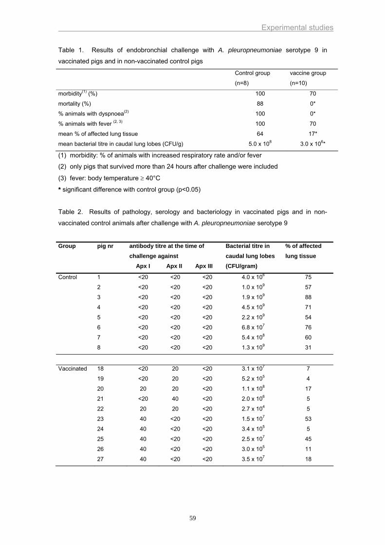
Experimental studies
Table 1. Results of endobronchial challenge with A. pleuropneumoniae serotype 9 in
vaccinated pigs and in non-vaccinated control pigs
Control group
(n=8)
vaccine group
(n=10)
morbidity(1) (%) 100 70
mortality (%) 88 0*
% animals with dyspnoea(2) 100 0*
% animals with fever (2, 3) 100 70
mean % of affected lung tissue 64 17*
mean bacterial titre in caudal lung lobes (CFU/g) 5.0 x 10⁸ 3.0 x 106*
(1) morbidity: % of animals with increased respiratory rate and/or fever
(2) only pigs that survived more than 24 hours after challenge were included
(3) fever: body temperature ≥ 40°C
* significant difference with control group (p<0.05)
Table 2. Results of pathology, serology and bacteriology in vaccinated pigs and in non-
vaccinated control animals after challenge with A. pleuropneumoniae serotype 9
Group pig nr antibody titre at the time of challenge against Apx I Apx II Apx III
Bacterial titre in caudal lung lobes (CFU/gram)
% of affected lung tissue
Control 1
2
3
4
5
6
7
8
<20
<20
<20
<20
<20
<20
<20
<20
<20
<20
<20
<20
<20
<20
<20
<20
<20
<20
<20
<20
<20
<20
<20
<20
4.0 x 10⁶
1.0 x 10⁹
1.9 x 10⁹
4.5 x 10⁹
2.2 x 10⁹
6.8 x 10⁷
5.4 x 10⁸
1.3 x 10⁹
75
57
88
71
54
76
60
31
Vaccinated 18
19
20
21
22
23
24
25
26
27
<20
<20
20
<20
20
40
40
40
40
40
20
20
20
40
20
<20
<20
<20
<20
<20
<20
<20
<20
<20
<20
<20
<20
<20
<20
<20
3.1 x 10⁷
5.2 x 10⁵
1.1 x 10⁸
2.0 x 10⁶
2.7 x 10⁴
1.5 x 10⁷
3.4 x 10⁵
2.5 x 10⁷
3.0 x 10⁵
3.5 x 10⁷
7
4
17
5
5
53
5
45
11
18
59
Experimental studies
DISCUSSION
Intranasal inoculation or aerosol exposure closely resemble the natural infection route of A.
pleuropneumoniae. These methods are difficult to standardize, however, because coughing,
sneezing, swallowing, breathing and mucociliary clearance can reduce the number of bacteria
that reach the alveolar region. In the present study, an endobronchial challenge model was
used. An advantage of this model is that it delivers an exact number of bacteria into the
bronchi, allowing standardization of experimental infections. Furthermore, this method allows
us to bring the bacteria directly close to the multiplication site of A. pleuropneumoniae namely
the alveoli and bronchioli (Haesebrouck et al., 1997). A possible disadvantage is that the
challenge strain is applicated not all over the lung but given more focal. Endobronchial
challenge is therefore more likely to break through low level immunity in the respiratory tract
than a more natural challenge route.
Seven of the 8 control animals died within 48 hours after infection. This demonstrates that a
severe challenge model was used.
None of the vaccinated animals died and none of them developed dyspnoea. Furthermore,
the mean % of affected lung tissue and the mean bacterial titre in the caudal lung lobes were
significantly lower than in the control animals. This indicates that this vaccine induced partial
protection against severe challenge.
It has been shown that the Apx toxins are essential vaccine components to confer protection
against challenge with A. pleuropneumoniae (Beaudet et al., 1994; Byrd et al., 1992; Van den
Bosch et al., 1992). However, in a previous study using a milder challenge model resulting in
no mortality in non-vaccinated control pigs, animals vaccinated with vaccines containing Apx
toxins were only partially protected against A. pleuropneumoniae serotype 9. It was
suggested that antibodies against other bacterial components are also required for protection
(Chiers et al., 1998). The vaccine used in the present studies also contained transferrin
binding proteins and induced significant protection against a very severe challenge. This
finding is in agreement with a study performed by Rossi et al. (1992). They found that pigs
vaccinated with a recombinant protein subunit vaccine containing Apx II toxin and a
transferrin binding protein of an A. pleuropneumoniae serotype 7 strain showed only minimal
clinical signs or lung lesions after a homologous challenge.
Although the vaccine contained Apx toxins, only low neutralizing antibody titres were detected
in the vaccinated animals. The reason for this finding is not clear. Interference with
maternally derived antibodies can not be excluded, since the first vaccination was carried out
at 7 weeks of age and low antibody titres against Apx toxins were detected at the time of first
vaccination. Furthermore, it is possible that the assay used here was of limited sensitivity,
although in previous studies with the same assay, high toxin-neutralizing titres were detected
in pigs infected with virulent A. pleuropneumoniae strains (Haesebrouck et al., 1996). In the
latter studies, animals were completely protected against challenge with the homologous
serotype. Further research is necessary to develop vaccines that induce high Apx toxin-
neutralizing antibody titres.
60

Experimental studies
In the present studies, animals were only challenged with a serotype 9 strain. Since Apx
toxins and transferrin binding proteins were included in the vaccine, protection against other
serotypes can be expected (Van den Bosch et al., 1992; Wilke et al., 1997). This should,
however, be confirmed in challenge studies with other serotypes.
61
Experimental studies
REFERENCES
Beaudet, R., McSween, G., Boulay, G., Rousseau, P., Bisaillon, J. G., Descoteaux, J.
P., Ruppanner, R., 1994. Protection of mice and swine against infection with
Actinobacillus pleuropneumoniae by vaccination. Vet. Microbiol. 39, 71-81.
Byrd, W., Harmon, B. G., Kadis, S., 1992. Protective efficacy of conjugate vaccines
against experimental challenge with porcine Actinobacillus pleuropneumoniae.
Vet. Immunol. Immunopathol. 34, 307-24.
Chiers, K., Van Overbeke, I., De Laender, P., Ducatelle, R., Carel, S. Haesebrouck , F.,
1998. Effects of endobronchial challenge with Actinobacillus pleuropneumoniae
serotype 9 of pigs vaccinated with inactivated vaccines containing the Apx
toxins. Vet. Quart. 20, 65-9.
Dom, P., Haesebrouck, F., 1992. Comparitive virulence of NAD dependent and NAD
independent Actinobacillus pleuropneumoniae strrains. J. Vet. Med. 39, 303-6.
Dom, P., Haesebrouck, F., Kamp, E. M., Smits, M. A., 1994. NAD-independent
Actinobacillus pleuropneumoniae strains: production of RTX toxins and
interactions with porcine phagocytes. Vet. Microbiol. 39, 205-18.
Fenwick, B., Henry, S., 1994. Porcine pleuropneumoniae. J. Am. Vet. Med. Assoc.
204, 1334-1340.
Frey, J., Bosse, J. T., Chang, Y. F., Cullen, J. M., Fenwick, B., Gerlach, G. F., Gygi, D.,
Haesebrouck, F., Inzana, T. J., Jansen, R., Kamp, E. M., Macdonald, J.,
MacInnes, J. I., Mittal, K. R., Nicolet, J., Rycroft, A., Segers, R. P. A. M., Smits,
M. A., Stenbaek, E., Struck, D. K., Van den Bosch, J. F., Willson, P. J., Young,
R., 1993. Actinobacillus pleuropneumoniae-RTX-toxins: Uniform designation of
haemolysins, cytolysins, pleurotoxin and their genes. J. Gen. Microbiol. 139,
1723-8.
Frey, J., Beck, M., Nicolet, J., 1994. RTX-toxins of Actinobacillus pleuropneumoniae.
Zbl. Bakt. Suppl. 24, 322-32.
Haesebrouck, F., Van de Kerkhof, A., Dom, P., Chiers, K., Ducatelle R., 1996. Cross
protection between Actinobacillus pleuropneumoniae biotypes-serotypes in pigs.
Vet. Microbiol. 52, 277-84.
Haesebrouck, F., Chiers, K., Van Overbeke, I., Ducatelle, R., 1997. Actinobacillus
pleuropneumoniae infections in pigs: the role of virulence factors in pathogenesis
and protection. Vet. Microbiol. 58, 239-249.
Hannan, P. C. T., Bhogal, B. S., Fish, J. P., 1982. Tylosin tartrate and tiamutilin effects
on experimental piglet pneumonia induced with pneumonic pig lung homogenate
containing mycoplasmas, bacteria and viruses. Res. Vet. Sci. 33, 76-88.
62
Experimental studies
Kamp, E. M., Popma, J. K., Anakotta, J., Smits, M. A., 1991. Identification of hemolytic
and cytotoxic proteins of Actinobacillus pleuropneumoniae by use of monoclonal
antibodies. Infect. Immun. 59, 3079-85.
Lium, B., Brun, E., Fjetland, O., Lund, A., 1998. A field study of the efficacy of an
Actinobacillus pleuropneumoniae subunit vaccine in the control of
pleuropneumonia caused by Actinobacillus serotype 6. Proc. 15th International
Pig Veterinary Society Congress, Birmingham, England, 256.
López, J. V., Font, J., Prat, M., Cantin, C., 1998. Technical and economic efficacy of
Porcilis App: a field trial. Proc. 15th International Pig Veterinary Society
Congress, Birmingham, England, 264.
Martelli, P., Guadagnini, P. F., Foccoli, E., Ballarini, G., 1996. Efficacy of an
Actinobacillus pleuropneumoniae subunit vaccine in the control of
pleuropneumonia: a field trial. Proc. 14th International Pig Veterinary Society
Congress, Bologna, Italy, 214.
Nicolet, J., 1992: Actinobacillus pleuropneumoniae, Diseases of swine. A.D. Leman, B.
Straw, W.L. Mengeling, S. D'Allaire and D.J. Taylor (Eds.). Ames, Iowa Stae
University Press, 401-8.
Pommier, P., Ridremont, B., Wessel-Robert, S., Keïta, A., 1996. Field study into
efficacy of a new Actinobacillus pleuropneumoniae subunit vaccine. Proc. 14th
International Pig Veterinary Society Congress, Bologna, Italy, 206.
Rossi, C.A., Anderson, C., Gerlach, G.F., Klashinsky, S., Potter, A.A., Willson, P.J.,
1992. Immunization of pigs against Actinobacillus pleuropneumoniae with two
recombinant protein preparations. Vaccine 10, 512-518.
Smits, M.A., Briaire, J., Jansen, R., Smith, H.E., Kamp, E.M., Gielkens, A. L. J., 1991.
Cytolysins of Actinobacillus pleuropneumoniae serotype 9. Infect. Immun. 59,
4497-4504.
Valks, M. M. H., Nell, T., Van den Bosch, J. F., 1996. A clinical field trial in finishing
pigs to evaluate the efficacy of a new APP subunit vaccine. Proc. 14th
International Pig Veterinary Society Congress, Bologna, Italy, 208.
Van den Bosch, J.F., Jongenelen, I.M.C.A., Pubben, N.B., van Vugt, F.G.A., Segers,
R.P.A.M., 1992. Protection induced by a trivalent Actinobacillus
pleuropneumoniae subunit vaccine. Proc. 12th International Pig Veterinary
Society Congress, The Hague, The Netherlands, 194.
Wilke, M., Franz, B., Gerlach G.F., 1997. Characterization of a large transferrin-binding
protein from Actinobacillus pleuropneumoniae serotype 7. J. Vet. Med. B 44, 73-
86.
63

Experimental studies
64
Experimental studies
CHAPTER 2
ADHESION OF ACTINOBACILLUS
PLEUROPNEUMONIAE TO PORCINE ALVEOLAR EPITHELIAL CELLS IN VITRO AND IN VIVO
65

Experimental studies
66
Experimental studies
Characterization of the in vitro adhesion of Actinobacillus pleuropneumoniae to swine alveolar epithelial cells
Ingrid Van Overbeke1, Koen Chiers1, Gerard Charlier2, Isabel Vandenberghe3, Jozef
Van Beeumen3, Richard Ducatelle1, Freddy Haesebrouck1
1Laboratory of Veterinary Bacteriology and Mycology and Laboratory of Veterinary Pathology,
Department of Pathology, Bacteriology and Poultry diseases, Faculty of Veterinary Medicine,
University of Ghent, Salisburylaan 133, B-9820 Merelbeke, Belgium
2Veterinary Agrochemical Research Institute (CODA), Groeselenberg 99, B 1180 Brussels,
Belgium
3 Department of Biochemistry, Fysiology and microbiology, Laboratory of Protein Biochemistry
and Protein Engineering, Faculty of Science, University of Ghent, K.L. Ledeganckstraat 35, B
9000 Ghent, Belgium
Veterinary Microbiology 88 (2002): 59-74.
67
Experimental studies
SUMMARY
A. pleuropneumoniae biovar 1 serotype 2, 5a, 9 and 10 strains were tested for their ability to
adhere to alveolar epithelial cells in culture. For the serotype 5a, 9 and 10 strains, optimal
adherence was observed after growth of bacterial cells in a NAD-restricted medium (0.001%
NAD). This condition was also associated with the expression of a 55 kDa outer membrane
protein and of fimbriae. For the serotype 2 strain, adherence and expression of fimbriae and
a 55 kDa outer membrane protein was less influenced by the growth conditions. The N-
terminal amino acid sequence of the 55 kDa outer membrane protein had no homology with
any known sequence, suggesting that it is an as yet unknown protein. Adherence capabilities
were significantly reduced following treatment of the bacteria with proteolytic enzymes or
heat. These findings suggest that proteins are involved in adhesion. The hydrophobic bond
breaking agent tetramethylurea was unable to inhibit the adherence of A. pleuropneumoniae
to alveolar epithelial cells. Treatment of the bacteria with sodium metaperiodate resulted in
lower adhesion scores for the serotype 2 and 9 strains but the inhibition of adhesion was
clearly lower than after treatment with proteolytic enzymes. This indicates that, besides
proteins, carbohydrates might also be involved in adhesion of A. pleuropneumoniae to
alveolar epithelial cells. The finding that inhibition of adhesion was very high when bacteria
were treated with a combination of sodium metaperiodate and pronase also suggests that
more than one adhesin is involved.
68
Experimental studies
INTRODUCTION
Actinobacillus pleuropneumoniae is a Gram-negative bacterium causing contagious
pleuropneumonia in pigs. Based on NAD requirements, A. pleuropneumoniae can be divided
into biovar 1 strains, which are NAD-dependent, and biovar 2 strains, which are NAD-
independent. So far 15 serotypes have been described (Blackall et al., 2002). Serotypes 1
and 5 are subdivided into 1a, 1b and 5a, 5b (Jolie et al., 1994; Nielsen, 1986).
In previous studies, it was shown that A. pleuropneumoniae adheres to the epithelium of the
alveoli or the cilia of the terminal bronchioli of experimentally infected pigs (Dom et al., 1994).
It has also been shown that A. pleuropneumoniae adheres to porcine tracheal rings
maintained in culture (Bélanger et al., 1990), to porcine frozen tracheal and lung sections
(Jacques et al., 1991), to erythrocytes from various animal species (Jacques et al., 1988), to
tonsillar epithelial cells (Chiers et al., 1999), to swine buccal epithelial cells (Hamer et al.,
1999) and to type III swine-lung collagen (Enriquez et al., 1999).
Adherence to host tissue is generally regarded as an important prerequisite for colonization
and virulence manifestation of bacteria (Ofek and Beachy, 1980). Adherence enables
colonization to occur and allows the bacterium to exert its pathogenic effects. Several surface
structures have been described in the family of Pasteurellaceae that are involved in adhesion,
including the capsule (Confer et al., 1995), fimbriae (Read et al., 1992), lipopolysaccharides
(Bélanger et al., 1990) and outer membrane proteins (Confer et al., 1995). Concerning A.
pleuropneumoniae, only few adhesion factors have been described. Lipopolysaccharides
seem to be involved in the in vitro adhesion to porcine tracheal rings (Bélanger et al., 1990)
and mucus (Bélanger et al., 1992; 1994). Recently, it was shown that the capsule is not
responsible for adherence to tracheal frozen sections (Jacques, 1999). Type 4 fimbriae have
been shown on A. pleuropneumoniae (Dom et al., 1994a; Zhang et al., 2000) but their role in
adherence needs to be elucidated.
Since in vivo studies indicated that adherence of A. pleuropneumoniae to alveolar epithelial
cells constitutes an important initial step in pathogenesis (Dom et al., 1994; Haesebrouck et
al., 1997), the association of the bacterium with these host cells was here further
characterized, using an in vitro adhesion model.
MATERIALS AND METHODS
Bacterial strains and culture conditions
Reference strains of A. pleuropneumoniae serotype 9 (13261) and 10 (D13039), as well as a
serotype 2 (266/7656) and 5a (1736) strain, both obtained from the lungs of pigs that died
during outbreaks of pleuropneumonia in Belgium (Dom et al., 1994b), were used in this study.
All 4 strains were NAD-dependent (biovar 1). Stock suspensions were stored at -70°C in
RPMI 1640 medium supplemented with 10% non-essential amino acids (Gibco Europe,
Merelbeke, Belgium), 10% glutamine, 10% foetal calf serum (FCS) and 1% sodium pyruvate
(Gibco) (leukocyte medium). To study the influence of culture conditions on adhesion,
69
Experimental studies
bacteria were grown on Columbia agar supplemented with 1% horse serum and 0.001% NAD
for 20 hours at 37°C (NAD-restricted medium), on Columbia agar supplemented with 3%
horse serum, 0.001% NAD and 5% yeast extract for 20 hours at 37°C (NAD-restricted
medium with yeast extract) or on Columbia agar supplemented with 3% horse serum, 0.03%
NAD and 5% yeast-extract for 6 hours at 37°C (NAD-rich medium). To study the expression
of surface antigens, bacteria were grown on the NAD-restricted and NAD-rich media
described above. In all other studies, bacteria were grown for 20 hours on the NAD-restricted
medium. All incubations were done in an atmosphere with 5% CO2. The number of colony
forming units (CFU) was determined by turbidimetry (optical density at 450 nm).
Adherence assay
Alveolar epithelial cells (AEC) were obtained as previously described (Van de Kerkhof et al.,
1996). For the adherence assay, 105 viable cells in 100 µl of supplemented Modified Eagle
Medium (MEM, Gibco) were added to wells of 24-well microtiter plates with coverslips and
incubated for 3 hours at 37°C in an atmosphere with 5% CO2. Then, 1 ml of supplemented
MEM was added to each well and the plates were further incubated for 2 days.
Subsequently, plates were washed once with phosphate buffered saline solution (PBSS) and
cells were fixed with 1 ml of methanol 100%. After washing the cells with distilled water,
adhesion tests were performed. In these tests, 108 CFU of the A. pleuropneumoniae strains
in 1 ml inoculum were added to the cultures. The microtiter plates were centrifuged at 110×g
for 10 minutes at 37°C and further incubated at 37°C for 30 minutes in an atmosphere with
5% CO2. The plates were washed 4 times with PBSS, stained with Haemacolor® staining
reagents (Merck, Darmstadt, Germany) and microscopically examined.
In each assay, one hundred cells were examined for the presence of adhering bacteria. The
following scores were used for each alveolar epithelial cell: score 0 = no adhesion, score 1 =
1 to 20 bacteria adhered to the cell, score 2 = more than 20 bacteria adhered to the cell. The
number of alveolar epithelial cells with score 0, 1 or 2 was determined.
Influence of culture conditions on adhesion to AEC and expression of capsule, fimbriae and outer membrane proteins
Influence of culture conditions on adhesion
To study the influence of the culture conditions on adhesion, adhesion tests were performed
with A. pleuropneumoniae serotype 2, 5a, 9 and 10 strains grown on either the NAD-restricted
medium, NAD-restricted medium with yeast extract or NAD-rich medium. These experiments
were performed at least 3 times.
Influence of culture conditions on expression of capsule
A. pleuropneumoniae serotype 2, 5a and 9 strains were grown on the NAD-restricted and the
NAD-rich medium. After incubation, bacteria were harvested, washed once with PBSS and
resuspended in PBSS at a concentration of 108 CFU/ml. In order to avoid collapse of the
70

Experimental studies
capsules during the electron microscopic examination, bacterial suspensions were exposed to
undiluted homologous antiserum for 1 hour at 4°C. The antisera were raised in rabbits
against whole bacterial cells. The titers of agglutinating antibodies of the rabbit antisera varied
between 1:8 and 1:32 (Russel W.M.S. and Burch R.L., 1959). Bacterial cells were then
suspended in cacodylate buffer (0.1M, pH 7.0) containing 5% glutaraldehyde and 0.15% (w/v)
ruthenium red. Fixation was done for 2 hours at 20°C. Thereafter, bacterial cells were
immobilized in 3% agar, washed five times in cacodylate buffer with 0.05% ruthenium red and
postfixed with 1% osmium tetroxide and 0.05% ruthenium red for 2 hours. Washings were
repeated as above, and the samples were dehydrated in a graded series of acetone washes.
All the solutions used in processing the specimen, from the wash after glutaraldehyde fixation
to dehydration with the 70% acetone solution, contained 0.05% ruthenium red. Samples were
then washed twice in propylene oxide and embedded in Spurr low-viscosity resin. Thin
sections were cut and stained with uranyl acetate and lead citrate. They were examined with
a transmission electron microscope (Philips 201) at an accelerating voltage of 60 kV. The
capsule thickness was measured on micrographs taken during the electron microscopic
observations. The cell wall thickness was measured between the inner edge of the capsule
layer and the outer leaflet of the plasma membrane.
Influence of culture conditions on expression of fimbriae
A. pleuropneumoniae serotype 2, 5a, 9 and 10 strains were grown on the NAD-restricted and
the NAD-rich medium, washed once in PBSS and then suspended in a minimal amount of
distilled water at a concentration of 108 CFU/ml. The cultures were examined for the
presence of fimbriae by negative staining and shadowing. For negative staining, a Formvar-
coated copper grid was floated on the surface of the bacterial suspension for 10 minutes.
The grid was then floated for 10 seconds on a solution of 2% uranyl acetate in distilled water.
After staining, the grid was washed twice and examined with a Philips 201 transmission
electron microscope (TEM). For shadowing, the bacterial suspension was absorbed on
carbon coated grids and rotary shadowed with platinum-carbon at a small angle (5-6°) with
the Balgers shadowing apparatus. The grid was examined with a Philips 201 TEM. The size
of the fimbriae was measured on the micrographs taken during electron microscopic
observations.
Influence of culture conditions on expression of outer membrane proteins
Sarcosyl insoluble outer membrane proteins (OMPs) of A. pleuropneumoniae serotype 2, 5a,
9 and 10 strains grown on the NAD-restricted and the NAD-rich medium were prepared
(Deneer and Potter, 1989). Briefly, bacteria were suspended in 100 ml of 10 mM HEPES
buffer (pH 7.5, Gibco, Ghent, Belgium) at a concentration of 2×10⁹ CFU/ml. Cells were
disrupted by ultrasound with 10 bursts, 20 seconds each, with cooling on ice. Debris was
removed by centrifugation at 2,000×g for 20 minutes and 50 ml 2% (w/v) sarcosyl (Sigma
Chemical Co, St Louis, Mo, USA) was added to the supernatant. After incubation for 10
minutes at room temperature, the outer membrane fraction was pelleted by centrifugation at
71
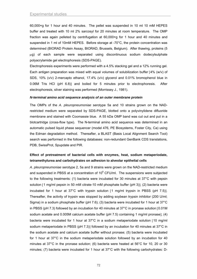
Experimental studies
60,000×g for 1 hour and 40 minutes. The pellet was suspended in 10 ml 10 mM HEPES
buffer and treated with 10 ml 2% sarcosyl for 20 minutes at room temperature. The OMP
fraction was again pelleted by centrifugation at 60,000×g for 1 hour and 40 minutes and
suspended in 1 ml of 10mM HEPES. Before storage at -70°C, the protein concentration was
determined (BIORAD Protein Assay, BIORAD, Brussels, Belgium). After thawing, proteins (5
μg) of each sample were separated using discontinuous sodium dodecylsulphate
polyacrylamide gel electrophoresis (SDS-PAGE).
Electrophoresis experiments were performed with a 4.5% stacking gel and a 12% running gel.
Each antigen preparation was mixed with equal volumes of solubilization buffer [4% (w/v) of
SDS, 10% (v/v) 2-mercapto ethanol, 17.4% (v/v) glycerol and 0.01% bromophenol blue in
0.06M Tris HCl (pH 6.8)] and boiled for 5 minutes prior to electrophoresis. After
electrophoresis, silver staining was performed (Morrissey J., 1981).
N-terminal amino acid sequence analysis of an outer membrane protein
The OMPs of the A. pleuropneumoniae serotype 5a and 10 strains grown on the NAD-
restricted medium were separated by SDS-PAGE, blotted onto a polyvinylidene difluoride
membrane and stained with Coomassie blue. A 55 kDa OMP band was cut out and put in a
blotcartridge (cross-flow type). The N-terminal amino acid sequence was determined in an
automatic pulsed liquid phase sequencer (model 476, PE Biosystems, Foster City, Ca) using
the Edman degradation method. Thereafter, a BLAST (Basis Local Alignment Search Tool)
search was performed in the following databases: non-redundant GenBank CDS translations,
PDB, SwissProt, Spupdate and PIR.
Effect of pretreatment of bacterial cells with enzymes, heat, sodium metaperiodate, tetramethylurea and carbohydrates on adhesion to alveolar epithelial cells
A. pleuropneumoniae serotype 2, 5a and 9 strains were grown on the NAD-restricted medium
and suspended in PBSS at a concentration of 108 CFU/ml. The suspensions were subjected
to the following treatments: (1) bacteria were incubated for 30 minutes at 37°C with pepsin
solution [1 mg/ml pepsin in 50 mM citrate-10 mM phosphate buffer (pH 3)]; (2) bacteria were
incubated for 1 hour at 37°C with trypsin solution [1 mg/ml trypsin in PBSS (pH 7.5)].
Thereafter, the activity of trypsin was stopped by adding soybean trypsin inhibitor (200 U/ml;
Sigma) in a sodium phosphate buffer (pH 7.6); (3) bacteria were incubated for 1 hour at 37°C
in PBSS (pH 7.3) followed by an incubation for 40 minutes at 37°C in pronase solution [0.01M
sodium acetate and 0.005M calcium acetate buffer (pH 7.5) containing 1 mg/ml pronase]; (4)
bacteria were incubated for 1 hour at 37°C in a sodium metaperiodate solution [10 mg/ml
sodium metaperiodate in PBSS (pH 7.3)] followed by an incubation for 40 minutes at 37°C in
the sodium acetate and calcium acetate buffer without pronase; (5) bacteria were incubated
for 1 hour at 37°C in the sodium metaperiodate solution followed by an incubation for 40
minutes at 37°C in the pronase solution; (6) bacteria were heated at 56°C for 10, 20 or 30
minutes; (7) bacteria were incubated for 1 hour at 37°C with the following carbohydrates: D-
72
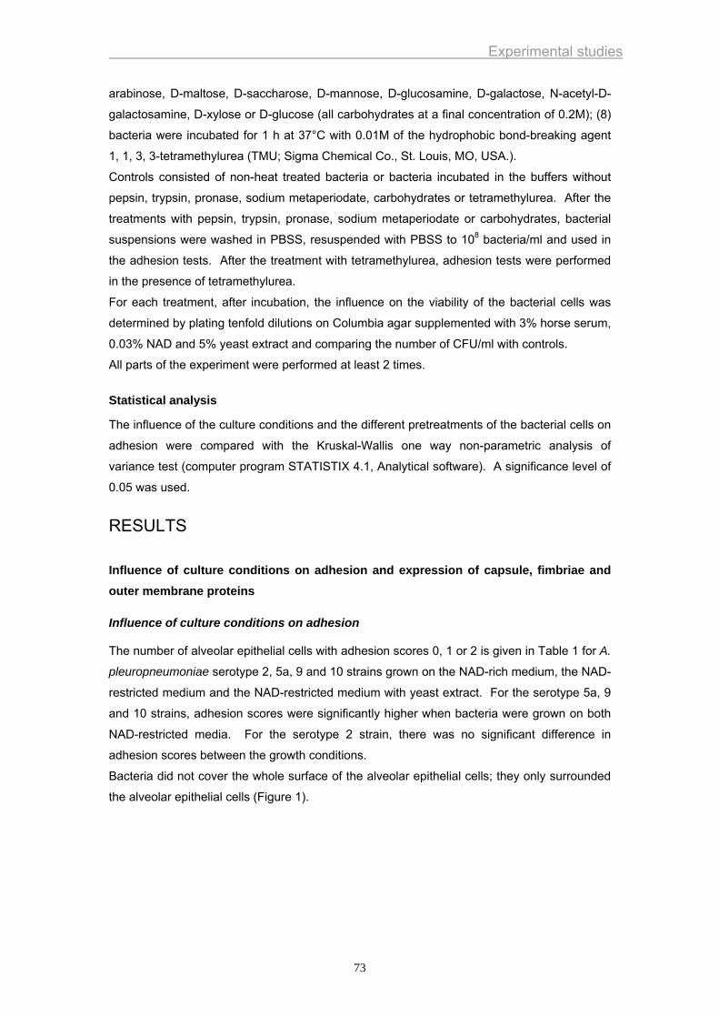
Experimental studies
arabinose, D-maltose, D-saccharose, D-mannose, D-glucosamine, D-galactose, N-acetyl-D-
galactosamine, D-xylose or D-glucose (all carbohydrates at a final concentration of 0.2M); (8)
bacteria were incubated for 1 h at 37°C with 0.01M of the hydrophobic bond-breaking agent
1, 1, 3, 3-tetramethylurea (TMU; Sigma Chemical Co., St. Louis, MO, USA.).
Controls consisted of non-heat treated bacteria or bacteria incubated in the buffers without
pepsin, trypsin, pronase, sodium metaperiodate, carbohydrates or tetramethylurea. After the
treatments with pepsin, trypsin, pronase, sodium metaperiodate or carbohydrates, bacterial
suspensions were washed in PBSS, resuspended with PBSS to 108 bacteria/ml and used in
the adhesion tests. After the treatment with tetramethylurea, adhesion tests were performed
in the presence of tetramethylurea.
For each treatment, after incubation, the influence on the viability of the bacterial cells was
determined by plating tenfold dilutions on Columbia agar supplemented with 3% horse serum,
0.03% NAD and 5% yeast extract and comparing the number of CFU/ml with controls.
All parts of the experiment were performed at least 2 times.
Statistical analysis
The influence of the culture conditions and the different pretreatments of the bacterial cells on
adhesion were compared with the Kruskal-Wallis one way non-parametric analysis of
variance test (computer program STATISTIX 4.1, Analytical software). A significance level of
0.05 was used.
RESULTS
Influence of culture conditions on adhesion and expression of capsule, fimbriae and outer membrane proteins
Influence of culture conditions on adhesion
The number of alveolar epithelial cells with adhesion scores 0, 1 or 2 is given in Table 1 for A.
pleuropneumoniae serotype 2, 5a, 9 and 10 strains grown on the NAD-rich medium, the NAD-
restricted medium and the NAD-restricted medium with yeast extract. For the serotype 5a, 9
and 10 strains, adhesion scores were significantly higher when bacteria were grown on both
NAD-restricted media. For the serotype 2 strain, there was no significant difference in
adhesion scores between the growth conditions.
Bacteria did not cover the whole surface of the alveolar epithelial cells; they only surrounded
the alveolar epithelial cells (Figure 1).
73

Experimental studies
74
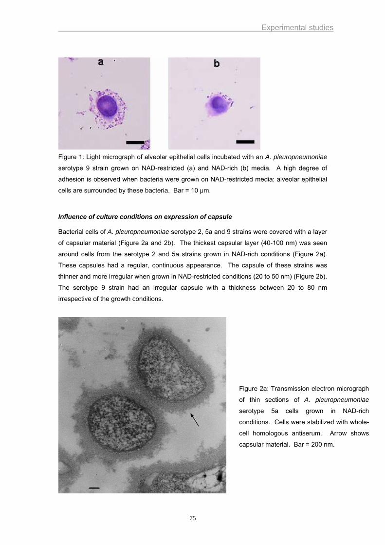
Experimental studies
Figure 1: Light micrograph of alveolar epithelial cells incubated with an A. pleuropneumoniae
serotype 9 strain grown on NAD-restricted (a) and NAD-rich (b) media. A high degree of
adhesion is observed when bacteria were grown on NAD-restricted media: alveolar epithelial
cells are surrounded by these bacteria. Bar = 10 µm.
Influence of culture conditions on expression of capsule
Bacterial cells of A. pleuropneumoniae serotype 2, 5a and 9 strains were covered with a layer
of capsular material (Figure 2a and 2b). The thickest capsular layer (40-100 nm) was seen
around cells from the serotype 2 and 5a strains grown in NAD-rich conditions (Figure 2a).
These capsules had a regular, continuous appearance. The capsule of these strains was
thinner and more irregular when grown in NAD-restricted conditions (20 to 50 nm) (Figure 2b).
The serotype 9 strain had an irregular capsule with a thickness between 20 to 80 nm
irrespective of the growth conditions.
Figure 2a: Transmission electron micrograph
of thin sections of A. pleuropneumoniae
serotype 5a cells grown in NAD-rich
conditions. Cells were stabilized with whole-
cell homologous antiserum. Arrow shows
capsular material. Bar = 200 nm.
75
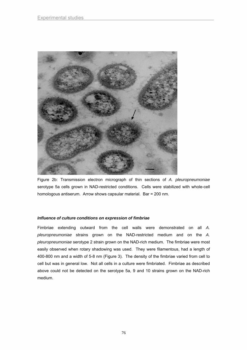
Experimental studies
Figure 2b: Transmission electron micrograph of thin sections of A. pleuropneumoniae
serotype 5a cells grown in NAD-restricted conditions. Cells were stabilized with whole-cell
homologous antiserum. Arrow shows capsular material. Bar = 200 nm.
Influence of culture conditions on expression of fimbriae
Fimbriae extending outward from the cell walls were demonstrated on all A.
pleuropneumoniae strains grown on the NAD-restricted medium and on the A.
pleuropneumoniae serotype 2 strain grown on the NAD-rich medium. The fimbriae were most
easily observed when rotary shadowing was used. They were filamentous, had a length of
400-800 nm and a width of 5-8 nm (Figure 3). The density of the fimbriae varied from cell to
cell but was in general low. Not all cells in a culture were fimbriated. Fimbriae as described
above could not be detected on the serotype 5a, 9 and 10 strains grown on the NAD-rich
medium.
76
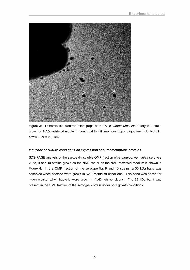
Experimental studies
Figure 3: Transmission electron micrograph of the A. pleuropneumoniae serotype 2 strain
grown on NAD-restricted medium. Long and thin filamentous appendages are indicated with
arrow. Bar = 200 nm.
Influence of culture conditions on expression of outer membrane proteins
SDS-PAGE analysis of the sarcosyl-insoluble OMP fraction of A. pleuropneumoniae serotype
2, 5a, 9 and 10 strains grown on the NAD-rich or on the NAD-restricted medium is shown in
Figure 4. In the OMP fraction of the serotype 5a, 9 and 10 strains, a 55 kDa band was
observed when bacteria were grown in NAD-restricted conditions. This band was absent or
much weaker when bacteria were grown in NAD-rich conditions. The 55 kDa band was
present in the OMP fraction of the serotype 2 strain under both growth conditions.
77
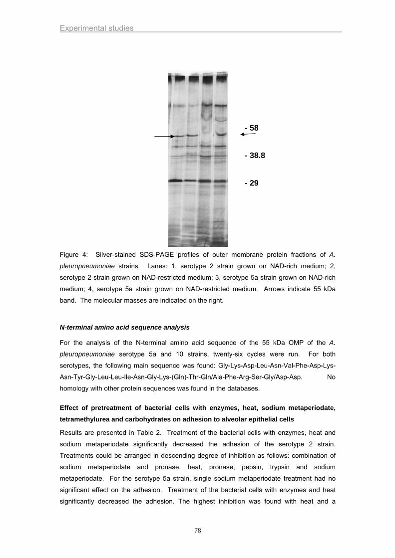
Experimental studies
- 58 - 38.8 - 29
Figure 4: Silver-stained SDS-PAGE profiles of outer membrane protein fractions of A.
pleuropneumoniae strains. Lanes: 1, serotype 2 strain grown on NAD-rich medium; 2,
serotype 2 strain grown on NAD-restricted medium; 3, serotype 5a strain grown on NAD-rich
medium; 4, serotype 5a strain grown on NAD-restricted medium. Arrows indicate 55 kDa
band. The molecular masses are indicated on the right.
N-terminal amino acid sequence analysis
For the analysis of the N-terminal amino acid sequence of the 55 kDa OMP of the A.
pleuropneumoniae serotype 5a and 10 strains, twenty-six cycles were run. For both
serotypes, the following main sequence was found: Gly-Lys-Asp-Leu-Asn-Val-Phe-Asp-Lys-
Asn-Tyr-Gly-Leu-Leu-Ile-Asn-Gly-Lys-(Gln)-Thr-Gln/Ala-Phe-Arg-Ser-Gly/Asp-Asp. No
homology with other protein sequences was found in the databases.
Effect of pretreatment of bacterial cells with enzymes, heat, sodium metaperiodate, tetramethylurea and carbohydrates on adhesion to alveolar epithelial cells
Results are presented in Table 2. Treatment of the bacterial cells with enzymes, heat and
sodium metaperiodate significantly decreased the adhesion of the serotype 2 strain.
Treatments could be arranged in descending degree of inhibition as follows: combination of
sodium metaperiodate and pronase, heat, pronase, pepsin, trypsin and sodium
metaperiodate. For the serotype 5a strain, single sodium metaperiodate treatment had no
significant effect on the adhesion. Treatment of the bacterial cells with enzymes and heat
significantly decreased the adhesion. The highest inhibition was found with heat and a
78

Experimental studies
combination of sodium metaperiodate and pronase followed by pronase, pepsin and trypsin.
For the serotype 9 strain, all treatments significantly inhibited the adherence. In descending
order, inhibition was observed with heat, combination of sodium metaperiodate and pronase,
pronase, pepsin, trypsin and sodium metaperiodate.
Tetramethylurea or carbohydrate treatment did not reduce the level of adhesion. Pronase,
trypsin, TMU and carbohydrate treatment of the bacterial cells did not have any effect on the
viability. Treatment of the bacteria with heat, pepsin and sodium metaperiodate resulted in a
decrease in viability.
79

Experimental studies
80
Experimental studies
DISCUSSION
In previous studies, it has been shown that A. pleuropneumoniae adheres to alveolar
epithelial cells of experimentally infected pigs (Dom et al., 1994 & 1995). Most probably,
adherence of A. pleuropneumoniae to lower respiratory tract epithelial cells constitutes an
important initial step in pathogenesis. Therefore, in the present work, porcine alveolar
epithelial cells were used to study the adhesion of A. pleuropneumoniae. The alveolar
epithelial cells were allowed to attach to coverslips whereafter they were fixed with methanol.
Fixation was used to prevent destruction of the alveolar epithelial cells by the Apx toxins of A.
pleuropneumoniae (Van de Kerkhof et al., 1996).
Advantages of using an in vitro system include reproducibility between assays, ease and
simplicity of experimentation and animal welfare. However, there are also several
disadvantages. Surfaces of in vitro cells may present proteins different from in vivo cells,
especially in terms of distribution and accessibility of the receptors. Notably, the surfactant
layer covering the alveolar epithelial cells in vivo is absent in the in vitro adhesion model. For
this reason, data obtained in in vitro studies should be confirmed by in vivo studies.
The functional expression of most bacterial adhesins is not an essential activity for the
survival of the bacteria. In fact, most organisms display differences in adhesive qualities
during growth. These qualities can be modulated by pH and composition and physical state
of growth medium (Ofek and Doyle, 1994). In this study, three growth media were used: a
medium containing 0.03% NAD which was defined as NAD-rich and two media containing
0.001% NAD which were defined as NAD-restricted. For the serotype 5a, 9 and 10 strains,
we found that growth on the NAD-restricted media resulted in a higher degree of adhesion,
indicating that adhesins are more expressed. NAD-restricted conditions may reflect the in
vivo situation. It is generally accepted that A. pleuropneumoniae is inhaled and enters the
lung alveoli directly via the trachea and bronchi. In the lumen of the lung alveoli, extracellular
NAD-concentrations most probably are very low, since, based on molecular weight and
charge, it is unlikely that leakage of NAD out of normal living cells occurs. The NAD-restricted
conditions in the lumen of lung alveoli may stimulate the expression of adhesins. Adhesion of
A. pleuropneumoniae to alveolar epithelial cells may lead to a local high concentration of Apx
toxins at the surface of host cells resulting in lysis of these cells (Van de Kerkhof et al., 1996)
and release of growth components in the environment which may favor multiplication of the
bacteria in lung tissue. The adhesion degree of bacteria belonging to the serotypes 5a, 9 and
10 strains grown for 6 hours on the NAD-rich medium was very low, indicating that adhesins
were less expressed or less exposed at the bacterial surface.
Since adhesins should be located at the surface of the bacteria, electron microscopic
examination and sodium dodecyl sulphate-polyacrylamide gel electrophoresis of outer
membrane proteins was performed to study the surface components of the bacteria. For the
serotype 5a, 9 and 10 strains, production of fimbriae and a 55 kDa outer membrane protein
were observed when bacteria were grown under NAD-restricted conditions. These conditions
81
Experimental studies
also resulted in highest in vitro adhesion scores to alveolar epithelial cells. This relationship
between adherence levels and specific cell surface components might indicate that these
components could play a role in A. pleuropneumoniae adherence to alveolar epithelial cells.
In contrast to the A. pleuropneumoniae serotype 5a, 9 and 10 strains, a correlation between
the growth conditions and the adhesion of the serotype 2 strain was not observed. This strain
exhibited a high degree of in vitro adhesion grown under NAD-restricted and NAD-rich
conditions. Furthermore, fimbriae and a 55 kDa OMP were expressed under both growth
conditions. This confirms that these components could play a role in in vitro adhesion.
Recently, fimbriae of A. pleuropneumoniae were characterized as type 4 fimbriae (Zhang et
al., 2000). Type 4 fimbriae have been described in several other Gram-negative bacteria
including Haemophilus influenzae, Moraxella bovis, Pasteurella multocida and Pseudomonas
aeruginosa. A large number of studies have shown that they mediate attachment of these
bacteria to epithelial cells in vivo and in vitro (Sato and Okinaga, 1987; Doig et al., 1988;
Paranchych and Frost, 1988; Starr et al., 1999). Attempts should be made to try to isolate
these fimbriae from A. pleuropneumoniae and determine their role in adhesion.
The outer membrane is an excellent compartment for anchoring and positioning adhesins to
the outer surface. It has been demonstrated that outer membrane proteins, unrelated to
fimbriae, can play a role in the adhesion to animal cells. Examples of such interaction have
been found in the adhesion reactions of Neisseria gonorrhoeae (Dekker et al., 1990) and
Pseudomonas aeruginosa (Ramphal et al., 1991). Recently, a 80 kDa OMP has been
described that can play a role in the adherence of A. pleuropneumoniae to type III swine lung
collagen (Enriquez et al., 1999). In the absence of lesions, collagen is not present on the
surface of the alveoli, therefore adherence to collagen most probably is not involved in the
initial adhesion of A. pleuropneumoniae to alveolar epithelial cells in vivo.
Bacteria were grown for 20 hours on the NAD-restricted medium and only 6 hours on the
NAD-rich medium. Since the A. pleuropneumonia strains used in this study were NAD-
dependent, more time was needed for their growth on the NAD-restricted medium.
Differences in incubation time could result in phenotypic variations of the strains. However, in
preliminary studies, no differences in adhesion scores were observed between bacteria grown
for 6 or 20 hours on the NAD-rich medium (data not shown). Therefore, most likely, the
differences in adhesion observed in the present study were due to the altered growth media.
For many Gram-negative bacteria it has been shown that deprivation of nutrients results in
the alteration of the protein composition of their outer membrane (Brown and Williams, 1985).
In the present study, A. pleuropneumoniae serotype 2, 5a, 9 and 10 strains were deprived of
NAD, resulting in a decreased growth for all strains and the expression of a 55 kDa OMP in
serotype 5a, 9 and 10 strains. O’Reilly et al. (1991) demonstrated the expression of OMPs of
17, 31 and 69 kDa in A. pleuropneumoniae under pyridine nucleotide restricted conditions.
OMPs in the relative molecular size range of 55 kDa were also described in other studies.
OMPs of 54 kDa (Niven et al., 1989), 56 kDa (Gonzalez et al., 1990) and 60 kDa
(Archambault et al., 1999) were induced when cells were grown under iron-restricted
82
Experimental studies
conditions. The 60 kDa OMP was identified as a transferrin binding protein (Gerlach et al.,
1992). Since the media used in this study were not deprived of iron, most likely the 55 kDa
OMP does not correspond to an iron-regulated OMP. Furthermore, our finding that no
homology in N-terminal amino acid sequence is present in the databases suggests that the 55
kDa OMP we observed has not yet been described before.
The capsule of A. pleuropneumoniae is generally regarded as an important virulence factor.
It protects the pathogen against phagocytosis and lysis by complement (Haesebrouck et al.,
1997). Capsular polysaccharides are not responsible for adherence to tracheal frozen
sections, since a higher degree of adhesion was observed with non-capsulated mutant strains
(Bélanger et al., 1990; Jacques et al., 1991; Rioux et al., 2000). Furthermore, Bélanger et al.
(1992, 1994) demonstrated that heavily encapsulated A. pleuropneumoniae strains showed
no or less affinity for porcine respiratory tract mucus than strains with a thinner capsule. From
these studies, it was concluded that the capsule masks surface components, such as LPS,
responsible for the in vitro adhesion to tracheal frozen sections and mucus. In the present
study, the A. pleuropneumoniae serotype 5a strain grown under NAD–restricted conditions
had a more irregular, thinner capsule and a higher adhesion score. This could confirm the
finding that a thinner capsule may expose several adhesins. However, the serotype 9 strain
had a thin, irregular capsule irrespective of the growth conditions and a high degree of
adhesion to alveolar epithelial cells was only observed when the strain was grown on the
NAD-restricted medium. This finding indicates that growth conditions, besides affecting the
adhesion by altering the production of capsular material, may also influence the expression of
surface antigens that are directly involved in adhesion.
In the present study, treatment of bacteria with tetramethylurea had no effect on the binding.
Furthermore, hydrophobicity tests as described by Vercauteren et al. (1993) did not show
differences in hydrophobicity between bacteria grown under NAD-restricted and NAD-rich
conditions (data not shown). This indicates that, although hydrophobic interactions may
contribute in a subordinate way to the overall adherence mechanism, it is unlikely that
hydrophobicity per se is a major factor in adherence of A. pleuropneumoniae to alveolar
epithelial cells.
Using the in vitro adhesion model, it was noted that the adherence capacity was strongly
inhibited by treating the bacteria with proteolytic enzymes or heat. These findings suggest
that proteins are involved in the adhesion. Treatment of the bacteria with sodium
metaperiodate resulted in lower adhesion scores for the serotype 2 and 9 strains but the
inhibition of adhesion was clearly lower than after treatment with proteolytic enzymes.
Sodium metaperiodate is known to cleave the C-C bond between neighbouring hydroxyl
groups of sugar. This indicates that, besides proteins, carbohydrates could also be involved
in the adhesion of A. pleuropneumoniae to alveolar epithelial cells. Our finding that inhibition
of adhesion was very high when bacteria were treated with a combination of sodium
metaperiodate and pronase also suggests that more than one adhesin is involved.
83

Experimental studies
In conclusion, we present several lines of evidence that more than one adhesin is involved in
the in vitro adhesion of A. pleuropneumoniae to alveolar epithelial cells and that proteins play
a major role. The fimbriae and the 55 kDa OMP, both of protein nature, can be designated as
candidate-adhesins since bacteria expressing these structures had the highest adhesion
scores. The expression of these components was higher under NAD-restricted conditions for
the A. pleuropneumoniae serotype 5a, 9 and 10 strains, but not for the serotype 2 strain.
84
Experimental studies
REFERENCES
Archambault, M., Rioux, S., Jacques, M., 1999. Evaluation of the hemoglobin-binding
activity of Actinobacillus pleuropneumoniae using fluorescein-labeled pig
hemoglobin and flow cytometry. FEMS Microbiol. Lett. 173, 17-25.
Bélanger, M., Dubreuil, D., Harel, J., Girard, C., Jacques, M., 1990. Role of
lipopolysaccharides in adherence of Actinobacillus pleuropneumoniae to porcine
tracheal rings. Infect. Immun. 58, 3523-3530.
Bélanger, M., Dubreuil, D., Jacques, M., 1994. Proteins found within porcine respiratory
tract secretions bind lipopolysaccharides of Actinobacillus pleuropneumoniae.
Infect. Immun. 62, 868-873.
Bélanger, M., Rioux, S., Foiry, B., Jacques, M., 1992. Affinity for porcine respiratory
tract mucus is found in some isolates of Actinobacillus pleuropneumoniae. FEMS
Microbiol. Lett. 76, 119-125.
Blackall, P.J., Klaasen, H.L.B.M., Van Den Bosch, H., Kuhnert, P., Frey, J., 2002.
Proposal of a new serovar of Actinobacillus pleuropneumoniae: serovar 15. Vet.
Microbiol. 84, 47-52.
Brown M.R.W., Williams, P., 1985. The influence of environment on envelope
properties affecting survival of bacteria in infections. Ann. Rev. Microbiol. 39,
527-556.
Chiers, K., Haesebrouck, F., Van Overbeke, I., Charlier, G., Ducatelle, R., 1999. Early
in vivo interactions of Actinobacillus pleuropneumoniae with tonsils of pigs. Vet.
Microbiol. 68, 301-306.
Confer, A.W., Clinckenbeard, K., Murphy, G.L., 1995. Pathogenesis and virulence of
Pasteurella haemolytica in cattle: an analysis of current knowledge and future
approaches. In: Haemophilus, Actinobacillus and Pasteurella (Eds: W. Donachie,
F.A. Lainson and J.C. Hodgson). Proc. Haemophilus, Actinobacillus and
Pasteurella Conf., Edinburgh, Scotland, 51-62.
Dekker, N.P., Lammel, C.J., Mandrell, R.E., Brooks, G.F., 1990. Opa (protein II)
influences gonococcal organization in colonies, surface appearance, size and
attachment to human fallopian tube tissues. Microb. Pathog. 9, 19-31.
Deneer, H.G., Potter, A.A., 1989. Identification of a maltose-induced major outer
membrane protein in Actinobacillus (Haemophilus) pleuropneumoniae. Microb.
Pathog. 6, 425-432.
Doig, P.T.T., Sastry, P.A., Lee, K.K., Hodges, R.S., Paranchych, W., Irvin, R.T., 1988.
Role of pili in adhesion of Pseudomonas aeruginosa to human respiratory
epithelial cells. Infect. Immun. 56, 1641.
85
Experimental studies
Dom, P., Haesebrouck, F., Ducatelle, R., Charlier, G., 1994a. In vivo association of
Actinobacillus pleuropneumoniae serotype 2 with the respiratory epithelium of
pigs. Infect. Immun. 62, 1262-1267.
Dom, P., Hommez, J., Castryck, F., Devriese, L.A., Haesebrouck, F., 1994b.
Serotyping and quantitative determination of in vitro antibiotic susceptibility of
Actinobacillus pleuropneumoniae strains isolated in Belgium (July 1991-August
1992). Vet. Quart. 16, 10-13.
Dom, P., Haesebrouck, F., Ducatelle, R., Charlier, G., 1995. In vivo association of
Actinobacillus pleuropneumoniae with the respiratory epithelium of pigs. In:
Haemophilus, Actinobacillus and Pasteurella (Eds: W. Donachie, F.A. Lainson
and J.C. Hodgson). . Proc. Haemophilus, Actinobacillus and Pasteurella Conf.,
Edinburgh, Scotland, 204.
Enriquez, I., Guerrero, A.L., Serrano, J.J., Rosales, M.E., Hamer, R.C., Martinez, R.,
Godinez, D., de la Garza, M., 1999. Adhesion of Actinobacillus
pleuropneumoniae to type III swine lung collagen. Proc. Haemophilus,
Actinobacillus and Pasteurella Conf., Mabula, South-Africa, 52.
Gerlach, G.F., Klashinsky, S., Anderson, C., Potter, A.A., Willson, P.J., 1992.
Characterization of two genes encoding distinct transferrin-binding proteins in
different Actinobacillus pleuropneumoniae isolates. Infect. Imm. 60, 3253-3261.
Gonzalez, G.C., Caamano, D.L., Schryvers, A.B., 1990. Identification and
characterization of a porcine-specific transferrin receptor in Actinobacillus
pleuropneumoniae. Mol. Microbiol. 4, 1173-1179.
Haesebrouck, F., Chiers, K., VanOverbeke, I., Ducatelle, R., 1997. Actinobacillus
pleuropneumoniae infections in pigs: the role of virulence factors in pathogenesis
and protection. Vet. Microbiol. 58, 239-249.
Hamer, R.C., Enriquez, I., Godinez, D., Guerrero, A.L., Martinez, R., Talamas, P.,
Vaca, S., Garcia, C., de la Garza, M., 1999. Adhesion of Actinobacillus
pleuropneumoniae to swine buccal epithelial cells. Proc. Haemophilus,
Actinobacillus and Pasteurella Conf., Mabula, South-Africa, 51.
Jacques, M., 1999. Adherence of mini-Tn10-generated, surface polysaccharides
mutants of Actinobacillus pleuropneumoniae serotype 1. Proc. Haemophilus,
Actinobacillus and Pasteurella Conf., Mabula, South-Africa, 23.
Jacques, M., Bélanger, M., Roy, G., Foiry, B., 1991. Adherence of Actinobacillus
pleuropneumoniae to porcine tracheal epithelial cells and frozen lung sections.
Vet. Microbiol. 27, 133-143.
Jacques, M., Roy, G., Mittal, K.R., 1988. Hemagglutinating properties of Actinobacillus
pleuropneumoniae. Can. J. Microbiol. 34, 1046-1049.
86
Experimental studies
Jolie, R.A.V., Mulks, M.H., Thacker, B.J., 1994. Antigenic differences within
Actinobacillus pleuropneumoniae serotype 1. Vet. Microbiol. 38, 329-349.
Morrissey, J., 1981. Silver staining for proteins in polyacrylamide gels: a modified
procedure with enhanced uniform sensitivity. Anal. Biochem. 117, 307-310.
Nielsen, R., 1986. Serology of Haemophilus (Actinobacillus) pleuropneumoniae
serotype 5 strains: establishment of subtype A and B. Acta Vet. Scand. 27, 49-
58.
Niven, D.F., Donga, J., Archibald, F.S., 1989. Responses of Haemophilus
pleuropneumoniae to iron restriction: changes in the outer membrane protein
profile and the removal of iron from porcine transferrin. Mol. Microbiol. 3, 1083-
1089.
Ofek, I., Beachy, E.H., 1980. Bacterial adherence. Adv. Int. Med. 25, 505-532.
Ofek I., Doyle, R.J., 1994. Bacterial adhesion to cells and tissues. Chapman and Hall,
Inc., London, England.
O’Reilly, T., Niven, D.F., Brown, M.R.W., 1991. Phenotypic variations in the outer
membrane protein composition of Actinobacillus (Haemophilus)
pleuropneumoniae: non-specific effect of exogenous pyridine nucleotide supply.
Vet. Microbiol. 29, 159-172.
Paranchych, W., Frost, L.S., 1988. The physiology and biochemistry of pili. Adv.
Microb. Physiol. 29, 53.
Ramphal, R., Koo, L., Ishimoto, K.S., Totten, P.A., Lara, J.C., Lory, S., 1991. Adhesion
of Pseudomonas aeruginosa pilin-deficient mutants to mucin. Infect. Immun. 59,
1307-1311.
Read, R., Rutman, A., Jeffrey, P., Lund, V., Brain, A., Moxon, R., Cole, P., Wilson, R.,
1992. Interaction of capsulated Haemopilus influenzae with human airway
mucosa in vitro. Infect. Immun. 60, 3244-3252.
Rioux, S., Galarneau, C., Harel, J., Kobisch, M., Frey, J., Gottschalk, M., Jacques, M.,
2000. Isolation and characterization of a capsule-deficient mutant of
Actinobacillus pleuropneumoniae serotype 1. Microb. Pathogen. 28, 279-289.
Russel, W.M.S., Burch, R.L., 1959. The principles of humane experimental technique.
Methuen, London, England.
Sato, H., Okinaga, K., 1987. Role of pili in the adherence of Pseudomonas aeruginosa
to mouse epidermal cells. Infect. Immun. 55, 1774.
Starr, M., Adler, B., Ruffolo, C., Al-Hasani, K., Robins-Browne, R., 1999. htfA, A novel
gene mediating adhesion of nontypeable Haemophilus influenzae to HEP-2 cells.
Proc. Haemophilus, Actinobacillus and Pasteurella Conf., Mabula, South-Africa,
30.
87

Experimental studies
Van de Kerkhof, A., Haesebrouck, F., Chiers, K., Ducatelle, R., Kamp, E.M., Smits,
M.A., 1996. Influence of Actinobacillus pleuropneumoniae and its metabolites on
porcine alveolar epithelial cells. Infect. Immun. 64, 3905-3907.
Vercauteren, R., Dom, P., Haesebrouck, F., 1993. Virulence of bacteria in relation to
their hydrophobicity, adhesiveness and phagocytosis. Vlaams Diergen. Tijdschr.
62, 29-34.
Zhang, Y.M., Tennent, J.M., Ingham, A., Beddome, G., Prideaux, C., Michalski, W.P.,
2000. Identification of type 4 fimbriae in Actinobacillus pleuropneumoniae. FEMS
Microbiol. Lett. 189, 15-18.
88

Experimental studies
Effect of culture conditions of Actinobacillus pleuropneumoniae serotype 2 and 9 strains on in vivo adhesion to alveoli of pigs
Ingrid Van Overbeke, Koen Chiers, Freddy Haesebrouck, Richard Ducatelle
Laboratory of Veterinary Bacteriology and Mycology and Laboratory of Veterinary Pathology,
Department of Pathology, Bacteriology and Poultry diseases, Faculty of Veterinary Medicine,
University of Ghent, Salisburylaan 133, B-9820 Merelbeke, Belgium
89

Experimental studies
SUMMARY
The effect of culture conditions on adhesion of inactivated A. pleuropneumoniae biotype 1-
serotype 2 and 9 strains to alveoli of experimentally inoculated piglets was studied. Growing
the bacteria on a NAD-restricted or NAD-rich medium did not influence this adhesion and had
no effect on the in vitro association of the bacteria with surfactant proteins. Adhesion scores
in alveolar epithelial cell cultures were, however, much higher when the serotype 9 strain was
grown on the NAD-restricted medium. It is possible that different adhesins are involved in
colonisation of alveoli of infected pigs. Association with surfactant might be a first step that
precedes adhesion to alveolar epithelial cells.
90
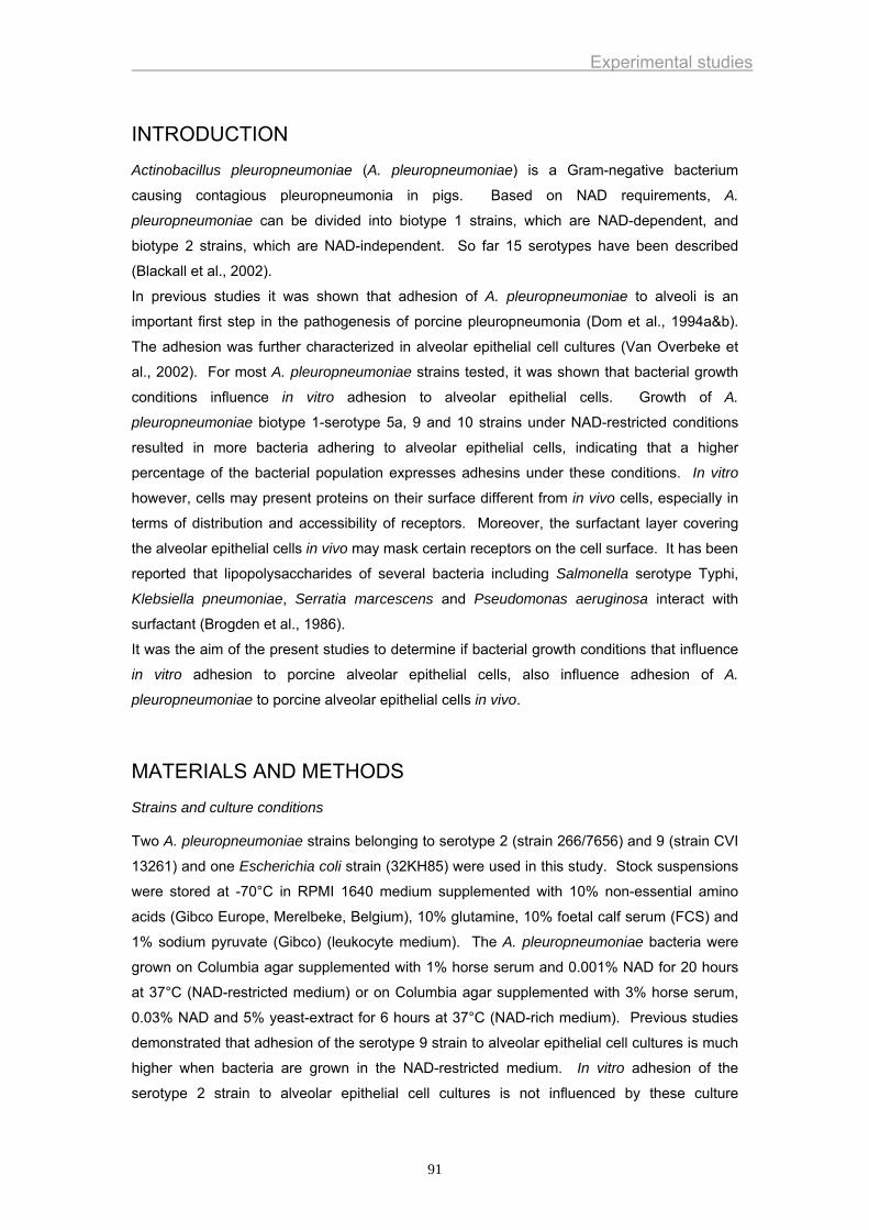
Experimental studies
INTRODUCTION
Actinobacillus pleuropneumoniae (A. pleuropneumoniae) is a Gram-negative bacterium
causing contagious pleuropneumonia in pigs. Based on NAD requirements, A.
pleuropneumoniae can be divided into biotype 1 strains, which are NAD-dependent, and
biotype 2 strains, which are NAD-independent. So far 15 serotypes have been described
(Blackall et al., 2002).
In previous studies it was shown that adhesion of A. pleuropneumoniae to alveoli is an
important first step in the pathogenesis of porcine pleuropneumonia (Dom et al., 1994a&b).
The adhesion was further characterized in alveolar epithelial cell cultures (Van Overbeke et
al., 2002). For most A. pleuropneumoniae strains tested, it was shown that bacterial growth
conditions influence in vitro adhesion to alveolar epithelial cells. Growth of A.
pleuropneumoniae biotype 1-serotype 5a, 9 and 10 strains under NAD-restricted conditions
resulted in more bacteria adhering to alveolar epithelial cells, indicating that a higher
percentage of the bacterial population expresses adhesins under these conditions. In vitro
however, cells may present proteins on their surface different from in vivo cells, especially in
terms of distribution and accessibility of receptors. Moreover, the surfactant layer covering
the alveolar epithelial cells in vivo may mask certain receptors on the cell surface. It has been
reported that lipopolysaccharides of several bacteria including Salmonella serotype Typhi,
Klebsiella pneumoniae, Serratia marcescens and Pseudomonas aeruginosa interact with
surfactant (Brogden et al., 1986).
It was the aim of the present studies to determine if bacterial growth conditions that influence
in vitro adhesion to porcine alveolar epithelial cells, also influence adhesion of A.
pleuropneumoniae to porcine alveolar epithelial cells in vivo.
MATERIALS AND METHODS
Strains and culture conditions
Two A. pleuropneumoniae strains belonging to serotype 2 (strain 266/7656) and 9 (strain CVI
13261) and one Escherichia coli strain (32KH85) were used in this study. Stock suspensions
were stored at -70°C in RPMI 1640 medium supplemented with 10% non-essential amino
acids (Gibco Europe, Merelbeke, Belgium), 10% glutamine, 10% foetal calf serum (FCS) and
1% sodium pyruvate (Gibco) (leukocyte medium). The A. pleuropneumoniae bacteria were
grown on Columbia agar supplemented with 1% horse serum and 0.001% NAD for 20 hours
at 37°C (NAD-restricted medium) or on Columbia agar supplemented with 3% horse serum,
0.03% NAD and 5% yeast-extract for 6 hours at 37°C (NAD-rich medium). Previous studies
demonstrated that adhesion of the serotype 9 strain to alveolar epithelial cell cultures is much
higher when bacteria are grown in the NAD-restricted medium. In vitro adhesion of the
serotype 2 strain to alveolar epithelial cell cultures is not influenced by these culture
91
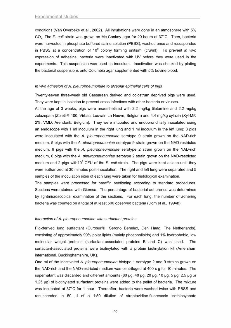
Experimental studies
conditions (Van Overbeke et al., 2002). All incubations were done in an atmosphere with 5%
CO2. The E. coli strain was grown on Mc Conkey agar for 20 hours at 37°C. Then, bacteria
were harvested in phosphate buffered saline solution (PBSS), washed once and resuspended
in PBSS at a concentration of 109 colony forming units/ml (cfu/ml). To prevent in vivo
expression of adhesins, bacteria were inactivated with UV before they were used in the
experiments. This suspension was used as inoculum. Inactivation was checked by plating
the bacterial suspensions onto Columbia agar supplemented with 5% bovine blood.
In vivo adhesion of A. pleuropneumoniae to alveolar epithelial cells of pigs
Twenty-seven three-week old Caesarean derived and colostrum deprived pigs were used.
They were kept in isolation to prevent cross infections with other bacteria or viruses.
At the age of 3 weeks, pigs were anaesthetized with 2.2 mg/kg tiletamine and 2.2 mg/kg
zolazepam (Zoletil® 100, Virbac, Louvain La Neuve, Belgium) and 4.4 mg/kg xylazin (Xyl-M®
2%, VMD, Arendonk, Belgium). They were intubated and endobronchially inoculated using
an endoscope with 1 ml inoculum in the right lung and 1 ml inoculum in the left lung: 8 pigs
were inoculated with the A. pleuropneumoniae serotype 9 strain grown on the NAD-rich
medium, 5 pigs with the A. pleuropneumoniae serotype 9 strain grown on the NAD-restricted
medium, 6 pigs with the A. pleuropneumoniae serotype 2 strain grown on the NAD-rich
medium, 6 pigs with the A. pleuropneumoniae serotype 2 strain grown on the NAD-restricted
medium and 2 pigs with109 CFU of the E. coli strain. The pigs were kept asleep until they
were euthanized at 30 minutes post-inoculation. The right and left lung were separated and 5
samples of the inoculation sites of each lung were taken for histological examination.
The samples were processed for paraffin sectioning according to standard procedures.
Sections were stained with Giemsa. The percentage of bacterial adherence was determined
by lightmicroscopical examination of the sections. For each lung, the number of adhering
bacteria was counted on a total of at least 500 observed bacteria (Dom et al., 1994b).
Interaction of A. pleuropneumoniae with surfactant proteins
Pig-derived lung surfactant (Curosurf®, Serono Benelux, Den Haag, The Netherlands),
consisting of approximately 99% polar lipids (mainly phospholipids) and 1% hydrophobic, low
molecular weight proteins (surfactant-associated proteins B and C) was used. The
surfactant-associated proteins were biotinylated with a protein biotinylation kit (Amersham
international, Buckinghamshire, UK).
One ml of the inactivated A. pleuropneumoniae biotype 1-serotype 2 and 9 strains grown on
the NAD-rich and the NAD-restricted medium was centrifuged at 400 x g for 10 minutes. The
supernatant was discarded and different amounts (80 µg, 40 µg, 20 µg, 10 µg, 5 µg, 2.5 µg or
1.25 µg) of biotinylated surfactant proteins were added to the pellet of bacteria. The mixture
was incubated at 37°C for 1 hour. Thereafter, bacteria were washed twice with PBSS and
resuspended in 50 μl of a 1:50 dilution of streptavidine-fluorescein isothiocyanate
92

Experimental studies
(streptavidine-FITC) in PBSS. After another hour of incubation at 37°C, bacteria were
washed twice with PBSS. Finally, samples were examined with a fluorescence microscope
(Leica DM RBE, Brussel, Belgium).
Controls consisted of A. pleuropneumoniae biotype 1-serotype 2 and 9 strains grown on the
NAD-rich and the NAD-restricted medium incubated with streptavidine-FITC.
RESULTS
In vivo adhesion of A. pleuropneumoniae to lung alveolar epithelial cells
A. pleuropneumoniae serotype 2 and 9 strains grown on the NAD-restricted or NAD-rich
media were able to adhere to alveolar cells. The mean percentage of bacteria adhering to the
alveolar epithelial cells was 72% and 66% for the serotype 9 strain grown on the NAD-rich
medium or the NAD-restricted medium, respectively. For the serotype 2 strain, 59% of the
bacteria grown on the NAD-rich medium and 77% of the bacteria grown on the NAD-restricted
medium were found in association with alveoli.
Adhesion was not observed in pigs inoculated with the E. coli strain.
Interaction of A. pleuropneumoniae with surfactant proteins
Fluorescence was not detected in controls consisting of the A. pleuropneumoniae biotype 1-
serotype 2 and 9 strains incubated with streptavidine-FITC.
Incubation of the A. pleuropneumoniae biotype 1-serotype 2 and 9 strains with biotinylated
surfactant and streptavidine-FITC resulted in a clear fluorescence, indicating that surfactant
associated proteins bound to the bacteria. The surfactant proteins bound to the serotype 2
and 9 strains grown on the NAD-restricted or NAD-rich media. The fluorescence decreased
when lower concentrations of biotinylated surfactant proteins were used.
DISCUSSION
Intranasal inoculation or aerosol exposure closely resembles the natural infection route of A.
pleuropneumoniae. These methods are difficult to standardize, however, because coughing,
sneezing, swallowing, breathing and mucociliary clearance can reduce the number of bacteria
that reach the alveolar region. In the present study, the bacteria were inoculated
endobronchially using an endoscope. Advantages of this approach are that it delivers an
exact number of bacteria in the bronchi and it delivers the bacteria at a choosen place in the
lung. This allows standardization of the experimental inoculations. Pigs were anaesthetized
before the inoculation and were kept asleep until they were euthanized. Coughing, sneezing
and swallowing was hereby avoided. It is to be expected that a certain amount of bacteria are
eliminated by breathing. However, this reduction of the number of bacteria is probably
equivalent for the different serotypes.
93
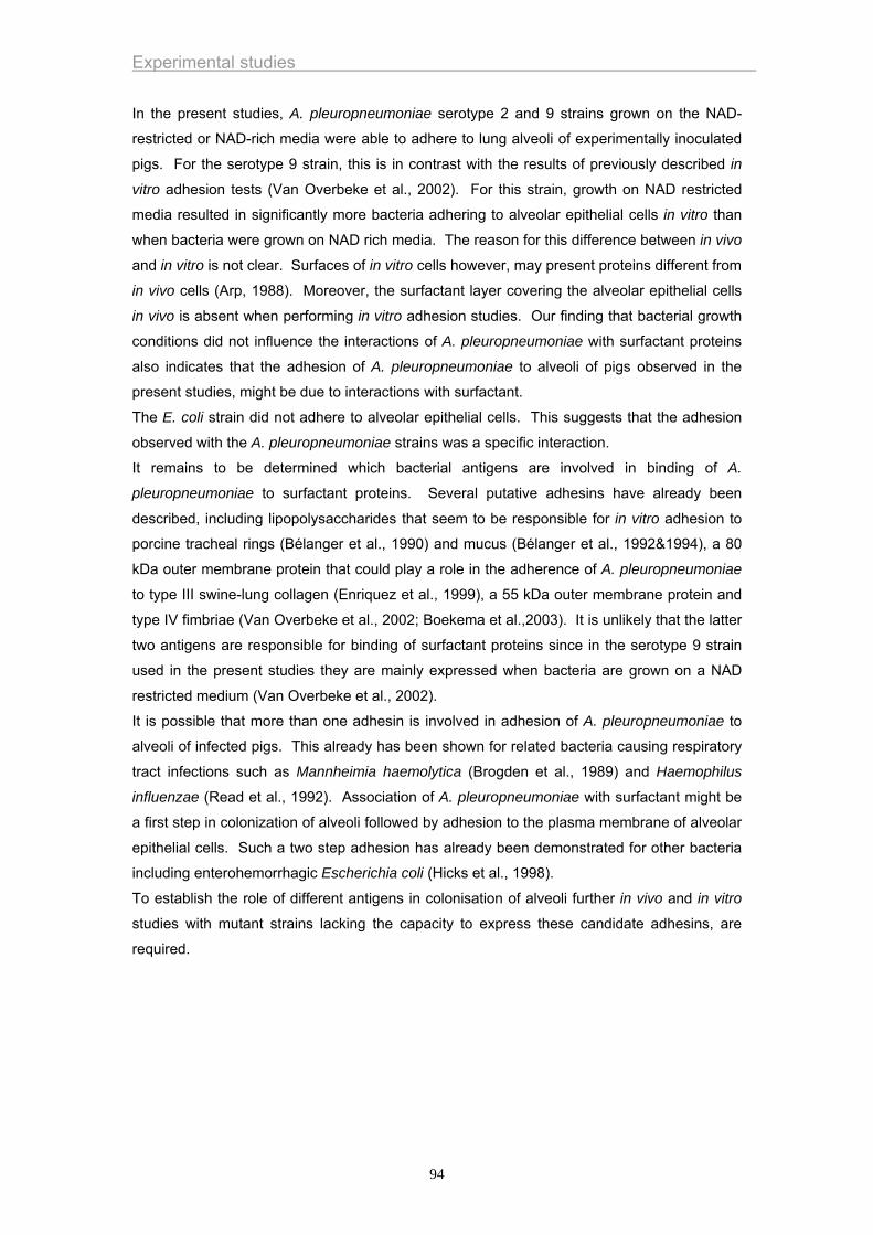
Experimental studies
In the present studies, A. pleuropneumoniae serotype 2 and 9 strains grown on the NAD-
restricted or NAD-rich media were able to adhere to lung alveoli of experimentally inoculated
pigs. For the serotype 9 strain, this is in contrast with the results of previously described in
vitro adhesion tests (Van Overbeke et al., 2002). For this strain, growth on NAD restricted
media resulted in significantly more bacteria adhering to alveolar epithelial cells in vitro than
when bacteria were grown on NAD rich media. The reason for this difference between in vivo
and in vitro is not clear. Surfaces of in vitro cells however, may present proteins different from
in vivo cells (Arp, 1988). Moreover, the surfactant layer covering the alveolar epithelial cells
in vivo is absent when performing in vitro adhesion studies. Our finding that bacterial growth
conditions did not influence the interactions of A. pleuropneumoniae with surfactant proteins
also indicates that the adhesion of A. pleuropneumoniae to alveoli of pigs observed in the
present studies, might be due to interactions with surfactant.
The E. coli strain did not adhere to alveolar epithelial cells. This suggests that the adhesion
observed with the A. pleuropneumoniae strains was a specific interaction.
It remains to be determined which bacterial antigens are involved in binding of A.
pleuropneumoniae to surfactant proteins. Several putative adhesins have already been
described, including lipopolysaccharides that seem to be responsible for in vitro adhesion to
porcine tracheal rings (Bélanger et al., 1990) and mucus (Bélanger et al., 1992&1994), a 80
kDa outer membrane protein that could play a role in the adherence of A. pleuropneumoniae
to type III swine-lung collagen (Enriquez et al., 1999), a 55 kDa outer membrane protein and
type IV fimbriae (Van Overbeke et al., 2002; Boekema et al.,2003). It is unlikely that the latter
two antigens are responsible for binding of surfactant proteins since in the serotype 9 strain
used in the present studies they are mainly expressed when bacteria are grown on a NAD
restricted medium (Van Overbeke et al., 2002).
It is possible that more than one adhesin is involved in adhesion of A. pleuropneumoniae to
alveoli of infected pigs. This already has been shown for related bacteria causing respiratory
tract infections such as Mannheimia haemolytica (Brogden et al., 1989) and Haemophilus
influenzae (Read et al., 1992). Association of A. pleuropneumoniae with surfactant might be
a first step in colonization of alveoli followed by adhesion to the plasma membrane of alveolar
epithelial cells. Such a two step adhesion has already been demonstrated for other bacteria
including enterohemorrhagic Escherichia coli (Hicks et al., 1998).
To establish the role of different antigens in colonisation of alveoli further in vivo and in vitro
studies with mutant strains lacking the capacity to express these candidate adhesins, are
required.
94
Experimental studies
REFERENCES
Arp, L.H., 1988. Bacterial infection of mucosal surfaces: an overview of cellular and
molecular mechanisms. In: Roth, J.A. (Ed), Virulence mechanisms of bacterial
pathogens. Am. Soc. Microbiol., Washington.
Bélanger, M., Dubreuil, D., Harel, J., Girard, C., Jacques, M., 1990. Role of
lipopolysaccharides in adherence of Actinobacillus pleuropneumoniae to porcine
tracheal rings. Infect. Immun. 58, 3523-3530.
Bélanger, M., Rioux, S., Foiry, B., Jacques, M., 1992. Affinity for porcine respiratory
tract mucus is found in some isolates of Actinobacillus pleuropneumoniae. FEMS
Microbiol. Lett. 76, 119-125.
Bélanger, M., Dubreuil, D., Jacques, M., 1994. Proteins found within porcine
respiratory tract secretions bind lipopolysaccharides of Actinobacillus
pleuropneumoniae. Infect. Immun. 62, 868-873.
Blackall, P.J., Klaasen, H.L.B.M, Van den Bosch, H., Kuhnert, P., Frey, J., 2002.
Proposal of a new serovar of Actinobacillus pleuropneumoniae: serovar 15. Vet.
Microbiol. 84, 47-52.
Boekema, B.K.H.L., van Putten, J.P., Smith, H.E., 2003. Pathogenesis of Actinobacillus
pleuropneumoniae: role of toxins and fimbriae, PhD thesis, Lelystadt, The
Netherlands.
Brogden, K., Adlam, C., Lehmkuhl, H., Cutlip, R., Knights, J., Engen, R., 1989. Effect of
Pasteurella haemolytica (A1) capsular polysaccharide on sheep lung in vivo on
pulmonary surfactant in vitro. Am. J. Vet. Res. 50, 555-559.
Brogden, K., Cutlip, R., Lehmkuhl, H., 1986. Complexing of bacterial lipopolysaccharide
with lung surfactant. Infect. Immun. 52, 644-649.
Chiers, K., Haesebrouck, F., Van Overbeke, I., Charlier, G., Ducatelle, R., 1999. Early
in vivo interactions of Actinobacillus pleuropneumoniae with tonsils of pigs. Vet.
Microbiol. 68, 301-306.
Dom, P., Haesebrouck, F., Ducatelle, R., Charlier, G., 1994a. In vivo association of
Actinobacillus pleuropneumoniae with the respiratory epithelium of pigs. Proc.
Haemophilus, Actinobacillus and Pasteurella Congress, Edinburgh, Scotland,
204.
Dom, P., Haesebrouck, F., Ducatelle, R., Charlier, G., 1994b. In vivo association of
Actinobacillus pleuropneumoniae serotype 2 with the respiratory epithelium of
pigs. Infect. Immun. 62, 1262-1267.
Enriquez, I., Guerrero, A.L., Serrano, J.J., Rosales, M.E., Hamer, R.C., Martinez, R.,
Godinez, D., de la Garza, M., 1999. Adhesion of Actinobacillus
95
Experimental studies
pleuropneumoniae to type III swine lung collagen. Proc. Haemophilus,
Actinobacillus and Pasteurella Congress, Mabula, South Africa, 52.
Hicks, S., Frankel, G., Kaper, J.B., Dougan, G., Phillips A.D, 1998. Role of intimin and
bundle-forming pili in enteropathogenic Escherichia coli adhesion to pediatric
intestinal tissue in vitro. Infect. Immun. 66, 1570-1578.
Read, R., Rutman, A., Jeffrey, P., Lund, V., Brain, A., Moxon, R., Cole, P., Wilson, R.,
1992. Interaction of capsulated Haemopilus influenzae with human airway
mucosa in vitro. Infect. Immun. 60, 3244-3252.
Van Overbeke, I., Chiers, K., Charlier, G., Vandenberghe, I., Van Beeumen, J.,
Ducatelle, R., Haesebrouck, F., 2002. Characterization of the in vitro adhesion of
Actinobacillus pleuropneumoniae to swine alveolar epithelial cells. Vet. Microbiol.
88, 59-74.
96
Experimental studies
CHAPTER 3
EVALUATION OF THE EFFICACY OF A VACCINE CONTAINING CANDIDATE-ADHESINS
97

Experimental studies
98
Experimental studies
Effect of endobronchial challenge with Actinobacillus pleuropneumoniae serotype 10 of pigs vaccinated with bacterins consisting of Actinobacillus pleuropneumoniae serotype 10 grown under NAD-rich and NAD-restricted conditions
Ingrid Van Overbeke, Koen Chiers, Eef Donné, Richard Ducatelle, Freddy
Haesebrouck
Laboratory of Veterinary Bacteriology and Mycology and Laboratory of Veterinary Pathology,
Department of Pathology, Bacteriology and Poultry diseases, Faculty of Veterinary Medicine,
University of Ghent, Salisburylaan 133, B-9820 Merelbeke, Belgium
Journal of Veterinary Medicine Series B 50 (2003): 289-293
99
Experimental studies
SUMMARY
The efficacy of two bacterins containing an Actinobacillus pleuropneumoniae serotype 10
strain was evaluated. The bacterial cells constituting bacterin 1 and 2 were grown under
NAD-rich (low adherence capacitiy to alveolar epithelial cell cultures) and NAD-restricted
(high adherence capacity to alveolar epithelial cell cultures) conditions, respectively. Ten pigs
were vaccinated twice with the bacterin 1 and 9 pigs with the bacterin 2. Ten control animals
were injected twice with a saline solution. Three weeks after the second vaccination, all pigs
were endobronchially inoculated with 106.5 colony-forming units (CFU) of an A.
pleuropneumoniae serotype 10 strain. In the bacterin 1 and 2 group, three and two pigs died
after inoculation, respectively. Only two pigs of the control group survived challenge.
Surviving pigs were killed at 7 days after challenge. The percentage of pigs with severe lung
lesions (>10% of the lung affected) was 100% in the control group, 70% in the bacterin 1
group and 22% in the bacterin 2 group. A. pleuropneumoniae was isolated from the lungs of
all animals. The mean bacterial titres of the caudal lung lobes were 7.0x10⁶ CFU/g in the
control group, 6.3x10⁵ CFU/g in the bacterin 1 group and 1.3x10⁶ CFU/g in the bacterin 2
group. It was concluded that both bacterins induced partial protection against severe
challenge. Furthermore, there are indications that the bacterin 2, containing A.
pleuropneumoniae bacteria grown under conditions resulting in high in vitro adhesion,
induced better protection than the bacterin 1.
100
Experimental studies
INTRODUCTION
Actinobacillus pleuropneumoniae causes porcine contagious pleuropneumonia, which is
distributed world wide and results in serious losses in the pig rearing industry. It also causes
severe animal suffering. Infected pigs may develop acute hemorrhagic necrotizing
pneumonia and fibrinous pleuritis or chronic localized lung lesions and adhesive pleuritis
(Taylor, 1999). To control outbreaks of this disease, vaccination may be useful. Vaccines
containing the A. pleuropneumoniae Apx toxins have become commercially available and in
previous studies, partial protection against endobronchial challenge in animals vaccinated
with these vaccines was demonstrated (Chiers et al., 1998; Van Overbeke et al., 2001). Field
trials carried out in France (Pommier et al., 1996), the Netherlands (Valks et al., 1996), Italy
(Martelli et al., 1996), Spain (López et al., 1998) and Norway (Lium et al., 1998) confirmed
that vaccination with Apx-containing vaccines can result in reduction of clinical symptoms and
lung lesions of acute and chronic pleuropneumonia and improvement of performance (growth,
feed conversion, cost of medication).
Apx toxins are not the only antigens that could play a role in protection against porcine
pleuropneumonia. Capsular antigens, cell wall lipopolysaccharides, outer membrane
proteins, fimbriae, transferrin binding proteins and adhesins also are important virulence
factors (Haesebrouck et al., 1997).
Adherence to host tissue is generally regarded as an important prerequisite for colonization
and virulence manifestation of bacteria (Ofek and Beachy, 1980).
In previous studies, it was shown that A. pleuropneumoniae adheres to alveolar epithelial
cells of experimentally infected pigs (Dom et al., 1994a; 1995). It has also been shown that
A. pleuropneumoniae adheres to porcine tracheal rings maintained in culture (Bélanger et al.,
1990), to porcine frozen tracheal and lung sections Jacques et al., 1991), to erythrocytes from
various animal species (Jacques et al., 1988), to porcine tonsillar epithelial cells (Chiers et al.,
1999), to swine buccal epithelial cells (Hamer et al., 1999), to type III swine-lung collagen
(Enriqueze et al., 1999) and to a culture of alveolar epithelial cells (Van Overbeke et al.,
2002). In the latter study, it has been demonstrated that adhesive qualities of A.
pleuropneumoniae to alveolar epithelial cells were influenced by growth conditions. Growth
of A. pleuropneumoniae under NAD-restricted conditions results in more bacteria adhering to
alveolar epithelial cells, indicating that a higher percentage of the bacterial population
expresses adhesines under these conditions. The high adhesion scores of bacteria grown
under NAD-restricted conditions have been related to expression of fimbriae and a 55 kDa
outer membrane protein.
The present study was undertaken to determine if pigs, vaccinated with a bacterin consisting
of bacteria grown under NAD-restricted conditions (high in vitro adherence capacity), were
better protected or not against an A. pleuropneumoniae infection than pigs vaccinated with a
bacterin consisting of bacteria grown under NAD-rich conditions (low in vitro adherence
capacitiy).
101
Experimental studies
MATERIALS AND METHODS
Challenge strain
An A. pleuropneumoniae serotype 10 reference strain D13039 was used. Bacteria were
grown for 6 hours (log phase of growth) on Columbia agar (Columbia Agar Base, Lab M,
Bury, Great Britain) supplemented with 3% horse serum, 0.03% NAD (Sigma Chemical Co, St
Louis, Mo, USA), and 5% yeast extract at 37°C in a humid atmosphere with 5% CO2.
Bacteria were harvested in phosphate-buffered saline solution (PBSS, pH 7.3), centrifuged at
400 x g for 20 minutes, and suspended in RPMI (Roswell Park Memorial Institute) 1640
supplemented with 10% non-essential amino acids, 10% glutamine, 10% fetal calf serum, and
1% sodium pyruvate (Dom et al., 1994a). The suspension was checked for purity and the
number of colony-forming units (CFU) was determined by plating tenfold dilutions on
Columbia agar supplemented with 3% horse serum, 0.03% NAD, and 5% yeast extract.
Bacterial suspensions were stored overnight at 4°C. The next day, they were used in the
challenge experiments.
Pigs
Twenty-nine conventional pigs were obtained by using a medicated segregated early weaning
programme (Chiers et al., 1998). They were weaned at 18 days of age and kept in isolation
until used in the experiments.
Vaccine
Two bacterins were used. Bacterin 1 contained A. pleuropneumoniae serotype 10 strain
D13039 grown on Columbia agar supplemented with 3% horse serum, 0.03% NAD and 5%
yeast-extract for 6 hours at 37°C (NAD-rich medium). Bacterin 2 contained the A.
pleuropneumoniae serotype 10 strain D13039 grown on Columbia agar supplemented with
1% horse serum and 0.001% NAD for 20 hours at 37°C (NAD-restricted medium).
The bacteria were harvested from the agar in PBSS, washed once with PBSS and
resuspended in PBSS to achieve a suspension of 109 CFU/ml. Bacteria in each suspension
were inactivated by UV-light. This inactivation procedure does not affect in vitro adhesion to
alveolar epithelial cell cultures (data not shown). After inactivation, the bacterins were
emulsified in an equal volume of a mineral oil adjuvans (De Herdt et al., 1999).
Experimental design
At the age of 20 and 23 weeks, pigs were intramuscularly injected: 10 pigs with 2 ml of the
bacterin 1, 9 pigs with 2 ml of the bacterin 2 and 10 pigs with 2 ml of a saline solution. Three
weeks after the second injection, all pigs were experimentally infected. The pigs were
anaesthetized with Zoletil® 100 (2.2 mg/kg tiletamine and 2.2 mg/kg zolazepam, Virbac,
Louvain La Neuve, Belgium) and Xyl-M® 2% (4.4 mg/kg xylazin, VMD, Arendonk, Belgium).
102
Experimental studies
They were inoculated endobronchially with 106.5 CFU of the challenge strain in 5 ml inoculum
(Van Overbeke et al., 2001). All pigs were examined clinically. Pigs that died were autopsied
immediately; those that survived the challenge were killed 7 days after inoculation. At
necropsy, the lungs were examined macroscopically and the percentage of affected lung
tissue was determined. For this purpose, a diagram was used that divides the lung into 74
equal triangles (Hannan et al., 1982). The percentage of affected lung tissue was determined
by summing up the triangles showing pneumonia and/or pleuritis divided by 74. Samples
from lungs and tracheobronchial lymph nodes, were taken for bacteriological examination.
Sera were collected before the first and the second vaccination and before the challenge.
Clinical examination
The pigs were examined for signs of pneumonia, characterized by increased respiratory rate
(>40 inspirations/min), dyspnoea, sneezing, coughing, and the presence of a bloody foam on
mouth and/or nostrils. Rectal temperature was measured 1 day before challenge inoculation,
just before inoculation, and during the 7 days after inoculation. Other indications of infection
were vomiting and a depressed appearance.
Bacteriological examination
Twenty per cent (w/v) suspensions of the right and the left caudal lung lobes were made in
PBSS. The samples were tested for the presence of A. pleuropneumoniae by inoculating the
suspensions onto Columbia agar supplemented with 5% bovine blood with a Staphylococcus
intermedius streak. The number of cfu was determined by plating tenfold dilutions of the
suspensions on Columbia agar supplemented with 3% equine serum, 0.03% NAD and 5%
yeast extract. Samples from tracheobronchial lymph nodes were tested for the presence of
A. pleuropneumoniae by making an incision in these tissues and taking a sample with an
inoculation loop. These samples were inoculated onto Columbia agar supplemented with 5%
bovine blood with a Staphylococcus intermedius streak. Identification of A.
pleuropneumoniae was based on the CAMP reaction and the inhanced growth around the
Staphylococcus intermedius streak.
Serology
All pig sera were tested for presence of ApxI neutralizing antibodies using a bioassay based
on neutral red uptake by viable pulmonary alveolar macrophages, as described previously
(Dom et al., 1994b) and for the presence of antibodies against cell-surface antigens of A.
pleuropneumoniae serotype 10 strain D13039 grown on the NAD-rich and NAD-restricted
media using an ELISA assay. The bacteria were harvested from the agar in PBSS, washed
once with PBSS and resuspended in PBSS to achieve a suspension of 109 CFU/ml. After
inactivation of the bacteria by UV-light, the suspension was centrifuged and resuspended in
coatingbuffer (0.05M carbonate-bicarbonate buffer, pH 9.6) to achieve a suspension of 107
103

Experimental studies
CFU/ml. Wells of a microtiter plate (Nunc-Immuno Plate MaxiSorp™, Gibco, Ghent, Belgium)
were coated with 150 µl coatingbuffer containing the bacteria and incubated overnight at 4°C.
The next day, plates were washed four times with PBSS containing 0.05% Tween-20 (VWR
International, Leuven, Belgium) (PBSS-T, pH 7.2). Non-specific binding was blocked by
incubating the plates with 5% (w/v) glycine (VWR International) in PBSS-T. Plates were
washed twice with PBSS-T, and were incubated with 1/200 dilution of sera. Bound antibodies
were detected using 1/800 diluted peroxidase-conjugated rabbit anti-pig immunoglobulin
serum (Sigma). Aminosalicylic acid and H2O2 (Sigma) were used as enzyme substrate.
Reactions were read at 405 nm using a microELISA reader (Titertek Multiscan Plus). The OD
values of wells devoided of sera served as background values.
Sera collected at the time of challenge were also tested for the presence of antibodies against
a 55 kDa outer membrane protein of A. pleuropneumoniae serotype 10 strain D13039.
Sarcosyl-insoluble outer membrane proteins of the A. pleuropneumoniae serotype 10 strain
grown on the NAD-restricted medium were prepared as previously described (Van Overbeke
et al., 2002). The outer membrane proteins were separated using discontinuous sodium
dodecylsulphate polyacrylamide gel electrophoresis (SDS-PAGE). Electrophoresis
experiments were performed with a 4.5% stacking gel and a 12% running gel. The antigen
preparation was mixed with equal volumes of solubilization buffer [4% (w/v) of SDS, 10% (v/v)
2-mercapto ethanol, 17.4% (v/v) glycerol and 0.01% bromophenol blue in 0.06M Tris HCl (pH
6.8)] and boiled for 5 minutes prior to electrophoresis. After electrophoresis, antigens were
electroblotted onto an immobilon polyvinylidene difluoride membrane (Millipore, Bedford,
Mass., USA) using a semi-dry blotting system (190 mA, 1h, Bio-Rad Laboratories, Richmond,
USA). Blotted proteins were subsequently probed with 1/100 diluted pig sera and 1/800
diluted anti-pig IgG serum conjugated with peroxidase (Sigma). Recognised antigens were
visualised with chloronaftol and H2O2. To stop the reactions, the strips were washed three
times in distilled water. A standard solution of prestained proteins with a molecular range
from 14.2-97 kDa was used as a marker (Sigma, SDS-6B).
Statistical analysis
Statistical analysis was performed on the following variables: percentage of mortality,
percentage of animals with fever, percentage of animals with dyspnoea, percentage of pigs
with severe lung lesions (>10% of the lung affected), mean percentage of affected lung tissue
and logarithmic mean of bacterial titre in caudal lung lobes.
A logistic regression analysis was used to compare the percentage of mortality, percentage of
animals with fever, percentage of animals with dyspnoea, mean percentage of affected lung
tissue and percentage of pigs with severe lung lesions (>10% of the lung affected). A
Wilcoxon signed rank test was used to compare the log mean bacterial titres in caudal lung
lobes between groups.
104
Experimental studies
RESULTS
Results are summarized in Table 1.
Clinical examination
None of the animals showed disease signs before challenge and at the time of challenge.
In the control group (n=10), 6 pigs died and 2 animals were euthanised within 48 h after
inoculation. The two remaining pigs showed dyspnoea and fever on day 1. Thereafter, one
pig had dyspnoea and occasional coughing for another day and fever (>40°C) for 6 days.
The other pig had an increased respiratory rate during 4 days after challenge.
In the bacterin 1 group (n=10), one pig was euthanised one day after challenge and 2 other
animals were euthanised on day 2. Respiratory signs (dyspnoea, increased respiratory rate
or coughing) and fever could be detected in five animals. The duration of these signs varied
between one day and 6 days. Two pigs had only an increased respiratory rate during 2 and 4
days. Three of the animals were depressed for 1 to 2 days.
In the bacterin 2 group (n=9), two pigs showed very severe symptoms and were euthanised.
Four animals developed respiratory signs and fever. Duration of these symptoms varied
between 1 and 7 days. Two animals had fever without respiratory symptoms. One pig
showed an increased respiratory rate for 1 day. Two animals were depressed for 2 days.
A significant difference (p<0.05) was found for the percentage of mortality between the control
group and the bacterin 1 group (low adhesin expression) (p=0.033) and bacterin 2 (high
adhesin expression) group (p=0.009). There was also a significant difference for the
percentage of animals with dyspnoea between the control group and the bacterin 2 (high
adhesin expression) group (p=0.049).
Necropsy findings
In all animals lung lesions, when present, were characterized by a haemorrhagic necrotizing
pneumonia and fibrinous pleuritis .
All animals of the control group had severe lung lesions (>10% of the lungs affected). The
mean percentage of affected lung tissue was 56% (variation 16-100%).
In the bacterin 1 group, seven animals had severe lung lesions. One animal had very mild
lung lesions and 2 animals had no lung lesions. The percentage of affected lung tissue varied
between 0% and 57% (mean: 25%).
In the bacterin 2 group, two animals had severe lung lesions. Six animals had very mild lung
lesions and 1 animal had no lung lesions. The percentage of affected lung tissue varied
between 0% and 88% (mean: 16%).
A significant difference was found for the percentage of animals with severe lung lesions
between the bacterin 2 (high adhesin expression) group and the control group (p=0.007) and
between the bacterin 2 (high adhesin expression) group and the bacterin 1 (low adhesin
expression) group (p=0.047). The mean percentage of affected lung tissue differed
significantly between both bacterin groups and the control group (p<0.001) and between the
105
Experimental studies
bacterin 2 (high adhesin expression) group and the bacterin 1 (low adhesin expression) group
(p<0.001).
Bacteriology
The results are summarized in Table 1.
In the control pigs, A. pleuropneumoniae was isolated from all lung lobes. The number of
CFU/gram lung tissue of the caudal lobes varied between 3.0 x 10⁵ and 2.8 x 10⁸, with a
logarithmic mean of 7.0 x 10⁶. The challenge strain was isolated from the tracheobronchial
lymphnodes of 8 pigs.
In the bacterin 1 group, A. pleuropneumoniae was isolated from all lung lobes. The number
of CFU/gram lung tissue of the caudal lobes varied between 5.2 x 10² and 1.8 x 10⁸, with a
logarithmic mean of 6.3 x 10⁵. A. pleuropneumoniae was isolated from the tracheobronchial
lymphnodes of 1 pig.
In the bacterin 2 group, A. pleuropneumoniae was isolated from all lung lobes. The number
of CFU/gram lung tissue of the caudal lobes varied between 3.6 x 10⁴ and 9.4 x 10⁷, with a
logarithmic mean of 1.3 x 10⁶. A. pleuropneumoniae was isolated from the tracheobronchial
lymphnodes of 2 pigs.
No significant difference (p<0.05) was found between the logarithmic mean of bacterial titres
in the caudal lung lobes for the different groups.
Serology
In none of the animals, neutralizing antibodies against the Apx I toxin were detected.
Antibodies against cell-surface antigens were detected in both bacterin groups. OD values
were in the same range for both antigen preparations. At the time of first vaccination, a mean
OD value (standard deviation) was found of 0.132 (0.050) for the bacterin 1 group, 0.121
(0.059) for the bacterin 2 group and 0.124 (0.041) for the control group. At the time of second
vaccination, a mean OD value (standard deviation) was found of 0.410 (0.103) for the
bacterin 1 group, 0.408 (0.138) for the bacterin 2 group and 0.113 (0.23) for the control group.
At the time of challenge, a mean OD value (standard deviation) was found of 0.454 (0.103) for
the bacterin 1 group, 0.509 (0.161) for the bacterin 2 group and 0.132 (0.029) for the control
group. The background OD values were in the same range as the OD values of the control
group.
Antibodies against a 55 kDa outer membrane protein could not be detected in the sera
collected at time of challenge.
Serology
In none of the animals, neutralizing antibodies against the Apx I toxin were detected.
Antibodies against cell-surface antigens were detected in both bacterin groups. OD values
were in the same range for both antigen preparations. At the time of first vaccination, a mean
OD value (standard deviation) was found of 0.132 (0.050) for the bacterin 1 group, 0.121
(0.059) for the bacterin 2 group and 0.124 (0.041) for the control group. At the time of second
106
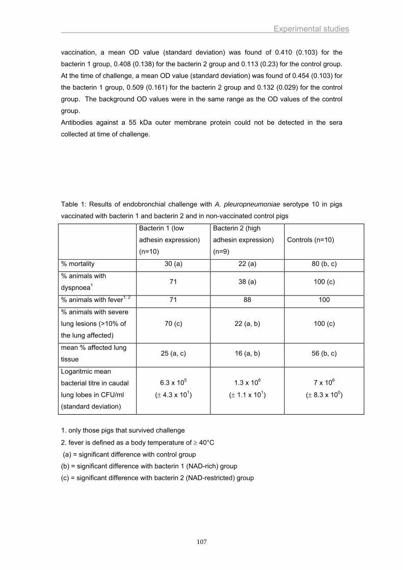
Experimental studies
vaccination, a mean OD value (standard deviation) was found of 0.410 (0.103) for the
bacterin 1 group, 0.408 (0.138) for the bacterin 2 group and 0.113 (0.23) for the control group.
At the time of challenge, a mean OD value (standard deviation) was found of 0.454 (0.103) for
the bacterin 1 group, 0.509 (0.161) for the bacterin 2 group and 0.132 (0.029) for the control
group. The background OD values were in the same range as the OD values of the control
group.
Antibodies against a 55 kDa outer membrane protein could not be detected in the sera
collected at time of challenge.
Table 1: Results of endobronchial challenge with A. pleuropneumoniae serotype 10 in pigs
vaccinated with bacterin 1 and bacterin 2 and in non-vaccinated control pigs
Bacterin 1 (low
adhesin expression)
(n=10)
Bacterin 2 (high
adhesin expression)
(n=9)
Controls (n=10)
% mortality 30 (a) 22 (a) 80 (b, c)
% animals with
dyspnoea1 71 38 (a) 100 (c)
% animals with fever1; 2 71 88 100
% animals with severe
lung lesions (>10% of
the lung affected)
70 (c) 22 (a, b) 100 (c)
mean % affected lung
tissue 25 (a, c) 16 (a, b) 56 (b, c)
Logaritmic mean
bacterial titre in caudal
lung lobes in CFU/ml
(standard deviation)
6.3 x 10⁵
(± 4.3 x 101)
1.3 x 10⁶
(± 1.1 x 101)
7 x 10⁶
(± 8.3 x 100)
1. only those pigs that survived challenge
2. fever is defined as a body temperature of ≥ 40°C
(a) = significant difference with control group
(b) = significant difference with bacterin 1 (NAD-rich) group
(c) = significant difference with bacterin 2 (NAD-restricted) group
107
Experimental studies
DISCUSSION
Intranasal inoculation or aerosol exposure closely resemble the natural infection route of A.
pleuropneumoniae. However, these methods are difficult to standardize because coughing,
sneezing, swallowing, breathing and mucociliary clearance can reduce the number of bacteria
that reach the alveolar region. In the present study, an endobronchial challenge model was
used. An advantage of this model is that it delivers an exact number of bacteria into the
bronchi, allowing standardization of experimental infections. Furthermore, this method allows
us to bring the bacteria directly close to the multiplication site of A. pleuropneumoniae,
namely the alveoli and bronchioli (Haesebrouck et al., 1997). A possible disadvantage is that
the challenge strain is applicated not all over the lung but is given more focal. Endobronchial
challenge is therefore more likely to break through low level immunity in the respiratory tract
than a more natural challenge route.
Eight of the 10 control animals died after infection. This demonstrates that a severe
challenge model was used.
There was a significant difference in the percentage of mortality and the mean percentage of
affected lung tissue between the control group and the bacterin groups. This indicates that
both bacterins induced partial protection against severe challenge.
The percentage of animals with severe lung lesions and the mean percentage of affected lung
tissue were significant lower in the bacterin 2 (high adhesin expression) group than in the
bacterin 1 (low adhesin expression) group. Furthermore, there was also a significant
difference for the percentage of animals with dyspnoea between the control group and the
bacterin 2 (high adhesin expression) group. This could indicate that the bacterin 2, i.e. a
bacterin containing bacteria grown under NAD-restricted conditions resulting in high in vitro
adhesion to a culture of alveolar epithelial cells, provides better protection. Further studies
are necessary to determine which antigens could be involved.
Bacterins were based on UV-inactivated and washed A. pleuropneumoniae bacteria. In sera
of vaccinated animals, antibodies against the Apx I toxin were not found, suggesting that this
toxin is not cell-associated but is only secreted into the culture medium. This is also
mentioned by Frey and Nicolet (1991), but is in contrast with the reports by Fedorka-Cray
(1989; 1993).
In previous studies, it was demonstrated that a 55 kDa outer membrane protein was
expressed by the serotype 10 strain used in the present studies when bacteria were grown
under NAD-restricted conditions (Van Overbeke et al., 2002). However, antibodies against
this protein were not detected by Western blot analysis in sera of vaccinated animals at the
time of challenge. The reason for this finding is not clear. It is possible that the
immunogenicity of the 55 kDa outer membrane protein is low or that antibodies do not
recognise the denaturated protein used in Western Blot analysis. Antibody reactions
occurred between other outer membrane protein components and sera of vaccinated animals
at the time of challenge (data not shown). However, there was no marked difference between
the groups.
108

Experimental studies
In the present studies, animals were only challenged with a serotype 10 strain. It has been
shown that whole-cell bacterins based on bacteria grown in NAD-rich conditions generally do
not confer protection against challenge with heterologous serotypes (Fenwick and Henry,
1994). An explanation for the limited protection might be the absence of secreted and certain
bacteria-associated virulence factors in these bacterins. Further challenge studies with
heterologous serotypes are necessary to determine if a vaccine containing bacteria grown in
NAD-restricted conditions induces protection against other serotypes.
109
Experimental studies
REFERENCES
Bélanger, M., Dubreuil, D., Harel, J., Girard, C., Jacques, M., 1990 . Role of
lipopolysaccharides in adherence of Actinobacillus pleuropneumoniae to porcine
tracheal rings. Infect. Immun. 58, 3523-3530.
Chiers, K., Van Overbeke, I., De Laender, P., Ducatelle, R., Carel, S., Haesebrouck,
F., 1998. Effects of endobronchial challenge with Actinobacillus
pleuropneumoniae serotype 9 of pigs vaccinated with inactivated vaccines
containing the Apx toxins. Vet. Quart. 20, 65-9.
Chiers, K., Haesebrouck, F., Van Overbeke, I., Charlier, G., Ducatelle, R., 1999. Early
in vivo interactions of Actinobacillus pleuropneumoniae with tonsils of pigs. Vet.
Microbiol. 68, 301-306.
De Herdt, P., Vanrobaeys, M., Devriese, L.A., Ducatelle, R., Haesebrouck, F., 1999.
Efficacy of inactivated whole-cell vaccines against streptococcosis in pigeons.
Av. Pathol. 28, 355-361.
Dom, P., Haesebrouck, F., 1992. Comparitive virulence of NAD dependent and NAD
independent Actinobacillus pleuropneumoniae strrains. J. Vet. Med. 39, 303-6.
Dom, P., Haesebrouck, F., Kamp, E.M., Smits, M.A., 1994a. NAD-independent
Actinobacillus pleuropneumoniae strains: production of RTX toxins and
interactions with porcine phagocytes. Vet. Microbiol. 39, 205-18.
Dom, P., Haesebrouck, F., Ducatelle, R., Charlier, G., 1994b. In vivo association of
Actinobacillus pleuropneumoniae serotype 2 with the respiratory epithelium of
pigs. Infect. Immun. 62, 1262-1267.
Dom, P., Haesebrouck, F., Ducatelle, R., Charlier, G., 1995. In vivo association of
Actinobacillus pleuropneumoniae with the respiratory epithelium of pigs. In:
Haemophilus, Actinobacillus and Pasteurella (Eds: W. Donachie, F.A. Lainson
and J.C. Hodgson). Proc. Haemophilus, Actinobacillus and Pasteurella Conf.,
Edinburgh, Scotland, 204.
Enriquez, I., Guerrero, A.L., Serrano, J.J., Rosales, M.E., Hamer, R.C., Martinez, R. ,
Godinez, D., De la Garza, M., 1999 . Adhesion of Actinobacillus
pleuropneumoniae to type III swine lung collagen. Proc. Haemophilus,
Actinobacillus and Pasteurella Conf., Mabula, South-Africa, 52.
Fedorka-Cray, P.J., 1989. Actinobacillus (Haemophilus) pleuropneumoniae virulence
factors: Partial characterization and efficacy studies in swine. University of
Nebraska, Ph.D. Thesis, 217 pp.
Fedorka-Cray, P.J., Stine, D.L., Greenwald, J.M., Gray, J.T., Huether, M.J., Anderson,
G.A., 1993. The importance of secreted virulence factors in Actinobacillus
pleuropneumoniae bacterin preparation: a comparison. Vet. Microbiol. 37, 85-
100.
110
Experimental studies
Fenwick, B., Henry, S., 1994. Porcine pleuropneumonia. J. Am. Vet. Med. Assoc. 204,
1334-1340.
Frey, J., Nicolet, J., 1991. Immunological properties of Actinobacillus
pleuropneumoniae haemolysin I. Vet. Microbiol. 28, 61-73.
Haesebrouck, F., Chiers K., Van Overbeke, I., Ducatelle, R., 1997. Actinobacillus
pleuropneumoniae infections in pigs: the role of virulence factors in pathogenesis
and protection. Vet. Microbiol. 58, 239-249.
Hamer, R.C., Enriquez, I., Godinez, D., Guerrero, A.L., Martinez, R., Talamas, P.,
Vaca, S., Garcia, C., de la Garza, M., 1999 . Adhesion of Actinobacillus
pleuropneumoniae to swine buccal epithelial cells. Proc. Haemophilus,
Actinobacillus and Pasteurella Conf., Mabula, South-Africa, 51.
Hannan, P.C.T., Bhogal, B.S., Fish, J.P., 1982. Tylosin tartrate and tiamutilin effects on
experimental piglet pneumonia induced with pneumonic pig lung homogenate
containing mycoplasmas, bacteria and viruses. Res. Vet. Sci. 33, 76-88.
Jacques, M., Roy, G., Mittal, K.R., 1988. Hemagglutinating properties of Actinobacillus
pleuropneumoniae. Can. J. Microbiol. 34, 1046-1049.
Jacques, M., Bélanger, M., Roy, G., Foiry, B., 1991 . Adherence of Actinobacillus
pleuropneumoniae to porcine tracheal epithelial cells and frozen lung sections.
Vet. Microbiol. 27, 133-143.
Lium, B., Brun, E., Fjetland, O., Lund, A., 1998. A field study of the efficacy of an
Actinobacillus pleuropneumoniae subunit vaccine in the control of
pleuropneumonia caused by Actinobacillus serotype 6. Proc. 15th International
Pig Veterinary Society Congress, Birmingham, England, 256.
López, J.V., Font, J., Prat, M., Cantin, C., 1998. Technical and economic efficacy of
Porcilis App: a field trial. Proc. 15th International Pig Veterinary Society
Congress, Birmingham, England, 264.
Martelli, P., Guadagnini, P.F., Foccoli, E., Ballarini, G., 1996. Efficacy of an
Actinobacillus pleuropneumoniae subunit vaccine in the control of
pleuropneumonia: a field trial. Proc. 14th International Pig Veterinary Society
Congress, Bologna, Italy, 214.
Nicolet, J., 1992. Actinobacillus pleuropneumoniae, Diseases of swine. A.D. Leman, B.
Straw, W.L. Mengeling, S. D'Allaire and D.J. Taylor (Eds.). Ames, Iowa Stae
University Press, 401-8.
Ofek, I., Beachy, E.H., 1980. Bacterial adherence. Adv. Int. Med. 25, 505-532.
Pommier, P., Ridremont, B., Wessel-Robert, S., Keïta, A., 1996. Field study into
efficacy of a new Actinobacillus pleuropneumoniae subunit vaccine. Proc. 14th
International Pig Veterinary Society Congress, Bologna, Italy, 206.
111

Experimental studies
Valks, M. M. H., Nell, T., Van den Bosch, J.F., 1996. A clinical field trial in finishing pigs
to evaluate the efficacy of a new APP subunit vaccine. Proc. 14th International
Pig Veterinary Society Congress, Bologna, Italy, 208.
Van Overbeke, I., Chiers, K., Ducatelle, R., Haesebrouck, F., 2001. Effect of
endobronchial challenge with Actinobacillus pleuropneumoniae serotype 9 of
pigs vaccinated with a vaccine containing Apx toxins and transferrin-binding
proteins. J. Vet. Med. B 48, 15-20.
Van Overbeke, I., Chiers, K., Charlier, G., Vandenberghe, I., Van Beeumen, J.,
Ducatelle, R., Haesebrouck, F., 2002. Characterization of the in vitro adhesion of
Actinobacillus pleuropneumoniae to swine alveolar epithelial cells. Vet.
Microbiol. 88, 59-74.
112
General discussion
GENERAL DISCUSSION
A. pleuropneumoniae causes severe losses in the pig rearing industry (Nicolet et al., 1992).
Although the disease can be controlled by antimicrobial agents, the use of these products has
several disadvantages including induction of acquired resistance in pathogenic bacteria and
bacteria belonging to the normal flora of pigs. The latter bacteria may act as a reservoir of
antimicrobial resistance genes for bacteria that cause disease in humans or animals.
Excessive use of antimicrobials may also result in the presence of residues in meat.
Therefore, prevention is a better option for the control of the disease. Vaccines could be very
useful to control porcine pleuropneumonia. Rational design of effective antibacterial
vaccines, however, requires knowledge of the virulence factors of the bacterium and the
pathogenesis of the disease. Pathogenesis of porcine pleuropneumonia is very complex and
several virulence factors have been described. Some of these virulence factors, such as the
Apx toxins, have been extensively studied. Virulence attributes involved in lung colonisation
are, however, less understood. The need for research in the first steps of pathogenesis
provided the main rationale for this thesis.
Recovery from naturally acquired infection with A. pleuropneumoniae provides solid, serotype
cross-protective immunity. Evidence from field and experimental studies indicates that
infection with one serotype of A. pleuropneumoniae provides complete protection against
subsequent infection with the homologous serotype and at least partial protection against
heterologous infection (Nielsen et al., 1984; Haesebrouck et al., 1996). Preferably, protection
induced by vaccines should approach protection induced by natural infection.
The first vaccines that were developed against porcine pleuropneumonia were whole-cell
bacterins, consisting of inactivated bacterial cell suspensions and supplemented with an
adjuvant. Although these vaccines have the advantage of presenting a complex array of
antigenic determinants to the immune system, inactivation procedures may alter the antigenic
patterns of a bacterin and may be responsible for the loss of relevant immunogenic epitopes.
This can be overcome by using ghost vaccines. Bacterial ghosts are empty cell envelopes
achieved by the expression of a cloned bacteriophage lysis gene and, unlike classical
bacterins, suffer no denaturating steps during their production. These properties may lead to
a superior presentation of surface antigens to the immune system (Hensel et al., 2000).
Whole-cell bacterins may reduce mortality after infection with the homologous serotype, but
they generally do not confer protection against challenge with heterologous serotypes. An
explanation for the limited protection might be the absence of Apx toxins. Apx toxins are
indeed thought to be of particular importance for the induction of protective immunity. Frey
and Nicolet (1991) reported that Apx toxins are secreted by the bacterium in the culture
medium and that they are not cell-associated. However, this is in contrast with reports by
Fedorka-Cray (1989; 1993). Results of our studies also suggest that the Apx toxins are not
113
General discussion
cell-associated since antibodies against Apx toxins were not detected in sera of animals
vaccinated with bacterins based on UV-inactivated and washed A. pleuropneumoniae
bacteria. Improved protection has been obtained by using subunit vaccines containing the Apx toxins.
Among the different serotypes of A. pleuropneumoniae, 4 of these exotoxins have been
described. In contrast to ApxI, ApxII and ApxIII toxins, each of which is produced by some
but not all serotypes, the ApxIV toxin is produced by all serotypes (Cho and Chae, 2001;
Schaller et al., 1999). Apparently, the ApxIV toxin is only produced in vivo (Schaller et al.,
1999). Therefore, the current commercially available vaccines do not contain this ApxIV
toxin. Results of the present thesis and field trials demonstrated that vaccination with
vaccines containing ApxI, ApxII and ApxIII can result in a reduction of the clinical symptoms
and lung lesions of acute and chronic pleuropneumonia and improvement of performance
(growth, feed conversion, cost of medication) (Pommier et al., 1996; Valks et al., 1996;
Martelli et al., 1996; Lopez et al., 1998; Lium et al., 1998).
Keeping in mind the very complex pathogenesis of porcine pleuropneumonia, inclusion of
other bacterial virulence factors in subunit vaccines might be of value. Results of studies
described in this thesis demonstrated that a vaccine containing both Apx toxins and
transferrin binding proteins induced better protection against severe challenge than a vaccine
solely based on Apx toxins. Recently, it was shown that antibodies induced against purified
PalA, the most immuno-predominant outer membrane protein of A. pleuropneumoniae,
aggravated the consequences of an experimental infection and counteracted the protective
effect of anti-Apx antibodies (van den Bosch and Frey, 2003). Therefore, care should be
taken which antigens are included in subunit vaccines. In whole-cell preparations of bacterial
cultures, the concentration of PalA may vary depending on the mode of cultivation and
preparation of the bacteria. This may be an explanation for the variation in protective efficacy
of different bacterins.
Bacterial virulence factors that, most probably, would be very valuable to include in vaccines
are adhesins. Indeed, A. pleuropneumoniae first adheres to alveolar epithelial cells before it
causes lung lesions (Dom et al., 1992). Antibodies that interfere with adhesion could block
this first step in the pathogenesis of porcine pleuropneumonia and protect pigs against lung
infection. In chapter 3 of this thesis it was shown that a vaccine containing inactivated A.
pleuropneumoniae bacteria grown under circumstances resulting in high in vitro adhesion
induced better protection than a vaccine containing inactivated A. pleuropneumoniae bacteria
grown under circumstances resulting in low in vitro adhesion.
When we started our studies, no reliable adherence model was available to study the way A.
pleuropneumoniae interacts with alveolar epithelial cells. The in vitro adherence model
developed during these studies enabled us to investigate the first interactions between A.
pleuropneumoniae and the alveolar epithelial cells. We were also able to pinpoint the
adherence mechanisms and to characterize some candidate-adhesins.
114
General discussion
There are different methods for studying bacterial adherence to tissue culture cells: (1)
Colony count: the cell layer can be lysed with an appropriate detergent which does not affect
bacterial viability. Appropriate dilutions are then plated on agar plates for enumeration of cell-
associated bacteria by colony count (Robbins-Browne and Bennett-Wood, 1992). The
number of cell-associated bacteria is most commonly expressed as a percentage of the
original bacterial inoculum. (2) Radioactivity count: bacteria can be labelled with a radioactive
isotope before investigation by the adhesion assay. The number of cell-associated bacteria
can then be measured by lysing the cell monolayer, and measuring the radioactivity of the
lysate (Dunkle et al., 1986). (3) Enzyme linked immunosorbent assay: the number of cell-
associated bacteria can be quantitated by ELISA, by either using specific antibodies to the
adherent bacteria followed by enzyme-conjugated secondary antibody, or by using
biotinylated bacteria and avidin-enzyme as the detecting agent (Ofek et al., 1986). (4)
Fluorescence flow cytometry, (5) Light and electron microscopy as was used in our studies.
Microscopic examination has several advantages. It allows us to determine the pattern of
adherence of bacteria and the proportion of host cells with adherent bacteria. Also, an
estimation of the number of bacteria adherent to each host cell can be made and changes in
cell morphology as a result of bacterial adherence can be examined (Nataro et al., 1987;
Zafriri et al., 1989).
Cultured cells provide a simple and easily controlled model for investigating the host-
bacterium interaction. Cells can be grown under reproducible conditions and only one cell
type is represented. Due to the variation in expression of cell surface epitopes known to exist
between species and tissues/organs, as well as among cells originating from the same tissue
as a function of developmental and differentiation stages (Falk et al., 1994a; Falk et al.,
1994b), careful consideration must be made to the type of cultured cell selected for the
adhesion assays. The cultured cell chosen in these studies, i.e. the alveolar epithelial cell, is
the naturally colonized cell with which the organism interacts.
The in vitro adhesion model offers a valuable alternative for the use of live animals, in line
with the Three Rs concept (replacement, reduction and refinement) of Russell & Burch
(1959). Any experimental system which does not entail the use of a whole, living animal is
considered to be a replacement alternative. The in vitro adhesion model developed here is a
relative replacement, as it still entails the humane killing of an animal for the purpose of
obtaining cells for subsequent in vitro studies. Although animals are needed to collect the
cells, the animals are used more economically, because a single animal provides cells for a
number of cultures. To collect the alveolar epithelial cells, pain and suffering was minimised
as the animals were anaesthetised and euthanised prior to the collection of the cells. In that
way, the animals merely act as organ donors and are not subjected to painful infectivity
studies.
115
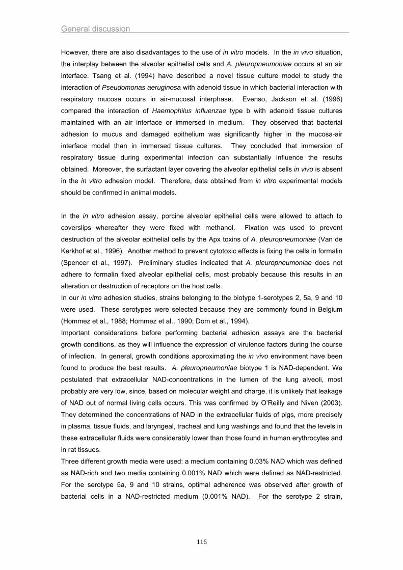
General discussion
However, there are also disadvantages to the use of in vitro models. In the in vivo situation,
the interplay between the alveolar epithelial cells and A. pleuropneumoniae occurs at an air
interface. Tsang et al. (1994) have described a novel tissue culture model to study the
interaction of Pseudomonas aeruginosa with adenoid tissue in which bacterial interaction with
respiratory mucosa occurs in air-mucosal interphase. Evenso, Jackson et al. (1996)
compared the interaction of Haemophilus influenzae type b with adenoid tissue cultures
maintained with an air interface or immersed in medium. They observed that bacterial
adhesion to mucus and damaged epithelium was significantly higher in the mucosa-air
interface model than in immersed tissue cultures. They concluded that immersion of
respiratory tissue during experimental infection can substantially influence the results
obtained. Moreover, the surfactant layer covering the alveolar epithelial cells in vivo is absent
in the in vitro adhesion model. Therefore, data obtained from in vitro experimental models
should be confirmed in animal models.
In the in vitro adhesion assay, porcine alveolar epithelial cells were allowed to attach to
coverslips whereafter they were fixed with methanol. Fixation was used to prevent
destruction of the alveolar epithelial cells by the Apx toxins of A. pleuropneumoniae (Van de
Kerkhof et al., 1996). Another method to prevent cytotoxic effects is fixing the cells in formalin
(Spencer et al., 1997). Preliminary studies indicated that A. pleuropneumoniae does not
adhere to formalin fixed alveolar epithelial cells, most probably because this results in an
alteration or destruction of receptors on the host cells.
In our in vitro adhesion studies, strains belonging to the biotype 1-serotypes 2, 5a, 9 and 10
were used. These serotypes were selected because they are commonly found in Belgium
(Hommez et al., 1988; Hommez et al., 1990; Dom et al., 1994).
Important considerations before performing bacterial adhesion assays are the bacterial
growth conditions, as they will influence the expression of virulence factors during the course
of infection. In general, growth conditions approximating the in vivo environment have been
found to produce the best results. A. pleuropneumoniae biotype 1 is NAD-dependent. We
postulated that extracellular NAD-concentrations in the lumen of the lung alveoli, most
probably are very low, since, based on molecular weight and charge, it is unlikely that leakage
of NAD out of normal living cells occurs. This was confirmed by O’Reilly and Niven (2003).
They determined the concentrations of NAD in the extracellular fluids of pigs, more precisely
in plasma, tissue fluids, and laryngeal, tracheal and lung washings and found that the levels in
these extracellular fluids were considerably lower than those found in human erythrocytes and
in rat tissues.
Three different growth media were used: a medium containing 0.03% NAD which was defined
as NAD-rich and two media containing 0.001% NAD which were defined as NAD-restricted.
For the serotype 5a, 9 and 10 strains, optimal adherence was observed after growth of
bacterial cells in a NAD-restricted medium (0.001% NAD). For the serotype 2 strain,
116
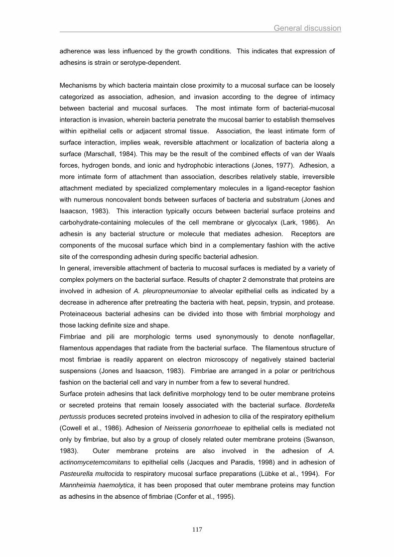
General discussion
adherence was less influenced by the growth conditions. This indicates that expression of
adhesins is strain or serotype-dependent.
Mechanisms by which bacteria maintain close proximity to a mucosal surface can be loosely
categorized as association, adhesion, and invasion according to the degree of intimacy
between bacterial and mucosal surfaces. The most intimate form of bacterial-mucosal
interaction is invasion, wherein bacteria penetrate the mucosal barrier to establish themselves
within epithelial cells or adjacent stromal tissue. Association, the least intimate form of
surface interaction, implies weak, reversible attachment or localization of bacteria along a
surface (Marschall, 1984). This may be the result of the combined effects of van der Waals
forces, hydrogen bonds, and ionic and hydrophobic interactions (Jones, 1977). Adhesion, a
more intimate form of attachment than association, describes relatively stable, irreversible
attachment mediated by specialized complementary molecules in a ligand-receptor fashion
with numerous noncovalent bonds between surfaces of bacteria and substratum (Jones and
Isaacson, 1983). This interaction typically occurs between bacterial surface proteins and
carbohydrate-containing molecules of the cell membrane or glycocalyx (Lark, 1986). An
adhesin is any bacterial structure or molecule that mediates adhesion. Receptors are
components of the mucosal surface which bind in a complementary fashion with the active
site of the corresponding adhesin during specific bacterial adhesion.
In general, irreversible attachment of bacteria to mucosal surfaces is mediated by a variety of
complex polymers on the bacterial surface. Results of chapter 2 demonstrate that proteins are
involved in adhesion of A. pleuropneumoniae to alveolar epithelial cells as indicated by a
decrease in adherence after pretreating the bacteria with heat, pepsin, trypsin, and protease.
Proteinaceous bacterial adhesins can be divided into those with fimbrial morphology and
those lacking definite size and shape.
Fimbriae and pili are morphologic terms used synonymously to denote nonflagellar,
filamentous appendages that radiate from the bacterial surface. The filamentous structure of
most fimbriae is readily apparent on electron microscopy of negatively stained bacterial
suspensions (Jones and Isaacson, 1983). Fimbriae are arranged in a polar or peritrichous
fashion on the bacterial cell and vary in number from a few to several hundred.
Surface protein adhesins that lack definitive morphology tend to be outer membrane proteins
or secreted proteins that remain loosely associated with the bacterial surface. Bordetella
pertussis produces secreted proteins involved in adhesion to cilia of the respiratory epithelium
(Cowell et al., 1986). Adhesion of Neisseria gonorrhoeae to epithelial cells is mediated not
only by fimbriae, but also by a group of closely related outer membrane proteins (Swanson,
1983). Outer membrane proteins are also involved in the adhesion of A.
actinomycetemcomitans to epithelial cells (Jacques and Paradis, 1998) and in adhesion of
Pasteurella multocida to respiratory mucosal surface preparations (Lübke et al., 1994). For
Mannheimia haemolytica, it has been proposed that outer membrane proteins may function
as adhesins in the absence of fimbriae (Confer et al., 1995).
117
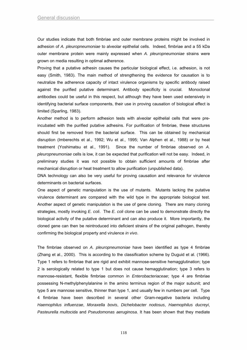
General discussion
Our studies indicate that both fimbriae and outer membrane proteins might be involved in
adhesion of A. pleuropneumoniae to alveolar epithelial cells. Indeed, fimbriae and a 55 kDa
outer membrane protein were mainly expressed when A. pleuropneumoniae strains were
grown on media resulting in optimal adherence.
Proving that a putative adhesin causes the particular biological effect, i.e. adhesion, is not
easy (Smith, 1983). The main method of strengthening the evidence for causation is to
neutralize the adherence capacity of intact virulence organisms by specific antibody raised
against the purified putative determinant. Antibody specificity is crucial. Monoclonal
antibodies could be useful in this respect, but although they have been used extensively in
identifying bacterial surface components, their use in proving causation of biological effect is
limited (Sparling, 1983).
Another method is to perform adhesion tests with alveolar epithelial cells that were pre-
incubated with the purified putative adhesins. For purification of fimbriae, these structures
should first be removed from the bacterial surface. This can be obtained by mechanical
disruption (Imberechts et al., 1992; Wu et al., 1995; Van Alphen et al., 1988) or by heat
treatment (Yoshimatsu et al., 1991). Since the number of fimbriae observed on A.
pleuropneumoniae cells is low, it can be expected that purification will not be easy. Indeed, in
preliminary studies it was not possible to obtain sufficient amounts of fimbriae after
mechanical disruption or heat treatment to allow purification (unpublished data).
DNA technology can also be very useful for proving causation and relevance for virulence
determinants on bacterial surfaces.
One aspect of genetic manipulation is the use of mutants. Mutants lacking the putative
virulence determinant are compared with the wild type in the appropriate biological test.
Another aspect of genetic manipulation is the use of gene cloning. There are many cloning
strategies, mostly invoking E. coli. The E. coli clone can be used to demonstrate directly the
biological activity of the putative determinant and can also produce it. More importantly, the
cloned gene can then be reintroduced into deficient strains of the original pathogen, thereby
confirming the biological property and virulence in vivo.
The fimbriae observed on A. pleuropneumoniae have been identified as type 4 fimbriae
(Zhang et al., 2000). This is according to the classification scheme by Duguid et al. (1966).
Type 1 refers to fimbriae that are rigid and exhibit mannose-sensitive hemagglutination; type
2 is serologically related to type 1 but does not cause hemagglutination; type 3 refers to
mannose-resistant, flexible fimbriae common in Enterobacteriaceae; type 4 are fimbriae
possessing N-methylphenylalanine in the amino terminus region of the major subunit; and
type 5 are mannose sensitive, thinner than type 1, and usually few in numbers per cell. Type
4 fimbriae have been described in several other Gram-negative bacteria including
Haemophilus influenzae, Moraxella bovis, Dichelobacter nodosus, Haemophilus ducreyi,
Pasteurella multocida and Pseudomonas aeruginosa. It has been shown that they mediate
118
General discussion
attachment of these bacteria to epithelial cells in vivo and in vitro (Sato and Okinaga, 1987;
Doig et al., 1988; Paranchyn and Frost, 1988; Starr et al., 1999).
Recently, Stevenson et al. (2003) reported the identification and characterization of the type 4
fimbrial structural gene (apfA) of A. pleuropneumoniae. In addition a number of open reading
frames were identified that have significant homology to type 4 fimbrial biogenesis genes from
other species. These type 4 fimbrial biogenesis genes showed the strongest homology to
putative type 4 fimbrial genes of Haemophilus ducreyi. Recombinant fimbriae produced by
expressing type 4 subunits from Moraxella bovis (Beard et al., 1990) and Dichelobacter
nodosus (Elleman et al., 1986) in Pseudomonas aeruginosa have been used as vaccine
antigens and this type of approach might be interesting for an A. pleuropneumoniae ApfA
based vaccine. Furthermore, it was shown by Boekema et al. (2003) that contact with
epithelial cells is a trigger for fimbriae production and that there is an in vivo induced activity
of the fimbriae promoter shortly after infection, suggesting that fimbriae play a role in the
colonisation and the pathogenesis.
Surface hydrophobicity of bacteria is an important physical factor for adhesion, especially
when the substrata are either hydrophilic or hydrophobic. Generally, bacteria with
hydrophobic properties prefer materials with hydrophobic surfaces, those with hydrophilic
characteristics prefer hydrophilic surfaces (Hogt et al., 1983; Satou et al., 1988;
Vacheethasanee et al., 1998). To assess the role of hydrophobic interactions in adherence,
we studied the influence of the presence of the hydrophobic bond-breaking agent
tetramethylurea. This technique was also used by Bélanger et al. (1994) in relation to A.
pleuropneumoniae adherence to crude mucus. Hydrophobic interactions did not seem to
participate in the binding of A. pleuropneumoniae to alveolar epithelial cells in vitro since
tetramethylurea had no effect on binding.
Many adhesins act as lectins, recognizing specific carbohydrate moieties on host cell
surfaces. Therefore, bacterial cells were subjected to treatments with various sugars to
determine whether carbohydrate-specific lectins on the surface of A. pleuropneumoniae cells
are involved in adherence. The various sugars we used were mono-and disacharides. The
finding that none of these sugars caused decreased adherence does not mean that A.
pleuropneumoniae can not interact with specific carbohydrates in adherence processes.
Indeed, adhesins can recognize complex carbohydrates as has been described for the P
fimbriae of Escherichia coli. These fimbriae recognize complex carbohydrates such as
globotriasoylceramide and globoside (Bock et al., 1985). Moreover, it can not be excluded
that different types of lectins are present on the bacterial cells or that a heterogeneous
population of bacterial cells is present, each bearing a different lectin (Ofek and Sharon,
1990). In such cases, a combination of sugars is a more effective inhibitor than individual
ones. This, however, remains to be tested.
119
General discussion
Treatment of A. pleuropneumoniae strains with sodium metaperiodate resulted in decreased
adhesion of the bacteria to alveolar epithelial cells, but inhibition was clearly lower than after
treatment of bacteria with proteolytic enzymes. The finding that inhibition of adhesion was
very high when bacteria were treated with a combination of sodium metaperiodate and
pronase suggests that more than one adhesin is involved. Indeed, although adhesion of
bacteria to host cells may depend on the binding of a unique bacterial adhesin to a unique
host receptor, most frequently bacterial adhesion depends on simultaneous interaction
between multiple adhesins with different cell receptors. The inhibition of a single pathway
therefore may have no effect on overall adhesion or at best may produce a partial inhibition.
Most Pasteurellaceae organisms can employ multiple molecular mechanisms of adherence to
initiate infection (Jacques and Paradis, 1998). Proteins (e.g. fimbriae, fibrils, outer membrane
proteins) and/or polysaccharides were shown to play a role in adherence of several members
of this bacterial family. These multiple adhesins may be used in a stepwise fashion during
colonization. It seems reasonable that evolutionary pressures have selected organisms that
are capable of using more than one mechanism of adherence (Ofek and Doyle, 1994).
As stated before, it is very important to confirm data obtained from in vitro models in animal
models. This was certainly recommended in our studies because the surfactant layer
covering the alveolar epithelial cells in vivo is absent in the in vitro adhesion model.
Pulmonary surfactant is a lipoprotein complex that is synthesized and secreted by the alveolar
type II epithelial cell and the airway Clara cell (Gourke, 1998). Once in the extracellular space,
surfactant reduces surface tension at the air-liquid interface of the lung, a function that
requires an appropriate mix of surfactant lipids and the hydrophobic proteins, surfactant
protein B (SP-B) and SP-C (Whitsett, 1995). Phospholipids of several types are the major
components of surfactant. Phospholipids are amphipathic molecules with a polar head group
and a nonpolar tail. These hydrophobic tails are extended into the air. The single most
abundant phospholipid is dipalmitoylphosphatidylcholine (Hawgood, 1991). Other
phospholipids are: unsaturated phosphatidylcholines, phosphatidylglycerol,
phosphatidylethanolamine and phosphatidylserine. It is possible that A. pleuropneumoniae
interacts with surfactant as a first step in the colonization. We demonstrated that A.
pleuropneumoniae binds to surfactant proteins. Moreover, in vivo adhesion studies resulted
in adherence of A. pleuropneumoniae to lung alveoli irrespective of the growth conditions of
the bacteria. This was in contrast with the in vitro adhesion studies whereby growth on NAD
restricted media resulted in significantly higher adhesion scores in serotype 5, 9 and 10
strains. Hydrophobicity tests as described by Vercauteren et al. (1993) did not show
differences in hydrophobicity between bacteria grown under different growth conditions
(unpublished results). All these results indicate that adhesion of A. pleuropneumoniae to
alveoli is a two step adhesion. A first step could be a reversible association of A.
pleuropneumoniae with surfactant. Lipopolysaccharides might be involved in this association
since Jeannotte et al. (2003) described that A. pleuropneumoniae can bind to
120
General discussion
phosphatidylethanolamine. Such association has also been described for other pathogenic
bacteria including Chlamydia pneumoniae and Chlamydia trachomatis (Krivan et al., 1991),
Helicobacter pylori (Lingwood et al., 1992), Helicobacter mustelae (Gold et al. 1993),
Haemophilus influenzae (Busse et al., 1997) and Campylobacter upsaliensis (Busse et al.,
1997).
A second step could be an adhesion step to alveolar epithelial cells i.e. a relatively stable,
irreversible attachment mediated by specialized complementary molecules in a ligand-
receptor fashion. The above described candidate-adhesins i.e. fimbriae and a 55 kDa outer
membrane protein could play a role in this adhesion.
Surfactant also plays a role in host defence against infection and inflammation (Wright,
1997&2003). Two of the surfactant proteins, SP-A and SP-D, are members of the collectin
protein family (Crouch and Wright, 2001; Crouch et al., 2000). The most well-defined function
of the collectins is their ability to opsonize pathogens, including bacteria and viruses, and to
facilitate phagocytosis by innate immune cells such as macrophages and monocytes. It is
unlikely that opsonization of A. pleuropneumoniae and subsequent phagocytosis by
macrophages and monocytes will result in killing of A. pleuropneumoniae. Indeed, Cruijsen et
al. (1992) demonstrated that porcine alveolar macrophages effectively phagocytise virulent
strains of A. pleuropneumoniae in the presence of convalescent pig serum but they are not
able to kill the bacteria intracellularly. This is most probably due to intracellular released Apx
toxins (Cruijsen et al., 1992). Data presented by Wu and coworkers (2003) show convincingly
that, in addition to facilitating pathogen uptake and killing by immune cells, SP-A and SP-D
are directly antimicrobial, that is, they kill bacteria in the absence of immune effector cells.
The effect of these surfactant proteins on A. pleuropneumoniae has not yet been studied.
In conclusion, results of the present thesis confirm that the currently available subunit
vaccines, based on Apx toxins, induce partial protection. Inclusion of adhesins in such
vaccines might improve efficacy. Adhesion of A. pleuropneumoniae to alveolar epithelial cells
is a very complex phenomenon and several bacterial antigens seem to be involved including
outer membrane proteins and fimbriae. Additional studies are necessary to further
characterize virulence factors responsible for adhesion of A. pleuropneumoniae to host cells.
121
General discussion
REFERENCES
Beard, M.K., Mattick, J.S., Moore, L.J. Mott, M.R. Marrs, C.F., 1990. Egerton J.R.
Morphogenic expression of Moraxella bovis fimbriae (pili) in Pseudomonas
aeruginosa. J. Bacteriol. 172, 2601-2607.
Bélanger, M., Dubreuil, D., Jacques, M., 1994. Proteins found within porcine respiratory
tract secretions bind lipopolysaccharides of Actinobacillus pleuropneumoniae.
Infect. Immun. 62, 868-873.
Bock, K., Breimer, M.E., Brignole, A., Hansson, G.C., Karlsson, K.A., Larson, G.,
Leffler, H., Samuelsson, B.E., Stromberg, N., Svanborg-Edén, C., Thurin, J.,
1985. Specificity of binding of a strain of uropathogenic Escherichia coli to
Galα1-4Gal containing glycosphingolipids. J. Biol. Chem. 260, 8545.
Boekema, B.K.H.L., Stockhofe-Zurwieden, N., Smith, H.E., Kamp, E.M., van Putten,
J.P., Verheiijden, J.H., 2003. Adherence of Actinobacillus pleuropneumoniae to
primary cultures of porcine lung epithelial cells. Vet. Microbiol. 93, 133-144.
Brinton, C.C., 1965. The structure, function, synthesis and genetic control of bacterial
pili and a molecular model for DNA and RNA transport in Gram negative
bacteria. Trans. N.Y. Acad. Sci 27, 1003-1054.
Busse, J., Hartmann, E., Lingwood, C.A., 1997. Receptor affinity purification of a lipid-
binding adhesin from Haemophilus influenzae. J. Infect. Dis. 175, 77-83.
Cho, W.S., Chae, C., 2001. Expression of the apxIV gene in pigs naturally infected with
Actinobacillus pleuropneumoniae. J. Comp. Path. 125, 34-40.
Confer, A.W., Clinckenbeard, K., Murphy, G.L., 1995. Pathogenesis and virulence of
Pasteurella haemolytica in cattle: an analysis of current knowledge and future
approaches. In: Haemophilus, Actinobacillus and Pasteurella (Eds: W. Donachie,
F.A. Lainson and J.C. Hodgson). Proc. Haemophilus, Actinobacillus and
Pasteurella Conf., Edinburgh, Scotland, 51-62.
Cowell, J.L., Urisu, A., Zhang, J.M., Steven, A.C., Manclark, C.R., 1986. Filamentous
hemagglutinin and fimbriae of Bordetella pertussis: properties and roles in
attachment. In: L. Leive (ed), Microbiology-1986. Am. Soc.Microb., Washington,
D.C.
Crouch, E., Hartshorn, K., Ofek, I., 2000. Collectins and pulmonary innate immunity.
Immunol. Rev. 173, 52-65.
Crouch, E., Wright, J.R., 2001. Surfactant proteins A and D and pulmonary host
defense. Annu. Rev. Physiol. 63, 521-554.
Cruijsen, T., Van Leengoed, L.A.M.G., Dekker-Nooren, T.C., Schoevers, J.H.,
Verheijden, J.H., 1992. Phagocytosis and killing of Actinobacillus
122
General discussion
pleuropneumoniae by alveolar macrophages and polymorphonuclear leukocytes
isolated from pigs. Infect. Immun. 60, 4867-4871.
De Graaf, F.K., Roorda I., 1982. Production, purification, and characterization of the
fimbrial adhesive antigen F41 isolated from calf enteropathogenic Escherichia
coli strain B41M. Infect. Immun. 36, 751-758.
Doig, P.T.T., Sastry, P.A., Lee, K.K., Hodges, R.S., Paranchych, W., Irvin, R.T., 1988.
Role of pili in adhesion of Pseudomonas aeruginosa to human respiratory
epithelial cells. Infect. Immun. 56, 1641.
Dom, P., Haesebrouck, F., Kamp, E., Smits, M.A., 1992. Influence of Actinobacillus
pleuropneumoniae serotype 2 and its cytolysins on porcine neutrophil
chemiluminescence. Infect. Immun. 60, 4328-4334.
Dom, P., Haesebrouck, F., Kamp, E.M., Smits, M.A., 1994. NAD-independent
Actinobacillus pleuropneumoniae strains: production of RTX toxins and
interactions with porcine phagocytes. Vet. Microbiol. 39, 205-218.
Doughty, S.W., Ruffolo, C.G., Adler, B., 2000. The type 4 fimbrial subunit gene of
Pasteurella multocida. Vet. Microbiol. 72, 79-90.
Duguid, J. P., E. S. Anderson, and I. Campbell. 1966. Fimbriae and adhesive
properties in Salmonellae. J. Pathol. Bacteriol. 92, 107-138
Dunkle, L.M., Blair, L.L., Fortune, K.P., 1986. Transformation of a plasmid encoding an
adhesin of Staphyococcus aureus into a nonadherent staphylococcal strain. J.
Infect. Dis. 153, 670-675.
Elleman, T.C., Hoyne, P.A., Stewart, D.J., McKern, N.M., Peterson, J.E., 1986.
Expression of pili from Bacteroides nodosus in Pseudomonas aeruginosa. J.
Bacteriol. 168, 574-580.
Falk, P., Roth, K., Gordon, J., 1994a. Lectins are sensitive tools for defining the
differentiation programs of epithelial cell lineages in the development of adult
mouse gastrointestinal tract. Am. J. Physiol. 266, G987-1003.
Falk, P., Boren, T. Haslam, D., 1994b. Bacterial adhesion and colonization assays. In:
Russell D. (ed.) Microbes as tools for cell biology. Academic press, San Diego,
CA, 165-192.
Fedorka-Cray, P.J., 1989. Actinobacillus (Haemophilus) pleuropneumoniae virulence
factors: Partial characterization and efficacy studies in swine. University of
Nebraska, Ph.D. Thesis, 217 pp.
Fedorka-Cray, P.J., D.L. Stine, J.M. Greenwald, J.T. Gray, M.J. Huether, G.A.
Anderson, 1993. The importance of secreted virulence factors in Actinobacillus
pleuropneumoniae bacterin preparation: a comparison. Vet. Microbiol. 37, 85-
100.
123

General discussion
Frey, J., Nicolet, J., 1991. Immunological properties of Actinobacillus
pleuropneumoniae hemolysin I. Vet. Microbiol. 28, 61-73.
Goerke, J., 1998. Pulmonary surfactant : functions and molecular composition.
Biochem. Biophys. Acta 1408, 79-89.
Gold, B.D., Huesca, M., Sherman, P.M., Lingwood, C.A., 1993. Helicobacter mustelae
and Helicobacter pylori bind to common lipid receptors in vitro. Infect. Immun. 61,
2632-2638.
Haesebrouck, F., Van de Kerkhof, A., Dom, P., Chiers, K., Ducatelle, R., 1996. Cross-
protection between Actinobacillus pleuropneumoniae biotypes-serotypes in pigs.
Vet. Microbiol. 52, 277-84.
Hawgood, S., 1991. Surfactant : composition, structure and metabolism. In : The Lung :
Scientific Foundations, Crystal R.G., West J.B. (ed.), Raven Press, Ltd, New
York, 247-261.
Hensel, A., Huter, V., Katinger, A., Raza, P., Strnistischie, C., Roesler, U., Brand, E.,
Lubitz, W., 2000. Intramuscular immunization with genetically inactivated
(ghosts) Actinobacillus pleuropneumoniae serotype 9 protect pigs against
homologous aerosol challenge and prevents carrier state. Vaccine 18, 2945-
2955.
Hogt, A.H., Dankert, J., De Vries, J.A., 1983. Adhesion of coagulase-negative
staphylococci to biomaterials. J. Gen. Microbiol. 129, 2959-2968.
Hommez, J., Devriese, L.A., Cassimon, P., Castryck, F., 1988. Serotypes and antibiotic
sensitivity of Actinobacillus (Haemophilus) pleuropneumoniae strains isolated in
Belgium. Vl. Diergen. Tijdschr. 57, 46-52.
Hommez, J., Devriese, L.A., Castryck, F., Cassimon, P., 1990. Slide precipitation: a
simple method to type Actinobacillus (Haemophilus) pleuropneumoniae. Vet.
Microbiol. 24, 123-126.
Imberechts, H., De Greve, H., Schlicker, C., Bouchet, H., Pohl, P., Charlier, G.,
Bertschinger, H., Wild, P., Vandekerckhove, J., Van Damme, J., Van Montagu,
M., Lintermans, P., 1992. Characterization of F107 fimbriae of Escherichia coli
107/86, wich causes edema disease in pigs, and nucleotide sequence of the
F107 major fimbrial subunit gene, fedA. Infect. Immun. 60, 1963-1971.
Isaacson, R.E., 1977. K99 surface antigen of Escherichia coli: purification and partial
characterization. Infect. Immun. 15, 272-279.
Isaacson, R.E., Richter, P., 1981. Escherichia coli 987P pilus: purification and partial
characterization. J. Bacteriol. 146, 784-789.
Jackson, A.D., Cole, P.J. Wilson, R., 1996. Comparison of Haemophilus influenza type
b interaction with respiratory mucosa organ cultures maintained with an air
interface or immersed in medium. Infect. Immun. 64, 2353-2355.
124
General discussion
Jacques, M., Paradis S.E., 1998. Adhesin-receptor interactions in Pasteurellaceae.
FEMS Microbiol. Rev. 22, 45-59.
Jeannotte, M.E., Abul-Milh, M., Dubreuil, J.D., Jacques, M., 2003. Binding of
Actinobacillus pleuropneumoniae to phosphatidylethanolamine. Infect. Immun.
71, 4657-4663.
Jones, G.W., 1977. The attachment of bacteria to the surfaces of animal cells. In: J.L.
Reissing (ed), Microbial interactions. Chapman & Hall, Ltd., London.
Jones, G.W., Isaacson R.E., 1983. Proteinaceous bacterial adhesions and their
receptors. Crit. Rev. Microbiol. 10, 229-260.
Krivan, H.C., Nilsson, B., Lingwood, C.A., Ryu, H., 1991. Chlamydia trachomatis and
Chlamydia pneumoniae bind specifically to phosphatidylethanolamine in HeLa
cells and to GalNAcβ1-4Galβ1-4Glc sequences found in asialo-GM1 and asialo-
GM2. Biochem. Biophys. Res. Commun. 175, 1082-1089.
Lark, D.L., 1986. Protein-carbohydrate interactions in biological systems. Academic
Press, Inc., New York.
Levine, M.M., Ristaino, P., Marley, G., Smyth, C., Knutton, S., Boedeker, E., Black, R.,
Young, C., Clements, M.L., Cheney, C., Patnaik, R., 1984. Coli surface antigens
1 and 3 of colonization factor antigen II-positive enterotoxigenic Escherichia coli:
morphology, purification, and immune responses in humans. Infect. Immun. 44,
409-420.
Lingwood, C.A., Huesca, M., Kuksis, A., 1992. The glycerolipid receptor for
Helicobacter pylori (and exoenzyme S) is phosphatidylethanolamine. Infect.
Immun. 60, 2470-2474.
Lium, B., Brun, E., Fjetland, O., Lund A., 1998. A field study of the efficacy of an
Actinobacillus pleuropneumoniae subunit vaccine in the control of
pleuropneumonia caused by Actinobacillus serotype 6. Proc. 15th International
Pig Veterinary Society Congress, Birmingham, England, 256.
López, J. V., Font, J., Prat, M., Cantin, C., 1998. Technical and economic efficacy of
Porcilis App: a field trial. Proc. 15th International Pig Veterinary Society
Congress, England, 264.
Lübke, A., Hartmann, L., Schröder, W., Hellmann E., 1994. Isolation and partial
characterization of the major protein of the outer membrane of Pasteurella
haemolytica and Pasteurella multocida. Zent. Bl. Bakt. 281, 45-54.
Martelli, P., Guadagnini, P.F., Foccoli, E., Ballarini G., 1996. Efficacy of an
Actinobacillus pleuropneumoniae subunit vaccine in the control of
pleuropneumonia: a field trial. Proc. 14th International Pig Veterinary Society
Congress, Bologna, Italy, 214.
125
General discussion
Marshall, K.C., Stout,R., Mitchell, R., 1971. Mechanism of the initial events in the
sorption of marine bacteria to surfaces. J. Gen. Microbiol 68, 337-348.
Marshall, K.C., 1984. Microbial adhesion and aggregation. Springer-Verlag, New York,
397-399.
Nataro, P., Kaper, J.B., Robins-Browne, R., 1987. Patterns of adherence of
diarrheagenic Escherichia coli to Hep-2 cells. Pediatr. Infect. Dis. J. 6, 829-831.
Nicolet, J., 1992. Actinobacillus pleuropneumoniae. In: A.D. Leman, B. Straw, W.L.
Mengeling, S. D'Allaire and D.J. Taylor (Eds.). Diseases of swine. Ames, Iowa
State University Press, 401-408.
Nielsen, R., 1984. Haemophilus pleuropneumoniae serotypes: Cross protection
experiments. Nord. Vet. Med. 36, 221-234.
Ofek, I., Courtney,H.S., Schifferli, D.M., 1986. Enzyme linked immunosorbent assay for
adherence of bacteria to animal cells. J. Clin. Microbiol. 24, 512-516.
Ofek, I, Sharon, N., 1990. Adhesins as lectins : specificity and role in infection.
Curr.Top. Microbiol. Immunol. 151, 91-108.
Ofek, I., Doyle, R.J., 1994. Bacterial adhesion to cells and tissues. Chapman and Hall,
Inc., London, England.
O’Reilly, T., Niven, D.F., 2003. Levels of nicotinamide adenine dinucleotide in
extracellular body fluids of pigs may be growth-limiting for Actinobacillus
pleuropneumoniae and Haemophilus parasuis. Can J. Vet. Res. 67, 229-231.
Paranchych, W., Frost, L.S., 1988. The physiology and biochemistry of pili. Adv.
Microb. Physiol. 29, 53.
Pommier, P, Ridremont, B, Wessel-Robert, S, Keïta, A., 1996. Field study into efficacy
of a new Actinobacillus pleuropneumoniae subunit vaccine. Proc. 14th
International Pig Veterinary Society Congress, Bologna, Italy, 206.
Robbins-Browne, R., Bennet-Wood, V., 1992. Quantitative assessment of the ability of
Escherichia coli to invade cultured animal cells. Microb. Pathog. 12, 159-164.
Russell, W.M.S., Burch, R.L., 1959. The principles of human experimental technique.
Methuen, London, England.
Salit, I.E., Gotschlich E.C. 1977. Hemagglutination by purified type 1 Escherichia coli
pili. J. Exp. Med. 146, 1169-1181.
Sato, H., Okinaga, K., 1987. Role of pili in the adherence of Pseudomonas aeruginosa
to mouse epidermal cells. Infect. Immun. 55, 1774.
Satou, N., Satou, J., Shintani, H., 1988. Adherence of streptococci to surface-modified
glass. J. Gen. Microbiol. 134, 1299-1305.
Schaller, A., Kuhn, R., Kuhnert, P., Nicolet, J., Anderson, T.J., MacInnes, J.I., Segers,
R.P., Frey, J., 1999. Characterization of apxIVA, a new RTX determinant of
Actinobacillus pleuropneumoniae. Microbiol. 145, 2105-2116.
126
General discussion
Smith, H., 1983. The elusive determinants of bacterial interference with non-specific
host defences. Philos. Trans. R. Soc. London Ser. B 303, 99-113.
Sparling, P.F., 1983. Applications of genetics to studies of bacterial virulence. Philos.
Trans. R. Soc. London Ser. B 303, 199-207.
Spencer, J., Chart, H., Smith, H.R., 1997. Improved detection of enteroaggregative
Escherichia coli using formalin-fixed HEp-2 cells. Lett. Appl. Microbiol. 25, 325-
326.
Starr, M., Adler, B., Ruffolo, C., Al-Hasani, K., Robins-Browne, R., 1999. htfA, A novel
gene mediating adhesion of nontypeable Haemophilus influenzae to HEP-2 cells.
Proc. Haemophilus, Actinobacillus and Pasteurella Conf., Mabula, South-Africa,
30.
Stevenson, A., Macdonald, J., Roberts, M., 2003. Cloning and characterization of type
4 fimbrial genes from Actinobacillus pleuropneumoniae. Vet. Microbiol. 92, 121-
134.
Swanson, J., 1983. Gonococcal adherence: selected topics. Rev. Infect. Dis. 5, S678-
S684.
Tsang, K.W.T., Rutman, A., Tanaka, E., 1994. Interaction of Pseudomonas aeruginosa
with human respiratory mucosa in vitro. Eur. Respir. J. 7, 1746-1753.
Vacheethasanee, K., Temenoff, J.S., Higashi, J.M., 1998. Bacterial surface properties
of clinically isolated Staphylococcus epidermidis strains determine adhesion on
polyethylene. J. Biomed. Mater Res. 42, 425-432.
Valks, MMH, Nell, T, Van den Bosch, JF., 1996. A clinical field trial in finishing pigs to
evaluate the efficacy of a new APP subunit vaccine. Proc. 14th International Pig
Veterinary Society Congress, Bologna, Italy, 208.
Van Alphen, L., Van den Berghe, N., Geelen-van den Broek, L., 1988. Interaction of
Haemophilus influenzae with human enterocytes and oropharyngeal epithelial
cells is mediated by a common fimbrial epitope. Infect. Immun. 56, 1800-1806.
Van den Bosch, H., Frey J., 2003. Interference of outer membrane protein PalA with
protective immunity against Actinobacillus pleuropneumoniae infections in
vaccinated pigs. Vaccine 21, 3601-3607.
Van de Kerkhof, A., Haesebrouck, F., Chiers, K., Ducatelle, R., Kamp, E.M., Smits,
M.A., 1996. Influence of Actinobacillus pleuropneumoniae and its metabolites on
porcine alveolar epithelial cells. Infect. Immun. 64, 3905-3907.
Vercauteren, R., Dom, P., Haesebrouck, F., 1993. Virulence of bacteria in relation to
their hydrophobicity, adhesiveness and phagocytosis. Vlaams Diergen. Tijdschr.
62, 29-34.
127
General discussion
Whitsett, J.A., Nogee, L.M., Weaver, T.E., Horowitz, A.D., 1995. Human surfactant
protien B: structure, function, regulation and genetic disease. Physiol. Rev. 75,
749-757.
Wright, J.R., 1997. Immunomodulatory functions of surfactant. Physiol. Rev. 77, 931-
962.
Wright, J.R., 2003. Pulmonary surfactant : a front line of lung host defense. J. Clin.
Invest. 111, 1453-1455.
Wu, X., Gupta, S.K., Hazlett, L.D., 1995. Characterization of Pseudomonas aeruginosa
pili binding human corneal epithelial proteins. Curr. Eye Res. 14, 969-977.
Wu, H., 2003. Surfactant proteins A and D inhibit the growth of Gram negative bacteria
by increasing membrane permeability. J. Clin. Invest. 111, 1589-1602.
Yoshimatsu, K., Yuyama, Y., Ono, E., Syuto, B., Naiki, M., 1991. New methods for
isolation of K99 fimbriae from enterotoxigenic Escherichia coli. J. Vet. Med. Sci.
53, 1119-1121.
Zafriri, D., Ofek, I. Adar, R., 1989. Inhibitory activity of cranberry juice on adherence of
type 1 and type P fimbriated Escherichia coli to eukaryotic cells. Antimicrob.
Agents Chemother. 33, 92-98.
Zhang, Y.M., Tennent, J.M., Ingham, A., Beddome, G., Prideaux, C., Michalski, W.P.,
2000. Identification of type 4 fimbriae in Actinobacillus pleuropneumoniae. FEMS
Microbiol. Lett. 189, 15-18.
Zobell, C.E., 1943. The effect of solid surfaces upon bacterial activity. J. Bacteriol. 46,
39-56.
128
Summary
SUMMARY
A. pleuropneumoniae causes severe losses in the pig rearing industry. Although the disease
can be controlled by antimicrobial agents, the use of these products has several
disadvantages including induction of acquired resistance in pathogenic bacteria and bacteria
belonging to the normal flora of pigs. Therefore, prevention instead of therapy is an
increasing requirement in the control of the disease. Vaccines could be very useful to control
porcine pleuropneumonia. Rational design of effective antibacterial vaccines, however,
requires knowledge of the virulence factors of the bacterium and the pathogenesis of the
disease. Pathogenesis of porcine pleuropneumonia is very complex and several virulence
factors have been described. Some of these virulence factors, such as the Apx toxins, have
been extensively studied. Virulence attributes involved in lung colonisation are, however, less
well known. The need for research in these first steps of pathogenesis provided the main
rationale for this thesis.
The experiments performed in this thesis are divided in 3 Chapters. In Chapter 1, the efficacy
of vaccines containing Apx toxins was evaluated. Thereafter, the adhesion of A.
pleuropneumoniae to porcine alveolar epithelial cells in vitro and in vivo was studied (Chapter
2). Finally, the efficacy of a vaccine containing candidate-adhesins was evaluated (Chapter
3).
In a first study described in Chapter 1, the efficacy of two inactivated vaccines containing the
Apx toxins of Actinobacillus pleuropneumoniae (Hemopig™, Biokema, Lausanne, Switzerland
and Porcilis™ App, Intervet, Boxmeer, The Netherlands) was determined. Therefore, ten pigs
were vaccinated twice with Hemopig™ and eight pigs with Porcilis™ App. Ten control
animals were injected twice with a saline solution. Three weeks after the second vaccination,
all pigs were endobronchially inoculated with 105 colony-forming units (CFU) of an A.
pleuropneumoniae serotype 9 strain. Increased respiratory rate and/or fever were observed
in all vaccinated and control pigs after challenge. One pig of the Hemopig™ group and of the
Porcilis™ App group died, whereas all pigs of the control group survived the challenge.
Surviving pigs were killed at 7 days after challenge. The mean percentage of affected lung
tissue was 34% in the control group, 16% in the Hemopig™ group, and 17% in the Porcilis™
App group. A. pleuropneumoniae was isolated from the lungs of all 10 control animals, from 7
of the 10 animals vaccinated with Hemopig™ and from 5 of the 8 animals vaccinated with
Porcilis™ App. The mean bacterial titres of the caudal lung lobes were 1.4x10⁶ CFU/g in the
control group, 1.7x10³ CFU/g in the Hemopig™ group, and 4.8x10
3 CFU/g in the Porcilis™
App group. In both vaccinated groups the mean number of days with dyspnoea, the mean
number of days with fever, the mean percentage of affected lung tissue, and the mean
129
Summary
bacterial titre in the caudal lung lobes were significantly lower than in the control group.
Significant differences between the two vaccinated groups were not observed. It was
concluded that both vaccines induced partial protection.
In a second study, the efficacy of a subunit vaccine containing the Apx toxins and transferrin
binding proteins was determined. Therefore, ten pigs were vaccinated twice with the vaccine.
Eight control animals were injected twice with a saline solution. Three weeks after the second
vaccination, all pigs were endobronchially inoculated with 106.5
colony-forming units (CFU) of
an A. pleuropneumoniae serotype 9 strain. In the vaccine group, none of the pigs died after
inoculation. Only one pig of the control group survived challenge. Surviving pigs were killed
at 7 days after challenge. The mean percentage of affected lung tissue was 64% in the
control group and 17% in the vaccine group. A. pleuropneumoniae was isolated from the
lungs of all animals. The mean bacterial titres of the caudal lung lobes were 5.0x10⁸ CFU/g in
the control group and 3.0x10⁶ CFU/g in the vaccine group. It was concluded that the vaccine
induced partial protection against severe challenge.
Keeping in mind the very complex pathogenesis of porcine pleuropneumonia, inclusion of
other bacterial virulence factors in subunit vaccines might be of value. Bacterial virulence
factors that, most probably, would be very valuable to include in vaccines are adhesins.
Indeed, A. pleuropneumoniae first adheres to alveolar epithelial cells before it causes lung
lesions. Antibodies that interfere with adhesion could block this first step in the pathogenesis
of porcine pleuropneumonia and protect pigs against lung infection. Therefore, the adhesion
of A. pleuropneumoniae to cells in vitro and in vivo was studied (Chapter 2).
In a first study, the adhesion ability of A. pleuropneumoniae biovar 1 serotype 2, 5a, 9 and 10
strains was tested to alveolar epithelial cells in culture. For the serotype 5a, 9 and 10 strains,
optimal adherence was observed after growth of bacterial cells in a NAD-restricted medium
(0.001% NAD). This condition was also associated with the expression of a 55 kDa outer
membrane protein and of fimbriae. For the serotype 2 strain, adherence and expression of
fimbriae and a 55 kDa outer membrane protein was less influenced by the growth conditions.
The N-terminal amino acid sequence of the 55 kDa outer membrane protein had no homology
with any known sequence, suggesting that it is an as yet unknown protein. Adherence
capabilities were significantly reduced following treatment of the bacteria with proteolytic
enzymes or heat. These findings suggest that proteins are involved in adhesion. The
hydrophobic bond breaking agent tetramethylurea was unable to inhibit the adherence of A.
pleuropneumoniae to alveolar epithelial cells. Treatment of the bacteria with sodium
metaperiodate resulted in lower adhesion scores for the serotype 2 and 9 strains but the
inhibition of adhesion was clearly lower than after treatment with proteolytic enzymes. This
indicates that, besides proteins, carbohydrates might also be involved in adhesion of A.
pleuropneumoniae to alveolar epithelial cells. The finding that inhibition of adhesion was very
130
Summary
high when bacteria were treated with a combination of sodium metaperiodate and pronase
also suggests that more than one adhesin is involved.
Secondly, the effect of culture conditions on adhesion of inactivated A. pleuropneumoniae
biotype 1-serotype 2 and 9 strains to alveoli of experimentally inoculated piglets was studied.
Growing the bacteria on a NAD-restricted or NAD-rich medium did not influence this adhesion
and had no effect on the in vitro association of the bacteria with surfactant proteins. Adhesion
scores in alveolar epithelial cell cultures were, however, much higher when the serotype 9
strain was grown on the NAD-restricted medium. It is possible that different adhesins are
involved in colonisation of alveoli of infected pigs. Association with surfactant might be a first
step that precedes adhesion to alveolar epithelial cells.
Finally, in Chapter 3, the efficacy of two bacterins containing an A. pleuropneumoniae
serotype 10 strain was evaluated. The bacterial cells constituting bacterin 1 and 2 were
grown under NAD-rich (low adherence capacitiy to alveolar epithelial cell cultures) and NAD-
restricted (high adherence capacity to alveolar epithelial cell cultures) conditions, respectively.
Ten pigs were vaccinated twice with the bacterin 1 and 9 pigs with the bacterin 2. Ten control
animals were injected twice with a saline solution. Three weeks after the second vaccination,
all pigs were endobronchially inoculated with 106.5 colony-forming units (CFU) of an A.
pleuropneumoniae serotype 10 strain. In the bacterin 1 and 2 group, three and two pigs died
after inoculation, respectively. Only two pigs of the control group survived challenge.
Surviving pigs were killed at 7 days after challenge. The percentage of pigs with severe lung
lesions (>10% of the lung affected) was 100% in the control group, 70% in the bacterin 1
group and 22% in the bacterin 2 group. A. pleuropneumoniae was isolated from the lungs of
all animals. The mean bacterial titres of the caudal lung lobes were 7.0x10⁶ CFU/g in the
control group, 6.3x10⁵ CFU/g in the bacterin 1 group and 1.3x10⁶ CFU/g in the bacterin 2
group. It was concluded that both bacterins induced partial protection against severe
challenge. Furthermore, there are indications that the bacterin 2, containing A.
pleuropneumoniae bacteria grown under conditions resulting in high in vitro adhesion,
induced better protection than the bacterin 1.
131

Summary
132
Samenvatting
SAMENVATTING
A. pleuropneumoniae veroorzaakt grote economische verliezen in de varkenshouderij. Bij de
bestrijding van pleuropneumonie wordt vaak gebruik gemaakt van antibiotica. Alhoewel
antibiotica hun nut bewezen hebben, zijn er aan het overvloedig gebruik ervan verschillende
nadelen verbonden zoals het optreden van verworven resistentie. Het is duidelijk dat dient
gezocht te worden naar alternatieve bestrijdingsstrategiën. Hierbij wordt in de eerste plaats
gedacht aan vaccinatie. Bij de ontwikkeling van effectieve antibacteriële vaccins, is een
grondige kennis van de virulentiefactoren van de kiem en de pathogenese van de ziekte
noodzakelijk. De pathogenese van pleuropneumonie is zeer complex en er werden reeds
verschillende virulentiefactoren beschreven. Sommige van deze virulentiefactoren, zoals de
Apx toxines, werden in detail bestudeerd. Virulentiefactoren die betrokken zijn in de
kolonisatie van de long zijn echter minder gekend. Onderzoek naar deze eerste stappen in
de pathogenese was dan ook het hoofdobjectief van dit proefschrift.
Het experimentele gedeelte van dit proefschrift wordt in 3 hoofdstukken ingedeeld. Vooreerst
werd de doeltreffendheid van vaccins die Apx toxines bevatten, bepaald. Daarna werd de
adhesie van A. pleuropneumoniae aan alveolaire epitheelcellen in vitro en in vivo bestudeerd
(Hoofdstuk 2). Tenslotte werd de doeltreffendheid bepaald van een vaccin dat kandidaat-
adhesines bevat (Hoofdstuk 3).
In een eerste studie die beschreven werd in Hoofdstuk 1, werd de efficaciteit bepaald van
twee geïnactiveerde vaccins die de Apx toxines van A. pleuropneumoniae bevatten
(Hemopig™ Biokema, Lausanne, Zwitserland en Porcilis™ App, Intervet, Boxmeer,
Nederland). Hiertoe werden 10 varkens tweemaal, met een interval van 3 weken ingespoten
met Hemopig™ en 8 varkens werden tweemaal, met een interval van 3 weken gevaccineerd
met Porcilis™ App. Tien andere biggen werden met een gebufferde zoutoplossing op
analoge wijze geïnoculeerd en dienden als controles. Drie weken na de tweede vaccinatie
werden alle dieren endobronchiaal geïnoculeerd met 105 kolonie-vormende eenheden (kve)
van een A. pleuropneumoniae serotype 9 stam. Alle dieren werden klinisch gevolgd. Dieren
die overleefden, werden 7 dagen na de inoculatie geëuthanaseerd. Een verhoogde
ademhalingsfrequentie en/of koorts kon worden waargenomen bij alle gevaccineerde dieren
en controledieren. Alle controledieren overleefden de besmetting. Zowel in de Hemopig™
groep als de Porcilis™ App groep stierf er 1 dier na de besmetting. Het gemiddeld
percentage aangetast longweefsel bedroeg 34% in de controlegroep, 16% in de Hemopig™
groep en 17% in de Porcilis™ App groep. A. pleuropneumoniae kon uit de longen van alle
controledieren geïsoleerd worden en de gemiddelde bacteriële titer in de caudale
longkwabben bedroeg 1.4x10⁶ kve/gram. Bij de Hemopig™ groep kon A. pleuropneumoniae
bij 7 van de 10 dieren geïsoleerd worden en de gemiddelde bacteriële titer bedroeg 1.7x10³
133
Samenvatting
kve/gram. Bij de Porcilis™ App groep kon A. pleuropneumoniae bij 5 van de 8 dieren
geïsoleerd worden en de gemiddelde bacteriële titer bedroeg 4.8x10³ kve/gram. In beide
vaccingroepen was het gemiddeld aantal dagen met dyspnee, het gemiddeld aantal dagen
met koorts, het gemiddeld percentage aangetast longweefsel en de gemiddelde bacteriële
titer in de caudale longkwabben significant lager dan bij de controlegroep. Er werden geen
significante verschillen gevonden tussen de 2 vaccingroepen. Er werd geconcludeerd dat
beide vaccins partiële bescherming induceerden.
In een tweede studie werd de doeltreffendheid bepaald van een vaccin dat niet alleen Apx
toxines maar ook transferrine bindende proteïnes bevat. Hiertoe werden 10 dieren tweemaal
gevaccineerd met het vaccin met een interval van 4 weken. Acht controledieren werden
tweemaal ingespoten met een gebufferde zoutoplossing. Drie weken na de tweede
vaccinatie werden alle dieren endobronchiaal geïnoculeerd met 106.5 kolonie-vormende
eenheden (kve) van een A. pleuropneumoniae serotype 9 stam. In de vaccingroep stierf
geen enkel dier. Slechts 1 dier overleefde de besmetting in de controlegroep. De dieren die
de besmetting overleefden, werden na 7 dagen geëuthanaseerd. Het gemiddeld percentage
aangetast longweefsel bedroeg 64% in de controlegroep en 17% in de vaccingroep. A.
pleuropneumoniae kon uit de longen van alle dieren geïsoleerd worden. De gemiddelde
bacteriële titers in de caudale longkwabben bedroeg 5.0x10⁸ kve/gram bij de controlegroep
en 3.0x10⁶ kve/gram bij de vaccingroep. Er werd geconcludeerd dat het vaccin partiële
bescherming induceerde tegen een ernstige besmetting.
Aangezien de pathogenese van pleuropneumonie zeer complex is, kan het insluiten van
andere bacteriële virulentiefactoren in subunit vaccins waardevol zijn. Hierbij kan zeker
gedacht worden aan adhesines. Het is immers zo dat A. pleuropneumoniae zich vasthecht
aan de alveolaire epitheelcellen vooraleer er longletsels ontstaan. Aanwezigheid van
antistoffen die deze adhesie verhinderen, zouden de eerste stap in de pathogenese kunnen
blokkeren en het dier beschermen tegen longinfectie. Om deze reden werd de adhesie van
A. pleuropneumoniae aan alveolaire epitheelcellen in vitro en in vivo bestudeerd (Hoofdstuk
2).
In een eerste studie werd de adhesie van A. pleuropneumoniae biovar 1 serotype 2, 5a, 9 en
10 aan in vitro culturen van alveolaire epitheelcellen onderzocht. Voor de serotypes 5a, 9 en
10 stammen kon een optimale adhesie slechts bekomen worden wanneer de kiemen werden
opgegroeid op een NAD-arm milieu (0.001% NAD). Deze groeicondities waren ook
gerelateerd met de expressie van een 55 kDa buitenste membraan eiwit en fimbriae. Voor de
serotype 2 stam waren adhesie en expressie van de fimbriae en het 55 kDa buitenste
membraan eiwit minder afhankelijk van de groeicondities. De N-terminale aminozuur
sequentie van het 55 kDa buitenste membraan eiwit werd bepaald en er werd geen
homologie gevonden met andere gekende sequenties. Dit duidt erop dat het hier gaat om
een tot nu toe ongekend eiwit. De adhesie capaciteit van de kiemen was significant lager na
134
Samenvatting
behandeling van de bacteriën met proteolytische enzymes of hitte. Deze bevindingen
suggereren dat eiwitten betrokken zijn in de adhesie. Behandeling met tetramethylureum,
een product dat hydrofobe verbindingen verbreekt, kon de adhesie niet inhiberen.
Behandeling van de bacteriën met natriummetaperiodaat resulteerde in lagere adhesiescores
voor de serotype 2 en 9 stammen maar de inhibitie was duidelijk lager dan de inhibitie
bekomen na behandeling met proteolytische enzymes. Dit duidt erop dat, naast eiwitten, ook
suikers een rol spelen in de adhesie van A. pleuropneumoniae aan alveolaire epitheelcellen.
Het feit dat behandeling van de bacteriën met een combinatie van natriummetaperiodaat en
pronase resulteerde in de hoogste inhibitie suggereert eveneens dat er meer dan één
adhesine betrokken is.
In een tweede studie werd het effect bestudeerd van de cultuurcondities op adhesie van
geïnactiveerde A. pleuropneumoniae biotype 1-serotype 2 en 9 stammen aan de alveolen van
experimenteel besmette biggen. Er kon geen verschil in adhesie gevonden worden tussen
bacteriën die opgegroeid waren op een NAD-arm of een NAD-rijk medium. Deze
groeicondities hadden ook geen effect op de in vitro associatie van de bacteriën met
surfactant eiwitten. Het is mogelijk dat verschillende adhesines betrokken zijn in de
kolonisatie van de alveolen bij geïnfecteerde varkens. In een eerste stap zouden de kiemen
zich kunnen associëren met surfactant. Daarna zou een meer stabiele adhesie aan
alveolaire epitheelcellen kunnen optreden.
Tenslotte werd de doeltreffendheid van twee bacterins die een A. pleuropneumoniae serotype
10 stam bevatten, nagegaan (Hoofdstuk 3). Bacterin 1 bestond uit bacteriën die opgegroeid
waren in een NAD-rijk milieu (lage adhesie capaciteit aan alveolaire epitheelcelculturen),
bacterin 2 uit bacteriën die opgegroeid waren in een NAD-arm milieu (hoge adhesie capaciteit
aan alveolaire epitheelcelculturen). Tien dieren werden tweemaal gevaccineerd met het
bacterin 1 en 9 dieren tweemaal met het bacterin 2, telkens met een interval van 3 weken.
Tien controledieren werden tweemaal ingespoten met gebufferde zoutoplossing. Drie weken
na de tweede vaccinatie werden alle dieren endobronchiaal geïnoculeerd met 106.5 kolonie-
vormende eenheden (kve) van een A. pleuropneumoniae serotype 10 stam. Drie dieren van
de bacterin 1 groep en 2 dieren van de bacterin 2 groep stierven na de besmetting. Slechts 2
van de controledieren overleefden de besmetting. De dieren die de besmetting overleefden,
werden na 7 dagen geëuthanaseerd. Het percentage dieren met erge longletsels (>10% van
de long aangetast) bedroeg 100% bij de controlegroep, 70% bij de bacterin 1 groep en 22%
bij de bacterin 2 groep. A. pleuropneumoniae kon uit de longen van alle dieren geïsoleerd
worden. De gemiddelde bacteriële titer in de caudale longkwabben bedroeg 7.0x10⁶
kve/gram bij de controlegroep, 6.3x10⁵ kve/gram bij de bacterin 1 groep en 1.3x10⁶ kve/gram
bij de bacterin 2 groep. Er werd geconcludeerd dat beide bacterins een partiële bescherming
bieden tegen een ernstige besmetting. Bovendien waren er indicaties dat het bacterin 2, dat
135

Samenvatting
A. pleuropneumoniae kiemen bevat opgegroeid onder condities die resulteerden in hoge in
vitro adhesie, een betere bescherming geeft dan het bacterin 1.
136
Dankwoord
DANKWOORD
“Anyone who has never made a mistake has never tried anything new”
-Albert Einstein-
In de eerste plaats wil ik mijn promoter, Prof. Dr. F. Haesebrouck, bedanken voor zijn
begeleiding, geduld en aansporingen tijdens deze studies. Professor, uw vele nuttige
discussies, ideeën en contacten waren onontbeerlijk in dit onderzoek. U was altijd bereid een
luisterend oor te bieden en ons te motiveren verder te experimenteren. De geur van de
varkentjes kon u bovendien altijd verblijden; en dat is niet zo evident...Nogmaals bedankt voor
het vertrouwen dat u in mij stelde en voor de aanmoedigingen die geleid hebben tot dit
proefschrift.
Prof. Dr. R. Ducatelle, mijn co-promotor, zou ik willen bedanken voor de vele vernieuwende
ideeën en zijn welgekend enthousiasme. Professor, uw opmerkingen en vragen deden ons
wat kritischer naar ons werk kijken. Het was zeer aangenaam met u samen te werken,
bedankt.
Dit onderzoek werd financieel gesteund door het Ministerie van Middenstand en Landbouw,
waarvoor dank. Dergelijke bijdragen zijn van essentieel belang, niet enkel voor de economie,
maar komen ook mens, dier en milieu ten goede. Laat ons hopen dat dit in de toekomst kan
worden verder gezet. In het bijzonder wil ik Ir. J. Weerts en Dr. X. Van Huffel graag
bedanken voor hun vertrouwen in ons onderzoek en de begeleiding van het project.
Mijn dank gaat ook uit naar alle leden van de begeleidingscommisie: Prof. Dr. M. Pensaert,
Prof. Dr. P. Deprez, Prof. Dr. J. Mainil en Prof. Dr. H. De Greve voor hun constructieve
opmerkingen die de kwaliteit en de wetenschappelijke waarde van dit proefschrift zeker ten
goede zijn gekomen.
De verschillende studies van deze thesis waren slechts mogelijk door de hulp van talrijke
mensen waaronder de heer G. Charlier, Dr. L. Devriese, Prof. Dr. F. Gasthuys. Ook de
mensen werkzaam aan de Vakgroep Pathologie, Bacteriologie en Pluimveeziekten mogen
hierbij niet vergeten worden. Zij zorgden voor een aangename sfeer in de laboratoria en
omgeving. Voornamelijk diegenen die werkzaam waren op de APP projecten verdienen een
extra bedanking: N. Van Loocke, S. Mekeirle, N. Van Rijsselberghe, D. D’Halluin, G.
Massaer, J. Decraene, E. Verleysen, L. Hautekeete. We konden altijd rekenen op jullie hulp
en inzet om het App’tje en de OMP’tjes aan te pakken. Laboratoriumwerk, varkenswerk,
veldwerk, alles is de revue gepasseerd. Jullie waren bovendien altijd bereid om om het even
137
Dankwoord
wat uit te proberen, ook als het soms wat tegenviel. Ook voor jullie geldt dan ook een dikke
proficiat met jullie werk dat resulteert in dit proefschrift. Ik wens jullie nog veel geluk toe in
jullie verdere leven. Ook C. Puttevils wil ik nog eens extra bedanken voor het maken van de
vele coupes.
Dr. J. Jourquin en Dr. W. Neirynck van de firma Ratllerow Seghers zorgden ervoor dat wij
altijd konden rekenen op het leveren van de geschikte biggetjes, waarvoor dank.
Bovendien wil ik ook nog alle varkentjes die zich opgeofferd hebben voor deze studies
bedanken. Zonder hen was het hetzelfde niet... Proeven uitvoeren met levende dieren is niet
altijd evident. Het is altijd zoeken naar een evenwicht tussen wetenschappelijke correctheid
en ethische correctheid. Ik hoop dan ook dat de resultaten van deze studies zullen leiden tot
een vooruitgang in de bestrijding van pleuropneumonie in de varkenssector. Ook hun
verzorgers Magda en Gilbert verdienen een pluimpje. Zij zorgden ervoor dat de dieren alles
kregen wat nodig was. Bovendien kregen zij de stallen keer op keer kraaknet en dat is een
hele prestatie.
En dan ook nog deze mensjes:
Koen Chiers en Eef Donné (ons ‘Beefie’) voor de vele uurtjes die we samen doorbrachten in
onze toren en bij onze beesten. Koen, bedankt dat ik altijd bij jou terecht kon met mijn vele
vragen. Kooeeennn...was een veelgehoorde uitroep voor jouw hulp. Jij hebt me wegwijs
gemaakt in het kluwen van de App beestjes en in het werk met de varkens. Beefie, ook
bedankt voor alles! Lief en leed werd gedeeld daarboven en ik kon altijd rekenen op jullie
hulp en raad. We hebben samen vele uren doorgebracht met onze kleine en grote beesten
en daarbij hebben we zeker ook goed gelachen. Bedankt allebei voor jullie hulp, steun en
vriendschap in goede en minder goede tijden. Ik ben ervan overtuigd dat dit proefschrift er
niet zou zijn zonder jullie hulp.
Anneleen Van Acker en Sofie Taghon. Goeie vriendinnetjes...
Natuurlijk ook de vele vrienden en familie voor hun steun en interesse tijdens de afgelopen
jaren. Pepe, spijtig dat je er niet bij kan zijn. Ik weet zeker dat je trots op mij zou geweest
zijn.
Mijn lieve ouders. Zonder jullie hulp en steun zou dit proefschrift er niet zijn. Jullie gaven me
de gelegenheid mijn dierenliefde te volgen; een gegeven waar het allemaal mee begon.
Jullie hebben mij altijd aangemoedigd, zowel in mijn studies als tijdens de ‘werkjaren’. Moe
en pa, ik hoop dat jullie nu wat beter begrijpen waarmee ik al die jaren bezig was. Ook
138

Dankwoord
bedankt voor de ‘logistieke’ steun in ons huishouden, voor het verzorgen van Oribie en voor
het passen op Hanna en Wolfje.
En...natuurlijk ook mijn lievelingen Niek, Hanna en Wolf... If not for you,
Babe, I couldn't find the door,
Couldn't even see the floor,
I'd be sad and blue,
If not for you.
If not for you,
Babe, I'd lay awake all night,
Wait for the mornin' light
To shine in through,
But it would not be new,
If not for you.
If not for you
My sky would fall,
Rain would gather too.
Without your love I'd be nowhere at all,
I'd be lost if not for you,
And you know it's true.
If not for you
My sky would fall,
Rain would gather too.
Without your love I'd be nowhere at all,
Oh! What would I do
If not for you.
If not for you,
Winter would have no spring,
Couldn't hear the robin sing,
I just wouldn't have a clue,
Anyway it wouldn't ring true,
If not for you.
-Bob Dylan-
139

Dankwoord
140
Curriculum vitae
CURRICULUM VITAE
Ingrid Van Overbeke werd op 1 februari 1971 geboren te Gent. In 1989 beeïndigde zij haar
humanoria-opleiding aan het Maria Middelares Instituut te Aalter, richting wiskunde-latijn, en
werd de studie Diergeneeskunde aangevat aan de Universiteit Gent. In 1993 werd het
diploma van “Kandidaat in de Diergeneeskundige Wetenschappen” behaald. Vervolgens
werd in 1996 het diploma “Dierenarts” behaald met onderscheiding.
Na haar studies trad zij in dienst bij de vakgroep Pathologie, Bacteriologie en
Pluimveeziekten als wetenschappelijk medewerker in het kader van een IWONL project
(Ministerie van Landbouw). Dit onderzoek omvatte studies naar de karakterisatie van de
adhesines van Actinobacillus pleuropneumoniae en hun rol bij de bescherming van varkens
tegenover pleuropneumonie. Deze studies werden uitgevoerd onder leiding van Prof. Dr. F.
Haesebrouck en Prof. Dr. R. Ducatelle.
Van oktober 1998 tot juli 2000 volgde zij de specialisatieopleiding proefdierkunde en
behaalde het diploma van “Gediplomeerde in de gespecialiseerde studies proefdierkunde”
met grote onderscheiding. Tevens volgde zij de doctoraatsopleiding in de diergeneeskundige
wetenschappen waarvan het getuigschrift in 2001 bekomen werd.
Sinds 2003 werkt zij aan een onderzoeksmandaat van het Vlaams Instituut voor de
bevordering van het Wetenschappelijk-Technologisch Onderzoek in de Industrie.
Ingrid Van Overbeke is auteur of mede-auteur van meerdere publicaties in internationale en
nationale tijdschriften en zij was spreker op meerdere internationale congressen.
141
Curriculum vitae
Publicaties in nationale en internationale wetenschappelijke tijdschriften
Haesebrouck F., Chiers K., Van Overbeke I., Ducatelle R. (1997). Actinobacillus
pleuropneumoniae infections in pigs: the role of virulence factors in pathogenesis and
protection. Vet. Microbiol. 58, 239-249.
Chiers K., Haesebrouck F., Van Overbeke I., Ducatelle R. (1998). Actinobacillus
pleuropneumoniae bij varkens: een overzicht met nadruk op de virulentiefactoren. Vlaams
Diergeneeskundig Tijdschrift 67, 162-167.
Chiers K., Van Overbeke I., De Laender P., Ducatelle R., Carel S., Haesebrouck F. (1998).
Effects of endobronchial challenge with Actinobacillus pleuropneumoniae serotype 9 of pigs
vaccinated with inactivated vaccines containing the Apx toxins. Vet. Quart. 20, 65-69.
Chiers K., Haesebrouck F., Van Overbeke I., Charlier G., Ducatelle R. (1999). Early in vivo
interactions of Actinobacillus pleuropneumoniae with tonsils of pigs. Vet. Microbiol. 68, 301-
306.
Van Overbeke I., Chiers K., Ducatelle R., Haesebrouck F. (2000). Effect of endobronchial
challenge with Actinobacillus pleuropneumoniae serotype 9 of pigs vaccinated with a vaccine
containing Apx toxins and transferrin binding proteins. J. Vet. Med. B, 48, 15-20.
Chiers K., Haesebrouck F., Mateusen B., Van Overbeke I., Ducatelle R. (2001).
Pathogenicity of Actinobacillus minor, Actinobacillus indolicus and Actinobacillus porcinus
strains for gnotobiotic piglets. J. Vet. Med. B, 48, 127-131.
Chiers K., Van Overbeke I., Donné E., Baele M., Ducatelle R., De Baere T., Haesebrouck F.
(2001). Detection of Actinobacillus pleuropneumoniae in cultures from nasal and tonsillar
swabs of pigs by a PCR assay based on the nucleotide sequence of a DsbE-like gene. Vet.
Microbiol. 83, 147-159.
Chiers K., Donné E., Van Overbeke I., Ducatelle R., Haesebrouck F. (2002). Actinobacillus
pleuropneumoniae infections in closed swine herds:infection patterns and serological profiles.
Vet. Microbiol. 85, 343-352.
Van Overbeke I., Chiers K., Charlier G., Vandenberghe I., Van Beeumen J., Ducatelle R.,
Haesebrouck F. (2002). Characterization of the in vitro adhesion of Actinobacillus
pleuropneumoniae to swine alveolar epithelial cells. Vet. Microbiol., 88, 59-74.
142
Curriculum vitae
Chiers K., Donné E., Van Overbeke I., Ducatelle R. and Haesebrouck F. (2002). Evaluation
of serology, bacteriological isolation and polymerase chain reaction for the detection of pigs
carrying Actinobacillus pleuropneumoniae in the upper respiratory tract after experimental
infection. Vet. Microbiol., 88, 385-392.
Van Overbeke I., Chiers K., Donné E., Ducatelle R., Haesebrouck F. (2003). Effect of
endobronchial challenge with Actinobacillus pleuropneumoniae serotype 10 of pigs
vaccinated with bacterins consisting of A. pleuropneumoniae serotype 10 grown under NAD-
rich and NAD-restricted conditions. J. Vet. Med. B, 50, 289-293.
Abstracts en proceedings van internationale congressen
Van Overbeke I., Chiers K., De Laender P., Ducatelle R., Carel S., Haesebrouck F. (1997).
Effect of endobronchial challenge of pigs vaccinated with inactivated Actinobacillus
pleuropneumoniae vaccines containing the Apx toxins. Proc. 15th Veterinary Respiratory
Symposium, Luik, p/6.6.
Chiers K., Haesebrouck F., Van Overbeke I., Ducatelle R. (1998). Serological response to
different antigens of Actinobacillus pleuropneumoniae in experimentally produced carrier pigs.
Proc. 15th International Pig Veterinary Society Congress, Birmingham, England, 160.
Chiers K., Haesebrouck F., Van Overbeke I., Charlier G., Ducatelle R. (1998). In vivo
interactions of Actinobacillus pleuropneumoniae with tonsils of pigs. Proc. 15th International
Pig Veterinary Society Congress, Birmingham, England, 161.
Chiers K., Haesebrouck F., Van Overbeke I., Charlier G., Ducatelle R. (1998). Actinobacillus
pleuropneumoniae induced tonsillitis in pigs. 16th meeting of the European Society of
Veterinary Pathology, Lillehammer, Noorwegen, 42.
De Groote D., Ducatelle R., Chiers K., Van Overbeke I., Haesebrouck F. (1998). Early
interactions of Helicobacter-like organisms with the gastric mucosa of SPF-piglets. Proc. 15th
International Pig Veterinary Society Congress, Birmingham, England, 84.
Van Overbeke I., Chiers K., De Laender P., Ducatelle R., Carel S., Haesebrouck F. (1998).
Effects of endobronchial challenge with Actinobacillus pleuropneumoniae serotype 9 of pigs
vaccinated with inactivated vaccines containing the Apx toxins. Proc. 15th International Pig
Veterinary Society Congress, Birmingham, England, 162.
Chiers K., Van Overbeke I., Ducatelle R., Haesebrouck F. (1999). Pathogenicity of
Actinobacillus minor, Actinobacillus indolicus and Actinobacillus porcinus for gnotobiotic
143

Curriculum vitae
piglets. Haemophilus, Actinobacillus and Pasteurella Congress 1999, Mabula, South-Africa,
A2.4.
Chiers K., Haesebrouck F., Van Overbeke I., Charlier G., Ducatelle R. (1999). Early in vivo
interactions of Actinobacillus pleuropneumoniae with tonsils of pigs. Haemophilus,
Actinobacillus and Pasteurella Congress 1999, Mabula, South-Africa, A2.6.
Chiers K., Haesebrouck F., Van Overbeke I., Ducatelle R. (1999). Development of a model
for Actinobacillus pleuropneumoniae carrier pigs. Haemophilus, Actinobacillus and
Pasteurella Congress 1999, Mabula, South-Africa, A6.3.
Van Overbeke I., Chiers K., Ducatelle R. and Haesebrouck F. (2000). Effect of endobronchial
challenge with Actinobacillus pleuropneumoniae serotype 9 of pigs vaccinated with a vaccine
containing Apx toxins and transferrin binding proteins. Proc. 16th International Pig Veterinary
Society Congress, Melbourne, Australia, 473.
Van Overbeke I., Chiers K. Ducatelle R. and Haesebrouck F. (2000). Characterisation of in
vitro adhesion of Actinobacillus pleuropneumoniae to alveolar epithelial cells. Proc. 16th
International Pig Veterinary Society Congress, Melbourne, Australia, 504.
Chiers K., Donné E., Van Overbeke I., Ducatelle R., Haesebrouck F. (2002). Actinobacillus
pleuropneumoniae infections in closed swine herds: infection patterns and serological
profiles. Proc. 17th International Pig Veterinary Society Congress, Ames, USA, 181.
Van Overbeke I., Chiers K., Ducatelle R., Haesebrouck F. (2002). Characterization of in vivo
adhesion of Actinobacillus pleuropneumoniae to lung alveoli of pigs. Proc. 17th International
Pig Veterinary Society Congress, Ames, USA, 184.
Van Overbeke I., Chiers K., Ducatelle R., Haesebrouck F. (2002). Characterization of in vivo
adhesion of Actinobacillus pleuropneumoniae to lung alveoli of pigs. Proc. 20th Meeting of the
European Society of Veterinary Pathology, Turin, Italy, 80.
Brochures uitgegeven door het Ministerie van Middenstand en Landbouw (DG-6)
Haesebrouck F., Chiers K., Van Overbeke I., Ducatelle R. (1999). Actinobacillus
pleuropneumoniae bij het varken: nieuwe inzichten in de kiem-gastheer interactie die kunnen
leiden tot het ontwikkelen van betere bestrijdingsstrategiën. Brochure uitgebracht door het
Ministerie van Middenstand en Landbouw, Bestuur voor Onderzoek en Ontwikkeling-DG 6,
104 pp.
144

Curriculum vitae
Chiers K., Donné E., Van Overbeke I., Ducatelle R., Haesebrouck F. (2001). Actinobacillus
pleuropneumoniae infecties bij het varken: ontwikkeling van nieuwe diagnostische
technieken. Brochure uitgebracht door het Ministerie van Middenstand en Landbouw,
Bestuur voor Onderzoek en Ontwikkeling-DG 6, 71 pp.
145