
A NTI - ARRHYTHMIC DRUGS Giuseppe Biondi-Zoccai Division of Cardiology University of Turin...
53
ANTI-ARRHYTHMIC DRUGS Giuseppe Biondi-Zoccai Division of Cardiology University of Turin [email protected]
-
Upload
ophelia-thomas -
Category
Documents
-
view
214 -
download
0
Transcript of A NTI - ARRHYTHMIC DRUGS Giuseppe Biondi-Zoccai Division of Cardiology University of Turin...

CONTENT Physiology of normal cardiac rhythm Definition and mechanisms of
arrhythmias Classification of drugs to treat
arrhythmias Important anti-arrhythmic drugs
(mechanism and pharmacological characteristics)
Arrhythmias in clinical practice

PHYSIOLOGY OF CARDIAC RATE AND RHYTHM
Cardiac myocytes are electrically excitable Resting intracellular voltage of myocardial
cells is negative -90mV (SA node is -40mV) Resting state - K+ inside and Na+ outside cell
(Na+/K+ pump) Action potential occurs when Na+ enters the
cell and sets up a depolarising current Stimulation of a single muscle fibre causes
electrical activity to spread across the myocardium

PHASES OF ACTION POTENTIAL OF CARDIAC CELLS Phase 0 rapid depolarisation
(inflow of Na+)
Phase 1 partial repolarisation (inward Na+ current deactivated, outflow of K+)
Phase 2 plateau (slow inward calcium current)
Phase 3 repolarisation (calcium current inactivates, K+ outflow)
Phase 4 pacemaker potential (Slow Na+ inflow, slowing of K+ outflow) ‘autorhythmicity’
Refractory period (phases 1-3)
Phase 4
Phase 0
Phase 1
Phase 2
Phase 3
0 mV
-80mV
II
IIII
IV
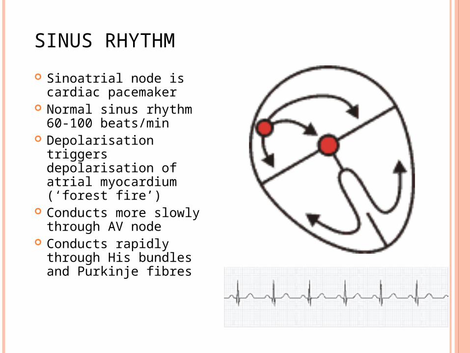
SINUS RHYTHM
Sinoatrial node is cardiac pacemaker
Normal sinus rhythm 60-100 beats/min
Depolarisation triggers depolarisation of atrial myocardium (‘forest fire’)
Conducts more slowly through AV node
Conducts rapidly through His bundles and Purkinje fibres

Sinoatrial rate controlled by autonomic nervous system
Parasympathetic system predominates (M2 muscarinic receptors)
Sympathetic system (ß1 receptors) Increased heart rate (positive chronotropic
effect) Increased automaticity Facilitation of conduction of AV node
SINUS RHYTHM

ECG
Recording of electrical activity of the heart
Net sum of depolarisation and repolarisation potentials of all myocardial cells
P-QRS-T pattern P - atrial depolarisation QRS - ventricular depolarisation T - ventricular repolarisation

ECG
Recording of electrical activity of the heart
Net sum of depolarisation and repolarisation potentials of all myocardial cells
P-QRS-T pattern P - atrial depolarisation QRS - ventricular depolarisation T - ventricular repolarisation
P
q
R
s
T

DEFINITION OF ARRHYTHMIA
Cardiac arrhythmia is an abnormality of the heart rhythm
Bradycardia – heart rate slow (<55-60 beats/min)
Tachycardia – heart rate fast (>100 beats/min)

CLINICAL CLASSIFICATION OF ARRHYTHMIAS
Heart rate (increased/decreased) Heart rhythm (regular/irregular) Site of origin (supraventricular / ventricular) Complexes on ECG (narrow/broad)
MECHANISMS OF ARRHYTHMIA PRODUCTION
Re-entry (refractory tissue reactivated due to conduction block, causes abnormal continuous circuit; eg accessory pathways linking atria and ventricles in Wolff-Parkinson-White syndrome)
Abnormal pacemaker activity in non-conducting/conducting tissue (eg ischemia)
Delayed after-depolarisation (automatic depolarisation of cardiac cell triggers ectopic beats, can be caused by drugs; eg digoxin)

VAUGHAN WILLIAMS CLASSIFICATION OF ANTIARRHYTHMIC DRUGS
Class I: block sodium channels Ia (quinidine, procainamide,
disopyramide) AP Ib (lidocaine, mexiletine,
phenytoin) AP Ic (flecainide, propafenone) AP
Class II: ß-adrenoceptor antagonists (atenolol, sotalol)
Class III: prolong action potential and prolong refractory period (suppress re-entrant rhythms) (amiodarone, dronedarone, sotalol)
Class IV: Calcium channel antagonists. Impair impulse propagation in nodal and damaged areas (verapamil)
Phase 4
Phase 0
Phase 1
Phase 2
Phase 3
0 mV
-80mV
II
IIII
IV

MANAGEMENT OF ARRHYTHMIAS
Acute management (clinical assessment of patient and diagnosis)
Prophylaxis Non-pharmacological Pharmacological (some antiarrhythmics are
also proarrhythmic)

NON-PHARMACOLOGICAL TREATMENT
Acute Vagal manoeuvres (Valsalva, carotid sinus
massage) DC cardioversion
Prophylaxis Radiofrequency ablation Implantable defibrillator
Pacing (external, temporary, permanent)

LIDOCAINE
Class Ib (blocks Na+ channels, reduces AP duration)
Ventricular arrhythmias (acute Rx) IV infusion only (2 hour half life, high
first pass metabolism) Hepatic metabolism (inhibited by
cimetidine, propranolol) SE mainly CNS - drowsiness,
disorientation, convulsions, hypotension

o Besides Na blocking effects, without affecting AP (class Ic), has weak Ca-blocker (class IV) and weak β-blocker effects (class II)
o It is metabolized to active metabolites, but slow metabolizers may have even 2x higher Cmax and 3x longer T1/2
o Indicated for SV tachyarrhythmias (WPW, AV node re-entry tachycardia, paroxysmal atrial fibrillation) and some ventricular arrhythmias
o Adverse effectso Cardiac – AV or bundle branch blockades,
ventricular tachyarrhythmias -> avoid in CAD or depressed LVEF
o GIT, CNS
PROPAFENONE

FLECAINIDE
Class Ic (block Na+ channels, no change to AP)
Slows conduction in all cardiac cells Acute Rx /prophylaxis Useful for
Supraventricular tachycardiasParoxysmal atrial fibrillation
Avoid in ventricular tachycardias, CAD, depressed LVEF
Oral/IV Long acting (T1/2 14 hours) Hepatic metabolism, urinary elimination

FLECAINIDE
CAST (Cardiac Arrhythmia Suppression Trial) 1989 – increased mortality post MI (VF arrest)
A COMPLICATION COMMON HOWEVER TO ALL CLASS I AGENTS!!!
other SE- cardiac failure, ventricular arrhythmias, blurred vision, abdominal discomfort, nausea, paraesthesia, dizzyness, tremor, metallic taste

o Decrease resting membrane potential (it is more negative; class II) – negative bathmotropic effects
o Pacemakers: decrease the rate of the spontaneous firing
o Prolong AV conductiono Clinical correlates: TF, impulse conduction
to ventricles, threshold for ventricular fibrillation, improved prognosis of patient after M.I. (sudden death prevention – antiarrhythmic effects)
o PK differences – t1/2!o Esmolol < Propranolol < Atenolol < Metoprolol
BETA-BLOCKERS

AMIODARONE
Class III - increases refractory period and AP Major effect acutely is depression of AV node Acute Rx/prophylaxis Atrial and ventricular arrhythmias Oral or IV (central line) Loading and maintenance doses T1/2 roughly 54 days Large volume of distribution Accumulates Hepatic metabolism- biliary and intestinal
excretion

AMIODARONE – ADVERSE EFFECTS
Photosensitive rashes Grey/blue discolouration of skin Thyroid abnormalities 2% Pulmonary fibrosis Corneal deposits CNS/GI disturbance Pro-arrhythmic effects (torsade de pointe) Heart block Nightmares 25% Abnormal LFT 20% Interacts with digoxin, warfarin ->
(reduces clearance)

o L-isomer (non selective b-blocker without ISA),o PROLONG AP: block rapid outward K current
repolarization phase is slowed – i.e., prolonged, longer is also effective refractory period (EPR)
o IND: i.v. - serious ventricular and SV arrhythmias; p.o. - effective in prophylaxis of recurrent SV arrhythmias; to keep the sinus rhythm after cardioversion of AF
o PK: relatively simple and predictable (p.o. i i.v.) – limited risk of drug interactions
o Adverse reaction: generally relatively tolerable drug;
• If induction of the long QT due to the possibility of TdP
occurrence (the risk in approx. in 3-4 % patients)
• Bradycardia, HF precipitation, hypotension,
bronchoconstriction, sleep disturbances (CI: severe HF, asthma..)
SOTALOL

o The effects on slow response structures (SA and AV nodes; class IV)
• conduction is based on Ca++
• depresses spontaneous depolarization of SA node
• decreases AV node conduction decreases ventricular response in AF and flutter suppresses AV nodal re-entry tachyarrhythmia
• no major impact on ventricular tachyarrhythmiaso Rapid response structures (the rest of the
myocardium)
Ca2+ channel block (L- type) in 2nd AP phase
less Ca for contraction - negative inotropic response
CA CHANNEL BLOCKERS

VERAPAMIL
Class IV (calcium channel blocker) Prolongs conduction and refractoriness
in AV node, slows rate of conduction of SA node
Acute Rx /prophylaxis Used IV/oral SUPRAVENTRICULAR NOT
VENTRICULAR ARRHYTHMIAS (cardiovascular collapse)
Do not use IV verapamil with ß- blocker (heart block)
T1/2 6-8 hours

VERAPAMIL- ADVERSE EFFECTS
Heart failure Constipation Bradycardia Nausea

ADENOSINE
Not in Vaughan Williams class Purine nucleotide (activates adenosine
receptors) Slows AV nodal conduction Acute Rx Termination of SVT/ diagnosis of VT Given IV only (rapid bolus) T1/2 < 2seconds

ADENOSINE- ADVERSE EFFECTS
Feeling of impending doom! Flushing, dyspnoea, chest pain, transient
arrhythmias Contraindicated in asthma, heart block

DIGOXIN
Not in Vaughan Williams class Cardiac glycoside (digitalis, foxglove) Act on Na/K-ATPase of cell membrane
(inhibits Na+/K+ pump, increases intracellular Na+ and calcium)/ increases vagal activity
Increase cardiac contraction and slows AV conduction by increasing AV node refractory period

DIGOXIN
Atrial fibrillation or flutter (controls ventricular rate)
Acute Rx/prophylaxis Oral/IV Loading and maintenance doses T1/2 36 hours Excreted by kidneys Narrow therapeutic index Therapeutic drug monitoring Reduce dose in elderly/renal impairment

DIGOXIN – ADVERSE EFFECTS Arrhythmias, heart block, anorexia, nausea,
diarrhoea, xanthopsia, gynaecomastia, confusion, agitation
AE potentiated by hypokalaemia and hypomagnesaemia
Overdose –Digibind (digoxin binding antibody fragments), phenytoin for ventricular arrhythmias, pacing, atropine

SOME TYPICAL DOSAGES Amiodarone (150 mg vials): VT/VF refractory to DC shock ->
300 mg bolus in 20 ml D5%; continuous IV infusion 600-900 mg in 500 mL D5% @ 21 mL/h
Digoxin (0.5 mg vials): 10-15 mcg/kg 50% stat, 25% in 6 h, and 25% afterwards; continuous IV infusion @ 0.0625-0.5 mg/d (eg 1/8 to 1 vial in 500 mL @ 21 mL/h)
Diltiazem (50 mg vials): 0.25 mg/kg in 2 min, then 0.35 mg/kg in 5-10 min; continuous IV infusion @ 5-15 mg/h (eg 3 vials in 500 mL @ 17 mL/h)
Lidocaine: IV infusion: 1-4 mg/min; IV bolus: 50-100 mg initially, can be repeated up to 3 mg/kg in 1 h (reduce bolus dose to 50% if elderly/CHF/hepatic disease)
Metoprolol (5 mg vials): 5 mg every 2 min, max 3 doses Propronolol (5 mg vials): 1 mg in slow bolus, every 10 min, max
total dose of 0.15 mg/kg Verapamil (5 or 125 mg vials): 2.5-10 mg in 2 min, then 10 mg
in 30 min, max 20 mg; continuous IV infusion @ 5-10 mg/h (eg 125-mg vial in 500 mL @ 20 mL/h)

CLINICAL CASES

A COMMON APPROACHWhat is the average heart rate? Is the rhythm regular or irregular? Is the QRS narrow or large? Is there any evidence of sinus
rhythm? Otherwise, what is the source of rhythm?
How is AV conduction? Is there any evidence of ongoing ischemia or
prior infarction? Is there any evidence of strain pattern or
electrolyte disturbance?

81 YEAR OLD WOMAN WITH ASTHMA HAS ‘THUMPING IN CHEST’

81 YEAR OLD WOMAN WITH ASTHMA HAS ‘THUMPING IN CHEST’
NORMAL EKG
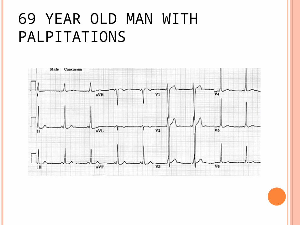
69 YEAR OLD MAN WITH PALPITATIONS

69 YEAR OLD MAN WITH PALPITATIONS
WPW

81 YEAR WOMAN WITH DIZZINESS

81 YEAR WOMAN WITH DIZZINESS
2:1 ATRIAL TACHYCARDIA

72 YEAR MAN WITH FATIGUE

72 YEAR MAN WITH FATIGUE
2:1 ATRIAL FLUTTER

76 YEAR OLD MAN WITH RECENT STROKE

76 YEAR OLD MAN WITH RECENT STROKE
ATRIAL FIBRILLATION

74 YEAR OLD WOMAN COLLAPSES 24 HOURS POST MI

74 YEAR OLD WOMAN COLLAPSES 24 HOURS POST MI
VENTRICULAR TACHYCARDIA

94 YEAR WOMAN AWAITING ELECTIVE HIP SURGERY

94 YEAR WOMAN AWAITING ELECTIVE HIP SURGERY
2:1 ATRIAL FLUTTER WITH PRE-EXISTENT LBBB

71 YEAR OLD PULSELESS MAN

71 YEAR OLD PULSELESS MAN
VENTRICULAR FIBRILLATION

71 YEAR OLD PULSELESS MAN
VENTRICULAR FIBRILLATION
AND DC SHOCK

ANY QUESTIONS?

SUMMARY
Anti-arrhythmic drugs are classified by their effect on the cardiac action potential
Not all drugs fit this classification In clinical practice treatment of
arrhythmias is determined by the type of arrhythmia (SVT, VT), clinical condition of the patient, and therapeutic window of each specific treatment means
Anti-arrhythmic drugs are efficacious but may have serious adverse effects
Not all arrhythmias are treated with drug therapy alone
For these and further slides on these topics please feel free to visit the
metcardio.org website:
http://www.metcardio.org/slides.html