A Novel Niosome-Encapsulated Essential Oil Formulation to ...
13
toxins Article A Novel Niosome-Encapsulated Essential Oil Formulation to Prevent Aspergillus flavus Growth and Aflatoxin Contamination of Maize Grains During Storage Marta García-Díaz, Belén Patiño , Covadonga Vázquez and Jessica Gil-Serna * Department of Genetics, Physiology and Microbiology, Faculty of Biology, University Complutense of Madrid, Jose Antonio Novais 12, 28040 Madrid, Spain; [email protected] (M.G.-D.); [email protected] (B.P.); [email protected] (C.V.) * Correspondence: [email protected] Received: 18 October 2019; Accepted: 5 November 2019; Published: 6 November 2019 Abstract: Aflatoxin (AF) contamination of maize is a major concern for food safety. The use of chemical fungicides is controversial, and it is necessary to develop new effective methods to control Aspergillus flavus growth and, therefore, to avoid the presence of AFs in grains. In this work, we tested in vitro the effect of six essential oils (EOs) extracted from aromatic plants. We selected those from Satureja montana and Origanum virens because they show high levels of antifungal and antitoxigenic activity at low concentrations against A. flavus. EOs are highly volatile compounds and we have developed a new niosome-based encapsulation method to extend their shelf life and activity. These new formulations have been successfully applied to reduce fungal growth and AF accumulation in maize grains in a small-scale test, as well as placing the maize into polypropylene woven bags to simulate common storage conditions. In this latter case, the antifungal properties lasted up to 75 days after the first application. Keywords: essential oils; Satureja montana; Origanum virens; Aspergillus flavus; aflatoxin; corn; nanoparticles Key Contribution: A safe, ecofriendly, novel strategy was developed to prevent aflatoxin contamination of maize during storage. This method uses niosome-encapsulated EOs extracted from Satureja montana and Origanum virens and is able to control Aspergillus flavus growth for long periods. 1. Introduction Aflatoxins (AFs) are secondary metabolites produced primarily by Aspergillus flavus and Aspergillus parasiticus. Aflatoxin B 1 ,B 2 ,G 1 , and G 2 (AFB 1 , AFB 2 , AFG 1 , and AFG 2 ) are the most important ones, with AFB 1 being the most toxic naturally occurring human carcinogen [1,2]. The International Agency for Research on Cancer (IARC) has classified the “naturally occurring mixes of aflatoxins” as Group 1 carcinogens in humans [3]. AFs contaminate a variety of staple crops including cereals (maize, sorghum, barley, oat, rye, rice, and wheat), soya, dry nuts (nuts, pistachios, almonds, and hazelnuts), cottonseed, coffee, cacao, and spices [4]. According to the Food and Agriculture Organization of the United Nations (FAO) [5], maize is one of the most important cereals, with an annual worldwide production of 1134 million tons in 2017, and most of it is intended for direct human and animal consumption. Moreover, maize and its derivatives are considered the main source of AFs worldwide [6]. For all these reasons, the European Union established strict regulations regarding maximum permitted AF levels for maize [7]. Toxins 2019, 11, 646; doi:10.3390/toxins11110646 www.mdpi.com/journal/toxins
Transcript of A Novel Niosome-Encapsulated Essential Oil Formulation to ...

toxins
Article
A Novel Niosome-Encapsulated Essential OilFormulation to Prevent Aspergillus flavus Growthand Aflatoxin Contamination of Maize GrainsDuring Storage
Marta García-Díaz, Belén Patiño , Covadonga Vázquez and Jessica Gil-Serna *
Department of Genetics, Physiology and Microbiology, Faculty of Biology, University Complutense of Madrid,Jose Antonio Novais 12, 28040 Madrid, Spain; [email protected] (M.G.-D.); [email protected] (B.P.);[email protected] (C.V.)* Correspondence: [email protected]
Received: 18 October 2019; Accepted: 5 November 2019; Published: 6 November 2019�����������������
Abstract: Aflatoxin (AF) contamination of maize is a major concern for food safety. The use ofchemical fungicides is controversial, and it is necessary to develop new effective methods to controlAspergillus flavus growth and, therefore, to avoid the presence of AFs in grains. In this work, we testedin vitro the effect of six essential oils (EOs) extracted from aromatic plants. We selected those fromSatureja montana and Origanum virens because they show high levels of antifungal and antitoxigenicactivity at low concentrations against A. flavus. EOs are highly volatile compounds and we havedeveloped a new niosome-based encapsulation method to extend their shelf life and activity. Thesenew formulations have been successfully applied to reduce fungal growth and AF accumulation inmaize grains in a small-scale test, as well as placing the maize into polypropylene woven bags tosimulate common storage conditions. In this latter case, the antifungal properties lasted up to 75 daysafter the first application.
Keywords: essential oils; Satureja montana; Origanum virens; Aspergillus flavus; aflatoxin; corn; nanoparticles
Key Contribution: A safe, ecofriendly, novel strategy was developed to prevent aflatoxin contaminationof maize during storage. This method uses niosome-encapsulated EOs extracted from Satureja montanaand Origanum virens and is able to control Aspergillus flavus growth for long periods.
1. Introduction
Aflatoxins (AFs) are secondary metabolites produced primarily by Aspergillus flavus and Aspergillusparasiticus. Aflatoxin B1, B2, G1, and G2 (AFB1, AFB2, AFG1, and AFG2) are the most important ones,with AFB1 being the most toxic naturally occurring human carcinogen [1,2]. The International Agencyfor Research on Cancer (IARC) has classified the “naturally occurring mixes of aflatoxins” as Group 1carcinogens in humans [3].
AFs contaminate a variety of staple crops including cereals (maize, sorghum, barley, oat, rye, rice,and wheat), soya, dry nuts (nuts, pistachios, almonds, and hazelnuts), cottonseed, coffee, cacao, andspices [4].
According to the Food and Agriculture Organization of the United Nations (FAO) [5], maizeis one of the most important cereals, with an annual worldwide production of 1134 million tons in2017, and most of it is intended for direct human and animal consumption. Moreover, maize and itsderivatives are considered the main source of AFs worldwide [6]. For all these reasons, the EuropeanUnion established strict regulations regarding maximum permitted AF levels for maize [7].
Toxins 2019, 11, 646; doi:10.3390/toxins11110646 www.mdpi.com/journal/toxins
Toxins 2019, 11, 646 2 of 13
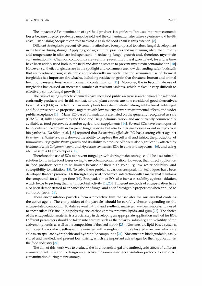
The impact of AF contamination of agri-food products is significant. It causes important economiclosses because infected products cannot be sold and the contamination also raises veterinary and healthcosts. Establishing adequate controls to avoid AFs in the food chain is thus essential [8].
Different strategies to prevent AF contamination have been proposed to reduce fungal developmentin the field or during storage. Applying good agricultural practices and maintaining adequate humidityand temperature in silos are indispensable in reducing fungal growth and, therefore, mycotoxincontamination [9]. Chemical compounds are useful in preventing fungal growth and, for a long time,have been widely used both in the field and during storage to prevent mycotoxin contamination [10].However, synthetic fungicides are in the spotlight and consumers are now demanding safer foodstuffsthat are produced using sustainable and ecofriendly methods. The indiscriminate use of chemicalfungicides has important drawbacks, including residue on grain that threatens human and animalhealth or causes extensive environmental contamination [11]. Moreover, the indiscriminate use offungicides has caused an increased number of resistant isolates, which makes it very difficult toeffectively control fungal growth [12].
The risks of using synthetic chemicals have increased public awareness and demand for safer andecofriendly products and, in this context, natural plant extracts are now considered good alternatives.Essential oils (EOs) extracted from aromatic plants have demonstrated strong antibacterial, antifungal,and food preservative properties, together with low toxicity, fewer environmental effects, and widerpublic acceptance [13]. Many EO-based formulations are listed on the generally recognized as safe(GRAS) list, fully approved by the Food and Drug Administration, and are currently commerciallyavailable as food preservatives and/or agricultural supplements [14]. Several EOs have been reportedto not only reduce growth in toxigenic fungal species, but also to interfere to some extent in mycotoxinbiosynthesis. Da Silva et al. [15] reported that Rosmarinus officinalis EO has a strong effect againstFusarium verticillioides, as it showed the ability to rupture the cell wall and inhibit the production offumonisins. Aspergillus flavus growth and its ability to produce AFs were also significantly affected bytreatment with Origanum virens and Ageratum conyzoides EOs in corn and soybeans [16], and usingMentha spicata EO in chickpeas [17].
Therefore, the use of EOs to prevent fungal growth during maize storage could be a sustainablesolution to minimize food losses owing to mycotoxin contamination. However, their direct applicationin food products seems to be limited because of their high volatility, low water solubility, andsusceptibility to oxidation [18]. To solve these problems, various encapsulation techniques have beendeveloped that can preserve EOs through a physical or chemical interaction with a matrix that maintainsthe compounds for a longer time [19]. Encapsulation of EOs also increases stability against oxidation,which helps to prolong their antimicrobial activity [18,20]. Different methods of encapsulation havealso been demonstrated to enhance the antifungal and antiaflatoxigenic properties when applied tocontrol A. flavus [21].
These encapsulation particles form a protective film that isolates the nucleus that containsthe active agent. The composition of the particles should be carefully chosen depending on theencapsulated compound. To date, several natural and synthetic matrices have been successfully usedto encapsulate EOs including polyethylene, carbohydrates, proteins, lipids, and gum [22]. The choiceof the encapsulation material is a crucial step in developing an appropriate application method for EOs.Different parameters should be taken into account such as the polarity, solubility, and volatility of theactive compounds, as well as the composition of the food matrix [23]. Niosomes are lipid-based systems,composed by non-toxic self-assembly vesicles, with a single or multiple layered structure, which areable to encapsulate hydrophobic and hydrophilic compounds [24]. Niosomes are biodegradable, easilystored and handled, and present low toxicity, which are important advantages for their application inthe food industry [24].
The aim of this work was to evaluate the in vitro antifungal and antitoxigenic effects of differentaromatic plant EOs and to design an effective niosome-based encapsulation protocol to avoid AFcontamination during maize storage.
Toxins 2019, 11, 646 3 of 13
2. Results
2.1. The Efficacy of Plant Essential Oils Against Fungal Growth and Mycotoxin Production
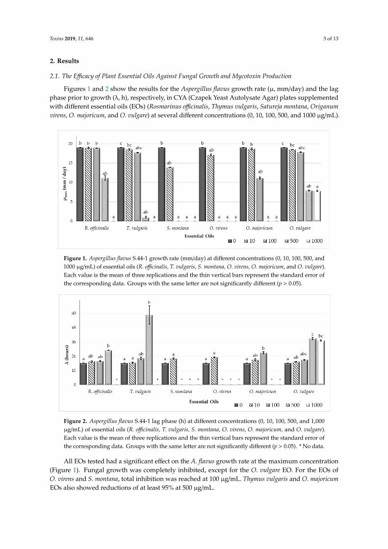
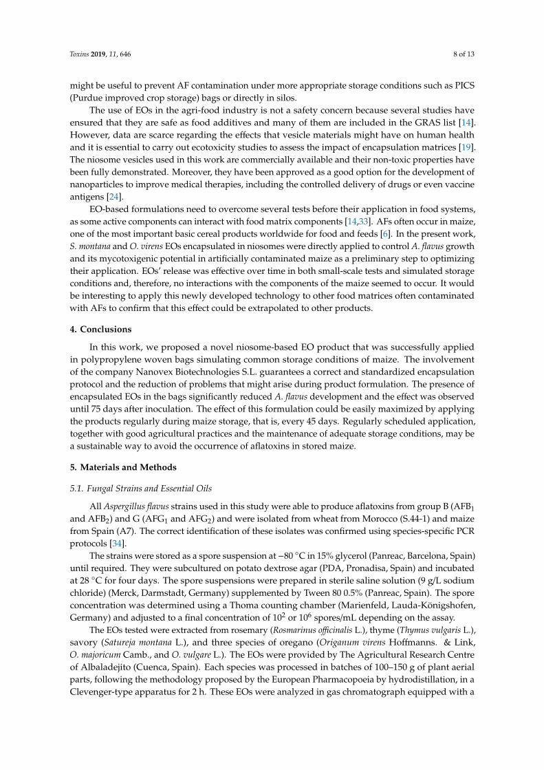
Figures 1 and 2 show the results for the Aspergillus flavus growth rate (µ, mm/day) and the lagphase prior to growth (λ, h), respectively, in CYA (Czapek Yeast Autolysate Agar) plates supplementedwith different essential oils (EOs) (Rosmarinus officinalis, Thymus vulgaris, Satureja montana, Origanumvirens, O. majoricum, and O. vulgare) at several different concentrations (0, 10, 100, 500, and 1000 µg/mL).
Figure 1. Aspergillus flavus S.44-1 growth rate (mm/day) at different concentrations (0, 10, 100, 500, and1000 µg/mL) of essential oils (R. officinalis, T. vulgaris, S. montana, O. virens, O. majoricum, and O. vulgare).Each value is the mean of three replications and the thin vertical bars represent the standard error ofthe corresponding data. Groups with the same letter are not significantly different (p > 0.05).
Figure 2. Aspergillus flavus S.44-1 lag phase (h) at different concentrations (0, 10, 100, 500, and 1,000µg/mL) of essential oils (R. officinalis, T. vulgaris, S. montana, O. virens, O. majoricum, and O. vulgare).Each value is the mean of three replications and the thin vertical bars represent the standard error ofthe corresponding data. Groups with the same letter are not significantly different (p > 0.05). * No data.
All EOs tested had a significant effect on the A. flavus growth rate at the maximum concentration(Figure 1). Fungal growth was completely inhibited, except for the O. vulgare EO. For the EOs ofO. virens and S. montana, total inhibition was reached at 100 µg/mL. Thymus vulgaris and O. majoricumEOs also showed reductions of at least 95% at 500 µg/mL.

Toxins 2019, 11, 646 4 of 13
The lag phase got longer as the EO concentration increased (Figure 2). It was not possible tocalculate the lag phase when the EO treatment completely inhibited growth in the plates.
Aflatoxin production (AFB1, AFB2, AFG1 y AFG2) was significantly reduced compared with thecontrol group in all treatments at the highest concentration tested (1000 µg/mL). No AFs were detectedat these concentrations (Table 1) in any case, except for the O. vulgare EO treatment, which achievedreductions of more than 97% in all toxins.
Table 1. Aflatoxin (AF) concentration (B1, B2, G1, and G2) in Czapek Yeast Autolysate Agar (CYA)plates supplemented with different concentrations (0, 10, 100, 500, and 1000 µg/mL) of essential oils(EOs) (R. officinalis, T. vulgaris, S. montana, O. virens, O. majoricum, and O. vulgare). Each value is themean of three replications ± standard error. Groups with the same letter are not significantly different(p > 0.05). ND: non detected.
EOs µg/mL AFB1 (µg/g agar) AFB2 (µg/gagar)
AFG1 (µg/gagar)
AFG2 (µg/gagar)
R. officinalis
0 10.754 ± 0.925 c 0.201 ± 0.021 c 0.485 ± 0.055 c 0.088 ± 0.014 b
10 5.205 ± 1.033 bc 0.1 ± 0.022 bc 0.213 ± 0.045 bc ND a
100 5.223 ± 0.171 abc 0.11 ± 0.006 bc 0.216 ± 0.012 abc ND a
500 1.09 ± 0.152 ab 0.017 ± 0.002 ab 0.058 ± 0.007 ab ND a
1000 ND a ND a ND a ND a
T. vulgaris
0 10.754 ± 0.925 b 0.201 ± 0.021 b 0.485 ± 0.055 b 0.088 ± 0.014 b
10 7.04 ± 0.977 b 0.122 ± 0.019 ab 0.256 ± 0.059 ab ND a
100 5.994 ± 0.554 ab 0.117 ± 0.019 ab 0.264 ± 0.069 ab ND a
500 ND a ND a ND a ND a
1000 ND a ND a ND a ND a
S. montana
0 10.754 ± 0.925 b 0.201 ± 0.021 b 0.485 ± 0.055 b 0.088 ± 0.014 b
10 8.27 ± 0.686 ab 0.151 ± 0.011 ab 0.314 ± 0.029 ab ND a
100 ND a ND a ND a ND a
500 ND a ND a ND a ND a
1000 ND a ND a ND a ND a
O. virens
0 10.754 ± 0.925 b 0.201 ± 0.021 bc 0.485 ± 0.055 b 0.088 ± 0.014 b
10 10.52 ± 1.334 b 0.245 ± 0.039 c 0.508 ± 0.065 b ND a
100 0.033 ± 0.044 ab 0.003 ± 0 ab 0.004 ± 0.002 ab ND a
500 ND a ND a ND a ND a
1000 ND a ND a ND a ND a
O. majoricum
0 10.754 ± 0.925 b 0.201 ± 0.021 ab 0.485 ± 0.055 b 0.088 ± 0.014 b
10 10.939 ± 0.21 b 0.234 ± 0.008 b 0.611 ± 0.057 b ND a
100 7.999 ± 0.628 ab 0.186 ± 0.022 ab 0.375 ± 0.028 ab ND a
500 0.003 ± 0 a ND a ND a ND a
1000 0.008 ± 0.008 a ND a ND a ND a
O. vulgare
0 10.754 ± 0.925 c 0.201 ± 0.021 b 0.485 ± 0.055b 0.088 ± 0.014 b
10 10.143 ± 0.86 bc 0.192 ± 0.015 b 0.475 ± 0.056 b ND a
100 7.867 ± 0.409 abc 0.168 ± 0.015 ab 0.466 ± 0.08 ND500 0.738 ± 0.08 ab 0.012 ± 0.002 a 0.043 ± 0.003 ND1000 0.298 ± 0.068 a 0.004 ± 0.003 a 0.014 ± 0.004 ND
AFB1 production was significantly affected at 500 µg/mL, with reductions of nearly 100% in all ofthe EOs tested. The same results were obtained when O. virens and S. montana EOs were applied at100 µg/mL.
AFB2 production was also significantly reduced by least 90% compared with the control platesafter treatment with R. officinalis, T. vulgaris, O. vulgare, and O. majoricum EOs at 500 µg/mL. The mosteffective treatments, reaching complete inhibition of AFB2 production at 100 µg/mL, were O. virens andS. montana EOs.

Toxins 2019, 11, 646 5 of 13
Aflatoxin G1 was not detected on S. montana and O. virens at 100 µg/mL. The rest of the EOsshowed a reduction greater than 80% at 500 µg/mL. In all cases, AFG2 concentration was below thedetection limit (<0.0025 µg/g agar), except in the case of control plates without treatment.
2.2. Techniques for the Application of Essential Oils to Prevent Fungal Growth and Mycotoxin Production
The main nanocapsule characteristics of the O. virens and S. montana niosomes can be found inTable 2. According to these data, both encapsulation processes yield high quality niosomes with lowaggregation of nanoparticles. The particle size of both samples was approximately 140 nm with apolydipersity index (PDI) of 0.251 and a ζ-potential of –14 mV.
Table 2. Characterization of O. virens and S. montana essential oil particles encapsulated in niosomes.
ZETASICER NANOSIGHT
PDI Z-AVERAGE(nm)
Toxins 2019, 11, x FOR PEER REVIEW 5 of 13
effective treatments, reaching complete inhibition of AFB2 production at 100 µg/mL, were O. virens and S. montana EOs.
Aflatoxin G1 was not detected on S. montana and O. virens at 100 µg/mL. The rest of the EOs showed a reduction greater than 80% at 500 µg/mL. In all cases, AFG2 concentration was below the detection limit (<0.0025 µg/g agar), except in the case of control plates without treatment.
2.2. Techniques for the Application of Essential Oils to Prevent Fungal Growth and Mycotoxin Production
The main nanocapsule characteristics of the O. virens and S. montana niosomes can be found in Table 2. According to these data, both encapsulation processes yield high quality niosomes with low aggregation of nanoparticles. The particle size of both samples was approximately 140 nm with a polydipersity index (PDI) of 0.251 and a ζ-potential of –14 mV.
Table 2. Characterization of O. virens and S. montana essential oil particles encapsulated in niosomes.
ZETASICER NANOSIGHT
PDI Z-AVERAGE (nm) POTENCIAL-ʐ (mV) Size (nm) CONCENTRATI
ON (particle/mL) O. virens 0.251 ± 0.019 156.2 ± 3.9 −14.5 ± 0.5 142.4 ± 1.0 (2.96 ± 0.12) × 1014
S. montana 0.251 ± 0.011 153.3 ± 2.8 −14.6 ± 2.3 140.6 ± 3.8 (1.86 ± 0.07) × 1014 PDI: polydipersity index.
2.2.1. Small-Scale Assay
Satureja montana and O. virens EOs applied by direct contact to artificially inoculated maize grains and incubated at 28 °C reduced A. flavus growth compared with the control group without EOs after seven days of incubation, although no significant differences were observed (Figure 3a). When these EOs were applied in their niosome-encapsulated form and incubated for seven days, colony forming units (CFU) per gram values were slightly increased, although no significant differences were found. When incubation time was extended to 21 days, the opposite effect was observed (Figure 3b). EOs applied by direct contact seemed to lose their effect over time. However, when these compounds were encapsulated in niosomes, S. montana EO reduced fungal growth by 58% and, in the case of O. virens, this reduction was 32% with respect to the control group.
Figure 3. Effect of S. montana (SM) and O. virens (OV) by direct contact (essential oil, EO) and immobilized in niosomes (EO-NIO) on corn grains inoculated with A. flavus, incubated for 7 (a) and 21 days (b). Each values is the mean of three replications and the thin vertical bars represent the standard error of the corresponding data. Groups with the same letter are not significantly different (p > 0.05). CFU, colony forming units.
AFB1 production was low at the short incubation time (7 days) and very significant for longer periods (21 days) (Figure 4). In the thin layer chromatography (TLC) analysis, the intensity and thickness of the fluorescent band is related to AFB1 concentration. Therefore, in the seven-day long assay, AFB1 concentration was reduced in all cases with respect to control. When plates were incubated over 21 days, a reduction in AFB1 concentration was observed in plates treated directly
Size (nm) CONCENTRATION(Particle/mL)
O. virens 0.251 ± 0.019 156.2 ± 3.9 −14.5 ± 0.5 142.4 ± 1.0 (2.96 ± 0.12) × 1014
S. montana 0.251 ± 0.011 153.3 ± 2.8 −14.6 ± 2.3 140.6 ± 3.8 (1.86 ± 0.07) × 1014
PDI: polydipersity index.
2.2.1. Small-Scale Assay
Satureja montana and O. virens EOs applied by direct contact to artificially inoculated maize grainsand incubated at 28 ◦C reduced A. flavus growth compared with the control group without EOs afterseven days of incubation, although no significant differences were observed (Figure 3a). When theseEOs were applied in their niosome-encapsulated form and incubated for seven days, colony formingunits (CFU) per gram values were slightly increased, although no significant differences were found.When incubation time was extended to 21 days, the opposite effect was observed (Figure 3b). EOsapplied by direct contact seemed to lose their effect over time. However, when these compounds wereencapsulated in niosomes, S. montana EO reduced fungal growth by 58% and, in the case of O. virens,this reduction was 32% with respect to the control group.
Figure 3. Effect of S. montana (SM) and O. virens (OV) by direct contact (essential oil, EO) andimmobilized in niosomes (EO-NIO) on corn grains inoculated with A. flavus, incubated for 7 (a) and21 days (b). Each values is the mean of three replications and the thin vertical bars represent thestandard error of the corresponding data. Groups with the same letter are not significantly different(p > 0.05). CFU, colony forming units.
AFB1 production was low at the short incubation time (7 days) and very significant for longerperiods (21 days) (Figure 4). In the thin layer chromatography (TLC) analysis, the intensity andthickness of the fluorescent band is related to AFB1 concentration. Therefore, in the seven-day longassay, AFB1 concentration was reduced in all cases with respect to control. When plates were incubatedover 21 days, a reduction in AFB1 concentration was observed in plates treated directly with O. virens

Toxins 2019, 11, 646 6 of 13
EO as well as both EO niosome-encapsulated. However, no differences were found after S. montanaEO application.
Figure 4. Effect of S. montana (SM) and O. virens (OV) by direct contact (EO) and encapsulatedin niosomes (EO-NIO) on aflatoxin (AF) B1 concentration of corn grains inoculated with A. flavus,incubated for 7 and 21 days. The standard corresponds to the application of purified AFB1 (0.05 mg/mL).
2.2.2. Polypropylene Woven Bags Assays
Figure 5 shows the results in CFU/g of A. flavus inoculated corn stored in polypropylene wovenbags at all incubation times and after treatment with EOs encapsulated in niosomes.
After 45, 60, and 75 days of incubation, both of the EOs encapsulated in niosomes were able tocontrol fungal growth, with a maximum reduction of up to 79% and 69% for S. montana and O. virensEOs, respectively. However, this effect seems to be lost over time and no significant differences withrespect to the control group were found after 90 days of incubation.
Figure 5. Effect of S. montana (NIO-SM) and O. virens (NIO-OV) EO encapsulated in niosomes onA. flavus growth in corn grains incubated for 45, 60, 75, and 90 days. Each value is the mean of threereplications and the thin vertical bars represent the standard error of the corresponding data. Groupswith the same letter are not significantly different (p > 0.05).
The results regarding AFB1 detected on inoculated control bags or after niosome-encapsulatedEO treatments are shown in Figure 6. The band intensity of each treatment was apparently lower withrespect to their corresponding control in all cases except for NIO-OV after 90 days of incubation whena slight increase in production was detected. Treatment using niosome-encapsulated S. montana EOwas the most effective to control AFB1 production by A. flavus.
Temperature and humidity were recorded during the experiment with ranges of 25–27 ◦C and75–85%, respectively.

Toxins 2019, 11, 646 7 of 13
Figure 6. AFB1 detection by thin layer chromatography (TLC) in polypropylene woven bags inoculatedwith A. flavus after S. montana (NIO-SM) and O. virens (NIO-OV) niosome treatment of corn incubatedfor 45, 60, 75, and 90 days. The intensity and thickness of the fluorescent band are related to theconcentration of toxin. The standard corresponds to the application of purified AFB1 (0.05 mg/mL).
3. Discussion
Aflatoxin (AF) presence poses a high risk to food security and most countries have establishedmaximum levels of these contaminants allowed in food products [3]. Appropriate control mechanismsare needed to keep these toxins from entering the food chain. In recent years, essential oils (EOs) havecome to be considered as a safe, ecofriendly, renewable, and easily biodegradable option to be used as afood supplement [13]. Moreover, many EOs have been described as potent antifungal compounds thatare able to interfere in mycotoxin synthesis [15,25–27]. In this work, we selected EOs extracted fromRosmarinus officinalis, Thymus vulgaris, Satureja montana, Origanum virens, O. majoricum, and O. vulgareto determine if they were able to control Aspergillus flavus growth and if they reduced AF productionby this fungus. To some extent, all of the EOs tested modified the fungal growth rate and extendedthe lag phase at high concentrations. However, at lower doses, the EOs extracted from S. montanaand O. virens were the most effective at reducing both fungal growth and AF production, and theywere selected to perform subsequent studies. Chromatographic characterization of the EOs usedin this study were carried out in the Agricultural Research Centre of Albaladejito (data not shown)and the results revealed that S. montana and O. virens EOs are highly rich in carvacrol and thymol,respectively. These compounds have been reported to be able to interact with the cell membrane,disrupt cell permeability, and produce cell death [28]. Pure extracts of both carvacrol and thymolhave been reported to inhibit the growth of important mycotoxin-producing species such as A. niger,A. flavus, A. ochraceus, and F. graminearum [29,30].
Different authors consider that EO-based formulations could be safe, ecofriendly preservatives toavoid post-harvest losses due to mycotoxin contamination [14]. Therefore, taking into account thepotent antifungal properties of EOs, many studies have focused on developing successful applicationprotocols to minimize their drawbacks, which limit their direct use in food products [18] and, therefore,it is essential to protect them to extend their shelf life and activity [19]. The controlled liberationof EOs and their encapsulation in nanoparticles made of different materials are considered a goodoption [19,23]. These technologies attempt to reduce the rapid loss of their active principles. In general,EOs are a complex mix of lipophilic compounds and, therefore, lipid nanoparticle systems such asliposomes are the most appropriate [23,31]. In our study, the small-scale experiments showed that theeffectiveness of EOs to control A. flavus growth diminished over time. However, when both EOs wereencapsulated in niosomes, a significant reduction in fungal viable counts with respect to the untreatedcontrol group were found even after 21 days of incubation. Hence, it seems that the niosome-basednanoparticles were able to reduce the loss of the EOs’ active principles and produce a controlled releaseof their compounds. Similar results were obtained in the larger-scale experiments using maize stored inpolypropylene woven bags. This type of storage is very common in African countries because it offerslow-cost protection for grain from pests. However, a higher contamination by toxigenic fungi andmycotoxins in the grains has been reported owing to the change in moisture content, which increasesthe relative humidity inside the bags [32]. Even under the worst conditions for maize storage, both ofthe EOs encapsulated in niosomes were able to control fungal development, significantly reducingaflatoxin levels, and their effect was extended for up to 75 days. Hence, these promising approaches
Toxins 2019, 11, 646 8 of 13
might be useful to prevent AF contamination under more appropriate storage conditions such as PICS(Purdue improved crop storage) bags or directly in silos.
The use of EOs in the agri-food industry is not a safety concern because several studies haveensured that they are safe as food additives and many of them are included in the GRAS list [14].However, data are scarce regarding the effects that vesicle materials might have on human healthand it is essential to carry out ecotoxicity studies to assess the impact of encapsulation matrices [19].The niosome vesicles used in this work are commercially available and their non-toxic properties havebeen fully demonstrated. Moreover, they have been approved as a good option for the development ofnanoparticles to improve medical therapies, including the controlled delivery of drugs or even vaccineantigens [24].
EO-based formulations need to overcome several tests before their application in food systems,as some active components can interact with food matrix components [14,33]. AFs often occur in maize,one of the most important basic cereal products worldwide for food and feeds [6]. In the present work,S. montana and O. virens EOs encapsulated in niosomes were directly applied to control A. flavus growthand its mycotoxigenic potential in artificially contaminated maize as a preliminary step to optimizingtheir application. EOs’ release was effective over time in both small-scale tests and simulated storageconditions and, therefore, no interactions with the components of the maize seemed to occur. It wouldbe interesting to apply this newly developed technology to other food matrices often contaminatedwith AFs to confirm that this effect could be extrapolated to other products.
4. Conclusions
In this work, we proposed a novel niosome-based EO product that was successfully appliedin polypropylene woven bags simulating common storage conditions of maize. The involvementof the company Nanovex Biotechnologies S.L. guarantees a correct and standardized encapsulationprotocol and the reduction of problems that might arise during product formulation. The presence ofencapsulated EOs in the bags significantly reduced A. flavus development and the effect was observeduntil 75 days after inoculation. The effect of this formulation could be easily maximized by applyingthe products regularly during maize storage, that is, every 45 days. Regularly scheduled application,together with good agricultural practices and the maintenance of adequate storage conditions, may bea sustainable way to avoid the occurrence of aflatoxins in stored maize.
5. Materials and Methods
5.1. Fungal Strains and Essential Oils
All Aspergillus flavus strains used in this study were able to produce aflatoxins from group B (AFB1
and AFB2) and G (AFG1 and AFG2) and were isolated from wheat from Morocco (S.44-1) and maizefrom Spain (A7). The correct identification of these isolates was confirmed using species-specific PCRprotocols [34].
The strains were stored as a spore suspension at −80 ◦C in 15% glycerol (Panreac, Barcelona, Spain)until required. They were subcultured on potato dextrose agar (PDA, Pronadisa, Spain) and incubatedat 28 ◦C for four days. The spore suspensions were prepared in sterile saline solution (9 g/L sodiumchloride) (Merck, Darmstadt, Germany) supplemented by Tween 80 0.5% (Panreac, Spain). The sporeconcentration was determined using a Thoma counting chamber (Marienfeld, Lauda-Königshofen,Germany) and adjusted to a final concentration of 102 or 106 spores/mL depending on the assay.
The EOs tested were extracted from rosemary (Rosmarinus officinalis L.), thyme (Thymus vulgaris L.),savory (Satureja montana L.), and three species of oregano (Origanum virens Hoffmanns. & Link,O. majoricum Camb., and O. vulgare L.). The EOs were provided by The Agricultural Research Centreof Albaladejito (Cuenca, Spain). Each species was processed in batches of 100–150 g of plant aerialparts, following the methodology proposed by the European Pharmacopoeia by hydrodistillation, in aClevenger-type apparatus for 2 h. These EOs were analyzed in gas chromatograph equipped with a
Toxins 2019, 11, 646 9 of 13
flame ionization detector (FID) and capillary column VF-5 of 60 mm × 0.25 mm, 5% phenyl methylsiloxane. A temperature gradient of 70 to 240 ◦C was applied, with an increase of 3 ◦C per minute,maintaining the final temperature for 2 min. For the identification of the EO components, the relativeretention times of standards and the corresponding Kovats indices were used. The quantification ofthe components was performed according to the areas of their chromatographic peaks.
These compounds were filtered (pore size 0.2 µm) (Fisherbrand, Shanghai, China) and stored at−20 ◦C in amber glass vials (Thermo Scientific, Madrid, Spain) until required.
5.2. Effectiveness of Plant Essential Oils on Fungal Growth and Aflatoxin Production
The effect of EOs at different concentrations on A. flavus S.44-1 growth and its ability to produceAFs were evaluated on CYA medium (45.5 g/L of modified Czapek–Dox agar (Pronadisa, Spain), 5 g/Lof yeast extract (Pronadisa, Spain)). EOs were diluted in polyethylene glycol 400 (PEG (Acros, Geel,Belgium)) and added to the medium to obtain final concentrations of 10, 100, 500, and 1000 µg/mL.The same amount of PEG was included in the control plates instead of EO. CYA plates supplementedwith EOs were inoculated with 1.5 µL (4 mm of diameter) of a 106 spores/mL suspension on the centerof the plate, and incubated at 28 ◦C for five days. All the conditions were tested in triplicate.
Fungal colony diameters were measured daily in two directions at right angles to each otheruntil the medium was fully colonized (five days). Growth parameters were calculated from a linearmodel obtained by plotting the diameter (mm) against time (day). The parameters determined wereλ, representing the lag phase (days prior to mycelial growth), and µmax, representing the maximumgrowth rate (mm/day), for control plates and each EO concentration tested.
AFs were extracted from the plates after six days of incubation, as described elsewhere [35]. Threeagar plugs were removed from the centre, medium, and outer edge of the colony and toxins wereextracted with 1 mL of methanol (Merck, Spain). Samples were stored at−20 ◦C until analysis. AFs weremeasured by high performance liquid chromatography (HPLC) using the protocol described below.
5.3. Effect of Satureja Montana and Origanum Virens Essential Oils Encapsulated in Niosomes on FungalGrowth and Aflatoxin Contamination
5.3.1. Procedure for Microencapsulation of Essential Oils
EOs extracted from S. montana and O. virens were encapsulated in non-ionic surfactant-based lipidvesicles (niosomes). These particles were prepared by Nanovex Biotechnologies S.L. (Oviedo, Spain)starting from 40 mL of each type of EO. Niosomes were obtained using the thin film hydration (TFH)method with homogenization and sonication to obtain niosomes with a good polydipersity index (PDI)and a particle size between 100 and 200 µm, with an EO concentration of 12 µL/mL.
The characterization of the niosomes was performed with a Zetasizer Nano ZS particle sizeanalyzer (Malvern Panalytical Ltd., Malvern, UK), which uses dynamic light scattering (DLS) todetermine particle size, and the M3-PALS technique to calculate the ζ-potential.
A nanoparticle tracking analysis (NTA) was performed using a nano sight particle trackinganalyzer (Malvern Panalytical Ltd., Malvern, UK) to determine concentration and size distribution.
5.3.2. Effect of Niosome-Encapsulated Essential Oils on Fungal Growth and Aflatoxin Production onMaize Grains
Previously autoclaved maize grains were inoculated with A. flavus, strain A7. Then, 100 g of cornwas immersed for 2 h in 100 mL of spore suspension 104 spores/mL to obtain a final concentration of102 spores/g. Subsequently, the effect of niosome-encapsulated EOs was tested in a small-scale testin Petri dishes as well as in polypropylene woven bags simulating real storage conditions. At thebeginning of the experiments, grain moisture was measured using a Hygropalm HP23A (Rotronic,Bassersdorf, Switzerland) and water activity was 0.95 in all cases.
Toxins 2019, 11, 646 10 of 13
Small-Scale Assays
Ninety millimeter Petri dishes were filled with crystalized potassium sulphate (Acros, Spain) tomaintain aw at 0.97 [36]. A 50 mm Petri dish containing 7 g of inoculated maize was placed inside thelarger one. Satureja montana and O. virens EOs were applied directly to the grains or encapsulated inniosomes at 500 µg/g. Control assays, mock-inoculated with water, were also included. Incubationwas performed at 28 ◦C and the effect of niosome-encapsulated EOs or those directly applied on maizegrains was evaluated at 7 and 21 days.
After the incubation period, a sample of 3.5 g was taken from each treatment and a viable countwas performed using serial decimal dilutions and inoculation on Rose Bengal with Chloramphenicolmedium. Plates were incubated in darkness at 28 ◦C for two days. The fungal growth of maize grainwas expressed as colony forming units per gram of maize (CFU/g).
Afterwards, another sample of 3.5 g was taken from each treatment, and shaken for 20 min with35 mL of chloroform for AF extraction. AFB1 was measured by thin layer chromatography (TLC),as described below.
Polypropylene Woven Bag Assays
One-hundred grams of inoculated maize was placed in small polypropylene woven bags.Subsequently, niosome-encapsulated EOs (S. montana and O. virens) were added at a dose of 500 µg/gand mixed. Bags were incubated at room temperature in darkness for 90 days in independent plasticboxes for each treatment (40 × 40 × 30 cm). Inoculated maize grains without EOs were used as control.Temperature and relative humidity were registered using a data logger El-USB-1 (Easylog; LASCARelectronic, Salisbury, UK) every 8 h until the end of the assay.
After the incubation period, the bags were cut open and the maize was diluted in 900 mL of sterilesaline solution (9 g/L) containing 0.05% Tween 80. The mixes were incubated in an orbital shaker(140 rpm) at 4 ◦C for 60 min to release spores. Then, serial decimal dilutions and culture in Rose Bengalwith Chloramphenicol were used to estimate fungal growth as CFU per gram of maize.
A sample of 14 g of corn was taken from each beaker, and shaken for 20 min with 35 mL ofchloroform for AFB1 extraction and subsequent evaluation by TLC, as described below.
5.4. Detection of Mycotoxins
5.4.1. Detection of Mycotoxins by High Performance Liquid Chromatography (HPLC)
After AFB1 extraction with methanol, mycotoxin measurements were performed in the“Laboratorio Arbitral Agroalimentario” (Madrid, Spain) following its standardized protocols. AF wasmeasured by HPLC on a reverse phase C18 column (Inertsil ODS3; 5 µm, 4.6 mm× 250 mm; GL Sciences,Tokio, Japan) at 40 ◦C in a Waters chromatograph 515 HPLC coupled with a fluorescence detector 474(Waters, Milford, MA, USA) at excitation and emission wavelengths of 362 and 435 nm, respectively.The mobile phase contained water, methanol, and acetonitrile (60:20:20), and the flow rate was 1 mL/min.AF was eluted and quantified by comparison with a calibration curve generated from AF standards(OEKANAL®, Sigma–Aldrich, Steinheim, Germany). The detection limit of the technique was 2.5 ng/g.
5.4.2. Detection of Mycotoxins by Thin Layer Chromatography
After AFB1 extraction with chloroform, samples were filtered using 0.45 µm syringe filters(Fisherbrand, Spain) and an aliquot of 1 mL was evaporated in a vacuum concentrator, Eppendorf™Concentrator Plus with Pump and GB Plug (Fisher Scientific, Madrid, Spain).
Silica gel 60 chromatography plates (Merck, Germany) were used, and AFB1 presence wasdetermined according to the protocols described elsewhere [37,38].
Toxins 2019, 11, 646 11 of 13
Samples and AF standards were re-suspended with 500 µL toluene/acetonitrile (95:5) (Panreac,Spain). Then, 10 µL of each sample was spotted on the plate. Toluene/acetone/acetonitrile (1:1:1 (LabKem,Barcelona, Spain)) was used as a mobile phase. Toxins were visualized under ultraviolet light (Spectronics,Westbury, NY, USA).
5.5. Statistical Analysis
Statistical analysis was performed on the effect of EOs encapsulated in niosomes with StatsGraphicsCenturion XVII V.17.2.04 program (Statpoint Technologies Inc., Warrenton, VA, USA). The Shapiro–Wilkand Levene tests were used to check normality and homoscedasticity, respectively. Data were analysedusing analysis of variance (ANOVA).
When data did not meet normality and homoscedasticity criteria, a non-parametric Kruskall–Wallistest was performed. These analyses were performed using the software InfoStat/E 2011 (FCA, Córdoba,Argentina). This was necessary in the case of the fungal growth and aflatoxin production variablesindicated in Section 5.2 of Material and Methods.
In all cases, the significance level was set at p < 0.05.
Author Contributions: All authors conceived the experimental design. M.G.-D., J.G.-S., and B.P. helped withlaboratory analysis. M.G.-D. and J.G.-S. performed statistical analysis and wrote the original draft. B.P. and C.V.reviewed and edited the manuscript. All authors read and approved the final version of the document.
Funding: This research was supported by the Spanish Ministry of Science and Innovation, grant number AGL2014-53928-C2-2-R, and Marta García-Díaz was funded through an FPI fellowship by the Spanish Ministry ofScience and Innovation (BES-2015-074533).
Acknowledgments: The authors would like to thank the Agricultural Research Centre of Albaladejito forsupplying the purified essential oils, as well as “Laboratorio Arbitral Agroalimentario” for the measurements ofmycotoxins using HPLC. Nanovex Biotechnologies was our partner in the process of encapsulating essential oils.
Conflicts of Interest: The authors declare no conflict of interest.
References
1. Kensler, T.W.; Roebuck, B.D.; Wogan, G.N.; Groopma, J.D. Aflatoxin: A 50-year odyssey of mechanistic andtranslational toxicology. Toxicol. Sci. 2011, 120, S25–S48. [CrossRef] [PubMed]
2. Alshannaq, A.; Yu, J.H. Occurrence, toxicity, and analysis of major mycotoxins in food. Int. J. Environ. Res.Public Health 2017, 14, 632. [CrossRef] [PubMed]
3. Wu, F.; Stacy, S.L.; Kensler, T.W. Global risk assessment of aflatoxins in maize and peanuts: Are regulatorystandards adequately protective? Toxicol. Sci. 2013, 135, 251–259. [CrossRef] [PubMed]
4. Gil-Serna, J.; Vázquez, C.; Patiño, B. Mycotoxins/Toxicology. In Encyclopedia of Food Microbiology; AcademicPress: Cambridge, MA, USA, 2014.
5. Food and Agriculture Organization of the United Nations, Statistic Division. Available online: http://www.fao.org/faostat/en/#data/QC (accessed on 18 September 2019).
6. Battilani, P.; Toscano, P.; van der Fels-Klerx, H.J.; Jeggieri, M.C.; Brera, C.; Rortais, A.; Goumperis, T.;Robinson, T. Aflatoxin B1 contamination in maize in Europe increases due to climate change. Sci. Rep. 2016,6, 24328. [CrossRef] [PubMed]
7. European Commission. Regulation N◦ 165/2010 amending Regulation (EC) No 1881/2006 setting maximumlevels for certain contaminants in foodstuffs as regards aflatoxins. Off. J. Eur. Union 2010, 50, 8–12.
8. Hussein, H.S.; Brasel, J.M. Toxicity, metabolism, and impact of mycotoxins on humans and animals. Toxicology2001, 167, 101–134. [CrossRef]
9. Chulze, S. Strategies to reduce mycotoxin levels in maize during storage: A review. Food Addit. Contam.2010, 27, 651–657. [CrossRef]
10. Lagogianni, C.S.; Tsisigiannis, D.I. Effective chemical management for prevention of aflatoxins in maize.Phytopathol. Mediterr. 2018, 57, 186–197.
11. Ji, C.; Fan, J.; Zhao, L. Review on biological degradation of mycotoxins. Anim. Nutr. 2016, 2, 127–133.[CrossRef]
Toxins 2019, 11, 646 12 of 13
12. Da Cruz Cabral, L.; Pinto, V.F.; Patriarca, A. Application of plant derived compounds to control fungalspoilage and mycotoxin production in foods. Int. J. Food Microbiol. 2013, 166, 1–14. [CrossRef]
13. Pandey, A.K.; Kumar, P.; Singh, P.; Tripathi, N.N.; Bajpai, V.K. Essential oils: Sources of antimicrobials andfood preservatives. Front. Microbiol. 2017, 7, 2161. [CrossRef] [PubMed]
14. Kumar-Dwivedy, A.; Kumar, M.; Updhyay, N.; Prakash, B.; Kishore-Dubey, N. Plant essential oils againstfood borne fungi and mycotoxins. Curr. Opin. Food Sci. 2016, 11, 16–21. [CrossRef]
15. Da Silva, N.; Polis, L.; Faggion, J.; Yumie, C.; Galerani, S.A.; Grespan, R.; Botiao, S.; Augusto, C.; Abreu, B.A.;Machinski, M. Antifungal activity and inhibition of fumonisin production by Rosmarinus officinalis L. essentialoil in Fusarium verticillioides (Sacc.) Nirenberg. Food Chem. 2015, 166, 330–336. [CrossRef] [PubMed]
16. Esper, R.H.; Gonçalez, E.; Marques, M.O.M.; Felicio, R.C.; Felicio, J.D. Potential of essential oils for protectionof grains contaminated by aflatoxin produced by Aspergillus flavus. Front. Microbiol. 2014, 5, 269. [CrossRef][PubMed]
17. Kedia, A.; Kumar-Dwivedy, A.; Kumar-Jha, D.; Dubey, N.K. Efficacy of Mentha spicata essential oil insuppression of Aspergillus flavus and aflatoxin contamination in chickpea with particular emphasis to modeof antifungal action. Protoplasma 2016, 253, 647–653. [CrossRef]
18. Ribeiro-Santos, R.; Andrade, M.; Sanches-Silva, A. Application of encapsulated essential oils as antimicrobialagents in food packaging. Food Sci. 2017, 14, 78–84. [CrossRef]
19. Mäes, C.; Bouquillon, S.; Fauconnier, M.L. Encapsulation of essential oils for the development of biosourcedpesticides with controlled release: A review. Molecules 2019, 24, 2539. [CrossRef]
20. Donsí, F.; Annunziata, M.; Sessa, M.; Ferrari, G. Nanoencapsulation of essential oils to enhance theirantimicrobial activity in foods. LWT Food Sci. Technol. 2011, 44, 1908–1914. [CrossRef]
21. Nesci, A.; Passone, M.A.; Barra, P.; Girardi, N.; García, D.; Etcheverry, M. Prevention of aflatoxin contaminationin stored grains using chemical strategies. Curr. Opin. Food Sci. 2016, 11, 56–60. [CrossRef]
22. Da Silva, P.T.; Fries, L.L.M.; de Menezes, C.R.; Holken, A.T.; Schwan, C.L.; Wigmann, E.F.; Bastos, J.D.O.; daSilva, C.D.B. Microencapsulation: Concepts, mechanisms, methods and some applications in food technology.Cienc. Rural 2014, 44, 1304–1311. [CrossRef]
23. Prakash, B.; Kujur, A.; Yadav, A.; Kumar, A.; Singh, P.P.; Dubey, N.K. Nanoencapsulation: An efficienttechnology to boost the antimicrobial potential of plant essential oils in food system. Food Control 2018, 89,1–11. [CrossRef]
24. Amoabediny, G.; Haghiralsadat, F.; Naderinezhad, S.; Helder, M.N.; Kharanaghi, E.A.; Arough, J.M.;Zandieh-Doulabi, B. Overview of preparation methods of polymeric and lipid-based (niosome, solid lipid,liposome) nanoparticles: A comprehensive review. Int. J. Polym. Mater. Polym. Biomater. 2018, 67, 383–400.[CrossRef]
25. Císarová, M.; Tancinová, D.; Medo, J.; Kacaniová, M. The in vitro effect of selected essential oils on thegrowth and mycotoxin production of Aspergillus species. J. Environ. Sci. Health Part B 2016, 51, 668–674.[CrossRef] [PubMed]
26. Perczak, A.; Gwiazdowska, D.; Marchwinska, D.; Jus, K.; Gwiazdowski, R.; Waskiewicz, A. Antifungal activityof selected essential oils against Fusarium culmorum and F. graminearum and their secondary metabolites inwheat seeds. Arch. Microbiol. 2019, 201, 1085–1097. [CrossRef]
27. Wang, L.; Jiang, N.; Wang, D.; Wang, M. Effects of essential oil citral on the growth, mycotoxin biosynthesisand transcriptomic profile of Alternaria alternate. Toxins 2019, 11, 553. [CrossRef]
28. Prakash, B.; Kedia, A.; Mishra, P.K.; Dubey, N.K. Plant essential oils as food preservatives to control moulds,mycotoxin contamination and oxidative deterioration of agri-food commodities—Potentials and challenges.Food Control 2015, 47, 381–391. [CrossRef]
29. Abbaszadeh, S.; Sharifzadeh, A.; Shokri, H.; Khosravi, A.R.; Abbaszadeh, A. Antifungal efficacy of thymol,carvacrol, eugenol and menthol as alternative agents to control the growth of food-relevant fungi. J. Mycol.Med. 2014, 24, 51–56. [CrossRef]
30. Gao, T.; Zhou, H.; Zhow, W.; Hu, L.; Chen, J.; Shi, Z. The fungicidal activity of thymol against Fusariumgraminearum via inducing lipid peroxidation and disrupting ergosterol biosynthesis. Molecules 2016, 21, 770.[CrossRef]
31. Zeisig, R.; Cammerer, B. Liposomes in the food industry. In Nano-and Microencapsulation for Foods; Vilstrup, P.,Ed.; Leatherhead: London, UK, 2001; pp. 101–109.

Toxins 2019, 11, 646 13 of 13
32. Maina, A.W.; Wagacha, J.M.; Mwaura, F.B.; Muthomi, J.W.; Woloshuk, C.P. Postharvest practices of maizefarmers in Kaiti district, Kenya and the impact of hermetic storage on populations of Aspergillus spp. andaflatoxin contamination. J. Food Res. 2016, 5, 53–66. [CrossRef]
33. Perricone, M.; Arace, E.; Corbo, M.R.; Sinigaglia, M.; Bevilacqua, A. Bioactivity of essential oils: A review ontheir interaction with food components. Front. Microbiol. 2015, 6, 76. [CrossRef]
34. González-Salgado, N.; González-Jaén, M.T.; Vázquez, C.; Patiño, B. Highly sensitive PCR-based detectionmethod specific for Aspergillus flavus in wheat flour. Food Addit. Contam. 2008, 25, 758–764. [CrossRef][PubMed]
35. Bragulat, M.R.; Abarca, M.L.; Cabañes, F.J. An easy screening method for fungi producing ochratoxin A inpure culture. Int. J. Food Microbiol. 2001, 71, 139–144. [CrossRef]
36. Bernáldez-Rey, M.V. Desarrollo de Métodos de RT-PCR en Tiempo Real Para la Cuantificación de MohosToxigénicos Viables en Alimentos. Ph.D. Thesis, University of Extremadura, Badajoz, Spain, 2016.
37. Scott, P.M.; Lawrence, J.W.; van Walbeek, W. Detection of mycotoxins by thin-layer chromatography:Application to screening of fungal extracts. Appl. Microbiol. 1970, 20, 839–842. [PubMed]
38. Gimeno, A.; Martins, M.L. Rapid thin layer chromatography determination of patulin, citrinin, and aflatoxinin apples and pears, and their juices and jams. J. Assoc. Off. Anal. Chem. 1983, 66, 85–91.
© 2019 by the authors. Licensee MDPI, Basel, Switzerland. This article is an open accessarticle distributed under the terms and conditions of the Creative Commons Attribution(CC BY) license (http://creativecommons.org/licenses/by/4.0/).