A Novel Device to Exploit the Smartphone Camera for Fundus Photography
7
We are pleased to provide you with the following peer-reviewed research paper which appeared in the May 2015 issue of the Journal of Ophthalmology: by Andrea Russo, Francesco Morescalchi, Ciro Costagliola, Luisa Delcassi, and Francesco Semeraro, “A Novel Device to Exploit the Smartphone Camera for Fundus Photography,” Journal of Ophthalmology, vol. 2015, Article ID 823139, 5 pages, 2015. doi:10.1155/2015/823139. http://www.hindawi.com/journals/joph/2015/823139/ The authors conducted their research for this article at the EyeClinic, Department of Neurological and Vision Sciences, University of Brescia, Brescia, Italy; and at the Eye Clinic, Department of Health Sciences, University of Molise, Campobasso, Italy. The purpose of their research was to determine the efficacy of using D-EYE smartphone technology as a means of examining the retina. Their findings concluded that the D-EYE attachment for smartphones might be a promising alternative to the direct ophthalmoscope, as its portability and wireless connectivity present strong potential applica- tions such as telemedicine, even in nonhospital or rural settings. Copyright©2015 Andrea Russo
description
We are pleased to provide you with the following peer-reviewed research paper which appeared in the May 2015 issue of the Journal of Ophthalmology: by Andrea Russo, Francesco Morescalchi, Ciro Costagliola, Luisa Delcassi, and Francesco Semeraro, “A Novel Device to Exploit the Smartphone Camera for Fundus Photography,” Journal of Ophthalmology, vol. 2015, Article ID 823139, 5 pages, 2015. doi:10.1155/2015/823139. http://www.hindawi.com/journals/joph/2015/823139/
Transcript of A Novel Device to Exploit the Smartphone Camera for Fundus Photography
We are pleased to provide you with the following peer-reviewed research paper which appeared in the May 2015 issue of the Journal of Ophthalmology: by Andrea Russo, Francesco Morescalchi, Ciro Costagliola, Luisa Delcassi, and Francesco Semeraro, “A Novel Device to Exploit the Smartphone Camera for Fundus Photography,” Journal of Ophthalmology, vol. 2015, Article ID 823139, 5 pages, 2015. doi:10.1155/2015/823139. http://www.hindawi.com/journals/joph/2015/823139/
The authors conducted their research for this article at the EyeClinic, Department of Neurological and Vision Sciences, University of Brescia, Brescia, Italy; and at the Eye Clinic, Department of Health Sciences, University of Molise, Campobasso, Italy.
The purpose of their research was to determine the e�cacy of using D-EYE smartphone technology as a means of examining the retina. Their findings concluded that the D-EYE attachment for smartphones might be a promising alternative to the direct ophthalmoscope, as its portability and wireless connectivity present strong potential applica-tions such as telemedicine, even in nonhospital or rural settings.
Copyright©2015 Andrea Russo
Research ArticleA Novel Device to Exploit the Smartphone Camera forFundus Photography
Andrea Russo, 1 Francesco Morescalchi, 1 Ciro Costagliola, 2
Luisa Delcassi, 1 and Francesco Semeraro 1
Eye Clinic, Department of Neurological and Vision Sciences, University of Brescia, Piazzale Spedale Civili , Brescia, ItalyEye Clinic, Department of Health Sciences, University of Molise, Via de Santis, Campobasso, Italy
Correspondence should be addressed to Andrea Russo; [email protected]
Received January ; Revised May ; Accepted May
Academic Editor: Marcel N. Menke
Copyright © Andrea Russo et al. is is an open access article distributed under the Creative Commons Attribution License,which permits unrestricted use, distribution, and reproduction in any medium, provided the original work is properly cited.
Purpose. To construct an inexpensive, convenient, and portable attachment for smartphones for the acquisition of still and liveretinal images. Methods. A small optical device based on the principle of direct ophthalmoscopy was designed to be magneticallyattached to a smartphone. Representative images of normal and pathological fundi were taken with the device. Results. A eld-of-view up to � � was captured at a clinical resolution for each fundus image. e cross-polarization technique adopted in theoptical design dramatically diminished corneal Purkinje re ections, making it possible to screen patients even through undilatedpupils. Light emission proved to be well within safety limits. Conclusions. is optical attachment is a promising, inexpensive,and valuable alternative to the direct ophthalmoscope, potentially eliminating problems of poor exam skills and inexperiencedobserver bias. Its portability, together with the wireless connectivity of smartphones, presents a promising platform for screeningand telemedicine in nonhospital settings. Translational Relevance. Smartphones have the potential to acquire retinal imaging for aportable ophthalmoscopy.
1. Introduction
Retinal imaging has considerably improved since the rstphotographic images o� he ocular fundus were taken nearthe end o� he th century [ ].
Traditionally, this approach has relied upon expensiveand bulky tabletop units, operated by a trained technicianin a hospital/clinic setting. ese units are complex opticalassemblies that require patients to be seated upright, whichis o en di cult for hospitalized or bedridden patients [ ].Portable fundus cameras have recently become commerciallywidespread, but these are o en costly or remain di cult touse in an ergonomic, hand-held manner [ – ].
To overcome these limitations, we took advantage ofphysicians’ pervasive adoption of smartphones, which areequipped nowadays with state-of-the-art cameras. We devel-oped a small optical device, which is attached magneticallyto a smartphone, to conveniently examine and record videosor photographs o� he retina. is attachment, which we call
D-Eye, leverages the portability and wireless connectivity ofcurrent smartphones, making it possible to acquire retinalpictures even in remote areas. In this paper, we present thisportable and inexpensive solution, while demonstrating itsfeasibility in a variety of clinical settings, for the acquisitionof retinal images at clinical resolution.
2. Materials and Methods
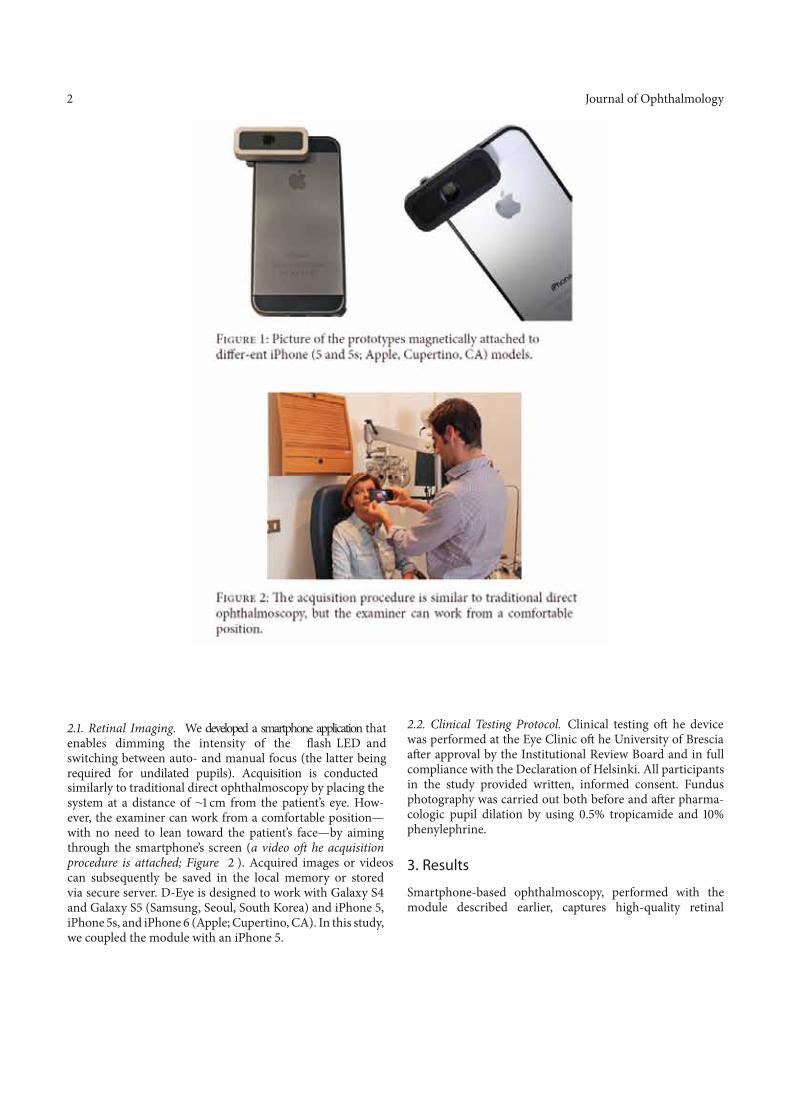
D-Eye works on the principles of direct ophthalmoscopy and exploits the smartphone camera’s autofocus capability to account for a patient’s refractive error. The smartphone requires no modification, and a bumper fitted with properly located neodymium magnets (N45) ensures the correct alignment of the device with the smart-phone’s camera andflash (Figure 1).
Hindawi Publishing CorporationJournal of OphthalmologyVolume 2015, Article ID 823139, 5 pageshttp://dx.doi.org/10.1155/2015/823139
Journal of Ophthalmology
. . Retinal Imaging. We developed a smartphone application that enables dimming the intensity of the �ash LED and switching between auto- and manual focus (the latter being required for undilated pupils). Acquisition is conducted
F 1: Picture of the prototypes magnetically attached to di er-ent iPhone ( and s; Apple, Cupertino, CA) models.
F 2: e acquisition procedure is similar to traditional direct ophthalmoscopy, but the examiner can work from a comfortable position.
similarly to traditional direct ophthalmoscopy by placing thesystem at a distance of cm from the patient’s eye. How-ever, the examiner can work from a comfortable position—with no need to lean toward the patient’s face—by aimingthrough the smartphone’s screen (a video o� he acquisitionprocedure is attached; Figure 2 ). Acquired images or videoscan subsequently be saved in the local memory or storedvia secure server. D-Eye is designed to work with Galaxy Sand Galaxy S (Samsung, Seoul, South Korea) and iPhone ,iPhone s, and iPhone (Apple; Cupertino, CA). In this study,we coupled the module with an iPhone .
. . Clinical Testing Protocol. Clinical testing o� he devicewas performed at the Eye Clinic o� he University of Bresciaa er approval by the Institutional Review Board and in fullcompliance with the Declaration of Helsinki. All participantsin the study provided written, informed consent. Fundusphotography was carried out both before and a er pharma-cologic pupil dilation by using . % tropicamide and %phenylephrine.
3. Results
Smartphone-based ophthalmoscopy, performed with themodule described earlier, captures high-quality retinal
Journal of Ophthalmology
(a) (b) (c)
(d) (e) (f)
(g) (h)
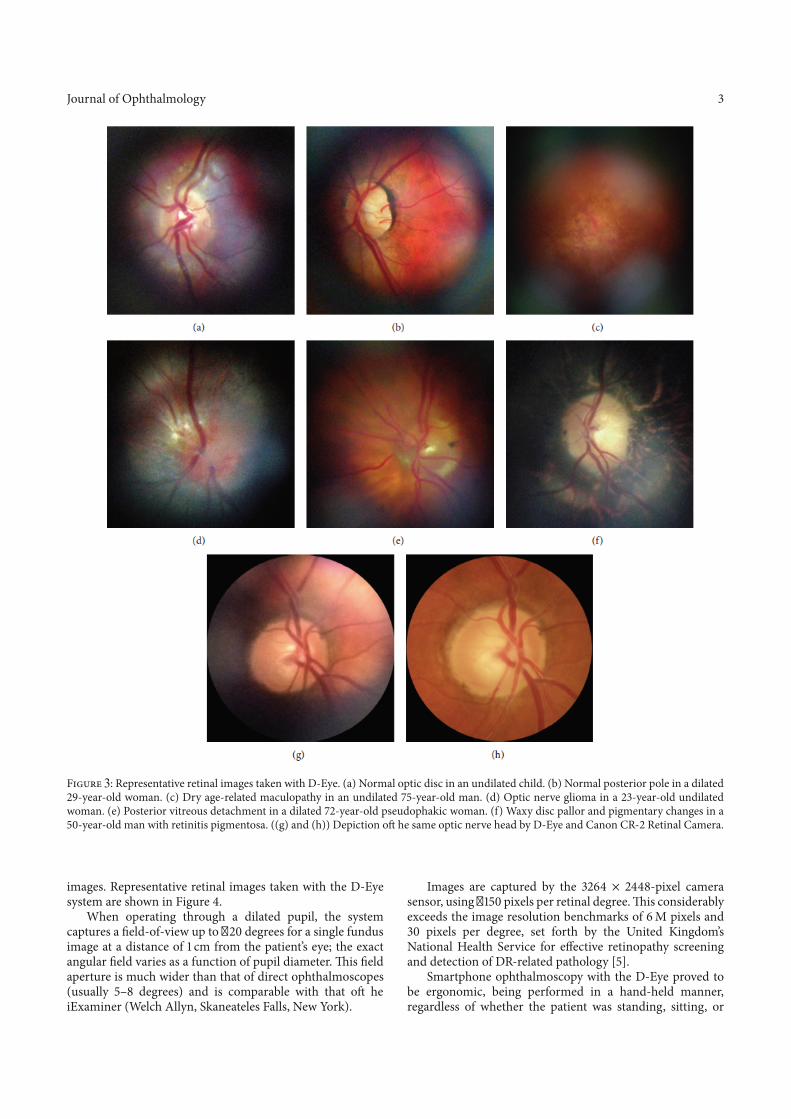
F : Representative retinal images taken with D-Eye. (a) Normal optic disc in an undilated child. (b) Normal posterior pole in a dilated-year-old woman. (c) Dry age-related maculopathy in an undilated -year-old man. (d) Optic nerve glioma in a -year-old undilated
woman. (e) Posterior vitreous detachment in a dilated -year-old pseudophakic woman. (f) Waxy disc pallor and pigmentary changes in a-year-old man with retinitis pigmentosa. ((g) and (h)) Depiction o� he same optic nerve head by D-Eye and Canon CR- Retinal Camera.
images. Representative retinal images taken with the D-Eyesystem are shown in Figure .
When operating through a dilated pupil, the systemcaptures a eld-of-view up to � degrees for a single fundusimage at a distance of cm from the patient’s eye; the exactangular eld varies as a function of pupil diameter. is eldaperture is much wider than that of direct ophthalmoscopes(usually – degrees) and is comparable with that o� heiExaminer (Welch Allyn, Skaneateles Falls, New York).
Images are captured by the × -pixel camerasensor, using � pixels per retinal degree. is considerablyexceeds the image resolution benchmarks of M pixels and
pixels per degree, set forth by the United Kingdom’sNational Health Service for e ective retinopathy screeningand detection of DR-related pathology [ ].
Smartphone ophthalmoscopy with the D-Eye proved tobe ergonomic, being performed in a hand-held manner,regardless of whether the patient was standing, sitting, or
3
Journal of Ophthalmology
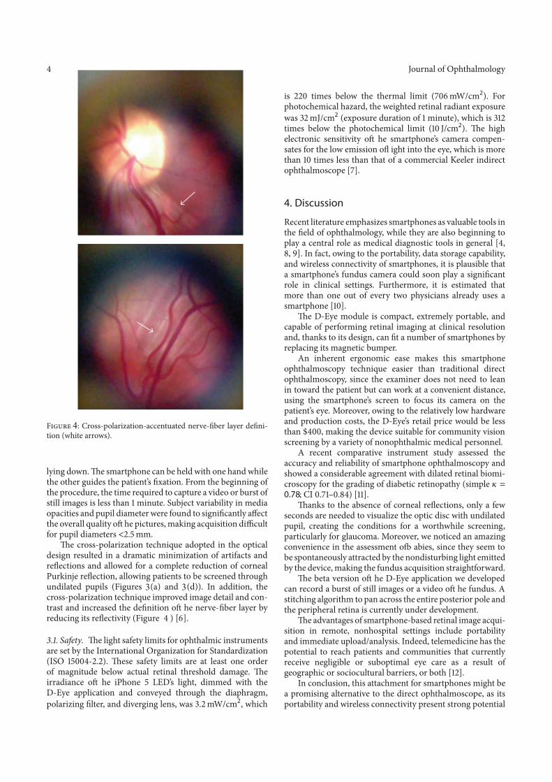
F : Cross-polarization-accentuated nerve- ber layer de ni-tion (white arrows).
lying down. e smartphone can be held with one hand whilethe other guides the patient’s xation. From the beginning ofthe procedure, the time required to capture a video or burst ofstill images is less than minute. Subject variability in mediaopacities and pupil diameter were found to signi cantly a ectthe overall quality o� he pictures, making acquisition di cultfor pupil diameters < . mm.
e cross-polarization technique adopted in the opticaldesign resulted in a dramatic minimization of artifacts andre ections and allowed for a complete reduction of cornealPurkinje re ection, allowing patients to be screened throughundilated pupils (Figures (a) and (d)). In addition, thecross-polarization technique improved image detail and con-trast and increased the de nition o� he nerve- ber layer byreducing its re ectivity (Figure 4 ) [ ].
. . Safety. e light safety limits for ophthalmic instrumentsare set by the International Organization for Standardization(ISO - . ). ese safety limits are at least one orderof magnitude below actual retinal threshold damage. eirradiance o� he iPhone LED’s light, dimmed with theD-Eye application and conveyed through the diaphragm,polarizing lter, and diverging lens, was . mW/cm2, which
is times below the thermal limit ( mW/cm2). Forphotochemical hazard, the weighted retinal radiant exposurewas mJ/cm2 (exposure duration of minute), which istimes below the photochemical limit ( J/cm2). e highelectronic sensitivity o� he smartphone’s camera compen-sates for the low emission o� ight into the eye, which is morethan times less than that of a commercial Keeler indirectophthalmoscope [ ].
4. Discussion
Recent literature emphasizes smartphones as valuable tools inthe eld of ophthalmology, while they are also beginning toplay a central role as medical diagnostic tools in general [ ,
, ]. In fact, owing to the portability, data storage capability,and wireless connectivity of smartphones, it is plausible thata smartphone’s fundus camera could soon play a signi cantrole in clinical settings. Furthermore, it is estimated thatmore than one out of every two physicians already uses asmartphone [ ].
e D-Eye module is compact, extremely portable, andcapable of performing retinal imaging at clinical resolutionand, thanks to its design, can t a number of smartphones byreplacing its magnetic bumper.
An inherent ergonomic ease makes this smartphoneophthalmoscopy technique easier than traditional directophthalmoscopy, since the examiner does not need to leanin toward the patient but can work at a convenient distance,using the smartphone’s screen to focus its camera on thepatient’s eye. Moreover, owing to the relatively low hardwareand production costs, the D-Eye’s retail price would be lessthan , making the device suitable for community visionscreening by a variety of nonophthalmic medical personnel.
A recent comparative instrument study assessed theaccuracy and reliability of smartphone ophthalmoscopy andshowed a considerable agreement with dilated retinal biomi-croscopy for the grading of diabetic retinopathy (simple =0.78; CI . – . ) [ ].
anks to the absence of corneal re ections, only a fewseconds are needed to visualize the optic disc with undilatedpupil, creating the conditions for a worthwhile screening,particularly for glaucoma. Moreover, we noticed an amazingconvenience in the assessment o� abies, since they seem tobe spontaneously attracted by the nondisturbing light emittedby the device, making the fundus acquisition straightforward.
e beta version o� he D-Eye application we developedcan record a burst of still images or a video o� he fundus. Astitching algorithm to pan across the entire posterior pole andthe peripheral retina is currently under development.
e advantages of smartphone-based retinal image acqui-sition in remote, nonhospital settings include portabilityand immediate upload/analysis. Indeed, telemedicine has thepotential to reach patients and communities that currentlyreceive negligible or suboptimal eye care as a result ofgeographic or sociocultural barriers, or both [ ].
In conclusion, this attachment for smartphones might bea promising alternative to the direct ophthalmoscope, as itsportability and wireless connectivity present strong potential
3 3
4
Journal of Ophthalmology 5
applications such as telemedicine, even in nonhospital orrural settings.
Conflict of Interests
The authors declare that there is no conflict of interestsregarding the publication of this paper.
References
[1] M. D. Abramoff, M. K. Garvin, andM. Sonka, “Retinal imagingand image analysis,” IEEE Reviews in Biomedical Engineering,vol. 3, pp. 169–208, 2010.
[2] C. L. Shields,M.Materin, and J. A. Shields, “Panoramic imagingof the ocular fundus,”Archives of Ophthalmology, vol. 121, no. 11,pp. 1603–1607, 2003.
[3] D. S. W. Ting, M. L. Tay-Kearney, and Y. Kanagasingam, “Lightand portable novel device for diabetic retinopathy screening,”Clinical and Experimental Ophthalmology, vol. 40, no. 1, pp.e40–e46, 2012.
[4] R. N. Maamari, J. D. Keenan, D. A. Fletcher, and T. P. Margolis,“A mobile phone-based retinal camera for portable wide fieldimaging,” British Journal of Ophthalmology, vol. 98, no. 4, pp.438–441, 2014.
[5] K. Tran, T. A. Mendel, K. L. Holbrook, and P. A. Yates, “Con-struction of an inexpensive, hand-held fundus camera throughmodification of a consumer ‘point-and-shoot’ camera,” Inves-tigative Ophthalmology and Visual Science, vol. 53, no. 12, pp.7600–7607, 2012.
[6] A. Sommer,H.A. Kues, S. A.D’Anna, S. Arkell, A. Robin, andH.A. Quigley, “Cross-polarization photography of the nerve fiberlayer,” Archives of Ophthalmology, vol. 102, no. 6, pp. 864–869,1984.
[7] D. Y. Kim, F. Delori, and S. Mukai, “Smartphone photographysafety,” Ophthalmology, vol. 119, no. 10, pp. 2200–2201, 2012.
[8] L. J. Haddock, D. Y. Kim, and S. Mukai, “Simple, inexpensivetechnique for high-quality smartphone fundus photography inhuman and animal eyes,” Journal of Ophthalmology, vol. 2013,Article ID 518479, 5 pages, 2013.
[9] A. Bastawrous, “Smartphone fundoscopy,” Ophthalmology, vol.119, no. 2, pp. 432.e2–433.e2, 2012.
[10] R. K. Lord, V. A. Shah, A. N. san Filippo, and R. Krishna, “Noveluses of smartphones in ophthalmology,”Ophthalmology, vol. 117,no. 6, pp. 1274–1274.e3, 2010.
[11] A. Russo, F. Morescalchi, C. Costagliola, L. Delcassi, andF. Semeraro, “Comparison of smartphone ophthalmoscopywith slit-lamp biomicroscopy for grading diabetic retinopathy,”American Journal of Ophthalmology, vol. 159, no. 2, pp. 360.e1–364.e1, 2015.
[12] S.-P. Chow, L.M. Aiello, J. D. Cavallerano et al., “Comparison ofnonmydriatic digital retinal imaging versus dilated ophthalmicexamination for nondiabetic eye disease in persons with dia-betes,” Ophthalmology, vol. 113, no. 5, pp. 833–840, 2006.

Submit your manuscripts athttp://www.hindawi.com
Stem CellsInternational
Hindawi Publishing Corporationhttp://www.hindawi.com Volume 201
Hindawi Publishing Corporationhttp://www.hindawi.com Volume 201
MEDIATORSINFLAMMATION
of
Hindawi Publishing Corporationhttp://www.hindawi.com Volume 201
Behavioural Neurology
EndocrinologyInternational Journal of
Hindawi Publishing Corporationhttp://www.hindawi.com Volume 2014
Hindawi Publishing Corporationhttp://www.hindawi.com Volume 2014
Disease Markers
Hindawi Publishing Corporationhttp://www.hindawi.com Volume 2014
BioMed Research International
OncologyJournal of
Hindawi Publishing Corporationhttp://www.hindawi.com Volume 201
Hindawi Publishing Corporationhttp://www.hindawi.com Volume 2014
Oxidative Medicine and Cellular Longevity
Hindawi Publishing Corporationhttp://www.hindawi.com Volume 2014
PPAR Research
The Scientific World JournalHindawi Publishing Corporation http://www.hindawi.com Volume 2014
Immunology ResearchHindawi Publishing Corporationhttp://www.hindawi.com Volume 201
Journal of
ObesityJournal of
Hindawi Publishing Corporationhttp://www.hindawi.com Volume 201
Hindawi Publishing Corporationhttp://www.hindawi.com Volume 201
Computational and Mathematical Methods in Medicine
OphthalmologyJournal of
Hindawi Publishing Corporationhttp://www.hindawi.com Volume 201
Diabetes ResearchJournal of
Hindawi Publishing Corporationhttp://www.hindawi.com Volume 2014
Hindawi Publishing Corporationhttp://www.hindawi.com Volume 201
Research and TreatmentAIDS
Hindawi Publishing Corporationhttp://www.hindawi.com Volume 201
Gastroenterology Research and Practice
Hindawi Publishing Corporationhttp://www.hindawi.com Volume 2014
Parkinson’s Disease
Evidence-Based Complementary and Alternative Medicine
Volume 201Hindawi Publishing Corporationhttp://www.hindawi.com