A novel antiageing mechanism for retinol: induction of ... · A novel anti-ageing mechanism for...
10
A novel anti-ageing mechanism for retinol: induction of dermal elastin synthesis and elastin fibre formation D. Rossetti*, M. G. Kielmanowicz , S. Vigodman , Y. P. Hu*, N. Chen*, A. Nkengneà, T. Oddosà, D. Fischer , M. Seiberg* and C. B. Lin* *The Johnson & Johnson Skin Research Center, Consumer Product Worldwide, A Unit of Johnson & Johnson Consumer Companies, Inc., Skillman, NJ, USA, ColBar LifeScience Ltd., Industrial Park, Rehovot, Israel and àJohnson & Johnson Consumer France, Pharmacology & Skin Care Research Center Campus de Maigremont, Val de Reuil, France Received 18 November 2009, Accepted 18 January 2010 Keywords: collagen, elastin, extracellular matrix, retinol, skin ageing Synopsis Dermal elastic fibres are extracellular matrix pro- tein complexes produced by fibroblasts and involved in skin elasticity. Elastin fibres decrease with age as a result of reduced synthesis and increased degradation, resulting in skin sagging and reduced skin elasticity. In this study, we show that retinol (ROL), known to enhance dermal col- lagen production, is also enhancing elastin fibre formation. ROL induced elastin gene expression and elastin fibre formation in cultured human der- mal fibroblasts. Topical treatment of cultured human skin explants with a low dose (0.04%) of ROL increased mRNA and protein levels of tropo- elastin and of fibrillin-1, an elastin accessory protein, as documented by QPCR and immunohis- tochemistry staining. Luna staining confirmed the increased elastin fibre network in the ROL-treated skin explants, as compared with untreated con- trols. These data demonstrate that ROL exerts its anti-ageing benefits not only via enhanced epider- mal proliferation and increased collagen produc- tion, but also through an increase in elastin production and assembly. Re ´ sume ´ Les fibres e ´lastiques du derme sont des complexes de prote ´ines matricielles extracellulaires produits par des fibroblastes et implique ´es dans l’e ´lasticite ´ de la peau. Les fibres d’e ´lastine diminuent avec l’a ˆge en raison d’une synthe `se re ´duite et d’une de ´gradation accrue, qui aboutissent a ` l’affaisse- ment de peau et a ` la re ´duction de son e ´lasticite ´. Nous montrons que le re ´tinol (ROL), connu pour favoriser la production de collage `ne dermique, ame ´liore aussi la formation des fibres d’e ´lastine. ROL a induit l’expression du ge `ne de l’e ´lastine et la formation des fibres dans des fibroblastes dermi- ques humains en culture. Le traitement topique d’explants de peau humaines en culture avec une faible dose de ROL (0.04%) a augmente ´ le taux d’ARNm et le marquage immunohistochimique et QPCR a montre ´ l’augmentation du taux des prote ´- ines tropo e ´lastine et fibrilline-1, une prote ´ine associe ´e a ` l’e ´lastine. La coloration Luna a confirme ´ un re ´seau de fibres e ´lastiques accru sur les explants cutane ´s ROL-TRAITE ´ ES, en comparaison des explants non traite ´s. Ces donne ´es de ´montrent que ROL exerce ses proprie ´te ´s anti-a ˆges non seule- ment via une ame ´lioration de la prolife ´ration e ´pid- ermique et de l’augmentation de la production de collage `ne, mais aussi par une augmentation de la production d’e ´lastine et de l’organisation du re ´seau e ´lastique. Correspondence: Dr Connie B. Lin, The Johnson & John- son Skin Research Center, Consumer Product Worldwide, A Unit of Johnson & Johnson Consumer Companies, Inc., 199 Grandview Rd., Skillman, NJ 08558, USA. Tel.: 908-874-1532; fax: 908-874-1254; e-mail: blin1@its. jnj.com International Journal of Cosmetic Science, 2010, 1–10 doi: 10.1111/j.1468-2494.2010.00588.x ª 2010 Johnson & Johnson. Journal compilation ª 2010 Society of Cosmetic Scientists and the Socie ´te ´ Franc ¸aise de Cosme ´tologie 1
Transcript of A novel antiageing mechanism for retinol: induction of ... · A novel anti-ageing mechanism for...
A novel anti-ageing mechanism for retinol: induction
of dermal elastin synthesis and elastin fibre
formation
D. Rossetti*, M. G. Kielmanowicz�, S. Vigodman�, Y. P. Hu*, N. Chen*, A. Nkengne�, T. Oddos�,
D. Fischer�, M. Seiberg* and C. B. Lin*
*The Johnson & Johnson Skin Research Center, Consumer Product Worldwide, A Unit of Johnson & Johnson Consumer
Companies, Inc., Skillman, NJ, USA, �ColBar LifeScience Ltd., Industrial Park, Rehovot, Israel and �Johnson & Johnson
Consumer France, Pharmacology & Skin Care Research Center Campus de Maigremont, Val de Reuil, France
Received 18 November 2009, Accepted 18 January 2010
Keywords: collagen, elastin, extracellular matrix, retinol, skin ageing
Synopsis
Dermal elastic fibres are extracellular matrix pro-
tein complexes produced by fibroblasts and
involved in skin elasticity. Elastin fibres decrease
with age as a result of reduced synthesis and
increased degradation, resulting in skin sagging
and reduced skin elasticity. In this study, we show
that retinol (ROL), known to enhance dermal col-
lagen production, is also enhancing elastin fibre
formation. ROL induced elastin gene expression
and elastin fibre formation in cultured human der-
mal fibroblasts. Topical treatment of cultured
human skin explants with a low dose (0.04%) of
ROL increased mRNA and protein levels of tropo-
elastin and of fibrillin-1, an elastin accessory
protein, as documented by QPCR and immunohis-
tochemistry staining. Luna staining confirmed the
increased elastin fibre network in the ROL-treated
skin explants, as compared with untreated con-
trols. These data demonstrate that ROL exerts its
anti-ageing benefits not only via enhanced epider-
mal proliferation and increased collagen produc-
tion, but also through an increase in elastin
production and assembly.
Resume
Les fibres elastiques du derme sont des complexes
de proteines matricielles extracellulaires produits
par des fibroblastes et impliquees dans l’elasticite
de la peau. Les fibres d’elastine diminuent avec
l’age en raison d’une synthese reduite et d’une
degradation accrue, qui aboutissent a l’affaisse-
ment de peau et a la reduction de son elasticite.
Nous montrons que le retinol (ROL), connu pour
favoriser la production de collagene dermique,
ameliore aussi la formation des fibres d’elastine.
ROL a induit l’expression du gene de l’elastine et
la formation des fibres dans des fibroblastes dermi-
ques humains en culture. Le traitement topique
d’explants de peau humaines en culture avec une
faible dose de ROL (0.04%) a augmente le taux
d’ARNm et le marquage immunohistochimique et
QPCR a montre l’augmentation du taux des prote-
ines tropo elastine et fibrilline-1, une proteine
associee a l’elastine. La coloration Luna a confirme
un reseau de fibres elastiques accru sur les
explants cutanes ROL-TRAITEES, en comparaison
des explants non traites. Ces donnees demontrent
que ROL exerce ses proprietes anti-ages non seule-
ment via une amelioration de la proliferation epid-
ermique et de l’augmentation de la production de
collagene, mais aussi par une augmentation de la
production d’elastine et de l’organisation du reseau
elastique.
Correspondence: Dr Connie B. Lin, The Johnson & John-
son Skin Research Center, Consumer Product Worldwide,
A Unit of Johnson & Johnson Consumer Companies, Inc.,
199 Grandview Rd., Skillman, NJ 08558, USA. Tel.:
908-874-1532; fax: 908-874-1254; e-mail: blin1@its.
jnj.com
International Journal of Cosmetic Science, 2010, 1–10 doi: 10.1111/j.1468-2494.2010.00588.x
ª 2010 Johnson & Johnson. Journal compilation
ª 2010 Society of Cosmetic Scientists and the Societe Francaise de Cosmetologie 1
Introduction
Skin ageing is associated with wrinkles, sagging,
uneven pigmentation, skin roughness and laxity,
which are induced by chronological metabolic pro-
cesses, and by external insults such as solar irradi-
ation (photoageing). Epidermal ageing includes
epidermal thinning and pigmentary effects such as
unevenness or ‘age spots’. Dermal ageing includes
dermal thinning, as a result of a decrease in fibro-
blast number, reduced collagen synthesis and
increased UV-induced collagen degradation
(reviewed in [1–4]). Additionally, the dermal elas-
tin network is destroyed by reduced synthesis of
elastin and its accessory proteins, by increased
elastin fibre degradation, and by UV-induced solar
elastosis (reviewed in [1, 5]), resulting in reduced
skin elasticity and resilience.
Elastin fibres are an essential element of the der-
mal connective tissue. Elastic fibres are produced
from extracellular matrix proteins that are synthe-
sized and secreted by dermal fibroblasts. This is fol-
lowed by the assembly of the elastin protein and
numerous microfibrillar components, to create a
fibre network that can stretch and relax ([6–8]
and reviewed in [9–11]). The mature elastin fibre
is made primarily of the elastin protein, which is
encoded by a single gene. Tropoelastin, a 72 000-
dalton polypeptide, is secreted as a soluble protein
and is then cross-linked by the enzyme lysyl oxi-
dase (LOX) [12–14]. Microfibrils consist mainly of
fibrillins, but they also include, or are associated
with microfibril-associated glycoproteins, fibulins
and EMILIN-1 [9–11, 15]. The microfibrils direct
the deposition of tropoelastin during elastic fibre
production and facilitate the alignment of the elas-
tin monomers prior to their cross-linking by LOX.
The controlled and balanced synthesis of elastin
and the microfibril components, as well as their
interactions, are essential for the formation of nor-
mal elastin fibres. Aberrations in the structure,
metabolism or assembly of elastin result in herita-
ble and acquired diseases, affecting skin and other
connective tissues, such as cutis laxa, pseudoxant-
homa elasticum and elastosis perforans serpiginosa
[16]. Mutations in fibrillin-1, the main structural
component of microfibrils, lead to Marfan syn-
drome, a heritable disease with severe skeleton,
skin and joints defects [17].
The elastin fibre network in the skin performs best
in adolescence and early adulthood, and declines
thereafter. Tissue regeneration slows upon ageing,
and fibroblasts increase the release of human neu-
trophil and macrophage elastases, which are known
to degrade mature elastic fibres [18–20]. Addition-
ally, environmental factors, and in particular
ultraviolet (UV) exposure, induce a massive accu-
mulation of non-functional elastotic material in the
upper and middle dermis, termed solar elastosis [1,
20]. Known strategies aimed to prevent or reverse
skin ageing include sun avoidance and sunscreens,
the use of anti-oxidant combinations, and the use of
retinoids to inhibit collagenases and to promote col-
lagen production, yet strategies aiming at enhanc-
ing the elastin fibre network are limited (reviewed
in [3]). Recently, the use of dill [21] and non-dena-
tured soybean [22] extracts were suggested for
enhancing skin elasticity.
The anti-ageing effects of retinol (ROL) on the
skin are extensively documented. Clinically, ROL
was shown to enhance keratinocyte proliferation
[23–25], to increase the production of collagen
[23, 26], and to reduce the formation and the pig-
mentary levels of dyspigmentation [27]. Recent
studies of the clinical effects of ROL on wrinkles
and photo ageing document an increase in skin
elasticity, as assessed by dermatologists [28, 29].
Retinoic acid, but not ROL, was shown to increase
elastin synthesis in chick embryonic skin fibro-
blasts [30], and topical tretinoin treatment was
shown to increase the tropoelastin content of
photoaged hairless mouse skin [31]. These data
might suggest that the topical application of ROL
may also stimulate elastin synthesis in human
skin; however, definitive research to that effect is
currently lacking. Here, we report that ROL is able
to stimulate elastin and fibrillin-1 gene expression
and elastin fibres formation in normal human der-
mal fibroblasts, in 3D dermal cultures and in
human skin explants. These data point to a novel
mechanism of action, adding to the anti-ageing
effects of ROL.
Materials and methods
Chemicals
Unless otherwise specified, all chemicals were from
Sigma-Aldrich (St Louis, MO, U.S.A.). Human skin
Etna-Elastin was purchased from Elastin Products
Company, Inc. (Owensville, MO, U.S.A.). TGF-bwas purchased from PeproTech Inc. (Rocky Hill,
NJ, U.S.A.). ROL was dissolved in DMSO as stock
solution of 10)1 M and was further diluted in
ª 2010 Johnson & Johnson. Journal compilation
ª 2010 Society of Cosmetic Scientists and the Societe Francaise de Cosmetologie
International Journal of Cosmetic Science, 1–102
Retinol induces elastin synthesis D. Rossetti et al.

culture media. An oil-in-water emulsion contain-
ing 0.04% ROL was used for topical treatments.
Monolayer dermal fibroblast culture
Primary normal human adult dermal fibroblasts
were purchased from PromoCell (Sickingenstr 63/
65, Heidelberg, Germany), and were maintained
according to manufacturer’s instructions. Cells were
plated at 1 · 104 cells/96 well in Dulbecco’s modi-
fied Eagle’s medium (DMEM; Invitrogen Corp., Carls-
bad, CA, U.S.A.) with 5% fetal bovine serum (FBS;
Biological Industries, Beit Haemek, Israel) and were
incubated for 48–72 h. Cells were then treated, in
triplicates, with ROL at the concentrations indi-
cated, in DMEM supplemented with 1% FBS, and
incubated for additional 72 h. Cells were washed
thrice with 300 lL of 1· phosphate-buffered saline
(PBS; Neopharm, Emek Hefer Industrial Park, Israel)
and were lysed with 100 lL of lysis buffer (0.1 M
Tris, 0.15 M NaCl, 0.5% Triton X-100) for analysis
of elastin protein levels by ELISA.
3D dermal fibroblast cultures
A 3D collagen scaffold was prepared by ColBar Life-
Science Ltd (Rehovot, Israel) using collagen Type I
purified from porcine tendons and cross-linked by
glycation [32]. The collagen scaffolds (5 ·1 · 5 mm) were placed in 6-well plates and were
seeded drop-wise with 2 · 106 human adult der-
mal fibroblasts, suspended in 50 lL growth med-
ium (DMEM; Invitrogen Corp.), supplemented with
10% FBS (Biological Industries). Cultures were kept
at room temperature in a Laminar Flow Hood to
allow cell adherence. After 2 h, the volume of cul-
ture medium was increased to 5 mL, and the 3D
dermal fibroblast cultures were placed in a 37�C,
5% CO2 incubator on an orbital shaker, for 2 days.
After the 2 days pre-incubation, the medium was
replaced, and ROL or TGF-b, at the indicated con-
centrations, were added, in DMEM supplemented
with 1% FBS, for additional incubation of 2 weeks.
Media and treatments were refreshed thrice a week.
Following 2 weeks of incubation, the 3D cultures
were placed into 4% buffered formalin solution and
were processed for histological analysis.
Human skin explants
Human skins were obtained with informed consent
from abdominal skins of healthy individuals under-
going plastic surgery (The Johns Hopkins Outpa-
tient Center, Plastic Surgery, Baltimore, MD,
U.S.A.). Patient identities were not disclosed to
preserve confidentiality, in compliance with US
HIPAA regulations. Punch biopsies (12 mm) were
disinfected at room temperature for 30 min with
DMEM with high sucrose content (Invitrogen
Corp.), supplemented with Pen/Strep, fungizone
and gentamycine (all from Invitrogen). Explants
were then rinsed with DMEM and placed in a
1 : 1 mixture of DMEM and F12 nutrient mixture
(F-12) (Invitrogen Corp.) supplemented with 2%
FBS (Invitrogen Corp.) and a cocktail of growth
factors [33]. After an overnight incubation in a
humidified chamber, in a 5% CO2 atmosphere at
32�C, explants were transferred to 37�C for the
rest of the culture duration. Explants were treated
topically with a ROL formulation (0.04%, 5 lL) at
days 1 and 3, and were harvested at different time
points for RNA extraction or histological staining.
Culture media were refreshed daily, except week-
ends. Experiments were repeated thrice.
ELISA
Cells were lysed using TritonX-100 based lysis buf-
fer and were tested by a direct ELISA using rabbit
anti-Elastin polyclonal antibody (Santa Cruz Bio-
technology, Inc., Santa Cruz, CA, U.S.A.), and a
biotinylated goat anti-rabbit IgG (Abcam Inc.,
Cambridge, MA, U.S.A.). Human skin Etna-Elastin
was used as a standard. Elastin standard (1 mg/
mL) was diluted in PBS with 0.5% Bovine Serum
Albumin to obtain working solutions. The wells of
the ELISA plates were coated with the appropri-
ately diluted cell lysates (1 : 31 or 1 : 100 in PBS
with 0.5% BSA for TGF-b-treated cells; 1 : 1 or
1 : 31 for ROL-treated cells) to obtain elastin con-
centrations within the linear portion of the stan-
dard curve.
Histological and immuno-fluorescence staining
3D dermal cultures were placed in 4% buffered for-
malin solution (Richard-Allan Scientific, Kalama-
zoo, MI, U.S.A.), dehydrated and then embedded in
paraffin. Two, 5 lm thick consecutive sections
were collected from each block at 150 lm inter-
vals. Skin tissues were fixed overnight in 10% buf-
fered formalin, and followed by embedding into
paraffin blocks and sectioning (5 lm) using stan-
dard procedures. Sections were stained with
ª 2010 Johnson & Johnson. Journal compilation
ª 2010 Society of Cosmetic Scientists and the Societe Francaise de Cosmetologie
International Journal of Cosmetic Science, 1–10 3
Retinol induces elastin synthesis D. Rossetti et al.
Herovici staining, for documenting newly synthe-
sized collagen [34] or with Luna staining, for doc-
umenting Elastin fibres [35]. Images of 3D
cultures were captured using Nikon microscope
ECLIPSE 50i (Nikon Instruments, Melville, NY,
U.S.A.) and Nikon Digita Sight DS-5M; Software:
NIS Elements AR 3.0 SP4 Build 384 (Nikon
Instruments). Images of skin explants were
obtained using Leica microscope (Leitz DM1L,
Leica, Allendale, NJ, U.S.A.) and a QiCAM camera
(QIMAGING, Surrey, BC, Canada).
For immuno-fluorescence staining, human skin
explants were embedded in Tissue-Tek OCT com-
pound (EMS, Washington, PA, U.S.A.), flash frozen
in liquid nitrogen and stored at )80�C. Frozen sec-
tions, 7 lm thick, were cut with a Leica CM 1950
cryostat (Leica, Microsystems GmbH, Wetzlar, Ger-
many) and stored at )80�C until used. Slides were
heated on a slide warmer to 65�C for 30 min, then
fixed in acetone at )20�C for 10 min, and then
allowed to air dry for 20 min. Sections were incu-
bated with 5% donkey normal serum (Jackson
ImmunoResearch Laboratories, West Grove, PA,
U.S.A.) in 1· PBS prior to incubating with primary
antibodies. A tropoelastin rabbit polyclonal anti-
body (Abcam Inc.) diluted 1 : 200 and a fibrillin-1
mouse monoclonal (Lab Vision Corporation, Fre-
mont, CA, U.S.A.) diluted 1 : 100 were applied to
the tissue sections for 1 h at room temperature. The
tissues were washed twice in 1· PBS at room
temperature for 5 min and then incubated with a
1 : 200 dilution of donkey anti-rabbit Cyanine-
2-conjugated and donkey anti-mouse Cyanine-
3-conjugated secondary antibodies (Jackson
ImmunoResearch Laboratories, Inc.) in 1· PBS and
DAPI (4¢,6-diamidino-2-phenylindole dihydrochlo-
ride; Sigma, St Louis, MO, U.S.A.) at room tempera-
ture for 30 min prior to being mounted with cover
slips. The slides were left to air dry for 24 h before
being viewed with a fluorescence microscope (Leica
DMIRE2, Leica, Microsystems GmbH) at wave-
lengths of 492 nm for Cy2 (green for tropoelastin)
and 550 nm for Cy3 (orange for fibrillin-1).
RNA extraction and QPCR
Total RNA was extracted from skin explants using
TRIzol (Invitrogen), followed by a purification step
using Mini-Column Clean-Up (Qiagen, Valencia,
CA). The purified RNA was converted to cDNA
using Superscript� III reverse transcriptase (Invitro-
gen, Carlsbad, CA, U.S.A.). QPCR analyses were set
up using a custom-made template by SABiosciences
Corp. (Frederick, MD, U.S.A.). The template was
coated with primers for: Elastin, fibrillin-1, collagen
I, and 18S RNA. Each 25 lL reaction contained
12.5 lL master mix (SABiosciences), 11 lL cDNA
(�6 ng) and 1.5 lL of H2O. QPCR was performed
on 7500 Realtime PCR system (Applied Biosystems,
Foster City, CA, U.S.A.).
Statistical analysis
Statistical analysis was performed with Student’s
t-test. All results are presented as the mean ± SE.
Results
ROL increases elastin protein levels and elastin
fibres staining in cultured dermal fibroblasts
Normal human adult dermal fibroblasts were cul-
tured in monolayers, and were remained untreated
or were treated with ROL (10)5 and 10)6 M) for
72 h. Cells were then lysed and their elastin pro-
tein content was quantified using an ELISA assay.
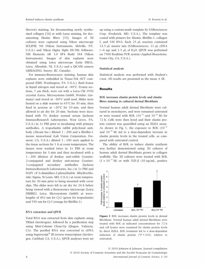
As shown in Fig. 1, the exposure to ROL (10)5
and 10)6 M) led to a dose-dependent increase in
elastin protein levels in the treated cells, as com-
pared with untreated control.
The ability of ROL to induce elastin synthesis
was further demonstrated using 3D cultures of
human adult dermal fibroblasts grown in collagen
scaffolds. The 3D cultures were treated with ROL
(1 · 10)5 M) or with TGF-b (10 ng/mL, positive
Figure 1 ROL increases elastin protein levels in dermal
fibroblasts. Normal human adult dermal fibroblasts were
treated with ROL at indicated concentrations for 72 h
and cell lysates were examined for elastin protein levels
by direct ELISA. ROL treatment led to a dose-dependent
induction of elastin protein (*P < 0.01, relative to
untreated).
ª 2010 Johnson & Johnson. Journal compilation
ª 2010 Society of Cosmetic Scientists and the Societe Francaise de Cosmetologie
International Journal of Cosmetic Science, 1–104
Retinol induces elastin synthesis D. Rossetti et al.

control) [36], or remained untreated for 2 weeks.
Histological staining of the 3D cultures (Fig. 2)
was used to document the induction of collagen
synthesis (a known ROL activity, shown by Hero-
vici staining) and the enhancement of elastin fibre
formation (shown by Luna staining). As expected,
TGF-b induced the formation of new collagen
fibres (Fig. 2B, compared with untreated control
Fig. 2A), and also increased the formation of new
elastin fibres (Fig. 2E, compared with untreated
control Fig. 2D). ROL (10)5 M) induced collagen
synthesis in this new 3D culture system (Fig. 2C,
compared with untreated control Fig. 2A), con-
firming both the metabolic activity of the culture
system and the activity of ROL under the tested
conditions. Additionally, ROL was shown, for the
first time, to enhance the formation of an elastic
fibre network in vitro (Fig. 2F, compared with
untreated control Fig. 2D). These results suggest
that ROL can induce elastin protein synthesis and
elastin fibre formation by adult skin fibroblasts.
ROL enhances the elastin fibre network of human
skin explants
To further confirm the elastin-enhancing effect of
ROL, studies were performed using full-thickness
human skin explants, which better represent the
physiological complexity of the skin. To study the
effect of ROL on human skin, in a way that mimics
the biological activity in human, topical treatment
of ROL was used in skin explants. Human skin
biopsies, obtained with informed consent from
healthy donors undergoing abdominal surgeries,
were topically treated with ROL (0.04%), at days 1
and 3, or remained untreated. Explants were har-
vested at different time points for gene expression
analyses and for histological staining. As a control
for the metabolic activity of the explants and for
the known activity of ROL, a collagen staining
(Herovici) was performed. As expected, at day 7
the ROL-treated explants (Fig. 3B) showed more
newly synthesized pro-collagen (blue staining) and
mature collagen (red staining) than their corre-
sponding untreated controls (Fig. 3A), suggesting
that the explants are metabolically active and
responsive to ROL, suggesting that the explants are
metabolically active and responsive to ROL. Note
the documented increase in mature collagen, but
not in procollagen staining, at day 7, suggesting
the correct assembly of the newly synthesized colla-
gen fibres in the cultured skin explants.
The elastic fibre network of the skin explants
was visualized at day 7 using Luna staining.
A B C
D E F
Figure 2 ROL increases elastin fibre staining in 3D dermal fibroblast cultures. 3D dermal fibroblast cultures, established
in collagen scaffolds, were treated with TGF-b (10 ng/mL), or with ROL (1 · 10)5 M), or remained untreated for
2 weeks. Representative Herovici-stained (collagen), and Luna-stained (elastin) histological images of control, ROL and
TGF-b-treated samples are shown. (A–C) Collagen staining of control (A), TGF-b (B) and ROL-treated sample (C). Newly
synthesized pro-collagen is stained as fine blue fibres (open arrow) and collagen scaffolds are stained as thick red/blue
fibres (solid arrow); (D–F) Elastin staining of control (D), TGF-b (E) and ROL-treated sample (F). Newly synthesized
elastin fibres are stained as fine purple/brown fibres (open arrow) and collagen scaffolds are stained as thick yellow-
brownish fibres (solid arrow). Bar = 10 lm.
ª 2010 Johnson & Johnson. Journal compilation
ª 2010 Society of Cosmetic Scientists and the Societe Francaise de Cosmetologie
International Journal of Cosmetic Science, 1–10 5
Retinol induces elastin synthesis D. Rossetti et al.
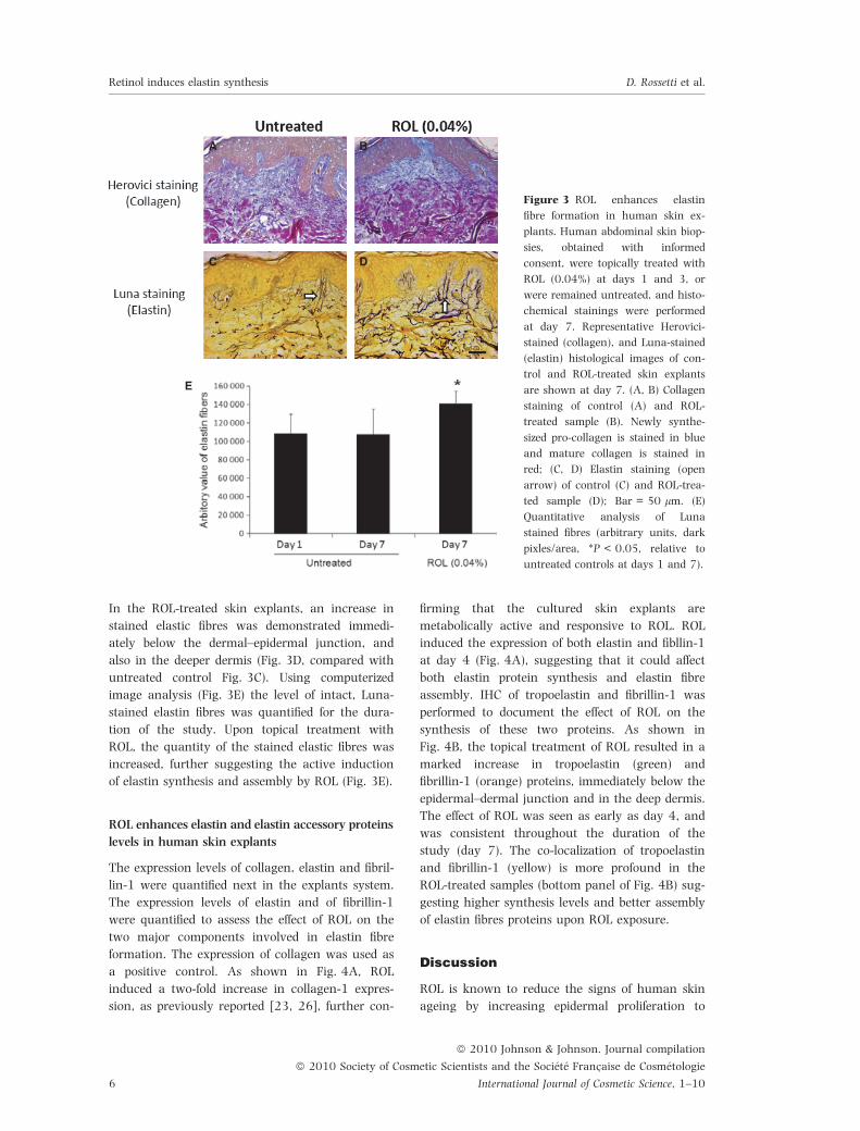
In the ROL-treated skin explants, an increase in
stained elastic fibres was demonstrated immedi-
ately below the dermal–epidermal junction, and
also in the deeper dermis (Fig. 3D, compared with
untreated control Fig. 3C). Using computerized
image analysis (Fig. 3E) the level of intact, Luna-
stained elastin fibres was quantified for the dura-
tion of the study. Upon topical treatment with
ROL, the quantity of the stained elastic fibres was
increased, further suggesting the active induction
of elastin synthesis and assembly by ROL (Fig. 3E).
ROL enhances elastin and elastin accessory proteins
levels in human skin explants
The expression levels of collagen, elastin and fibril-
lin-1 were quantified next in the explants system.
The expression levels of elastin and of fibrillin-1
were quantified to assess the effect of ROL on the
two major components involved in elastin fibre
formation. The expression of collagen was used as
a positive control. As shown in Fig. 4A, ROL
induced a two-fold increase in collagen-1 expres-
sion, as previously reported [23, 26], further con-
firming that the cultured skin explants are
metabolically active and responsive to ROL. ROL
induced the expression of both elastin and fibllin-1
at day 4 (Fig. 4A), suggesting that it could affect
both elastin protein synthesis and elastin fibre
assembly. IHC of tropoelastin and fibrillin-1 was
performed to document the effect of ROL on the
synthesis of these two proteins. As shown in
Fig. 4B, the topical treatment of ROL resulted in a
marked increase in tropoelastin (green) and
fibrillin-1 (orange) proteins, immediately below the
epidermal–dermal junction and in the deep dermis.
The effect of ROL was seen as early as day 4, and
was consistent throughout the duration of the
study (day 7). The co-localization of tropoelastin
and fibrillin-1 (yellow) is more profound in the
ROL-treated samples (bottom panel of Fig. 4B) sug-
gesting higher synthesis levels and better assembly
of elastin fibres proteins upon ROL exposure.
Discussion
ROL is known to reduce the signs of human skin
ageing by increasing epidermal proliferation to
A B
C D
E
Figure 3 ROL enhances elastin
fibre formation in human skin ex-
plants. Human abdominal skin biop-
sies, obtained with informed
consent, were topically treated with
ROL (0.04%) at days 1 and 3, or
were remained untreated, and histo-
chemical stainings were performed
at day 7. Representative Herovici-
stained (collagen), and Luna-stained
(elastin) histological images of con-
trol and ROL-treated skin explants
are shown at day 7. (A, B) Collagen
staining of control (A) and ROL-
treated sample (B). Newly synthe-
sized pro-collagen is stained in blue
and mature collagen is stained in
red; (C, D) Elastin staining (open
arrow) of control (C) and ROL-trea-
ted sample (D); Bar = 50 lm. (E)
Quantitative analysis of Luna
stained fibres (arbitrary units, dark
pixles/area, *P < 0.05, relative to
untreated controls at days 1 and 7).
ª 2010 Johnson & Johnson. Journal compilation
ª 2010 Society of Cosmetic Scientists and the Societe Francaise de Cosmetologie
International Journal of Cosmetic Science, 1–106
Retinol induces elastin synthesis D. Rossetti et al.

restore epidermal thickness, inhibiting tyrosinase
activity to reduce the pigmentary changes associ-
ated with ‘age spots’, and enhancing collagen syn-
thesis to restore age-induced dermal thinning [23,
25–29]. Here, we show that ROL also enhances
elastin synthesis and elastin fibre assembly, which
could result in enhanced skin elasticity.
The effect of retinoic acid on skin elasticity had
been documented experimentally and in the clinic
[30, 37]. However, no such effect has been
reported for ROL. On the contrary, Tajima et al.
documented retinoic-acid induced upregulation of
elastin secretion into conditioned media of chick
embryo fibroblasts, but could not detect any induc-
tion when the cells were treated with ROL [30].
Similarly, Hayashi et al. documented that retinoic
acid, but not ROL, stimulated elastin gene expres-
sion in chick embryo vascular smooth muscle cells
[38]. Using human dermal fibroblasts, we were
able to document an increase in elastin protein
levels in lysates of ROL-treated cells. This detection
was aided by the development of a novel ELISA
assay for elastin, which enabled the detection of
as low as 20 ng/mL of elastin. The detection of
ROL-induced elastin protein production was
confirmed with histological and IHC staining of
tropoelastin and of mature elastin fibres.
ROL-induced elastin synthesis is shown in
well-defined 2D and 3D dermal fibroblast culture
systems, as well as in the more physiologically
complex in vitro system of human skin explants.
We verified the dermal metabolic activity of our
in vitro systems by reproducing the known effect of
ROL on collagen synthesis [23, 26]. As the induc-
tion of collagen by ROL was confirmed in the
clinic we believe that our findings of elastin
enhancement by ROL are relevant to the enhance-
ment of human skin elasticity.
The reduced functionality of the dermal elastic
network provides a major contribution to the sag-
ging, loose and lax appearance of aged skin. How-
ever, most searches for the fountain of youth have
centered on dermal collagen enhancement and
neglected to specifically address the elastin net-
work. TGF-b induces elastin expression in cultured
fibroblasts [39] and in transgenic mice [36, 40],
but there are no published reports searching for
TGF-b-inducing agents for reducing the signs of
A
B
Figure 4 ROL increases mRNA and
protein levels of elastin and fibrillin-
1 in human skin explants. Human
abdominal skin biopsies, obtained
with informed consent, were topi-
cally treated with ROL (0.04%) at
days 1 and 3 or were remained
untreated. (A) Gene expression anal-
yses (QPCR) of elastin, fibrillin-1,
collagen-1 and 18S RNA. The
expression of elastin, fibrillin-1 and
collagen-1, was increased at day 4
following the ROL treatment. 18S
RNA was used as housekeeping,
control gene; (B) IHC staining of
tropoelastin and fibrillin-1 in skin
explants at days 4 and 7. Both trop-
oelastin (green) and fibrillin-1
(orange) protein levels were
increased in the ROL-treated skin
explants, compared with their corre-
sponding untreated controls. The
composite images of tropoelastin/
fibrillin-1 (co-localization is yellow)
are shown at the lower panel.
Bar = 50 lm.
ª 2010 Johnson & Johnson. Journal compilation
ª 2010 Society of Cosmetic Scientists and the Societe Francaise de Cosmetologie
International Journal of Cosmetic Science, 1–10 7
Retinol induces elastin synthesis D. Rossetti et al.
skin ageing. Zhao et al. [22] demonstrated that
non-denatured soybean extracts induce elastin
expression and inhibit elastase activity in vitro,
and documented enhanced elastic fibre network
in vivo upon topical treatment with this extract. A
recent clinical study documents enhanced skin
elasticity by dermatologist’s evaluation, as a part
of an overall assessment of the effect of ROL in
photoageing [28].
The induction of elastin synthesis is essential,
but is not sufficient, for the formation of elastic
fibres (reviewed in [15]). Cenizo et al. had identified
a dill extract that stimulates LOXL gene expression
in culture, and propose its use in enhancing
human skin elasticity [21]. Watson et al. reported
the enhanced IHC staining of fibrillin-1, upon
12-days occlusive treatment of human forearm
skin with either retinoic acid or a commercially
available cosmetic product with an undisclosed
composition [41]. These results were correlated
with an improvement in facial wrinkles following a
6-month topical treatment period. Here, we show
enhanced tropoelastin and fibrillin-1 staining in
cultured human skin explants topically treated
with ROL, suggesting that ROL-enhanced elastin
synthesis and elastin fibre assembly could contri-
bute to facial wrinkle reduction.
Retinoic acid induces tropoelastin expression in
cultured human fibroblasts and in mouse model
systems [30, 31], but reduces the UV-induced
enhancement in elastin gene expression in similar
experimental systems [42]. It would be interesting
to investigate the effect of ROL on UV-induced
elastin production and to identify if, similar to reti-
noic acid, ROL could be effective in the prevention
of solar elastosis.
It has been well established that topical retinoic
acid is metabolized by human skin and dermal
fibroblasts [43, 44]. On the other hand, the
metabolism of topical ROL by dermal fibroblasts is
not completely understood. Randolph reported
that dermal fibroblasts actively metabolize retinoic
acid but not ROL [43] and that ROL can be
metabolized by human keartinocytes [45]. How-
ever, Bailly et al. reported that low but significant
amounts of retinoic acid were detected in the epi-
dermis and dermis 24 h after topical ROL treat-
ment of skin explants. Additionally, they reported
that retinoic acid and other metabolites were
detected in cultured human dermal fibroblasts
treated with ROL at 24 h [44]. Therefore, it is
plausible that the elastin enhancement, anti-
ageing effect induced by the topical treatment of
ROL in our model systems is mediated by its
active metabolite, retinoic acid.
Anti-ageing skin care treatments range from
cosmetic preparations, fillers and neurotoxins, to
surgical intervention and laser skin resurfacing
[46, 47]. However, adverse effects have been asso-
ciated with many of these methods, and the desire
for safe and effective products that reduce wrinkles
and improve skin elasticity remains high. Our
studies show that significant stimulation of colla-
gen and elastin could be achieved by using low
levels of ROL (0.04%) compared with previously
published studies where ROL was used at 1% [23].
Low levels of ROL are considered safe and non-
irritating [25, 48]. Our findings that low levels of
ROL enhance not only collagen synthesis, but also
elastin synthesis and elastin fibre formation, pro-
vide an additional mechanism for the anti-ageing
effects of ROL.
Acknowledgements
This research was supported and funded by John-
son & Johnson Consumer Product Worldwide, a
unit of Johnson & Johnson Consumer Companies
Inc. The authors are employed by the company.
References
1. Uitto, J. The role of elastin and collagen in cutaneous
aging: intrinsic aging versus photoexposure. J. Drugs
Dermatol. 7, s12–s16 (2008).
2. Leyden, J.J. Clinical features of ageing skin. Br. J.
Dermatol. 122(Suppl. 35), 1–3 (1990).
3. Baumann, L. Skin ageing and its treatment. J. Pathol.
211, 241–251 (2007).
4. Uitto, J. Connective tissue biochemistry of the aging
dermis. Age-related alterations in collagen and elas-
tin. Dermatol. Clin. 4, 433–446 (1986).
5. Pasquali-Ronchetti, I. and Baccarani-Contri, M. Elas-
tic fiber during development and aging. Microsc. Res.
Tech. 38, 428–435 (1997).
6. Gray, W.R., Sandberg, L.B. and Foster, J.A. Molecu-
lar model for elastin structure and function. Nature
246, 461–466 (1973).
7. Sakai, L.Y., Keene, D.R. and Engvall, E. Fibrillin, a new
350-kD glycoprotein, is a component of extracellular
microfibrils. J. Cell Biol. 103, 2499–2509 (1986).
8. Zhang, H., Apfelroth, S.D., Hu, W. et al. Structure
and expression of fibrillin-2, a novel microfibrillar
component preferentially located in elastic matrices.
J. Cell Biol. 124, 855–863 (1994).
ª 2010 Johnson & Johnson. Journal compilation
ª 2010 Society of Cosmetic Scientists and the Societe Francaise de Cosmetologie
International Journal of Cosmetic Science, 1–108
Retinol induces elastin synthesis D. Rossetti et al.
9. Kielty, C.M., Sherratt, M.J. and Shuttleworth, C.A.
Elastic fibres. J. Cell Sci. 115, 2817–2828 (2002).
10. Mecham, R.P. Elastin synthesis and fiber assembly.
Ann. N. Y. Acad. Sci. 624, 137–146 (1991).
11. Kozel, B.A., Rongish, B.J., Czirok, A. et al. Elastic
fiber formation: a dynamic view of extracellular
matrix assembly using timer reporters. J. Cell Physiol
207, 87–96 (2006).
12. Csiszar, K. Lysyl oxidases: a novel multifunctional
amine oxidase family. Prog. Nucleic Acid Res. Mol.
Biol. 70, 1–32 (2001).
13. Kagan, H.M. and Li, W. Lysyl oxidase: properties,
specificity, and biological roles inside and outside of
the cell. J. Cell Biochem. 88, 660–672 (2003).
14. Lucero, H.A. and Kagan, H.M. Lysyl oxidase: an oxi-
dative enzyme and effector of cell function. Cell Mol.
Life Sci. 63, 2304–2316 (2006).
15. Wagenseil, J.E. and Mecham, R.P. New insights into
elastic fiber assembly. Birth Defects Res. C. Embryo.
Today 81, 229–240 (2007).
16. Uitto, J. Biochemistry of the elastic fibers in normal
connective tissues and its alterations in diseases.
J. Invest Dermatol. 72, 1–10 (1979).
17. Robinson, P.N., Booms, P., Katzke, S. et al. Mutations
of FBN1 and genotype–phenotype correlations in
Marfan syndrome and related fibrillinopathies. Hum.
Mutat. 20, 153–161 (2002).
18. Godeau, G. and Hornebeck, W. Morphometric analy-
sis of the degradation of human skin elastic fibres by
human leukocyte elastase (EC 3-4-21-37) and
human skin fibroblast elastase (EC 3-4-24). Pathol.
Biol. (Paris) 36, 1133–1138 (1988).
19. Werb, Z., Banda, M.J., McKerrow, J.H. and Sand-
haus, R.A. Elastases and elastin degradation. J. Invest
Dermatol. 79(Suppl 1), 154s–159s (1982).
20. Rijken, F. and Bruijnzeel, P.L. The pathogenesis of
photoaging: the role of neutrophils and neutrophil-
derived enzymes. J. Investig. Dermatol. Symp. Proc.
14, 67–72 (2009).
21. Cenizo, V., Andre, V., Reymermier, C., Sommer, P.,
Damour, O. and Perrier, E. LOXL as a target to
increase the elastin content in adult skin: a dill
extract induces the LOXL gene expression. Exp. Der-
matol. 15, 574–581 (2006).
22. Zhao, R., Bruning, E., Rossetti, D., Starcher, B., Sei-
berg, M. and Iotsova-Stone, V. Extracts from glycine
max (soybean) induce elastin synthesis and inhibit
elastase activity. Exp. Dermatol. 18, 883–886 (2009).
23. Varani, J., Warner, R.L., Gharaee-Kermani, M. et al.
Vitamin A antagonizes decreased cell growth and
elevated collagen-degrading matrix metalloproteinas-
es and stimulates collagen accumulation in naturally
aged human skin. J. Invest Dermatol. 114, 480–486
(2000).
24. Kang, S., Duell, E.A., Fisher, G.J. et al. Application of
retinol to human skin in vivo induces epidermal
hyperplasia and cellular retinoid binding proteins
characteristic of retinoic acid but without measur-
able retinoic acid levels or irritation. J. Invest Derma-
tol. 105, 549–556 (1995).
25. Bellemere, G., Stamatas, G.N., Bruere, V., Bertin, C.,
Issachar, N. and Oddos, T. Antiaging action of reti-
nol: from molecular to clinical. Skin Pharmacol. Phys-
iol 22, 200–209 (2009).
26. Kafi, R., Kwak, H.S., Schumacher, W.E. et al.
Improvement of naturally aged skin with vitamin A
(retinol). Arch. Dermatol. 143, 606–612 (2007).
27. Ortonne, J.P. Retinoid therapy of pigmentary disor-
ders. Dermatol. Ther. 19, 280–288 (2006).
28. Tucker-Samaras, S., Zedayko, T., Cole, C., Miller, D.,
Wallo, W. and Leyden, J.J. A stabilized 0.1% retinol
facial moisturizer improves the appearance of photo-
damaged skin in an eight-week, double-blind, vehi-
cle-controlled study. J. Drugs Dermatol. 8, 932–936
(2009).
29. Kawada, A., Konishi, N., Momma, T., Oiso, N. and
Kawara, S. Evaluation of anti-wrinkle effects of a
novel cosmetic containing retinol using the guideline
of the Japan Cosmetic Industry Association. J. Derma-
tol. 36, 583–586 (2009).
30. Tajima, S., Hayashi, A. and Suzuki, T. Elastin expres-
sion is up-regulated by retinoic acid but not by reti-
nol in chick embryonic skin fibroblasts. J. Dermatol.
Sci. 15, 166–172 (1997).
31. Schwartz, E. and Kligman, L.H. Topical tretinoin
increases the tropoelastin and fibronectin content of
photoaged hairless mouse skin. J. Invest Dermatol.
104, 518–522 (1995).
32. Tanaka, S., Avigad, G., Eikenberry, E.F. and Brodsky,
B. Isolation and partial characterization of collagen
chains dimerized by sugar-derived cross-links. J. Biol.
Chem. 263, 17650–17657 (1988).
33. Bellemere, G., Von, S.O. and Oddos, T. Retinoic acid
increases aquaporin 3 expression in normal human
skin. J. Invest Dermatol. 128, 542–548 (2008).
34. Herovici, C. [Picropolchorme. Histoligical technic for
the study of supporting tissue.]. Pathol. Biol. (Paris)
9, 387–388 (1961).
35. Luna, L. Manual of Histological Staining Methods of the
Armed Forces Institute of Pathology, pp. 114–115.
McGraw-Hill, New York (1968).
36. Katchman, S.D., Hsu-Wong, S., Ledo, I., Wu, M. and
Uitto, J. Transforming growth factor-beta up-regu-
lates human elastin promoter activity in transgenic
mice. Biochem. Biophys. Res. Commun. 203, 485–490
(1994).
37. Chen, Z., Shin, M.H., Moon, Y.J. et al. Modulation of
elastin exon 26A mRNA and protein expression in
human skin in vivo. Exp. Dermatol. 18, 378–386
(2009).
38. Hayashi, A., Suzuki, T. and Tajima, S. Modulations of
elastin expression and cell proliferation by retinoids in
ª 2010 Johnson & Johnson. Journal compilation
ª 2010 Society of Cosmetic Scientists and the Societe Francaise de Cosmetologie
International Journal of Cosmetic Science, 1–10 9
Retinol induces elastin synthesis D. Rossetti et al.

cultured vascular smooth muscle cells. J. Biochem.
117, 132–136 (1995).
39. Choi, W.S., Mitsumoto, A. and Kochevar, I.E.
Involvement of reactive oxygen species in TGF-beta1-
induced tropoelastin expression by human dermal
fibroblasts. Photochem. Photobiol 85, 1425–1433
(2009).
40. Uitto, J., Hsu-Wong, S., Katchman, S.D., Bashir,
M.M. and Rosenbloom, J. Skin elastic fibres: regula-
tion of human elastin promoter activity in transgenic
mice. Ciba Found. Symp. 192, 237–253 (1995).
41. Watson, R.E., Ogden, S., Cotterell, L.F., Bowden, J.J.,
Bastrilles, J.Y., Long, S.P. and Griffiths, C.E. A cos-
metic ‘anti-ageing’ product improves photoaged skin:
a double-blind, randomized controlled trial. Br. J.
Dermatol. 161, 419–426 (2009).
42. Lee, K.S., Kim, S.J., Ryoo, Y.W. and Kim, B.C. All-
trans-retinoic acid down-regulates elastin promoter
activity elevated by ultraviolet B irradiation in cul-
tured skin fibroblasts. J. Dermatol. Sci. 17, 182–189
(1998).
43. Randolph, R.K. and Simon, M. Dermal fibroblasts
actively metabolize retinoic acid but not retinol.
J. Invest Dermatol. 111, 478–484 (1998).
44. Bailly, J., Crettaz, M., Schifflers, M.H. and Marty, J.P.
In vitro metabolism by human skin and fibroblasts of
retinol, retinal and retinoic acid. Exp. Dermatol. 7,
27–34 (1998).
45. Randolph, R.K. and Simon, M. Characterization of ret-
inol metabolism in cultured human epidermal kerati-
nocytes. J. Biol. Chem. 268, 9198–9205 (1993).
46. Puizina-Ivic, N. Skin aging. Acta Dermatovenerol. Alp
Panonica. Adriat. 17, 47–54 (2008).
47. Glaser, D.A. Anti-aging products and cosmeceuticals.
Facial. Plast. Surg. Clin. North Am. 12, 363–372, vii
(2004).
48. Nohynek, G.J., Meuling, W.J., Vaes, W.H. et al. Repe-
ated topical treatment, in contrast to single oral
doses, with Vitamin A-containing preparations does
not affect plasma concentrations of retinol, retinyl
esters or retinoic acids in female subjects of child-
bearing age. Toxicol. Lett. 163, 65–76 (2006).
ª 2010 Johnson & Johnson. Journal compilation
ª 2010 Society of Cosmetic Scientists and the Societe Francaise de Cosmetologie
International Journal of Cosmetic Science, 1–1010
Retinol induces elastin synthesis D. Rossetti et al.


![Free Radic Biol Med. The induction of human superoxide dismutase … · 2018-02-01 · superoxide dismutase (SOD) and catalase, by virtue of their ... retinol [12], malathion [13],](https://static.fdocuments.in/doc/165x107/5f41fa3e32b0d31ee07cf958/free-radic-biol-med-the-induction-of-human-superoxide-dismutase-2018-02-01-superoxide.jpg)