A novel and highly specific phage endolysin cell wall ...kangtaejoon.com/PDF/26.pdf · A novel and...
10
1 3 Eur Biophys J (2015) 44:437–446 DOI 10.1007/s00249-015-1044-7 ORIGINAL PAPER A novel and highly specific phage endolysin cell wall binding domain for detection of Bacillus cereus Minsuk Kong 1 · Jieun Sim 2,3,4 · Taejoon Kang 2,3,4 · Hoang Hiep Nguyen 2,4 · Hyun Kyu Park 5 · Bong Hyun Chung 2,3,4 · Sangryeol Ryu 1 Received: 13 March 2015 / Revised: 7 May 2015 / Accepted: 13 May 2015 / Published online: 5 June 2015 © European Biophysical Societies’ Association 2015 inhibition assay based on the pre-incubation of B. cereus and CBDs, removal of CBD-bound B. cereus, and SPR detection of the unbound CBDs. The present study suggests that the small and genetically engineered CBDs can be promising biological probes for B. cereus. We anticipate that the CBD- based SPR-sensing methods will be useful for the sensitive, selective, and rapid detection of B. cereus. Keywords Bacillus cereus · Bacteriophage endolysin · Biosensor · Cell wall binding domain · Surface plasmon resonance Introduction Foodborne diseases are one of the most widespread and overwhelming public health problems of the modern world. In the USA, foodborne diseases cause an estimated 48 mil- lion illnesses each year, including 9.4 million episodes of illness, 55,961 hospitalizations, and 1351 deaths caused by known pathogens (Gould et al. 2013; Scallan et al. 2011). Additionally, outbreaks of foodborne illnesses result in economic losses totaling several billions of dollars annu- ally (Abadian et al. 2014). Foodborne diseases can occur because of various kinds of bacteria, viruses, parasites, toxins, and so on. Among them, bacteria are the most com- mon causes of foodborne diseases (Arora et al. 2011). For the identification of bacteria, conventional culture-based biochemical assays (Leoni and Legnani 2001) and DNA- based detection methods (Bej et al. 1991; Caliendo 2011; Lazcka et al. 2007; Mothershed and Whitney 2006; Olsen 2000) have been predominantly used. Additionally, sev- eral optical, electrical, electrochemical, and mechanical sensing approaches have been developed as alternatives (Abdel-Hamid et al. 1999; Baeumner et al. 2003; Brewster Abstract Rapid, specific and sensitive detection of patho- genic bacteria is crucial for public health and safety. Bacillus cereus is harmful as it causes foodborne illness and a num- ber of systemic and local infections. We report a novel phage endolysin cell wall-binding domain (CBD) for B. cereus and the development of a highly specific and sensitive surface plasmon resonance (SPR)-based B. cereus detection method using the CBD. The newly discovered CBD from endolysin of PBC1, a B. cereus-specific bacteriophage, provides high specificity and binding capacity to B. cereus. By using the CBD-modified SPR chips, B. cereus can be detected at the range of 10 5 –10 8 CFU/ml. More importantly, the detection limit can be improved to 10 2 CFU/ml by using a subtractive M. Kong and J. Sim contributed equally to this work. Electronic supplementary material The online version of this article (doi:10.1007/s00249-015-1044-7) contains supplementary material, which is available to authorized users. * Bong Hyun Chung [email protected] * Sangryeol Ryu [email protected] 1 Department of Food and Animal Biotechnology, Department of Agricultural Biotechnology, Center for Food and Bioconvergence, Research Institute of Agriculture and Life Sciences, Seoul National University, Seoul 151-742, Korea 2 BioNanotechnology Research Center, KRIBB, Daejeon 305-806, Korea 3 BioNano Health Guard Research Center, KRIBB, Daejeon 305-806, Korea 4 Major of Nanobiotechnology, Korea University of Science and Technology, Daejeon 305-806, Korea 5 MicoBioMed Ltd, Daejeon 305-806, Korea
-
Upload
nguyenkhuong -
Category
Documents
-
view
218 -
download
0
Transcript of A novel and highly specific phage endolysin cell wall ...kangtaejoon.com/PDF/26.pdf · A novel and...

1 3
Eur Biophys J (2015) 44:437–446DOI 10.1007/s00249-015-1044-7
ORIGINAL PAPER
A novel and highly specific phage endolysin cell wall binding domain for detection of Bacillus cereus
Minsuk Kong1 · Jieun Sim2,3,4 · Taejoon Kang2,3,4 · Hoang Hiep Nguyen2,4 · Hyun Kyu Park5 · Bong Hyun Chung2,3,4 · Sangryeol Ryu1
Received: 13 March 2015 / Revised: 7 May 2015 / Accepted: 13 May 2015 / Published online: 5 June 2015 © European Biophysical Societies’ Association 2015
inhibition assay based on the pre-incubation of B. cereus and CBDs, removal of CBD-bound B. cereus, and SPR detection of the unbound CBDs. The present study suggests that the small and genetically engineered CBDs can be promising biological probes for B. cereus. We anticipate that the CBD-based SPR-sensing methods will be useful for the sensitive, selective, and rapid detection of B. cereus.
Keywords Bacillus cereus · Bacteriophage endolysin · Biosensor · Cell wall binding domain · Surface plasmon resonance
Introduction
Foodborne diseases are one of the most widespread and overwhelming public health problems of the modern world. In the USA, foodborne diseases cause an estimated 48 mil-lion illnesses each year, including 9.4 million episodes of illness, 55,961 hospitalizations, and 1351 deaths caused by known pathogens (Gould et al. 2013; Scallan et al. 2011). Additionally, outbreaks of foodborne illnesses result in economic losses totaling several billions of dollars annu-ally (Abadian et al. 2014). Foodborne diseases can occur because of various kinds of bacteria, viruses, parasites, toxins, and so on. Among them, bacteria are the most com-mon causes of foodborne diseases (Arora et al. 2011). For the identification of bacteria, conventional culture-based biochemical assays (Leoni and Legnani 2001) and DNA-based detection methods (Bej et al. 1991; Caliendo 2011; Lazcka et al. 2007; Mothershed and Whitney 2006; Olsen 2000) have been predominantly used. Additionally, sev-eral optical, electrical, electrochemical, and mechanical sensing approaches have been developed as alternatives (Abdel-Hamid et al. 1999; Baeumner et al. 2003; Brewster
Abstract Rapid, specific and sensitive detection of patho-genic bacteria is crucial for public health and safety. Bacillus cereus is harmful as it causes foodborne illness and a num-ber of systemic and local infections. We report a novel phage endolysin cell wall-binding domain (CBD) for B. cereus and the development of a highly specific and sensitive surface plasmon resonance (SPR)-based B. cereus detection method using the CBD. The newly discovered CBD from endolysin of PBC1, a B. cereus-specific bacteriophage, provides high specificity and binding capacity to B. cereus. By using the CBD-modified SPR chips, B. cereus can be detected at the range of 105–108 CFU/ml. More importantly, the detection limit can be improved to 102 CFU/ml by using a subtractive
M. Kong and J. Sim contributed equally to this work.
Electronic supplementary material The online version of this article (doi:10.1007/s00249-015-1044-7) contains supplementary material, which is available to authorized users.
* Bong Hyun Chung [email protected]
* Sangryeol Ryu [email protected]
1 Department of Food and Animal Biotechnology, Department of Agricultural Biotechnology, Center for Food and Bioconvergence, Research Institute of Agriculture and Life Sciences, Seoul National University, Seoul 151-742, Korea
2 BioNanotechnology Research Center, KRIBB, Daejeon 305-806, Korea
3 BioNano Health Guard Research Center, KRIBB, Daejeon 305-806, Korea
4 Major of Nanobiotechnology, Korea University of Science and Technology, Daejeon 305-806, Korea
5 MicoBioMed Ltd, Daejeon 305-806, Korea

438 Eur Biophys J (2015) 44:437–446
1 3
et al. 1996; Croci et al. 2001; Radke and Alocilja 2005; Stephan et al. 2003; Taitt et al. 2004; Tims and Lim 2004; Vaughan et al. 2001; Velusamy et al. 2010; Wong et al. 2002). However, there are limitations with these methods such as the requirement of enrichment steps, long detection time, and need for labeling (Abdel-Hamid et al. 1999; Bae-umner et al. 2003; Croci et al. 2001; Stephan et al. 2003; Taitt et al. 2004). Therefore, it is highly desired to develop simple, fast, sensitive, and accurate detection methods for pathogenic bacteria.
Antibodies are the most widely used biological probes for the detection of pathogenic bacteria (Singh et al. 2011). Production of specific antibodies, however, involves immu-nization of animals and maintenance of hybridoma cells, which are difficult, expensive, and time-consuming (Singh et al. 2012; Skottrup et al. 2008). Moreover, antibodies can lose their activity by changing temperature or pH and are prone to aggregation (Petrenko and Vodyanoy 2003; Wang et al. 2007). Accordingly, it is necessary to explore robust and specific biological probes. Bacteriophages, the natural enemy of bacteria, have been used as alternative probes for pathogenic bacteria sensing because they have extreme host specificity and strong resistance to heat, pH, and chemicals (Arya et al. 2011; Mahony et al. 2011; Tawil et al. 2014). Recently, bacteriophage-derived proteins have received more attention than whole bacteriophages since it is easy to modify the affinity and binding properties of these proteins (Brzozowska et al. 2015; Chibli et al. 2014; Javed et al. 2013; Poshtiban et al. 2013; Singh et al. 2011, 2010). Endolysin is a peptidoglycan hydrolase of bacterio-phage that lyses its bacterial host at the end of the phage life cycle (Fischetti 2010; Loessner 2005). Endolysins from gram-positive bacteria-infecting phages consist of modular proteins including the C-terminal cell wall-binding domain (CBD) and N-terminal catalytic domain (Callewaert et al. 2011; Loessner et al. 2002). CBDs feature high specificity and strong binding affinity (KD values of pico- to nanomo-lar range) to bacteria (Schmelcher et al. 2010, 2011). In addition, they are small (10–15 kDa), act as a monomer, and have no lytic activity. Furthermore, these superior fea-tures of CBDs can be improved by genetic engineering. Therefore, several recent studies have exploited CBDs as novel probes for the detection of bacteria (Kretzer et al. 2007; Tolba et al. 2012).
Herein, we report the highly sensitive and selective sur-face plasmon resonance (SPR)-based detection of Bacillus cereus by using a newly discovered phage endolysin CBD. SPR is a well-known technology that enables label-free, real-time, and quantitative detection of biomolecules, ful-filling the need for bacterial sensing (Abadian et al. 2014). B. cereus is harmful to humans because it causes foodborne illness (Bottone 2010); however, very few detection meth-ods have been reported (Kang et al. 2013; Pal et al. 2007;
Vaughan et al. 2003). Since the isolated CBD from endoly-sin of PBC1, a B. cereus-specific bacteriophage (Kong et al. 2012), exhibits high specificity and binding capac-ity to B. cereus, we could detect B. cereus at the range of 105–108 CFU/ml by using CBD-modified SPR chips. More importantly, B. cereus could be detected at the low concen-tration of 102 CFU/ml with a subtractive inhibition assay. This assay is based on the pre-incubation of B. cereus and CBDs, followed by removal of CBD-bound B. cereus, and SPR detection of the unbound CBDs. We anticipate that this novel CBD-based SPR sensing method will be useful for the sensitive, selective, and rapid detection of B. cereus.
Materials and methods
Bacteria culture conditions
B. cereus ATCC 21768, Bacillus subtilis, Staphylococcus aureus, Salmonella typhi, Salmonella typhimurium, Sal-monella enteritidis, Escherichia coli O157:H7, Listeria monocytogenes, Vibrio vulnificus, Yersinia enterocolit-ica, and Shigella sonnei were used in this study. Bacteria were grown by streaking onto a Luria-Bertani (LB, Difco, Detroit, MI, USA) agar plate and incubated overnight at 37 °C. Next, a single colony from the agar plate was inocu-lated into 3 ml of LB media and incubated at 37 °C for 18 h with shaking at 200 rpm. One milliliter of bacterial culture was cultivated in a 250-ml flask with 100 ml of LB broth at 37 °C with shaking at 200 rpm. After 2 h, the optical den-sity (OD) of the bacterial culture was measured at 600 nm by UV/Vis spectroscopy (Beckman Coulter, DU-800, Indianapolis, IN, USA). The read mode of spectroscopy was absorbance, and the average read time was 0.5 s. The CFU was calculated by the OD of the bacterial cul-ture and the growth curve made by a plate count method. For SPR experiments, bacteria were centrifuged for 5 min at 16,000g, and the pellet was suspended in phosphate-buffered saline (PBS; 137 mM NaCl, 2.7 mM KCl, 8 mM Na2HPO4, and 2 mM KH2PO4; pH 7.4, Life Technologies, Seoul, Korea) after removing the supernatant. This step was performed in triplicate.
In silico analyses
Putative CBDs of LysPBC1 were identified through bioin-formatic analyses. Amino acid sequence alignments of the reported CBDs were conducted using ClustalX2 (Larkin et al. 2007). A conserved domain search was conducted using BLASTP (Altschul et al. 1990), InterProScan (Zdob-nov and Apweiler 2001), and NCBI Conserved Domain Database (Marchler-Bauer et al. 2007). Secondary and tertiary structures of PBC1 endolysin (LysPBC1) were

439Eur Biophys J (2015) 44:437–446
1 3
predicted using Phyre2 and the ESyPred3D server (Kelley and Sternberg 2009; Lambert et al. 2002). A gene fragment encoding 85 amino acids from the C-terminal of LysPBC1 was regarded as CBD, and the coding sequence was ampli-fied by polymerase chain reaction (PCR) for subsequent steps. A phylogenetic tree of CBDs from reported Bacillus phage endolysins was generated with the MEGA5 program using the neighbor-joining method with P-distance values (Tamura et al. 2011).
Construction of recombinant proteins
The enhanced green fluorescent protein (EGFP) gene was amplified by PCR using pEGFP (Clontech, Palo Alto, CA, USA) as a template. The native stop codon was omitted for translational fusions. The amplified DNA product was double-digested using NdeI and BamHI and ligated into a pET28a vector (Novagen, Madison, WI, USA). The gene fragment encoding the CBD was digested with BamHI and HindIII and subcloned into the EGFP-containing pET28a. For SPR experiments, glutathione S-transferase (GST)-CBD fusion protein was employed. The GST coding gene was amplified by PCR using pGST parallel 1 as a template (Sheffield et al. 1999). A flexible linker region encoding six Gly and Ser residues was added between the GST and CBD genes to enable efficient protein folding. The sequences were verified for all constructs.
Expression and purification of CBD fusion proteins
N-Terminal six-His-tagged GFP-CBD fusion protein was produced in E. coli BL21 (DE3) (Invitrogen, Carlsbad, CA, USA). Freshly transformed cells were grown in LB at 37 °C to an OD600 of 0.7, and isopropyl-β-d-thiogalactoside (IPTG) (0.5 mM) was added. After incubation for 20 h at 18 °C, the cells were harvested by centrifugation, resus-pended in a lysis buffer containing 200 mM NaCl and 50 mM Tris-Cl (pH 8.0) and frozen at −20 °C. After thaw-ing, cells were lysed by sonication at a duty cycle of 25 % and output control of 5 (Sonifier 250, Branson, Danbury, CT, USA). After centrifugation (21,000g, 1 h, 4 °C) and sterilization by a 0.20-μm filter (Sartorious, Goettingen, Germany), the soluble protein was purified by immobilized metal affinity chromatography (Poly-Prep® chromatog-raphy column, catalog no. 731-1550, Bio-Rad, Hercules, CA, USA) using Ni-NTA agarose (Qiagen, Valencia, CA, USA). For affinity chromatography, the protein solution (5 ml) was incubated with 0.5 ml of the Ni-NTA resin for 1 h and followed by column purification by gravity flow. After washing the resin twice with the lysis buffer contain-ing imidazole (10–20 mM), the protein was eluted in elu-tion buffer (200 mM NaCl, 50 mM Tris-Cl, and 240 mM imidazole; pH 8.0) and divided into four 500-µl aliquots.
The purity of the CBD fusion proteins was confirmed by sodium dodecyl sulfate-polyacrylamide gel electropho-resis (SDS-PAGE) with protein size marker (GenDEPOT, Barker, TX, USA). The purified protein was stored at −20 °C after buffer exchange to the storage buffer (50 mM Tris-Cl, 200 mM NaCl, and 50 % glycerol; pH 8.0) using PD Miditrap G-25 (GE Healthcare, Waukesha, WI, USA). The GST-CBD fusion protein was also produced by the same method as the GFP-CBD fusion protein. Before the SPR experiments, we exchanged the storage buffer into PBS by the PD-10 column (GE Healthcare, Seoul, Korea).
Cell‑binding assay with fluorescence microscopy
The binding property of GFP-CBD fusion protein was examined as previously described (Loessner et al. 2002). Briefly, 1 ml of exponentially grown bacterial cells was centrifuged (16000g, 1 min) and resuspended in 1 ml of PBS. Next, 100 μl of cells was incubated together with 0.4 μM GFP-CBD fusion protein at room temperature for 5 min. The cells were washed twice with PBS buffer and observed by epifluorescence microscopy (DE/Axio Imager A1 microscope, Carl Zeiss, Oberkochen, Germany) with a GFP filter (470/40 nm excitation, 495 nm dichroic, 525/50 nm emission).
SPR analysis
The SPR instrument used in this study was the Biacore X (GE Healthcare, Pittsburgh, PA, USA). PBS was used as a running buffer. The baseline was established by flow-ing PBS for 300 s at the flow rate of 10 μl/min. For the preparation of the glutathione (GSH) chip, a bare Au chip (GE Healthcare, Pittsburgh, PA, USA) was cleaned by an N2 stream and submerged in 1 mM GSH solution in dime-thyl sulfoxide (DMSO). The GSH-attached Au chip was washed by DMSO and ethanol, then dried under an N2 stream. Fifty microliters of 0.12 mg/ml GST-CBD in PBS was injected into the sample loop of the SPR instrument at a flow rate of 10 μl/min in order to attach the GST-CBD onto the GSH chip. PBS was continuously injected to remove the unbound GST-CBD. Finally, 50 μl of 1 mg/ml bovine serum albumin (BSA; reagent grade, Fisher, USA) in PBS was applied to prevent the non-specific binding. For the detection of bacteria, 50 μl of sample was injected across the chip surface at a flow rate of 10 μl/min, and the chip was washed in PBS.
To compare the binding capacity of the commercial anti-body and CBD, the same molar concentration (3.2 μM) of anti-Bacillus cereus antibody (ab20556, Abcam, Seoul, Korea) and GST-CBD were used. For the immobilization of antibody molecules on the SPR chip, 50 μl of 0.1 mg/ml protein G (Bioprogen, Daejeon, Korea) was first applied

440 Eur Biophys J (2015) 44:437–446
1 3
to a bare Au chip at a flow rate of 10 μl/min. Recombinant protein G is a widely used antibody-binding protein that has specific interactions with the Fc region of immunoglob-ulin G (Lee et al. 2007). After injection of protein G, 50 μl of 3.2 μM anti-Bacillus cereus antibody was applied to the SPR chip, and PBS was injected for 5 min to remove the unbound antibody. BSA in PBS was also injected across the chip to prevent the non-specific binding. The samples were finally injected.
In a subtractive inhibition assay, 500 μl of 5 μg/ml GST-CBD and B. cereus was mixed and incubated for 30 min at room temperature with occasional inverting. After incuba-tion, the solution was centrifuged for 1-min intervals with a gradual speed increase (100, 120, 200, 450, 1000, and 1600g). Fifty microliters of supernatant was gathered and injected across the GSH chip at a flow rate of 10 μl/min. For the preparation of the B. cereus-contaminated food sample, 10 g of cooked rice was mixed with 90 ml of PBS and shaken on a shaker for 1 h. Then, 100 μl of B. cereus (108 CFU/ml) was added to 900 μl of the cooked rice sam-ple. This B. cereus-inoculated sample was serially diluted ten-fold with the cooked rice sample to vary the concentra-tion of B. cereus. For the detection of B. cereus in a food sample, 500 μl of 5 μg/ml GST-CBD and B. cereus-inoc-ulated sample were mixed and incubated. After incubation, the solution was centrifuged, and 50 μl of supernatant was injected across the GSH chip.
Results and discussion
CBD from endolysin of B. cereus phage PBC1
PBC1 is a B. cereus-specific Siphoviridae phage with a 41,164-kb dsDNA genome (Kong et al. 2012). BLAST and domain searches showed that LysPBC1 consists of the N-acetylmuramoyl-l-alanine amidase with N-terminal Amidase_3 (PF01520) catalytic domain and the C-termi-nal Amidase02_C (PF12123) CBD (Fig. 1a). The N-ter-minal catalytic domain of LysPBC1 is significantly con-served with PlyPSA, while the putative CBD sequences of LysPBC1 and PlyPSA are totally different, suggesting that the putative CBD may determine host specificity. Phyloge-netic analysis of the putative CBDs of the B. cereus group phages indicates that the CBD of LysPBC1 is unrelated to other CBDs except for the putative CBD of phage 12826 (Loessner et al. 1997) (Fig. 1b). The amino acid sequence identity between the CBD of LysPBC1 and the putative CBD of phage 12826 is 76 %. Because the putative CBD of phage 12826 has not been experimentally proven to date, we selected the CBD of LysPBC1 as a novel recogni-tion element for B. cereus. Figure 1c shows a 3D model of LysPBC1 based on the structure of PlyPSA, an endolysin
of Listeria phage PSA. This protein structure prediction enabled us to define the putative CBD region (magenta) in the endolysin sequence.
To examine the binding activity of CBD of LysPBC1, we genetically fused EGFP to the N-terminus of the CBD (Fig. 2a). The GFP-CBD fusion proteins were overex-pressed in E. coli in the soluble form and easily purified by Ni-NTA affinity chromatography because of the N-terminal hexahistidine tag of the fusion proteins (Fig. 2b, c). The purified GFP-CBDs were added to intact B. cereus strain ATCC 21,768 cells, and the optical and fluorescent images were observed (Fig. 2d). The images clearly show that GFP-CBDs were uniformly attached to B. cereus cell sur-faces. For comparison, only GFP was added to B. cereus. Figure 2e shows the optical and fluorescent images of B. cereus after the addition of GFP, indicating negligible bind-ing of GFP to B. cereus. We further examined the binding specificity of the GFP-CBDs by mixing them with various bacterial species. None of the other bacterial species except B. cereus were labeled with GFP-CBDs, confirming that
Fig. 1 a Schematic representation of PBC1 endolysin (LysPBC1). The N-terminal (orange) and C-terminal (magenta) domains are pre-sented. b A phylogenetic tree indicating the relationships between CBDs examined in the present study. For calculation of the tree, the respective amino acid sequences, including the putative linker regions, were used. c Predicted 3D structure of LysPBC1 based on the crystal structure of Listeria phage endolysin PlyPSA (PDB 1D: 1XOV)

441Eur Biophys J (2015) 44:437–446
1 3
the binding of GFP-CBDs was highly specific to B. cereus (Table 1 and Fig. S1). This result demonstrates that the CBDs of LysPBC1 can be highly specific biological probes for B. cereus detection.
SPR detection of B. cereus using CBDs
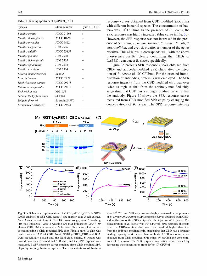
For SPR-based bacterial sensing, the use of CBDs is advantageous because the oriented immobilization of CBDs onto SPR chips can lead to the increased bacterial capture efficiency (Turkova 1999), whereas most antibod-ies are immobilized randomly onto SPR chips via reactive free amine coupling. To immobilize the CBDs onto a GSH chip, we genetically engineered the CBDs by adding GST genes to the N-terminal of CBDs (Fig. 3a). Since GST can bind to GSH with reasonable affinity and specificity
(Chen et al. 2009; Pan et al. 2011; Singh et al. 2011; Tes-sema et al. 2006), GST-CBDs could be immobilized on the GSH chip in an oriented manner. Furthermore, GST-tag has been known to increase the recombinant protein solubility and to stabilize the protein by protecting the intracellular protease cleavage (Terpe 2003; Zhou and Wagner 2010). The recombinant GST-CBD fusion pro-teins were expressed solubly in E. coli, and the N-terminal hexahistidine tag facilitated the purification of the fusion proteins (Fig. 3b). Figure 3c shows the schematic illustra-tion of B. cereus detection by using a CBD-modified SPR chip. First, a bare Au chip was coated with a self-assem-bled monolayer (SAM) of GSH. Next, GST-CBDs and BSA were flowed onto the GSH chip. Finally, B. cereus was flowed onto the CBD-modified SPR chip, and the SPR response was measured. Figure 3d shows the SPR
Fig. 2 a Schematic representa-tion of GFP-LysPBC1_CBD. b SDS-PAGE analysis of GFP-CBD [lane 1 size marker, lane 2 cell extract, lane 3 supernatant, lane 4 Ni-NTA flow-through, lane 5 washing (10 mM imida-zole), lane 6 washing (20 mM imidazole), lane 7–10 elution (240 mM imidazole)]. c Image of the purified GFP-LysPBC1_CBD. d,e Optical and fluores-cent images of B. cereus after the addition of GFP-LysPBC1_CBD and GFP. The fluorescent images show that B. cereus cell surfaces were evenly decorated with GFP-LysPBC1_CBD while GFP alone was not
442 Eur Biophys J (2015) 44:437–446
1 3
response curves obtained from CBD-modified SPR chips with different bacterial species. The concentration of bac-teria was 107 CFU/ml. In the presence of B. cereus, the SPR response was highly increased (blue curve in Fig. 3d). However, the SPR response was not increased in the pres-ence of S. aureus, L. monocytogenes, S. sonnei, E. coli, Y. enterocolitica, and even B. subtilis, a member of the genus Bacillus. This SPR result corresponds well with the above fluorescence results, clearly confirming that CBDs of LysPBC1 can detect B. cereus specifically.
Figure 3e presents SPR response curves obtained from CBD- and antibody-modified SPR chips after the injec-tion of B. cereus of 107 CFU/ml. For the oriented immo-bilization of antibodies, protein G was employed. The SPR response intensity from the CBD-modified chip was over twice as high as that from the antibody-modified chip, suggesting that CBD has a stronger binding capacity than the antibody. Figure 3f shows the SPR response curves measured from CBD-modified SPR chips by changing the concentrations of B. cereus. The SPR response intensity
Table 1 Binding spectrum of LysPBC1_CBD
Species Strain number LysPBC1_CBD
Bacillus cereus ATCC 21768 +Bacillus thuringiensis ATCC 10792 −Bacillus mycoides ATCC 6462 −Bacillus megaterium JCM 2506 −Bacillus subtilis ATCC 23857 −Bacillus pumilus JCM 2508 −Bacillus licheniformis JCM 2505 −Bacillus sphaericus JCM 2502 −Bacillus circulans JCM 2504 −Listeria monocytogenes Scott A −Listeria innocua ATCC 33090 −Staphylococcus aureus ATCC 29213 −Enterococcus faecalis ATCC 29212 −Escherichia coli MG1655 −Salmonella Typhimurium SL1344 −Shigella flexineri 2a strain 2457T −Cronobacter sakazakii ATCC 29544 −
Fig. 3 a Schematic representation of GST-LysPBC1_CBD. b SDS-PAGE analysis of GST-CBD [lane 1 size marker, lane 2 cell extract, lane 3 supernatant, lane 4 Ni-NTA flow-through, lane 5 washing (10 mM imidazole), lane 6 washing (20 mM imidazole), lane 7–10 elution (240 mM imidazole)]. c Schematic illustration of B. cereus detection using a CBD-modified SPR chip. First, a bare Au chip was coated with a SAM of GSH. Next, GST-LysPBC1_CBD and BSA were sequentially flowed onto the GSH chip. Finally, B. cereus was flowed onto the CBD-modified SPR chip, and the SPR response was measured. d SPR response curves obtained from CBD-modified SPR chips by varying bacterial species. The concentrations of bacteria
were 107 CFU/ml. SPR response was highly increased in the presence of B. cereus (blue curve). e SPR response curves obtained from CBD- and antibody-modified SPR chips after the injection of B. cereus. The concentration of B. cereus was 107 CFU/ml. SPR response intensity from the CBD-modified chip was over two-fold higher than that from the antibody-modified chip, suggesting that CBD has a stronger binding capacity to B. cereus than antibody. f SPR response curves obtained from CBD-modified SPR chips by varying the concentra-tions of B. cereus. The SPR response intensities were reduced by decreasing the concentration from 108 to 105 CFU/ml

443Eur Biophys J (2015) 44:437–446
1 3
decreased as the concentration of B. cereus was reduced from 108 to 105 CFU/ml. At 104 CFU/ml, the SPR response was indistinguishable. Therefore, the detection limit of this method was estimated to be 105 CFU/ml.
Improving B. cereus detection sensitivity by subtractive inhibition assay
In SPR sensing, the penetration depth of the evanescent field arising under conditions of total internal reflection cannot exceed 300 nm (Skottrup et al. 2008). Since most bacteria are larger than 300 nm, direct capture of bacte-ria can prevent effective penetration of the evanescent field, and thus SPR-based bacterial detection methods have limited sensitivity (Leonard et al. 2004). In order to improve the sensitivity of the SPR-based B. cereus detec-tion method, we adopted a subtractive inhibition assay. Figure 4a shows a schematic illustration of B. cereus detec-tion using a subtractive inhibition assay. GST-CBDs and B. cereus were incubated for 30 min at room temperature with occasional inverting. After incubation, unbound GST-CBDs were separated from GST-CBD-attached B. cereus by sequential centrifugation. The centrifugation speed was
increased gradually to distinguish the unbound GST-CBDs and the GST-CBD-attached B. cereus. We carefully gath-ered the supernatant containing the unbound GST-CBDs. The supernatant was flowed onto GSH chips, and the SPR response was measured. Figure 4b shows the SPR response curves obtained from GSH chips by varying the concen-tration of B. cereus in a subtractive inhibition assay. In the absence of B. cereus, the response intensity was highest. By increasing the concentration of B. cereus, the intensity decreased because the amount of unbound GST-CBDs was inversely proportional to the concentration of B. cereus. At a concentration as low as 102 CFU/ml, B. cereus could be detected by employing a subtractive inhibition assay. Fig-ure 4c is the plot of R/R0 versus concentration of B. cereus. R is the mean SPR response intensity at each concentra-tion of B. cereus, and R0 is the mean SPR response inten-sity in the absence of B. cereus. The plot verifies that the quantitative detection of B. cereus is possible at the range of 102–107 CFU/ml. Compared to the direct sensing result in Fig. 3f, the detection limit is improved three orders of magnitude.
Lastly, we tried to detect B. cereus in a food sample using subtractive inhibition assay. Cooked rice was used
Fig. 4 a Schematic illustration of B. cereus detection using a sub-tractive inhibition assay. GST-CBD and B. cereus were incubated for 30 min, and unbound GST-CBD was separated from GST-CBD attached to B. cereus by centrifugation. Next, the supernatant was flowed onto a GSH chip, and the SPR response was measured. b SPR response curves obtained from GSH chips by varying the concentra-tion of B. cereus in a subtractive inhibition assay. SPR response inten-sities decreased with increasing concentrations of B. cereus because the amount of unbound GST-CBD is inversely proportional to the
concentration of B. cereus. By employing the subtractive inhibi-tion assay, B. cereus could be detected at a concentration as low as 102 CFU/ml. c Plot of R/R0 versus the concentration of B. cereus. R is a mean SPR response intensity at each concentration of B. cereus. R0 is a mean SPR response intensity in the absence of B. cereus. A magenta linear fit line shows the dynamic range of 102–107 CFU/ml. Data represent the mean plus standard deviation from three measure-ments

444 Eur Biophys J (2015) 44:437–446
1 3
as a food matrix because it is the food most commonly polluted by B. cereus (Rangan 2008). Figure 5a shows the SPR response curves obtained from GSH chips by vary-ing the concentration of B. cereus in a cooked rice sample. One thousand (103) CFU/ml of B. cereus could be detected in a cooked rice sample. Plot of R/R0 versus the concentra-tion of B. cereus in a cooked rice sample shows that the quantitative detection of B. cereus is possible at the range of 103–107 CFU/ml (Fig. 5b). Compared with the results of pure B. cereus detection, the sensitivity was depressed. This may be attributed to the non-specific adsorption of carbohydrates, proteins, etc. The successful detection of B. cereus in a cooked rice sample demonstrates the feasibility of the CBD-based SPR sensing method for the detection of B. cereus in complex food samples.
B. cereus causes not only foodborne illness, but also a number of systemic and local infections, yet very few detection methods employing mechanical, electrical, or electrochemical sensors have been developed (Kang et al. 2013; Pal et al. 2007; Vaughan et al. 2003). This is the first report of SPR-based B. cereus sensing, which enables sen-sitive, specific, and convenient detection of B. cereus.
Conclusion
We report a novel phage endolysin CBD for B. cereus and development of a highly specific and sensitive SPR-based B. cereus detection method using the CBD. The present study has the following important features. First, the newly discovered CBD is very specific to B. cereus and exhibits a higher binding capacity than a commercial antibody. Therefore, the CBD can be used as an excellent
biological probe for the detection of B. cereus. Second, to the best of our knowledge, this is the first report of SPR-based B. cereus detection. We detected 105 CFU/ml of B. cereus by CBD-modified SPR chips, and the detection limit could be lowered to 102 CFU/ml using a subtractive inhibition assay. Third, B. cereus in a food sample could be detected at the low concentration of 103 CFU/ml. We anticipate that this result will advance us a step closer to rapid, sensitive, and selective detection of B. cereus.
Acknowledgments This research was supported by the Public Wel-fare & Safety research program (NRF-2012M3A2A1051684, NRF-2012M3A2A1051682) through the National Research Foundation of Korea funded by the Ministry of Science, ICT and Future Planning (MSIP), Global Frontier Project (H-GUARD_2013M3A6B2078950, H-GUARD_2014M3A6B2060489) through the Center for Bio-Nano Health-Guard funded by the MSIP, and KRIBB initiative Research Program.
References
Abadian PN, Kelley CP, Goluch ED (2014) Cellular analysis and detection using surface plasmon resonance techniques. Anal Chem 86:2799–2812
Abdel-Hamid I, Ivnitski D, Atanasov P, Wilkins E (1999) Flow-through immunofiltration assay system for rapid detection of E. coli O157:H7. Biosens Bioelectron 14:309–316
Altschul SF, Gish W, Miller W, Myers EW, Lipman DJ (1990) Basic local alignment search tool. J Mol Biol 215:403–410
Arora P, Sindhu A, Dilbaghi N, Chaudhury A (2011) Biosensors as innovative tools for the detection of food borne pathogens. Bios-ens Bioelectron 28:1–12
Arya SK, Singh A, Naidoo R, Wu P, McDermott MT, Evoy S (2011) Chemically immobilized T4-bacteriophage for specific Escheri-chia coli detection using surface plasmon resonance. Analyst 136:486–492
Fig. 5 Detection of B. cereus in a cooked rice sample by using a subtractive inhibition assay. a SPR response curves obtained from GSH chips by varying the concentration of B. cereus in a cooked rice sample. SPR response intensities decreased with increasing con-centrations of B. cereus. In a cooked rice sample, B. cereus could be detected at a concentration as low as 103 CFU/ml. b Plot of R/R0 ver-
sus the concentration of B. cereus. R is a mean SPR response inten-sity at each concentration of B. cereus. R0 is a mean SPR response intensity in the absence of B. cereus. A magenta linear fit line shows the dynamic range of 103–107 CFU/ml. Data represent the mean plus standard deviation from three measurements

445Eur Biophys J (2015) 44:437–446
1 3
Baeumner AJ, Cohen RN, Miksic V, Min J (2003) RNA biosensor for the rapid detection of viable Escherichia coli in drinking water. Biosens Bioelectron 18:405–413
Bej AK, Mahbubani MH, Dicesare JL, Atlas RM (1991) Polymer-ase chain reaction-gene probe detection of microorganisms by using filter-concentrated samples. Appl Environ Microbiol 57:3529–3534
Bottone EJ (2010) Bacillus cereus, a volatile human pathogen. Clin Microbiol Rev 23:382–398
Brewster JD, Gehring AG, Mazenko RS, Houten LJV, Crawford CJ (1996) Immunoelectrochemical assays for bacteria: use of epi-fluorescence microscopy and rapid-scan electrochemical tech-niques in development of an assay for Salmonella. Anal Chem 68:4153–4159
Brzozowska E, Smietana M, Koba M, Gorska S, Pawlik K, Gamian A, Bock WJ (2015) Recognition of bacterial lipopolysaccharide using bacteriophage-adhesin-coated long-period gratings. Bios-ens Bioelectron 67:93–99
Caliendo AM (2011) Multiplex PCR and emerging technologies for the detection of respiratory pathogens. Clin Infect Dis 52(Suppl 4):S326–S330
Callewaert L, Walmagh M, Michiels CW, Lavigne R (2011) Food applications of bacterial cell wall hydrolases. Curr Opin Biotech 22:164–171
Chen LH, Wang Q, Hou WG (2009) The utilization of BSA-modified chip on the investigation of ligand/protein interaction with sur-face plasma resonance. Afr J Biotechnol 8:7148–7155
Chibli H, Ghali H, Park S, Peter YA, Nadeau JL (2014) Immobilized phage proteins for specific detection of staphylococci. Analyst 139:179–186
Croci L, Delibato E, Volpe G, Palleschi G (2001) A rapid electro-chemical ELISA for the detection of Salmonella in meat sam-ples. Anal Lett 34:597–2607
Fischetti VA (2010) Bacteriophage endolysins: a novel anti-infec-tive to control Gram-positive pathogens. Int J Med Microbiol 300:57–362
Gould LH, Walsh KA, Vieira AR, Herman K, Williams IT, Hall AJ, Cole D (2013) Surveillance for foodborne disease outbreaks—United States, 1998–2008. MMWR Surveill Summ 62:1–34
Javed MA, Poshtiban S, Arutyunov D, Evoy S, Szymanski CM (2013) Bacteriophage receptor binding protein based assays for the simultaneous detection of Campylobacter jejuni and Campylobacter coli. PLoS One 8:e69770. doi:10.1371/journal.pone.0069770
Kang XB, Pang GC, Chen QS, Liang XY (2013) Fabrication of Bacillus cereus electrochemical immunosensor based on dou-ble-layer gold nanoparticles and chitosan. Sens Actuat B-Chem 177:1010–1016
Kelley LA, Sternberg MJ (2009) Protein structure prediction on the Web: a case study using the Phyre server. Nat Protoc 4:63–371
Kong M, Kim M, Ryu S (2012) Complete genome sequence of Bacil-lus cereus bacteriophage PBC1. J Virol 86:6379–6380
Kretzer JW, Lehmann R, Schmelcher M, Banz M, Kim KP, Korn C, Loessner MJ (2007) Use of high-affinity cell wall-binding domains of bacteriophage endolysins for immobilization and separation of bacterial cells. Appl Environ Microb 73:992–2000
Lambert C, Leonard N, De Bolle X, Depiereux E (2002) ESy-Pred3D: prediction of proteins 3D structures. Bioinformatics 18:1250–1256
Larkin MA, Blackshields G, Brown NP, Chenna R, McGettigan PA, McWilliam H, Valentin F, Wallace IM, Wilm A, Lopez R, Thompson JD, Gibson TJ, Higgins DG (2007) Clustal W and Clustal X version 2.0. Bioinformatics 23:2947–2948
Lazcka O, Del Campo FJ, Munoz FX (2007) Pathogen detection: a perspective of traditional methods and biosensors. Biosens Bio-electron 22:1205–1217
Lee JM, Park HK, Jung Y, Kim JK, Jung SO, Chung BH (2007) Direct immobilization of protein G variants with various numbers of cysteine residues on a gold surface. Anal Chem 79:2680–2687
Leonard P, Hearty S, Quinn J, O’Kennedy R (2004) A generic approach for the detection of whole Listeria monocytogenes cells in contaminated samples using surface plasmon resonance. Bios-ens Bioelectron 19:1331–1335
Leoni E, Legnani PP (2001) Comparison of selective procedures for isolation and enumeration of Legionella species from hot water systems. J Appl Microbiol 90:27–33
Loessner MJ (2005) Bacteriophage endolysins—current state of research and applications. Curr Opin Microbiol 8:480–487
Loessner MJ, Maier SK, Daubek-Puza H, Wendlinger G, Scherer S (1997) Three Bacillus cereus bacteriophage endolysins are unre-lated but reveal high homology to cell wall hydrolases from dif-ferent bacilli. J Bacteriol 179:2845–2851
Loessner MJ, Kramer K, Ebel F, Scherer S (2002) C-terminal domains of Listeria monocytogenes bacteriophage murein hydro-lases determine specific recognition and high-affinity binding to bacterial cell wall carbohydrates. Mol Microbiol 44:335–349
Mahony J, McAuliffe O, Ross RP, van Sinderen D (2011) Bacterio-phages as biocontrol agents of food pathogens. Curr Opin Bio-tech 22:157–163
Marchler-Bauer A, Anderson JB, Derbyshire MK, DeWeese-Scott C, Gonzales NR, Gwadz M, Hao L, He S, Hurwitz DI, Jackson JD, Ke Z, Krylov D, Lanczycki CJ, Liebert CA, Liu C, Lu F, Lu S, Marchler GH, Mullokandov M, Song JS, Thanki N, Yamashita RA, Yin JJ, Zhang D, Bryant SH (2007) CDD: a conserved domain database for interactive domain family analysis. Nucl Acids Res 35:D237–D240
Mothershed EA, Whitney AM (2006) Nucleic acid-based methods for the detection of bacterial pathogens: present and future consid-erations for the clinical laboratory. Clin Chim Acta 363:206–220
Olsen JE (2000) DNA-based methods for detection of food-borne bacterial pathogens. Food Res Int 33:257–266
Pal S, Alocilja EC, Downes FP (2007) Nanowire labeled direct-charge transfer biosensor for detecting Bacillus species. Biosens Bioel-ectron 22:2329–2336
Pan Y, Long MJC, Li XM, Shi Jf, Hedstrom L, Xu B (2011) Glu-tathione (GSH)-decorated magnetic nanoparticles for binding glutathione-S-transferase (GST) fusion protein and manipulating live cells. Chem Sci 2:945–948
Petrenko VA, Vodyanoy VJ (2003) Phage display for detection of bio-logical threat agents. J Microbiol Meth 53:253–262
Poshtiban S, Javed MA, Arutyunov D, Singh A, Banting G, Szyman-ski CM, Evoy S (2013) Phage receptor binding protein-based magnetic enrichment method as an aid for real time PCR detec-tion of foodborne bacteria. Analyst 138:5619–5626
Radke SM, Alocilja EC (2005) A high density microelectrode array biosensor for detection of E. coli O157:H7. Biosens Bioelectron 20:1662–1667
Rangan C (2008) Bacillus cereus. In: Barceloux DG (ed) Medical toxicology of natural substances: foods, fungi, medicinal herbs, plants, and venomous animals. Wiley, New Jersey, pp 89–95
Scallan E, Hoekstra RM, Angulo FJ, Tauxe RV, Widdowson MA, Roy SL, Jones JL, Griffin PM (2011) Foodborne illness acquired in the United States—major pathogens. Emerg Infect Dis 17:7–15
Schmelcher M, Shabarova T, Eugster MR, Eichenseher F, Tchang VS, Banz M, Loessner MJ (2010) Rapid multiplex detection and differentiation of Listeria cells by use of fluorescent phage endolysin cell wall binding domains. Appl Environ Microb 76:5745–5756
Schmelcher M, Tchang VS, Loessner MJ (2011) Domain shuf-fling and module engineering of Listeria phage endolysins for enhanced lytic activity and binding affinity. Microb Biotechnol 4:651–662

446 Eur Biophys J (2015) 44:437–446
1 3
Sheffield P, Garrard S, Derewenda Z (1999) Overcoming expression and purification problems of RhoGDI using a family of “paral-lel” expression vectors. Protein Expr Purif 15:34–39
Singh A, Arya SK, Glass N, Hanifi-Moghaddam P, Naidoo R, Szy-manski CM, Tanha J, Evoy S (2010) Bacteriophage tailspike proteins as molecular probes for sensitive and selective bacterial detection. Biosens Bioelectron 26:131–138
Singh A, Arutyunov D, McDermott MT, Szymanski CM, Evoy S (2011) Specific detection of Campylobacter jejuni using the bacteriophage NCTC 12673 receptor binding protein as a probe. Analyst 136:4780–4786
Singh A, Arutyunov D, Szymanski CM, Evoy S (2012) Bacteriophage based probes for pathogen detection. Analyst 137:3405–3421
Skottrup PD, Nicolaisen M, Justesen AF (2008) Towards on-site path-ogen detection using antibody-based sensors. Biosens Bioelec-tron 24:339–348
Stephan R, Schumacher S, Zychowska MA (2003) The VIT® technol-ogy for rapid detection of Listeria monocytogenes and other Lis-teria spp. Int J Food Microbiol 89:287–290
Taitt CR, Golden JP, Shubin YS, Shriver-Lake LC, Sapsford KE, Rasooly A, Ligler FS (2004) A portable array biosensor for detecting multiple analytes in complex samples. Microb Ecol 47:175–185
Tamura K, Peterson D, Peterson N, Stecher G, Nei M, Kumar S (2011) MEGA5: molecular evolutionary genetics analysis using maximum likelihood, evolutionary distance, and maximum par-simony methods. Mol Biol Evol 28:2731–2739
Tawil N, Sacher E, Mandeville R, Meunier M (2014) Bacteriophages: biosensing tools for multi-drug resistant pathogens. Analyst 139:1224–1236
Terpe K (2003) Overview of tag protein fusions: from molecular and biochemical fundamentals to commercial systems. Appl Micro-biol Biotechnol 60:523–533
Tessema M, Simons PC, Cimino DF, Sanchez L, Waller A, Pos-ner RG, Wandinger-Ness A, Prossnitz ER, Sklar LA (2006)
Glutathione-S-transferase-green fluorescent protein fusion pro-tein reveals slow dissociation from high site densiy beads and measures free GSH. Cytom Part A 69A:326–334
Tims TB, Lim DV (2004) Rapid detection of Bacillus anthracis spores directly from powders with an evanescent wave fiber-optic biosensor. J Microbiol Meth 59:127–130
Tolba M, Ahmed MU, Tlili C, Eichenseher F, Loessner MJ, Zourob M (2012) A bacteriophage endolysin-based electrochemical imped-ance biosensor for the rapid detection of Listeria cells. Analyst 137:5749–5756
Turkova J (1999) Oriented immobilization of biologically active pro-teins as a tool for revealing protein interactions and function. J Chromatogr B 722:11–31
Vaughan RD, O’Sullivan CK, Guilbault GG (2001) Development of a quartz crystal microbalance (QCM) immunosensor for the detec-tion of Listeria monocytogenes. Enzym Microb Tech 29:635–638
Vaughan RD, Carter RM, O’Sullivan CK, Guilbault GG (2003) A quartz crystal microbalance (QCM) sensor for the detection of Bacillus cereus. Anal Lett 36:731–747
Velusamy V, Arshak K, Korostynska O, Oliwa K, Adley C (2010) An overview of foodborne pathogen detection: in the perspective of biosensors. Biotechnol Adv 28:232–254
Wang W, Singh S, Zeng DL, King K, Nema S (2007) Antibody struc-ture, instability, and formulation. J Pharm Sci-Us 96:1–26
Wong YY, Ng SP, Ng MH, Si SH, Yao SZ, Fung YS (2002) Immu-nosensor for the differentiation and detection of Salmonella spe-cies based on a quartz crystal microbalance. Biosens Bioelectron 17:676–684
Zdobnov EM, Apweiler R (2001) InterProScan—an integration plat-form for the signature-recognition methods in InterPro. Bioinfor-matics 17:847–848
Zhou P, Wagner G (2010) Overcoming the solubility limit with solu-bility-enhancement tags: successful applications in biomolecular NMR studies. J Biomol NMR 46:23–31