Site-specific climate analysis elucidates revegetation challenges for ...

lable at ScienceDirect
Biomaterials 115 (2017) 19e29
Contents lists avai
Biomaterials
journal homepage: www.elsevier .com/locate/biomater ia ls
A novel 3D in vitro metastasis model elucidates differential invasivestrategies during and after breaching basement membrane
Asja Guzman, Víctor S�anchez Alemany, Yen Nguyen, Catherine Ruiqi Zhang,Laura J. Kaufman*
Columbia University, Department of Chemistry, New York, NY 10027, United States
a r t i c l e i n f o
Article history:Received 24 September 2016Received in revised form8 November 2016Accepted 14 November 2016Available online 15 November 2016
Keywords:MetastasisSpheroidCollagenInvasionModel systemCancer
* Corresponding author.E-mail address: [email protected] (L.J.
http://dx.doi.org/10.1016/j.biomaterials.2016.11.0140142-9612/© 2016 Elsevier Ltd. All rights reserved.
a b s t r a c t
Invasive breast cancer and other tumors of epithelial origin must breach a layer of basement membrane(BM) that surrounds the primary tumor before invading into the adjacent extracellular matrix. To analyzeinvasive strategies of breast cancer cells during BM breaching and subsequent invasion into a collagen I-rich extracellular matrix (ECM), we developed a physiologically relevant 3D in vitro model that recreatesthe architecture of a solid tumor with an intact, degradable, cell-assembled BM layer embedded in acollagen I environment. Using this model we demonstrate that while the BM layer fully preventsdissemination of non-malignant cells, cancer cells are capable of breaching it and invading into thesurrounding collagen, indicating that the developed system recreates a hallmark of invasive disease. Wedemonstrate that cancer cells exhibiting individual invasion in collagen matrices preferentially adopt aspecific mode of collective invasion when transmigrating a cell-assembled BM that is not observed in anyother tested fibrillar, non-fibrillar, or composite ECM. Matrix-degrading enzymes are found to be crucialduring BM breaching but not during subsequent invasion in the collagen matrix. It is further shown thatmulticellular transmigration of the BM is less susceptible to pharmacological MMP inhibition thanmulticellular invasion in composite collagen/basement membrane extract matrices. The newly devel-oped in vitro model of metastasis allows 3D cancer cell invasion to be studied not only as a function of aparticular tumor's genetics but also as a function of its heterogeneous environment and the differentstages of invasion. As such, this model is a valuable new tool with which to dissect basic mechanisms ofinvasion and metastasis and develop new therapeutic approaches in a physiologically relevant, yetinexpensive and highly tunable, in vitro setting.
© 2016 Elsevier Ltd. All rights reserved.
1. Introduction
Breast cancer deaths occur primarily from metastatic diseasethat compromises function of critical organs. In carcinomas(epithelium-derived cancers), the most common type of breastcancer, metastasis requires tumor cells to breach the basementmembrane (BM), a subtype of extracellular matrix (ECM) thatsurrounds the primary tumor, invade collagen I- and fat-rich ECMof the adjacent soft tissue, and intravasate into blood or lymphvessels where they will be transported to distant sites [1]. While acomplex interplay of genetic and epigenetic changes underlies themulti-step metastatic cascade, dynamic interactions between tu-mor cells and the ECM are increasingly recognized as a key aspect of
Kaufman).
metastatic progression [2,3].The BM is a specialized cell-adherent ECM produced jointly by
normal and/or pathological epithelial, endothelial, and stromalcells. It is formed in a multi-step process initiated by cells bindinglaminin at the cell surface and subsequent accumulation of thenon-fibrillar collagen IV at the nascent laminin scaffold. This pro-cess leads to a dense sheet-like matrix that under normal circum-stances separates the epithelium or endothelium from the adjacentstroma [4,5]. BM deposition and turnover are often perturbed incancers, resulting in matrices that are less crosslinked and thusmore accessible to degradation and remodeling [4,6,7]. Disconti-nuities of BM surrounding primary tumors are caused by alteredexpression and crosslinking of BM components as well as enhancedenzymatic degradation, all hallmarks of aggressive cancers andeach of established prognostic value [8e12].
In contrast to the non-fibrillar BM, stromal ECM in most organs

A. Guzman et al. / Biomaterials 115 (2017) 19e2920
and connective tissues is dominated by collagen I, a fibrillarcollagen [13]. The stromal ECM also displays abnormalities incomposition and organization during carcinogenesis, which lead tochanges in biomechanical properties and matrix architecture. Thehigh breast tissue density associated with poor prognosis in pa-tients with breast cancer is due in part to enhanced deposition ofmostly fibrillar collagens [14e18]. Moreover, highly linearized andaligned collagen at tumor boundaries has been found to contributeto tumor invasion and linked to poor prognosis [19,20].
At the molecular level, cancer progression and metastasis havelong been associated with the epithelial-mesenchymal transition(EMT). This process includes aberrant activation of transcriptionfactors, altered expression and reorganization of cell surface andcytoskeletal proteins, and production of ECM-degrading enzymes,together resulting in a pro-migratory cellular phenotype [21,22]. Thecontribution of BM/ECM biomechanics to tumor progression hasalso been recognized, and several studies have reported stiffness-driven induction of EMT [23,24] and dramatic changes in invasivebehavior in response to matrix stiffness and architecture [25e27].
Still, the cellular processes that lead to and occur alongside tu-mor cells traversing the BM layer and entering the surroundingECM as invasive entities are insufficiently understood. Thisincomplete understanding is caused in part by the considerabledifficulties of studying these processes in vivo and in vitro. In vivo,studies are hindered by limitations related to microscopic obser-vations at the tumor site including imaging depth, resolution andoverall imaging quality degraded by light scattering and physio-logical motion [28,29]. In contrast, while in vitro approaches offergood optical accessibility, they often use models of limited physi-ological relevance. Such in vitro studies typically rely on either 2Dmodels, which do not recapitulate the dimensionality and biome-chanics of the tumor microenvironment, or cells seeded in 3Dmatrices that do not mimic the tumor architecture or the hetero-geneous nature of the tumor environment at the BM/ECM interface.While studies employing multicellular tumor spheroids (MTSs orspheroids) embedded in biopolymer matrices overcome some ofthese issues and represent a good model for cancer cell invasion insoft tissue [30], they do not recapitulate the initial invasive events,namely transmigration of the BM. Indeed, there are very fewstudies that address cancer cells consecutively migrating throughBM and invading into stromal ECM as occurs in vivo [31,32].
Herewepresent a novel, biochemicallywell-defined andopticallyaccessible 3D in vitro model for analysis of tumor cells during BMbreaching and subsequent invasion into collagen I-rich environ-ments. The newly established protocol allows spheroids to be sur-rounded with a BM layer of tunable thickness that consists ofexogenously added BM components bound and assembled into ascaffold in a cell-mediated process. These “shelled” spheroids maythen be embedded in 3D collagenmatrices of variable biomechanicalproperties for extended culture and monitoring. Using this newmodel,wedemonstrate that the presenceof a BM layer is sufficient toinduce a switch from individual to multicellular invasion, thusrecapitulating a fundamental feature of the metastatic processin vivo. This newmodel allows tumor cell invasion to be investigatednot only as a function of the distinct genetic characteristics of thetumor cells but also as a function of a tumor's heterogeneous envi-ronment and the different stages of invasion, thus offering a new toolfor delineating basic mechanisms of invasion and developing newtherapeutic approaches in a physiologically relevant setting.
2. Materials and methods
2.1. Cell lines and reagents
MDA-MB468 (referred to as MB468) breast cancer cells were
obtained from the American Type Culture Collection (Manassas,VA). MCF10A and MCF10A-HRas cells were a gift from ProfessorCarol Prives (Columbia University, NY). All cell culture reagentsunless otherwise stated were obtained from Gibco (Grand Island,NY). Ultra-low attachment plates were obtained from NOF Amer-ican Corporation (Lipidure microplates) (Irvine, CA) or fromThermo Fisher Scientific (Nunclon Sphera microplates, pre-treatedwith 2% bovine serum albumin (BSA) to block protein absorption toplate surface) (Waltham, MA). Pepsin-treated (PT) bovine collagen Iwas obtained from Advanced BioMatrix (San Diego, CA) as a5.9e6.1 mg/ml solution. Growth factor-reduced, phenol red-freebasement membrane extract (BME)/Matrigel was obtained as an8.9e10 mg/ml solution from BD Biosciences (San Jose, CA).Fluorescein-conjugated DQ type IV collagenwas obtained from LifeTechnologies (Carlsbad, CA), dissolved in distilled, deionized H2O(ddH2O) and used as a 1 mg/ml solution. HiLite488-conjugatedlaminin was obtained from Cytoskeleton Inc. (Denver, CO), dis-solved in ddH2O and used as a 1 mg/ml solution. 10� DMEM so-lution, sterile NaOH (1 N) and sodium bicarbonate solution (7.5%)were purchased from Sigma-Aldrich (St. Louis, MO). Gibco 4-(2-hydroxyethyl)-1-piperazineethanesulfonic acid (HEPES) buffer(1 M) was obtained from Invitrogen (Carlsbad, CA). Protease in-hibitor cocktail (P1860) was obtained from Sigma-Aldrich. Triton-Xand marimastat (BB-2516) were obtained from EMD MilliporeChemicals (Billerica, MA). 10% buffered formalin phosphate wasobtained from Fisher Scientific (Pittsburgh, PA). AlexaFluor-conjugated phalloidin was obtained from Invitrogen Life Technol-ogies (Grand Island, NY). Fluorescent carboxy-modified micro-spheres (FluoSpheres 1 mm, lex/em ¼ 535/575 nm, 2% solids) wereobtained from Thermo Fisher Scientific.
2.2. Cell culture
MCF10A and MCF10A-HRas cells were cultured in 1� DMEM/F-12medium supplementedwith 5% (v/v) horse serum,1% (v/v) 100�penicillin/streptomycin/amphotericin B solution (MP Biomedicals,Solon, OH), 0.5 mg/ml hydrocortisone (Sigma-Aldrich), 10 mg/mlinsulin (Sigma-Aldrich), 0.1 mg/ml cholera toxin (Sigma-Aldrich)and 20 ng/ml EGF (Sigma-Aldrich) at 37 �C with 5% carbon dioxide.MB468 cells were cultured in 1� high glucose DMEM mediumsupplemented with 10% (v/v) fetal bovine serum, 1% (v/v) 100�penicillin/streptomycin/amphotericin B solution and 1% (v/v) 100�non-essential amino acids solution at 37 �C with 5% carbon dioxide.All cells were sub-cultured when 70e80% confluent.
2.3. Generation of multicellular tumor spheroids
Shell-free spheroids were formed using a centrifugationmethoddescribed previously [33]. In brief, cells were brought into sus-pension in culture medium containing 0.2575 mg/ml BME andcentrifuged at 4 �C for 10 min at 1000e1200 g in a Sorvall desktopcentrifuge in ultra-low adhesion U-bottom culture plates. Cultureplates were then transferred to an incubator for 24 h at 37 �C with5% carbon dioxide, allowing spheroid compaction. Spheroids werethen treated with Cell Recovery Solution (Corning, Corning, NY) for45e75 min at 4 �C (time depending on cell type) prior to embed-ding in 3D matrices.
Prior to treatment with Cell Recovery Solution, a non-enzymaticmild chaotropic agent, spheroids prepared as described above had alayer of BME of variable thickness, density, and continuity, makingthem unsuitable for study of cells breaching BM. Thus, a variation ofthis method was developed to prepare fully shelled spheroids. Toprepare spheroids surrounded by a continuous BM layer, cells werebrought into suspension in ice-cold culture medium containing0.2575 mg/ml total extracellular matrix proteins, consisting of

A. Guzman et al. / Biomaterials 115 (2017) 19e29 21
0.2500e0.2565 mg/ml BME and 0.0010e0.0075 mg/ml collagentype IV. Care was taken to ensure uniform distribution of BME andcollagen IV in the solution, and perturbation of the solution afterpipetting it into the culture plates was kept to a minimum. Forformation of spheroids uniformly surrounded with a continuousBM, preventing adsorption of soluble matrix proteins onto thesubstrate was found to be critical. As such, Lipidure- or NunclonSphera-coated U-bottom 96-well plates additionally blocked withBSA were used. Centrifugation and transfer to the incubator wasperformed as described above for shell-free spheroids. Perturbationduring transfer to the incubator was also kept to a minimum toprevent formation of irregularly shaped cell aggregates. Spheroidand shell were allowed to form for 24 h under standard cell cultureconditions. For preparation of fluorescently labeled spheroids,adherent cells were incubated with Vybrant DiD cell labeling so-lution (Thermo Fischer Scientific), diluted 1:200 in growthmediumfor 1 h at 37 �C, rinsed twicewith PBS and processed as described inthe spheroid preparation protocol above.
2.4. Preparation of hydrogel-embedded spheroids
Spheroids with or without a BM shell were prepared asdescribed in the section above. Single spheroids were placed intoone of three types of biopolymer solution (collagen I, BME, orcomposite collagen I/BME), each of which could then be gelledaround the spheroid. Spheroids without a BM shell were placed inthe solution directly after treatment with Cell Recovery Solution.Spheroids with a BM shell were washed with pre-warmed PBS5min at room temperature to remove loosely bound BM and debrisbefore placement into the solution. Collagen I solutions at 1 mg/mlwere prepared by diluting a high-concentration collagen stock so-lution. Appropriate amounts of collagen stock solution were pre-paredwith 10% (v/v) 10� DMEM, 2.5% (v/v) HEPES buffer, 2.5% (v/v)sodium bicarbonate and ddH2O. All solutions were held and mixedat 4 �C. NaOH was added to adjust the pH to 7.4, and 200 ml of theneutralized collagen solution was immediately added to a chamberconsisting of a 5 mm glass cylinder glued to a coverslip-bottom cellculture dish. A nylon mesh was placed on the inner circumferenceof the cylinder to anchor the gel. A single spheroid in 5 ml liquid wasadded to the liquid collagen. The gel chamber was then transferredto the 37 �C incubator. The collagen gels were overlaid with 50 mlgrowth medium after completion of gelation (t ¼ 1 h) and sur-rounded by 700e1000 ml medium to prevent drying duringextended monitoring following the incubation period. To prepareBME matrices loaded with a single spheroid, BME stock solution(8.9e10 mg/ml) was diluted with ice cold 1� DMEM to the finalconcentration of 3 mg/ml, 200 ml of the solution was added to a gelchamber and a single spheroid was added as described above. Allsteps were performed at 4 �C with pre-chilled solutions and in-struments and transferred immediately to the 37 �C incubator. Thegels were overlaid and surrounded with growth medium after 1 has described above. For composite collagen I/BME gels, first 10�DMEM, HEPES buffer, and sodium bicarbonate were mixed. Then,the required amount of BME stock solution was added to reach thefinal concentration of 3 mg/ml. The BME replaced a proportion ofthe ddH2O that would be added in the equivalent pure collagen gel.Subsequently the collagen stock solution was added to achieve aconcentration of 1 mg/ml, and the solution was brought to pH 7.4by adding NaOH. After careful mixing, the solution was transferredto the chamber, a single spheroid was added and gelation and liquidoverlay was performed as described above.
2.5. Cell treatments
Inhibition of endogenous proteases for cells cultured in 3D
environments was achieved through addition of the followingagents (as described in Ref. [34]): aprotinin (targets serine pro-teases), bestatin (targets aminopeptidases), E-64 (targets cysteineproteases), leupeptin (targets serine and cysteine proteases), pep-statin: (targets acid proteases) and marimastat (targets MMP-1, -2,-3, -7, -9 and -14 (MT1-MMP)). All inhibitors except marimastatwere contained in the P1860 inhibitor cocktail (Sigma-Aldrich),which was supplemented with 100 mM marimastat. The MMP in-hibitors were dissolved in DMSO, whichwas used as solvent controlin all MMP inhibition experiments.
Spheroids were pre-treated with inhibitors or the respectivesolvent control diluted in growth medium for 2 h at 37 �C in ultra-low adhesion plates before Cell Recovery Solution treatment (forspheroids without BM) and before washing steps (for spheroidswith a BM layer). Both the collagen solution and the growth me-dium added on top of the 3D collagen matrix were supplementedwith inhibitors at the same concentration as the pre-treatmentsolution.
2.6. Microscopy
Spheroids and individual cells in 3D matrices were imaged witha 10� (NA ¼ 0.4) air and/or 60� (NA ¼ 1.42) oil objective on aninverted confocal laser-scanning microscope (Olympus Fluoview300) in either scanning transmittance, confocal reflectance, orconfocal fluorescencemode. An Argon ion laser at 488 nmwas usedfor excitation of fluorescein and HiLite488a and a Helium-Neonlaser at 543 nm was used for excitation of AlexaFluor568. Fluo-rescence was detected on photo-multiplier tube detectors (PMT).Unlabeled collagen I was imaged via confocal reflectance micro-scopy (CRM) with the 60� oil objective using the 488 nm laser forexcitation and a PMT for detection. Live cell imaging was performedusing a custom-built microscope incubation chamber and objectiveheater to keep cells at 37 �C and 5% CO2.
2.7. Quantification of imaging data
For quantitative assessment of invasion, spheroids were imagedin transmittance mode at 2 h and 24 h after implantation, withparticular number of spheroids assessed noted in the figure cap-tions. From the 10� magnification spheroid images, invasive areafor each spheroid was determined. Invasive area was defined as thedifference between the area of the 2D projection of the spheroid att ¼ 2 h and t ¼ 24 h. In cases with extensive individual cell invasione as observed for spheroids without BM shells in pure collagenmatrices e a circle was used to quantify invasive area (Fig. S1a). Incases with primarily collective invasion, as observed for spheroidswithout BM shells in composite collagen/BME matrices, invasivemulticellular strand boundaries were traced, and the area of theresulting shape was used for further calculations (Fig. S1b). Toassess the significance of differences observed between groups of5< n< 20, the non-parametrical Mann-Whitney-Wilcoxon test wasapplied as described in Ref. [35].
3. Results
To investigate cellular migratory behavior during the initialsteps of invasion under physiologically relevant and biochemicallydefined conditions, we developed and used a novel experimentalmodel for multicellular cancer cell invasion that allows monitoringcancer cells breaching a cell-bound basement membrane andsubsequently invading into a three-dimensional collagen-richmatrix.
We have previously addressed breast cancer invasion usingmulticellular tumor spheroids embedded in 3D collagen, BME, or
A. Guzman et al. / Biomaterials 115 (2017) 19e2922
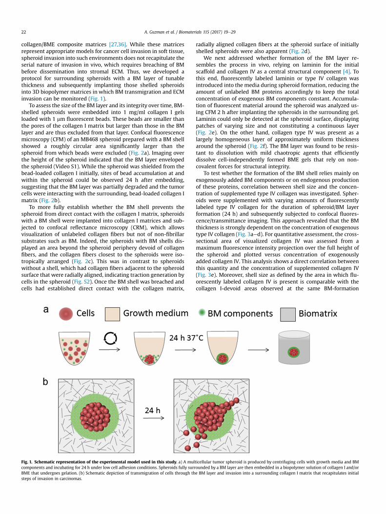
collagen/BME composite matrices [27,36]. While these matricesrepresent appropriate models for cancer cell invasion in soft tissue,spheroid invasion into such environments does not recapitulate theserial nature of invasion in vivo, which requires breaching of BMbefore dissemination into stromal ECM. Thus, we developed aprotocol for surrounding spheroids with a BM layer of tunablethickness and subsequently implanting those shelled spheroidsinto 3D biopolymer matrices in which BM transmigration and ECMinvasion can be monitored (Fig. 1).
To assess the size of the BM layer and its integrity over time, BM-shelled spheroids were embedded into 1 mg/ml collagen I gelsloaded with 1 mm fluorescent beads. These beads are smaller thanthe pores of the collagen I matrix but larger than those in the BMlayer and are thus excluded from that layer. Confocal fluorescencemicroscopy (CFM) of an MB468 spheroid prepared with a BM shellshowed a roughly circular area significantly larger than thespheroid from which beads were excluded (Fig. 2a). Imaging overthe height of the spheroid indicated that the BM layer envelopedthe spheroid (Video S1). While the spheroid was shielded from thebead-loaded collagen I initially, sites of bead accumulation at andwithin the spheroid could be observed 24 h after embedding,suggesting that the BM layer was partially degraded and the tumorcells were interacting with the surrounding, bead-loaded collagen Imatrix (Fig. 2b).
To more fully establish whether the BM shell prevents thespheroid from direct contact with the collagen I matrix, spheroidswith a BM shell were implanted into collagen I matrices and sub-jected to confocal reflectance microscopy (CRM), which allowsvisualization of unlabeled collagen fibers but not of non-fibrillarsubstrates such as BM. Indeed, the spheroids with BM shells dis-played an area beyond the spheroid periphery devoid of collagenfibers, and the collagen fibers closest to the spheroids were iso-tropically arranged (Fig. 2c). This was in contrast to spheroidswithout a shell, which had collagen fibers adjacent to the spheroidsurface that were radially aligned, indicating traction generation bycells in the spheroid (Fig. S2). Once the BM shell was breached andcells had established direct contact with the collagen matrix,
Fig. 1. Schematic representation of the experimental model used in this study. a) A mulcomponents and incubating for 24 h under low cell adhesion conditions. Spheroids fully surrBME that undergoes gelation. (b) Schematic depiction of transmigration of cells through thsteps of invasion in carcinomas.
radially aligned collagen fibers at the spheroid surface of initiallyshelled spheroids were also apparent (Fig. 2d).
We next addressed whether formation of the BM layer re-sembles the process in vivo, relying on laminin for the initialscaffold and collagen IV as a central structural component [4]. Tothis end, fluorescently labeled laminin or type IV collagen wasintroduced into the media during spheroid formation, reducing theamount of unlabeled BM proteins accordingly to keep the totalconcentration of exogenous BM components constant. Accumula-tion of fluorescent material around the spheroid was analyzed us-ing CFM 2 h after implanting the spheroids in the surrounding gel.Laminin could only be detected at the spheroid surface, displayingpatches of varying size and not constituting a continuous layer(Fig. 2e). On the other hand, collagen type IV was present as alargely homogeneous layer of approximately uniform thicknessaround the spheroid (Fig. 2f). The BM layer was found to be resis-tant to dissolution with mild chaotropic agents that efficientlydissolve cell-independently formed BME gels that rely on non-covalent forces for structural integrity.
To test whether the formation of the BM shell relies mainly onexogenously added BM components or on endogenous productionof these proteins, correlation between shell size and the concen-tration of supplemented type IV collagen was investigated. Spher-oids were supplemented with varying amounts of fluorescentlylabeled type IV collagen for the duration of spheroid/BM layerformation (24 h) and subsequently subjected to confocal fluores-cence/transmittance imaging. This approach revealed that the BMthickness is strongly dependent on the concentration of exogenoustype IV collagen (Fig. 3aed). For quantitative assessment, the cross-sectional area of visualized collagen IV was assessed from amaximum fluorescence intensity projection over the full height ofthe spheroid and plotted versus concentration of exogenouslyadded collagen IV. This analysis shows a direct correlation betweenthis quantity and the concentration of supplemented collagen IV(Fig. 3e). Moreover, shell size as defined by the area in which flu-orescently labeled collagen IV is present is comparable with thecollagen I-devoid areas observed at the same BM-formation
ticellular tumor spheroid is produced by centrifuging cells with growth media and BMounded by a BM layer are then embedded in a biopolymer solution of collagen I and/ore BM layer and invasion into a surrounding collagen I matrix that recapitulates initial

Fig. 2. Presence and composition of the basement membrane layer a, b) Confocal fluorescence (CFM) images of a representative MB468 spheroid surrounded by a BM layer at (a)2 h and (b) 24 h post-implantation in a fluorescent bead-loaded collagen I gel. Cells are dyed with Vybrant DiD live cell-labeling solution and false colored in red, while beads appearin white. The BM layer is represented by the area free from bead and cell fluorescence. Its size and integrity change over the course of 24 h, and beads can be seen within thespheroid at that time point. Scale bar ¼ 200 mm. c, d) Confocal reflectance (CRM) images of a representative MB468 spheroid surrounded by a BM layer and embedded in a collagen Igel (c) 2 h and (d) 24 h after embedding. At 2 h, the spheroid surface is isolated from the collagen network through the BM layer. The area devoid of collagen fibers (marked by apunctate red line) reflects the thickness of the BM. At 24 h, cell contact with collagen fibers and radial alignment of these fibers (red arrow), indicating traction generation, isevident. In (c, d), image processing software has been used to remove an optical artifact (a bright spot covering ~ 9 � 103 square pixels in the image) present in confocal reflectanceimages. Scale bar ¼ 50 mm e) Representative confocal fluorescence maximum projection constructed from a z-scan over 150 mm of a shelled MB468 spheroid (red) with a BM layercontaining fluorescently labeled laminin (green). Distinctive laminin accumulations at the surface of the spheroid are apparent. Scale bar ¼ 200 mm. At right, a higher magnificationmaximum projection over 60 mm of a region of the spheroid is shown. Scale bar ¼ 50 mm. f) Representative confocal fluorescence maximum projection constructed from a z-scanover 18 mm of a shelled MB468 spheroid containing fluorescently labeled type IV collagen (green) shows a dense scaffold fully surrounding the spheroid. Scale bar ¼ 200 mm. (Forinterpretation of the references to colour in this figure legend, the reader is referred to the web version of this article.)
A. Guzman et al. / Biomaterials 115 (2017) 19e29 23
conditions in bead-exclusion experiments as shown in Fig. 2a(Fig. 3f), demonstrating that collagen IV is the main structuralcomponent of the BM shell.
Time lapse imaging revealed the time course of the shell for-mation process. Fluorescently labeled collagen IV accumulatedaround the spheroid as early as 4e5 h after process initiation. Atsuch early time points, collagen IV was present in irregularly sha-ped veil-like structures emanating from the spheroid surface(Fig. S3). These structures were compacted into a denser, moreuniform layer within 24 h at collagen IV concentrations0.003e0.006 mg/ml (Fig. 3). We note that at concentrations higherthan 0.006 mg/ml collagen IV, complete compaction did not alwaysoccur and irregular shell extensions sometimes remained after 24 h(data not shown). Observation of the BM structures at these earlytime points reveals a similar correlation between basementmembrane density and size and collagen IV concentration asobserved at the later time point as shown in Fig. 3. Taken together,these results indicate that the BM surrounding spheroids is pri-marily formed from the exogenously supplemented BMcomponents.
We next interrogatedwhether the BM layer around the spheroidmimics the function and behavior of BM in vivo, where this layerseparates healthy cells from the surrounding tissue e containingthem within its boundary e but can be degraded and traversed bycancerous cells. To this end, spheroids with BM shells weregenerated from non-tumorigenic and from oncogenically trans-formed breast epithelial cells with the same genetic background,namely MCF10A and MCF10A-HRas cells. These spheroids were
embedded into 3D collagen I matrices and monitored for theintegrity of the BM layer as well as cell invasion into the collagenmatrices up to 48 h after embedding. BM shell structure wasvisualized via confocal fluorescence microscopy of labeled type IVcollagen, while spheroid architecture and cell dissemination werevisualized either via transmitted light imaging or CFM followingimmunofluorescent staining of actin cytoskeleton. As a control,spheroids without a BM layer were used. Collagen-embedded non-cancerous MCF10A spheroids with no BM layer exhibited sheet-likeexpansion with a closed cell front and no individual cell invasioninto the collagen (Fig. 4a). In contrast, spheroids with BM shellsremained fully confined within the boundary defined by the BMthroughout the monitoring period and exhibited negligible in-crease in spheroid cross-sectional area over 24 h (Fig. 4b); completeabrogation of cell dissemination into collagen I was effectedthrough the introduction of the BM layer (Fig. 4c). This behaviorwas also observed for spheroids with a BM layer formed at thelowest collagen IV concentration yielding a complete shell(0.003 mg/ml), indicating that the presence of even the thinnestBM layer does not merely reduce but abolishes the migratory ac-tivity of MCF10A cells (Fig. S4).
In contrast to MCF10A spheroids, the oncogenically transformedMCF10A-HRas spheroids were not contained by the presence of theBM layer and exhibited multiple BM breaching events anddissemination of cells into the collagen matrix within 24 h afterembedding (Fig. 5a). Compared toMCF10A-HRas spheroids with noBM layer embedded in collagen I, the density of invading cells wasgreatly diminished (Figs. 5a and 6a). This is consistent with the
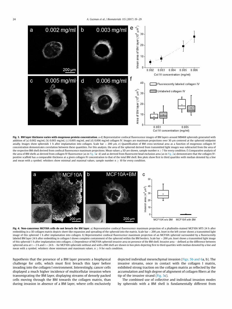
Fig. 3. BM layer thickness varies with exogenous protein concentration. a-d) Representative confocal fluorescence images of BM layers around MB468 spheroids generated withaddition of (a) 0.002 mg/ml, (b) 0.003 mg/ml, (c) 0.005 mg/ml, and (d) 0.006 mg/ml collagen IV. Images are maximum projections over 30 mm centered at the spheroid midpointaxially. Images show spheroids 1 h after implantation into collagen. Scale bar ¼ 200 mm. e) Quantification of BM cross-sectional area as a function of exogenous collagen IVconcentration demonstrates correlation between these quantities. For this analysis, the area of the spheroid derived from transmitted light images was subtracted from the area ofthe respective BM shell derived from confocal fluorescence maximum projections. Mean values ± SD are shown, sample number n � 7 for every condition. f) Comparative analysis ofthe area of BM shells as derived from collagen IV fluorescence (as in Fig. 3aed) and as derived from fluorescent bead exclusion area (as in Fig. 2a) demonstrates that the collagen IV-positive scaffold has a comparable thickness at a given collagen IV concentration to that of the total BM shell. Box plots show first to third quartiles with median denoted by a lineand mean with a symbol; whiskers show minimal and maximal values, sample number n � 10 for every condition.
Fig. 4. Non-cancerous MCF10A cells do not breach the BM layer. a) Representative confocal fluorescence maximum projection of a phalloidin-stained MCF10A MTS 24 h afterembedding in a 3D collagen matrix depicts sheet-like expansion and spreading of the spheroid into the matrix. Scale bar ¼ 200 mm. Inset in the left corner shows a transmitted lightimage of this spheroid 1 h after implantation into collagen. b) Representative confocal fluorescence maximum projection of an MCF10A spheroid surrounded by a fluorescentlylabeled BM layer 24 h after embedding in collagen I shows complete containment of the spheroid within the BM borders. Scale bar ¼ 200 mm. Inset shows a transmitted light imageof this spheroid 1 h after implantation into collagen. c) Dependence of MCF10A spheroid invasive area on presence of the BM shell. Invasive area e defined as the difference betweenspheroid area at t ¼ 2 h and t ¼ 24 h e for MCF10A spheroids without and with a BM shell are shown in box plots depicting first to third quartiles with median denoted by a line andmean with a symbol; whiskers show minimum and maximum values. n � 9 for each condition.
A. Guzman et al. / Biomaterials 115 (2017) 19e2924
hypothesis that the presence of a BM layer presents a biophysicalchallenge for cells, which must first breach this layer beforeinvading into the collagen I environment. Interestingly, cancer cellsdisplayed a much higher incidence of multicellular invasion whentransmigrating the BM layer, displaying streams of densely packedcells moving through the BM towards the collagen matrix, thanduring invasion in absence of a BM layer, where cells exclusively
depicted individual mesenchymal invasion (Figs. 5b and 6a, b). Theinvasive streams, once in contact with the collagen I matrix,exhibited strong traction on the collagen matrix as reflected by theaccumulation and high degree of alignment of collagen fibers at thetip of the invasive strand (Fig. 5c).
The combined use of collective and individual invasion modesby spheroids with a BM shell is fundamentally different from

Fig. 5. Oncogenically transformed MCF10A-Ras cells effectively transmigrate the BM layer. a) Representative confocal fluorescence maximum projection of a BM-shelledphalloidin-stained MCF10A-HRas spheroid 24 h after embedding in a 3D collagen I matrix shows a combination of individual and collective invasion of cells into the surround-ings. The white square defines a site of collective invasion that is shown at higher magnification in b) and c). Cells are shown in red and BM layer is shown in green. Scalebar ¼ 200 mm. b, c) High magnification (b) confocal fluorescence and (c) reflectance maximum projections of MCF10A-Ras spheroid shown in (a). Panel (b) shows dense packing ofcells during collective transmigration of the BM layer and subsequent dissemination as individual cells beyond the BM shell. Panel (c) shows extensive collagen reorganization andalignment by the multicellular invasive stream at the interface of the BM and collagen matrix. Scale bar ¼ 50 mm. (For interpretation of the references to colour in this figure legend,the reader is referred to the web version of this article.)
Fig. 6. MCF10A-Ras spheroids show distinct invasive modes in different matrices. a, b) Representative confocal fluorescence maximum projection of a phalloidin-stainedMCF10A-HRas spheroid without a BM layer 24 h after embedding in a 3D collagen I matrix (1PT) at (a) lower and (b) higher magnification shows extensive individual invasionof cancer cells into the surroundings. Scale bar (a) ¼ 200 mm. Scale bar (b) ¼ 50 mm. c, d) Representative confocal fluorescence maximum projection of a phalloidin-stained MCF10A-HRas spheroid without a BM layer 24 h after embedding in a 3D BME matrix (3BME) at (c) lower and (d) higher magnification shows spherical outgrowth from the spheroid bodywithout any cells leaving the bulk spheroid. Scale bar (c) ¼ 200 mm. Scale bar (d) ¼ 50 mm. e, f) Representative confocal fluorescence maximum projection of a phalloidin-stainedMCF10A-HRas spheroid without a BM layer 24 h after embedding in a composite 3D collagen I/BME matrix (1PT3BME) at (e) lower and (f) higher magnification shows multicellularinvasion with strongly polarized leader cells. Scale bar (e) ¼ 200 mm. Scale bar (f) ¼ 50 mm.
A. Guzman et al. / Biomaterials 115 (2017) 19e29 25
invasion observed for 3D-embedded spheroids in the absence of aBM layer. While collagen I matrices support strong individual in-vasion and mesenchymal cell morphology (Fig. 6a, b), a matrixcomposed solely of basement membrane extract (BME) does notsupport any invasive behavior over the assessed time scales (Fig. 6c,d). In composite collagen I/BME matrices, the cells show collectiveinvasion as well as strong cell polarity similar to the initial
migratory phenotype of shelled spheroids during invasion throughthe BM layer (Fig. 6e, f); however this matrix does not support theswitch to individual invasion at any point.
Next, we investigated whether the observed induction of col-lective invasion in shelled spheroids relative to unshelled spheroidsin collagen I environments is a cell-type specific response to theseexperimental conditions or a more general behavior of tumorigenic

A. Guzman et al. / Biomaterials 115 (2017) 19e2926
cells in the presence of a BM layer. Thus, invasion studies similar tothose performed on MCF10A-HRas cells were performed on cancercells of different origin, namely MB468 breast cancer cells. MB468spheroids prepared without a BM layer and introduced intocollagen I matrices showed individual cell invasion into the sur-roundings, no invasion in pure BME matrices, and multicellularinvasion in composite collagen I/BME matrices (Fig. S5), parallelingthe invasion pattern observed in response to these matrices forMCF10A-HRas cells. When prepared with BM shells, the cells suc-cessfully traversed the BM layer and invaded into collagen (Fig. S6),as also found for MCF10A-HRas cells. While the two cancerous celllines differ in origin, cell morphology (mesenchymal MCF10A-HRasand grape-like MB468) and time required for dissemination (24 hfor MCF10A-HRas, 24e48 h for MB468), they were similar in theirability to breach and transmigrate the surrounding BM layer, incontrast to the non-tumorigenic MCF10A cells. Thus, this newexperimental model recapitulates the fundamentally differentphysiological behavior of cancerous and non-cancerous cell ag-gregates surrounded by a cell-bound BM.
We next investigated molecular activity required for cells totraverse the BM layer, in particular whether matrix metal-loproteinases (MMPs) were required for BM breaching and/orsubsequent invasion in collagen I. To this end, MCF10A-HRasspheroids prepared with or without BM layers were pre-treatedwith an MMP-inhibitor cocktail targeting MMP-1, -2, -3, -7, -9and -14 (MT1-MMP) as well as aminopeptidases and serine- andcysteine-proteases and embedded in collagen I matrices supple-mented with the same inhibitors. We note that MCF10A-HRas hasbeen reported to have upregulated expression of both MMP-2and MMP-9, with the former regulated by MT1-MMP, relativeto MCF10A [37,38]. After 24 h, samples were fixed and subjectedto actin cytoskeleton staining and confocal fluorescence imagingas described earlier. It was found that the presence of the BMlayer strongly modulated the cellular response to MMP inhibi-tion. Collagen I invasion of spheroids without a BM layer was onlymildly affected by MMP inhibition, with no observable differ-ences of invasion mode or cell morphology and no significantreduction of invasive distance (Fig. 7aec). In contrast, the inva-sive behavior of BM-shelled spheroids was strongly compromisedby MMP inhibition. Here, the MMP inhibition led to a greaterthan two-fold reduction of invasion incidence (from 100% to38.5%), as characterized by the presence of individual cancer cellsin the collagen matrix, and a nearly twenty-fold reduction in themean number of individual invasive cells (57e2.5) (Fig. 7def).MMP inhibition in BM-shelled spheroids did not fully preventformation of multicellular streams, while it did completelyabolish formation of invasive structures (and invasion) in BM-freespheroids embedded in composite collagen I/BME matrices, thecondition that induces multicellular invasion in the absence ofMMP inhibition (Fig. 7gei). While the multicellular structures inBM-enveloped spheroids still formed under MMP inhibition, thisoccurred in reduced numbers per spheroid (from median 6 to 2per spheroid). Moreover, 50% of the multicellular streams failedto breach the BM layer within 24 h, indicating that the efficiencyof BM breaching is strongly dependent on MMP-mediated BMdegradation. The fact that multicellular invasive streams formedunder MMP inhibition in BM-enveloped spheroids while theirformation was completely abrogated in bare spheroids embeddedin composite matrix suggests that ECM comprising a cell-assembled non-fibrillar BM adjacent to fibrillar collagen matrixevokes a particular collective invasion mode that is more resis-tant to pharmacological MMP inhibition than spheroid invasion(without a BM shell) in a composite matrix consisting of amixture of both components.
4. Discussion
Despite decades of study, the cellular events that allow an in situcircumscribed tumor to become an invasive entity and the molec-ular mechanisms underlying the penetration of cancer cellsthrough the BM and adjacent ECM are not fully understood. Here,we present an optically accessible 3D model that recapitulatesdiverse dynamic cell-cell and cell-ECM interactions that exist ascells traverse a dense, sheet-like BM layer in advance of invasioninto adjacent ECM. The experimental model presented in this studyconsists of spheroids containing several thousands of benign ortumorigenic cells surrounded by a BM layer and embedded into abiomechanically tunable 3D matrix (Fig. 1). Importantly, we foundthat non-tumorigenic cells were confined by the BM layer (Fig. 4b,Fig. S4), while spheroids composed of various cancerous cell linesbreached the BM layer and invaded into the adjacent matrix within24 h after embedding (Fig. 5, Fig. S6). This recapitulates a criticalprocess in the progression of metastatic disease. When a dysplasticcarcinoma in situ acquires the ability to traverse the BM, the lesionis classified as a malignant carcinoma [39e41]. For probing theinvasive behavior of cancer cells, the model presented here is su-perior to embedding spheroids in pure collagen I matrices, as somenon-cancerous epithelial cells spread from the spheroid in 3Dcollagen I matrices despite their benign character (Fig. 4a). Whilethe cells maintain tight cell-cell contacts and a closed cell frontmore reminiscent of sheet expansion than true invasion in acollagen matrix, the spheroid e in absence of a BM layer e loses itsoriginal architecture and does not fully reflect its non-tumorigenicnature. This is avoided by surrounding the MTS with a dense layerof BM (compare Fig. 4a and b).
While many models suggest metastasis begins with individualcells undergoing the epithelial-mesenchymal transition (EMT) andleaving the boundaries of the primary tumor, analysis of tumor-stroma interfaces in clinical samples has revealed that it is thepresence of invasive cell clusters [42], also termed tumor buds, thatcorrelates with metastatic progression and poor prognosis invarious solid tumor types [43e47]. This highlights the importanceof understanding the cellular and molecular underpinnings ofcollective cancer cell invasion and the need for physiologicallyrelevant in vitro models supporting this crucial mode of invasion.To date, in vitro settings for the study of collective cancer cellmigration have relied primarily on 2D scratch/wound assays or onassays using spheroids or organoids embedded in 3D matrices,typically composed of collagen I or BME [48e52]. We and othershave reported differential invasive behavior for cancerous cells infibrillar (collagen I) vs. non-fibrillar (BME) 3D matrices, withcollagen I typically being more supportive of invasion than is non-fibrillar BME, which did not lead to invasion in either spheroids ororganoids of known tumorigenic breast, ovarian and prostatecancer cells [27,52e54]. Recently, we demonstrated that one breastcancer cell line showed individual invasion in collagen I matrices,no invasion in BME, and a primarily collective mode of invasion in acomposite collagen I/BME matrix [27]. These results mirror thosefound in theMCF10A-HRas andMB468 cell lines shown here (Fig. 6,Fig. S5). Although the composite matrix appears to be a bettersystem to evoke and study collective invasion than do homoge-neous collagen I or BME matrices, a homogeneous environmentdoes not recapitulate the in vivo setting in which cells are facedwith distinct ECM components serially, as epithelial based tumorsmust first breach a cell-bound BM layer to subsequently migratethrough stromal ECM.
Thus, the presence of the BM-ECM interface and the structure ofthe BM layer, which is intimately related to the mechanism of itsgeneration, are a critical advantage of the shelled spheroid systemdescribed here. Conventional assays probing cell invasion in BM

Fig. 7. Differential effects of MMP inhibition as a function of ECM. a, b) Representative confocal fluorescence maximum projections of (a) solvent control and (b) MMP inhibitor-treated phalloidin-stained MCF10A-HRas spheroids without a BM layer 24 h after embedding in a 3D collagen I matrix. Scale bar ¼ 200 mm. c) Quantitative analysis of MCF10A-HRasspheroid invasion in collagen I under MMP inhibition reveals no significant difference (p < 0.05) in invasive distance between the treated and control groups. Invasive distances areshown with box plots depicting first to third quartiles with median denoted by a line and mean with a symbol; whiskers show minimum and maximum values, sample numbern � 12 for every condition. d, e) Representative confocal fluorescence maximum projections of (d) solvent control and (e) MMP inhibitor-treated phalloidin-stained MCF10A-HRasspheroids with a BM layer 24 h after embedding in a 3D collagen I matrix. Scale bar ¼ 200 mm. f) Quantitative analysis of BM-shelled MCF10A-HRas spheroid invasion in collagen Iunder MMP inhibition reveals significant differences in number of collective invasion sites and number of individual cells invaded in the collagen matrix per spheroid with p < 0.05obtained in the non-parametrical Mann-Whitney test. Histograms show the percentage of spheroids with stated number of collective invasion sites or individual invaded cells in theinhibitor-treated versus the control group. n � 17 for every condition. g, h) Representative confocal fluorescence maximum projections of (g) solvent control and (h) MMP inhibitor-treated phalloidin-stained MCF10A-HRas spheroids without a BM layer 24 h after embedding in a composite collagen I/BME matrix show abrogation of invasion under MMPinhibition. Scale bar ¼ 200 mm. i) Quantitative analysis of MCF10A-HRas spheroid invasion in composite collagen I/BME matrix under MMP inhibition reveals significant differencein invasive area between the treated and control groups, with p < 0.05 obtained in the Mann-Whitney test. Invasive areas for inhibitor and control groups are shownwith box plotsdepicting first to third quartiles with median denoted by a line and mean with a symbol; whiskers show minimum and maximum values, sample number n � 6 for every condition.
A. Guzman et al. / Biomaterials 115 (2017) 19e29 27
commonly use basement membrane extract polymerized in a cell-independent manner. In contrast, in the shelled spheroid protocolpresented here, the BM layer is assembled from supplementedcomponents in a cell-mediated process. This process requiresfunctional b1 integrin receptors since antibody-mediated b1integrin inhibition strongly compromised the formation of acontinuous and dense BM layer (data not shown). This is in accor-dance with the b1 integrin-dependent mechanism reported for BMformation in mice [55] and suggests that the formation of the BMlayer in the presented experimental system requires similar cellularmechanisms to the respective process in vivo. This hypothesis issupported by the observation that in the shelled spheroid model
presented here, laminin is bound and forms a thin patchy layerdirectly at the spheroid surface (Fig. 2e) while collagen IV is poly-merized into a complex network that constitutes the bulk of the BMstructure (Figs. 2f and 3). This closely recapitulates the reportedmolecular mechanisms of BM assembly in vivo, where laminin ispolymerized at the cell surface and serves as the initial template forscaffold formation through type IV collagen polymerization [56,57].
BME polymerized in a cell-independent manner is not onlymore compliant than endogenous BM [58,59], it also lacks somehallmarks of mature BM structure, such as covalently cross-linkedcollagen IV [53,60,61]. Cells thus may use distinct invasion strate-gies in traversing BME relative to those used in cell-assembled BM

A. Guzman et al. / Biomaterials 115 (2017) 19e2928
in vivo or in the shelled spheroid model presented here. This hy-pothesis is supported by our finding that cancer cells that are non-invasive in 3D BME gels can efficiently transmigrate the BM layer inthe presented model (compare Figs. 5a and 6c, Figs. S5b and S6).This finding is also consistent with studies showing that matrixstiffness and architecture determine cancer cell invasion efficiencyand strategy and regulate dynamic switching between invasivemodes, such as mesenchymal and amoeboid invasion [27,62e64].For individually invading cancer cells, some 3D environmentspreferentially induce mesenchymal invasion requiring cellematrixadhesion andMMP-mediated ECM proteolysis, while others inducemigratory modes relying on ROCK-mediated cortical actomyosincontractility, such as MMP-dependent lobopodial migration andMMP-independent amoeboid migration [64,65]. For collective in-vasion, mechanistically distinct invasive modes have not been aswell-characterized. Indeed, the biomechanical determinants (suchas matrix stiffness and porosity) and cellular determinants (such ascell contractility and adhesion and protease requirements) of col-lective invasion modes are yet to be fully elucidated. For example, itis possible that the existence of a confining BM shell around agrowing spheroid may drive collective invasion by generatingelevated pressure within the spheroid that mimics the high inter-stitial pressure observed in solid tumors, a characteristic that hasbeen linked to altered migratory behavior in vitro and increasedmetastasis and poor prognosis in vivo [66e69].
Interestingly, we find that in MCF10A-HRas spheroids sur-rounded by a layer of cell-assembled BM, the formation of multi-cellular streams and a degree of successful invasion occurs underMMP inhibition targeting both secreted MMPs and the membrane-bound MT1-MMP. In contrast, this cellular behavior is completelyabolished in unshelled spheroids embedded in composite matrices(Fig. 7). The lower sensitivity to MMP inhibition suggests that thecells utilize different invasion mechanisms during transmigrationof cell-assembled BM than when confronted with BME polymer-ized in a cell-independent manner. A study utilizing decellularizedperitoneal BM demonstrated that transmigration of native ex vivoBM has different requirements for MMP activity, relying exclusivelyon membrane-bound MMPs (MT1-3 MMPs) than invasion ofin vitro reconstituted BME that utilized secretedMMPs [61]. Thus, itis particularly important to address the mechanisms of BMbreaching in a system that not only recapitulates the biochemicalcomposition of BM but also more closely resembles its biome-chanical properties in vivo. Whether the multicellular invasionmode seen in this study uses membrane-bound MMPs (such asMT2- or MT3-MMP) that were not specifically targeted by theprotease inhibitors used here or whether the cell-assembled BMprovides mechano-transductory stimuli inducing a less MMP-dependent collective invasion mode remains to be clarified. How-ever, our finding of limited MMP dependence in the shelledspheroid model, together with the largely MMP-independentmigratory mode of individual cancer cells in fibrillar collagen Imatrix, offers an explanation for the inefficiency of MMP inhibitionas a treatment strategy for late stage cancers [70].
5. Conclusion
We have developed a novel experimental model inwhich tumorspheroids surrounded by a cell-bound BM of tunable thickness aregenerated and may be subsequently embedded in a secondbiopolymer matrix such that the cells serially encounter multiple,adjacent extracellular environments. Using this model, centralinitial events of metastatic progression were recapitulated in aphysiologically relevant setting. First, we showed that tumorigenicbreast cancer cell lines of two different subtypes can breach this BMwithin 24 h, while non-cancerous breast epithelial cells were fully
retained within BM borders, thus reproducing an early hallmark ofmetastatic behavior. We also demonstrated selective cancer cellutilization of collective migration for transmigration of the physi-cally challenging BM layer. Moreover, this study revealed that whileBM breaching, in contrast to collagen I invasion, is an MMP-dependent process, it is less susceptible to pharmacological MMPinhibition then collective invasion in homogeneous compositematrices and cannot be fully abolished by such. Thus, we showedthat the heterogeneous environment comprising a distinct non-fibrillar BM and an adjacent fibrillar ECM evoked a complex inva-sive phenotype that differed from any homogeneous ECM condi-tion tested and that the described model represents aphysiologically highly relevant setting for addressing cellularcharacteristics and treatment responses in metastasizing solidtumors.
Acknowledgements
This work was supported in part by the National ScienceFoundation and the National Institutes of Health via PESO 1227297.
Appendix A. Supplementary data
Supplementary data related to this article can be found at http://dx.doi.org/10.1016/j.biomaterials.2016.11.014.
References
[1] S. Valastyan, R.A. Weinberg, Tumor metastasis: molecular insights andevolving paradigms, Cell 147 (2011) 275e292.
[2] S. Kumar, V.M. Weaver, Mechanics, malignancy, and metastasis: the forcejourney of a tumor cell, Cancer Metastasis Rev. 28 (2009) 113e127.
[3] P. Lu, V.M. Weaver, Z. Werb, The extracellular matrix: a dynamic niche incancer progression, J. Cell Biol. 196 (2012) 395e406.
[4] R. Kalluri, Basement membranes: structure, assembly and role in tumourangiogenesis, Nat. Rev. Cancer 3 (2003) 422e433.
[5] P.D. Yurchenco, Basement membranes: cell scaffoldings and signaling plat-forms, Cold Spring Harb. Perspect. Biol. 3 (2011).
[6] L.A. Liotta, K. Tryggvason, S. Garbisa, I. Hart, C.M. Foltz, S. Shafie, Metastaticpotential correlates with enzymatic degradation of basement membranecollagen, Nature 284 (1980) 67e68.
[7] A. Martinez-Hernandez, P.S. Amenta, The basement membrane in pathology,Lab. Investig. 48 (1983) 656e677.
[8] J.V. Frei, The fine structure of the basement membrane in epidermal tumors,J. Cell Biol. 15 (1962) 335e342.
[9] F.T. Bosman, M. Havenith, J.P. Cleutjens, Basement membranes in cancer,Ultrastruct. Pathol. 8 (1985) 291e304.
[10] S. Spaderna, O. Schmalhofer, F. Hlubek, G. Berx, A. Eger, S. Merkel, et al.,A transient, EMT-linked loss of basement membranes indicates metastasisand poor survival in colorectal cancer, Gastroenterology 131 (2006) 830e840.
[11] A. Bergamaschi, E. Tagliabue, T. Sorlie, B. Naume, T. Triulzi, R. Orlandi, et al.,Extracellular matrix signature identifies breast cancer subgroups withdifferent clinical outcome, J. Pathol. 214 (2008) 357e367.
[12] K. Polyak, Molecular markers for the diagnosis and management of ductalcarcinoma in situ, J. Natl. Cancer Inst. Monogr. 2010 (2010) 210e213.
[13] J.K. Mouw, G. Ou, V.M. Weaver, Extracellular matrix assembly: a multiscaledeconstruction, Nat. Rev. Mol. Cell Biol. 15 (2014) 771e785.
[14] G.G. Zhu, L. Risteli, M. Makinen, J. Risteli, A. Kauppila, F. Stenback, Immuno-histochemical study of type I collagen and type I pN-collagen in benign andmalignant ovarian neoplasms, Cancer 75 (1995) 1010e1017.
[15] S. Kauppila, F. Stenback, J. Risteli, A. Jukkola, L. Risteli, Aberrant type I and typeIII collagen gene expression in human breast cancer in vivo, J. Pathol. 186(1998) 262e268.
[16] I.J. Huijbers, M. Iravani, S. Popov, D. Robertson, S. Al-Sarraj, C. Jones, et al.,A role for fibrillar collagen deposition and the collagen internalization re-ceptor endo180 in glioma invasion, PLoS One 5 (2010) e9808.
[17] S. Alowami, S. Troup, S. Al-Haddad, I. Kirkpatrick, P.H. Watson, Mammo-graphic density is related to stroma and stromal proteoglycan expression,Breast Cancer Res. 5 (2003) R129eR135.
[18] Y.P. Guo, L.J. Martin, W. Hanna, D. Banerjee, N. Miller, E. Fishell, et al., Growthfactors and stromal matrix proteins associated with mammographic densities,Cancer Epidemiol. Biomarker. Prev. 10 (2001) 243e248.
[19] P.P. Provenzano, K.W. Eliceiri, J.M. Campbell, D.R. Inman, J.G. White, P.J. Keely,Collagen reorganization at the tumor-stromal interface facilitates local inva-sion, BMC Med. 4 (2006) 38.
[20] M.W. Conklin, J.C. Eickhoff, K.M. Riching, C.A. Pehlke, K.W. Eliceiri,

A. Guzman et al. / Biomaterials 115 (2017) 19e29 29
P.P. Provenzano, et al., Aligned collagen is a prognostic signature for survivalin human breast carcinoma, Am. J. Pathol. 178 (2011) 1221e1232.
[21] R. Kalluri, R.A. Weinberg, The basics of epithelial-mesenchymal transition,J. Clin. Investig. 119 (2009) 1420e1428.
[22] S. Gurzu, S. Turdean, A. Kovecsi, A.O. Contac, I. Jung, Epithelial-mesenchymal,mesenchymal-epithelial, and endothelial-mesenchymal transitions in malig-nant tumors: an update, World J. Clin. Cases 3 (2015) 393e404.
[23] J.L. Leight, M.A. Wozniak, S. Chen, M.L. Lynch, C.S. Chen, Matrix rigidity reg-ulates a switch between TGF-beta1-induced apoptosis and epithelial-mesenchymal transition, Mol. Biol. Cell 23 (2012) 781e791.
[24] S.C. Wei, L. Fattet, J.H. Tsai, Y. Guo, V.H. Pai, H.E. Majeski, et al., Matrix stiffnessdrives epithelial-mesenchymal transition and tumour metastasis through aTWIST1-G3BP2 mechanotransduction pathway, Nat. Cell Biol. 17 (2015)678e688.
[25] K.R. Levental, H. Yu, L. Kass, J.N. Lakins, M. Egeblad, J.T. Erler, et al., Matrixcrosslinking forces tumor progression by enhancing integrin signaling, Cell139 (2009) 891e906.
[26] N.R. Lang, K. Skodzek, S. Hurst, A. Mainka, J. Steinwachs, J. Schneider, et al.,Biphasic response of cell invasion to matrix stiffness in three-dimensionalbiopolymer networks, Acta Biomater. 13 (2015) 61e67.
[27] A. Guzman, M.J. Ziperstein, L.J. Kaufman, The effect of fibrillar matrix archi-tecture on tumor cell invasion of physically challenging environments, Bio-materials 35 (2014) 6954e6963.
[28] J. Condeelis, J.E. Segall, Intravital imaging of cell movement in tumours, Nat.Rev. Cancer 3 (2003) 921e930.
[29] S.I. Ellenbroek, J. van Rheenen, Imaging hallmarks of cancer in living mice, Nat.Rev. Cancer 14 (2014) 406e418.
[30] C.R. Thoma, M. Zimmermann, I. Agarkova, J.M. Kelm, W. Krek, 3D cell culturesystems modeling tumor growth determinants in cancer target discovery,Adv. Drug Deliv. Rev. 69e70 (2014) 29e41.
[31] E. Katz, S. Dubois-Marshall, A.H. Sims, P. Gautier, H. Caldwell, R.R. Meehan, etal., An in vitro model that recapitulates the epithelial to mesenchymal tran-sition (EMT) in human breast cancer, PLoS One 6 (2011) e17083.
[32] M. Schoumacher, R.D. Goldman, D. Louvard, D.M. Vignjevic, Actin, microtu-bules, and vimentin intermediate filaments cooperate for elongation ofinvadopodia, J. Cell Biol. 189 (2010) 541e556.
[33] A. Ivascu, M. Kubbies, Rapid generation of single-tumor spheroids for high-throughput cell function and toxicity analysis, J. Biomol. Screen 11 (2006)922e932.
[34] K. Wolf, I. Mazo, H. Leung, K. Engelke, U.H. von Andrian, E.I. Deryugina, et al.,Compensation mechanism in tumor cell migration: mesenchymal-amoeboidtransition after blocking of pericellular proteolysis, J. Cell Biol. 160 (2003)267e277.
[35] M.P. Fay, M.A. Proschan, Wilcoxon-Mann-Whitney or t-test? On assumptionsfor hypothesis tests and multiple interpretations of decision rules, Stat. Surv. 4(2010) 1e39.
[36] M.J. Ziperstein, A. Guzman, L.J. Kaufman, Breast Cancer cell line aggregatemorphology does not predict invasive capacity, PLoS One 10 (2015) e0139523.
[37] A. Moon, M.S. Kim, T.G. Kim, S.H. Kim, H.E. Kim, Y.Q. Chen, et al., H-ras, but notN-ras, induces an invasive phenotype in human breast epithelial cells: a rolefor MMP-2 in the H-ras-induced invasive phenotype, Int. J. Cancer 85 (2000)176e181.
[38] D. Al-Raawi, H. Abu-El-Zahab, M. El-Shinawi, M.M. Mohamed, Membranetype-1 matrix metalloproteinase (MT1-MMP) correlates with the expressionand activation of matrix metalloproteinase-2 (MMP-2) in inflammatory breastcancer, Int. J. Clin. Exp. Med. 4 (2011) 265e275.
[39] D. Hanahan, R.A. Weinberg, The hallmarks of cancer, Cell 100 (2000) 57e70.[40] R.G. Rowe, S.J. Weiss, Breaching the basement membrane: who, when and
how? Trends Cell Biol. 18 (2008) 560e574.[41] F.T. Bosman, The borderline: basement membranes and the transition from
premalignant to malignant neoplasia, Microsc. Res. Tech. 28 (1994) 216e225.[42] D. Hanahan, R.A. Weinberg, Hallmarks of cancer: the next generation, Cell 144
(2011) 646e674.[43] N. Ohike, I. Coban, G.E. Kim, O. Basturk, T. Tajiri, A. Krasinskas, et al., Tumor
budding as a strong prognostic indicator in invasive ampullary adenocarci-nomas, Am. J. Surg. Pathol. 34 (2010) 1417e1424.
[44] B. Mitrovic, D.F. Schaeffer, R.H. Riddell, R. Kirsch, Tumor budding in colorectalcarcinoma: time to take notice, Mod. Pathol. 25 (2012) 1315e1325.
[45] E. Karamitopoulou, I. Zlobec, B. Gloor, A. Kondi-Pafiti, A. Lugli, A. Perren, Lossof Raf-1 kinase inhibitor protein (RKIP) is strongly associated with high-gradetumor budding and correlates with an aggressive phenotype in pancreaticductal adenocarcinoma (PDAC), J. Transl. Med. 11 (2013) 311.
[46] F. Liang, W. Cao, Y. Wang, L. Li, G. Zhang, Z. Wang, The prognostic value oftumor budding in invasive breast cancer, Pathol. Res. Pract. 209 (2013)
269e275.[47] Y. Sun, F. Liang, W. Cao, K. Wang, J. He, H. Wang, et al., Prognostic value of
poorly differentiated clusters in invasive breast cancer, World J. Surg. Oncol.12 (2014) 310.
[48] T. Das, K. Safferling, S. Rausch, N. Grabe, H. Boehm, J.P. Spatz, A molecularmechanotransduction pathway regulates collective migration of epithelialcells, Nat. Cell Biol. 17 (2015) 276e287.
[49] M.L. Graves, J.A. Cipollone, P. Austin, E.M. Bell, J.S. Nielsen, C.B. Gilks, et al., Thecell surface mucin podocalyxin regulates collective breast tumor budding,Breast Cancer Res. 18 (2016) 11.
[50] L.J. Kaufman, C.P. Brangwynne, K.E. Kasza, E. Filippidi, V.D. Gordon,T.S. Deisboeck, et al., Glioma expansion in collagen I matrices: analyzingcollagen concentration-dependent growth and motility patterns, Biophys. J.89 (2005) 635e650.
[51] Y.L. Yang, S. Motte, L.J. Kaufman, Pore size variable type I collagen gels andtheir interaction with glioma cells, Biomaterials 31 (2010) 5678e5688.
[52] K.V. Nguyen-Ngoc, K.J. Cheung, A. Brenot, E.R. Shamir, R.S. Gray, W.C. Hines, etal., ECM microenvironment regulates collective migration and local dissemi-nation in normal and malignant mammary epithelium, Proc. Natl. Acad. Sci. U.S. A. 109 (2012) E2595eE2604.
[53] K.L. Sodek, T.J. Brown, M.J. Ringuette, Collagen I but not Matrigel matricesprovide an MMP-dependent barrier to ovarian cancer cell penetration, BMCCancer 8 (2008) 223.
[54] V. Harma, J. Virtanen, R. Makela, A. Happonen, J.P. Mpindi, M. Knuuttila, et al.,A comprehensive panel of three-dimensional models for studies of prostatecancer growth, invasion and drug responses, PLoS One 5 (2010) e10431.
[55] S. Raghavan, C. Bauer, G. Mundschau, Q. Li, E. Fuchs, Conditional ablation ofbeta1 integrin in skin. Severe defects in epidermal proliferation, basementmembrane formation, and hair follicle invagination, J. Cell Biol. 150 (2000)1149e1160.
[56] K.K. McKee, D. Harrison, S. Capizzi, P.D. Yurchenco, Role of laminin terminalglobular domains in basement membrane assembly, J. Biol. Chem. 282 (2007)21437e21447.
[57] S. Li, P. Liquari, K.K. McKee, D. Harrison, R. Patel, S. Lee, et al., Laminin-sulfatidebinding initiates basement membrane assembly and enables receptorsignaling in Schwann cells and fibroblasts, J. Cell Biol. 169 (2005) 179e189.
[58] S.S. Soofi, J.A. Last, S.J. Liliensiek, P.F. Nealey, C.J. Murphy, The elastic modulusof Matrigel as determined by atomic force microscopy, J. Struct. Biol. 167(2009) 216e219.
[59] W. Halfter, P. Oertle, C.A. Monnier, L. Camenzind, M. Reyes-Lua, H. Hu, et al.,New concepts in basement membrane biology, FEBS J. 282 (2015) 4466e4479.
[60] S. Even-Ram, K.M. Yamada, Cell migration in 3D matrix, Curr. Opin. Cell Biol.17 (2005) 524e532.
[61] K. Hotary, X.Y. Li, E. Allen, S.L. Stevens, S.J. Weiss, A cancer cell metal-loprotease triad regulates the basement membrane transmigration program,Genes Dev. 20 (2006) 2673e2686.
[62] K. Wolf, M. Te Lindert, M. Krause, S. Alexander, J. Te Riet, A.L. Willis, et al.,Physical limits of cell migration: control by ECM space and nuclear defor-mation and tuning by proteolysis and traction force, J. Cell Biol. 201 (2013)1069e1084.
[63] M.H. Zaman, L.M. Trapani, A.L. Sieminski, D. Mackellar, H. Gong, R.D. Kamm, etal., Migration of tumor cells in 3D matrices is governed by matrix stiffnessalong with cell-matrix adhesion and proteolysis, Proc. Natl. Acad. Sci. U. S. A.103 (2006) 10889e10894.
[64] R.J. Petrie, N. Gavara, R.S. Chadwick, K.M. Yamada, Nonpolarized signalingreveals two distinct modes of 3D cell migration, J. Cell. Biol. 197 (2012)439e455.
[65] V. Sanz-Moreno, C.J. Marshall, The plasticity of cytoskeletal dynamics un-derlying neoplastic cell migration, Curr. Opin. Cell Biol. 22 (2010) 690e696.
[66] J.M. Tse, G. Cheng, J.A. Tyrrell, S.A. Wilcox-Adelman, Y. Boucher, R.K. Jain, et al.,Mechanical compression drives cancer cells toward invasive phenotype, Proc.Natl. Acad. Sci. U. S. A. (2012) 911e916.
[67] M. Milosevic, A. Fyles, D. Hedley, M. Pintilie, W. Levin, L. Manchul, et al.,Interstitial fluid pressure predicts survival in patients with cervix cancer in-dependent of clinical prognostic factors and tumor oxygen measurements,Cancer Res. 61 (2001) 6400e6405.
[68] T. Hompland, C. Ellingsen, K.M. Ovrebo, E.K. Rofstad, Interstitial fluid pressureand associated lymph node metastasis revealed in tumors by dynamiccontrast-enhanced MRI, Cancer Res. 72 (2012) 4899e4908.
[69] W.J. Polacheck, A.E. German, A. Mammoto, D.E. Ingber, R.D. Kamm, Mecha-notransduction of fluid stresses governs 3D cell migration, Proc. Natl. Acad.Sci. U. S. A. 111 (2014) 2447e2452.
[70] L.M. Coussens, B. Fingleton, L.M. Matrisian, Matrix metalloproteinase in-hibitors and cancer: trials and tribulations, Science 295 (2002) 2387e2392.