A NOSY NEIGHBOR
10
A NOSY NEIGHBOR Purification and Functional Characterization of Lpg2149 Abstract Ubiquitination is a process that marks proteins for various cell-signaling pathways, namely protein degradation and other processes. ese pathways are essential in a wide array of cellular processes, including defense mechanisms against invading pathogens. e ubiquitination process is universally found in all eukaryotic organisms, including plants and animals, and thus plays a vital role in cellular homeostasis. Recently, more discoveries have been made on prokaryotic effector proteins that hijack the ubiquitination system even when they do not possess a ubiquitin system of their own. MavC, also known as lpg2147 (Gan, Nakayasu, Hollenbeck, & Luo, 2019; Puvar et al., 2020; Valleau et al., 2018), has been found to be a ubiquitinating enzyme that ubiquitinates UbE2N by bypassing the usual E1, E2, E3 ubiquitination pathway. MavC plays an important role in the infection of Legionella pneumophila, which is the culprit of Legionnaires’ disease. rough earlier molecular biology analysis, it has been discovered that a neighboring gene on the same locus as MavC encodes lpg2149, which has been characterized to inhibit MavC’s function. Given the novelty of this protein, this research project aims to achieve cloning, expression, and purification of lpg2149 so that in its inhibitory complex lpg2149 can be captured by X-ray crystallography. An attempt was made to crystallize and obtain the structure of lpg2149 with MavC. With the optimization of lpg2149 production demonstrated in this project, a better understanding of L. pneumophila pathogenesis can be obtained, which would help in our understanding of regulating L. pneumophila infection. Keywords Legionella pneumophila, inhibitor, MavC (lpg2147), lpg2149, UbE2N, crystallography http://dx.doi.org/10.7771/2158-4052.1502
Transcript of A NOSY NEIGHBOR
A NOSY NEIGHBORPurification and Functional Characterization of Lpg2149
AbstractUbiquitination is a process that marks proteins for various cell-signaling pathways, namely protein degradation and other processes. Th ese pathways are essential in a wide array of cellular processes, including defense mechanisms against invading pathogens. Th e ubiquitination process is universally found in all eukaryotic organisms, including plants and animals, and thus plays a vital role in cellular homeostasis. Recently, more discoveries have been made on prokaryotic eff ector proteins that hijack the ubiquitination system even when they do not possess a ubiquitin system of their own. MavC, also known as lpg2147 (Gan, Nakayasu, Hollenbeck, & Luo, 2019; Puvar et al., 2020; Valleau et al., 2018), has been found to be a ubiquitinating enzyme that ubiquitinates UbE2N by bypassing the usual E1, E2, E3 ubiquitination pathway. MavC plays an important role in the infection of Legionella pneumophila, which is the culprit of Legionnaires’ disease. Th rough earlier molecular biology analysis, it has been discovered that a neighboring gene on the same locus as MavC encodes lpg2149, which has been characterized to inhibit MavC’s function. Given the novelty of this protein, this research project aims to achieve cloning, expression, and purifi cation of lpg2149 so that in its inhibitory complex lpg2149 can be captured by X-ray crystallography. An attempt was made to crystallize and obtain the structure of lpg2149 with MavC. With the optimization of lpg2149 production demonstrated in this project, a better understanding of L. pneumophila pathogenesis can be obtained, which would help in our understanding of regulating L. pneumophila infection.
KeywordsLegionella pneumophila, inhibitor, MavC (lpg2147), lpg2149, UbE2N, crystallography
http://dx.doi.org/10.7771/2158-4052.1502

Journal of Purdue Undergraduate Research: Volume 11, Fall 202164
Student AuthorAshley M. Holahan is a third-year undergraduate student at Purdue University majoring in biochemistry. Her involvement with undergraduate research began in her first semester at Purdue when she joined the Das Lab. Through her research experience,
Holahan has been trained in various biomolecular tech-niques and procedures and has been optimizing the production of lpg2149. For her research, she has received the Office of Undergraduate Research Scholarship. Holahan plans to pursue a doctorate after she graduates from Purdue.
MentorsSebastian Kenny is a third-year PhD student in the
Das Lab. He received his bachelor of science degree in biochemistry from Purdue University. During his undergraduate research, Kenny received training in organic synthe-sis. His current research focuses on understanding the mechanism of
p53 ubiquitination by human papillomavirus using various biophysical techniques. Some of these biophysi-cal techniques include biolayer interferometry and isothermal titration calorimetry, which shed light on the protein-protein binding, allowing for better mechanistic understanding. Additionally, Kenny is being trained in cryo-electron microscopy with the hope of determining the structure of a five-protein complex.
Dr. Chittaranjan Das is an associate professor in the Department of Chemistry at Purdue University. He received his PhD from the Indian Institute of Science, Bangalore (India). Following his PhD education, Das became a postdoc in Dr. Greg Petsko’s group at Harvard Medical School and Brandeis
University. Dr. Das’s lab is interested in the ability of pathogens to produce pathogenic effectors that hijack the ubiquitination system. To study this system, he employs structural and biophysical techniques that elucidate the mechanism of the effector proteins that these pathogens produce. In addition, Das has established numerous collaborations with other labs across different universities to take advantage of different expertise.
INTRODUCTION
Ubiquitination is a posttranslational modification that plays a role in many processes in eukaryotic cells, including protein degradation, protein trafficking, protein localization, and protein regulation. During ubiquitination, proteins are modified with the addition of a single ubiquitin or ubiquitin protein chains. The conformation and number of ubiquitin attached to the protein act as a code that determines that protein’s fate in the cell, one example being protein degradation. Three enzymes known as the E1, E2, and E3 enzymes take part in ubiquitination. E1s are ubiquitin-activating enzymes that catalyze the first step of ubiquitination, wherein adenosine triphosphate is used to generate an active ubiquitin that is primed for conjugation with E2. E2s are ubiquitin-conjugating enzymes that transfer ubiquitin to their partner E3s. Finally, E3s are ubiquitin-ligating enzymes that give specificity to the substrate proteins. More ubiquitin molecules can then be subsequently added to the initial ubiquitin, forming a polyubiquitin chain on the substrate protein. The conformation and number of ubiquitin attached to the substrate protein ultimately affect what happens to the protein. In humans, there are 2 E1s, 40 E2s, and more than 600 E3s (Komander & Rape, 2012). To replenish the cellular ubiquitin pool and to remove cellular response signals that arise from polyubiquitination, there are enzymes that can cleave the isopeptide bond formed between a specific protein and ubiquitin so the ubiquitin can be reused. These enzymes are deubiquitinating enzymes, and they essentially reverse ubiquitination (Clague, Urbé, & Komander, 2019).
Bacteria are prokaryotic organisms that do not possess the ubiquitination system of their own. However, many of these pathogens have evolved to secrete effector proteins that affect cellular pathways, like ubiquitination, in the host cells it invades. Effector proteins are biological molecules that act to hijack or inhibit host proteins, which in turn changes and regulates the function of that particu-lar host protein. Legionella pneumophila is one of the bacteria that expresses effectors that target ubiquitination pathways in the host cell. Among the 330 effector proteins secreted by L. pneumophila is MavC, also known as lpg2147 (Puvar et al., 2020; Valleau et al., 2018). MavC aids in deactivating NF-κB, a major immune regulatory protein. When MavC is not present, the

A Nosy Neighbor 65
ubiquitin-conjugating E2 enzyme UbE2N aids in activat-ing a protein (NEMO) required to phosphorylate IκBα. In this phosphorylated state, IκBα dissociates from NF-κB, so NF-κB is activated (Sato, Yoshikawa, Yamashita, Yamagata, & Fukai, 2009). When MavC is active it adds ubiquitin to UbE2N, thus deactivating UbE2N, which leads to the downstream effect of no phosphorylation on IκBα (Gan et al., 2019; Valleau et al., 2018). IκBα remains attached to NF-κB, which prevents the immune response from kicking in, indicating that L. pneumophila inhibits NF-κB to suppress and bypass the host immune response.
Interestingly, MavC is regulated by another L. pneu-mophila effector known as lpg2149. Lpg2149 acts as an inhibitor to MavC and also inhibits another protein, MvcA (lpg2148), which has a homologous structure to MavC. MvcA is a ubiquitin specific deamidase that targets ubiquitinated UbE2N (UbE2N-Ub), the reaction product of MavC (Valleau et al., 2018). These three effectors of L. pneumophila play an important role in the regulation of UbE2N during L. pneumophila infection. UbE2N plays a role in other cellular pathways outside of the NF-κB pathway, such as its importance in mitosis and p53 regulation. These other pathways related to UbE2N are necessary for the intracellular replication of L. pneumophila. Additionally, even though L. pneumoph-ila needs to inactivate NF-κB to bypass the host immune response, prolonged deactivation of the NF-κB pathway negatively affects L. pneumophila intracellular replica-tion. As a result, the bacteria need to be able to regulate ubiquitination of UbE2N in order to reproduce in the host as well as avoid the host immune response. Lpg2149 is an interesting target for further study due to its important role in enzyme regulation of UbE2N manipu-lators. Lpg2149 optimization is important for future studies that aim to better understand lpg2149 and L. pneumophila pathogenesis. In this project, a protocol to produce lpg2149 has been optimized so that crystalliza-tion studies with its binding partners can be performed and for further studies of lpg2149 inhibition activity.
METHODS AND RESULTS
To produce the protein on a large scale, a recombinant protein production method was employed. First, the lpg2149 gene from L. pneumophila was cloned into an expression vector, followed by transformation into an
Escherichia coli strain for protein expression. Protein expression was proceeded by affinity chromatography using the recombinantly introduced tag on the protein.
Site-Directed Mutagenesis and Cloning
Cloning a gene into a vector requires amplification by a process called polymerase chain reaction (PCR), which can be broken down into three steps: denaturation, annealing, and extension. Denaturation occurs at 95°C, a temperature that is high enough to break the hydrogen bonds between deoxyribonucleic acid (DNA) strands so they can separate. The temperature is then reduced to a value that is approximately 5°C lower than the theoreti-cal melting point of the primers. This is the optimal temperature at which the primers anneal to the template DNA as the temperature is low enough so that the primers do not denature from the template DNA but high enough that they only bind to their specific comple-mentary sequences. Generally, the temperature for annealing is around 45–65°C. The next step of PCR is extension, which occurs at 72°C. This is the optimal temperature at which the Taq polymerase enzyme functions. During extension, the polymerase enzyme extends the primer sequence by adding nucleotide bases to the 3′ end. PCR is conducted in a thermocycler that cycles through the different temperature settings of each step so that the three main steps of PCR can repeat. The thermocycler typically runs about 25–35 cycles, leading to the synthesis of millions of copies of the original DNA template (Figure 1a). Before conducting PCR, primers were designed to insert the lpg2149 gene into the expression vector pGEX-6p-1. These primers were designed with overhangs that insert into the multiple cloning site of the expression vector, hence forming the plasmid with our lpg2149 insert (Sarkar & Sommer, 1990). Using a program called SnapGene, a primer was designed to specifically anneal to a particular DNA sequence. After the primers were designed and synthe-sized, PCR was conducted to amplify the plasmid with the lpg2149 gene insert. Alternatively, PCR could also be used to perform site-directed mutagenesis to specifically mutate residues of interest for activity studies. With the primer, the codon of the desired mutation would be inserted in place of the wild-type amino acid codon, and the extension time would span the time it takes to elongate to the full length of the vector with insert.

Journal of Purdue Undergraduate Research: Volume 11, Fall 202166
After PCR, DpnI enzyme was added to the reaction to cut up the parental template DNA that does not contain the desired gene. The parental DNA is methylated so the DpnI enzyme can digest parental DNA strands upon recognition of methylation patterns. This ensured that only the desired plasmid (with insert for cloning or with mutation for mutagenesis) was present. After DpnI digestion, the plasmids were transformed into DH5α bacteria. DH5α is a specific strain of E. coli that does not contain endA or recA genes. EndA encodes an enzyme, Endonuclease A, that digests DNA that is not a part of the bacteria. RecA encodes RecA, an enzyme involved in DNA recombination. The lack of these two enzymes allowed the plasmid to be reproduced without being cut up by the endonuclease A or recombined with the bacterial DNA by the RecA enzyme.
Transformation involves a process of incubating, heating, and cooling to enable the E. coli to take up the plasmid. First, the mutant plasmids were incubated with DH5α cells so that the plasmids could equally distribute around the E. coli (see Figure 1b). Once the plasmids had been incubated with the E. coli, the bacteria were subjected to heat shock by being placed in a water bath set at 42°C. The heat opened transient pores in the cell membrane of the E. coli,
allowing the plasmids to enter the cell. The transformation mix was then cooled on ice for a few minutes to allow the pores to close before being incubated for an hour in Lysogeny broth (LB) media without antibiotics at 37°C. This period of outgrowth allowed the cells transformed with the plasmid to replicate. After the hour-long incuba-tion, cells were plated on an LB agar plate supplemented with the antibiotic corresponding to the selectable antibi-otic resistance gene in the plasmid (see Figure 1c), which allowed only the bacterial cells transformed with the plasmid to survive. The plate was then placed in a 37°C incubator overnight so the bacteria could grow and form colonies. The next day one of the colonies was transferred to a tube containing 10mL of LB media to set up an overnight culture (see Figure 1d). The 10 mL overnight culture was incubated at 37°C, and after about 16 hours the cells were pelleted for plasmid DNA extraction using a commercially available miniprep kit (see Figure 1e).
DNA extraction began by pelleting the bacteria and removing the LB media before resuspending it in the P1 buffer, which contains 50 mM Tris-Cl at pH 8.0, 10 mM ethylenediaminetetraacetic acid and 100 µg/ml of ribonu-clease A. Each of these components played an important
FIGURE 1. The process of cloning a gene into an expression vector uses various techniques including (a) PCR, (b–d) DH5α bacteria transformation and cultures, (e) DNA plasmid extraction, and (f) DNA sequencing.

A Nosy Neighbor 67
role in DNA extraction. The Tris-Cl buffer helps to maintain a constant pH of around 8. Ethylenediamine-tetra acetic acid chelates the metal ions, which decrease the activity of enzymes with metal ion cofactors such as deoxyribonuclease. As a result, this protected the plasmid DNA from being degraded by deoxyribonuclease. The ribonuclease A in the P1 buffer (Qiagen) helps to remove ribonucleic acid from the plasmid preparation by cutting up and degrading the bacterial ribonucleic acid. After the cells had been resuspended in 250 µL of P1 buffer, 250 µL of P2 buffer was added to lyse the cells, and the lysis reaction was incubated at room temperature for 5 min-utes. P2 contains 1% sodium dodecyl sulfate (SDS) and 200 mM NaOH. SDS is a detergent that punctures cell and nuclear membranes while also solubilizing the proteins and lipids that make up the membranes. The NaOH in the lysis buffer caused osmotic damage to the bacterial cell walls so that the plasmid DNA could leave the cell and come into the solution. After the lysis reaction, 350 µL of N3 buffer was added to neutralize the reaction. N3 buffer neutralizes the NaOH and SDS, allowing the cell debris, chromosomal DNA, and bacteria proteins to precipitate out. Additionally, N3 buffer has guanidine HCl to dena-ture soluble proteins. After the reaction was neutralized, the sample was centrifuged for 10 minutes so that the cell debris, chromosomal DNA, and proteins were spun down into a pellet. The supernatant was then transferred to a
spin column. It was centrifuged for 1 minute, and the flow-through was discarded. The column was then washed with 750 µL of PE buffer, which contains ethanol and removes excess salt from the column. The column was centrifuged for 1 minute. The flow-through was then discarded, and the column was centrifuged for an addi-tional minute to remove any leftover wash buffer. The spin column was then placed into a clean 1.5 mL microcentri-fuge tube, and 50 μl of EB buffer (10 mM Tris-Cl, pH 8.5) was added to elute the DNA. The solution sat on the membrane for 1 minute and then was centrifuged for 1 minute. The spin column was then discarded, and the plasmid DNA was collected in the microcentrifuge tube. The extracted DNA could then be sequenced to ensure that it had the correct sequence (see Figure 1f).
Protein Expression
After the desired gene had been cloned into the vector of choice and the sequence was verified, the resultant plasmid needed to be introduced into BL21(DE3) bacteria so that the protein of interest could be recombi-nantly expressed. BL21(DE3) is an optimized E. coli strain for protein expression. The process of BL21(DE3) transformation mirrored the DH5α transformation (Figures 2a and 2b).
FIGURE 2. Protein expression flowchart. The plasmid DNA is transformed into BL21(DE3) bacteria, and a bacterial culture is produced (a–d). IPTG is added to induce protein expression of the gene of interest (e). Then the cells are harvested by centrifuging them and removing the liquid LB media (f).
Journal of Purdue Undergraduate Research: Volume 11, Fall 202168
After colonies were grown on the plate, one of the colonies was transferred into a tube containing 10mL of liquid LB media to set up an overnight culture (see Figure 2c). After the bacteria had been incubated in the LB media overnight, the 10 mL overnight culture was added to a 1L flask of LB media (see Figure 2d). This was inoculated for 2–3 hours, and then 350μL of 1M Isopropyl β-D-1-thiogalactopyranoside (IPTG, final concentration of 350μM) was added to the culture. IPTG was the reagent used to induce protein expression of the gene under the control of the lac operon. When IPTG was added, protein expression was induced because the lac repressor protein binds to the IPTG and dissociates from the lac operon. The incubation temperature was adjusted based on the protein being expressed. For this project, protein expression was induced at 18°C for 16 to 18 hours (see Figure 2e). After protein expression, the cells were harvested by centrifuging for 6 minutes at 3,000 rpm. Centrifuging the cultures caused the cells to spin down and collect as a pellet. The remaining liquid LB media was then removed so that all that remained were the pelleted cells (see Figure 2f).
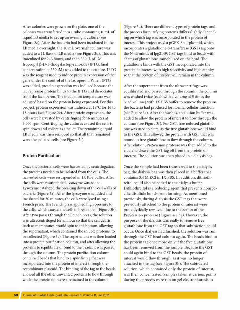
Protein Purification
Once the bacterial cells were harvested by centrifugation, the proteins needed to be isolated from the cells. The harvested cells were resuspended in 1X PBS buffer. After the cells were resuspended, lysozyme was added. Lysozyme catalyzed the breaking down of the cell walls of bacteria (Figure 3a). After the lysozyme was added and incubated for 30 minutes, the cells were lysed using a French press. The French press applied high pressure to the cells, which caused the cells to break open (Figure 3b). After two passes through the French press, the solution was ultracentrifuged for an hour so that the cell debris, such as membranes, would spin to the bottom, allowing the supernatant, which contained the soluble proteins, to be collected (Figure 3c). The supernatant was then loaded into a protein purification column, and after allowing the proteins to equilibrate or bind to the beads, it was passed through the column. The protein purification column contained beads that bind to a specific tag that was incorporated into the protein of interest through the recombinant plasmid. The binding of the tag to the beads allowed all the other unwanted proteins to flow through while the protein of interest remained in the column
(Figure 3d). There are different types of protein tags, and the process for purifying proteins differs slightly depend-ing on which tag was incorporated in the protein of interest. This project used a pGEX-6p-1 plasmid, which incorporates a glutathione-S-transferase (GST) tag onto the N-terminus of lpg2149. GST tags bind to beads with chains of glutathione immobilized on the bead. The glutathione binds with the GST incorporated into the protein of interest with high selectivity and high affinity so that the protein of interest will remain in the column.
After the supernatant from the ultracentrifuge was equilibrated and passed through the column, the column was washed twice (each with a volume of 5 times the bead volume) with 1X PBS buffer to remove the proteins the bacteria had produced for normal cellular function (see Figure 3e). After the washes, an elution buffer was added to allow the protein of interest to flow through the column (see Figure 3f). For GST, free reduced glutathi-one was used to elute, as the free glutathione would bind to the GST. This allowed the protein with GST that was bound to free glutathione to flow through the column. After elution, PreScission protease was then added to the eluate to cleave the GST tag off from the protein of interest. The solution was then placed in a dialysis bag.
Once the sample had been transferred to the dialysis bag, the dialysis bag was then placed in a buffer that contains 0.4 M KCl in 1X PBS. In addition, dithioth-reitol could also be added to the dialysis buffer. Dithiothreitol is a reducing agent that prevents nonspe-cific disulfide bonds from forming. As mentioned previously, during dialysis the GST tags that were previously attached to the protein of interest were proteolytically removed due to the action of the PreScission protease (Figure see 3g). However, the purpose of the dialysis was really to remove free glutathione from the GST tag so that subtraction could occur. Once dialysis had finished, the solution was run through the GST bead column again. The beads bind to the protein tag once more only if the free glutathione has been removed from the sample. Because the GST could again bind to the GST beads, the protein of interest would flow through, as it was no longer attached to the tag (see Figure 3h). The subtracted solution, which contained only the protein of interest, was then concentrated. Samples taken at various points during the process were run on gel electrophoresis to
A Nosy Neighbor 69
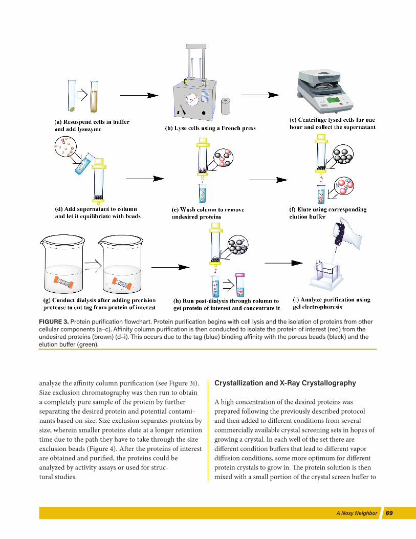
analyze the affinity column purification (see Figure 3i). Size exclusion chromatography was then run to obtain a completely pure sample of the protein by further separating the desired protein and potential contami-nants based on size. Size exclusion separates proteins by size, wherein smaller proteins elute at a longer retention time due to the path they have to take through the size exclusion beads (Figure 4). After the proteins of interest are obtained and purified, the proteins could be analyzed by activity assays or used for struc-tural studies.
Crystallization and X-Ray Crystallography
A high concentration of the desired proteins was prepared following the previously described protocol and then added to different conditions from several commercially available crystal screening sets in hopes of growing a crystal. In each well of the set there are different condition buffers that lead to different vapor diffusion conditions, some more optimum for different protein crystals to grow in. The protein solution is then mixed with a small portion of the crystal screen buffer to
FIGURE 3. Protein purification flowchart. Protein purification begins with cell lysis and the isolation of proteins from other cellular components (a–c). Affinity column purification is then conducted to isolate the protein of interest (red) from the undesired proteins (brown) (d–i). This occurs due to the tag (blue) binding affinity with the porous beads (black) and the elution buffer (green).
Journal of Purdue Undergraduate Research: Volume 11, Fall 202170
form a drop on a cover slip. Th e cover slip is then inverted over the well and sealed. Th e concentration in the drop equilibrates with the concentration via vapor diff usion, hopefully allowing a crystal to form (Figure 5a). When the proteins in the drop packs favorably, a crystal is formed. Th is crystal can then be looped and diff racted by X-ray diff raction (Figures 5b–d). In this process an X-ray beam is directed to the crystal, and by rotating the crystal, a complete sphere of data can be obtained. As the crystal is rotated, the beam will be diff racted at diff erent angles, thus forming a set of diff raction patterns. Th e set of diff raction patterns can be correlated to the distribution of electrons of individual atoms that make up the protein or protein complexes in the crystal. Th e diff raction pattern set is analyzed using computer soft ware, such as Phenix and CCP4, to give an outline of the electron density map (Figure 5e). Th e structure can then be solved by molecular replacement if a model is available or by stepwise amino acid fi tting to the density map if no model is present and atomic resolution data is available. Once the structure is solved, it can then be deposited to the Protein Data Bank, an
online database that contains biological structures that have been solved to date (Figure 5f).
DISCUSSION AND FUTURE DIRECTION
As shown in Figure 4, a substantial amount of lpg2149 was successfully produced following this optimized protocol. With this optimized protocol and lpg2149 construct identifi ed, we wanted to see how lpg2149 inhibits MavC and MvcA by obtaining a crystal structure. However, this was solved by another group before we obtained our structure (Wang et al., 2020). One of the future directions of this project is to study the mechanism of inhibition between lpg2149 and MavC. An inhibition assay, which involves the ubiquitination of UbE2N with the aid of MavC in the presence of diff ering concentra-tions of lpg2149, can be used to observe the extent of wild-type lpg2149 inhibition on MavC (Figure 6a). As shown in the inhibition assay, with increasing amounts of lpg2149 a reduced amount of UbE2N-Ub is observed. Th ese inhibition assays can be used to analyze key
FIGURE 4. Protein purification gel. The aff inity column purification gel lanes contain the protein ladder, the supernatant after French press lysis (load), the first flow-through, the wash, the eluate, the postdialysis, the subtracted sample, and the concentrated sample, respectively (a). Size exclusion is conducted, and a gel is run with fractions that appear only to contain the protein of interest (b).

A Nosy Neighbor 71
residues of lpg2149 by performing the assay with mutants of lpg2149. If specifi c residues of lpg2149 were mutated and then tested with the inhibition assay, the eff ects of these mutations in inhibiting MavC can be compared to those of wild-type lpg2149. If the ubiquitination of
UbE2N is impaired, it can be deduced that the specifi c mutation of lpg2149 is of low importance, since lpg2149 is still inhibiting MavC despite the mutation and vice versa. Additionally, the assay would test diff erent concen-trations of lpg2149 with each mutation to see how the
FIGURE 5. Crystallization and X-ray diff raction. Crystal screening is conducted to obtain a crystal (a–b). The crystal’s structure is analyzed and solved using X-ray diff raction and online programs (c–f).
FIGURE 6. Inhibition assays and residue sites. The results of an inhibition assay with an increasing concentration of lpg2149 are shown in the electrophoresis gel (a). Lpg2149 is shown with residue sites highlighted in violet that could be potentially mutated to test their importance in the binding of lpg2149 with MavC (b).
Journal of Purdue Undergraduate Research: Volume 11, Fall 202172
concentration of mutated lpg2149 aff ected the reactions. Th e diff erent concentrations of lpg2149 were 0μM, 0.1μM, 0.25μM, 0.5μM, 1μM, 2μM and 5μM. As a future direction, mutants that are presented on the MavC interface with lpg2149 (highlighted in violet, Figure 6b) can be the fi rst ones to be tested as residues that may play an important role in the binding of lpg2149 to MavC. Additionally, biolayer interferometry can be used to study the binding affi nity by measuring the ratio of association to dissociation of lpg2149 with its binding partner. Th ese analysis methods studying the interactions of lpg2149 with other L. pneumophila eff ectors would be benefi cial in improving our understanding of lpg2149 function and L. pneumophila pathogenesis.
ACKNOWLEDGMENTS
We thank the Offi ce of Undergraduate Research for providing funding support to Ashley Holahan for the research stipend. We also thank the National Institute of General Medical Sciences predoctoral fellowship (T32 GM132024) for the funding to Sebastian Kenny, allowing further mentorship for Holahan. Finally, we thank the members of the Das Lab, who have made research something to look forward to each week.
All fi gures were generated by Holahan using ChemDraw, Microsoft Word, and PyMOL.
REFERENCES
Clague, M. J., Urbé, S., & Komander, D. (2019). Breaking the chains: Deubiquitylating enzyme specifi city begets function.
Nature Reviews Molecular Cell Biology, 20(6), 338–352. https://doi.org/10.1038/s41580-019-0099-1
Gan, N., Nakayasu, E. S., Hollenbeck, P. J., & Luo, Z. Q. (2019). Legionella pneumophila inhibits immune signalling via MavC-mediated transglutaminase-induced ubiquitination of UBE2N. Nature Microbiology, 4(1), 134–143. https://doi.org/10.1038/s41564-018-0282-8
Komander, D., & Rape, M. (2012). Th e ubiquitin code. Annual Review of Biochemistry, 81, 203–229. https://doi.org/10.1146/annurev-biochem-060310-170328
Puvar, K., Iyer, S., Fu, J., Kenny, S., Negrón Terón, K. I., Luo, Z. Q., Brzovic, P. S., Klevit, R. E., & Das, C. (2020). Legionella eff ector MavC targets the Ube2N~Ub conjugate for nonca-nonical ubiquitination. Nature Communications, 11(1). https://doi.org/10.1038/s41467-020-16211-x
Sarkar, G., & Sommer, S. S. (1990). Th e “megaprimer” method of site-directed mutagenesis. BioTechniques, 8(4), 404–407.
Sato, Y., Yoshikawa, A., Yamashita, M., Yamagata, A., & Fukai, S. (2009). Structural basis for specifi c recognition of Lys 63–linked polyubiquitin chains by NZF domains of TAB2 and TAB3. EMBO Journal, 28(24), 3903–3909. https://doi.org/10.1038/emboj.2009.345
Valleau, D., Quaile, A. T., Cui, H., Xu, X., Evdokimova, E., Chang, C., Cuff , M. E., Urbanus, M. L., Houliston, S., Arrowsmith, C. H., Ensminger, A. W., & Savchenko, A. (2018). Discovery of ubiquitin deamidases in the pathogenic arsenal of Legionella pneumophila. Cell Reports, 23(2), 568–583. https://doi.org/10.1016/j.celrep.2018.03.060
Wang, Y., Zhan, Q., Wang, X., Li, P., Liu, S., Gao, G., & Gao, P. (2020). Insights into catalysis and regulation of non-canoni-cal ubiquitination and deubiquitination by bacterial deami-dase eff ectors. Nature Communications, 11(1), 1–13. https://doi.org/10.1038/s41467-020-16587-w


![Mcq in Pharma Cog Nosy and Phytochemistry[1]](https://static.fdocuments.in/doc/165x107/543f01c4b1af9f780b8b4cd0/mcq-in-pharma-cog-nosy-and-phytochemistry1.jpg)