A Nonenzymatic and Automated Closed-Cycle Process for the ... · Valentina Coccè ,1 Anna Brini...
11
Research Article A Nonenzymatic and Automated Closed-Cycle Process for the Isolation of Mesenchymal Stromal Cells in Drug Delivery Applications Valentina Coccè , 1 Anna Brini , 1,2 Aldo Bruno Giannì , 1,3 Valeria Sordi , 4 Angiola Berenzi, 5 Giulio Alessandri, 6 Carlo Tremolada , 7 Silvia Versari, 8 Antonio Bosetto, 9 and Augusto Pessina 1 1 CRC StaMeTec, Department of Biomedical, Surgical and Dental Sciences, University of Milan, Milan, Italy 2 I.R.C.C.S Galeazzi Orthopedic Institute, Milan, Italy 3 Maxillofacial and Dental Unit, Fondazione IRCCS Cà Granda Ospedale Maggiore Policlinico, Via Commenda 10, 20122 Milan, Italy 4 Diabetes Research Institute, IRCCS San Raffaele Scientific Institute, Milan, Italy 5 University Research Center “Integrated Models for Prevention and Protection in Environmental and Occupational Health” (MISTRAL), Brescia, Italy 6 Cellular Neurobiology Laboratory, Department of Cerebrovascular Diseases, Fondazione IRCCS Neurological Institute C. Besta, Milan, Italy 7 Image Institute, Milan, Italy 8 Lipogems International Spa, Milan, Italy 9 Hydra srl, Mirandola, Modena, Italy Correspondence should be addressed to Augusto Pessina; [email protected] Received 12 October 2017; Accepted 25 December 2017; Published 20 February 2018 Academic Editor: Massimo Petruzzi Copyright © 2018 Valentina Coccè et al. This is an open access article distributed under the Creative Commons Attribution License, which permits unrestricted use, distribution, and reproduction in any medium, provided the original work is properly cited. The adipose tissue is a good source of mesenchymal stromal cells that requires minimally invasive isolation procedures. To ensure reproducibility, efficacy, and safety for clinical uses, these procedures have to be in compliant with good manufacturing practices. Techniques for harvesting and processing human adipose tissue have rapidly evolved in the last years, and Lipogems® represents an innovative approach to obtain microfragmented adipose tissue in a short time, without expansion and/or enzymatic treatment. The aim of this study was to assess the presence of mesenchymal stromal cells in the drain bag of the device by using a prototype Lipogems processor to wash the lipoaspirate in standardized condition. We found that, besides oil and blood residues, the drain bag contained single isolated cells easy to expand and with the typical characteristics of mesenchymal stromal cells that can be loaded with paclitaxel to use for drug-delivery application. Our findings suggest the possibility to replace the drain bag with a “cell culture chamber” obtaining a new integrated device that, without enzymatic treatment, can isolate and expand mesenchymal stromal cells in one step with high good manufacturing practices compliance. This system could be used to obtain mesenchymal stromal cells for regenerative purposes and for drug delivery. 1. Introduction Mesenchymal stromal cells (MSCs) can be easily isolated from several human organs and tissues and, because of their self-renewing capacity and multipotent differentiation properties, they are important tools for treating immune disorder and for tissue repair. In particular, adipose- derived MSCs (ASCs) can be harvested in high amounts by minimally invasive procedures with good viability, dif- ferentiating potential, and paracrine activity [1–3]. These Hindawi Stem Cells International Volume 2018, Article ID 4098140, 10 pages https://doi.org/10.1155/2018/4098140
Transcript of A Nonenzymatic and Automated Closed-Cycle Process for the ... · Valentina Coccè ,1 Anna Brini...

Research ArticleA Nonenzymatic and Automated Closed-Cycle Process for theIsolation of Mesenchymal Stromal Cells in DrugDelivery Applications
Valentina Coccè ,1 Anna Brini ,1,2 Aldo Bruno Giannì ,1,3 Valeria Sordi ,4
Angiola Berenzi,5 Giulio Alessandri,6 Carlo Tremolada ,7 Silvia Versari,8 Antonio Bosetto,9
and Augusto Pessina 1
1CRC StaMeTec, Department of Biomedical, Surgical and Dental Sciences, University of Milan, Milan, Italy2I.R.C.C.S Galeazzi Orthopedic Institute, Milan, Italy3Maxillofacial and Dental Unit, Fondazione IRCCS Cà Granda Ospedale Maggiore Policlinico, Via Commenda 10, 20122 Milan,Italy4Diabetes Research Institute, IRCCS San Raffaele Scientific Institute, Milan, Italy5University Research Center “Integrated Models for Prevention and Protection in Environmental and Occupational Health”(MISTRAL), Brescia, Italy6Cellular Neurobiology Laboratory, Department of Cerebrovascular Diseases, Fondazione IRCCS Neurological Institute C. Besta,Milan, Italy7Image Institute, Milan, Italy8Lipogems International Spa, Milan, Italy9Hydra srl, Mirandola, Modena, Italy
Correspondence should be addressed to Augusto Pessina; [email protected]
Received 12 October 2017; Accepted 25 December 2017; Published 20 February 2018
Academic Editor: Massimo Petruzzi
Copyright © 2018 Valentina Coccè et al. This is an open access article distributed under the Creative Commons AttributionLicense, which permits unrestricted use, distribution, and reproduction in any medium, provided the original work isproperly cited.
The adipose tissue is a good source of mesenchymal stromal cells that requires minimally invasive isolation procedures. To ensurereproducibility, efficacy, and safety for clinical uses, these procedures have to be in compliant with good manufacturing practices.Techniques for harvesting and processing human adipose tissue have rapidly evolved in the last years, and Lipogems® represents aninnovative approach to obtain microfragmented adipose tissue in a short time, without expansion and/or enzymatic treatment. Theaim of this study was to assess the presence of mesenchymal stromal cells in the drain bag of the device by using a prototypeLipogems processor to wash the lipoaspirate in standardized condition. We found that, besides oil and blood residues, the drainbag contained single isolated cells easy to expand and with the typical characteristics of mesenchymal stromal cells that can beloaded with paclitaxel to use for drug-delivery application. Our findings suggest the possibility to replace the drain bag with a“cell culture chamber” obtaining a new integrated device that, without enzymatic treatment, can isolate and expandmesenchymal stromal cells in one step with high good manufacturing practices compliance. This system could be used to obtainmesenchymal stromal cells for regenerative purposes and for drug delivery.
1. Introduction
Mesenchymal stromal cells (MSCs) can be easily isolatedfrom several human organs and tissues and, because oftheir self-renewing capacity and multipotent differentiation
properties, they are important tools for treating immunedisorder and for tissue repair. In particular, adipose-derived MSCs (ASCs) can be harvested in high amountsby minimally invasive procedures with good viability, dif-ferentiating potential, and paracrine activity [1–3]. These
HindawiStem Cells InternationalVolume 2018, Article ID 4098140, 10 pageshttps://doi.org/10.1155/2018/4098140

cells can be also used as drug carrier (as already demon-strated for MSCs derived from bone marrow) because,when primed with high doses of the chemotherapeuticdrug paclitaxel (PTX), they are capable to uptake the drugand release it in big amounts inhibiting the in vitro prolif-eration of different tumour cell lines [4, 5]. Their defini-tion as advanced-therapy medicinal products (ATMPs)according to the European Medicines Agency (EMA) andthe US Food and Drug Administration requirements fortheir production and use implies the application of pro-duction processes that should be in accordance with goodmanufacturing practices (GMPs). To increase safety andreproducibility and to implement the many clinical trialsusing ASCs, several methods based on reducing themanipulation of human tissue-based products have beensuggested, in particular by using nonenzymatic treatments[6, 7]. Among the many, an innovative closed and sterilesystem (named Lipogems), designed to harvest, process,and transfer a refined and not-expanded adipose tissuecharacterized by a great regenerative potential and optimalhandling, has been developed. By the aid of this technol-ogy, and without the addition of enzymes or any otheradditives, fat tissue is microfragmented in a completelyclose liquid environment and washed from proinflamma-tory oil and blood residues [8]. This closed and steriledevice reduces the size of the adipose tissue clusters bymeans of two filters [9]. A drain bag, connected to thesecond filter of the device, collects the waste fluid contain-ing red blood cells and oil residues. We used a prototypeLipogems processor (PLG-P) to wash and process thelipoaspirate in a standardized condition and we found
that, besides oil and blood residues, the drain bag con-tained single isolated cells easy to expand and with thetypical characteristics of ASCs. These cells can be alsoloaded with paclitaxel to provide a cell-mediated drug-delivering tool.
2. Materials and Methods
2.1. Ethics Statements. Samples from adult donors werecollected after signed informed consent of no objection forthe use for research of surgical tissues (otherwise destinedfor destruction) in accordance with the Declaration ofHelsinki. The approval for their use was obtained fromthe Institutional Ethical Committee of Milan University(n.59/15, C.E.UNIMI, 09.1115).
2.2. Prototype Lipogems Processor (PLG-P). The prototypeprocessor (Figure 1(a)) equipped with the Lipogems device(Figure 1(b)) is able to guarantee digitally controlled move-ments according to these main parameters: oscillation ampli-tude and frequency and pitch movements and saline washingflux. 25ml of lipoaspirate was loaded in the Lipogems deviceand washed according to a standardized procedure: processflow 120ml/min, oscillation frequency 2Hz, pitch frequency0.3Hz, pitch angle 30°, and pitch axis 90° vertical (exitbottom). In a first preliminary experiment, the tissue wasprocessed for 5 minutes and cells were collected one step into600ml of saline. In a second experiment, the collection wasfractioned and performed at 1 (120ml), 2.5 (180ml), and 5minutes (300ml).
(a)
Lipoaspirate
Drain bag
F1 F2
Lipogems device
Pellet seeded in flask
(b)
Figure 1: DB-MSC isolation and expansion. (a) Prototype Lipogems processor (PLG-P) mounting a Lipogems device. In this closed system,the lipoaspirate is microfragmented by using mild mechanical forces without the addition of collagenase or other enzymes/additives. (b)Schematic representation of Lipogems device. The lipoaspirate (blue circle) is subjected to a cluster reduction by pushing the fat into thedevice through the first filter (F1). LGP-P shaking transmits a mechanical force to the stainless steel marbles contained in the device thatresults into a temporary fat emulsion washed by a flux of buffer. This allows the washing buffer to cross a second filter (F2) andaccumulate into the drain bag (red circle) together with oil, red blood cells, and mesenchymal stromal cells (ASCs). The pellet obtainedafter centrifugation was seeded in 25 cm2
flask in StemMACS medium (Miltenyi Biotec, USA) (see Materials and Methods for details).
2 Stem Cells International

2.3. ASC Isolation from Drain Bag (DB-ASCs). 100ml of eachsample was centrifuged at 2500×g for 15 minutes. Pellet wastreated twice with 0.85% ammonium chloride (5 minutes at+4°C) to partially remove red cells. Then, after three washeswith PBS 1x through centrifugation (800×g, 10 minutes),pellet was plated on 25cm2
flask (Corning, USA) with 5mlof Stem MACS MSC Expansion Medium (Miltenyi Biotec,Germany) and incubated at 37°C, 5% CO2. The primaryculture was expanded with 1 : 2 passages until passage num-ber (pn) 6. At pn 2, the population doubling time (PDT)was calculated as previously described [10].
The clonogenicity of the ASCs was evaluated as colony-forming efficiency (CFE) in multiwell plates (SPL LifeSciences, Korea) by serial dilution (from 50 cells/well to 1cell/well) in DMEM LG media with 10% fetal bovine serum(EuroClone, UK). After 10 days, cell colonies fixed andlabeled with Crystal Violet (0.5%, Fluka, Switzerland) werecounted at the microscope (a colony formed by at least 25cells) and the CFE as the following: CFE=average of coloniesformed× 100/number of seeded cells.
2.4. Immunohistochemical Analysis of DB-MSCs. Mesenchy-mal cell monolayer (pn 2) was detached with trypsin-EDTA(EuroClone, UK) and 1.7∗106 cells were placed in a 10mlglass conical tube in 1x PBS and centrifuged at 800×g for10 minutes. Pellet was treated for immunohistochemicalcharacterization as previously reported [11]. Briefly, the cen-trifuged pellet was treated 20 minutes at room temperaturewith a solution of 75% methanol, 20% chloroform, and 5%glacial acetic acid. The pellet compacted in a solid mass wasplaced in an immunohistochemical cassette and immersedin a 10% buffered formaldehyde solution and, after dehydra-tion in ethanol and paraffin fixation, the pellet was cut in sec-tions of 4-5μm that were stored at −20°C. For phenotypiccharacterization, monoclonal or polyclonal antibodies wereused using the streptavidin-biotin method with diaminoben-zidine as chromogen. The following antibodies were used:antismooth muscle actin (SMA), anti-CD14, anti-CD105,and anti-neural/glial antigen 2 (NG2) (Santa Cruz Biotech-nology, USA); anti-CD44 (Monosan, Holland); antivascularendothelial growth factor (VEGF), anti-CD90 (Dako, Italy);anti-CD45, anti-CD146, anti-CD31, and anti-CD34 (LeicaBiosystems, Germany).
The expression of CD73 (CD73-PE Becton Dickinson,USA) was evaluated by flow cytometry (FacsVantage SE,Becton Dickinson, USA). Cells were washed twice inPBS, diluted to a final concentration of 106 cells/ml, andincubated with the dark antibody at +4°C for 30 minutes.The cells were then washed with PBS and analysed bycytofluorometry by acquisition of 10,000 events per sam-ple. The results were analysed with CellQuest Pro software(Becton Dickinson, USA).
2.5. DB-ASC Osteo-/Adipo-Differentiation Capacity. DB-ASCs at pn 2 have been evaluated for their osteogenic andadipogenic differentiation using a standardized procedure[12]. As positive control, MSCs obtained from human bonemarrow (BM-MSCs) were isolated and expanded in our lab-oratory as previously reported [5]. As negative control, MSCs
were cultured without the addition of supplements. The dif-ferentiation was performed by plating cells in 35mm Petri(Nunc, Germany) at a density of 50 cells/cm2 in 1ml ofSTEM MACS MSC Expansion Medium (Miltenyi Biotec,Germany). After 72 hours of incubation, the cell mediumwas removed and replaced with a specific medium forcell differentiation. To induce osteogenic differentiation[13], cells were incubated for 14 days in DMEM mediumLG+20% fetal bovine serum in the presence of the following:desametasone 10 nM, glycerol 2–10mM, and phosphate andascorbic acid 300nM (all reagents, Sigma-Aldrich, USA).The differentiation medium was replaced every 3-4 days withfresh medium. Cell monolayer was fixed with cold methanolfor 5 minutes at −20°C, then alkaline phosphatase (ALP) wasevaluated by SIGMA FAST BCIP/NTB substrate for ALP(Sigma-Aldrich, USA). The colorimetric reaction wasdetected by optical microscope. The presence of osteo-blasts at the mature stage was evaluated by staining withAlizarin Red S (Sigma-Aldrich, USA). To induce adipocy-tic differentiation, cells were cultured in DMEM LG+20%fetal bovine serum in the presence of indomethacin200μM (Alexis Biochemicals, USA), isobutylmethyl-xanthine 0.5mM (AppliChem, Germany), dexamethasone1μM, hydrocortisone 1μM insulin, and 10μg/ml (all fromSigma-Aldrich, USA). After 10 days of incubation, thecells were fixed with 10% buffered formalin (Sigma-Aldrich, USA) for 30 minutes at room temperature andthe adipocyte vacuoles present in the cell cytoplasm wereevaluated by using red lipophilic Oil Red O (Sigma-Aldrich, USA).
2.6. Ability of DB-ASCs to Uptake and Release Paclitaxel. Theability to incorporate and release drugs from DB-ASCshas been assessed with a procedure already applied onMSCs obtained from bone marrow, adipose tissue,human gingival tissue, human fibroblasts, and also bloodcells [4, 5, 10, 14–16]. The “uptake-release”method was per-formed using paclitaxel (PTX) that is an anticancer drug withalso antiangiogenic activity. Subconfluent ASC cultures(15,000 cells/cm2) were exposed to 2000 ng/ml of PTX(Fresenius Kabi, Italy) dissolved in culture media, andafter 24 hours of incubation (37°C, 5% CO2), the cellmonolayer was washed with PBS and the cells detached,washed by centrifugation in PBS, and subcultured in a25 cm2
flask with fresh culture media. After 48 hours,the conditioned medium (CM) was collected and its anti-tumour activity was evaluated in vitro. The CM fromuntreated cells was used as a negative control.
2.7. Cytokines in DB-ASC Secretome. The conditionedmedium of DB-ASCs (from untreated or treated PTX cells)was collected, and the cytokine content was evaluated using“multiplex bead-based xMAP technology” (Bio-Plex HumanCytokine 27-Plex Panel and Bio-Plex Human Group II Cyto-kine 21-Plex Panel, Bio-Rad Laboratories).
2.8. In Vitro Antiproliferative Assay on CFPAC-1 of CMfrom PTX-Loaded MSCs. The effect of pure paclitaxeland CM from PTX-treated ASCs (CM/PTX) against
3Stem Cells International

tumour cell proliferation has been studied in 96-multiwellplates (Sarstedt, Germany) by using as target pancreaticadenocarcinoma cells (CFPAC-1) [17]. Briefly, 1 : 2 serialdilutions of pure drug or ASC-CM were prepared in100μl of culture medium/well and then to each well wereadded 1000 tumour cells. After 7 days of culture at 37°Cand 5% CO2, cell growth was evaluated by MTT assay(3-(4,5-dimethyl-2-thiazolyl)-2,5-diphenyl-2-H-tetrazolium)as previously described [5, 18]. The inhibitory concentra-tions (IC50 and IC90) were determined according to theReed and Muench formula [19]. The antitumor activityof PTXCMwas compared to that of pure PTX and expressedas PTX equivalent concentration: PEC (ng/ml) = IC50PTX× 100/V50 (μl/well); IC50 PTX= the concentration ofpure PTX producing 50% of inhibition; V50 is the volumePTX-MSCs-CM able to inhibit cell proliferation by 50%.The PEC referred to a single primed ASC was calculated asratio between the total amount of PEC (PEC (ng/ml)×CMvolume (ml)) and the number of cells seeded: PE release(pg/cell) = PEC (ng tot)∗1000/number of cells seeded.Uptake release experiments were performed in triplicate.
2.9. Statistical Analysis. Experiments were performed usinglipoaspirate from three donors and repeated in triplicate.The reported data are expressed as mean± standard devia-tion. If necessary, appropriate statistical tests have beenperformed using GraphPad Software (GraphPad Inc., SanDiego, CA, USA). A p value≤ 0.05 was considered statisti-cally significant. The linearity of response and the correla-tion were studied using regression analysis, by Excel 2007software (Microsoft Inc.).
3. Results
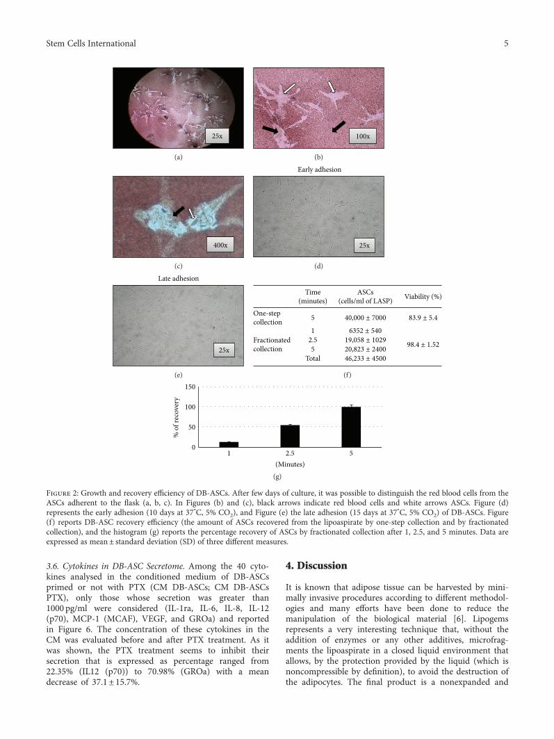
3.1. DB-ASC Isolation and Expansion. The waste mediumrecovered from the drain bag contained high amounts ofred cells that were removed, only partially, by the ammo-nium chloride treatment. However, after about 7 days, theadhered ASCs could be clearly identified, by transparency,surrounded by a compact rug of red cells (Figures 2(a)–2(c)). However, the adherent cells (washed every 3 days)improve their growth and after 10 days (early adhesion)or 15 days (late adhesion) of incubation, the cell mono-layer (Figures 2(d) and 2(e)) was detached and primaryculture (pn 0) was quantified for the number of cells.The MSC primary culture was subsequently expandedby 1 : 2 serial passage until passage number 6. MSC growthwas evaluated at pn 2 by evaluating the population dou-bling Time (PDT) and the colony-forming efficiency (CFE)that resulted, respectively, 55.2± 2.3 hours (PDT) and20.8%± 10.4% (CFE).
3.2. DB-ASC Recovery Efficiency. To evaluate the amount ofASCs recovered from the lipoaspirate, the washing fluidwas collected from the drain bag by two different ways. Ina first experiments (one-step collection), 600ml of salinewas collected after 5 minutes of washing; in a secondexperiment (fractionated collection), the collection wasperformed at 1 minute (120ml), 2.5 (180ml), and 5 minutes
(300ml) (Box 2F). In the one-step collection, the totalrecovery resulted 40,000± 7000 ASCs/ml of lipoaspiratewith a cell viability of 83.9± 5.4% as evaluated by trypanblue assay. By fractionated collection, the final recovery at5 minutes was 46,233± 4500 ASCs/ml with a viability of98.4± 1.52. As shown, ASCs accumulate into the drain bagaccording to a kinetic that collects 13.7% of cells at 1 min-ute, 54.9% at 2.5 minutes and, at the end of the process,the recovery (100%) is similar to what was observed in theone-step procedure but with a higher percentage of cellviability (Figure 2(g)).
3.3. Immunohistochemical Analysis of DB-ASCs. The immu-nohistochemistry analysis performed on ASCs showed thehigh positivity for CD44, CD90, and CD105 (>90%) andthe negativity for CD45, CD34, CD31, and CD14 hematopoi-etic markers (Figure 3(a)). The positivity for CD73 was eval-uated by FACS and expressed by the dot-blots (Figure 3(c)).Significant was also the expression of SMA (cytoplasmic andmembrane) and VEGF (cytoplasmic). Only a small percent-age of cells are positive for CD146, whereas the presence ofreactivity for NG2 seems to be mainly expressed at thenuclear level (Figure 3(a)).
3.4. DB-ASC Osteo-/Adipo-Differentiation Capability. Thedifferentiation capacity towards osteogenic and adipogeniclineages is reported in Figure 4.
A remarkable positivity of ALP (Figure 4(d)) confirmsthe presence of a significant enzymatic activity involved inthe mineralization of the bone matrix. The mature stage ofosteoblasts is confirmed by the presence of calcium depositsforming complex orange-colored bifurcations, evidenced bythe coloration with Alizarin Red S (Figure 4(b)). As shownin Figure 4(f), DB-ASCs are also able to differentiate in adi-pocytes as evidenced by the red intracellular inclusions. Thenegative controls (DB-ASCs cultured without the additionof supplements) do not give any evidence of differentiation(Figures 4(a), 4(c), and 4(e)).
3.5. Ability of DB-ASCs to Uptake and Release Paclitaxel. Theconditioned media from DB-ASCs, both primed with PTX(CM/PTX) or not (CM CTRL), were tested on the standardlaboratory tumour cell line CFPAC-1 (Figure 5). As expected,the CM CTRL does not inhibit neoplastic growth, whichremains stable to 80–100% of proliferation (Figure 5(a)).On the contrary, CM/PTX affected CFPAC-1 cell growthaccording to a dose-dependent antiproliferative effect that,even if less effective, reflects the inhibition produced by freePTX (from 0.39 to 50 ng/ml). The inhibitory kineticsanalysed by linear regression showed high coefficients ofdetermination (R2) ranging from 0.71 to 0.88 (Figure 5(b)).The biological dosage of the CM/PTX based on the standarddose-response regression of the pure drugs enabled the esti-mation of an equivalent paclitaxel concentration (PEC) of3.94± 0.32 ng/ml. The amount of PTX released by a singlecell (CPR), expressed as pg/cell, was 0.14± 0.01 pg/cell (box5C). The drug concentrations released by 106 DB-ASCs/PTX were 140ng of PTX that correspond to values of about10 times the IC50 value of the free drug (1.48± 0.35 ng/ml).
4 Stem Cells International
3.6. Cytokines in DB-ASC Secretome. Among the 40 cyto-kines analysed in the conditioned medium of DB-ASCsprimed or not with PTX (CM DB-ASCs; CM DB-ASCsPTX), only those whose secretion was greater than1000 pg/ml were considered (IL-1ra, IL-6, IL-8, IL-12(p70), MCP-1 (MCAF), VEGF, and GROa) and reportedin Figure 6. The concentration of these cytokines in theCM was evaluated before and after PTX treatment. As itwas shown, the PTX treatment seems to inhibit theirsecretion that is expressed as percentage ranged from22.35% (IL12 (p70)) to 70.98% (GROa) with a meandecrease of 37.1± 15.7%.
4. Discussion
It is known that adipose tissue can be harvested by mini-mally invasive procedures according to different methodol-ogies and many efforts have been done to reduce themanipulation of the biological material [6]. Lipogemsrepresents a very interesting technique that, without theaddition of enzymes or any other additives, microfrag-ments the lipoaspirate in a closed liquid environment thatallows, by the protection provided by the liquid (which isnoncompressible by definition), to avoid the destruction ofthe adipocytes. The final product is a nonexpanded and
(a) (b)
(c) (d)
(e) (f)
(g)
Figure 2: Growth and recovery efficiency of DB-ASCs. After few days of culture, it was possible to distinguish the red blood cells from theASCs adherent to the flask (a, b, c). In Figures (b) and (c), black arrows indicate red blood cells and white arrows ASCs. Figure (d)represents the early adhesion (10 days at 37°C, 5% CO2), and Figure (e) the late adhesion (15 days at 37°C, 5% CO2) of DB-ASCs. Figure(f) reports DB-ASC recovery efficiency (the amount of ASCs recovered from the lipoaspirate by one-step collection and by fractionatedcollection), and the histogram (g) reports the percentage recovery of ASCs by fractionated collection after 1, 2.5, and 5 minutes. Data areexpressed as mean± standard deviation (SD) of three different measures.
5Stem Cells International

(a)
(b) (c)
Figure 3: Immunohistochemical analysis of DB-ASCs. (a) Immunohistochemical analysis for CD14, CD31, CD34, CD45, CD90, CD105,CD44, CD146, NG2, SMA, and VEGF (all figures are 100x magnification). (b, c) FACS analysis on CD73: (b) = negative CTRL and(c) =CD73 expression.
6 Stem Cells International

minimally manipulated adipose tissue product suitable forclinical uses because it is in compliant with goodmanufactur-ing practices (GMPs). As recently reviewed, Lipogems hasbeen used in many surgical fields such as orthopaedic, recon-structive, and plastic surgery and oncology [20]. In order toobtain more standardized microfragmenting conditions, aprototype apparatus (PLG-P) has been designed that over-coming the manual operations provides a procedure withprogrammable parameters (angle and frequency of shakingand flux of washing). We mounted a Lipogems device intothe PLG-P equipped with a drain bag allowing thecollection of high volumes of the washing fluid object ofour investigations (Figure 1). Our study demonstrated thatthe “waste” material contained in the drain bag is surpris-ingly a rich source of ASCs. In fact, during the longmicrofragmentation process, the continuous shaking andwashing flux allows to detach mechanically many ASCsthat accumulate into the drain bag. Of course, the signifi-cant amount of single isolated ASCs is diluted into the bagdue to the extensive washing volume of buffer used. How-ever, these cells can be easily collected by centrifugationand if cultured, in the presence of contaminant red cells,
are able to adhere with a significant CFE and can be easilyexpanded with a good PDT (Figure 2). These cells are pos-itive for the expression of CD44, CD73, CD90, CD105,SMA, VEGF, and NG2 and negative for CD14, CD31,CD34, and CD45 and showed osteo-/adipogenic differenti-ation capability (Figures 3 and 4). Although it is knownthat the expression of these markers may depend onculture conditions, the pattern clearly confirm the mesenchy-mal stromal cell type of DB-ASCs. Besides the positivity forthe typical MSC markers CD90, CD105, and CD73[21],DB-ASCs are also positive for NG2 (about 50%) and SMA(90%) that are markers commonly expressed by pericytes indifferent amount depending from the type of vessel fromwhich they originated (capillaries, venules, or arterioles)[22, 23]. Lipoaspirate is very rich in microvessels, and,therefore, a significant amount of ASC progenitors can beobtained with our procedure. Very few cells were positivefor CD146, probably because the expression of this markeris reduced in the ASC population washed from lipoaspirate,in agreement with our previous study on MSCs in Lipogemssuggesting that CD146 could stain mostly cells of endothelialorigin rather than pericytes [24].
CTRL− CTRL−
CTRL−
Oil Red S staining
Alizarin Red S staining Alkaline phosphatase staining
(a) (b) (c)
(e) (f)
(d)
Figure 4: DB-ASC osteo-/adipo-differentiation. Osteogenic differentiation evaluated in Petri dish by Alizarin Red S staining (left) and byphosphatase alkaline staining (right). (a, c) DB-ASCs grown in control medium without supplements (negative control, CTRL−); (b, d)positive culture (200x magnification). Adipogenic differentiation evaluated by Oil Red staining (presence of red cytoplasmic inclusions;200x magnification). (e) Negative control (CTRL−) (200x magnification). (f) Positive culture (200x magnification).
7Stem Cells International

The possibility to estimate the cell content and, in partic-ular, the number of ASCs/ml of lipoaspirate give importanceto this technique. Our study suggests that washing the
lipoaspirate with the PLG-P allows to collect significantamounts of single isolated ASCs with a recovery efficiency,in terms of ASCs per ml of lipoaspirate processed, of
(a) (b)
IC50PTX (ng/ml) 1.48 ± 0.35
PEC (ng/ml) 3.94 ± 0.32
CPR (pg/cell) 0.14 ± 0.01
(c)
Figure 5: Ability of DB-ASCs to uptake and release paclitaxel. The graph reports the inhibitory activity exerted on pancreatic carcinomaCFPAC-1 cells by (a) the conditioned medium (CM) of nontreated cells (CM CTRL) and (b) the CM of DB-ASCs loaded with PTX (CM/PTX). The activity is compared to the inhibition exerted by pure PTX. The activity is expressed as percentage of CFPAC-1 proliferationreferred to that of untreated cells (100% proliferation). The regression and the correlation coefficient (R2) of the dose-response kinetics arealso reported. The box (c) reports the IC50 value referred to PTX (ng/ml), the paclitaxel equivalent concentration (PEC), and the singlecell paclitaxel release (CPR). See details in Materials and Methods. Values are expressed as mean± standard deviation (SD) of threeindependent experiments.
Cytokines(pg/ml) CMDB‐ASCs CM DB‐ASCsPTX
IL‐1raIL‐6 10278.09IL‐8 5832.56IL‐12 (p70)MCP‐1 (MCAF) 3433.47VEGF 4423.49GROaSCGF‐b 6306.34
1077.87 736.107242.883068.68869.601119.86
2309.903333.90863.372975.18
3906.46
(a) (b)
Figure 6: Cytokines in DB-ASC secretome. (a) The table shows the amounts of cytokines (expressed in pg/ml) secreted in the conditionedmedium of DB-ASCs before (CM DB-ASCs) and after treatment with PTX (CM DB-ASCs PTX). (b) The histogram reports the decreaseof cytokines secretion by PTX-treated DB-ASCs expressed as percentage of the amount produced by the untreated DB-ASCs.
8 Stem Cells International

46,233± 5500 ASCs/ml that is higher compared to what wasreported by other authors [25].
A further important observation is that the expandedASCs were able to uptake and then release paclitaxel(PTX). Although the PTX treatment modified the amountof some cytokines produced by DB-ASCs (Figure 6), thisaspect is not relevant because cells loaded with the drug areable to release PTX in an active form as shown by thein vitro anticancer activity of the conditioned medium(Figure 5). The uptake-release ability of paclitaxel by DB-MSCs is in line with our previous observations on MSCsfrom different sources (bone marrow, adipose tissue, humangingival tissue, human fibroblasts, and also blood cells) andconfirms that these cells can be considered an important toolfor cell-mediated drug delivery [4, 5, 10, 14–16].
5. Conclusions
In conclusion, this system minimally manipulate the adiposetissue thus bypassing the complex requirements of GMPguidelines, with a dramatic reduction of the costs for cell-based therapies on human patients. From a biotechnologicalpoint of view, our findings suggest the possible developmentof a new integrated device that operating without enzymesallows the isolation, expansion, and drug loading of ASCsin one single step. If equipped with selective filters and ifthe drain bag will be replaced by a “cell culture chamber,”the PLG-P+Lipogems device configures a “standardizedautomated collection system” to isolate mesenchymal cellswith a “minimal manipulation” compliant with the GMPstandards [26, 27]. A system as such has the potential toobtain MSCs not only for regenerative medicine purposesbut also for cell-mediated drug delivery.
Conflicts of Interest
Carlo Tremolada is the president and founder of LipogemsInternational SpA. Silvia Versari is employed in LipogemsInternational SpA. All other authors decline any conflict ofinterests.
Acknowledgments
The authors would like to thank Loredana Cavicchini(University of Milan, technician) and Eng. Paolo Pirazzoli(Hydra srl) for the technical assistance.
References
[1] A. I. Caplan, “Adult mesenchymal stem cells for tissue engi-neering versus regenerative medicine,” Journal of CellularPhysiology, vol. 213, no. 2, pp. 341–347, 2007.
[2] A. I. Caplan, “Why are MSCs therapeutic? New data: newinsight,” The Journal of Pathology, vol. 217, no. 2, pp. 318–324, 2009.
[3] A. Schäffler and C. Büchler, “Concise review: adipose tissue-derived stromal cells—basic and clinical implications for novelcell-based therapies,” Stem Cells, vol. 25, no. 4, pp. 818–827,2007.
[4] A. Bonomi, V. Coccè, L. Cavicchini et al., “Adipose tissue-derived stromal cells primed in vitro with paclitaxel acquireanti-tumor activity,” International Journal of Immunopathol-ogy and Pharmacology, vol. 26, no. Supplement 1, pp. 33–41,2013.
[5] A. Pessina, A. Bonomi, V. Coccè et al., “Mesenchymal stromalcells primed with paclitaxel provide a new approach for cancertherapy,” PLoS One, vol. 6, no. 12, article e28321, 2011.
[6] E. Oberbauer, C. Steffenhagen, C. Wurzer, C. Gabriel, H. Redl,and S. Wolbank, “Enzymatic and non-enzymatic isolation sys-tems for adipose tissue-derived cells: current state of the art,”Cell Regeneration, vol. 4, no. 1, p. 4:7, 2015.
[7] B. Bellei, E. Migliano, M. Tedesco, S. Caputo, and M. Picardo,“Maximizing non-enzymatic methods for harvesting adipose-derived stem from lipoaspirate: technical considerations andclinical implications for regenerative surgery,” ScientificReports, vol. 7, no. 1, article 10015, 2017.
[8] F. Bianchi, M. Maioli, E. Leonardi et al., “A new nonenzy-matic method and device to obtain a fat tissue derivativehighly enriched in pericyte-like elements by mild mechani-cal forces from human lipoaspirates,” Cell Transplantation,vol. 22, no. 11, pp. 2063–2077, 2013.
[9] C. Tremolada, G. Beltrami, and A. Magri, “Adipose mesenchy-mal stem cells and “regenerative adipose tissue graft” (LIPO-GEMS™) for musculoskeletal regeneration,” EuropeanJournal Of Musculoskeletal Disease, vol. 3, no. 2, pp. 57–67,2014.
[10] A. T. Brini, V. Coccè, L. M. Josè Ferreira et al., “Cell-mediateddrug delivery by gingival interdental papilla mesenchymalstromal cells (GinPa-MSCs) loaded with paclitaxel,” ExpertOpinion on Drug Delivery, vol. 13, no. 6, pp. 1–10, 2016.
[11] A. Berenzi, N. Steimberg, J. Boniotti, and G. Mazzoleni, “MRTletter: 3D culture of isolated cells: a fast and efficient methodfor optimizing their histochemical and immunocytochemicalanalyses,” Microscopy Research and Technique, vol. 78, no. 4,pp. 249–254, 2015.
[12] M. F. Pittenger, A. M. Mackay, S. C. Beck et al., “Multilineagepotential of adult human mesenchymal stem cells,” Science,vol. 284, no. 5411, pp. 143–147, 1999.
[13] P. Tropel, D. Noël, N. Platet, P. Legrand, A. L. Benabid, andF. Berger, “Isolation and characterisation of mesenchymalstem cells from adult mouse bone marrow,” Experimental CellResearch, vol. 295, no. 2, pp. 395–406, 2004.
[14] A. Bonomi, A. Silini, E. Vertua et al., “Human amniotic mes-enchymal stromal cells (hAMSCs) as potential vehicles fordrug delivery in cancer therapy: an in vitro study,” Stem CellResearch & Therapy, vol. 6, no. 1, p. 155, 2015.
[15] V. Coccè, A. Vitale, S. Colombo et al., “Human skin-derivedfibroblasts used as a ‘Trojan horse’ for drug delivery,” Clinicaland Experimental Dermatology, vol. 41, no. 4, pp. 417–424,2016.
[16] A. Pessina, V. Coccè, L. Pascucci et al., “Mesenchymal stromalcells primed with paclitaxel attract and kill leukaemia cells,inhibit angiogenesis and improve survival of leukaemia-bearing mice,” British Journal of Haematology, vol. 160,no. 6, pp. 766–778, 2013.
[17] J. C. McIntosh, R. A. Schoumacher, and R. E. Tiller, “Pancre-atic adenocarcinoma in a patient with cystic fibrosis,” TheAmerican Journal of Medicine, vol. 85, no. 4, p. 592, 1988.
[18] T. Mossman, “Rapid colorimetric assay for cellular growthand survival: application to proliferation and cytotoxicity
9Stem Cells International
assays,” Journal of Immunological Methods, vol. 65, no. 1-2,pp. 55–63, 1983.
[19] L. J. Reed and H. Muench, “A simple method of estimatingfifty per cent endpoints,” American Journal of Epidemiology,vol. 27, no. 3, pp. 493–497, 1938.
[20] C. Tremolada, V. Colombo, and C. Ventura, “Adipose tissueand mesenchymal stem cells: state of the art and Lipogems®technology development,” Current Stem Cell Reports, vol. 2,no. 3, pp. 304–312, 2016.
[21] M. Dominici, K. Le Blanc, I. Mueller et al., “Minimal cri-teria for defining multipotent mesenchymal stromal cells.The International Society for Cellular Therapy positionstatement,” Cytotherapy, vol. 8, no. 4, pp. 315–317, 2006.
[22] M. Crisan, S. Yap, L. Casteilla et al., “A perivascular origin formesenchymal stem cells in multiple human organs,” Cell StemCell, vol. 3, no. 3, pp. 301–313, 2008.
[23] M. Crisan, M. Corselli, W. C. Chen Corselli, C. WCW, andB. Péault, “Perivascular cells for regenerative medicine,”Journal of Cellular and Molecular Medicine, vol. 16, no. 12,pp. 2851–2860, 2012.
[24] V. Ceserani, A. Ferri, A. Berenzi et al., “Angiogenic and anti-inflammatory properties of micro-fragmented fat tissue andits derived mesenchymal stromal cells,” Vascular Cell, vol. 8,no. 1, p. 3, 2016.
[25] F. S. Shah, X. Wu, M. Dietrich, J. Rood, and J. M. Gimble, “Anon-enzymatic method for isolating human adipose tissue-derived stromal stem cells,” Cytotherapy, vol. 15, no. 8,pp. 979–985, 2013.
[26] N. Fekete, M. T. Rojewski, D. Fürst et al., “GMP-compliantisolation and large-scale expansion of bone marrow-derivedMSC,” PLoS One, vol. 7, no. 8, article e43255, 2012.
[27] M. L. Torre, E. Lucarelli, S. Guidi et al., “Ex vivo expandedmesenchymal stromal cell minimal quality requirements forclinical application,” Stem Cells and Development, vol. 24,no. 6, pp. 677–685, 2015.
10 Stem Cells International

Hindawiwww.hindawi.com
International Journal of
Volume 2018
Zoology
Hindawiwww.hindawi.com Volume 2018
Anatomy Research International
PeptidesInternational Journal of
Hindawiwww.hindawi.com Volume 2018
Hindawiwww.hindawi.com Volume 2018
Journal of Parasitology Research
GenomicsInternational Journal of
Hindawiwww.hindawi.com Volume 2018
Hindawi Publishing Corporation http://www.hindawi.com Volume 2013Hindawiwww.hindawi.com
The Scientific World Journal
Volume 2018
Hindawiwww.hindawi.com Volume 2018
BioinformaticsAdvances in
Marine BiologyJournal of
Hindawiwww.hindawi.com Volume 2018
Hindawiwww.hindawi.com Volume 2018
Neuroscience Journal
Hindawiwww.hindawi.com Volume 2018
BioMed Research International
Cell BiologyInternational Journal of
Hindawiwww.hindawi.com Volume 2018
Hindawiwww.hindawi.com Volume 2018
Biochemistry Research International
ArchaeaHindawiwww.hindawi.com Volume 2018
Hindawiwww.hindawi.com Volume 2018
Genetics Research International
Hindawiwww.hindawi.com Volume 2018
Advances in
Virolog y Stem Cells International
Hindawiwww.hindawi.com Volume 2018
Hindawiwww.hindawi.com Volume 2018
Enzyme Research
Hindawiwww.hindawi.com Volume 2018
International Journal of
MicrobiologyHindawiwww.hindawi.com
Nucleic AcidsJournal of
Volume 2018
Submit your manuscripts atwww.hindawi.com