A new synthetic protocol for labeled oligonucleotides, using a chemically cleavable universal linker
8
A new synthetic protocol for labeled oligonucleotides, using a chemically cleavable universal linker Shweta Mahajan, a S. Patnaik, a P. Kumar, a, * R. P. Gandhi b and K. C. Gupta a, * a Nucleic Acids Research Laboratory, Institute of Genomics and Integrative Biology, Mall Road, Delhi University Campus, Delhi 110 007, India b Dr. B.R. Ambedkar Center for Biomedical Research, University of Delhi, Delhi 110 007, India Received 23 November 2005; revised 21 January 2006; accepted 23 January 2006 Available online 21 February 2006 Abstract—A two-step general method for labeling of synthetic oligonucleotides is described. The protocol employs a cleavable universal linker, 5 0 -O-(4,4 0 -dimethoxytrityl)-3 0 -O-benzoyl-2 0 -O-(2-cyanoethyl-N,N-diisopropyl)-uridine phosphoramidite, to effect coupling to polymer-bound oligonucleotide chains. Sequentially, coupling with commercially available phosphoramidite reagent of an appropriate label (Biotin, HEX etc.) in an automated DNA synthesizer is carried out. The labeled oligomers, obtained after cleavage and deprotection reactions, are analyzed on RP-HPLC. A distinctive feature of this protocol is the recovery of free oligomers from their labeled analogs under mild conditions. The oligomers obtained are comparable to the corresponding standard oligonucleotides (HPLC). Ó 2006 Published by Elsevier Ltd. 1. Introduction As a result of intensifying concerns over hazards associ- ated with use and disposal of radionuclides, non-radio- labeled biopolymers are being used extensively for a wide variety of applications in modern biology. 1–3 La- beled oligonucleotides and nucleic acids find various applications in detection of amplification products of polymerase chain reactions, 4 solid-phase DNA sequenc- ing, 5 hybridization probes, 6–9 etc. Conventional approaches for synthesis of labeled synthetic oligonucle- otides involve introduction of a nucleophilic functional group in the synthetic scheme, followed by reaction with an appropriate electrophilic reporter moiety. 10–13 Also, there exist a few reporter phosphoramidite reagents and engineered supports, which allow one to label either 5 0 - or 3 0 -end. Literature records a few methods for per- manent attachment of reporter moieties to oligonucleo- tides, 14–19 albeit, in these protocols, the linker and/or the label cannot be selectively removed to generate free oligonucleotides from their labeled analogs. The strong affinity between biotin and streptavidin or avidin (association constant 10 15 /M) has proved to be useful for efficient isolation of biotinylated DNA from complex mixtures. 20,21 Biotinylation of target molecules can be achieved either chemically or enzymatically. A biotin moiety can be introduced into the oligonucleotide chain during solid phase synthesis using its phospho- ramidite 22–24 or by performing post-synthetic modifica- tion using appropriate biotinylating reagents such as biotin hydrazide and biotin N-hydroxysuccinimide es- ter. 21,25 Biotinylation, if it can be reversed, holds poten- tial as a general approach in molecular biology, since this leads to release of free oligonucleotide from the bio- tin–streptavidin/avidin complex. As such, a full-length biotinylated oligonucleotide can be isolated from failure sequences, generated during automated solid phase syn- thesis, using streptavidin/avidin affinity media followed by release of free (unmodified) oligonucleotide for sub- sequent use. Thus, we reasoned that introduction of a chemically cleavable spacer arm would be advantageous for removal of biotin moiety from a corresponding biotinylated oligonucleotide. Most of the labeling tech- niques are beset with disadvantage since an oligonucleo- tide cannot be recovered in its original form, due to absence of a cleavable linkage between oligonucleotide and label. The widespread use of biotin–streptavidin system has led to a need for methods which offer a cleavable bond between the label and nucleic acid. Bioorganic & Medicinal Chemistry 14 (2006) 4302–4309 0968-0896/$ - see front matter Ó 2006 Published by Elsevier Ltd. doi:10.1016/j.bmc.2006.01.063 Keywords: Universal linker; Oligonucleotides; Labeling; Cleavage conditions; Biotin–phosphoramidite; HEX–phosphoramidite; Deprotection. * Corresponding authors. Tel.: +91 11 27662491; fax: +91 11 27667471; e-mail: [email protected]
-
Upload
shweta-mahajan -
Category
Documents
-
view
213 -
download
0
Transcript of A new synthetic protocol for labeled oligonucleotides, using a chemically cleavable universal linker

Bioorganic & Medicinal Chemistry 14 (2006) 4302–4309
A new synthetic protocol for labeled oligonucleotides,using a chemically cleavable universal linker
Shweta Mahajan,a S. Patnaik,a P. Kumar,a,* R. P. Gandhib and K. C. Guptaa,*
aNucleic Acids Research Laboratory, Institute of Genomics and Integrative Biology, Mall Road,
Delhi University Campus, Delhi 110 007, IndiabDr. B.R. Ambedkar Center for Biomedical Research, University of Delhi, Delhi 110 007, India
Received 23 November 2005; revised 21 January 2006; accepted 23 January 2006
Available online 21 February 2006
Abstract—A two-step general method for labeling of synthetic oligonucleotides is described. The protocol employs a cleavableuniversal linker, 5 0-O-(4,4 0-dimethoxytrityl)-3 0-O-benzoyl-2 0-O-(2-cyanoethyl-N,N-diisopropyl)-uridine phosphoramidite, to effectcoupling to polymer-bound oligonucleotide chains. Sequentially, coupling with commercially available phosphoramidite reagentof an appropriate label (Biotin, HEX etc.) in an automated DNA synthesizer is carried out. The labeled oligomers, obtained aftercleavage and deprotection reactions, are analyzed on RP-HPLC. A distinctive feature of this protocol is the recovery of freeoligomers from their labeled analogs under mild conditions. The oligomers obtained are comparable to the corresponding standardoligonucleotides (HPLC).� 2006 Published by Elsevier Ltd.
1. Introduction
As a result of intensifying concerns over hazards associ-ated with use and disposal of radionuclides, non-radio-labeled biopolymers are being used extensively for awide variety of applications in modern biology.1–3 La-beled oligonucleotides and nucleic acids find variousapplications in detection of amplification products ofpolymerase chain reactions,4 solid-phase DNA sequenc-ing,5 hybridization probes,6–9 etc. Conventionalapproaches for synthesis of labeled synthetic oligonucle-otides involve introduction of a nucleophilic functionalgroup in the synthetic scheme, followed by reaction withan appropriate electrophilic reporter moiety.10–13 Also,there exist a few reporter phosphoramidite reagentsand engineered supports, which allow one to label either5 0- or 3 0-end. Literature records a few methods for per-manent attachment of reporter moieties to oligonucleo-tides,14–19 albeit, in these protocols, the linker and/or thelabel cannot be selectively removed to generate freeoligonucleotides from their labeled analogs.
0968-0896/$ - see front matter � 2006 Published by Elsevier Ltd.
doi:10.1016/j.bmc.2006.01.063
Keywords: Universal linker; Oligonucleotides; Labeling; Cleavage
conditions; Biotin–phosphoramidite; HEX–phosphoramidite;
Deprotection.* Corresponding authors. Tel.: +91 11 27662491; fax: +91 11
27667471; e-mail: [email protected]
The strong affinity between biotin and streptavidin oravidin (association constant 1015/M) has proved to beuseful for efficient isolation of biotinylated DNA fromcomplex mixtures.20,21 Biotinylation of target moleculescan be achieved either chemically or enzymatically. Abiotin moiety can be introduced into the oligonucleotidechain during solid phase synthesis using its phospho-ramidite22–24 or by performing post-synthetic modifica-tion using appropriate biotinylating reagents such asbiotin hydrazide and biotin N-hydroxysuccinimide es-ter.21,25 Biotinylation, if it can be reversed, holds poten-tial as a general approach in molecular biology, sincethis leads to release of free oligonucleotide from the bio-tin–streptavidin/avidin complex. As such, a full-lengthbiotinylated oligonucleotide can be isolated from failuresequences, generated during automated solid phase syn-thesis, using streptavidin/avidin affinity media followedby release of free (unmodified) oligonucleotide for sub-sequent use. Thus, we reasoned that introduction of achemically cleavable spacer arm would be advantageousfor removal of biotin moiety from a correspondingbiotinylated oligonucleotide. Most of the labeling tech-niques are beset with disadvantage since an oligonucleo-tide cannot be recovered in its original form, due toabsence of a cleavable linkage between oligonucleotideand label. The widespread use of biotin–streptavidinsystem has led to a need for methods which offer acleavable bond between the label and nucleic acid.

S. Mahajan et al. / Bioorg. Med. Chem. 14 (2006) 4302–4309 4303
We speculated that a cleavable linker would be usefulfor manipulation, purification and analysis of chemical-ly synthesized oligonucleotides. An earlier report in thisdirection relates to incorporation of, for example, a link-er containing disulfide linkage between biotin and oligo-nucleotide, which is then cleaved by a reducing agentsuch as DTT.26,27 However, the method admits of a lim-itation that, during the cleavage of the label from theoligomer, a part of the linker is left on the oligonucleo-tide, thus returning a modified oligonucleotide. Gildeaet al.28 reported an acid-labile linker for biotinylationof oligonucleotides. Though, this has been successfullyused for affinity purification of synthetic DNA, the syn-thesis of biotinylating reagent required many steps.Rothschild and co-workers reported a photocleavablephosphoramidite for 5 0-end labeling, but the formationof thymine–thymine dimer under UV-irradiation is apoint of concern and which becomes more significantwhen long sequences containing more thymidines are in-volved.29,30 Towards partial solution of these concerns,Fang and Bergstorm31 reported two fluoride cleavablebiotinylating phosphoramidites for 5 0-end labeling,which can be further used for affinity purification of oli-gonucleotides. However, this approach admits of twomain weaknesses: (i) the reagent involves base specificmultistep syntheses and (ii) the labeling strategy is limit-ed to single reporter group (biotin). The alternative ap-proach proposed by the same research group also resultsin 5 0-end phosphorylated DNA upon fluoride treat-ment.32 In this communication, we report on a newmethod for 5 0-end labeling of oligonucleotides employ-ing a cleavable universal linker and commercially avail-able reporter phosphoramidites, which are not basespecific. The highlighting feature of the proposed strate-gy is that the free oligonucleotide can be recovered back,if required, from its labeled analog under mildconditions.
2. Results and discussion
In designing the projected general method for introduc-ing reporter groups in synthetic oligonucleotidesthrough a cleavable linker, we were guided by the fol-lowing considerations: (a) the method should involve
PN
CH(CH3)2
CH(CH3)2
i ii
P =OCH2CH2CN
NH
O
ONO
OHOH
HH
HH
HO
NH
O
ONO
OHOBz
HH
HH
HO DM
(I)
Scheme 1. Preparation of cleavable universal phosphoramidite linker (III). R
rt, 10 min; (ii) DMTrCl, py, rt, 24 h; (iii) 2-cyanoethyl-N,N,N 0,N 0-tetraisopr
the use of commercially available reporter phospho-ramidites in machine-aided synthesis without deviationfrom the standard protocols, (b) the synthesis of thecleavable linker should be straightforward using com-monly available reagents and chemicals in minimalnumber of steps, (c) the labeled oligomer assembled inthe machine should be cleaved from its support alongwith cleavable linker without loss of reporter group,and (d) the recovery of free oligomer from its corre-sponding labeled analog should be accomplished rapidlywithout formation of any side product.
In this investigation, we have employed commerciallyavailable reporter phosphoramidites (Biotin and HEX)and designed a cleavable universal linker, 5 0-O-(4,4 0-dimethoxytrityl)-3 0-O-benzoyl-2 0-O-(2-cyanoethyl-N,N-diisopropyl)-uridine phosphoramidite (III) (Scheme 1).The strategy rests on the attachment of linker at 5 0-ter-minal of the growing oligonucleotide chain, followed bycoupling with the desired reporter phosphoramidite(Biotin, HEX phosphoramidite, etc.). The oligonucleo-tide sequence was then deprotected under two differentconditions33 to obtain labeled oligonucleotide in thesequel [(i) treatment with anhydrous tert-C4H9NH2
and methanol (1:1, v/v) for 12 h at rt and lyophilization,followed by deprotection with aq NH4OH and CH3NH2
(40%) (1:1, v/v) for 5 min at 65 �C or (ii) treatment with0.5 M DBU in MeCN for 2 h at rt and lyophilization,followed by deprotection with aq NH4OH and CH3NH2
(40%) (1:1, v/v) for 5 min at 65 �C]. The unmodifiedoligonucleotides were obtained from their labeledanalogs, by subjecting the labeled oligomers to mild con-dition [(iii) 0.2 M NaOH containing 0.5 M NaCl (200 ll)for 30 min at room temperature or (iv) 1.0 M sperminecontaining 1.5 M LiCl (200 ll) for 20 min at 60 �C,Scheme 2], already reported from this laboratory.34,35
The projection of the new linker (III) rests on our earlierwork on universal supports,34,35 designed for oligonu-cleotide synthesis, where we reported two sets of fastdeprotection conditions for liberation of free oligomersfrom cis-diol group bearing universal linker. It becomesapparent that introduction of this linker at the 5 0-end ofpolymer bound oligomer, followed by coupling with adesired reporter phosphoramidite, would result in a
iii
(II) (III)
NH
O
ONO
OHOBz
HH
HH
TrO
O
OPOBz
HH
HH
DMTrO
NH
O
ON
eagents and conditions: (i) a—Bu2SnO, reflux, MeOH; b—BzCl, TEA,
opylphosphoramidite, tetrazole, MeCN.
P Oligomer(III)
(B)(A)
(i) or (ii)
( IV ) ( V )
(i) or (ii)
O
OBzO
U
PP
O
OCH2CH2CN
O Oligomer
DMTrO
OPHEX
OCNE
O
OBzO
U
PO
OCH2CH2CN
O Oligomer 2
O
P
OPBIOTIN
OCNE
O
OBzO
U
PO
OCH2CH2CN
O Oligomer 1
O
P
OPBIOTIN
O-
O
OHO
U
PO
O-
O Oligomer 1
O
OPHEX
O-
O
OHO
U
PO
O-
O Oligomer 2
O
(iii) or (iv) (iii) or (iv)
+
O
OOP
O O-
OPU
O-
BIOTIN
O +
O
OOP
O O-
OPU
O-
HEX
Od ( TCG TTT TTC CT )Oligomer 1
d ( CTA CTA GTA GTA CTA )Oligomer 2
( VI ) ( VII )
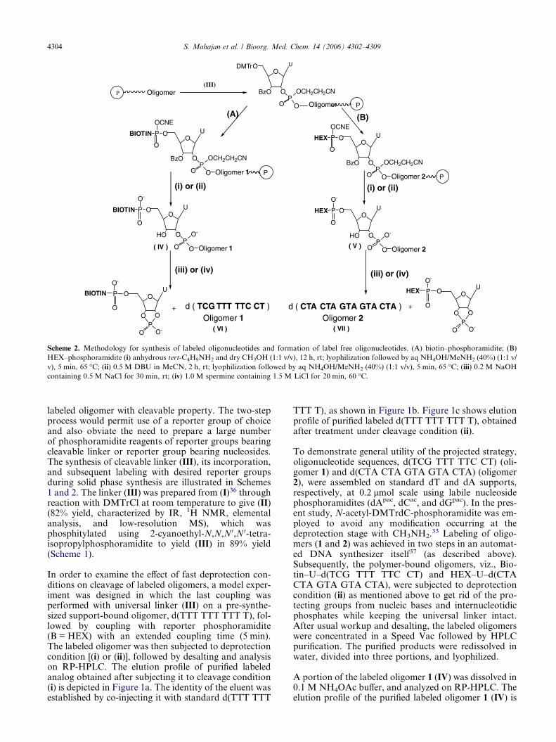
Scheme 2. Methodology for synthesis of labeled oligonucleotides and formation of label free oligonucleotides. (A) biotin–phosphoramidite; (B)
HEX–phosphoramidite (i) anhydrous tert-C4H9NH2 and dry CH3OH (1:1 v/v), 12 h, rt; lyophilization followed by aq NH4OH/MeNH2 (40%) (1:1 v/
v), 5 min, 65 �C; (ii) 0.5 M DBU in MeCN, 2 h, rt; lyophilization followed by aq NH4OH/MeNH2 (40%) (1:1 v/v), 5 min, 65 �C; (iii) 0.2 M NaOH
containing 0.5 M NaCl for 30 min, rt; (iv) 1.0 M spermine containing 1.5 M LiCl for 20 min, 60 �C.
4304 S. Mahajan et al. / Bioorg. Med. Chem. 14 (2006) 4302–4309
labeled oligomer with cleavable property. The two-stepprocess would permit use of a reporter group of choiceand also obviate the need to prepare a large numberof phosphoramidite reagents of reporter groups bearingcleavable linker or reporter group bearing nucleosides.The synthesis of cleavable linker (III), its incorporation,and subsequent labeling with desired reporter groupsduring solid phase synthesis are illustrated in Schemes1 and 2. The linker (III) was prepared from (I)36 throughreaction with DMTrCl at room temperature to give (II)(82% yield, characterized by IR, 1H NMR, elementalanalysis, and low-resolution MS), which wasphosphitylated using 2-cyanoethyl-N,N,N 0,N 0-tetra-isopropylphosphoramidite to yield (III) in 89% yield(Scheme 1).
In order to examine the effect of fast deprotection con-ditions on cleavage of labeled oligomers, a model exper-iment was designed in which the last coupling wasperformed with universal linker (III) on a pre-synthe-sized support-bound oligomer, d(TTT TTT TTT T), fol-lowed by coupling with reporter phosphoramidite(B = HEX) with an extended coupling time (5 min).The labeled oligomer was then subjected to deprotectioncondition [(i) or (ii)], followed by desalting and analysison RP-HPLC. The elution profile of purified labeledanalog obtained after subjecting it to cleavage condition(i) is depicted in Figure 1a. The identity of the eluent wasestablished by co-injecting it with standard d(TTT TTT
TTT T), as shown in Figure 1b. Figure 1c shows elutionprofile of purified labeled d(TTT TTT TTT T), obtainedafter treatment under cleavage condition (ii).
To demonstrate general utility of the projected strategy,oligonucleotide sequences, d(TCG TTT TTC CT) (oli-gomer 1) and d(CTA CTA GTA GTA CTA) (oligomer2), were assembled on standard dT and dA supports,respectively, at 0.2 lmol scale using labile nucleosidephosphoramidites (dApac, dCac, and dGpac). In the pres-ent study, N-acetyl-DMTrdC-phosphoramidite was em-ployed to avoid any modification occurring at thedeprotection stage with CH3NH2.33 Labeling of oligo-mers (1 and 2) was achieved in two steps in an automat-ed DNA synthesizer itself37 (as described above).Subsequently, the polymer-bound oligomers, viz., Bio-tin–U–d(TCG TTT TTC CT) and HEX–U–d(CTACTA GTA GTA CTA), were subjected to deprotectioncondition (ii) as mentioned above to get rid of the pro-tecting groups from nucleic bases and internucleotidicphosphates while keeping the universal linker intact.After usual workup and desalting, the labeled oligomerswere concentrated in a Speed Vac followed by HPLCpurification. The purified products were redissolved inwater, divided into three portions, and lyophilized.
A portion of the labeled oligomer 1 (IV) was dissolved in0.1 M NH4OAc buffer, and analyzed on RP-HPLC. Theelution profile of the purified labeled oligomer 1 (IV) is

0 5 10 15
0 5 10 15
0 5 10 15 20
13.791
13.910
10.281
Time (min) Time (min)
13.889
Time (min)
Abs
orba
nce
(254
nm
)
Abs
orba
nce
(254
nm
)
Abs
orba
nce
(254
nm
)
a b
c
Figure 1. RP-HPLC profiles of (a) purified oligomer, HEX–U–d(TTT TTT TTT T), obtained after cleavage [under condition (i)], (b) co-injection of
purified oligomer, HEX–U–d(TTT TTT TTT T) and standard d(TTT TTT TTT T), and (c) purified oligomer, HEX–U–d(TTT TTT TTT T),
obtained after cleavage [under condition (ii)].
S. Mahajan et al. / Bioorg. Med. Chem. 14 (2006) 4302–4309 4305
shown in Figure 2a. The same oligomer 1 (IV) was co-in-jected with the corresponding standard oligomer d(TCGTTT TTC CT) and the elution pattern is shown in Fig-ure 2b. The second portion of oligomer 1 (IV) was sub-jected to deprotection condition (iii) to cleave theuniversal linker along with biotin moiety.34 The cleavageof the universal linker from (IV) occurs presumably viaa cyclic phosphate (cf. Ref. 35). The resulting solution,after neutralization with acetic acid, was concentratedin a Speed Vac, redissolved in NH4OAc buffer and ana-lyzed on RP-HPLC (Fig. 2c). The third portion was sub-jected to mild condition (iv) followed by usual work-up,as described above. The residue was redissolved in bufferand analyzed on RP-HPLC. The elution profile of theoligomer 1 (VI) obtained after above treatment is shownin Figure 2d. In both cases, the free oligomer 1 (VI) elut-ed with a lower retention time as compared to labeledoligomer 1 (IV). The identity of the released free oligo-nucleotides (in both the cases) was further confirmedby co-injecting them with the corresponding standardoligomer, d(TCG TTT TTC CT). Figure 2e shows elu-tion pattern of a co-injection of oligomer 1 (VI), ob-tained after subjecting oligomer 1 (IV) to cleavagecondition [(iii), Scheme 2] with the corresponding stan-dard oligomer on RP-HPLC.
Likewise, the purified labeled oligomer 2 (V) was dis-solved in water, divided into three portions, and lyoph-ilized. A portion of oligomer 2 (V) was directlyredissolved in NH4OAc buffer and analyzed on RP-HPLC, whereas the remaining two portions of oligo-mer 2 (V) were subjected to different deprotection con-ditions34,35 and then analyzed on RP-HPLC. Figures
3a and b sketch elution profiles of oligomer 2 (V)and a co-injection of oligomer (V) with (VII), respec-tively. Figures 3c and d depict crude oligomer 2(VII) after cleavage under conditions (iii) and (iv),respectively. Figure 3(e) shows a co-injection of crudeoligomer 2 (VII), obtained after treatment of oligomer2 (V) to cleavage condition [(iv), Scheme 2] with stan-dard oligomer, d(CTA CTA GTA GTA CTA), on RP-HPLC.
In order to demonstrate the practical applicability ofthe cleavable linker (III) and usefulness of affinitypurification of biotinylated probes, the purified bio-tinylated oligomer 1 (IV) was incubated with mag-netic Dynabeads M-280 streptavidin in bindingbuffer. Subsequently, the beads were subjected tocleavage condition (iii) (Scheme 2). After usualwork-up, the cleaved oligomer 1 (VI) was analyzedon RP-HPLC. The oligomer 1 (VI), as anticipated,eluted with the standard oligomer when the co-injec-tion was analyzed on RP-HPLC, as also shown inFigure 4.
3. Conclusion
A new universal linker based on uridine nucleosidehas been synthesized and used to incorporate cleav-able property in labeled oligonucleotides. It hasbeen demonstrated that the linker phosphoramiditecan be coupled to the 5 0-end of synthetic oligonucle-otides efficiently in an automated DNA synthesizerwith subsequent coupling with the commercially

Time (min)
15.96
5 10 15 200 25
Time (min)Time (min)
0 5 10 15 20
15.81
25
16.10 18.25
0 5 10 15 20 25
Time (min)
18.67
Time (min)
0 5 10 15 20 25
2520151050
Abs
orba
nce
(254
nm)
Abs
orba
nce
(254
nm)
Abs
orba
nce
(254
nm)
Abs
orba
nce
(254
nm)
Abs
orba
nce
(254
nm)
a b
dc
e
Figure 2. RP-HPLC profiles of (a) purified oligomer, Biotin–U–d(TCG TTT TTC CT) (IV), obtained after cleavage [under condition (i)], (b) co-
injection of Biotin–U–d(TCG TTT TTC CT) (IV) and standard d(TCG TTT TTC CT), (c) d(TCG TTT TTC CT) (VI) obtained after treatment of
(IV) with (iii), (d) d(TCG TTT TTC CT) (VI) obtained after treatment of (IV) with (iv), and (e) co-injection of d(TCG TTT TTC CT) (VI), obtained
after treatment of (IV) with (iii), with standard d(TCG TTT TTC CT). ‘U’ denotes chemically cleavable linker (III).
4306 S. Mahajan et al. / Bioorg. Med. Chem. 14 (2006) 4302–4309
available desired labeled phosphoramidite. The linkerbetween the label and the oligonucleotide is suffi-ciently stable during post-synthesis work-up, howev-er, can readily be detached from the labeledoligonucleotides under mild condition, (iii) or (iv).The simplicity of the method, comprising usage ofcommercially available reporter phosphoramiditesand straightforward synthesis of cleavable linkerarm from commonly available chemical reagents ina minimal number of steps, is reflective of new pos-sibilities of manipulations in the DNA and RNAchemistry.
4. Experimental
4.1. 3 0-O-Benzoyluridine (I)
This was prepared according to the literature procedure36
and characterized by 1H NMR and IR spectroscopy.
4.2. Preparation of 5 0-O-(4,4 0-dimethoxytrityl)-3 0-O-ben-zoyluridine (II)
3 0(2 0)-O-Benzoyluridine (1.0 g, 2.87 mmol) was driedby co-evaporation with anhydrous pyridine and dis-

Abs
orba
nce
(254
nm
)
Abs
orba
nce
(254
nm
)
Abs
orba
nce
(254
nm
)
Abs
orba
nce
(254
nm
)
Abs
orba
nce
(254
nm
)
b
dc
e
a
Figure 3. RP-HPLC profiles of (a) purified oligomer, HEX–U–d(CTA CTA GTA GTA CTA) (V), obtained after cleavage [under condition (ii)], (b)
co-injection of HEX–U–d(CTA CTA GTA GTA CTA) (V) with standard d(CTA CTA GTA GTA CTA), (c) d(CTA CTA GTA GTA CTA) (VII)
obtained after treatment of (V) with (iii), (d) d(CTA CTA GTA GTA CTA) (VII) obtained after treatment of (V) with (iv), and (e) co-injection of
d(CTA CTA GTA GTA CTA) (VII), obtained after treatment of (V) with (iv), with standard d(CTA CTA GTA GTA CTA). ‘U’ denotes chemically
cleavable linker (III).
5 10 15
10.92
Time (min)
Abs
orba
nce
(254
nm)
Figure 4. RP-HPLC profile of d(TCG TTT TTC CT) (VI) obtained
after cleavage from Dynabeads M-280 Streptavidin under cleavage
condition (iii).
S. Mahajan et al. / Bioorg. Med. Chem. 14 (2006) 4302–4309 4307
solved in dry pyridine (10 ml). 4,4 0-Dimethoxytritylchloride (DMTrCl) (1.1 g, 3.2 mmol) was added andthe reaction mixture was stirred at room temperaturefor 6 h. Methanol (1 ml) was then added to quenchthe reaction and the solution was concentrated to anoil, which was re-dissolved in EtOAc (75 ml) andwashed with 5% aqueous NaHCO3 solution (3·25 ml) and later with saturated NaCl solution (2·25 ml). The organic phase was collected and concen-trated on a rotary evaporator to a syrupy material.It was redissolved in 1,2-dichloroethane (EDC) con-taining triethylamine (TEA) (1%, v/v) and purifiedby silica gel column chromatography using a stepwisegradient of CH3OH (0.5–2%) in EDC containing TEA(1%, v/v). The fractions containing the desired materi-al were pooled together and concentrated to obtainthe title compound as a light brown solid in 82%(1.49 g) yield.

4308 S. Mahajan et al. / Bioorg. Med. Chem. 14 (2006) 4302–4309
Rf : 0.51 (DCM/CH3OH 9:1). IR (thin film) m(cm�1) = 1033, 1255, 1682, 1702, 3049, 3502. 1H NMR(CDCl3) d (ppm) : 3.16 (d, 2H, 5 0-CH2), 3.79 (s, 6H,2· OCH3), 4.25 (m, 1H, 4 0-CH), 4.45 (br, 1H, 2 0-CH),4.95 (br, 1H, 3 0-CH), 5.95 (m, 1H, 1 0-CH), 6.70–7.60(m, 19H, Ar-H, 5-CH), 8.05 (d, 1H, 6-CH). MALDI-TOF = 673.34 (M+Na+). Anal. Calcd for C37H34N2O9:C, 68.31; H, 5.23; N, 4.31. Found : C, 66.21; H, 5.34;N, 4.17.
4.3. 5 0-O-(4,4 0-Dimethoxytrityl)-3 0-O-benzoyl-2 0-O-2-cyanoethyl-N,N 0-diisopropyl- uridine phosphoramidite(III)
A round-bottomed flask containing 5 0-O-(4,4 0-dimeth-oxytrityl)-3 0-O-benzoyluridine (500 mg, 0.76 mmol)was flushed with argon and charged with anhydrousCH3CN (10 ml). 2-Cyanoethyl-N,N,N 0,N 0-tetraisopro-pyl phosphoramidite (457.5 ll, 1.52 mmol) was addeddropwise via a syringe. A solution of 1H-tetrazole(53.2 mg, 0.76 mmol), dissolved in dry CH3CN(2 ml), was added dropwise over a period of 5 min.After stirring at room temperature for 2 h (completionmonitored on TLC), the reaction was quenched byadding methanol (1 ml). The reaction mixture was con-centrated under reduced pressure and the residue takenup in EtOAc (75 ml) containing TEA (0.5%, v/v). Theorganic phase was washed with saturated NaCl solu-tion (2· 25 ml) and dried over anhydrous Na2SO4.After filtration, the solvent was removed and the resi-due redissolved in EDC containing TEA (1%, v/v) andpurified on silica gel column. The fractions containingthe desired material were pooled together and concen-trated under reduced pressure to obtain the compound(III) in 89% (583 mg) yield. Rf: 0.68 (EtOAc/DCM/TEA 4.5:4.5:1).
4.4. N-1-(4,4 0-Dimethoxytrityl)biotinyl-aminohexanolphosphoramidite (A)
This was prepared according to the protocol publishedearlier from this laboratory.16
4.5. Oligonucleotide synthesis, deprotection, andpurification
The oligonucleotides were synthesized at 0.2 lmol scaleon a Pharmacia LKB Gene assembler Plus using stan-dard b-cyanoethyl phosphoramidite chemistry, withconventional (dAbz, dCac, and dGibu) and labile (dApac,dCac, and dGpac) nucleoside phosphoramidites. Fortethering, biotin at the 5 0-terminal of the synthesized oli-gomer, the universal linker phosphoramidite (III)(0.2 M) was dissolved in absolute CH3CN and coupledto 5 0-terminal hydroxyl groups of the oligomer sequencewith extended coupling time (5 min) followed by cou-pling of biotin–phosphoramidite (A) again with extend-ed coupling reaction time (5 min). Similarly, HEX–phosphoramidite (B) was attached to the oligomer se-quence bound to polymer support with an extended cou-pling time (5 min) after coupling of the universal linker(III). Subsequently, the support-bound oligomers weresubjected to treatment with either (i) anhydrous tert-
C4H9NH2 and methanol (1:1, v/v) for 12 h at rt andlyophilization, followed by deprotection with aqNH4OH and CH3NH2 (40%) (1:1, v/v) for 5 min at65 �C or (ii) 0.5 M DBU in MeCN for 2 h at rt andlyophilization, followed by deprotection with aqNH4OH and CH3NH2 (40%) (1:1, v/v) for 5 min at65 �C. These treatments resulted in cleavage of oligonu-cleotide chains from the support as well as removal ofthe protecting groups from nucleic bases and internu-cleotidic phosphates.
The oligomers were concentrated and subjected todesalting on a RP-18 silica gel column. Elution wasrealized with 30% acetonitrile in water and concen-trated in vacuo. After purification, these were redis-solved in water, divided into three portions, andlyophilized. One portion of the desalted oligomerswas dissolved in NH4OAc buffer, pH 7.1, and ana-lyzed on RP-HPLC. Other portions of oligomerswere subjected to following treatments in order toobtain free oligomers:
(a) Oligomer with Biotin label (IV) was dissolved in asolution (200 ll) of 0.2 M NaOH containing 0.5 MNaCl and kept at rt for 30 min. The solution was neu-tralized with acetic acid (20 ll) and concentrated in aSpeed Vac. The material was dissolved in water, desalt-ed, concentrated, redissolved in NH4Ac buffer, and ana-lyzed on RP-HPLC. A similar treatment of HEX-labeled oligomer (V) resulted in a label free oligomer(VII).(b) Alternatively, the oligomer with Biotin label (IV)was treated with a solution (200 ll) of spermine(1.0 M) and LiCl (1.5 M) at 60 �C for 20 min. After usu-al work-up, the fully deprotected oligomer (VI) was ana-lyzed on RP-HPLC. After a similar treatment, the HEX-labeled oligomer (V) was analyzed on RP-HPLC.
The above fully deprotected oligomers (VI and VII)were compared with corresponding oligomers, whichwere synthesized on standard supports and deprotectedfollowing standard protocol.15
4.6. Affinity purification of biotinylated probe
Oligomer sequence, Biotin–U–d(TCG TTT TTC CT)(oligomer 1) (IV), (0.4 O.D. A254), obtained afterdeprotection with aq NH4OH and CH3NH2, was dis-solved in a binding buffer, 10 mM Tris–HCl, pH 7.5,containing 1 mM EDTA and 1.0 M NaCl. It was thenmixed with magnetic Dynabeads M-280 Streptavidin(10 mg), pre-washed with PBS (0.1 M phosphate buffercontaining 0.5 M NaCl) buffer, pH 7.2 (2· 1.0 ml).After incubation for 1 h at rt, the Eppendorf tube con-taining the above suspension was kept on a magneticparticle concentrator. The supernatant liquid was re-moved and the beads were repeatedly washed withbinding buffer (3· 1 ml). The beads were treated witha solution of 0.2 N NaOH containing 0.5 M NaClfor 30 min at rt. The supernatant liquid was removed,neutralized, concentrated, and worked up as describedabove. The cleaved oligomer (VI) was then analyzedon RP-HPLC.

S. Mahajan et al. / Bioorg. Med. Chem. 14 (2006) 4302–4309 4309
Acknowledgments
Financial support from CSIR Task Force Project (NNI-OSB, COR010) is gratefully acknowledged. The author(S.M.) is thankful to Council of Scientific and IndustrialResearch (CSIR), New Delhi, India, for the award ofSenior Research Fellowship.
References and notes
1. Beck, S.; O’keeffe, T.; Coull, J. M.; Koster, H. NucleicAcids Res. 1989, 17, 5115–5123.
2. Smith, L. M.; Fung, S.; Hunkapiller, M. W.; Hunkap-iller, T. J.; Hood, L. E. Nucleic Acids Res. 1985, 13,2399–2412.
3. Haralambidis, J.; Chai, M.; Tregear, G. W. Nucleic AcidsRes. 1987, 15, 4857–4876.
4. Green, A.; Roopra, A.; Vaudin, M. Nucleic Acids Res.1990, 18, 6163–6164.
5. Hultman, T.; Stahl, S.; Hornes, E.; Uhlen, M. NucleicAcids Res. 1989, 17, 4937–4946.
6. De Vos, M.-J.; Cravador, A.; Lenders, J.-P.; Houard, S.;Bollen, A. Nucleosides Nucleotides 1990, 9, 259–273.
7. Bengtstrom, M.; Harju, L.; Syvanen, A.-C. NucleosidesNucleotides 1991, 10, 507–509.
8. Reyes-Engel, A.; Dieguez-Lucena, J. L. Nucleic Acids Res.1993, 21, 759–760.
9. Niemeyer, C. M.; Sano, T.; Smith, C. L.; Cantor, C. R.Nucleic Acids Res. 1994, 22, 5530–5539.
10. Gibson, K. J.; Benkovic, S. J. Nucleic Acids Res. 1987, 15,6455–6467.
11. Sproat, B. S.; Beijer, B.; Rider, P. Nucleic Acids Res. 1987,15, 6181–6196.
12. Agrawal, S.; Christodoulou, C.; Gait, M. J. Nucleic AcidsRes. 1986, 14, 6227–6245.
13. Sinha, N. D.; Cook, R. M. Nucleic Acids Res. 1988, 16,2659–2669.
14. Misiura, K.; Durrant, I.; Evans, M. R.; Gait, M. J. NucleicAcids Res. 1990, 18, 4345–4354.
15. Kumar, P.; Sharma, A. K.; Gupta, K. C. NucleosidesNucleotides 1996, 15, 1263–1273.
16. Kumar, P.; Bhatia, D.; Garg, B. S.; Gupta, K. C. BioMed.Chem. Lett. 1994, 4, 1761–1766.
17. Manoharan, M.; Inamati, G.; Tivel, K. L.; Wheeler, P.;Stecker, K.; Cook, P. D. Nucleosides Nucleotides 1997, 16,1411–1413.
18. Pieles, U.; Sproat, B. S.; Lamm, G. M. Nucleic Acids Res.1990, 18, 4355–4360.
19. Zhao, Z. Y.; Ackroyd, J. Nucleosides Nucleotides 1999, 18,1231–1234.
20. Wilchek, M.; Bayer, E. A. Methods Enzymol. 1990, 184,301–303.
21. McInnes, J. L.; Symons, R. H. In Nucleic Acids Probes;Symons, R. H., Ed.; CRC: Boca Raton, FL, 1989; pp 33–80.
22. Pon, R. T. Tetrahedron Lett. 1991, 32, 1715–1718.23. Cocuzza, A. J. Tetrahedron Lett. 1989, 30, 6287–6290.24. Alves, A. M.; Holland, D.; Edge, M. D. Tetrahedron Lett.
1989, 30, 3089–3092.25. Tijssen, P. Laboratory Techniques in Biochemistry and
Molecular Biology—Hybridization with Nucleic AcidProbes; Elsevier: New York, NY, 1993, Vol. 4.
26. Herman, T. M.; Lefever, E.; Shimkus, M. Anal. Biochem.1986, 156, 48–55.
27. Herman, T. M.; Fenn, B. J. Methods Enzymol. 1990, 184,584–588.
28. Gildea, B. D.; Coull, J. M.; Koster, H. Tetrahedron Lett.1990, 31, 7095–7098.
29. Olejnik, J.; Sonar, S.; Krzymanska-Olejnik, E.; Roths-child, K. J. Proc. Natl. Acad. Sci. U.S.A. 1995, 92, 7590–7594.
30. Olejnik, J.; Krzymanska-Olejnik, E.; Rothschild, K. J.Nucleic Acids Res. 1996, 24, 361–366.
31. Fang, S.; Bergstrom, D. E. Nucleic Acids Res. 2003, 31,708–715.
32. Fang, S.; Bergstrom, D. E. Bioconjugate Chem. 2003, 14,80–85.
33. Reddy, M. P.; Hanna, N. B.; Farooqui, F. TetrahedronLett. 1994, 35, 4311–4314.
34. Kumar, P.; Gupta, K. C. Nucleic Acids Res. 1999, 27, e2.35. Kumar, P.; Dhawan, G.; Chandra, R.; Gupta, K. C.
Nucleic Acids Res. 2002, 30, e130.36. Wagner, D.; Verheyden, J. P. H.; Moffatt, J. G. J. Org.
Chem. 1974, 39, 24–30.37. Gene Assembler Plus Manual, Uppsala, Sweden, 1988.