A new shading concept based on natural tooth color applied ...
12
VOLUME 37 • NUMBER 2 • FEBRUARY 2006 91 QUINTESSENCE INTERNATIONAL Resin composites nowadays occupy a para- mount position among restorative materials because they offer an excellent esthetic potential and acceptable longevity, with a much lower cost than equivalent ceramic restorations for the treatment of anterior teeth. 1–3 In addition, composite restorations allow for minimally invasive preparations or no preparation at all for the replacement of decayed or missing tissues. The identification of respective dentin and enamel optical characteristics is of consider- able interest for the development of tooth-col- ored materials 4,5 (Fig 1). Master ceramists and manufacturers of dental porcelains have made a lot of effort in developing specific powders that mimic the 2 main constituents of natural teeth, when placed in the specific configuration of a ceramic restoration. 6 However, ceramics are to be used for the veneering of a metal or ceramic framework, in thin layers and in a configuration that does not correspond to the arrangement of natural A new shading concept based on natural tooth color applied to direct composite restorations Didier Dietschi, DMD, PhD 1 /Stefano Ardu, DMD 2 /Ivo Krejci, DMD 3 Objective: Patient demands have prompted manufacturers to improve intrinsic optical properties of resin composites and clinicians to refine application procedures. The aim of this study is to present a shading concept based on colorimetric L*a*b* and contrast ratio data of human dentin and enamel. Method and materials: Extracted teeth of the A and B Vita shade groups (n = 8 per group) were sectioned according to 2 different planes to measure specific color (using the CIE L*a*b* system) and opacity (contrast ratio). Standardized samples of enamel and dentin shades of a new composite system (Miris, Coltène Whaledent) were submitted to the same colorimetric evaluation for comparison with natural tissues. Results: Comparison of teeth from the Vita groups A and B having the same chroma showed limited variations regarding a* (green to red) and b* (blue to yellow) values; the only significant variation was the increasing b* values (yellow) with increasing chroma (A1 to A4 and B1 to B3). As for dentin contrast ratio, limited differ- ences were reported, while enamel proved to increase in translucency with age (reduced contrast ratio). Conclusion: These data served as the foundation of the so-called natural layering concept, which makes use of 2 basic composite masses (dentin and enamel) that optically mimic natural tissues. This concept allows for simplified clinical application and layering of composite, as it uses only 1 universal dentin hue with several chroma levels and 3 enamel types for young, adult, and old patients, each exhibiting specific tints and translucency levels. (Quintessence Int 2006;37:91–102) Key words: CIE L*a*b*, contrast ratio, dentin color, natural layering technique 1 Senior Lecturer, Department of Cariology and Endodontics, School of Dentistry, University of Geneva, Switzerland; Adjunct Associate Professor, Department for the Practice of General Dentistry, Case Western University, Cleveland, Ohio. 2 Lecturer, Department of Cariology and Endodontics, School of Dentistry, University of Geneva, Switzerland. 3 Professor and Chair, Department of Cariology and Endodontics, School of Dentistry, University of Geneva, Switzerland. Reprint requests: Dr Didier Dietschi, Department of Cariology and Endodontics, School of Dentistry, University of Geneva, 19 Rue Barthélémy Menn, 1205 Geneva, Switzerland. Fax: +41.22 39 29 990. E-mail: [email protected]
Transcript of A new shading concept based on natural tooth color applied ...

VOLUME 37 • NUMBER 2 • FEBRUARY 2006 91
QUINTESSENCE INTERNATIONAL
Resin composites nowadays occupy a para-
mount position among restorative materials
because they offer an excellent esthetic
potential and acceptable longevity, with a
much lower cost than equivalent ceramic
restorations for the treatment of anterior
teeth.1–3 In addition, composite restorations
allow for minimally invasive preparations or
no preparation at all for the replacement of
decayed or missing tissues.
The identification of respective dentin and
enamel optical characteristics is of consider-
able interest for the development of tooth-col-
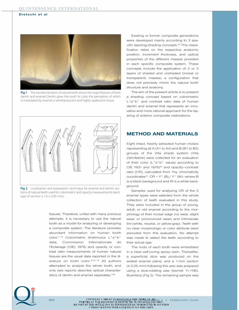
ored materials4,5 (Fig 1). Master ceramists
and manufacturers of dental porcelains have
made a lot of effort in developing specific
powders that mimic the 2 main constituents
of natural teeth, when placed in the specific
configuration of a ceramic restoration.6
However, ceramics are to be used for the
veneering of a metal or ceramic framework,
in thin layers and in a configuration that does
not correspond to the arrangement of natural
A new shading concept based on natural toothcolor applied to direct composite restorations Didier Dietschi, DMD, PhD1/Stefano Ardu, DMD2/Ivo Krejci, DMD3
Objective: Patient demands have prompted manufacturers to improve intrinsic optical
properties of resin composites and clinicians to refine application procedures. The aim of
this study is to present a shading concept based on colorimetric L*a*b* and contrast
ratio data of human dentin and enamel. Method and materials: Extracted teeth of the A
and B Vita shade groups (n = 8 per group) were sectioned according to 2 different planes
to measure specific color (using the CIE L*a*b* system) and opacity (contrast ratio).
Standardized samples of enamel and dentin shades of a new composite system (Miris,
Coltène Whaledent) were submitted to the same colorimetric evaluation for comparison
with natural tissues. Results: Comparison of teeth from the Vita groups A and B having
the same chroma showed limited variations regarding a* (green to red) and b* (blue to
yellow) values; the only significant variation was the increasing b* values (yellow) with
increasing chroma (A1 to A4 and B1 to B3). As for dentin contrast ratio, limited differ-
ences were reported, while enamel proved to increase in translucency with age (reduced
contrast ratio). Conclusion: These data served as the foundation of the so-called natural
layering concept, which makes use of 2 basic composite masses (dentin and enamel) that
optically mimic natural tissues. This concept allows for simplified clinical application and
layering of composite, as it uses only 1 universal dentin hue with several chroma levels
and 3 enamel types for young, adult, and old patients, each exhibiting specific tints and
translucency levels. (Quintessence Int 2006;37:91–102)
Key words: CIE L*a*b*, contrast ratio, dentin color, natural layering technique
1Senior Lecturer, Department of Cariology and Endodontics,
School of Dentistry, University of Geneva, Switzerland;
Adjunct Associate Professor, Department for the Practice of
General Dentistry, Case Western University, Cleveland, Ohio.
2Lecturer, Department of Cariology and Endodontics, School
of Dentistry, University of Geneva, Switzerland.
3Professor and Chair, Department of Cariology and
Endodontics, School of Dentistry, University of Geneva,
Switzerland.
Reprint requests: Dr Didier Dietschi, Department of
Cariology and Endodontics, School of Dentistry, University of
Geneva, 19 Rue Barthélémy Menn, 1205 Geneva, Switzerland.
Fax: +41.22 39 29 990. E-mail: [email protected]
92 VOLUME 37 • NUMBER 2 • FEBRUARY 2006
QUINTESSENCE INTERNATIONAL
Dietschi et a l
tissues. Therefore, unlike with many previous
attempts, it is necessary to use the natural
tooth as a model for analyzing or developing
a composite system. The literature provides
abundant information on human tooth
color.7–14 Colorimetric (tristimulus L*a*b*
data, Commission Internationale de
l’Eclairage [CIE] 1976) and opacity or con-
trast ratio measurements of human natural
tissues are the usual data reported in the lit-
erature on tooth color.10,14–16 All authors
attempted to analyze the whole tooth, and
only rare reports describe optical character-
istics of dentin and enamel separately.17,18
Existing or former composite generations
were developed mainly according to 3 spe-
cific layering/shading concepts.19 This classi-
fication relies on the respective anatomic
position, increment thickness, and optical
properties of the different masses provided
in each specific composite system. These
concepts include the application of 2 or 3
layers of shaded and unshaded (incisal or
transparent) masses, a configuration that
does not precisely mimic the natural tooth
structure and anatomy.
The aim of the present article is to present
a shading concept based on colorimetric
L*a*b* and contrast ratio data of human
dentin and enamel that represents an inno-
vative and more rational approach for the lay-
ering of anterior composite restorations.
METHOD AND MATERIALS
Eight intact, freshly extracted human molars
representing all A (A1 to A4) and B (B1 to B3)
groups of the Vita shade system (Vita
Zahnfabrik) were collected for an evaluation
of their color (L*a*b* values according to
CIE 1931 and 1976)20 and opacity—contrast
ratio [CR], calculated from Yxy chromaticity
coordinates21: CR = Y* (B) / Y* (W)—where B
is a black background and W is a white back-
ground.
Samples used for analyzing CR of the 3
enamel types were selected from the whole
collection of teeth evaluated in this study.
They were included in the group of young,
adult, or old enamel according to the mor-
phology of their incisal edge (no wear, slight
wear, or pronounced wear) and intrinsicate
tint (white, neutral, or yellow-gray). Teeth with
no clear morphologic or color attribute were
excluded from this evaluation. No attempt
was made to select the teeth according to
their actual age.
The roots of each tooth were embedded
in a clear self-curing epoxy resin. Thereafter,
a superficial slice was produced on the
widest enamel plane, and a 1-mm section
(± 0.05 mm) following this axis was prepared
using a slow-rotating saw (Isomet 11-1180,
Buehlers) (Fig 2). The remaining sample was
Fig 1 The transillumination of natural teeth shows the major features of bothdentin and enamel. Dentin gives the tooth its color, the perception of which is modulated by enamel, a semitranslucent and highly opalescent tissue.
Fig 2 Localization and preparation technique for enamel and dentin sec-tions of natural teeth used for colorimetric and opacity measurements (eachtype of section is 1.0 ± 0.05 mm).

VOLUME 37 • NUMBER 2 • FEBRUARY 2006 93
QUINTESSENCE INTERNATIONAL
Dietschi et a l
sectioned perpendicular to the tooth’s long
axis, 1.5 mm below the deepest point of the
occlusal surface, and a 1-mm section (± 0.05
mm) was made below this plane (see Fig 2).
Colorimetric measures of enamel (on the
inner side) and dentin samples (on the
occlusal side) were performed using a
reflectance colorimetric device (Minolta CR-
21, Minolta). Detailed evaluation protocol was
presented in previous reports.18,22
During a second phase, standardized
samples (1 � 15 � 15 mm ± 0.05 mm) made
of a composite developed according to the
data produced from human tissues (Miris,
Coltène Whaledent) were fabricated to
assess its optical properties (L*a*b* and
CR). Five samples of each dentin (S1 to S7)
and enamel (white bleach, WB; white regular,
WR; neutral regular, NR; neutral transparent,
NT; ivory regular, IR; ivory transparent IT)
shade were produced.
Descriptive statistics were computed for
either natural tissues or composite data
(mean and standard deviation). The Student
t test unpaired was used to explore the dif-
ferences in dentin L*, a*, and b* values
between A and B Vita shades, within the
same chroma range (1 to 3).
RESULTS
The L*a*b* and CR data of natural tissues
and Miris composite are presented in Tables
1 to 4 and Figs 3a and 3b.
Fig 3a CIE L*a*b* valuesof natural dentin samplesarranged according to theVita shades.
Fig 3b CIE L*a*b* valuesof Miris dentin shades,which were developedaccording to the naturallayering concept.
CIE
L*a
*b*
valu
esC
IE L
*a*b
* va
lues
S1 S2 S3 S4 S5 S6 S7
A1 B1 A2 B2 A3 B3 A3.5 A4
Vita shades
Miris dentin shades
90
80
70
60
50
40
30
20
10
0
-10
80
70
60
50
40
30
20
10
0
-10
94 VOLUME 37 • NUMBER 2 • FEBRUARY 2006
QUINTESSENCE INTERNATIONAL
Dietschi et a l
Table 4 Mean L* and CR values of Miris enamel shades (n = 5 per shade)
Miris enamel shade L* CR
White regular (WR) 65.11 0.44White bleach (WB) 64.82 0.46Neutral regular (NR) 62.59 0.42Neutral transparent (NT) 65.82 0.29Ivory regular (IR) 64.09 0.375Ivory transparent (IT) 62.62 0.335
Table 3 Mean L* and CR values of natural enamel (n = 5 per shade)
Enamel age/type L* CR
Young/white 75.89 0.485Adult/neutral 66.77 0.434Old/yellow-gray 71.84 0.402Mean value 70.83 0.435
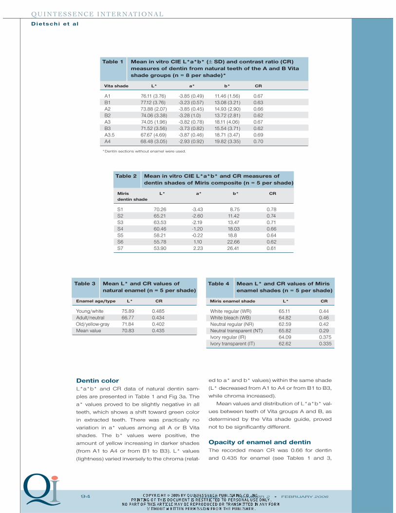
Dentin colorL*a*b* and CR data of natural dentin sam-
ples are presented in Table 1 and Fig 3a. The
a* values proved to be slightly negative in all
teeth, which shows a shift toward green color
in extracted teeth. There was practically no
variation in a* values among all A or B Vita
shades. The b* values were positive, the
amount of yellow increasing in darker shades
(from A1 to A4 or from B1 to B3). L* values
(lightness) varied inversely to the chroma (relat-
ed to a* and b* values) within the same shade
(L* decreased from A1 to A4 or from B1 to B3,
while chroma increased).
Mean values and distribution of L*a*b* val-
ues between teeth of Vita groups A and B, as
determined by the Vita shade guide, proved
not to be significantly different.
Opacity of enamel and dentin The recorded mean CR was 0.66 for dentin
and 0.435 for enamel (see Tables 1 and 3,
Table 2 Mean in vitro CIE L*a*b* and CR measures ofdentin shades of Miris composite (n = 5 per shade)
Miris L* a* b* CRdentin shade
S1 70.26 -3.43 8.75 0.78S2 65.21 -2.60 11.42 0.74S3 63.53 -2.19 13.47 0.71S4 60.46 -1.20 18.03 0.66S5 58.21 -0.22 18.8 0.64S6 55.78 1.10 22.66 0.62S7 53.90 2.23 26.41 0.61
Table 1 Mean in vitro CIE L*a*b* (± SD) and contrast ratio (CR)measures of dentin from natural teeth of the A and B Vitashade groups (n = 8 per shade)*
Vita shade L* a* b* CR
A1 76.11 (3.76) -3.85 (0.49) 11.46 (1.56) 0.67B1 77.12 (3.76) -3.23 (0.57) 13.08 (3.21) 0.63A2 73.88 (2.07) -3.85 (0.45) 14.93 (2.90) 0.66B2 74.06 (3.38) -3.28 (1.0) 13.72 (2.81) 0.62A3 74.05 (1.96) -3.82 (0.78) 18.11 (4.06) 0.67B3 71.52 (3.56) -3.73 (0.82) 15.54 (3.71) 0.62A3.5 67.67 (4.69) -3.87 (0.46) 18.71 (3.47) 0.69A4 68.48 (3.05) -2.93 (0.92) 19.82 (3.35) 0.70
*Dentin sections without enamel were used.
VOLUME 37 • NUMBER 2 • FEBRUARY 2006 95
QUINTESSENCE INTERNATIONAL
Dietschi et a l
Implications of natural tooth color
measurements in the development
of a clinical conceptThe analysis of L*a*b* measures, including
their standard deviation, led to the conclusion
that an ideal dentin replacement material
should exhibit the following characteristics: sin-
gle hue, single opacity, and large chroma scale.
Actually, the variations of a* and b* values
between A and B Vita shades do not seem to
justify the use of distinct dentin colors, at
least for a direct composite restorative sys-
tem. Likewise, the variations of the contrast
ratio within a single shade group do not sup-
port the use of different dentin opacities (ie,
translucent, regular, and opaque dentins).
However, chroma (related to a* and b* val-
ues) proved to increase from light to dark
shades (A1 to A4 or B1 to B3) and then sup-
port the concept of a large chroma scale cov-
ering all variations of natural dentitions, plus
some specific conditions like sclerotic dentin
(as found underneath decay, restorations, or
cervical lesions).
As for enamel, differences in L* and CR
values proved to vary in relation to tooth age,
which confirmed the clinical concept of 3
specific enamel types25:
• Young enamel: white tint, high opales-
cence, less translucency
• Adult enamel: neutral tint, less opales-
cence, and intermediate translucency
• Old enamel: yellow tint and higher translu-
cency
This whole interpretation of CIE L*a*b*
and CR data of natural tissues led to a clini-
cal approach called the natural layering con-
cept, which embraces more accurately the
optical and anatomic characteristics of natu-
ral teeth2,18,26 (Fig 4). It actually defines the fea-
tures of an optimal restorative material aimed
to replace dentin and enamel, respectively.
Dentin shades should be available in a single
hue (Vita A or universal dentin shade) with a
large range of chroma (usually expending
the existing Vita shade range) and present-
ing opacity close to that of natural dentin.
Enamel shades should present different tints
and opacity levels, tentatively replicating all
variations found in nature. Products that pro-
vide such shade features are Miris,
Vitalescence (Ultradent), and Ceram-X duo
(Dentsply).
The optical properties of the product test-
ed in this study (Miris) mimic those of the nat-
ural tissues quite closely . However, the color
of dentin masses had to be slightly modified
based on the clinical experience gained dur-
ing the product development; actually, the
influence of surrounding soft tissues and vital
pulp on color perception proved different
between natural tissues and the restorative
material. Therefore, a* and b* values were
adjusted to obtain a better color match in vivo;
this is why no attempt was made to correlate
statistically L*a*b* and CR values of the com-
posite and natural tissues.
Influence of the natural layering
concept on shade recording The quality of the final restoration, of course,
depends on a correct shade recording.
According to the natural layering concept,
only 3 steps are involved: (1) selection of
respectively). The CR of enamel also proved
to vary according to age and type: young,
white (0.485); adult, neutral (0.434); old,
gray-yellow (0.402) (see Table 3).
Composite colorimetric data The L*a*b* and CR data of Miris are pre-
sented in Table 2 and Fig 3b (dentin) and
Table 4 (enamel). The dentin shades present
a moderate increase of a* values (shift from
green toward red) with increasing chroma
(S1 to S7); likewise, b* values are increasing
with chroma (S1 to S7). L* values diminish
with chroma (S1 to S7) (Fig 3b).
DISCUSSION
Only A1 to A4 and B1 to B3 shades of the
Vita system were evaluated, those being the
most common shades found within the col-
lection of extracted teeth. Regarding the other
Vita shades (C and D), it proved impossible to
collect samples in sufficient number to evalu-
ate them. These 2 shades proved to be rarely
observed in the natural dentition.12,13,23,24

96 VOLUME 37 • NUMBER 2 • FEBRUARY 2006
QUINTESSENCE INTERNATIONAL
Dietschi et a l
Fig 4 Description of the natural layering concept with specific features of young, adult, and old teeth and how itinfluences the choice of related composite shades.
EnamelVarying tint and increasing translucency
Adult
OldOld
Adult
Young Young

VOLUME 37 • NUMBER 2 • FEBRUARY 2006 97
QUINTESSENCE INTERNATIONAL
Dietschi et a l
DentinSame Hue and Opacity
Spacial relationshipsVarying enamel coverage at incisal edge
Young
Adult Adult
Old Old
Young

98 VOLUME 37 • NUMBER 2 • FEBRUARY 2006
QUINTESSENCE INTERNATIONAL
Dietschi et a l
Figs 5a to 5f Shade recording according to the natural layering concept. (a) The new shade guide of the Miris system comprisesshade tabs with anatomic shape, dimensions, and thicknesses. Glycerin gel, which has a refractive index close to that of resin com-posites resins, must be applied in between both shade tabs to allow for an accurate shade selection. (b) Teeth are cleaned with aprophylactic nonfluoridated paste. (c) For the selection of dentin, a serial of dentin tabs is first placed in front of the teeth to selectthe most likely dentin chroma. (d) The choice of dentin chroma is confirmed by placing the composite tab close to the cervicalarea, where there is the least amount of enamel, allowing for a more precise selection. (e) The enamel shade, which was visuallyselected, was placed over the selected dentin tab. Combined samples are then brought to the mouth and placed incisal edge toincisal edge for shade comparison. If shade match is not adequate, another enamel tab can be tested. (f) Completed restorationsshowing good color and translucency integration of the restoration.
a b
c d
e f

VOLUME 37 • NUMBER 2 • FEBRUARY 2006 99
QUINTESSENCE INTERNATIONAL
Dietschi et a l
dentin chroma in the cervical area, where
enamel is the thinnest, using samples of the
composite material, (2) selection of enamel
tint and translucency, by simple visual obser-
vation, and (3) preparation of a simplified
chromatic map (visually or through an intra-
oral photograph) to decide whether the appli-
cation of effect materials is needed. The
most useful effect materials are the blue (rein-
forcement of composite natural opales-
cence), gold-yellow (for a local increase of
restoration chroma), and white (for simulating
white spots or hypocalcifications). A new
technique to record shade has been recently
introduced to facilitate this clinical procedure
and make it more accurate, by superimpos-
ing the dentin and enamel samples (Fig 5).
Clinical application of the natural
layering concept
A 50-year-old patient shows discolored and
worn incisal composite reconstructions that
necessitate a replacement for esthetic and
functional reasons (Fig 6a). Shade selection
is always performed first, to avoid any inter-
ference in chroma and opacity evaluation
due to tissue dehydration (see Fig 6a). A
mockup is performed to restore the normal
tooth length and width; an esthetic, function-
al, and phonetic check can be performed
before proceeding with further restorative
steps (Fig 6b). As soon as the incisal edge
position is confirmed, a silicone index is fab-
ricated that will be used during the next steps
and also at the time of finishing (Fig 6c).
The selected enamel composite is applied
directly on the silicone index (Miris, Neutral
Regular), which is then placed against the
teeth. This allows the lingual buildup of
enamel to be performed easily and precisely
(Fig 6d). The reference of the new incisal
edge serves for a 3-dimensional placement
of dentin (Miris, dentin S3); with this “lin-
guobuccal” incremental approach, the space
needed for the subsequent application of
effect materials and enamel is maintained
with superior accuracy (Fig 6e). A little incre-
ment of blue-tinted composite (Miris, Blue
effect) is placed on top of the dentin buildup
to mimic opalescence of natural enamel (Fig
6f). This was judged necessary since the
intrinsic opalescence of the composite
enamel was insufficient in this case. Finally,
unshaded, translucent enamel mass (same
as that used on the lingual surface) is applied
to complete the proximal and buccal profiles
and provide desired translucency and bright-
ness (Figs 6g and 6h).
The application of the natural layering
concept through a logical application of 2
separate composite masses that mimic natu-
ral tooth anatomy presents clear advantages
for the clinician; it makes the whole proce-
dure more efficient and predictable.
Effect of tooth aging on opticalproperties of dentin and enamel Special attention has to be paid to the mor-
phologic changes that affect the incisal edge
structure as a result of tissue aging and func-
tional wear. Actually, in addition to the
increase in dentin chroma and enamel
translucency, the progressive thinning of the
enamel layer and exposure of dentin at the
incisal edge necessitates an adaptation of
the basic layering technique (see Fig 4).
CONCLUSION
The traditional restorative objectives have not
changed over time; they simply were imple-
mented by the esthetic demands of an
increasing number of patients. Resin com-
posite then became the material of choice for
young patients and less privileged people, or
for any case that requires a strictly conserva-
tive approach. The contemporary practition-
er is ultimately challenged to replace the
missing tissues or eventually modify their
configuration by applying on the patient’s
teeth an artificial material, which has to simu-
late the appearance of natural tissues.
The natural layering concept has enabled
this objective to be achieved in a predictable
way by incorporating newly acquired knowl-
edge about natural tissue optical properties
into contemporary composite systems. This
advance can be regarded as a milestone in
operative dentistry, as it will give direct com-
posite application a tremendous advantage,
enabling a larger number of patients to receive
more conservative and esthetic restorations.

100 VOLUME 37 • NUMBER 2 • FEBRUARY 2006
QUINTESSENCE INTERNATIONAL
Dietschi et a l
Figs 6a to 6h Buildup of 2 large Class 4 cavities according to the natural layering concept. (a) Preoperative view andshade selection with dual Miris shade guide. (b) Freehand mockup that reproduces the normal length and width of theincisal edge. (c) A silicone index records this information to facilitate further procedures. (d) The lingual enamel walls arebuilt up directly against the index.
a b
c d

VOLUME 37 • NUMBER 2 • FEBRUARY 2006 101
QUINTESSENCE INTERNATIONAL
Dietschi et a l
(e) Dentin can be applied and placed precisely in relation with the future incisal edge, respecting the specific tooth andage configurations. (f) Effect masses are applied in small quantities on top of dentin to mimic specific light effects suchas opalescence (blue tinted). (g) A final enamel layer is applied on proximal and buccal surfaces to complete the restora-tion. (h) Completed restorations after tissue rehydration.
f
h
e
g

102 VOLUME 37 • NUMBER 2 • FEBRUARY 2006
QUINTESSENCE INTERNATIONAL
Dietschi et a l
REFERENCES
1. Osborne JW, Normann RD, Gale EN. A 12-year clini-
cal evaluation of two composite resins.
Quintessence Int 1990;21:111–114.
2. Dietschi D. Free-hand composite resin restorations:
A key to anterior aesthetics. Pract Periodontics
Aesthetic Dent 1995;7:15–25.
3. Fahl N. Optimizing the esthetics of Class IV restora-
tions with composite. J Can Dent Assoc 1997;63:
108–115.
4. Winter R. Visualizing the natural dentition. J Esthet
Dent 1993;5:103–117.
5. Chiche G, Pinault A. Esthetics of Anterior Fixed
Prosthodontics. Chicago: Quintessence, 1994.
6. Sieber C. Voyage. Chicago: Quintessence, 1994.
7. Sproull RC. Color matching in dentistry. Part I: The
three-dimensional nature of color. J Prosthet Dent
1973;29:416–424.
8. Sproull RC. Color matching in dentistry. Part II:
Practical applications of the organization of color. J
Prosthet Dent 1973;29:556–566.
9. Macentee M, Lakowski R. Instrumental color meas-
urement of vital and extracted human teeth. J Oral
Rehabil 1981;8:203–208.
10. Clarke FJJ. Measurement of color of human teeth.
In: McLean JW (ed). Dental Ceramics: Proceedings of
the First International Symposium on Ceramics.
Chicago: Quintessence, 1983:441–488.
11. Goodkind RJ, Schwabacher WB. Use of a fiber-optic
colorimeter for in vivo color measurements of 2830
anterior teeth. J Prosthet Dent 1987;58:535–542.
12. Miller L. Shade matching. J Esthet Dent 1993;5:
143–152.
13. Miller L. Shade selection. J Esthet Dent 1994;6: 47–60.
14. O’Brien WJ, Hemmendinger H, Boenke KM, Linger
JB, Groh CL. Color distribution of three regions of
extracted human teeth. Dent Mat 1997;13:179–185.
15. Crisp S, Abel G, Wilson A.The quantitative measure-
ment of the opacity of dental filling materials. J
Dent Res 1979;58:1585–1596.
16. Inokoshi S, Kataumi M, Pereira PNR, Yamada T,
Tagami J. Appearance of composite resins in poste-
rior teeth. In: Dondi dall’Orologio G, Prati C (eds).
Factors Influencing the Quality of Composite
Restorations: Theory and Practice. [Proceedings of
the Bologna International Symposium, 22–23 Nov
1996, Bologna, Italy]. Haarlem, The Netherlands:
Cavex,1995:141–154.
17. Cook WD, McAree DC. Optical properties of esthetic
restorative materials and natural dentition. J
Biomed Mat Res 1985;19:469–488.
18. Dietschi D, Ardu S, Krejci I. Exploring the layering
concepts for anterior teeth. In: Roulet JF, Degrange
M (eds). Adhesion: The Silent Revolution in
Dentistry. Berlin: Quintessence, 2000:235–251.
19. Dietschi D. Layering concepts in anterior composite
restorations. J Adhes Dent 2001;3:71–80.
20. Chamberlain GJ, Chamberlain DG. Colour: Its meas-
urement, computation and application. London:
Heyden & Son, 1980:60–61.[Au: Is this the full range
of pages?]
21. Hunter RS, Harold RW. The Measurement of
Appearance,ed 2.New York: John Wiley & Sons,1987.
22. Rossier S. In vitro colorimetric evaluation of the effica-
cy of various bleaching methods and products [the-
sis]. Geneva, Switzerland: University of Geneva, 2005.
23. Schwabacher WB, Goodkind RJ. Three-dimensional
color coordinates of natural teeth compared with
three shade guides. J Prosthet Dent 1990;64:
425–431.
24. Hasegawa A, Ikeda I, Kawaguchi S. Color and
translucency of in vivo natural central incisors. J
Prosthet Dent 2000;83:418–423.
25. Ubassy G. Shape and Color: The Key to Successful
Ceramic Restorations. Berlin: Quintessenz, 1993.
26. Dietschi D. Free-hand bonding in esthetic treat-
ment of anterior teeth: Creating the illusion.J Esthet
Dent 1997;9:156–164.