A NEW HIGH-PERFORMANCE REAGENT AND PROCEDURE FOR …€¦ · A NEW HIGH-PERFORMANCE REAGENT AND...
20
81 Can. Soc. Forensic Sci. J. Vol. 39. No 3 (2006) pp. 81–100 A NEW HIGH-PERFORMANCE REAGENT AND PROCEDURE FOR LATENT BLOODSTAIN DETECTION BASED ON LUMINOL CHEMILUMINESCENCE. L. J. BLUM 1 , PHILIPPE ESPERANÇA 2 , STÉPHANIE ROCQUEFELTE 3 ABSTRACT A new occult blood revealing agent was designed such that DNA material would be preserved and a DNA profile could be obtained. Indeed, there were uncertainties about previous chemicals that might alter blood DNA content and have adverse effects on DNA typing. BlueStar ® is a chemiluminescent bloodstain detecting agent resulting from our research. It combines ease-of-use with minimum health risks and does not have any harmful effects on DNA profile determination. RÉSUMÉ Résultat de nos recherches, le BlueStar ® est un produit chimique de révélation de traces de sang latentes. Alors que l’action de certains produits ayant la même fonc- tion reste incertaine sur l’ADN, celui-ci ne perturbe pas la détermination d’un profil génétique. A cette qualité essentielle s’ajoute une facilité d’utilisation avec des risques minimisés sur la santé de l’utilisateur. INTRODUCTION Bloodstains can be found anywhere a violent crime has occurred. Bloodstain patterns on the floor (from a dripping wound, for example) or spattered on the walls can be interpret- ed for crime scene reconstruction. DNA typing analysis can establish the genetic profile(s) of the participant(s) in a violent crime. Consequently, bloodstains are among the most use- ful evidence for court. This fact is becoming well-known, as criminals now often attempt to clean up the crime scene. A wide variety of chemicals may be used as presumptive tests to detect evidence of these cleanings. Blood detection methods are either based on the detection of hemoglobin and its derivatives (catalytic test) or on the detection of proteins and amino acids (non- covalently bound to proteins). However, the use of some chemicals may have adverse effects on subsequent Short Tandem Repeat (STR) DNA typing and also has been shown to be potential health hazards. For example, benzidine and ortho-tolidine have potential carcinogenic properties (1–2) and leucomalachite green does not allow for DNA typing (3). 1. Directeur UMR 5013 CNRS, Laboratoire de Génie Enzymatique et Biomoléculaire, Université Claude Bernard Lyon 1 - Bâtiment CPE, 43, boulevard du 11 novembre 1918 - 69622 Villeurbanne cedex - FRANCE 2. Institut de Recherche Criminelle de la Gendarmerie Nationale (IRCGN), 1 boulevard Théophile Sueur - 93111 Rosny sous bois cedex FRANCE. Phone : +33 1 49 35 58 86. Email : [email protected] To whom correspondence should be addressed. 3. Unité Fonctionnelle d’Empreintes Génétiques, Centre Hospitalier Universitaire de Nantes - Institut de Biologie, 9, Quai Moncousu - 44093 Nantes cedex 01 FRANCE
Transcript of A NEW HIGH-PERFORMANCE REAGENT AND PROCEDURE FOR …€¦ · A NEW HIGH-PERFORMANCE REAGENT AND...

81
Can. Soc. Forensic Sci. J. Vol. 39. No 3 (2006) pp. 81–100
A NEW HIGH-PERFORMANCE REAGENT AND PROCEDUREFOR LATENT BLOODSTAIN DETECTION BASED ON
LUMINOL CHEMILUMINESCENCE.
L. J. BLUM 1, PHILIPPE ESPERANÇA 2 , STÉPHANIE ROCQUEFELTE 3
ABSTRACT
A new occult blood revealing agent was designed such that DNA material would bepreserved and a DNA profile could be obtained. Indeed, there were uncertaintiesabout previous chemicals that might alter blood DNA content and have adverseeffects on DNA typing. BlueStar® is a chemiluminescent bloodstain detecting agentresulting from our research. It combines ease-of-use with minimum health risksand does not have any harmful effects on DNA profile determination.
RÉSUMÉ
Résultat de nos recherches, le BlueStar® est un produit chimique de révélation detraces de sang latentes. Alors que l’action de certains produits ayant la même fonc-tion reste incertaine sur l’ADN, celui-ci ne perturbe pas la détermination d’un profilgénétique. A cette qualité essentielle s’ajoute une facilité d’utilisation avec desrisques minimisés sur la santé de l’utilisateur.
INTRODUCTION
Bloodstains can be found anywhere a violent crime has occurred. Bloodstain patterns onthe floor (from a dripping wound, for example) or spattered on the walls can be interpret-ed for crime scene reconstruction. DNA typing analysis can establish the genetic profile(s)of the participant(s) in a violent crime. Consequently, bloodstains are among the most use-ful evidence for court.
This fact is becoming well-known, as criminals now often attempt to clean up the crimescene. A wide variety of chemicals may be used as presumptive tests to detect evidence ofthese cleanings. Blood detection methods are either based on the detection of hemoglobinand its derivatives (catalytic test) or on the detection of proteins and amino acids (non-covalently bound to proteins). However, the use of some chemicals may have adverseeffects on subsequent Short Tandem Repeat (STR) DNA typing and also has been shownto be potential health hazards. For example, benzidine and ortho-tolidine have potentialcarcinogenic properties (1–2) and leucomalachite green does not allow for DNA typing (3).
1. Directeur UMR 5013 CNRS, Laboratoire de Génie Enzymatique et Biomoléculaire, Université ClaudeBernard Lyon 1 - Bâtiment CPE, 43, boulevard du 11 novembre 1918 - 69622 Villeurbanne cedex - FRANCE2. Institut de Recherche Criminelle de la Gendarmerie Nationale (IRCGN), 1 boulevard Théophile Sueur -93111 Rosny sous bois cedex FRANCE. Phone : +33 1 49 35 58 86.Email : [email protected] To whom correspondence should be addressed.3. Unité Fonctionnelle d’Empreintes Génétiques, Centre Hospitalier Universitaire de Nantes - Institut deBiologie, 9, Quai Moncousu - 44093 Nantes cedex 01 FRANCE

Luminol (5-amino-2,3-dihydrophthalazine-1,4-dione) has become the most popular pre-sumptive test for blood in crime scene investigation (4–6). The chemiluminescent proper-ties of luminol were first reported in 1928 by Albrecht (7). In aqueous medium, an oxida-tion system and an oxidative catalyst are required in addition to alkaline conditions.Transition metal cations, either free or complexed to organic or inorganic ligands, catalyzethe luminol chemiluminescence oxidation. This is why heme-containing proteins andhemoglobin are able to catalyze the chemiluminescence of luminol in the presence of anoxidant. To establish a working assay, the choice of reactants (oxidant and alkaline medi-um) and reaction conditions (pH and reactant concentrations) must be carefully considered.
Two formulations exist, Luminol I described by Grodsky (8) and Luminol II describedby Weber (9). Previous studies have shown that this test did not disturb DNA typing (10–12)but its use remained difficult due to the short duration of the chemiluminescence (13–14).
The goal of the present study was to develop a new Luminol formulation, BlueStar®,which would be easy-to-use at a crime scene or in a laboratory while affording a brighterand more long-lasting chemiluminescence emission, with no damaging effects on DNAtyping, nor any hazard to the crime scene investigator. The first part of the study aimed tooptimise the concentrations of the various components. The second part of the studylooked at the performance of the newly formulated blood reagent.
MATERIALS AND METHODS:
Reagents
Luminol and hydrogen peroxide were supplied by Sigma-Aldrich (Lyon, France).Potassium carbonate, sodium carbonate, potassium hydroxide, sodium hydroxide andsodium perborate were purchased from Prolabo (Fontenay-sous-Bois, France). Aqueoussolutions were prepared with distilled demineralized water.
Instrumentation and procedure for chemiluminescence measurements
• Light measurements for the optimization of the reaction conditions
The assays were performed by adding 90 µL of suitable reagents (oxidant, base andluminol) at the appropriate concentrations to 10µL of blood in the wells of 96 well platesfrom Nunc (purchased from Fisher Bioblock Scientific, Illkirch, France). The light inten-sity, expressed in arbitrary units, was measured using a plate reader luminometer(Luminoscope from Labsystems).
• Light measurements using commercial bloodstain chemiluminescent detection kits
Micro-drops (0.3 µL) of undiluted or diluted blood were deposited on white wall-tilesand dried at ambient temperature. For each kit, the chemiluminescent solutions were pre-pared as recommended by the supplier. The solutions were sprayed from a distance of 50cm on the wall-tiles which were then immediately introduced in a high-sensitive cooledcharge coupled device (CCD) light measurement system (Intelligent Dark Box II, FujiFilm). On the pictures obtained, light intensity was quantified in arbitrary units with a Fujifilm image analysis program (Image Gauge 3.12).
• Performance of the Bluestar® bloodstain detection kit and comparison with com-mercial luminol solution
In order to estimate the sensitivity of the Bluestar® chemiluminescent bloodstain detec-tion kit, different dilutions, ranging from 1:5 to 1:10 000, of blood in isotonic solution
82

were prepared. Afterwards, 0.3 µl of diluted blood were deposited on white wall-tiles anddried at ambient temperature. After spraying the Bluestar® solution and the commercialluminol solution on the bloodstains, the light intensity was measured. These tiles wereintroduced in the charge coupled device (CCD) light measurement system and the lightemission was monitored for 10 minutes.
Instrumentation and procedure for DNA typing
• DNA Extraction and Quantification
All DNA extractions were performed using the organic extraction method (ProteinaseK with an organic solvent). All extracted DNA samples were quantitfied by slot blot analy-sis (15) using the Quantiblot kit (Applied Biosystems, a division of Perkin Elmer,Branchburg, NJ).
• STR Amplification and DNA typing
Polymerase chain reaction (PCR) amplification of template DNA was performed usingthe Perkin Elmer GeneAmp 9700 system (Applied Biosystems, a division of Perkin Elmer,Branchburg, NJ). Approximately 0.5 ng of DNA was amplified using AmpFlSTR® SGMPlus® PCR Amplification Kit (Applied Biosystems). This kit co-amplifies ten short tan-dem repeats (STR): D3S1358, vWA, D16S539, D2S1338, D8S1179, D21S11, D18S51,D19S433, THO1, FGA, plus amelogenin. DNA typing was performed by capillary elec-trophoresis on the ABI PrismTM Genetic Analyzer (Applied Biosystems) according tomanufacturer’s recommendations. The system was prerun for approximately 180 seconds(15 000V, 0.1 mA, 4.5 W, 60°C) prior to sample injection. The system was allowed to runfor approximately 1500 seconds (15 000 V, 0.1 mA, 4.5 W, 60°C). Allele sizes were ana-lyzed in real time using the local Southern Method by GeneScan® Analysis SoftwareVersion 3.7 (Applied Biosystems).
Chemistry
• Choice of the oxidizing agent
For bloodstain detection, perborate and hydrogen peroxide are the most frequently usedoxidants. However, due to the instability of the perborate solutions, measurements withthis oxidant are not reproducible. Consequently all experiments were performed withhydrogen peroxide as the oxidizing agent.
• Choice of the alkaline solution and pH
The maximum light intensity can be obtained in a pH range varying from 10.5 to 13,depending on both the catalyst and the oxidizing reagent used (16). Such strong alkalineconditions can be obtained using carbonate, in the form of either sodium or potassium salt,or a strong base such as sodium hydroxide or potassium hydroxide. However, to allowDNA typing on bloodstains detected with a chemiluminescent method, the pH of thesprayed solution must be around 11.5. In order to avoid problems that could occur with anexcessively high pH, a value of 11 was chosen and different alkaline solutions were pre-pared having a pH close to this specified value. The exact pH value was measured in thefinal reacting solutions containing hydrogen peroxide and luminol in addition to the alka-line compound. The different solutions were sprayed on identical dried bloodstains and theresults were compared by measuring the maximum light intensity.
83

• Optimization of the luminol and hydrogen peroxide concentrations
To determine the optimum concentrations of luminol and hydrogen peroxide, solutionscontaining 25 mM NaOH and varying concentrations of luminol (1–10 mM) and hydro-gen peroxide (5–100 mM) were prepared.
DNA typing procedures
• BlueStar® Preparation
BlueStar® tablets (all tablet kit) were diluted in 125 mL of deionised water. The mixturewas sprayed with an atomizer at a distance of about 50 cm. Two controls were included tocheck each preparation: a positive control (a piece of material with three blood deposits atdilution ratios of 1:10, 1:100, 1:1000) and a negative control (a piece of material withoutblood or other biological products).
• Micro volumes sampling, detection with BlueStar® and genotyping
Minute volumes of blood were taken with a pipette tip by capillary action. This tech-nique was used in cases A, B, C:
A and B: two identical tests which consisted of three deposits of blood, performed in orderto ensure the repeatability of the process.
C: Six deposits of blood.
Five other volumes of blood were tested (0.1 µL, 0.5 µL, 1 µL, 2.5 µL and 5 µL) in orderto estimate the influence of blood amount on DNA typing (Table 2 and Figures 3 and 4)
• False positive test
Six different tests with varying blood and bleach ratios (Table 3) were repeated threetimes. These 18 spots were extracted and analysed by DNA typing.
• Testing on different kinds of support materials
Various support materials were tested: absorbents (wallpaper, various kinds of paper,wool, cloth for lining, sheet, various woods, fitted carpet, various cardboards), non-absorbents (cork, tube, different kinds of plastic, glass bottle, various plants, leaves (dead),emery paper, a piece of metal with acrylic painting, chisels, rusted metal, gravel), andporous (terracotta ware, earthenware, sugar, shell). Each support material was divided intothree areas. On two of the areas, a bloodstain (20 µL) was deposited, while the third arearemained empty. Two identical tests, which consisted of three deposits of blood were usedto allow for recreation of the testing procedures. BlueStar® was then spread over the threeareas, the third one serving as a negative control.
RESULTS
Chemistry
• Choice of the alkaline solution and pH
As shown in Table 1, the light intensity measured in the presence of 25 mM NaOH washigher than in the presence of other alkaline compounds. More precisely, the intensity ofthe emitted light according to the nature of the alkaline compound was in the order: NaOH> KOH > Na2CO3 > K2CO3. These results are in agreement with other measurementsperformed at different pH values and for different values of the other reactant concentrations.
84

As a matter of fact, at a given pH value and whatever the reaction conditions, the lightintensity measured in the presence of sodium or potassium hydroxide was higher than inthe presence of sodium or potassium carbonate. Moreover, sodium ions, either withhydroxide solutions or with carbonate solutions, seemed to produce a higher light emis-sion intensity than potassium ions in the corresponding solutions (Table 1).
• Optimization of the luminol and hydrogen peroxide concentrations
The selected results (Fig. 1–2) show that the optimum light measurement conditionswere obtained with a hydrogen peroxide concentration of 50 mM and a luminol concen-tration of 10 mM. However, for this latter compound, a two-fold increase of the luminolconcentration, from 5 mM to 10 mM produced an increase of the light intensity of only8%. Consequently, a 5 mM luminol concentration was chosen.
Finally the optimum reaction conditions determined for bloodstain detection were a 25mM NaOH solution containing 5 mM luminol and 50 mM hydrogen peroxide. With thealkaline conditions chosen (pH=11), the preferable reactant final concentrations are :Luminol : 5 mM, NaOH : 25 mM, H2O2 : 50 mM (17).
• Microvolume detection with BlueStar® and genotyping
In all of these tests, the bloodstains were clearly visualized at the crime scene withBlueStar® and samples could be taken from those detected bloodstains. In addition, itappeared also possible to use BlueStar® in the laboratory so as to detect micro-bloodstainson different kinds of objects.
Figures 3 and 4 show the average fluorescence intensity as a function of the 10 ShortTandem Repeats (STR) and amelogenin in the presence of different volumes of blood.Successful STR detection was obtained upon genotyping bloodstains with different quan-tities of blood (cases A, B & C and with different volumes : 0.1 µL, 0.5 µL, 1 µL, 2.5 µLand 5 µL). This detection allowed DNA typing of 10 STR and amelogenin with satisfac-tory intensities (Fig. 5).
85
TABLE 2
Optimal STR detection obtained upon genotyping different quantities of bloodstains (variation of blood volume deposited)
Volume A : 3* B : 3* C : 6* 0.1 µL 0.5 µL 1µL 2.5 µL 5 µL
Number ofSTR detected
10 10 10 10 10 10 10 10
Amelogenin 1 1 1 1 1 1 1 1
* number of micro bloodstain deposits
TABLE 1
Maximum light intensity measured after spraying different alkaline chemiluminescent solutions on dried bloodstains. Each solution contained 5 mM luminol and 50mM hydrogen peroxide
K2CO3 Na2CO3 KOH NaOHAlkaline compound 0.47 M 0.47 M 25 mM 25 mM
pH measured in the 10.95 10.90 11.10 11.10final solutionLight Intensity (a.u.) 26400 30970 34300 39740

86
0
1000
2000
3000
4000
5000
6000
7000
8000
9000
10000
0 2 4 6 8 10 12
[Luminol] (mM)
Lig
ht
Inte
nsi
ty (
a.u
.)
Figure 1. Light intensity as a function of luminol concentration in the presence of 50 mMhydrogen peroxide and 25 mM NaOH. Light was measured after spraying the differ-ent chemiluminescent solutions on identical dried bloodstains.
0
500
1000
1500
2000
2500
3000
3500
4000
4500
5000
0 20 40 60 80 100 120
[Hydrogen peroxide] (mM)
Lig
ht
Inte
nsi
ty (
a.u
.)
Figure 2. Variation of the light intensity as a function of the hydrogen peroxide concentrationin the sprayed solution. The chemiluminescent solutions containing 25 mM NaOHand 5 mM luminol were sprayed on identical dried bloodstains.

87
TABLE 3
Parameters chosen to test the influence of bleach concentration and blood: bleach ratio on bloodstain detection.
Bleach Type 9.6% 0.48%
Blood: Bleach Ratio (1:1) (1:2) (1:3) (1:1) (1:2) (1:3)
Bloodstain Volume (µL) 20 20 20 20 20 20Bleach Volume (µL) 20 40 60 20 40 60
0
500
1000
1500
2000
2500
3000
3500
4000
4500
D2
long
D18 FGA vWA D16
medium
D21 THO1 D3 D8 D19
short
AME
Systems
Ave
rag
e fl
uo
resc
ence
inte
nsi
ty (
a.u
.)
A B C
Figure 3. Average fluorescence intensity as a function of 10 short tandem repeats (STR) andamelogenin in the presence of different volumes of blood (Table 3).
Figure 4. Average fluorescence intensity as a function of 10 short tandem repeats (STR) andamelogenin in the presence of different volumes of blood (Table 2).
0
500
1000
1500
2000
2500
3000
3500
4000
4500
D2
long
D18 FGA vWA D16
medium
D21 THO1 D3 D8 D19
short
AME
Systems
Ave
rag
e fl
uo
resc
ence
inte
nsi
ty (
a.u
.)

• False positive test
Bleach gives a positive reaction with BlueStar®. However, this reaction is visually dif-ferent from the reaction with a bloodstain. An experienced eye is able to see immediatelythe difference between a bleach stain (diffused aspect) and a bloodstain (stain clearlydefined). Moreover, the bleach dilutes the blood and makes the detection of the latter eas-ier. In 18 cases (Table 3) the bleach with blood produced chemiluminescence withBlueStar®. In all cases, 10 STRs and amelogenin gave a positive result with an intensityfor amelogenin averaging 1980 a.u (Fig. 5).
The bleach concentration did not impair DNA typing, and this chemical prevents nei-ther BlueStar® detection nor DNA typing.
• Testing on different kinds of support materials
Bloodstains can be detected with BlueStar® on different materials. However, specialattention must be paid with hydrophobic materials because the spraying of BlueStar® leadsto blood dispersion. The technician’s competence is then crucial to accurately localize thebloodstain.
Comparison with other commercial luminol solution
The light intensities at t0, i.e. immediately after spraying, were 344 600 a.u. with theBluestar® solution and 143 500 a.u. with a standard Grodsky formula-based commercialkit. The results (Fig. 6) show that after 1 minute, the light intensity was close to 0 with thestandard commercial kit whereas with the Bluestar® solution, the light intensity was 83%of the initial value measured at t0. Moreover, after 7 minutes with the Bluestar® solution,the measured light was still 10% of the initial value and after 10 minutes, even if the lumi-nous signal was only 1% of the initial one, it was still quantifiable (3 100 a.u.).
88
0
500
1000
1500
2000
2500
D2
long
D18 FGA vWA
medium
D16 D21 THO1 D3
shor t
D8 D19 AME
Systems
Flu
ores
cenc
e In
tens
ity
(a.u
.)
0.48% 1:1 9.60% 1:1 0.48% 1:2 9.60% 1:2 0.48% 1:3 9.60% 1:3
Figure 5. Influence of the bleach concentration and blood: bleach ratio on the DNA typingfluorescence detection.

As expected, the lower the dilution factor, the lower the light intensity (Fig. 7). It isnoteworthy to observe that for the highly diluted blood (1:10 000), light emission was stillquantifiable (1 730 a.u.).
Performance of the Bluestar® bloodstain detection kit
BlueStar® allows detection of diluted bloodstains up to 1:1000 (blood diluted in sterilewater). However, DNA typing is possible with dilutions from 1:250 to 1:500. In conclu-sion, the information given by BlueStar® is very promising for the detection of blood-stains.
89
0 1 2 3 4 5 6 7 89
10
0
50,000
100,000
150,000
200,000
250,000
300,000
350,000
Lig
ht
Inte
nsi
ty (
a.u
.)
Time (min)
Figure 6. Light intensity as a function of time measured with a Grodsky formula-based stan-dard kit (Standard L) and the Bluestar® formula. Both chemiluminescent solutionswere sprayed on identical bloodstains.
Figure 7. Photographs of the light emission obtained upon spraying the Bluestar® chemilumi-nescent solution on bloodstains of different dilutions.

CONCLUSION
This new BlueStar® luminol mixture enables occult blood detection in washed areas.
Whether or not the substrate is porous and whatever the cleaning agent used, BlueStar®
gives a positive reaction; the reaction is observed, under dim conditions, in the form of ashort yet renewable blue chemiluminescence that is longer and stronger than the Grodskyformula-based commercial kit. Technicians can thus take pictures under optimum condi-tions (Apppendix 1).
False positive reactions may be eliminated after DNA analysis is carried out. Thereremain some limitations due to the quantity of blood and to the thresholds of each tech-nique. The threshold for detection of DNA is higher than the threshold of this new formu-la. Therefore, a visual detection does not ensure a succesful DNA typing.
In conclusion, this new luminol reagent has been shown to be very efficient in forensicfields to localize washed/dilute bloodstains and is now used routinely in France.
ACKNOWLEDGEMENTS
We are very grateful to Dr. Brian Yamashita for his review of this paper and would liketo thank M. Noël Chambellan for his help in the production of the photography method.We are indebted to Dr. Alexandra Schlenck, DNA criminal expert, for her help in thepreparation of this paper and also thank Bernard Auffray and Sylvie Launay, for their tech-nical assistance. Thanks are due to Agnès Degiuli from Laboratoire de Génie Enzymatiqueet Biomoléculaire for her help and her technical assistance during this work.
REFERENCES1. Lee H.C. Benzidine or O-tolidine. Identification News 1984; 34(1): 13–14.
2. Cox M.A. Study of the sensitivity and specificity of four presumptive tests of blood. Journal of ForensicSciences 1991; 36: 1503–1511.
3. Hochmeister M.N., Budowle B., and Baechtel F.S. Effects of presumptive reagents on the ability toobtain restriction fragment lenth polymorphism (RFLP) patterns from human blood and semen stains.J. Forensic Sci. 1991; 36: 656–661.
4. Proescher F. and Moody A.M. Detection of blood by means of chemiluminescence. Journal ofLaboratory and Clinical Medicine 1939; 24: 1183–1189.
5. Caporusso M.K. Use of Luminol – a case study. Presented at: The 18th Annual MAFS Meeting.Fairview Heights, 1989.
6. Laux D.L. Luminol: Chemical sheds new light on crime scenes. Law Enforcement Technology. 1991:26–27.
7. Albrecht H.O. Über die Chemiluminescenz des Amonophthalsäurehydrazids. Z. Phys. Chem. 1928;136: 321–330.
8. Grodsky M., Wright K., and Kirk P.L. Simplified preliminary blood testing. An improved technique andcomparative study of methods. Journal of Criminal Law and Criminal Police Science. 1951; 42:94–104.
9. Weber K. Die andwendung der chemiluminescenz des Luminols in der gerichtlichen medizin und tox-icologie. I. Der nachweis von blutspuren. Dtsch Z Gesamte Gerichtl Med. 1966; 57: 410–423.
10. Gross A.M., Harris K.A., and Kaldun G.L. The effet of Luminol on presumptive tests and DNA analy-sis using the polymerase chain reaction. J. Forensic Sci. 1999; 44(4): 837–840.
11. Grispino R.R.J. The effects of luminol on serological analysis of dried bloodstains. Crime LaboratoryDigest. 1990; 17(1): 13–23.
90
12. Lee H.C., Gaensslen R.E., Pagliaro E.M., Buman M.B., Berka K.M., Keith T.P., and Phipps P. TheEffect of Presumptive Test, Latent Fingerprint and Some Other Reagents and Materials on SubsequentSerological Identification, Genetic Marker and DNA Testing in Bloodstains. J. Forensic Ident. 1989;39 (6): 339–358.
13. Everse K.E. and Menzel E.R. Blood print detection by fluorescence. Proceedings of SPIE. 1987; 743:184–189.
14. Thornton J.I. and Heye C.L. Fluorescence detection of bloodstain patterns. Proceedings of SPIE. 1987;743: 190.
15. Waye J.S., Presley L.A., Budowle B., Schutler G.G., Fourney R.M. A simple and sensitive method forquantifying human genomic DNA in forensic specimen extracts. Biotechniques. 1989; 7: 852–855.
16. Roswell D.F. and White, E.H. The chemiluminescence of luminol and related hydrazides. MethodsEnzymol. 1979; 57: 409–423.
17. Method of detecting and locating traces of blood and compounds for detecting traces of blood (patentnumber FR2839155 and international extensions WO03091687)
18. Gimeno F.E. and Rini G.A. Fill flash photo luminescence to photograph luminol bloodstain patterns.J. Forensic Sci. 1989; 39(3): 149–156.
19. Zweidinger R.A. et al. Photography of bloodstains visualized by luminol. J. Forensic Sci. 1973; 18(4):296–302.
91

APPENDIX 1: BLUESTAR® PROCEDURE FOR BLOODSTAIN DETECTION
BlueStar® has been used in different situations (crime scenes or laboratory) in France andhas not interfered with subequent DNA typing. The procedure is quite simple, but must befollowed step by step.
Preliminary steps :
• Crime scenes
First and foremost, the area must be darkened, although there is no need to seal off allambient light. Total darkness, which can be hazardous for the technician moving aroundthe scene, is not necessary.
Proper safety measures must be followed. Face masks should be worn when spraying ina confined area. Sterile officer protection suits are compulsory, not only to protect oneself,but also to prevent contamination due to the technician’s DNA.After making the active solution, the technician should check it using a known sample ofblood as a positive control.
• DNA typing laboratories
An area dedicated to BlueStar® manipulations is recommended. Proper safety measuresmust be followed (technician wears gloves, face mask, labcoat, mobcap). Moreover, thisarea should be cleaned before and after manipulation. First and foremost, the area must bedarkened, but not totally.
Spraying and sampling :
• Crime scenes
The mixture is sprayed from a distance of about 50 cm. A bluish luminescence indicatesa positive reaction. This will fade after about one minute, but the surface can be sprayedagain. To serve as a blank or negative control, the technician should apply the solution toan unstained surface.
Tests to confirm the stain as blood must be carried out in the laboratory, as must be theDNA analysis. Sampling at the scene must be carried out as with any biological traces.Using swabs may be inadequate due to the low concentration of blood that may be presentat a scene that has been cleaned. It may be preferable to excise or remove the sample.Bluestar® has been used at several crime scenes, and DNA profiles have been successful-ly obtained.
• Laboratory
In the laboratory, the potential bloodstains are often very small and/or washed.Therefore, a uniform and fine spraying has to be done in order to detect such bloodstains.When BlueStar® is applied to the sample, an efficient light emission is observed both interms of time and intensity. However, some limitations exist when BlueStar® is used on anon-porous material. In such a situation, BlueStar® solution is not absorbed, leading to adilution of the bloodstain.
Under these conditions, the accurate localization of bloodstains is more difficult. Someextra tools such as a pencil, chalk, or self-adhesive disc are helpful in localizing and sam-pling the bloodstains.
92
Photographic techniques
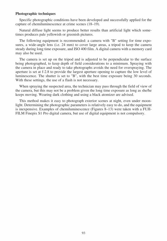
Specific photographic conditions have been developed and successfully applied for thecapture of chemiluminescence at crime scenes (18–19).
Natural diffuse light seems to produce better results than artificial light which some-times produces pale yellowish or greenish pictures.
The following equipment is recommended: a camera with "B" setting for time expo-sures, a wide-angle lens (i.e. 24 mm) to cover large areas, a tripod to keep the camerasteady during long time exposure, and ISO 400 film. A digital camera with a memory cardmay also be used.
The camera is set up on the tripod and is adjusted to be perpendicular to the surfacebeing photographed, to keep depth of field considerations to a minimum. Spraying withthe camera in place and ready to take photographs avoids the need for overspraying. Theaperture is set at f-2.8 to provide the largest aperture opening to capture the low level ofluminescence. The shutter is set to "B", with the best time exposure being 30 seconds.With these settings, the use of a flash is not necessary.
When spraying the suspected area, the technician may pass through the field of view ofthe camera, but this may not be a problem given the long time exposure as long as she/hekeeps moving. Wearing dark clothing and using a black atomizer are advised.
This method makes it easy to photograph exterior scenes at night, even under moon-light. Determining the photographic parameters is relatively easy to do, and the equipmentis inexpensive. Examples of chemiluminescence (Figures 8–13) were taken with a FUJI-FILM Finepix S1 Pro digital camera, but use of digital equipment is not compulsory.
93

94
Figure 8. Latent swiping on tile floor

95
Figure 9. Luminescent latent footwear impressions

96
Figure 10. Close-up photograph of latent luminescent traces on the footwear

97
Figure 11. Evidence of clean up of blood developed on tile floor.

98
Figure 12. Latent swiping on a armchair back

99
Figure 13. Evidence of a clean up of blood on a bathroom floor.


![Luminol [521-31-3] Review of Toxicological Literaturentp.niehs.nih.gov/ntp/htdocs/chem_background/exsumpdf/...Luminol [521-31-3] Review of Toxicological Literature Prepared for Errol](https://static.fdocuments.in/doc/165x107/6104962f4c41ca2cb7557b7b/luminol-521-31-3-review-of-toxicological-luminol-521-31-3-review-of-toxicological.jpg)