A new disease outbreak affecting two dominant sea urchin...
40
New disease outbreak affects two dominant sea urchin species associated with Australian temperate reefs Item type Article Authors Sweet, Michael J.; Bulling, Mark T.; Williamson, Jane E. Citation Sweet, M. et al (2016) 'New disease outbreak affects two dominant sea urchin species associated with Australian temperate reefs', Marine Ecology Progress Series, 551:171 DOI 10.3354/meps11750 Journal Marine Ecology Progress Series Rights Archived with thanks to Marine Ecology Progress Series Downloaded 8-May-2018 23:56:25 Item License http://creativecommons.org/licenses/by/4.0/ Link to item http://hdl.handle.net/10545/618800
Transcript of A new disease outbreak affecting two dominant sea urchin...

New disease outbreak affects two dominant sea urchin speciesassociated with Australian temperate reefs
Item type Article
Authors Sweet, Michael J.; Bulling, Mark T.; Williamson, Jane E.
Citation Sweet, M. et al (2016) 'New disease outbreak affects twodominant sea urchin species associated with Australiantemperate reefs', Marine Ecology Progress Series, 551:171
DOI 10.3354/meps11750
Journal Marine Ecology Progress Series
Rights Archived with thanks to Marine Ecology Progress Series
Downloaded 8-May-2018 23:56:25
Item License http://creativecommons.org/licenses/by/4.0/
Link to item http://hdl.handle.net/10545/618800

1
A new disease outbreak affecting two dominant
sea urchin species associated with Australian
temperate reefs.
Michael Sweet,1*
, Mark Bulling,1, Jane E. Williamson.
2,3
1Molecular Health and Disease Laboratory, Environmental Sustainability Research Centre, College of Life and
Natural Sciences, University of Derby, DE22 1GB, UK 2Department of Biological Sciences, Macquarie University, Sydney, 2109, Australia
3Sydney Institute of Marine Science, Mosman, New South Wales, Sydney, 2088, Australia
*Corresponding author: email: [email protected]

2
Abstract
Diseases of sea urchins have been implicated in dramatic transitions of marine ecosystems.
Although, no definitive causal agent has been found for many of these outbreaks, most are
hypothesised to be waterborne and bacterial. Here we show the first report of a novel
disease affecting at least two species of urchins off the south-eastern coast of Australia. The
etiological agent; identified via a range of molecular techniques, immuno-histology and
inoculation experiments, is found to be the opportunistic pathogen, Vibrio anguillarum. The
disease appears to be temperature-dependent, with a faster transmission rate and increase
in prevalence during experimental trials conducted at higher temperatures. Furthermore,
analysis of long term field data, suggests that it may have already reached epidemic
proportions (R0 = 1.09). With the increases in ocean temperatures brought about by climate
change, this novel urchin disease may pose a severe problem for the organisms associated
with the temperate reefs off Australia and/or the ecosystem as a whole.
KEY WORDS: Sea Urchin / Disease / Vibrio / Henle-Koch postulates
Introduction

3
Occurrences of diseases are reported to be increasing worldwide and, for a diversity of taxa,
large-scale outbreaks of disease have been associated with climate change and other
anthropogenic stresses (Harvell et al. 1999, Campbell et al. 2014). In particular, profound
and rapid changes are occurring at global and regional scales within marine environments.
Mean sea temperature has risen by 0.2 oC per decade (Group 2014) and oceanic pH has
decreased, on average, from 8.2 to 8.1 since the industrial revolution (Zeebe 2012). It is
hypothesised that these changes are stressing marine organisms, causing range shifts in
distribution in many taxa, and leading to increasing susceptibility of these organisms to
opportunistic pathogens (Ainsworth et al. 2010, Campbell et al. 2014). Echinoids, in
particular, have experienced substantial declines associated with disease in the past half
century (Feehan & Scheibling 2014). Urchins are often key components of marine
communities (Sammarco 1982, Scheibling & Hennigar 1997), limiting algal biomass by
extensive grazing and being an important food source for many predators. The result of the
balance between algal and urchin biomass can range from kelp forests to urchin-dominated
‘barrens’. However, transitions can occur between these states as a result of factors
associated with climate change that result in changes to nutrient supply, the number and
type of predators, variation in the frequency and intensities of storms, and increases in
incidence of diseases, for example (Sammarco 1982, Steinberg 1995, Scheibling & Hennigar
1997).
In some instances, urchin diseases are necessary to regulate populations in the absence of
natural predators such as many fish species, a result brought about by overfishing around
the world (Lafferty 2004). However, disease outbreaks can also have large impacts on urchin
populations and the ecosystems they inhabit. In 1983 for example, the dominant urchin of

4
Caribbean coral reefs, Diadema antillarum, experienced massive disease-induced mortality,
and the functional effects of this decline continue to persist today (Mumby et al. 2006).
Prior to the decline, D. antillarium could be found in densities exceeding 70 m-2
(Sammarco
1982) and was considered to be one of the most important grazers on Caribbean reefs
(Mumby et al. 2006; Carpenter 1986). It is now widely agreed, that it’s loss across much of
its range led to a dramatic decline in coral cover throughout the Caribbean (Sammarco
1982, Mumby et al. 2006) and the decline of this urchin is still considered to be the greatest
recorded mass mortality of a marine animal, with functional recovery remaining unlikely
more than 30 years on (Levitan et al. 2014).
On the other side of the world, Australia is a well-known diversity hotspot for sea urchin
species (Miskelly 2002), in particular the temperate waters off the south-eastern coast. Two
of the more abundant species in these ecosystems are Heliocidaris erythrogramma
(Valenciennes, 1846) and Holopneustes purpurascens (Agassiz, 1872). Both species inhabit
shallow subtidal kelp habitats but have different life history characteristics in the manner
that they use the habitat. H. erythrogramma commonly occurs on the substratum,
consuming a range of foliose and crustose algae (Wright et al. 2005), whereas H.
purpurascens lives largely enmeshed in the canopy of the algae it consumes, using it as a
habitat in addition to a food source, thus limiting its distribution to restrictions in diet
breadth (Williamson et al. 2004). Both species have the capacity to substantially impact the
demography and diversity of other flora and fauna in these habitats (Pederson and Johnson
2007, Bell et al. 2014). During regular surveys conducted in this region in 1996, a previously
un-reported disease was noted. The disease was first recorded after unusually high density
aggregations of H. erythrogramma occurred in the previous year (1995), and these high
5
densities continued for a further 3 years (Wright et al. 2000). In this study we document the
long term prevalence of the disease, assess the modes of transmission, aim to identify the
pathogen or pathogens responsible and assess the influence of temperature on
transmission and spread, which is in itself a well-documented abiotic factor increasing at
approximately four times the global average within the region (Ridgway 2007).
Methods
Long term monitoring and current prevalence of lesions
Trends in the prevalence of lesions in Holopneustes purpurascens were assessed using long
term monitoring records, which were collected from 1996 to 2013. Populations associated
with the shallow rocky reef habitat near Bare Island off the coast of Sydney, Australia,
(151°13′52″E, 33°59′32″S) were measured, taking into account; the size of the individual
(diameter), sex of the individual and the number of lesions present. During sampling,
random individuals were collected from the population as H. purpurascens wrap themselves
within the canopy of the algae they inhabit (Steinberg 1995), meaning that any lesions on
individuals are not always obvious at the time of collection in situ. Such random sampling
enables unbiased assessment of the numbers of diseased vs healthy individuals. Animals
were collected at various times throughout each year, usually in groups of 10 or 20.
However, the extent of sampling from year to year was inconsistent (minimum = 14,
maximum = 540, median = 111) and sampling was absent in some years (1999, 2000, 2002,
2007, 2011). To account for this heterogeneity and to ensure robust analysis, only records
from years when over 99 individuals were collected were included in the long term analysis.

6
In addition, we also wanted to assess if there where variations in disease abundance
between sexes. H. purpurascens is usually gonochoristic, with a sex ratio of 1:1 (Williamson
& Steinberg 2002), and this was found in our sampling (χ2 = 0.99, d.f. = 1, p = 0.32). To assess
if there was a sex bias in the presence of lesions, a Chi-squared goodness-of-fit test was
conducted on pooled (years) survey results. Relationships between size (diameter), sex and
presence of lesions were also assessed in a similar way, using logistic regression. Seemingly
both males and females were equally affected by lesions over the 17 years of sampling (Chi-
square = 0.88, 1 df, p = 0.35), and there was no significant relationship between the size of
the individual and the occurrence of lesions over time (Hosmer-Lemeshow = 3.70, 8 df, p =
0.88).
Identification of potential etiological agents
During 2012 and 2013, tissue samples from Holopneustes purpurascens and Heliocidaris
erythrogramma were taken for microbial and histological analysis to describe the
pathogenesis of the characteristic dark lesions (Fig 1) which were first observed to affect the
wild populations in 1996. The aim of this part of the study was to identify any ‘potential’
causal agents associated with the disease. Sterile swabs were used to collected surface
associated microbiota in situ from live animals, whilst a different subset of animals were
collected whole from the field. Both sample types were then stored in 100% ethanol until
extraction and further analysis. Healthy (n = 20) and diseased (n = 20) individuals from both
species were collected from two locations where the disease was prevalent; Bare Island in
Botany Bay (151°13′52″E, 33°59′32″S) and Fairlight in Port Jackson (151°16'32"E, 33°48'1"S).
7
For microbial and histological analysis, samples were separated into healthy tissue (n = 20
from individuals that were free of lesions), apparently healthy tissue (n = 20 tissue from
individuals with lesions present, adjacent to the lesion interface) and n =20 of the tissue
directly associated with the lesions. Tissues from these whole animals were subsequently
removed under sterile conditions and crushed using a mortar and pestle. A further set of n =
20 urchins (n = 10 from both healthy and diseased) from each species were dissected and
the coelomic fluid collected as outlined in (Jellett et al. 1989).
PCR and DGGE
Extraction of the different sample types (healthy tissue, apparently healthy tissue, diseased
tissue, swabs and coelomic fluid) were extracted using the QIAGEN DNeasy blood and tissue
kit. Bacterial 16S rRNA gene diversity was amplified using the primers 357-F (5’-
CAGCACGGGGGGCCTACGGGAGGCAGCAG-3’) and 518-R (5’-ATTACCGCGGCTGCTGG-3’).
Fungal diversity was assessed using a nested approach following the protocol laid out in
(Sweet et al. 2013). In brief, this consisted of an initial amplification using the primers ITS1-F
(5’-CTTGGTCATTTAGAGGAAGTAA-3’) and ITS4-R (5’-TCCTCCGCTTATTGATATGC-3’) followed
by a 1:100 dilution of the PCR product with the primers ITS3-F (5’-
GCATCGATGAAGAACGCAGC-3’) and ITS4-R (5’-TCCTCCGCTTATTGATATGC-3’). Protozoa 18S
rRNA gene diversity was amplified using primers Cil-F (5’-TGGTAGTGTATTGGACWACCA-3’)
and Cil-R-(5’- TGAAAACATCCTTGGCAACTG-3’). PCR protocols were the same as those
outlined in (Sweet & Bythell 2012; Sweet et al. 2013). A GC clamp
(CGCCCGCCGCGCCCCGCGCCCGGCCCGCCGCCCCCGCCCC) was inserted onto the 5’ end of the
primers; 357-F, ITS4-R and Cil-R enabling use in Denaturing Gradient Gel Electrophoresis

8
(DGGE). For each of the above primer pairs, 30 µl PCR mixtures containing; 1.5 mM MgCl2,
0.2 mM dNTP (PROMEGA), 0.5 mM of each primer, 2.5 Ul of Taq DNA polymerase
(QBiogene), incubation buffer and 20 ng of template DNA were utilised (Sweet & Bythell
2012). DGGE was conducted using a 10% (w/v) polyacrylamide gel for bacterial 16S rRNA
gene diversity and 8% (w/v) for fungal and protozoan diversity as in (Sweet & Bythell 2012;
Sweet et al. 2013). Bands of interest (those which explained the greatest
differences/similarities between samples) were excised from DGGE gels, re-amplified using
the same PCR protocols as described above with the same original primers minus the
additional GC clamps needed for DGGE analysis. These were then labelled using Big Dye
(Applied Biosystems) transformation sequence kit, and sent to Genevision (Newcastle
University, UK) for sequencing.
454 Sequencing and Analysis
After the initial results from DGGE analysis, 454 pyrosequencing was conducted for bacterial
diversity only using the same PCR primers as above; 357-F (5’-
CAGCACGGGGGGCCTACGGGAGGCAGCAG-3’) and 518-R (5’-ATTACCGCGGCTGCTGG-3’). PCR
mixtures and conditions were the same as above with the following minor amendments; the
primers used did not have the addition of the GC clamp and HotStarTaq polymerase was
utilised (Qiagen, Valencia, CA). PCR products for 454 were then cleaned using AMPure
magnetic beads (Beckman Coulter Genomics, Danvers, MA). Amplicon samples were then
quantified using the Qubit flourometer (Invitrogen, Carlsbad, CA, USA) and pooled to an
equimolar concentration. Sequences were run on 1/8th
of a 454 FLX Titanium pico-titer plate
at Newgene in the Centre for Life, Newcastle, UK.

9
Pyrosequences were processed using a QIIME pipeline (version 1.5.0) (Caporaso et al. 2010),
using the SILVA reference database (release version SSU/LSU 123). Sequences were filtered
and removed if they were; (i) < 50 nucleotides in length, (ii) contained ambiguous bases
(Ns), and/or (iii) contained primer mismatches. Analysis using 2% single-linkage pre-
clustering (SLP) and average-linkage clustering based on Pairwise alignments (PW-AL) was
performed to remove sequencing based errors. The remaining sequences were de-noised
within QIIME. The resulting reads were checked for chimeras (using UCHIME), and clustered
into >98% similarity operational taxonomic units (OTU) using the USEARCH algorithm also in
QIIME. All singletons (reads found only once in the whole data set) were excluded from
further analyses. After blast searches on GenBank, we retained the best BLAST outputs, i.e.
the most complete identifications, and compiled an OTU table, including all identified OTUs
and respective read abundances. A total of 748,892 raw nucleotide reads were produced
with an average length of 49 bp, corresponding to 223.7 Mb. After filtering, a total of
492,540 quality reads were acquired. The length of the remaining sequences varied from 95
bp to 151 bp, with an average length of 121 bp. Technical control samples were processed
in the same way as above, whereby samples consisting of just ethanol (used for
preservation of the samples) were extracted and sequenced. No DNA was detected either
after PCR or from the downstream processing.
An analysis of similarity (ANOSIM) was conducted to test differences in 16S rRNA gene
bacterial assemblages as described by both the DGGE and 454 sequencing. In addition,
percentage similarity analysis (SIMPER) was performed to determine which OTUs better
explained differences and similarities between the sample types and replicates for both
methods.

10
Culturing of the suspected pathogen and sequence/phylogentic analysis
Crushed samples of freshly acquired healthy and diseased tissue were plated directly (100
µl) in triplicate serial dilutions (1:1, 1:10 and 1:100) on Marine Agar (1.8% Marine Broth
(MB), Difco-2216 USA, 0.9% NaCl, 1.8% Agar Bacto, Difco-214010, USA, MA) and thiosulfate
citrate bile salts sucrose (TCBS) agar. Plates were incubated at 32 oC for 24 hrs as in (Sere et
al. 2015). Cultivable bacterial strains exhibiting a unique colony were then routinely sub-
cultured to purification under the same growth conditions for their subsequent use in
inoculation experiments. Individual colonies were sequenced using the primer pair; 27F
(AGAGTTTGATCMTGGCTCAG) and 1492R (GGTTACCTTGTTACGACTT) following the protocols
outlined in (Sere et al. 2015). Only certain bacteria identified as potential pathogens from
the previous DGGE and NGS were specifically targeted, i.e. from the genus Vibrio. To
confirm identification of the cultured Vibrios, we compared the best match achieved from
the 16S rRNA gene bacterial sequence to the best match achieved from the sequence of the
housekeeping gene pyrH. The housekeeping gene was sequenced using the primers; pyrH-F
(ATGASNACBAAYCCWAAACC) and pyrH-R (GTRAABGCNGMYARRTCCA) following the
protocols outlined in (Sere et al. 2015). The sequences obtained were aligned with reference
sequences related to multiple bacterial strains selected from NCBI using the GENEIOUS
alignment method. Additionally, sequences were concatenated and aligned against
corresponding concatenated sequences (Thompson et al. 2005). Phylogenetic trees were
built by the neighbour-joining method using the NKY model of GENEIOUSTM
Pro (V.6.1.5)
with bootstrap values based on 1000 replicates to further confirm identification of the
pathogenic agent (Supplementary Fig 3).

11
Association of potential pathogens with tissue damage
A further set of samples (n = 10 per sample type for each species) were collected as for
microbial analysis. However, in this instance, samples were preserved with 5%
paraformaldehyde for 24 hrs then stored in 100% EtOH at 4 oC. Half these samples were
embedded in LR white resin (Sigma-Aldrich) after dehydration steps consisting of 30 min
intervals in 25%, 35%, 45%, 55%, 65% & 75% acetone with a final two steps in 100% acetone
for 1 hr each. These samples were used for immuno-histological light microscopy and
transmission electron microscopy (TEM). Survey sections of 1 µm were cut and stained with
the general stain 1% Toluidine Blue in 1% Borax. For each tissue type, the locations of
microorganisms were recorded using 0.01% acridine orange (Smith et al. 2014). The stain
nigrosin (Smith et al. 2014), was also used for evaluating the extent of mass tissue necrosis.
All sections were viewed under epiflourescence microscopy with an FITC-specific filter block
(Nikon UK Ltd, Surrey, UK), and images were recorded using an integrated camera (Model
JVC KY-SSSB: Foster Findlay and Associates, Newcastle upon Tyne, UK).
For transmission electron microscopy (TEM), ultrathin sections (80 nm approx) were cut
using a diamond knife on a RMC MT-XL ultramicrotome. These were then stretched with
chloroform to eliminate compression and mounted on Pioloform filmed copper grids.
Staining was with 2% aqueous Uranyl Acetate and Lead Citrate (Leica). The grids were
examined using a Philips CM 100 Compustage (FEI) transmission electron microscope, and
digital images are collected using an AMT CCD camera (Deben). Bacterial abundance was
measured by sectioning samples (n = 3) of each tissue type of both urchin species and
imaging on the TEM, with fields of view (n = 20) at magnification of x2600.

12
The remainder of the samples (n = 5 per tissue type for each species) were utilised for
scanning electron microscopy (SEM). These were dehydrated in 25%, 35%, 45%, 55%, 65% &
75% ethanol (30 mins at each interval), then a further (2 x 1 hr) in 100% ethanol, with final
dehydration using carbon dioxide in a Baltec Critical Point Dryer. Specimens were then
mounted on an aluminium stub with Achesons Silver Dag, dried overnight and coated with
gold (standard 15nm) using a Polaron SEM Coating Unit. Specimens were examined using a
Stereoscan 240 SEM, and digital images collected by Orion 6.60.6 software.
Transmission of lesions
Experiment 1:
A laboratory experiment was conducted to estimate the potential rate of transmission of
the disease between individuals of Holopneustes purpurascens in the Australian winter (first
trial; June, 2012) and summer (second trial; January, 2013). Individuals (25 – 40 mm) were
collected from kelp (Ecklonia radiata) at Fairlight, immediately placed in aerated seawater
and transported to Macquarie University. Separate collections were carried out for each
trial. Experiments were run under two sets of temperature regimes; one representing
present conditions in the field at the respective time of year (Manly hydraulics data), and a
second set representing predicted future temperatures (a rise of 2 o
C) at the field site (the
IS92 A2 scenario by the IPCC (2007) for 2060; Ridgway 2007). Therefore, temperatures used
during the winter experiment were 19 and 21 o
C, and those used during the summer
experiment were 21 and 23 o
C.

13
At the start of each trial, eight urchins were added to 40 L aerated aquariums that had been
randomly allocated a specific treatment (three aquaria per treatment-health state
combination; temperature treatments (ambient temperature, predicted temperature) and
health states (“no lesion” and “lesion present”)). The number of urchins used per aquaria
was chosen to reflect the density range found naturally at the time of collection of
individuals on E. radiata at Fairlight (Bell et al. 2014). “No lesion” treatments contained
eight H. purpurascens that appeared healthy and had no lesions at the start of the trial.
“Lesion present” treatments included one individual out of the eight with a single small
lesion, with the other seven showing no signs of lesions. Urchins were fed kelp (Ecklonia
radiate) ad libitum, which is their preferred food source(Williamson & Steinberg 2012).
Water was changed approximately three to four times per week and there was no exchange
of water between tanks or treatments. Survivorship and the number of lesions on each
individual were measured at the end of each week. Individuals developing lesions were left
in the aquaria, but those that died were removed on a daily basis to avoid any perceivable
effects of decay processes. Trials were terminated after eight weeks.
Fulfilment of Henle-Koch postulates
Experiment 2:
For this part of the study, sixteen apparently healthy urchins from both species (H.
erythrogramma and H. purpurascens) were kept in individual tanks with the aim of avoiding
any possible confounding effects due to inter-urchin interactions. The tanks were randomly
assigned 1 of 5 treatments, either control (no inoculation), control (inoculation with a non-
14
pathogenic bacterium – Marinobacter aquaeolei [ATCC 700491]) or ‘low’, ‘medium’ or ‘high’
concentrations of the proposed pathogen, Vibrio anguillarum (see below). The non-
pathogenic M. aquaeolei was used solely as a means of testing if the inoculation procedure
affected the urchins in anyway and not a means of testing virulence of the proposed
pathogen. Colonies of V. anguillarum were cultured on vibrio specific growth media (TCBS
agar) (Frans et al. 2011), see above methodology. A single pure colony was then
subsequently cultured in Marine Broth 2216 (Difco), then diluted to ‘low’, ‘medium’ and
‘high’ concentrations corresponding to 102, 10
4 and 10
6 colony-forming units (CFU) ml
–1
(Sweet et al. 2015). Such concentrations were determined from direct plate counts and
comparison of culture turbidity with McFarland standards. Inoculation doses were tested in
the three concentrations as above (cultured concentrations).
Free flowing water was run through the tank systems during the entirety of the trial,
however during the inoculation process, the water flow was stopped, the appropriate
inoculum of cells added to each treatment and the aquaria were individually aerated for 5
hrs (Sweet et al. 2015). During this time, there was no detectable change in temperature of
the aquarium water (21 oC), which was monitored using Hobo Data Loggers. After a 5 hr
exposure, the water flow was re-started. The trial was conducted over 14 d, throughout the
experimental process, visual observations of the contraction and/or extent of external
lesions were recorded and compared to those occurring in the wild. At the end of the 14 d
trial, n = 8 tissue samples were taken for both histology and microbial analysis in the same
manner as that for the wild individuals (described above).
Estimation of R0

15
The estimation of the reproduction number (R0), i.e. the average number of secondary
infections caused by one infected individual, is a critical component of understanding
disease dynamics and making predictions in wild populations. It is usually assumed that a
reproduction number greater than 1 indicates a high probability of an epidemic developing.
However, such estimations of R0 are often difficult to make as they are based on few data
points. Furthermore, most methods of estimation require the generation time of the
pathogen, the mean time lag between infection in a primary case and that in a secondary
case (Obadia et al. 2012). Here, we did not have this information for the wild populations so
we utilised a simpler but less refined method, as outlined in (Diekmann et al. 2013). This
method is based on the estimation of the exponential growth rate, r. We assumed a closed
population and a susceptible-infected-removed (SIR) disease dynamic. Under these
circumstances, if the incidence grows exponentially, R0 is related to r in the following way,
where γ is the mortality rate;
r = (R0 - 1)γ (1)
The minimum period between collections of diseased individuals in the field was
approximately one year and, given the rapid mortality of specimens in controlled aquaria
experiments (see above), it is reasonable to assume a mortality rate of 1 between samples.
Exponential growth of disease incidence can then be described by;
incidence (t) = ert (2)
Where t is time and incidence (t), is the incidence at time t. By taking the natural log of both
sides of the equation we can obtain the linear form;
ln(incidence (t)) = rt (3)

16
Thus, r can be estimated using the beta coefficient (the slope) of a linear regression, with ln
(incidence) as the dependent variable and time as the independent variable. This value can
then be substituted into equation (1) to estimate R0. R0 is best estimated over the initial
spread of the disease. We therefore calculated two estimates, one based on records up to
and including 2005, and the second based on all records up to and including 2013, which
allowed us to also examine the consistency of the estimation.
Results and Discussion
Natural occurrence, transmission and spread of the disease
Two urchin species, Heliocidaris erythrogramma and Holopneustes purpurascens, are
prominent grazers throughout shallow subtidal rocky reefs around south-eastern Australia
(Wright et al. 2005). Although much is known of their ecology and biology (Williamson &
Steinberg 2012, Keesing 2013), disease has not been identified as an issue for concern until
this study. Diseased individuals of both species were first recorded during routine sampling
in 1996. Inflicted urchins exhibited symptoms consisting of distinctive dark mucoid lesions
on the epidermal tissue overlying the test and a loss of spines around the lesion (Fig1 a and
b). This pathology shares numerous similarities with the well documented diseases
described for D. antillarium in the 1980’s outbreak throughout the Caribbean and a more
recent disease outbreak for D. africanum in the subtropical eastern Atlantic(Lessios et al.
1984, Lessios 1988, Clemente et al. 2014). Here, we show that, from the first record of this
disease, incidence of the number of individuals displaying lesions have increased, although
the estimates of incidence have not always increased from year to year (Fig 2). Furthermore,
17
there is also a positive correlation between the number of diseased individuals observed
and increases in the SST within the region (Fig 2), again a result reflective of many other
diseases in the marine environment. In order to test if there is indeed a relationship
between increases in SST and disease occurrence (Fig 2), a laboratory experiment was
conducted to estimate the potential rate of transmission of the disease between individuals
of one of the species, H. purpurascens in both the Australian winter (first trial; June, 2012)
and summer (second trial; January, 2013). Experiments were run under two sets of
temperature regimes; one representing present conditions in the field at the respective
time of year (Manly hydraulics data), and a second set representing predicted future
temperatures (a rise of 2 o
C) at the field site (the IS92 A2 scenario by the IPCC (2007) for
2060). In these experimental trials, all urchins that were exposed to individuals with lesions
exhibited symptoms of the disease within an eight week period, regardless of the season
(Fig 3), highlighting that an increase in temperature is not required for individuals to
contract the disease. However, symptom development (as indicated by the presence of
lesions), was faster in treatments at elevated temperatures than those at ambient
temperatures (Fig 3). In all instances, urchins that developed lesions either developed a
greater number of lesions over time or died. It must be noted that, one individual developed
a lesion in the control tanks, however this did occur in the summer during a period of
elevated temperature (Fig 3) and the individual was most likely infected in the wild but not
identified as such before the experiment was initiated. Although, the limitations of
mesocosm experiments such as this one (Spivak et al. 2014) cannot be ignored, we believe
that the results shown here highlight the importance of temperature as a driver of disease
spread and/or transmission, with a faster rate of transmission and earlier mortality being
observed at higher temperatures (Fig 3). Although, we cannot say for sure that an increase

18
in temperature in the environment has resulted in the observed increase in disease
incidence, records do show that since the early 1990’s, sea surface temperature from
satellites positioned over the sample site at Bare Island, have risen on average by 0.52 oC to
date (Fig 2) (Wray-Barnes 2014). Furthermore, although the overall process of transmission
in our study cannot be concluded from experiments such as those conducted here, we
hypothesise that the causal agent in this study is waterborne as lesions appeared to spread
(during Experiment 1) without contact being necessary. Again, such a result mirrors initial
observations made during the 1980’s Diadema die off (Hunte et al. 1986, Clemente et al.
2014). However, to confirm such a hypothesis, further tests of the surrounding water-
column, both in the field and in any further experimental trials, would be necessary. This
would allow us to assign presence of the proposed pathogen within the environment.
Identification of potential pathogens associated with the disease
Both DGGE and 454 pyro-sequencing using rDNA amplicons were utilised to assess the
microbial communities associated with healthy urchins, and those associated with diseased
tissue, apparently healthy tissue (those associated with tissue on diseased individuals that
still appeared healthy) and the coelomic fluid of both H. erythrogramma and H.
purpurascens (see methodology). This was conducted in order to assign potential microbial
causal agents to this disease. No protozoans were detected in any of the tissues and/or the
coelomic fluid and only one fungal ribotype (a Pleosporales sp.) was detected on any of the
urchin tissues and this was consistently detected in all samples from both species. In
contrast, a much greater diversity of bacterial OTUs were detected from all sample types.
Furthermore, there were significant differences between the bacterial diversity of healthy,

19
apparently healthy and diseased tissue for urchins from both species (H. erythrogramma
and H. purpurascens; ANOSIM, R = 1, p = 0.001 and R = 1, p = 0.002 respectively – based on
454 pyrosequencing data). Both the DGGE profiles and 454 sequence analysis showed
similar significant differences between groupings, however we only report results from the
454 pyrosequencing from hereinafter. In total, eleven bacterial OTUs were detected on
healthy individuals. These were consistent between replicates of both species and are likely
to be surface associated as there were no significant differences between swab samples (i.e.
surface associated bacterium) and those of the ‘complete’ crushed tissue samples (R = 0.97,
p = 0.12). From these eleven bacterial OTUs found on healthy, non-diseased individuals (see
the heatmap in Supplementary Table 1 for full detail on NGS OTUs found between the
samples); a Mycobacterium, a Comamonadaceae and a Xanthomonadaceae were detected
in all replicates of the healthy tissue (non-diseased) samples and only in one of the species,
H. purpurascens (Supplementary Table 1). The bacterial community associated with these
‘apparently healthy’ tissues (i.e. visibly healthy tissues on a diseased individual) showed an
increase in the bacterial diversity yet were more similar (between replicates) to those
associated with healthy tissues than to diseased tissues, despite originating from an infected
individual (Supplementary Table 1). Differences were observed in the bacterial communities
between apparently healthy tissues from the two urchin species, with H. purpurascens
harbouring 31 individual OTUs compared to 25 in H. erythrogramma. Only four OTUs were
found to occur in the apparently healthy tissue and were absent in healthy tissues across
both urchin species, including; a Micrococcaceae sp, a Tenacibaculum sp, a
Kordiimonadaceae sp and a Vibrio sp (Supplementary Table 1). The diseased tissues
harboured an even greater diversity of bacteria with many bacterial OTUs present between
both species (Supplementary Table 1). However, only the Vibrio, was consistently detected

20
in both diseased urchins and across all replicate samples, as might be expected of a causal
agent. Vibrios are routinely detected as etiological agents and are ubiquitous in the marine
environment (Morris & Acheson 2003). Indeed, a recent study by Clemente et al. (2014)
highlighted that a potential causal agent for the recent mass mortality event effecting up to
65% of D. africanum is another member of this group, V. alginolyticus (Clemente et al. 2014).
Interestingly, we also detected bacteria present in the coelomic fluid of two of the diseased
urchins sampled in this study. However, only one bacterial species was detected within
these samples which was the same Vibrio detected in the diseased tissues. These same
samples showed a more advanced stage of the disease, evidenced by the histological
sections (Fig 4 d,e). In all other samples tested, there were no other detectable microbes
(bacteria, fungi or protozoan) within this area, leading us to suggest that in these individuals
a more advanced stage of the disease was present. In healthy urchins the coelomic fluid is
usually a sterile environment (Kaneshiro & Karp 1980), maintained by both bactericidal
properties of the coelomic fluid (Turton & Wardlaw 1987) and by coelomocytes that
phagocytise foreign organisms and particles (Dybas & Fankboner 1986). This finding
corresponds with previous studies which have found similar pathogenicity for two other
urchin diseases i.e. bald sea urchin disease (Maes & Jangoux 1984, Maes & Jangoux 1985)
and deep sea vibrio urchin disease (Bauer & Young 2000), whereby the coelomic fluid of
these urchins were also infected by bacteria during disease outbreaks.
Association of potential pathogens with tissue damage
21
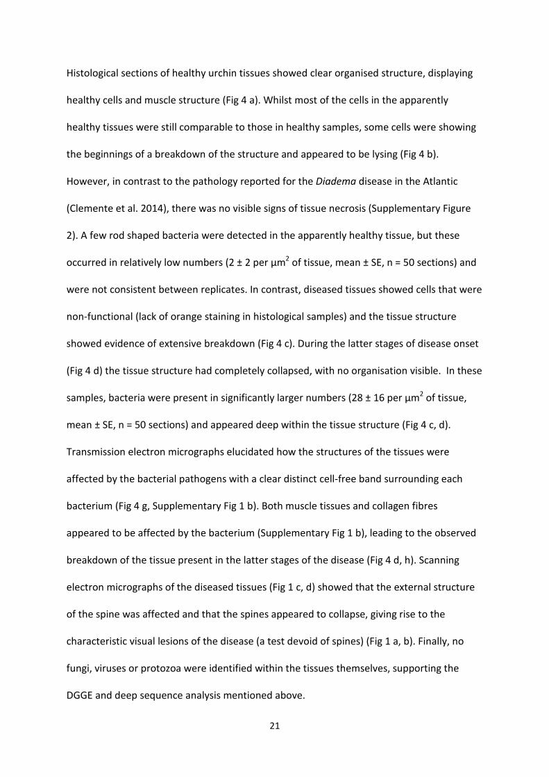
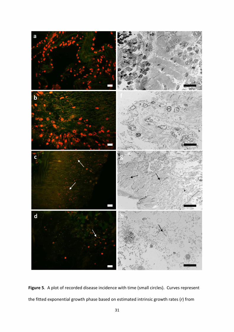
Histological sections of healthy urchin tissues showed clear organised structure, displaying
healthy cells and muscle structure (Fig 4 a). Whilst most of the cells in the apparently
healthy tissues were still comparable to those in healthy samples, some cells were showing
the beginnings of a breakdown of the structure and appeared to be lysing (Fig 4 b).
However, in contrast to the pathology reported for the Diadema disease in the Atlantic
(Clemente et al. 2014), there was no visible signs of tissue necrosis (Supplementary Figure
2). A few rod shaped bacteria were detected in the apparently healthy tissue, but these
occurred in relatively low numbers (2 ± 2 per µm2 of tissue, mean ± SE, n = 50 sections) and
were not consistent between replicates. In contrast, diseased tissues showed cells that were
non-functional (lack of orange staining in histological samples) and the tissue structure
showed evidence of extensive breakdown (Fig 4 c). During the latter stages of disease onset
(Fig 4 d) the tissue structure had completely collapsed, with no organisation visible. In these
samples, bacteria were present in significantly larger numbers (28 ± 16 per µm2 of tissue,
mean ± SE, n = 50 sections) and appeared deep within the tissue structure (Fig 4 c, d).
Transmission electron micrographs elucidated how the structures of the tissues were
affected by the bacterial pathogens with a clear distinct cell-free band surrounding each
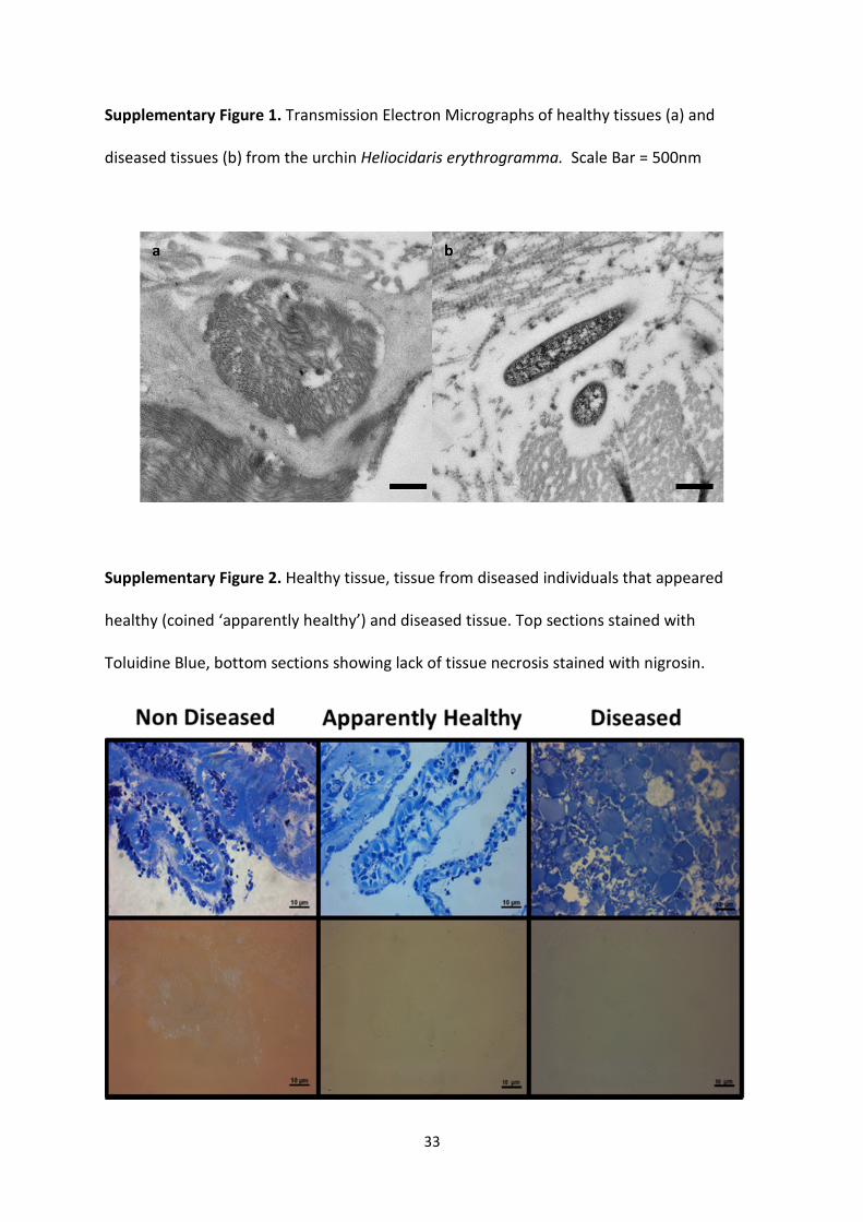
bacterium (Fig 4 g, Supplementary Fig 1 b). Both muscle tissues and collagen fibres
appeared to be affected by the bacterium (Supplementary Fig 1 b), leading to the observed
breakdown of the tissue present in the latter stages of the disease (Fig 4 d, h). Scanning
electron micrographs of the diseased tissues (Fig 1 c, d) showed that the external structure
of the spine was affected and that the spines appeared to collapse, giving rise to the
characteristic visual lesions of the disease (a test devoid of spines) (Fig 1 a, b). Finally, no
fungi, viruses or protozoa were identified within the tissues themselves, supporting the
DGGE and deep sequence analysis mentioned above.

22
Fulfilment of Henle-Koch postulates
The evidence from the bacterial profiling highlighted that a member of the genus Vibrio was
the most likely pathogen responsible for this disease (see above). This Vibrio OTU was
absent in healthy tissues and present in all apparently healthy and diseased tissues sampled,
from both species. Furthermore, Vibrios were only able to be cultured from diseased tissue
adding further support to this theory. A subset of the cultured Vibrio colonies were
sequenced and all were identified (using both the 16S and pyrH house-keeping gene – see
methodology) as V. anguillarum (submitted to GenBank under the unique Accession No.
KP001554). A pure culture of this bacterium was then utilised in the inoculation trials to
fulfil Koch’s postulates and confirm pathogenesis (Experiment 2 methodologies). Disease
signs were induced in less than 48 hrs after inoculation. Upon the appearance of visual
lesions (which were similar to those associated with the diseased individuals in the wild), all
urchins ultimately succumbed within a further 76 hrs of contracting the disease. None of the
un-inoculated controls or those inoculated with the non-pathogenic bacterium died during
the experiment and none showed any visible signs of ill health. However, as discussed
above, we cannot discount the potential effects the aquarium setup had on the urchins. It is
therefore likely that the rate of death in the wild would be slower than that observed in
these experiments and further monitoring of diseased individuals in situ must be conducted.
To fulfil Koch’s postulates, the same pathogen was re-isolated from experimentally diseased
individuals and matched to the original inoculum (Hogue 1971). This evidence further
supports the role of Vibrios in this disease, in particular highlighting V. anguillarum as the
likely causal agent in this study. This finding compliments two previous studies (Gilles &

23
Pearse 1986, Becker et al. 2007), which also showed that Vibrios are responsible for diseases
associated with two other urchin species; Strongylocentrotus purpuratus (Gilles & Pearse
1986) and Tripneustes gratilla (Becker et al. 2007). Furthermore, the pathogenicity of this
specific Vibrio, V. anguillarum has been established for other marine organisms from a
diverse range of taxa, including eels, oysters, fish and lobsters (Joseph et al. 2014, Tan et al.
2014, Wendling et al. 2014).
Modelling the current spread of the disease in situ
We also aimed to model the average number of secondary infections caused by any one
infected individual at any given time in situ (also known as the reproduction number or R0).
Such information is a critical component to understanding disease dynamics in any system.
A reproduction number greater than 1 for example, indicates a high probability of an
epidemic developing. In this instance the intrinsic rate of increase (r) was estimated at
0.0961 using the full temporal span of the incidence records (from 1996 to 2013), and
0.1019 using the data up to and including that for 2005 (see methodology). Incidence data
and fitted exponential curves suggested values of R0 as 1.096 (95% CI = ± 0.0139) and 1.10
(95% CI = ± 0.0405) (Fig5). This indicates a strong possibility that a disease epidemic may
occur, or already has. Although we acknowledge that such estimates for epidemics should
be treated with caution, namely as a number of assumptions have to be made during the
estimation process, our long-term sampling shows an increasing trend in disease incidence
over the 17 year period (although not always a year on year basis), with an incidence of over
0.1 recorded in 2012 (Fig 5). Moreover, for this model we also had to assume that the
population was closed, this is actually conservative and probably unlikely as both species are
24
free spawners. Open populations promote re-infection of recovered patches from infected
areas, therefore there is substantial potential for spatial dynamics to increase the likelihood
of disease persistence over and above our current ‘conservative’ estimate.
To conclude, this study reports a disease of two common sea urchins in south-eastern
Australia, and identifies the causal agent as Vibrio anguillarum. The symptomatology and
transmission characteristics of this reported disease share similarities with the two Diadema
die offs in the Caribbean and the Atlantic, and two further diseases reported to affect
another two urchin species; S. purpuratus and T. gratilla. Our results support the now
widely accepted view that transmission of many diseases in the marine environment and
virulence of specific pathogens such as those from the genus Vibrio increase at elevated
temperatures (Vezzulli et al. 2010, Frans et al. 2011). Therefore, as is the same with many
other marine diseases associated with other marine taxa, this disease is likely to increase in
severity and extent as sea surface temperatures rise, a result due to climate change.
However, we suggest that future studies should assess the pathogenicity of different
isolated strains of V. anguillarum, as this was not tested in this current study and may have
important implications regarding the spread of this disease.
Acknowledgements
Animals were collected under scientific collection permits from the Department of Primary
Industries, New South Wales. We thank Alexander Wray-Barnes for use of his long-term
dataset on sea surface temperature. This research was partly funded by the Department of
Biological Sciences at Macquarie University, and is contribution number 171 from the
Sydney Institute of Marine Science.
25
Author’s contributions
MS conducted the design of the experiment, conducted parts of the field work, carried out
the molecular lab work, data analysis, sequence filtering and alignment; MB carried out the
statistical analyses and modelling; JW collected field data and samples and conducted the
temperature challenge experiments. All authors contributed in the writing of the manuscript
and gave final approval for publication.
There are no conflicting/competing interests of the authors associated with this manuscript.
Data accessibility
The datasets supporting this article have been uploaded as part of the supplementary
material.
References
Ainsworth TD, Thurber RV, Gates RD (2010) The future of coral reefs: a microbial perspective. Trends
in Ecology & Evolution 25:233-240
Bauer JC, Young CM (2000) Epidermal lesions and mortality caused by vibriosis in deep-sea
Bahamian echinoids: a laboratory study. Diseases of aquatic organisms 39:193-199
Becker P, Gillan DC, Eeckhaut I (2007) Microbiological study of the body wall lesions of the echinoid
Tripneustes gratilla. Diseases of aquatic organisms 77:73-82
Bell JE, Bishop MJ, Taylor RB, Williamson JE (2014) Facilitation cascade maintains a kelp community.
Marine Ecology Progress Series 501:1-10
Campbell AH, Harder T, Nielsen S, Kjelleberg S, Steinberg PD (2011) Climate change and disease:
bleaching of a chemically defended seaweed. Global Change Biology 17:2958-2970
Campbell AH, Vergés A, Steinberg PD (2014) Demographic consequences of disease in a habitat-
forming seaweed and impacts on interactions between natural enemies. Ecology 95:142-152
Caporaso G, Kuczynski J, Stombaugh J, Bittinger K, FD B (2010) QIIME allows analysis of high-
throughput community sequencing data. Nature Methods 7:335-336
Carpenter RC (1986) Partitioning herbivory and its effects on coral reef algal communities. Ecological
Monogaphs. 56, 345–363

26
Clemente S, Lorenzo-Morales J, Mendoza J, López C, Sangil C, Alves F, Kaufmann M, Hernández J
(2014) Sea urchin Diadema africanum mass mortality in the subtropical eastern Atlantic: role
of waterborne bacteria in a warming ocean. Marine Ecology Progress Series 506:1-14
Diekmann O, Heesterbeek H, Britton T (2013) Book Review Mathematical Tools for Understanding
Infectious Disease Dynamics. IMA
Dybas L, Fankboner PV (1986) Holothurian survival strategies: Mechanisms for the maintenance of a
bacteriostatic environment in the coelomic cavity of the sea cucumber,
Parastichopuscalifornicus. Developmental & Comparative Immunology 10:311-330
Feehan CJ, Scheibling RE (2014) Effects of sea urchin disease on coastal marine ecosystems. Mar
Biol:1-19
Frans I, Michiels CW, Bossier P, Willems KA, Lievens B, Rediers H (2011) Vibrio anguillarum as a fish
pathogen: virulence factors, diagnosis and prevention. Journal of Fish Diseases 34:643-661
Gilles KW, Pearse JS (1986) Disease in sea urchins Strongylocentrotus purpuratus. Experimental
infection and bacterial virulence Diseases of Aquatic Organisms 1:105-114
Group W (2014) International Panel on climate change.
Harvell CD, Kim K, Burkholder JM, Colwell RR, Epstein PR, Grimes DJ, Hofmann EE, Lipp EK, Osterhaus
ADME, Overstreet RM, Porter JW, Smith GW, Vasta GR (1999) Emerging marine diseases -
Climate links and anthropogenic factors. Marine Ecology 285:1505-1510
Hogue RS (1971) DEMONSTRATION OF KOCHS POSTULATES. American Biology Teacher 33:174-&
Hunte W, Côté I, Tomascik T (1986) On the dynamics of the mass mortality of Diadema antillarum in
Barbados. Coral Reefs 4:135-139
Jellett JF, Wardlaw AC, Scheibling RE (1989) 8.3 Experimental infection of the sea urchin
(Strongylocentrotus droebachiensis) with a pathogenic amoeba (paramoeba invadens) :
quantitative changes in the coelomic fluid In vivo and cellular interaction in vitro.
Developmental and Comparative Immunology 13:428-429
Joseph FS, Latha N, Ravichandran S, Devi AS, Sivasubramanian K (2014) Shell disease in the
Freshwater crab, Barytelphusa cunicularis.
Kaneshiro ES, Karp RD (1980) The ultrastructure of coelomocytes of the sea star Dermasterias
imbricata. The Biological Bulletin 159:295-310
Keesing JK (2013) Heliocidaris erythrogramma. Sea Urchins: Biology and Ecology 38:369
Lafferty KD (2004) Fishing for lobsters indirectly increases epidemics in sea urchins. Ecological
Applications 14:1566-1573
Lessios H (1988) Mass mortality of Diadema antillarum in the Caribbean: what have we learned?
Annual Review of Ecology and Systematics:371-393
Lessios HA, Cubit JD, Robertson DR, Shulman MJ, Parker MR, Garrity SD, Levings SC (1984) Mass
mortality of Diadema antillarum on the Caribbean coast of Panama. Coral Reefs 3:173-182
Levitan DR, Edmunds PJ, Levitan KE (2014) What makes a species common? No evidence of density-
dependent recruitment or mortality of the sea urchin Diadema antillarum after the 1983–
1984 mass mortality. Oecologia:1-12
Maes P, Jangoux M (1984) The bald-sea-urchin disease: a biopathological approach. Helgoländer
Meeresuntersuchungen 37:217-224
Maes P, Jangoux M The bald-sea-urchin disease: a bacterial infection. Proc Echinodermata:
Proceedings of the 5th International Echinoderm Conference AA Balkema, Rotterdam
Miskelly (2002) https://www.bookdepository.com/Sea-Urchins-Australia-Indo-Pacific-Ashley-
Miskelly/9780957745568
Morris JG, Acheson D (2003) Cholera and other types of vibriosis: a story of human pandemics and
oysters on the half shell. Clinical Infectious Diseases 37:272-280
Mumby PJ, Hedley JD, Zychaluk K, Harborne AR, Blackwell PG (2006) Revisiting the catastrophic die-
off of the urchin Diadema antillarum on Caribbean coral reefs: Fresh insights on resilience
from a simulation model. Ecological Modelling 196:131-148

27
Obadia T, Haneef R, Boëlle P-Y (2012) The R0 package: a toolbox to estimate reproduction numbers
for epidemic outbreaks. BMC medical informatics and decision making 12:147
Pederson HG, Johnson CR (2007) Growth and age structure of sea urchins (Heliocidaris
erythrogramma) in complex barrens and native macroalgal beds in eastern Tasmania. ICES
Journal of Marine Science 65:1–11
Ridgway KR (2007) Long-term trend and decadal variability of the southward penetration of the East
Australian Current. Geophysical Research Letters 34: 13
Sammarco PW (1982) Echinoid grazing as a structuring force in coral communities: whole reef
manipulations. Journal of Experimental Marine Biology and Ecology 61:31-55
Scheibling RE, Hennigar AW (1997) Recurrent outbreaks of disease in sea urchins Strongylocentrotus
droebachiensis in Nova Scotia: Evidence for a link with large-scale meteorologic and
oceanographic events. Marine Ecology Progress series 152:155-165
Sere MG, Tortosa P, Chabanet P, Quod JP, Sweet MJ, Schleyer MH (2015) Identification of a bacterial
pathogen associated with Porites white patch syndrome in the Western Indian Ocean.
Molecular Ecology 24:4570-4581
Smith D, Leary P, Bendall M, Flach E, Jones R, Sweet M (2014) A Novel Investigation of a Blister-Like
Syndrome in Aquarium Echinopora lamellosa. PloS one 9:e97018
Spivak AC, Vanni MJ, Mette EM (2014) Moving on up: can results from simple aquatic mesocosm
experiments be applied across broad spatial scales? Freshwater Biology 56:279-291
Steinberg PD (1995) Interaction between the canopy dwelling echinoid Holopneustes purpurescens
and its host kelp. Marine Ecology Progress Series 127:169-181
Sweet M, Bulling M, Cerrano C (2015) A novel sponge disease caused by a consortium of micro-
organisms. Coral Reefs:1-13
Sweet M, Burn D, Croquer A, Leary P (2013) Characterisation of the Bacterial and Fungal
Communities Associated with Different Lesion Sizes of Dark Spot Syndrome Occurring in the
Coral Stephanocoenia intersepta. PloS one 8:e62580
Sweet MJ, Bythell J (2012) Ciliate and bacterial communities associated with White Syndrome and
Brown Band Disease in reef building corals. Environ Microbiol 14:2184-2199
Tan D, Gram L, Middelboe M (2014) Vibriophages and their interactions with the fish pathogen
Vibrio anguillarum. Applied and environmental microbiology 80:3128-3140
Thompson F, Gevers D, Thompson C, Dawyndt P, Naser S, Hoste B, Munn C, Swings J (2005)
Phylogeny and molecular identification of vibrios on the basis of multilocus sequence
analysis. Appl Environ Microbiol 71:5107-5115
Turton GC, Wardlaw AC (1987) Pathogenicity of the marine yeasts Metschnikowia zobelli and
Rhodotorula rubra for the sea urchin Echinus esculentus. Aquaculture 67:199-202
Vezzulli L, Previati M, Pruzzo C, Marchese A, Bourne DG, Cerrano C, VibrioSea C (2010) Vibrio
infections triggering mass mortality events in a warming Mediterranean Sea. Environmental
Microbiology 12:2007-2019
Wendling CC, Batista FM, Wegner KM (2014) Persistence, seasonal dynamics and pathogenic
potential of Vibrio communities from Pacific oyster hemolymph. PloS one 9:e94256
Williamson J, Steinberg P (2002) Reproductive cycle of the sea urchin Holopneustes purpurascens
(Temnopleuridae: Echinodermata). Mar Biol 140:519-532
Williamson JE, Carson DG, de Nys R, Steinberg PD (2004) Demographic consequences of an
ontogenetic shift by a sea urchin in response to host plant chemistry. Ecology 85:1355-1371
Williamson JE, Steinberg PD (2012) Fitness benefits of size-dependent diet switching in a marine
herbivore. Mar Biol 159:1001-1010
Wray-Barnes A (2014) Sea Surface Temperature off Bare Island, New South Wales, Australia
(Unpublished data). . University of Newcastle, Ourimbah, New South Wales, Australia
Wright J, De Nys R, Steinberg P (2000) Geographic variation in halogenated furanones from the red
alga Delisea pulchra and associated herbivores and epiphytes: Marine chemical ecology.
Marine ecology Progress series 207:227-241

28
Wright JT, Dworjanyn SA, Rogers CN, Steinberg PD, Williamson JE, Poore AG (2005) Density-
dependent sea urchin grazing: differential removal of species, changes in community
composition and alternative community states. Marine Ecology Progress Series 298:143-156
Zeebe RE (2012) History of seawater carbonate chemistry, atmospheric CO2, and ocean acidification.
Annual Review of Earth and Planetary Sciences 40:141-165
Figures
Figure 1. a. Diseased individual showing the characteristic dark mucoid lesions on the
epidermal tissue overlying the test, b. loss of spines often occur around the lesion and
spread outwards in a circular pattern. Photo by Paul Edward Duckett. c and d Scanning
Electron Micrographs of diseased tissues, showing the effect on the surface of the spines.
Figure 2. Illustrates the increase in proportion of individuals with lesions with sea surface
temperature, highlighting a positive correlation between the two.

29
Figure 3. Experimental trials, illustrating contraction of the disease (i.e., occurrence of
lesions) and survivorship for Holopneustes purpurascens. a and b illustrate the proportion of
live individuals with lesions over the 8 week trial period for the winter (a) and summer (b)
periods. c and d illustrate survivorship over the same 8 weeks for winter (c) and summer (d).
Open Circle = no lesions at t + 0°C, closed circle = lesions at t + 0°C, open triangle = no lesion
at t + 2°C, closed triangle = lesions at t + 2°C.
y = 5E-05x + 20.002
R² = 0.0031
0
1
2
3
4
5
6
7
8
9
10
14
16
18
20
22
24
19
90
19
91
19
92
19
93
19
94
19
95
19
96
19
97
19
98
19
99
20
00
20
01
20
02
20
03
20
04
20
05
20
06
20
07
20
08
20
09
20
10
20
11
20
12
20
13
Pro
po
rtio
n o
f in
did
ivu
als
wit
h l
esi
on
s (%
)
Se
a s
urf
ace
te
mp
era
ture
(°C
)
Year
SST (°C) % lesioned indidivuals Linear (SST (°C))

30
Figure 4. Representative histological sections of healthy, apparently healthy and two
different stages of diseased urchin tissues (a-d stained with acridine orange, e-h
Transmission Electron Micrographs). a and e represent healthy H. erythrogramma tissue, b
and f represent apparently healthy tissue from a diseased individual, c and g show the early
stage of disease taken at the disease lesion itself, and d and h show the later stages of
disease onset. Arrows highlight the presence of bacteria. Scale bar 10µm.
31
Figure 5. A plot of recorded disease incidence with time (small circles). Curves represent
the fitted exponential growth phase based on estimated intrinsic growth rates (r) from

32
linear regression, using all records to 2013 (solid line) and records up to and including 2005
(dashed line).
33
Supplementary Figure 1. Transmission Electron Micrographs of healthy tissues (a) and
diseased tissues (b) from the urchin Heliocidaris erythrogramma. Scale Bar = 500nm
Supplementary Figure 2. Healthy tissue, tissue from diseased individuals that appeared
healthy (coined ‘apparently healthy’) and diseased tissue. Top sections stained with
Toluidine Blue, bottom sections showing lack of tissue necrosis stained with nigrosin.

34
Supplementary Figure 3. Phylogenetic trees illustrating the cultured bacterium used in
inoculation trials in relation to reference sequences from NCBI; (a) represents sequences
from the 16S rRNA gene and (b) the pyrH housekeeping gene. All sequences were aligned
using GENEIOUS alignment. The trees were built by the neighbour-joining method using the
NKY model of GENEIOUSTM
Pro (V.6.1.5) with bootstrap values based on 1000 replicates to
further confirm identification of the pathogenic agent
Supplementary Table 1. Heatmap, showing a representative number of samples of the 16S
rRNA gene bacterial clone libraries of healthy (A), tissue from diseased individuals that
appeared healthy (AH), and diseased (DL) tissues of two species of urchins H.
erythrogramma and H. purpurascens. The heatmap contains the unique accession number
of the individual retrieved sequences, their closest ID to species level where possible and
their closets match to known sequences on GenBank. The darker the colour the higher the

35
abundance of sequences matching that ribotype were discovered present within each
sample. N = 4 replicates per sample type were show in this image for ease of viewing.

36
Heliocidaris erythrogramma Holopneustes purpurascens
Accession
Number ID
H
H
H
H
AH
AH
AH
AH
DL
DL
DL
DL
H
H
H
H
AH
AH
AH
AH
DL
DL
DL
DL
KF057002 Chloroflexi sp
KF057003 Methanotroph sp
KF057004 Trichodesmium sp
KF057005 Nostoc sp
KF057006 Roseiflexus sp
KF057007 Rhodobium sp
KF057008 Candidatus sp
KF057009 Nitrosococcus sp
KF057010 Desulfurococcus sp
KF057011 Methylobacterium sp
KF057012 Capnocytophaga sp
KF057013 Endozoicomonas sp
KF057014 Lysobacter sp
KF057015 Ideonella sp

37
KF057016 Teredinibacter sp
KF057017 Dethiosulfovibrio sp
KF057018 Meiothermus sp
KF057019 Flavobacterium sp
KF057020 Desulfosarcina sp
KF057021 Flavobacterium sp
KF057022 Marinobacter sp
KF057023 Vibrio sp
KF057024 Tindallia sp
KF057025 Leptospira sp
KF057026 Vibrio sp
KF057027 Mycobacterium sp
KF057028
Streptosporangiaceae
sp
KF057029 Comamonadaceae sp
KF057030 Pseudomonas sp
KF057031 Xanthomonadaceae sp
KF057032 Micrococcaceae sp
KF057033 Microscilla sp
KF057034 Flexithrix sp
KF057035 Reichenbachiella sp

38
KF057036 Roseivirga sp
KF057037 Fluviicola sp
KF057038 Polaribacter sp
KF057039 Tenacibaculum sp
KF057040 Kordiimonadaceae sp
KF057041 Rhizobiales sp
KF057042 Phaeobacter sp
KF057043 Sphingomonas sp

39