
A new bacterial blight of pomegranate caused by ... · Pashan,Pune– 411,021, Maharashtra, India...
6
A new bacterial blight of pomegranate caused by Pseudomonas sp. in Maharashtra, India Smita B. Jagdale 1 & Mahesh S. Sonawane 2 & Balu P. Kapadnis 1 Received: 24 January 2018 /Accepted: 30 July 2018 /Published online: 3 August 2018 # Australasian Plant Pathology Society Inc. 2018 Abstract In the past few decades, pomegranate production in India has greatly suffered from a highly catastrophic bacterial blight disease caused by Xanthomonas axonopodis pv. punicae. Various management strategies have failed to control the disease. During surveys carried out in Maharashtra, India in 2010–2015, an unusual yellow pigmented bacterium was consistently associated with pomegranate showing blight symptoms. Based on morphological, biochemical, 16S rRNA and gyrase B gene sequencing, the bacterium was identified as Pseudomonas sp. Pathogenicity was assessed both by a detached leaf assay method (in vitro) and in pomegranate plantlets (in vivo). To the best of our knowledge, we report Pseudomonas sp. as a cause of new bacterial blight disease of pomegranate in Maharashtra, India in addition to Xanthomonas axonopodis pv. punicae. Keywords Bacterial blight . Pomegranate . Pseudomonas sp. . India Pomegranate is one of the major cash fruit crops grown in India. The blight disease of pomegranate, when first reported, was known to be caused by Xanthomonas axonopodis pv. punicae (Hingorani and Mehta 1952; Hingorani and Singh 1960). Over the past few decades, bacterial blight has created havoc among pomegranate cultivators in India especially in Maharashtra, Andhra Pradesh and Karnataka states. The dis- ease results in a huge loss (60–80%) of economy due to de- struction of the fruit. Sometimes the bacterial blight results in complete loss of the orchards (Kumar et al. 2009; Mondal and Mani 2009). Current management practices have not been successful in controlling the disease and it is fast spreading throughout the pomegranate cultivating regions, mainly in Maharashtra, India. A total of 130 samples of infected pomegranate plant parts (leaves, fruits, stems and flowers) were collected from 10 differ- ent fields of various pomegranate growing regions of Maharashtra (Satana - 30, Malegaon - 31, Solapur - 32, Rahuri - 20, and Purandar - 17 samples), India (Supplementary Fig. S1). The infected pomegranate parts viz., leaves, fruits and stems (Supplementary Fig. S2) were disinfected using 2% sodium hypochlorite followed by thorough washing with distilled water (DW) and instant dip in 75% ethanol and washed twice with sterile DW. The early disease spots from disinfected leaves, fruits and stems were cut across the spot using a sterile blade and two drops of sterile saline were added at the cut surface to ensure release of the etiological agent in the form of ooze. The suspension from the infected plant parts in the form of ooze was streaked on Nutrient agar (NA) (HiMedia) plates. The plates were incubated at 30 ± 1 °C for 2–3 days and observed for the appearance of the bacterial colonies. Colonies from the plates were purified by streaking on nutrient agar plates. The stock cultures were maintained on YDCA agar (Yeast extract 10.0 g; Dextrose 20.0 g; CaCO 3 20.0 g; Agar 20.0 g; pH 7.0– 7.2 per litre of distilled water) plates and slants for short-term storage (Supplementary Fig. S3). For long- term storage, 20% glycerol stocks were kept at -80 °C. In all, 83 isolates obtained from different regions of Maharashtra, India, showed yellow colonies on nutrient agar plates after 3 days of incubation at 30 °C. Single colonies were restreaked on nutrient agar plates to check their purity. Initially colonies showed cadmium yellow colour that later darkened to yellow. Isolated colonies when streaked on YDCA medium showed yellow glistening mucoid colonies, which later showed rough morphology when incubated for longer periods Electronic supplementary material The online version of this article (https://doi.org/10.1007/s13314-018-0311-8) contains supplementary material, which is available to authorized users. * Balu P. Kapadnis [email protected] 1 Department of Microbiology, Savitribai Phule Pune University, Pune, Maharashtra 411007, India 2 National Centre for Microbial Resource, National Centre for Cell Science, Pashan, Pune, Maharashtra 411021, India Australasian Plant Disease Notes (2018) 13: 27 https://doi.org/10.1007/s13314-018-0311-8
Transcript of A new bacterial blight of pomegranate caused by ... · Pashan,Pune– 411,021, Maharashtra, India...

A new bacterial blight of pomegranate caused by Pseudomonas sp.in Maharashtra, India
Smita B. Jagdale1& Mahesh S. Sonawane2
& Balu P. Kapadnis1
Received: 24 January 2018 /Accepted: 30 July 2018 /Published online: 3 August 2018# Australasian Plant Pathology Society Inc. 2018
AbstractIn the past few decades, pomegranate production in India has greatly suffered from a highly catastrophic bacterial blight diseasecaused by Xanthomonas axonopodis pv. punicae. Various management strategies have failed to control the disease. Duringsurveys carried out in Maharashtra, India in 2010–2015, an unusual yellow pigmented bacterium was consistently associatedwith pomegranate showing blight symptoms. Based on morphological, biochemical, 16S rRNA and gyrase B gene sequencing,the bacterium was identified as Pseudomonas sp. Pathogenicity was assessed both by a detached leaf assay method (in vitro) andin pomegranate plantlets (in vivo). To the best of our knowledge, we report Pseudomonas sp. as a cause of new bacterial blightdisease of pomegranate in Maharashtra, India in addition to Xanthomonas axonopodis pv. punicae.
Keywords Bacterial blight . Pomegranate . Pseudomonas sp. . India
Pomegranate is one of the major cash fruit crops grown inIndia. The blight disease of pomegranate, when first reported,was known to be caused by Xanthomonas axonopodis pv.punicae (Hingorani and Mehta 1952; Hingorani and Singh1960). Over the past few decades, bacterial blight has createdhavoc among pomegranate cultivators in India especially inMaharashtra, Andhra Pradesh and Karnataka states. The dis-ease results in a huge loss (60–80%) of economy due to de-struction of the fruit. Sometimes the bacterial blight results incomplete loss of the orchards (Kumar et al. 2009; Mondal andMani 2009). Current management practices have not beensuccessful in controlling the disease and it is fast spreadingthroughout the pomegranate cultivating regions, mainly inMaharashtra, India.
A total of 130 samples of infected pomegranate plant parts(leaves, fruits, stems and flowers) were collected from 10 differ-ent fields of various pomegranate growing regions of
Maharashtra (Satana - 30, Malegaon - 31, Solapur - 32, Rahuri- 20, and Purandar - 17 samples), India (Supplementary Fig. S1).The infected pomegranate parts viz., leaves, fruits and stems(Supplementary Fig. S2) were disinfected using 2% sodiumhypochlorite followed by thorough washing with distilled water(DW) and instant dip in 75% ethanol and washed twice withsterile DW. The early disease spots from disinfected leaves,fruits and stems were cut across the spot using a sterile bladeand two drops of sterile saline were added at the cut surface toensure release of the etiological agent in the form of ooze. Thesuspension from the infected plant parts in the form of ooze wasstreaked on Nutrient agar (NA) (HiMedia) plates. The plateswere incubated at 30 ± 1 °C for 2–3 days and observed for theappearance of the bacterial colonies. Colonies from the plateswere purified by streaking on nutrient agar plates. The stockcultures were maintained on YDCA agar (Yeast extract10.0 g; Dextrose 20.0 g; CaCO3 20.0 g; Agar 20.0 g; pH 7.0–7.2 per litre of distilled water) plates and slants for short-termstorage (Supplementary Fig. S3). For long- term storage, 20%glycerol stocks were kept at −80 °C.
In all, 83 isolates obtained from different regions ofMaharashtra, India, showed yellow colonies on nutrient agarplates after 3 days of incubation at 30 °C. Single colonies wererestreaked on nutrient agar plates to check their purity. Initiallycolonies showed cadmium yellow colour that later darkenedto yellow. Isolated colonies when streaked on YDCAmediumshowed yellow glistening mucoid colonies, which latershowed rough morphology when incubated for longer periods
Electronic supplementary material The online version of this article(https://doi.org/10.1007/s13314-018-0311-8) contains supplementarymaterial, which is available to authorized users.
* Balu P. [email protected]
1 Department of Microbiology, Savitribai Phule Pune University,Pune, Maharashtra 411007, India
2 National Centre for Microbial Resource, National Centre for CellScience, Pashan, Pune, Maharashtra 411021, India
Australasian Plant Disease Notes (2018) 13: 27https://doi.org/10.1007/s13314-018-0311-8

(Supplementary Fig. S3). The cultures grown onYDCA slantseven showed growth at 4 °C. The biochemical characterisa-tion suggested that the isolated strains belonged to genusPseudomonas and Pantoea (Kodama et al. 1985; Schaad1988; Garrity et al. 2004).
The pathogenicity test for 83 isolates which showed yellowcoloured colonies; was performed by detached leaf assay meth-od (Randhawa and Civerolo 1985) with modifications and invivo on pomegranate plants of 1–3 months old. The healthypomegranate plants were first washed with sterile DW. Thesepomegranate plants were then disinfected with 70% ethanoland then washed thrice with DW to remove traces of ethanol.The plants were then allowed to air dry. For detached leaf assay,48 isolates were able to produce symptoms of bacterial blight
(oily spot) disease on pomegranate but differed in their viru-lence (Fig. 1). The infection occurred not only on thepinpricked spot of inoculation but also started in the area wherethe bacterial suspension (108 cfu/ml) was swabbed with thecotton bud, when leaves were incubated at 30 °C for 10 days.The experiment was repeated thrice with two replicates foreach isolate. In addition, the pathogenicity test for the isolatedstrains was also carried out using a lower dose of 103 cfu/ml.The isolated strains were able to show the symptoms even at alower dose of inoculum (Supplementary Fig. S4).
The remaining 35 yellow coloured isolates were not path-ogenic and belonged to the genera Pantoea, Brevibacillus andPseudomonas (data not shown). Among these 35 isolates, fourPseudomonas sp. isolates were not phytopathogenic.
Fig. 1 Different stage symptomsof the bacterial blight disease onpomegranate leaves caused byisolated strains from pomegranategrowing regions of Maharashtra,India. a control leaf; b initialwater soaked bacterial blight spot;c increased number of blightspots; d more than one spotsappeared and coalesce together toform large infected area on leafsurface
Fig. 2 Symptoms of the bacterialblight disease on leaves ofpomegranate plantlets caused byisolated strains from pomegranategrowing regions of Maharashtra,India. a control plant; b plantshowing initial bacterial blightspots on 3rd day; c plant showingincreased number of blight spotson 7th day. The white arrowsindicates blight spots
27 Page 2 of 6 Australasian Plant Dis. Notes (2018) 13: 27

Similarly, for in vivo pathogenicity on pomegranate plants,sterile 1.0 ml syringes were used for pricking the leaves andstems of the pomegranate plants at different locations. Further,the bacterial suspension was sprayed using a hand sprayeronto the syringe-pricked areas mostly then followed by thewhole pomegranate plant. A similar procedure was followedwith sterile DW, which served as a control. All the pomegran-ate plants were watered. Then, sterile autoclaving plastic bagswere placed over the pomegranate plants and pinned in orderto maintain humidity. Minute holes were also created on thesterile plastic bags for the exchange of gases. The pomegran-ate plants were then subjected to 12 h light period followed bydark in an incubator room at 30 °C over a period of 7 days.
It was observed that 48 isolates were able to show bac-terial blight symptoms when inoculated on pomegranateplantlets (Fig. 2). The infected leaf part was used for re-isolation of the etiological agent. The re-isolated etiologi-cal agent were again inoculated and incubated as describedabove and showed the bacterial blight symptoms, thusconfirming the pathogenicity of the isolates. The re-isolated strains showed yellow coloured smooth, convex,translucent colonies on nutrient agar plates after 3 days ofincubation at 30 °C and were confirmed by various bio-chemical and molecular tests. The experiment was repeatedthrice with two replicates for each isolates. The morpho-logical and biochemical characteristics (Kodama et al.1985; Schaad 1988; Garrity et al. 2004) of the isolatedpathogenic strains showing bacterial blight symptoms aredescribed in Table 1.
Single representative pathogenic strains isolated fromfive different regions of Maharashtra, India were selectedto carry out further study. The five representative pathogen-ic strains were deposited in National Centre for MicrobialResource (NCMR), National Centre for Cell Sciences,Pashan, Pune – 411,021, Maharashtra, India which is regis-tered by the World Federation for Culture Collections. Theaccession numbers of the respective strains are shown inTable 2. The bacterial cultures were grown on nutrient agarplates for 24–48 h at 30 °C. Single colonies were used forisolation of DNA using Qiagen DNA amplification kit(QIAamp® DNA Mini Kit, Cat. No.51304). Total DNAthus obtained was checked on 0.8% agarose gel and quan-tified using NanoDrop spectrophotometer (NanoDrop,USA). 16S rRNA gene of the isolates was amplified withuniversal primers 8F (5′- AGA GTT TGATCC TGG CTCAG - 3′) and 1525R (5′ - TTC TGC AGT CTA GAA GGAGGT GWT CCA GCC - 3′) using GeneAmplifier PCRSystem 9700 (Perkin Elmer, USA). The polymerase chainreaction (PCR) for 20 ng of DNA comprised mixture of 10XTaq polymerase buffer, 2 mM dNTPs, 10 pM primers, 1 unitTaq polymerase (Thermo Fisher Scientific). The 16S rRNAgene was amplified by following a protocol of initial dena-turation at 94 °C for 5 min, 35 cycles of denaturation at
Table 1 Biochemical characteristics of pathogenic strains showingbacterial blight symptoms isolated from different regions ofMaharashtra, India
Tests Resultsa
Gram’s staining Gram negative rods
KOH string +
Motility +
Catalase +
Oxidase –
Pigment colour (water insoluble) Yellow
King’s B medium No fluorescence
Hugh-Leifson /O-F Oxidative
Esculin hydrolysis –
H2S –
NA+ 5% Glucose Mucoid colonies
NB + 0.5% Glucose Pellicle formed
NA+ 6.5% NaCl +
Growth at 42 °C –
Growth on Mac Conkey’s agar +
Growth on SS agar +
0.1% TTZ +
0.2% Asparagine medium +
Starch hydrolysis +
Gelatin hydrolysis +
Casein hydrolysis +
Pectin hydrolysis +
Cellulose hydrolysis +
Lipid hydrolysis +
Indole –
Methyl red +
Voges Prauskar –
Simmon’s citrate agar +
Nitrate reduction –
Urease –
LOPAT test
Levan production –
Oxidase –
Pectolytic activity +
Arginine dihydrolase –
Tobacco hypersensitivity +
Utilization of carbohydrates
Glucose A
Sucrose A
Galactose A
Xylose A
Maltose A
Arabinose –
Lactose –
a ‘+’: positive test; ‘−’: negative test; ‘A’: Acid production
Australasian Plant Dis. Notes (2018) 13: 27 Page 3 of 6 27
94 °C for 1 min, annealing at 55 °C for 1 min and extensionat 72 °C for 1 min, final extension at 72 °C for 10 min withfinal hold at 20 °C for infinity. The PCR products thus ob-tained were checked on 1.0% agarose gel and then purifiedby PEG-NaCl (Polyethylene glycol-NaCl) method as de-scribed by Salunke et al. (2012).
The gyraseB gene is one amongst the house keeping genesthat are conserved and can be used for identification purpose.A highly virulent isolate SK 10was selected forgyraseB geneamplification and sequencing. Primers UP-1 (5′- CAY GCNGGN GGN AAR TTY GA -3′) and UP-2r (5′- CCR TCNACR TCN GCR TCN GTC AT -3′) illustrated by Yamamotoand Harayama (1995) were used for gyrase B gene amplifica-tion. PCR was performed as illustrated above.
Sequencing of the respective amplicons (16S rRNA andgyrase B gene) was carried out on ABI 3730 × l AutomatedSequencer using ‘ABI PRISM BigDye Terminator CycleSequencing Ready Reaction Kit’ (Perkin Elmer AppliedBiosystems Division, Foster City, CA). For gyrase B gene,sequencing primers UP-1S 5′- GAA GTC ATC ATG ACCGTT CTG CA -3′ and UP-2SR 5′- AGC AGG GTA CGGATG TGC GAG CC -3′ were used as described byYamamoto and Harayama (1995). Resulting raw sequenceswere corrected manually using Chromas Pro v 1.34(Technelysium Pty Ltd., Tewantin QLD, Australia) and se-quence homology checked by using EZ Taxon search toolwith type strains from database.
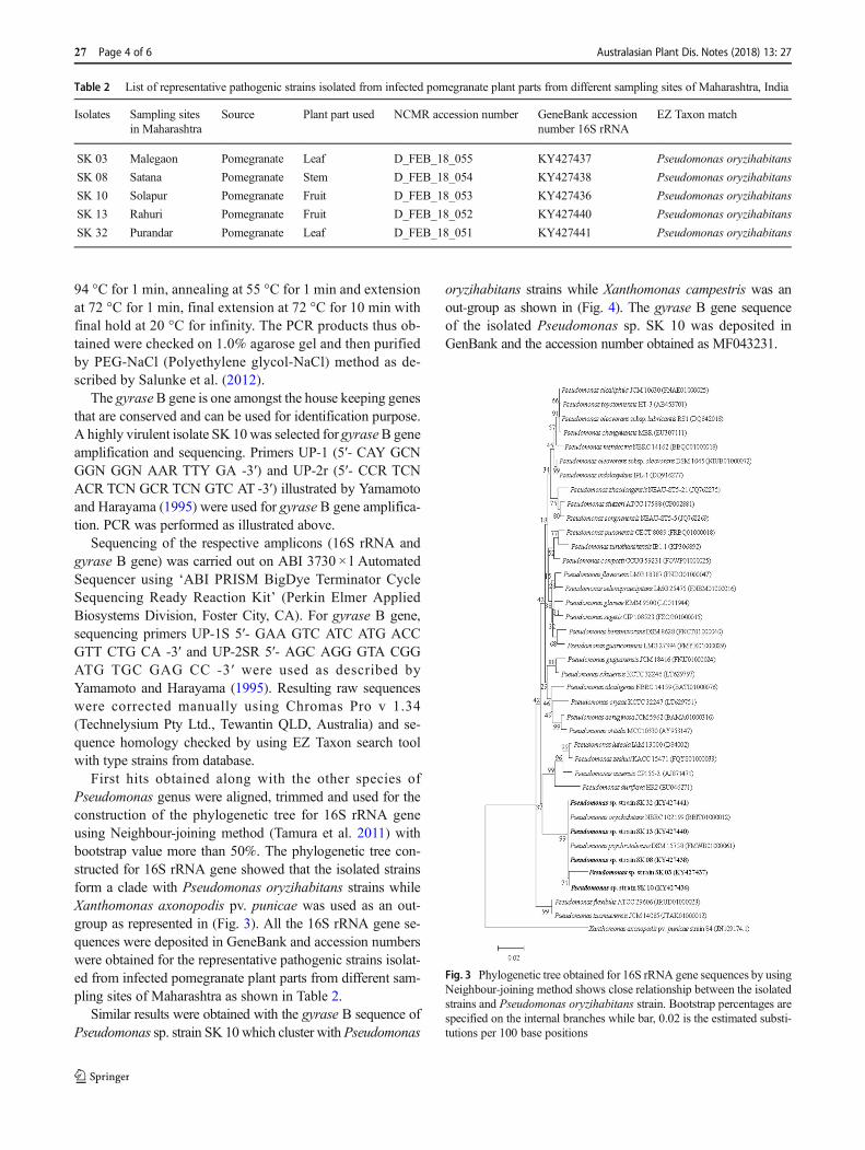
First hits obtained along with the other species ofPseudomonas genus were aligned, trimmed and used for theconstruction of the phylogenetic tree for 16S rRNA geneusing Neighbour-joining method (Tamura et al. 2011) withbootstrap value more than 50%. The phylogenetic tree con-structed for 16S rRNA gene showed that the isolated strainsform a clade with Pseudomonas oryzihabitans strains whileXanthomonas axonopodis pv. punicae was used as an out-group as represented in (Fig. 3). All the 16S rRNA gene se-quences were deposited in GeneBank and accession numberswere obtained for the representative pathogenic strains isolat-ed from infected pomegranate plant parts from different sam-pling sites of Maharashtra as shown in Table 2.
Similar results were obtained with the gyrase B sequence ofPseudomonas sp. strain SK 10which cluster with Pseudomonas
oryzihabitans strains while Xanthomonas campestris was anout-group as shown in (Fig. 4). The gyrase B gene sequenceof the isolated Pseudomonas sp. SK 10 was deposited inGenBank and the accession number obtained as MF043231.
Table 2 List of representative pathogenic strains isolated from infected pomegranate plant parts from different sampling sites of Maharashtra, India
Isolates Sampling sitesin Maharashtra
Source Plant part used NCMR accession number GeneBank accessionnumber 16S rRNA
EZ Taxon match
SK 03 Malegaon Pomegranate Leaf D_FEB_18_055 KY427437 Pseudomonas oryzihabitans
SK 08 Satana Pomegranate Stem D_FEB_18_054 KY427438 Pseudomonas oryzihabitans
SK 10 Solapur Pomegranate Fruit D_FEB_18_053 KY427436 Pseudomonas oryzihabitans
SK 13 Rahuri Pomegranate Fruit D_FEB_18_052 KY427440 Pseudomonas oryzihabitans
SK 32 Purandar Pomegranate Leaf D_FEB_18_051 KY427441 Pseudomonas oryzihabitans
Fig. 3 Phylogenetic tree obtained for 16S rRNA gene sequences by usingNeighbour-joining method shows close relationship between the isolatedstrains and Pseudomonas oryzihabitans strain. Bootstrap percentages arespecified on the internal branches while bar, 0.02 is the estimated substi-tutions per 100 base positions
27 Page 4 of 6 Australasian Plant Dis. Notes (2018) 13: 27

In the present study, Pseudomonas strains closely related to P.oryzihabitans were described as causal agents of a bacterialblight of pomegranate. To date, this disease was ascribed onlyto Xanthomonas axonopodis pv. punicae (Hingorani and Mehta1952; Hingorani and Singh 1960). These bacterial strains arecharacterised by yellow colonies a phenotype that has been al-ready described for a number of Pseudomonas, eg.Pseudomonas oryzihabitans (Kodama et al. 1985),Pseudomonas argentinensis sp. nov.(Peix et al. 2005),Pseudomonas helmanticensis sp. nov. (Ramírez-Bahena et al.2014), Pseudomonas oryzihabitans (Cottyn et al. 2009; Ruizaet al. 2011; Hardoim et al. 2012). P. oryzihabitans has beenalready associated with plants, as an endophyte of rice seeds(Cottyn et al. 2009; Ruiza et al. 2011; Hardoim et al. 2012) andin rice and paddy fields (Kodama et al. 1985). Strains frompomegranate were able to induce bacterial blight symptomswhen artificially inoculated in detached leaves and plantlets.Riley et al. (2002) reported in detail the plant disease diagnosisincluding various aspects of complete identification and diagno-sis of a particular plant disease and themajor factors related to thecause of disease. It may be possible in nature that two differentbacteria can show similar symptoms leading to the same disease(Riley et al. 2002). It has been reported that the Xanthomonasaxonopodis, Pseudomonas cichorii and few strains ofRhizobiaceae family exhibits typical symptoms causing bacterialblight in eucalyptus (Gonçalves et al. 2008) which supported theresult of Pomella et al. (1995) that leaf blight in Eucalyptusgrandis was also caused by Pseudomonas cichorii. In addition,walnut blight canker was earlier reported to be caused byXanthomonas campestris pv. juglandis however, isolation of
bacteria from the canker revealed the higher proportion of yellowpigmented bacteria known as Pseudomonas flavescens sp. nov(Hildebrand et al. 1994). In addition, the Pseudomonas sp. iso-lated in the present study is involved in the progression of bac-terial blight disease of pomegranate in Maharashtra, India.
To the best of our knowledge, this is the first report ofPseudomonas sp. as a cause of bacterial blight of pomegranatein Maharashtra, India. As the isolated pathogen, Pseudomonassp. is able to show the symptoms of the blight disease in pome-granate under laboratory conditions, further investigation of thepathogen is essential and hence is been currently undertakenincluding the measures to control the blight disease.
Acknowledgements Department of Science and Technology (DST),Government of India provided funds through DST-Women ScientistsScheme-A for carrying out this research work. Authors are grateful toDr. Yogesh Shouche, Principal Investigator, National Centre forMicrobial Resource, National Centre for Cell Science, Pashan, Pune -411021 for his continuous support and guidance in carrying out molecularidentification of the isolates. Thanks are due to the University GrantsCommission (UGC), India for the award of Emeritus Fellowship to thecorresponding author.
References
Cottyn B, Debode J, Regalado E, Mew TW, Swings J (2009) Phenotypicand genetic diversity of rice seed-associated bacteria and their role inpathogenicity and biological control. J ApplMicrobiol 107:885–897
Garrity GM, Bell JA, Lilburn TG (2004) Taxonomic outline of the pro-karyotes. Bergey's manual of systematic bacteriology, 2nd edn.Springer, New York, pp 94–247
Fig. 4 Phylogenetic tree obtained for gyrase B gene sequences by usingNeighbour-joining method shows close relationship between the isolatedstrain SK 10 and Pseudomonas oryzihabitans along with other
Pseudomonas species. Bootstrap percentages are specified on the internalbranches while bar, 0.1 is the estimated substitutions per 100 basepositions
Australasian Plant Dis. Notes (2018) 13: 27 Page 5 of 6 27

Gonçalves RC, Douglas L, Oliveira JR, Maffia LA, Cascardo J, AlfenasAC (2008) Etiology of bacterial leaf blight of eucalyptus in Brazil.Trop Plant Pathol 33:180–188
Hardoim PR, Hardoim CC, Van Overbeek LS, Van Elsas JD (2012)Dynamics of seed-borne rice endophytes on early plant growth stages.PLoS One 7:e30438. https://doi.org/10.1371/journal.pone.0030438
Hildebrand DC, Palleroni NJ, Hendson M, Toth J, Johnson JL (1994)Pseudomonas flavescens sp. nov., isolated from walnut blight can-kers. Int J Syst Evol Microbiol 44:410–415
Hingorani MK, Mehta PP (1952) Bacterial leaf spot of pomegranate.Indian Phytopathol 5:55–56
Hingorani MK, Singh NJ (1960) Xanthomonas punicae sp. nov. onPúnica granatum L. Indian J Agr Sci 29:45–48
Kodama K, Kimura N, Komagata K (1985) Two new species ofPseudomonas: P. oryzihabitans isolated from rice paddy and clinicalspecimens and P. luteola isolated from clinical specimens. Int J SystEvol Microbiol 35:467–474
Kumar R, Shamarao Jahagirdar MR, Yenjerappa ST, Patil HB (2009)Epidemiology andmanagement of bacterial blight of pomegranate causedby Xanthomonas axonopodis pv. punicae. Acta Hortic 818:291–296
Mondal KK, Mani C (2009) ERIC-PCR-generated genomic fingerprintsand their relationship with pathogenic variability of Xanthomonascampestris pv. punicae, the incitant of bacterial blight of pomegran-ate. Curr Microbiol 59:616–620
Peix A, Berge O, Rivas R, Abril A, Velázquez E (2005) Pseudomonasargentinensis sp. nov., a novel yellow pigment-producing bacterialspecies, isolated from rhizospheric soil in Córdoba, Argentina. Int JSyst Evol Microbiol 55:1107–1112
Pomella AWV, Romeiro RS, Ferreira FA, Oliveira JR (1995) Lesõesfoliares em viveiro de eucalipto incitadas por uma espéciefluorescente de Pseudomonas. Fitopatol Bras 20:374
Ramírez-Bahena M-H, Cuesta MJ, Flores-Félix JD, Mulas R, Rivas R,Castro-Pinto J et al (2014) Pseudomonas helmanticensis sp. nov.,isolated from forest soil. Int J Syst Evol Microbiol 64:2338–2345
Randhawa PS, Civerolo EL (1985) A detached-leaf bioassay forXanthomonas campestris pv. pruni. Phytopathol 75:1060–1063
RileyMB,WilliamsonMR,Maloy O (2002) Plant disease diagnosis. Theplant health instructor. https://doi.org/10.1094/PHI-I-2002-1021-01
Ruiza D, Agaras B, deWerra P,Wall LG, Valverde C (2011) Characterizationand screening of plant probiotic traits of bacteria isolated from rice seedscultivated in Argentina. J Microbiol 49:902–912
Salunke BK, Salunkhe RC, Dhotre DP, Walujkar SA, Khandagale AB,Chaudhari R, Chandode RK, Ghate HV, Patole MS, Werren JH,Shouche YS (2012) Determination of Wolbachia diversity in butter-flies from Western Ghats, India, by a multigene approach. ApplEnviron Microbiol 78:4458–4467
Schaad NW (1988) Laboratory guide for identification of plant pathogen-ic bacteria, 2nd edn. APS press, St. Paul, pp 60–93
Tamura K, Peterson D, Peterson N, Stecher G, Nei M, Kumar S (2011)MEGA5: molecular evolutionary genetics analysis using maximumlikelihood, evolutionary distance, and maximum parsimonymethods. Mol Biol Evol 28:2731–2739
Yamamoto S, Harayama S (1995) PCR amplification and direct sequenc-ing of gyr B genes with universal primers and their application to thedetection and taxonomic analysis of Pseudomonas putida strains.Appl Environ Microbiol 61:1104–1109
27 Page 6 of 6 Australasian Plant Dis. Notes (2018) 13: 27