A NANOFIBROUS HYDROGEL FOR BONE TISSUE … · A NANOFIBROUS HYDROGEL FOR BONE TISSUE ... Phase...
46
-
Upload
vuongthien -
Category
Documents
-
view
220 -
download
2
Transcript of A NANOFIBROUS HYDROGEL FOR BONE TISSUE … · A NANOFIBROUS HYDROGEL FOR BONE TISSUE ... Phase...

Umadevi Kandalam, PhD
Assistant Professor
Department of Pediatric Dentistry
College of Dental Medicine
Nova Southeastern University
Fort Lauderdale , Florida
A NANOFIBROUS HYDROGEL FOR BONE TISSUE ENGINEERING
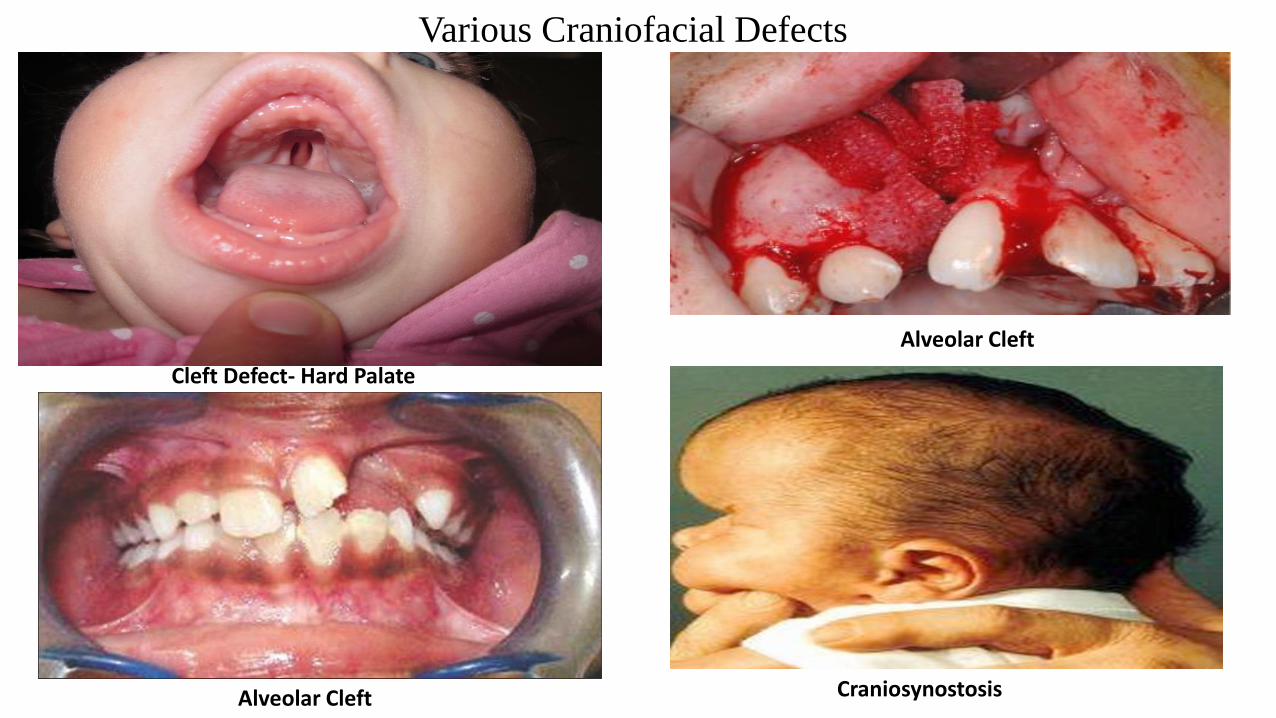
Various Craniofacial Defects
Cleft Defect- Hard Palate
Alveolar Cleft Craniosynostosis
Alveolar Cleft
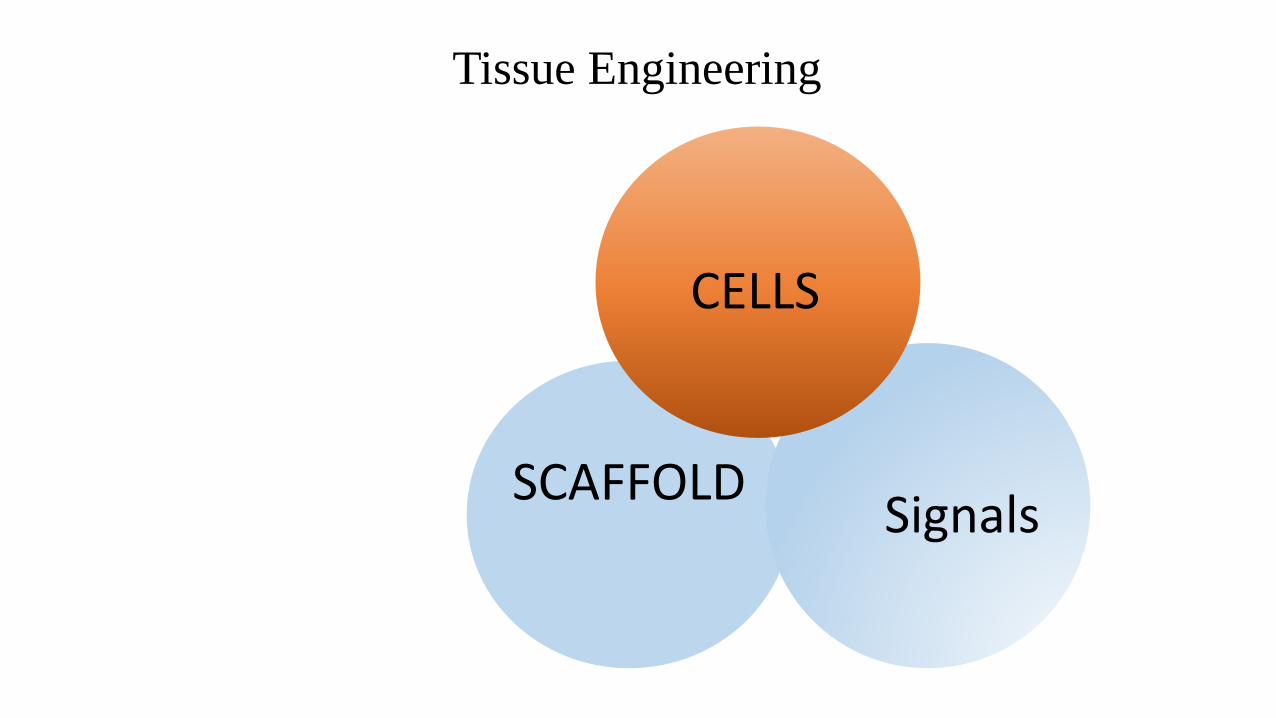
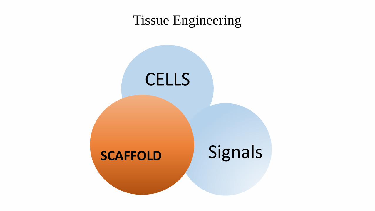
Tissue Engineering
SCAFFOLDSignals
CELLS
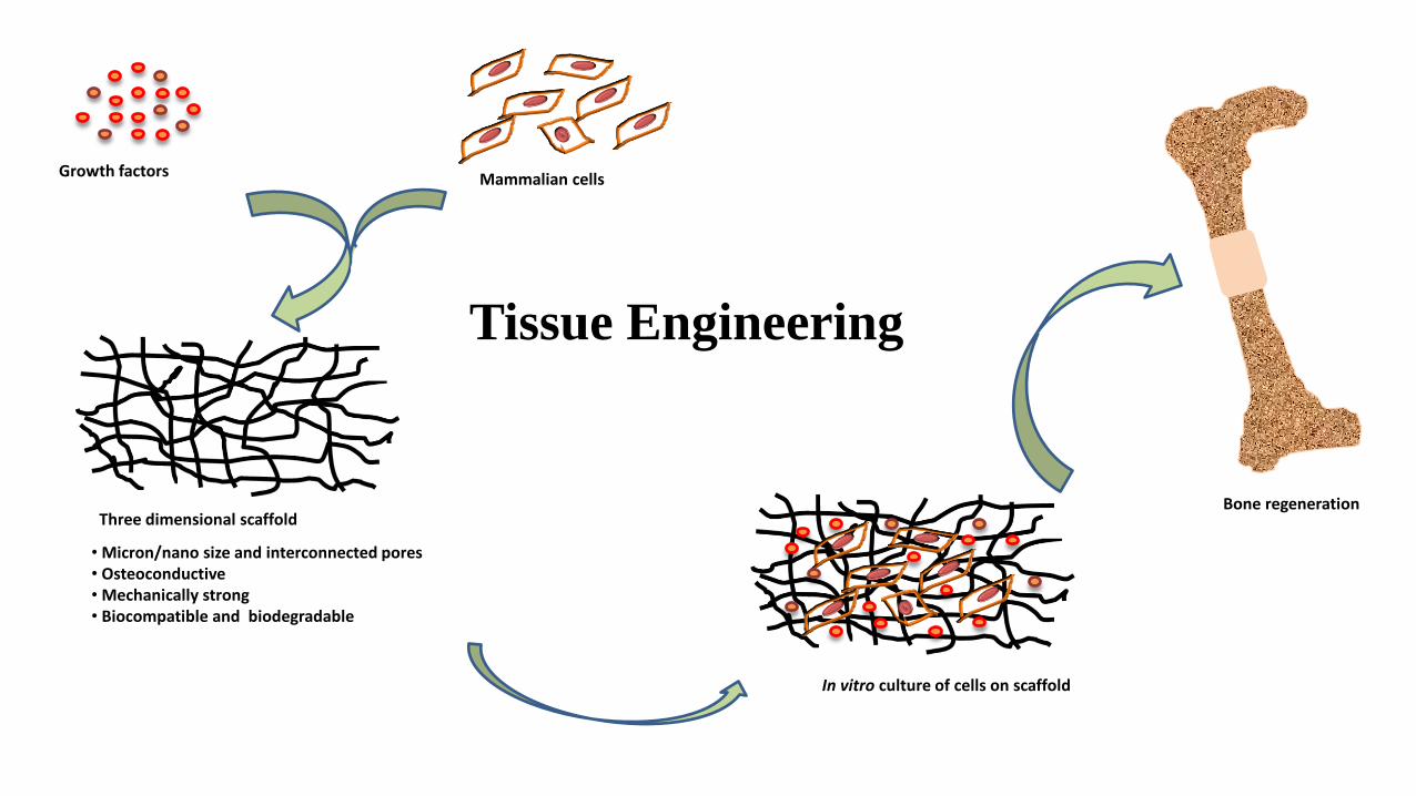
Growth factors Mammalian cells
Three dimensional scaffold
In vitro culture of cells on scaffold
Bone regeneration
• Micron/nano size and interconnected pores• Osteoconductive• Mechanically strong• Biocompatible and biodegradable
Tissue Engineering
REPAIR THE BONY DEFECT IN THE CRNIOFACIAL REGION USING TISSUE
ENGINEERING TECHNIQUES
GOAL
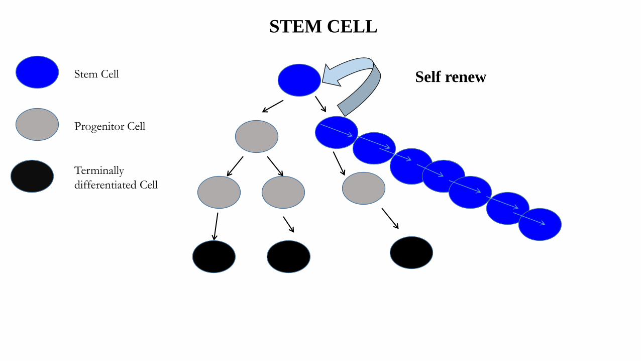
Stem Cell
Terminally
differentiated Cell
Progenitor Cell
STEM CELL
Self renew
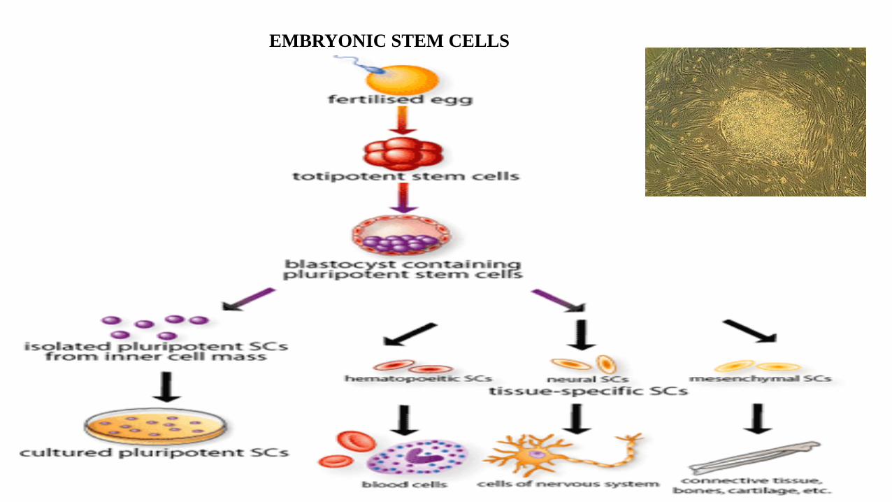
EMBRYONIC STEM CELLS
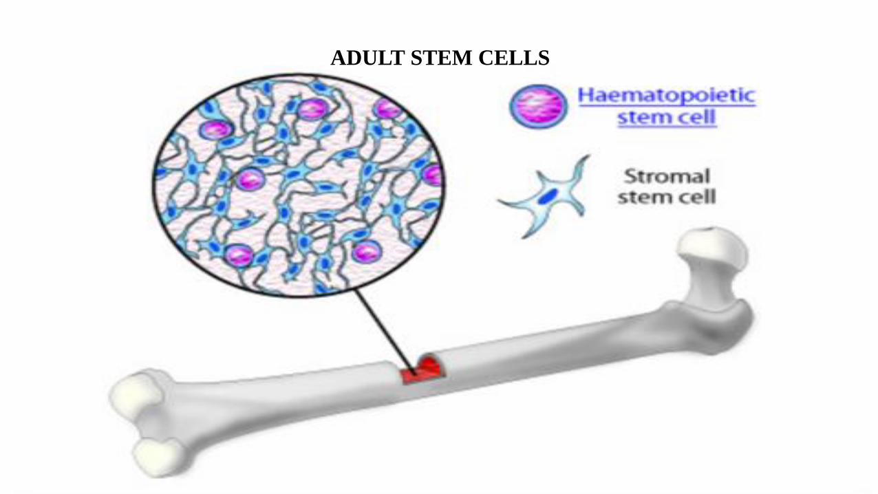
ADULT STEM CELLS
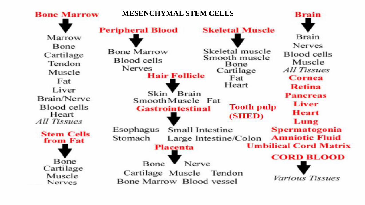
MESENCHYMAL STEM CELLS
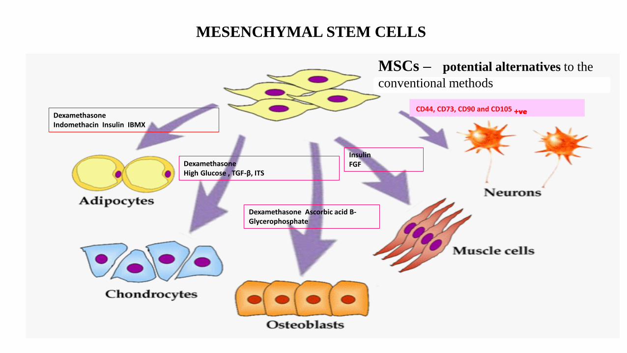
MESENCHYMAL STEM CELLS
CD44, CD73, CD90 and CD105 +veDexamethasoneIndomethacin Insulin IBMX
DexamethasoneHigh Glucose , TGF-β, ITS
Dexamethasone Ascorbic acid Β-Glycerophosphate
InsulinFGF
MSCs – potential alternatives to the
conventional methods
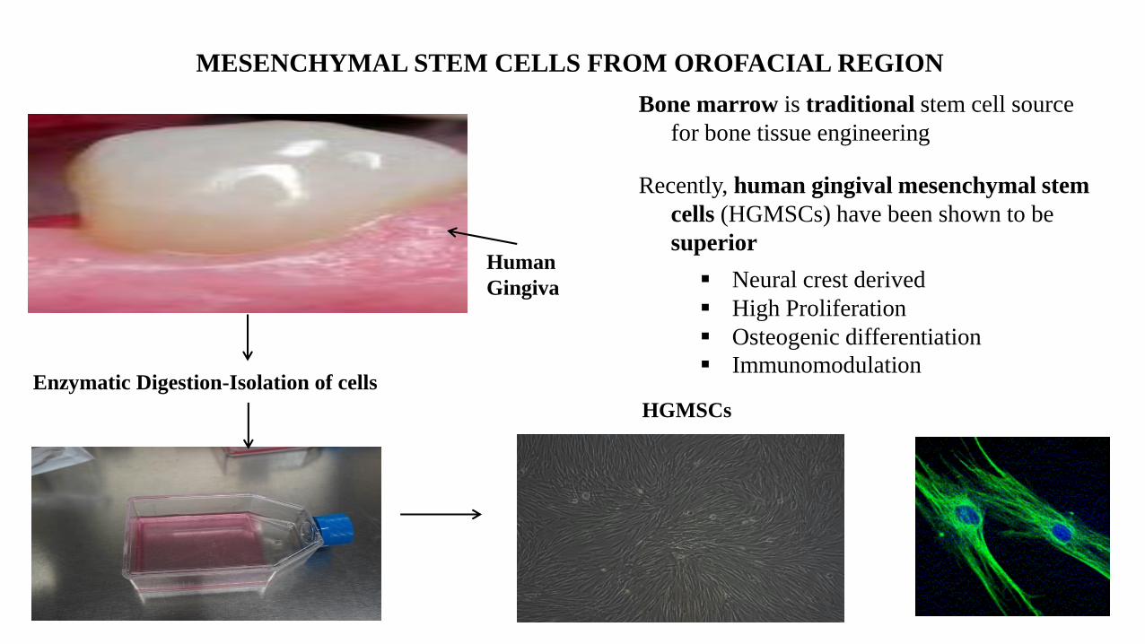
MESENCHYMAL STEM CELLS FROM OROFACIAL REGION
Human
Gingiva
Enzymatic Digestion-Isolation of cells
HGMSCs
Bone marrow is traditional stem cell source
for bone tissue engineering
Recently, human gingival mesenchymal stem
cells (HGMSCs) have been shown to be
superior
Neural crest derived
High Proliferation
Osteogenic differentiation
Immunomodulation

Tissue Engineering
CELLS
SignalsSCAFFOLD
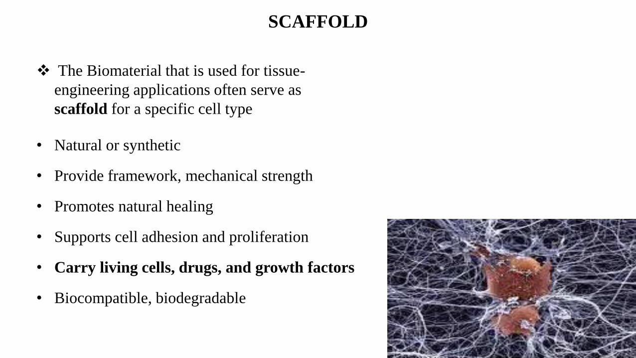
SCAFFOLD
The Biomaterial that is used for tissue-
engineering applications often serve as
scaffold for a specific cell type
• Natural or synthetic
• Provide framework, mechanical strength
• Promotes natural healing
• Supports cell adhesion and proliferation
• Carry living cells, drugs, and growth factors
• Biocompatible, biodegradable
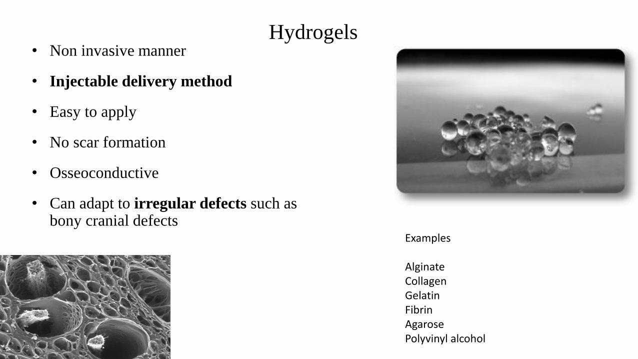
Hydrogels• Non invasive manner
• Injectable delivery method
• Easy to apply
• No scar formation
• Osseoconductive
• Can adapt to irregular defects such as bony cranial defects
Examples
AlginateCollagenGelatinFibrinAgarosePolyvinyl alcohol
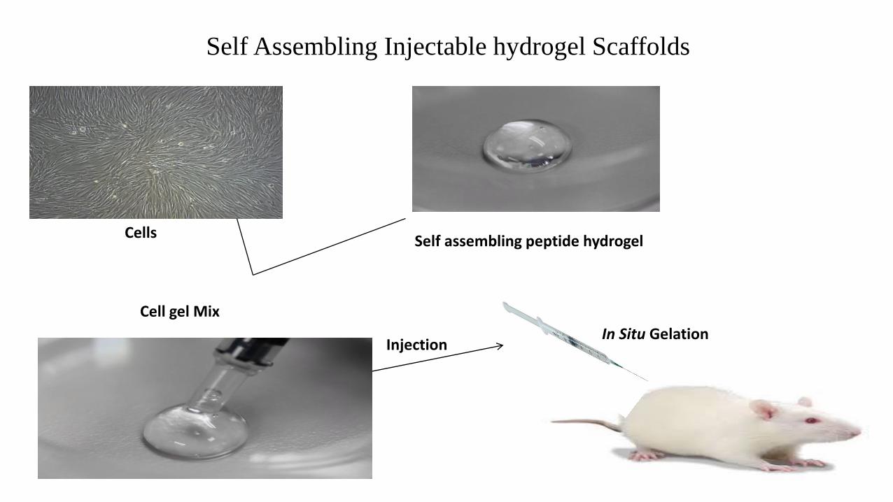
Self Assembling Injectable hydrogel Scaffolds
CellsSelf assembling peptide hydrogel
Cell gel Mix
InjectionIn Situ Gelation
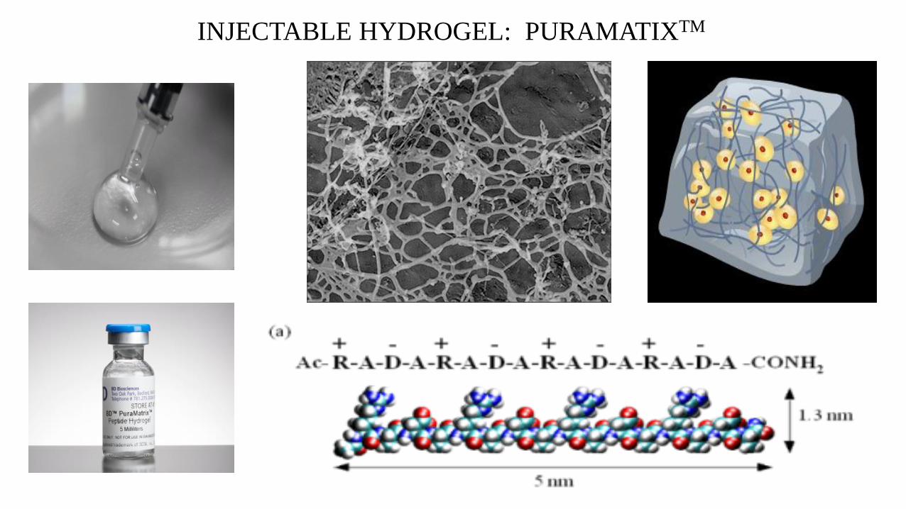
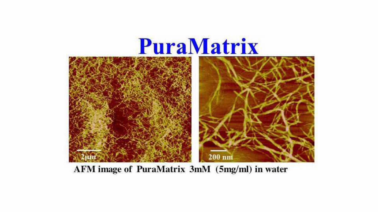
INJECTABLE HYDROGEL: PURAMATIXTM

CELL- SCAFFOLD SYSTEM
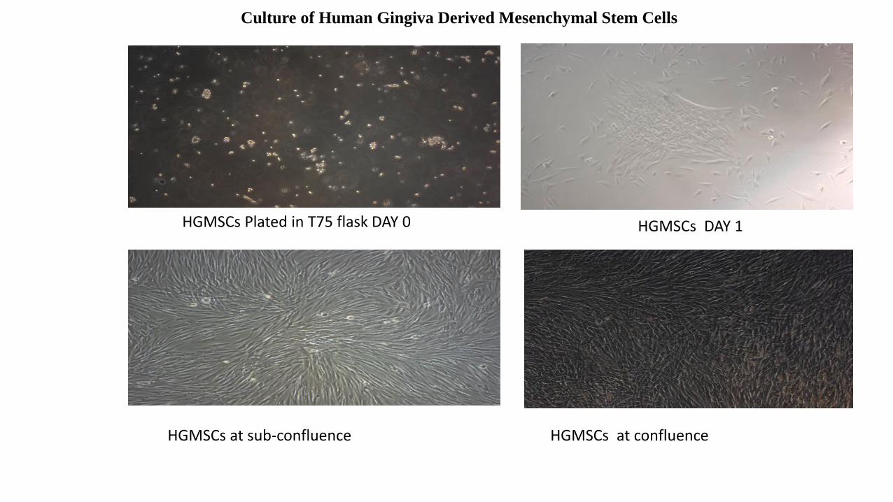
HGMSCs Plated in T75 flask DAY 0
HGMSCs at sub-confluence
HGMSCs DAY 1
HGMSCs at confluence
Culture of Human Gingiva Derived Mesenchymal Stem Cells
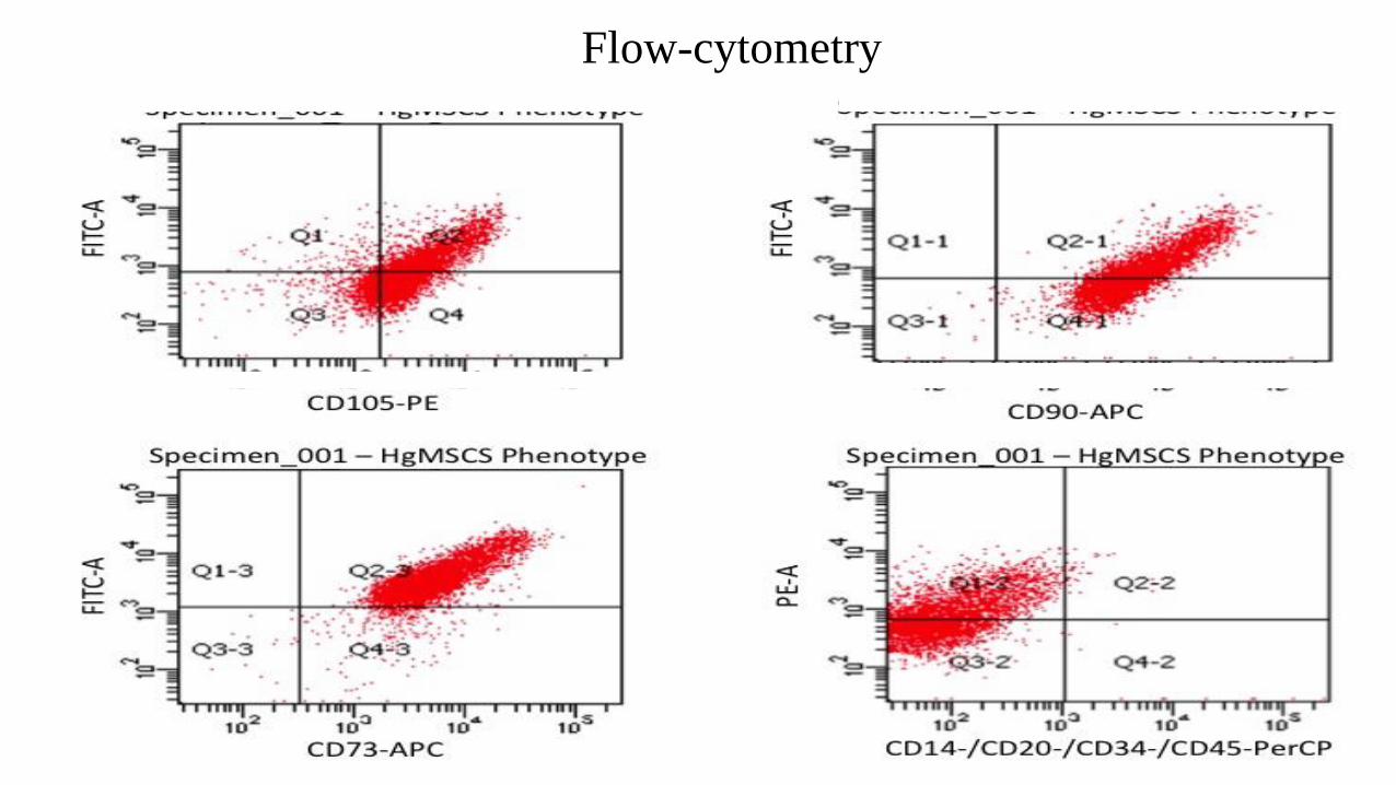
Flow-cytometry
0
20
40
60
80
100
120
140
160
180
200
1 week 2 weeks
Control
OM
*
*
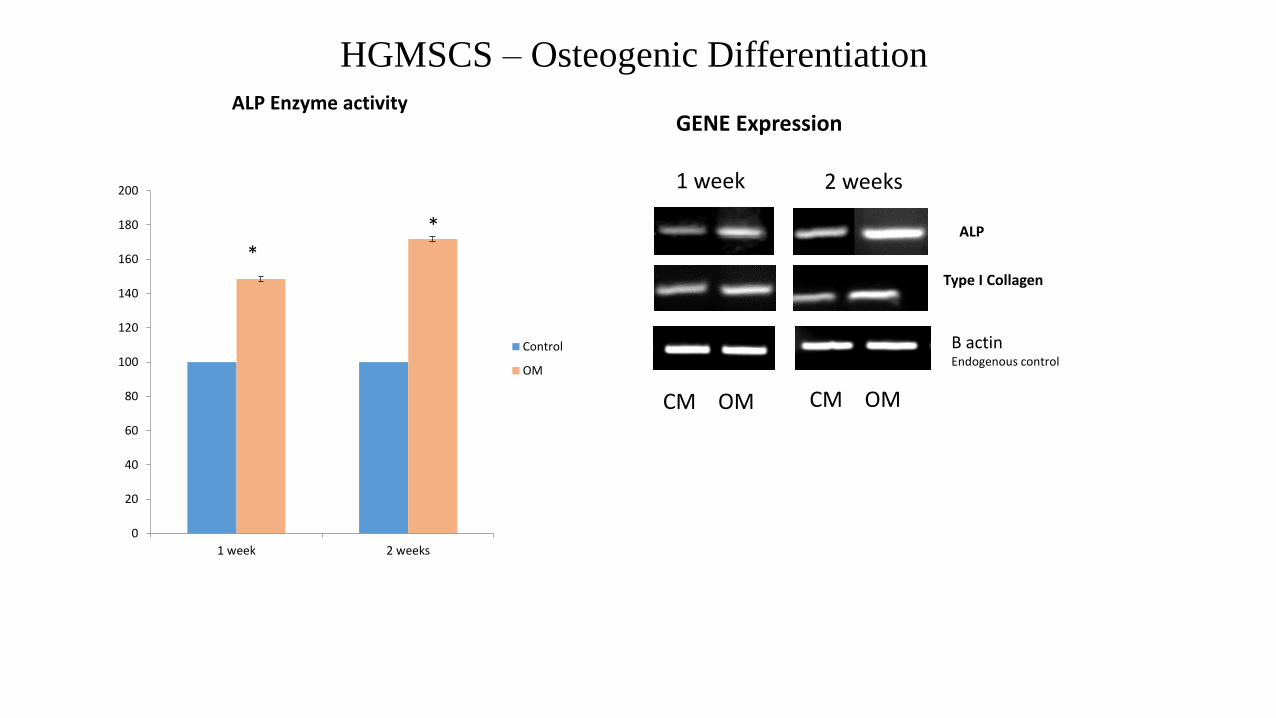
GENE Expression ALP Enzyme activity
ALP
Type I Collagen
1 week 2 weeks
B actin Endogenous control
HGMSCS – Osteogenic Differentiation
CM OM CM OM
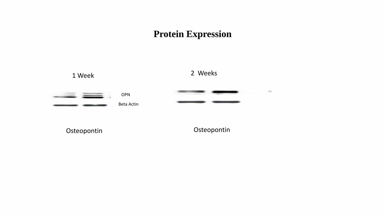
Protein Expression
1 Week 2 Weeks
Osteopontin Osteopontin
OPN
Beta Actin
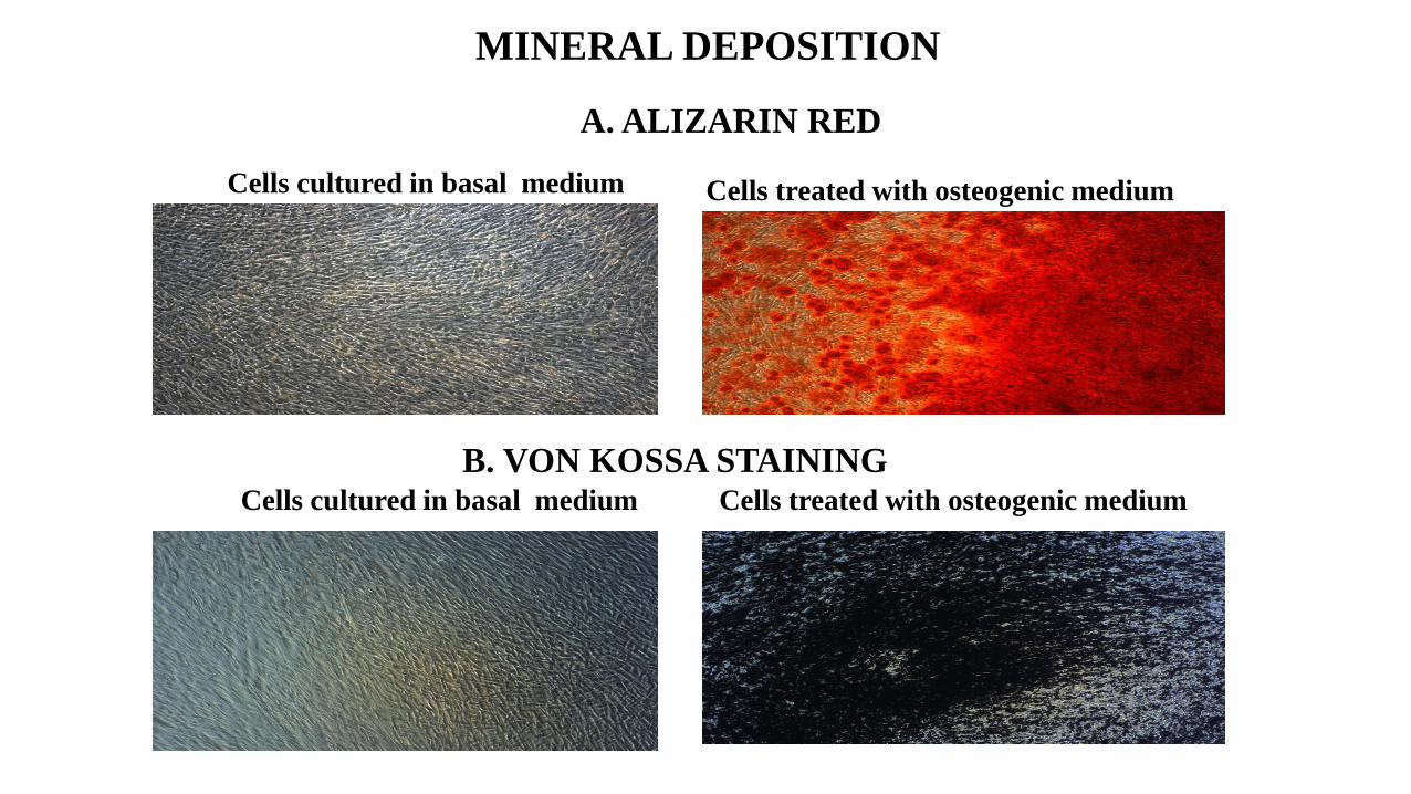
Cells treated with osteogenic mediumCells cultured in basal medium
MINERAL DEPOSITION
A. ALIZARIN RED
B. VON KOSSA STAINING Cells cultured in basal medium Cells treated with osteogenic medium
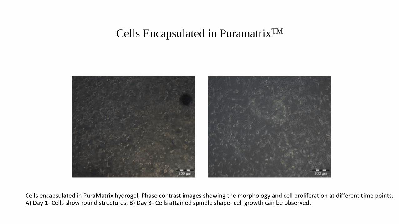
Cells encapsulated in PuraMatrix hydrogel; Phase contrast images showing the morphology and cell proliferation at different time points. A) Day 1- Cells show round structures. B) Day 3- Cells attained spindle shape- cell growth can be observed.
Cells Encapsulated in PuramatrixTM
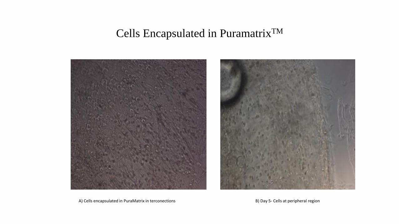
A) Cells encapsulated in PuraMatrix in terconections B) Day 5- Cells at peripheral region
Cells Encapsulated in PuramatrixTM
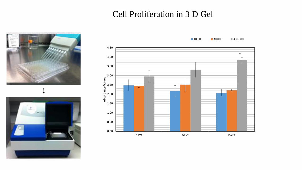
0.00
0.50
1.00
1.50
2.00
2.50
3.00
3.50
4.00
4.50
DAY1 DAY2 DAY3
Ab
sorb
ance
Val
ue
s
10,000 30,000 300,000
*
Cell Proliferation in 3 D Gel
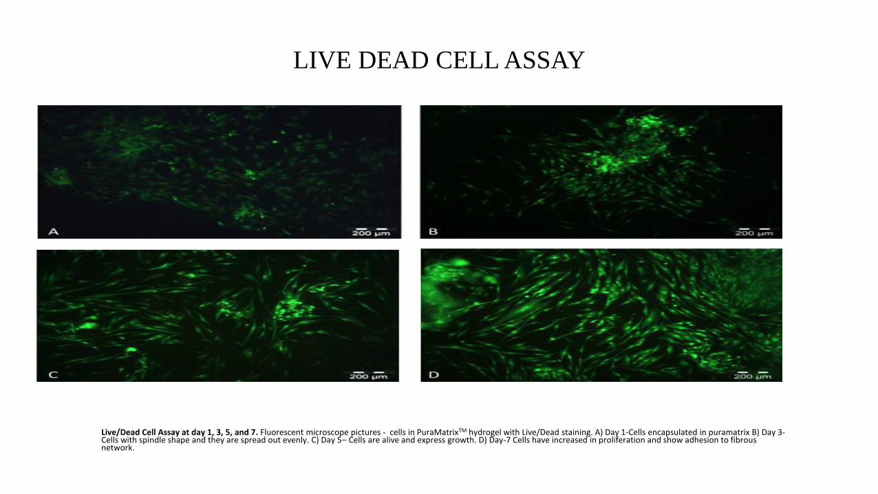
LIVE DEAD CELL ASSAY
Live/Dead Cell Assay at day 1, 3, 5, and 7. Fluorescent microscope pictures - cells in PuraMatrixTM hydrogel with Live/Dead staining. A) Day 1-Cells encapsulated in puramatrix B) Day 3-Cells with spindle shape and they are spread out evenly. C) Day 5– Cells are alive and express growth. D) Day-7 Cells have increased in proliferation and show adhesion to fibrous network.
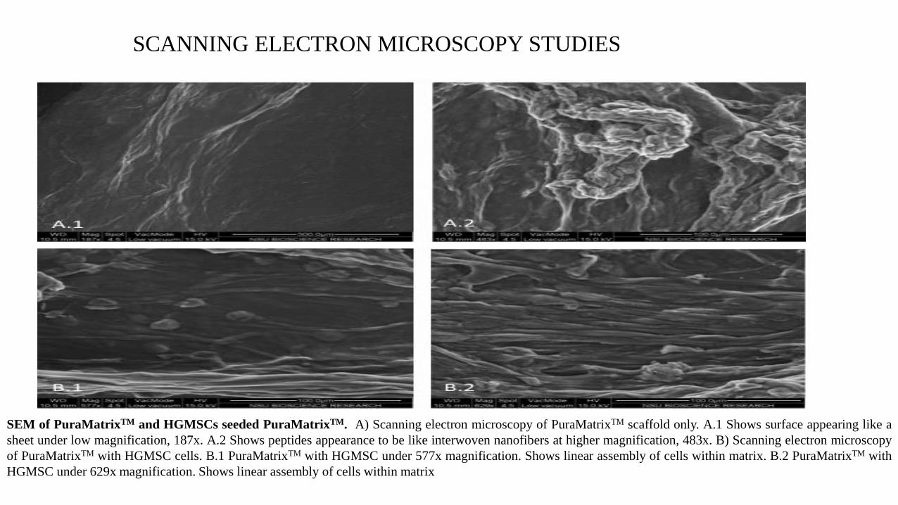
SCANNING ELECTRON MICROSCOPY STUDIES
SEM of PuraMatrixTM and HGMSCs seeded PuraMatrixTM. A) Scanning electron microscopy of PuraMatrixTM scaffold only. A.1 Shows surface appearing like a
sheet under low magnification, 187x. A.2 Shows peptides appearance to be like interwoven nanofibers at higher magnification, 483x. B) Scanning electron microscopy
of PuraMatrixTM with HGMSC cells. B.1 PuraMatrixTM with HGMSC under 577x magnification. Shows linear assembly of cells within matrix. B.2 PuraMatrixTM with
HGMSC under 629x magnification. Shows linear assembly of cells within matrix
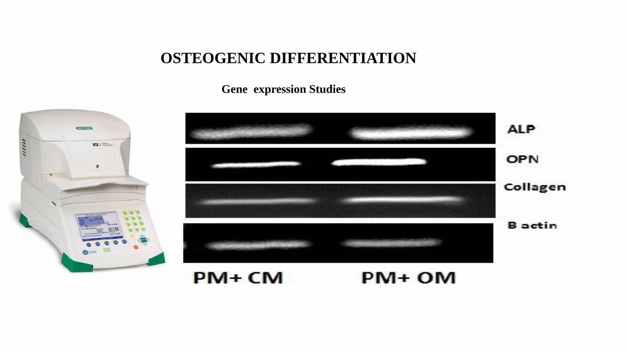
OSTEOGENIC DIFFERENTIATION
Gene expression Studies
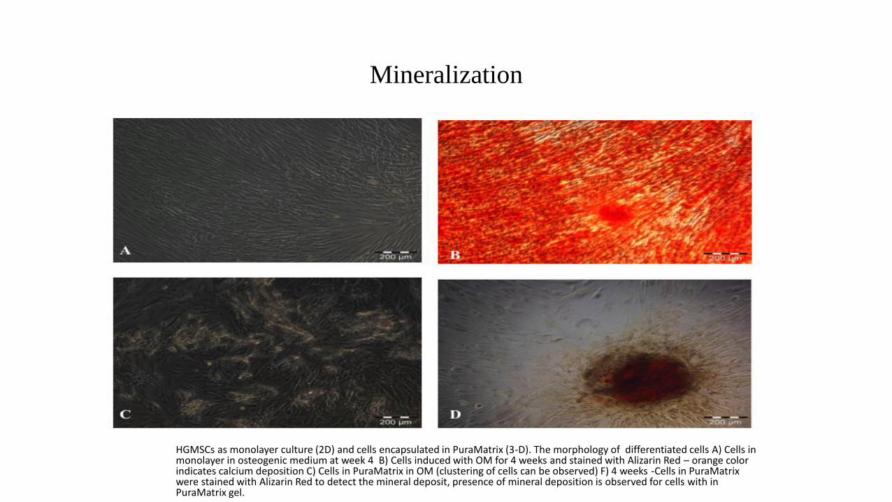
Mineralization
HGMSCs as monolayer culture (2D) and cells encapsulated in PuraMatrix (3-D). The morphology of differentiated cells A) Cells inmonolayer in osteogenic medium at week 4 B) Cells induced with OM for 4 weeks and stained with Alizarin Red – orange color indicates calcium deposition C) Cells in PuraMatrix in OM (clustering of cells can be observed) F) 4 weeks -Cells in PuraMatrix were stained with Alizarin Red to detect the mineral deposit, presence of mineral deposition is observed for cells with in PuraMatrix gel.
Summary of In Vitro Study
• HGMSCs demonstrated osteogenic differentiation
• The cells grown in PuraMatrix nanoscaffold showed high survival rate and cell growth was also observed in time dependent manner
• The cells attained spindle shape within 3 days of encapsulation and cell growth was observed from day 3
• Scanning electron microscope studies revealed that the cell bodies embedded in PuraMatrix started to develop a dense network process.
• The Live/Dead cell assay and WST assay revealed that PuraMatrix nanofibers are cytocompatible
• Cells expressed positive mineralization at 4 weeks
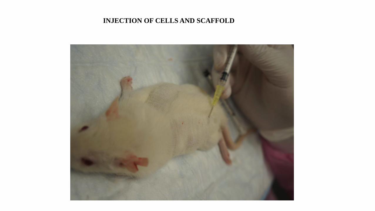
INJECTION OF CELLS AND SCAFFOLD
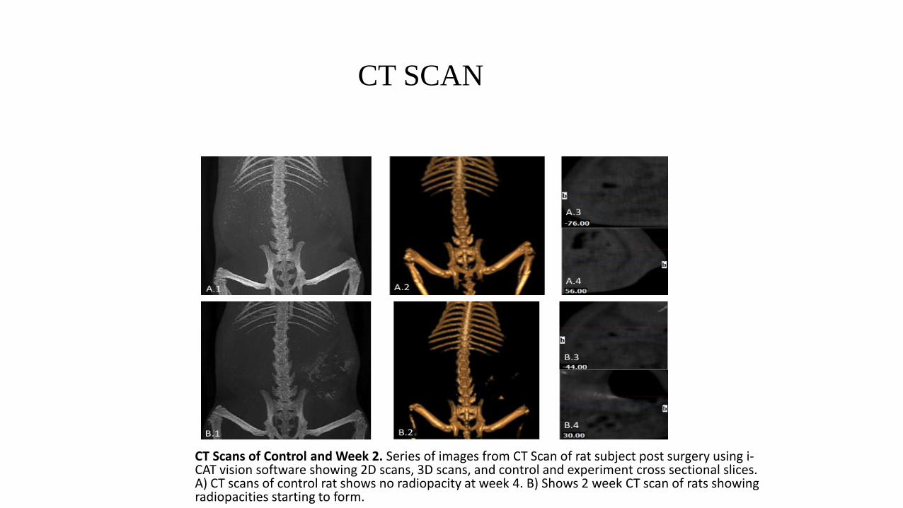
CT SCAN
CT Scans of Control and Week 2. Series of images from CT Scan of rat subject post surgery using i-CAT vision software showing 2D scans, 3D scans, and control and experiment cross sectional slices. A) CT scans of control rat shows no radiopacity at week 4. B) Shows 2 week CT scan of rats showing radiopacities starting to form.
CT SCAN
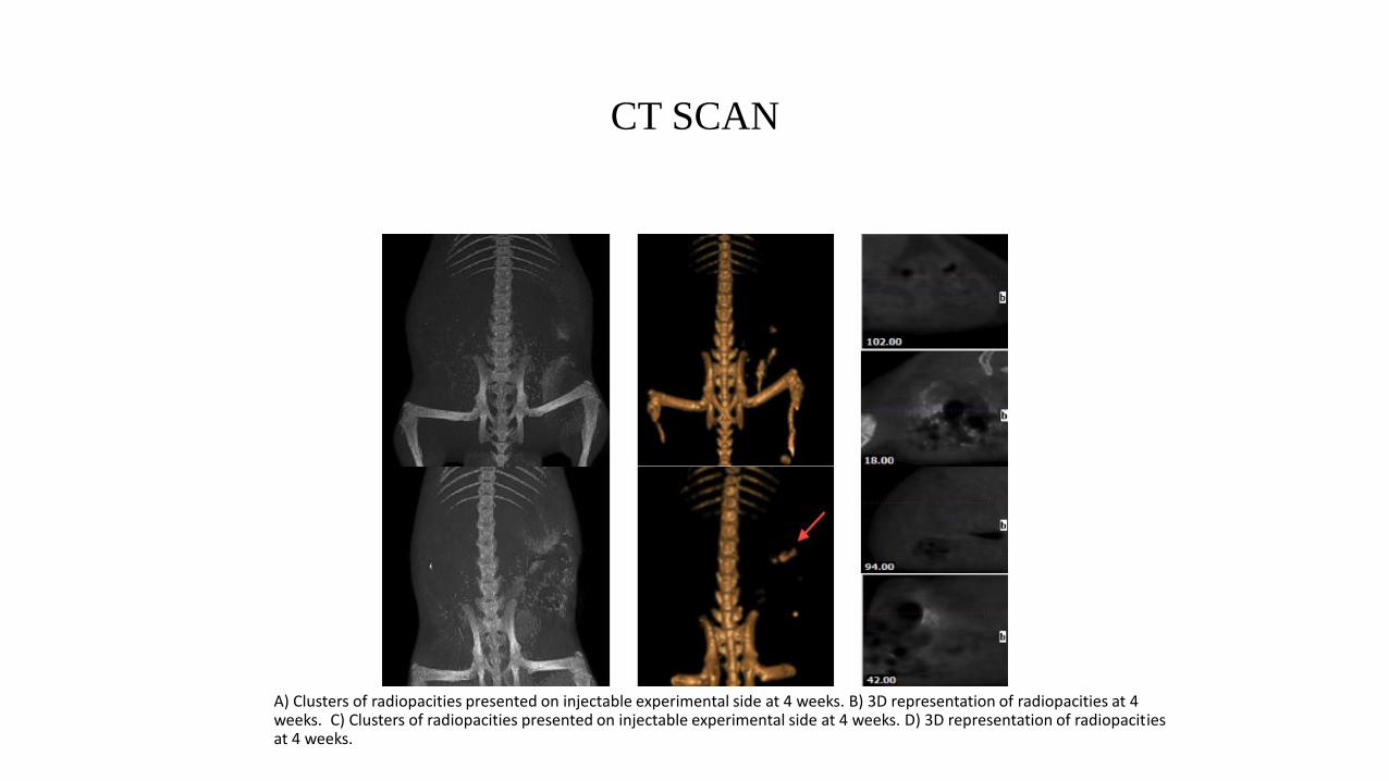
A) Clusters of radiopacities presented on injectable experimental side at 4 weeks. B) 3D representation of radiopacities at 4weeks. C) Clusters of radiopacities presented on injectable experimental side at 4 weeks. D) 3D representation of radiopacitiesat 4 weeks.
Ct Scan
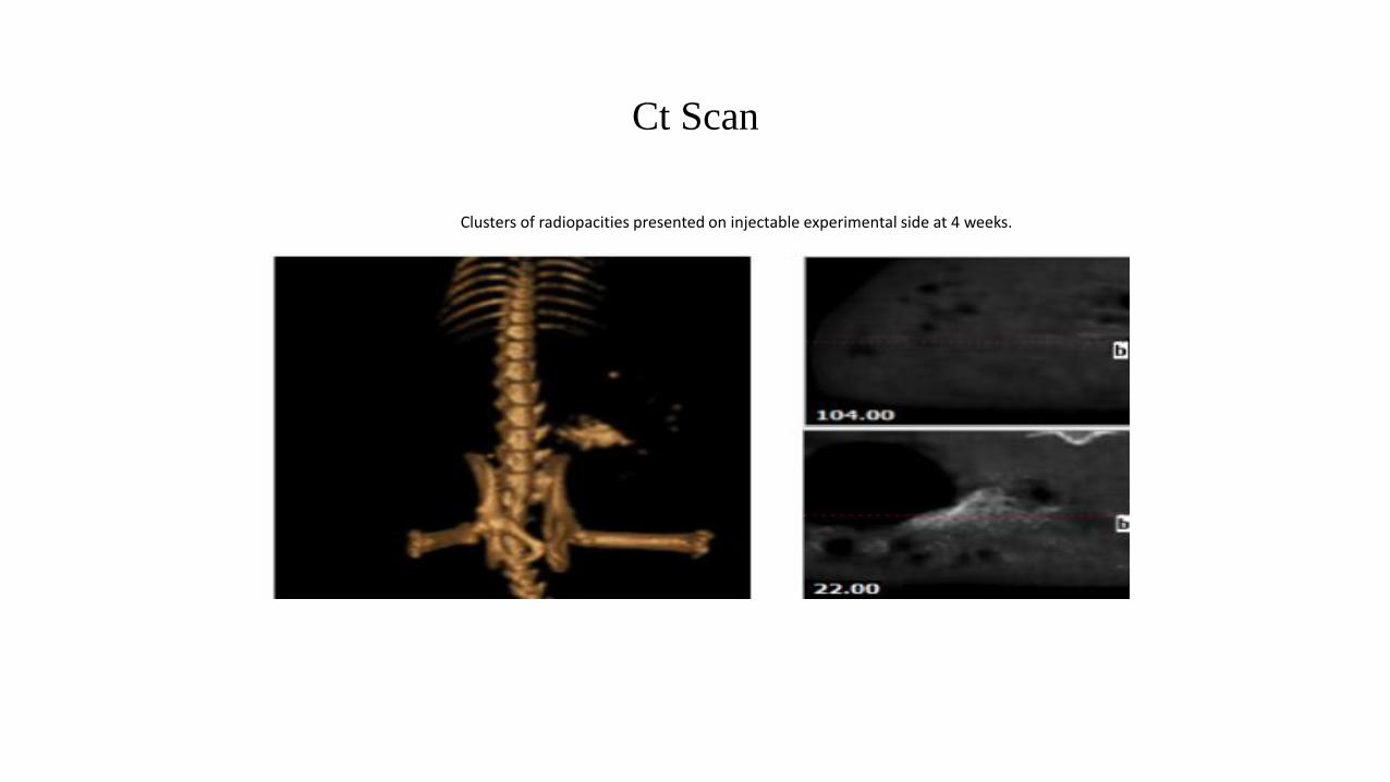
Clusters of radiopacities presented on injectable experimental side at 4 weeks.
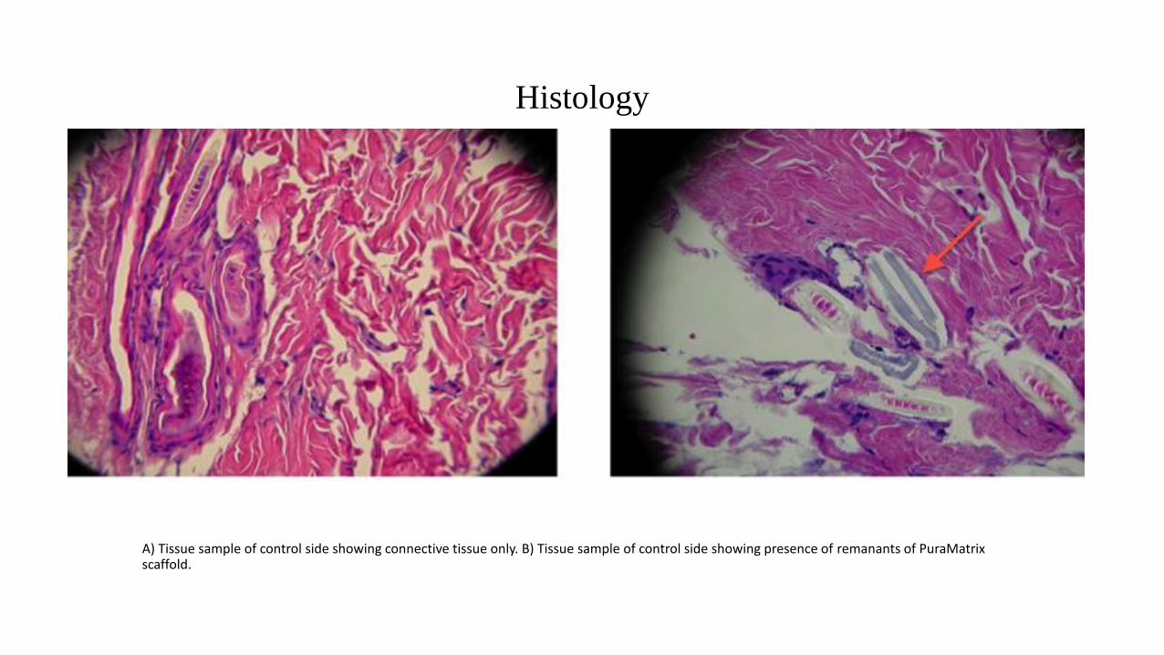
Histology
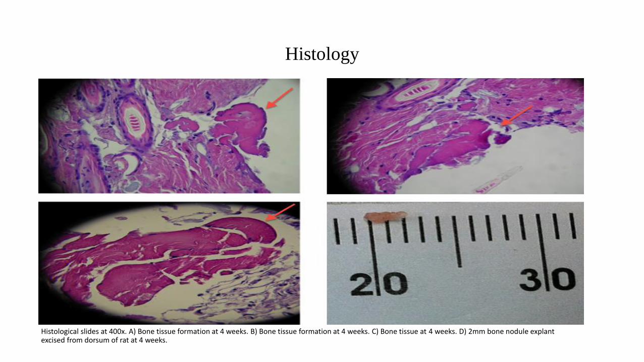
Histological slides at 400x. A) Bone tissue formation at 4 weeks. B) Bone tissue formation at 4 weeks. C) Bone tissue at 4 weeks. D) 2mm bone nodule explant excised from dorsum of rat at 4 weeks.
Histology
A) Tissue sample of control side showing connective tissue only. B) Tissue sample of control side showing presence of remanants of PuraMatrix scaffold.
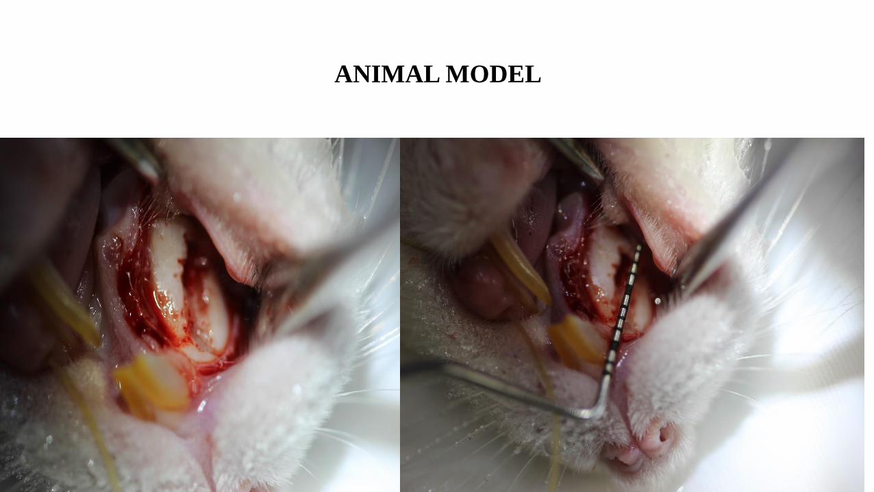
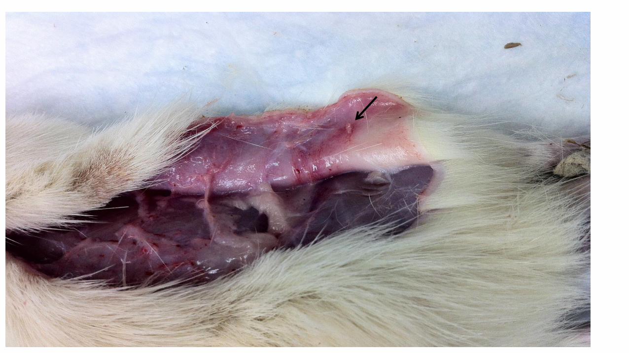
ANIMAL MODEL
Summary: In vivo Study
• In Vivo Study
• CT scans show radiopacities suggesting that PuraMatrix scaffold with HGMSC supported ectopic bone formation
• Histological studies demonstrated that PuraMatrix in combination HGMSCs supported the ectopic bone formation.
• We are establishing the animal model
CONCLUSIONS AND FUTURE STUDY
• The self-assembled, injectable PuraMatrix scaffold in combination with HGMSCs can support bone tissue formation
• Further research needed to investigate the bone growth in terms of density and volume at week 8 and later time points.
• Also, further studies will investigate the potential of HGMSCs seeded PuraMatrix combination for the repair of critical size defects in hard palate of rats.
• Using growth factors to enhance bone growth
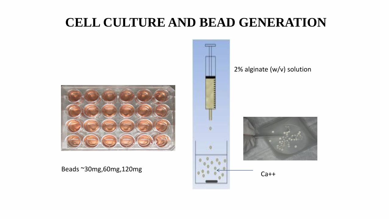
CELL CULTURE AND BEAD GENERATION
Ca++Beads ~30mg,60mg,120mg
2% alginate (w/v) solution
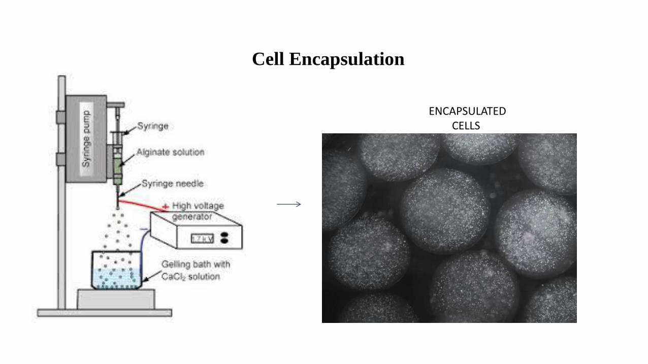
Cell Encapsulation
ENCAPSULATED CELLS

Acknowledgements
Don Do MDReem Almashat DDSNora Al Amer DDSCasey LynnDebbie StilesRoss BrockmanJason Portnof MD