A Multicenter Evaluation of Tests for Diagnosis of ... multicenter evaluation... · A Multicenter...
7
MAJOR ARTICLE A Multicenter Evaluation of Tests for Diagnosis of Histoplasmosis Chadi A. Hage, 1,2 Julie A. Ribes, 3 Nancy L. Wengenack, 4 Larry M. Baddour, 5 Maha Assi, 6 David S. McKinsey, 7 Kassem Hammoud, 8 Daisy Alapat, 3 N. Esther Babady, 9 Michelle Parker, 10 DeAnna Fuller, 10 Aliya Noor, 1 Thomas E. Davis, 10 Mark Rodgers, 11 Patricia A. Connolly, 11 Boutros El Haddad, 12 and L. Joseph Wheat 11 1 Pulmonary-Critical Care Medicine, Richard L. Roudebush Veterans Affairs Medical Center, Indianapolis, Indiana; 2 Infectious Diseases, Indiana University and Richard L. Roudebush Veterans Affairs Medical Center, Indianapolis, Indiana; 3 Department of Microbiology, University of Kentucky, Lexington, Kentucky; 4 Laboratory Medicine and Pathology, Mayo Clinic, Rochester, Minnesota; 5 Infectious Diseases, Mayo Clinic, Rochester, Minnesota; 6 Infectious Diseases, University of Kansas, Wichita, Kansas; 7 Infectious Diseases Associates of Kansas City, Missouri; 8 Infectious Diseases, University of Kansas Medical Center, Kansas City, Missouri; 9 Laboratory Medicine, Memorial Sloan-Kettering Cancer Center, New York, New York; 10 Pathology and Laboratory Medicine, Indiana University School of Medicine, Indianapolis, Indiana; 11 MiraVista Diagnostics and Mirabella Technologies, Indianapolis, Indiana; and 12 Department of Medicine, University of Kansas, Wichita, Kansas Background. The sensitivity of the MVista Histoplasma antigen enzyme immunoassay (MiraVista Diagnostics) has been evaluated in disseminated histoplasmosis in patients with AIDS and in the ‘‘epidemic’’ form of acute pneumonia. Moreover, there has been no evaluation of the sensitivity of antigenemia detection in disseminated histoplasmosis after the implementation of methods to dissociate immune complexes and denature released antibodies. The goal of this study was to determine the sensitivity of the current antigen assay in different categories of histoplasmosis. Methods. Urine and serum specimens obtained from 218 patients with histoplasmosis and 229 control subjects, including 30 with blastomycosis, were tested. Results. Antigenuria was detected in 91.8% of 158 patients with disseminated histoplasmosis, 83.3% of 6 patients with acute histoplasmosis, 30.4% of 46 patients with subacute histoplasmosis, and 87.5% of 8 patients with chronic pulmonary histoplasmosis; antigenemia was present in 100% of 31 tested cases of disseminated histoplasmosis. Among patients with disseminated cases, antigenuria was detected more often and at higher concentrations in immunocompromised patients and those with severe disease. Specificity was 99.0% for patients with nonfungal infections (n 5 130) and in healthy subjects (n 5 69), but cross-reactivity occurred in 90% of patients with blastomycosis. Conclusions. The sensitivity of antigen detection in disseminated histoplasmosis is higher in immunocom- promised patients than in immunocompetent patients and in patients with more severe illness. The sensitivity for detection of antigenemia is similar to that for antigenuria in disseminated infection. Antigen detection is widely used to diagnose progressive disseminated histoplasmosis (PDH). The original assay was a radioimmunoassay [1]; subsequently, an enzyme immunoassay (EIA) [2], designated the first-generation EIA, was used. A second-generation EIA was introduced in 2004 to reduce the number of false-positive results caused by human antirabbit antibodies [3], followed by a third-generation EIA in 2007, which permitted quantification [4]. Pretreatment of serum with ethylene diamine tetraacetic acid (EDTA) at 104°C increased the sensitivity for detection of antigenemia, by dissoci- ation of antigen-antibody complexes and denaturation of dissociated antibody, preventing it from interfering with antigen detection [5]. However, the performance characteristics of the current assay have not been fully assessed. Areas of need include determination of sensitivity in patients with PDH complicating diseases other than AIDS and in Received 17 February 2011; accepted 24 May 2011; electronically published 15 August 2011. Correspondence: Chadi A. Hage, MD, Pulmonary-Critical Care and Infectious Diseases, Roudebush VA Medical Center and Indiana University, 1481 W 10th St, 111P-IU, Indianapolis, IN 46202 ([email protected]). Clinical Infectious Diseases 2011;53(5):448–454 Ó The Author 2011. Published by Oxford University Press on behalf of the Infectious Diseases Society of America. All rights reserved. For Permissions, please email: [email protected]. This is an Open Access article distributed under the terms of the Creative Commons Attribution Non-Commercial License (http://creativecommons.org/licenses/by-nc/3.0/), which permits unrestricted non-commercial use, distribution, and reproduction in any medium, provided the original work is properly cited. 1058-4838/2011/535-0008$14.00 DOI: 10.1093/cid/cir435 448 d CID 2011:53 (1 September) d Hage et al
-
Upload
phungnguyet -
Category
Documents
-
view
225 -
download
0
Transcript of A Multicenter Evaluation of Tests for Diagnosis of ... multicenter evaluation... · A Multicenter...
M A J O R A R T I C L E
A Multicenter Evaluation of Tests for Diagnosisof Histoplasmosis
Chadi A. Hage,1,2 Julie A. Ribes,3 Nancy L. Wengenack,4 Larry M. Baddour,5 Maha Assi,6 David S. McKinsey,7
Kassem Hammoud,8 Daisy Alapat,3 N. Esther Babady,9 Michelle Parker,10 DeAnna Fuller,10 Aliya Noor,1 ThomasE. Davis,10 Mark Rodgers,11 Patricia A. Connolly,11 Boutros El Haddad,12 and L. Joseph Wheat11
1Pulmonary-Critical Care Medicine, Richard L. Roudebush Veterans Affairs Medical Center, Indianapolis, Indiana; 2Infectious Diseases, IndianaUniversity and Richard L. Roudebush Veterans Affairs Medical Center, Indianapolis, Indiana; 3Department of Microbiology, University of Kentucky,Lexington, Kentucky; 4Laboratory Medicine and Pathology, Mayo Clinic, Rochester, Minnesota; 5Infectious Diseases, Mayo Clinic, Rochester,Minnesota; 6Infectious Diseases, University of Kansas, Wichita, Kansas; 7Infectious Diseases Associates of Kansas City, Missouri; 8InfectiousDiseases, University of Kansas Medical Center, Kansas City, Missouri; 9Laboratory Medicine, Memorial Sloan-Kettering Cancer Center, New York,New York; 10Pathology and Laboratory Medicine, Indiana University School of Medicine, Indianapolis, Indiana; 11MiraVista Diagnostics and MirabellaTechnologies, Indianapolis, Indiana; and 12Department of Medicine, University of Kansas, Wichita, Kansas
Background. The sensitivity of the MVista Histoplasma antigen enzyme immunoassay (MiraVista Diagnostics) has
been evaluated in disseminated histoplasmosis in patients with AIDS and in the ‘‘epidemic’’ form of acute pneumonia.
Moreover, there has been no evaluation of the sensitivity of antigenemia detection in disseminated histoplasmosis
after the implementation of methods to dissociate immune complexes and denature released antibodies. The goal of
this study was to determine the sensitivity of the current antigen assay in different categories of histoplasmosis.
Methods. Urine and serum specimens obtained from 218 patients with histoplasmosis and 229 control subjects,
including 30 with blastomycosis, were tested.
Results. Antigenuria was detected in 91.8% of 158 patients with disseminated histoplasmosis, 83.3% of 6
patients with acute histoplasmosis, 30.4% of 46 patients with subacute histoplasmosis, and 87.5% of 8 patients with
chronic pulmonary histoplasmosis; antigenemia was present in 100% of 31 tested cases of disseminated
histoplasmosis. Among patients with disseminated cases, antigenuria was detected more often and at higher
concentrations in immunocompromised patients and those with severe disease. Specificity was 99.0% for patients
with nonfungal infections (n 5 130) and in healthy subjects (n 5 69), but cross-reactivity occurred in 90% of
patients with blastomycosis.
Conclusions. The sensitivity of antigen detection in disseminated histoplasmosis is higher in immunocom-
promised patients than in immunocompetent patients and in patients with more severe illness. The sensitivity for
detection of antigenemia is similar to that for antigenuria in disseminated infection.
Antigen detection is widely used to diagnose progressive
disseminated histoplasmosis (PDH). The original assay
was a radioimmunoassay [1]; subsequently, an enzyme
immunoassay (EIA) [2], designated the first-generation
EIA, was used. A second-generation EIA was introduced
in 2004 to reduce the number of false-positive results
caused by human antirabbit antibodies [3], followed by
a third-generation EIA in 2007, which permitted
quantification [4]. Pretreatment of serum with ethylene
diamine tetraacetic acid (EDTA) at 104�C increased
the sensitivity for detection of antigenemia, by dissoci-
ation of antigen-antibody complexes and denaturation
of dissociated antibody, preventing it from interfering
with antigen detection [5].
However, the performance characteristics of the
current assay have not been fully assessed. Areas of need
include determination of sensitivity in patients with
PDH complicating diseases other than AIDS and in
Received 17 February 2011; accepted 24 May 2011; electronically published 15August 2011.Correspondence: Chadi A. Hage, MD, Pulmonary-Critical Care and Infectious
Diseases, Roudebush VA Medical Center and Indiana University, 1481 W 10th St,111P-IU, Indianapolis, IN 46202 ([email protected]).
Clinical Infectious Diseases 2011;53(5):448–454� The Author 2011. Published by Oxford University Press on behalf of theInfectious Diseases Society of America. All rights reserved. For Permissions, pleaseemail: [email protected]. This is an Open Access article distributedunder the terms of the Creative Commons Attribution Non-Commercial License(http://creativecommons.org/licenses/by-nc/3.0/), which permits unrestrictednon-commercial use, distribution, and reproduction in any medium, provided theoriginal work is properly cited.1058-4838/2011/535-0008$14.00DOI: 10.1093/cid/cir435
448 d CID 2011:53 (1 September) d Hage et al
Vivien
Placed Image
those with pulmonary histoplasmosis [6]. In addition, the effect
of EDTA pretreatment of serum, first described in antigen
negative specimens from patients with AIDS [5], on sensitivity
for detection of antigenemia has only been studied in acute
pulmonary histoplasmosis, improving sensitivity by 38% over
testing of urine samples alone [7].
A multifaceted approach is commonly used for the diagnosis
of histoplasmosis, with combination of histopathological ex-
amination, culture, and antigen and antibody detection. The
sensitivities of these tests have not been examined since im-
provements were incorporated into the MVista Histoplasma
antigen EIA (MiraVista Diagnostics).
METHODS
Study CohortSpecimens were obtained from patients evaluated at 8 medical
centers during the period from November 2004 through De-
cember 2007 and were tested for Histoplasma and/or Blastomyces
antigen at MiraVista Diagnostics. The start date was chosen
because the second-generation MVista Histoplasma antigen as-
say was implemented on 8 November 2004. The results of the
other diagnostic tests (culture, cytology, pathology, and anti-
body detection using immunodiffusion [ID] and complement
fixation [CF]) were obtained by medical record review of tests
performed at the originating institution or other commercial
laboratories. The protocol was approved by the institutional
review committees at each institution.
The criteria for diagnosis included a positive result of culture,
antigen, histopathology, cytology, or Histoplasma antibody tests
demonstrating H or M precipitin bands by ID or titers of CF
antibodies of $1:8 [8]. Positive culture, cytology, or histopa-
thology results demonstrating yeast-like structures characteristic
of Histoplasma capsulatum were required for classification as
proven disease, whereas positive antigen or antibody test results
were required for classification as probable histoplasmosis,
combined with compatible clinical and radiographic findings.
Cases were excluded if there was no clinical information avail-
able, if histoplasmosis occurred before November 2004 or after
December 2007, if histoplasmosis was not diagnosed according to
the medical record, or if the original antigen test result was positive
but the specimen was not stored. There were also eligible cases
with negative antigen test results when originally tested in which
the specimens were not stored. To reduce bias in the evaluation
of sensitivity of antigen testing, these were included and con-
sidered to be negative for antigen, even though specimens were
not tested.
Control specimens were obtained from 130 patients evaluated
at Indiana University Health Medical Centers in whom the di-
agnosis of fungal infection was excluded, 30 patients with culture-
proven pulmonary and/or extrapulmonary blastomycosis, and
69 healthy commercial blood donors (SeraCare Life Sciences;
Milford, Massachusetts).
PDH was defined as the presence of clinical, laboratory, or
imaging evidence of extrapulmonary involvement. The diagnosis
of pulmonary histoplasmosis required respiratory symptoms
and pulmonary radiographs and/or computerized tomography
that demonstrated infiltrates and/or mediastinal lymphadenop-
athy, in the absence of evidence for PDH. Pulmonary cases were
further classified as acute, subacute, and chronic pulmonary
histoplasmosis [9], as follows: acute pulmonary histoplasmosis,
symptom duration of ,1 month and presence of diffuse or
multinodular pulmonary infiltrates; subacute pulmonary histo-
plasmosis, symptom duration of $1 month with presence of
focal infiltrates and/or mediastinal lymphadenopathy; and
chronic pulmonary histoplasmosis, symptom duration of .3
months, presence of cavitary pulmonary infiltrates, and pres-
ence of underlying emphysema.
Histoplasmosis was classified as severe if patients required
treatment in an intensive care unit, moderately severe if hos-
pitalization was required, and mild if hospitalization was not
required.
Antigen DetectionThe specimens were obtained during the period 2004–2007 and
stored frozen at MiraVista Diagnostics until 2010, when they
were retested for this study. Only 1 sample was tested for each
patient. The specimens were obtained at the time of diagnosis
in 202 patients, after the diagnosis was made and treatment
initiated in 12 patients, and before the time of diagnosis in 4
patients. The MVista Histoplasma antigen EIA [4] was modified
to permit quantification below the level of 0.6 ng/mL by in-
corporating 0.2 ng/mL and 0.4 ng/mL calibrators and removing
those of $19 ng/mL. Serum specimens were treated with 4%
EDTA at 104�C before testing [5].
Statistical AnalysisReceiver operator characteristic (ROC) curve analysis was per-
formed to determine the cutoff for positivity. The proportions
of patients with positive results were compared using the v2 test.
The student t test was used for pairwise comparisons of mean
antigen levels among the different clinical syndrome groups.
Multiple pairwise comparisons were adjusted using step-down
Bonferroni multiple comparison procedure. Ninety-five percent
confidence intervals (CIs) and proportions were calculated using
the Wilson score method for small sample with asymmetrical
distribution [10]. An overall significance level of a 5 .05 was
used for all comparisons.
RESULTS
PatientsOf 381 patients who were identified, 163 were excluded for
the following reasons: histoplasmosis outside the interval of
Diagnosis of Histoplasmosis d CID 2011:53 (1 September) d 449
the study, 100 patients; no laboratory documentation or inade-
quate information for classification, 29 patients each; and positive
specimen not stored, 5 patients. Of the 218 evaluable patients,
95 were from Indiana University Health (Indianapolis, Indiana),
41 were from the Mayo Clinic (Rochester, Minnesota), 33 were
from the University of Kentucky (Lexington, Kentucky), 20
were from the University of Kansas (Wichita, Kansas), 16 were
from the University of Kansas Medical Center (Kansas City,
Missouri), and 13 were from the Infectious Diseases Associates
of Kansas City (Kansas City, Missouri).
One hundred fifty-eight cases were classified as disseminated
infection and 60 were classified as pulmonary infection, in-
cluding 6 with acute, 46 with subacute, and 8 with chronic
pulmonary manifestations. One hundred fifty-seven patients
were immunocompromised, as follows: AIDS, 57 patients; re-
ceipt of solid-organ transplants, 26 patients; treatment with
tumor necrosis factor antagonists, 24 patients; treatment with
other immunosuppressive medications for hematologic or
inflammatory conditions, 34 patients; other immunodeficiency
states, 16 patients (including lymphoma in 5); primary immu-
nodeficiencies, 9 patients; and gastric bypass and prematurity,
1 patient each.
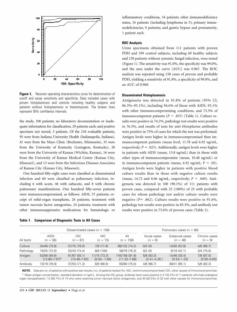
ROC AnalysisUrine specimens obtained from 111 patients with proven
PDH and 199 control subjects, including 69 healthy subjects
and 130 patients without systemic fungal infection, were tested
(Figure 1). The sensitivity was 91.0%, the specificity was 99.0%,
and the area under the curve (AUC) was 0.967. The ROC
analysis was repeated using 158 cases of proven and probable
PDH, yielding a sensitivity of 91.8%, a specificity of 99.0%, and
an AUC of 0.968.
Disseminated HistoplasmosisAntigenuria was detected in 91.8% of patients (95% CI,
86.3%–95.1%), including 94.6% of those with AIDS, 93.1%
with other immunocompromising conditions, and 73.3% of
immunocompetent patients (P 5 .837) (Table 1). Culture re-
sults were positive in 74.2%, pathology test results were positive
in 76.3%, and results of tests for anti-Histoplasma antibodies
were positive in 75% of cases for which the test was performed.
Antigen levels were higher in immunocompromised than im-
munocompetent patients (mean level, 11.78 and 6.92 ng/mL,
respectively; P5 .023). Additionally, antigen levels were higher
in patients with AIDS (mean, 13.8 ng/mL) than in those with
other types of immunocompromise (mean, 10.48 ng/mL) or
in immunocompetent patients (mean, 6.92 ng/mL; P , .05).
Antigen levels were higher in patients with positive blood
culture results than in those with negative culture results
(mean, 14.72 and 8.06 ng/mL, respectively; P 5 .049). Anti-
genuria was detected in 100 (90.1%) of 111 patients with
proven cases, compared with 25 (100%) of 25 with probable
cases for whom pathology test and/or culture results were
negative (P5 .862). Culture results were positive in 91.6%,
pathology test results were positive in 85.3%, and antibody test
results were positive in 73.6% of proven cases (Table 2).
Figure 1. Receiver operating characteristics curve for determination ofcutoff and assay sensitivity and specificity. Data included cases withproven histoplasmosis and controls including healthy subjects andpatients without histoplasmosis or blastomycosis. The broken linesrepresent 95% confidence intervals.
Table 1. Comparison of Diagnostic Tests in All Cases
Disseminated cases (n 5 158) Pulmonary cases (n 5 60)
All tests
AIDS
(n 5 56)
OIC
(n 5 87)
NIC
(n 5 15)
All
(n 5 158)
Acute cases
(n 5 6)
Subacute cases
(n 5 46)
Chronic cases
(n 5 8)
Culture 34/48 (70.8) 57/75 (76.0) 7/9 (77.8) 98/132 (74.2) 0/3 (0) 14/26 (53.8) 4/6 (66.7)
Pathology 18/25 (72.0) 32/43 (74.4) 8/8 (100) 58/76 (76.3) 0/2 (0) 8/19 (42.1) 3/4 (75.0)
Antigen 53/56 (94.6)[13.80–7.67]a
81/87 (93.1)[10.48–7.62]
11/15 (73.3)[6.92– 7.65]
145/158 (91.8)[11.32–7.88]
5/6 (83.3)[2.41–2.26 ]
14/46 (30.4)[0.53–1.23]
7/8 (87.5)[0.93–0.83]
Antibody 15/19 (78.9) 37/53 (71.2) 8/9 (88.9) 60/80 (75.0) 4/6 (66.7) 39/41 (95.1) 5/6 (83.3)
NOTE. Data are no. of patients with positive test results / no. of patients tested (%). NIC, nonimmunocompromised; OIC, other causes of immunocompromise.a Mean antigen concentration, standard deviation in ng/mL. Among the OIC group, antibody tests were positive in 2 (18.2%) of 11 patients who had undergone
organ transplantation, 12 (85.7%) of 14 who were receiving tumor necrosis factor antagonists, and 20 (62.5%) of 32 with other causes for immunocompromise.
450 d CID 2011:53 (1 September) d Hage et al
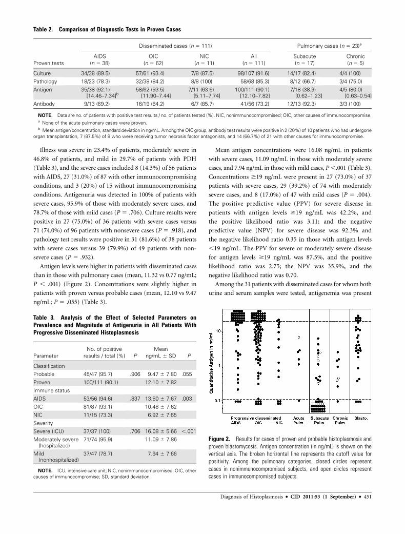
Illness was severe in 23.4% of patients, moderately severe in
46.8% of patients, and mild in 29.7% of patients with PDH
(Table 3), and the severe cases included 8 (14.3%) of 56 patients
with AIDS, 27 (31.0%) of 87 with other immunocompromising
conditions, and 3 (20%) of 15 without immunocompromising
conditions. Antigenuria was detected in 100% of patients with
severe cases, 95.9% of those with moderately severe cases, and
78.7% of those with mild cases (P 5 .706). Culture results were
positive in 27 (75.0%) of 36 patients with severe cases versus
71 (74.0%) of 96 patients with nonsevere cases (P 5 .918), and
pathology test results were positive in 31 (81.6%) of 38 patients
with severe cases versus 39 (79.9%) of 49 patients with non-
severe cases (P 5 .932).
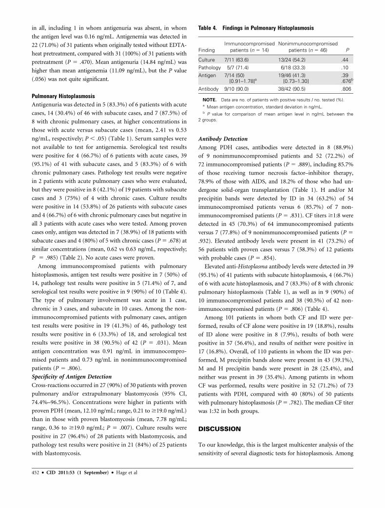
Antigen levels were higher in patients with disseminated cases
than in those with pulmonary cases (mean, 11.32 vs 0.77 ng/mL;
P , .001) (Figure 2). Concentrations were slightly higher in
patients with proven versus probable cases (mean, 12.10 vs 9.47
ng/mL; P 5 .055) (Table 3).
Mean antigen concentrations were 16.08 ng/mL in patients
with severe cases, 11.09 ng/mL in those with moderately severe
cases, and 7.94 ng/mL in those with mild cases, P,.001 (Table 3).
Concentrations $19 ng/mL were present in 27 (73.0%) of 37
patients with severe cases, 29 (39.2%) of 74 with moderately
severe cases, and 8 (17.0%) of 47 with mild cases (P 5 .004).
The positive predictive value (PPV) for severe disease in
patients with antigen levels $19 ng/mL was 42.2%, and
the positive likelihood ratio was 3.11; and the negative
predictive value (NPV) for severe disease was 92.3% and
the negative likelihood ratio 0.35 in those with antigen levels
,19 ng/mL. The PPV for severe or moderately severe disease
for antigen levels $19 ng/mL was 87.5%, and the positive
likelihood ratio was 2.75; the NPV was 35.9%, and the
negative likelihood ratio was 0.70.
Among the 31 patients with disseminated cases for whom both
urine and serum samples were tested, antigenemia was present
Table 2. Comparison of Diagnostic Tests in Proven Cases
Disseminated cases (n 5 111) Pulmonary cases (n 5 23)a
Proven tests
AIDS
(n 5 38)
OIC
(n 5 62)
NIC
(n 5 11)
All
(n 5 111)
Subacute
(n 5 17)
Chronic
(n 5 5)
Culture 34/38 (89.5) 57/61 (93.4) 7/8 (87.5) 98/107 (91.6) 14/17 (82.4) 4/4 (100)
Pathology 18/23 (78.3) 32/38 (84.2) 8/8 (100) 58/68 (85.3) 8/12 (66.7) 3/4 (75.0)
Antigen 35/38 (92.1)[14.46–7.34]b
58/62 (93.5)[11.90–7.44]
7/11 (63.6)[5.11–7.74]
100/111 (90.1)[12.10–7.82]
7/18 (38.9)[0.62–1.23]
4/5 (80.0)[0.63–0.54]
Antibody 9/13 (69.2) 16/19 (84.2) 6/7 (85.7) 41/56 (73.2) 12/13 (92.3) 3/3 (100)
NOTE. Data are no. of patients with positive test results / no. of patients tested (%). NIC, nonimmunocompromised; OIC, other causes of immunocompromise.a None of the acute pulmonary cases were proven.b Mean antigen concentration, standard deviation in ng/mL. Among the OIC group, antibody test results were positive in 2 (20%) of 10 patients who had undergone
organ transplantation, 7 (87.5%) of 8 who were receiving tumor necrosis factor antagonists, and 14 (66.7%) of 21 with other causes for immunocompromise.
Table 3. Analysis of the Effect of Selected Parameters onPrevalence and Magnitude of Antigenuria in All Patients WithProgressive Disseminated Histoplasmosis
Parameter
No. of positive
results / total (%) P
Mean
ng/mL 6 SD P
Classification
Probable 45/47 (95.7) .906 9.47 6 7.80 .055
Proven 100/111 (90.1) 12.10 6 7.82
Immune status
AIDS 53/56 (94.6) .837 13.80 6 7.67 .003
OIC 81/87 (93.1) 10.48 6 7.62
NIC 11/15 (73.3) 6.92 6 7.65
Severity
Severe (ICU) 37/37 (100) .706 16.08 6 5.66 ,.001
Moderately severe(hospitalized)
71/74 (95.9) 11.09 6 7.86
Mild(nonhospitalized)
37/47 (78.7) 7.94 6 7.66
NOTE. ICU, intensive care unit; NIC, nonimmunocompromised; OIC, other
causes of immunocompromise; SD, standard deviation.
Figure 2. Results for cases of proven and probable histoplasmosis andproven blastomycosis. Antigen concentration (in ng/mL) is shown on thevertical axis. The broken horizontal line represents the cutoff value forpositivity. Among the pulmonary categories, closed circles representcases in nonimmunocompromised subjects, and open circles representcases in immunocompromised subjects.
Diagnosis of Histoplasmosis d CID 2011:53 (1 September) d 451
in all, including 1 in whom antigenuria was absent, in whom
the antigen level was 0.16 ng/mL. Antigenemia was detected in
22 (71.0%) of 31 patients when originally tested without EDTA-
heat pretreatment, compared with 31 (100%) of 31 patients with
pretreatment (P 5 .470). Mean antigenuria (14.84 ng/mL) was
higher than mean antigenemia (11.09 ng/mL), but the P value
(.056) was not quite significant.
Pulmonary HistoplasmosisAntigenuria was detected in 5 (83.3%) of 6 patients with acute
cases, 14 (30.4%) of 46 with subacute cases, and 7 (87.5%) of
8 with chronic pulmonary cases, at higher concentrations in
those with acute versus subacute cases (mean, 2.41 vs 0.53
ng/mL, respectively; P , .05) (Table 1). Serum samples were
not available to test for antigenemia. Serological test results
were positive for 4 (66.7%) of 6 patients with acute cases, 39
(95.1%) of 41 with subacute cases, and 5 (83.3%) of 6 with
chronic pulmonary cases. Pathology test results were negative
in 2 patients with acute pulmonary cases who were evaluated,
but they were positive in 8 (42.1%) of 19 patients with subacute
cases and 3 (75%) of 4 with chronic cases. Culture results
were positive in 14 (53.8%) of 26 patients with subacute cases
and 4 (66.7%) of 6 with chronic pulmonary cases but negative in
all 3 patients with acute cases who were tested. Among proven
cases only, antigen was detected in 7 (38.9%) of 18 patients with
subacute cases and 4 (80%) of 5 with chronic cases (P5 .678) at
similar concentrations (mean, 0.62 vs 0.63 ng/mL, respectively;
P 5 .985) (Table 2). No acute cases were proven.
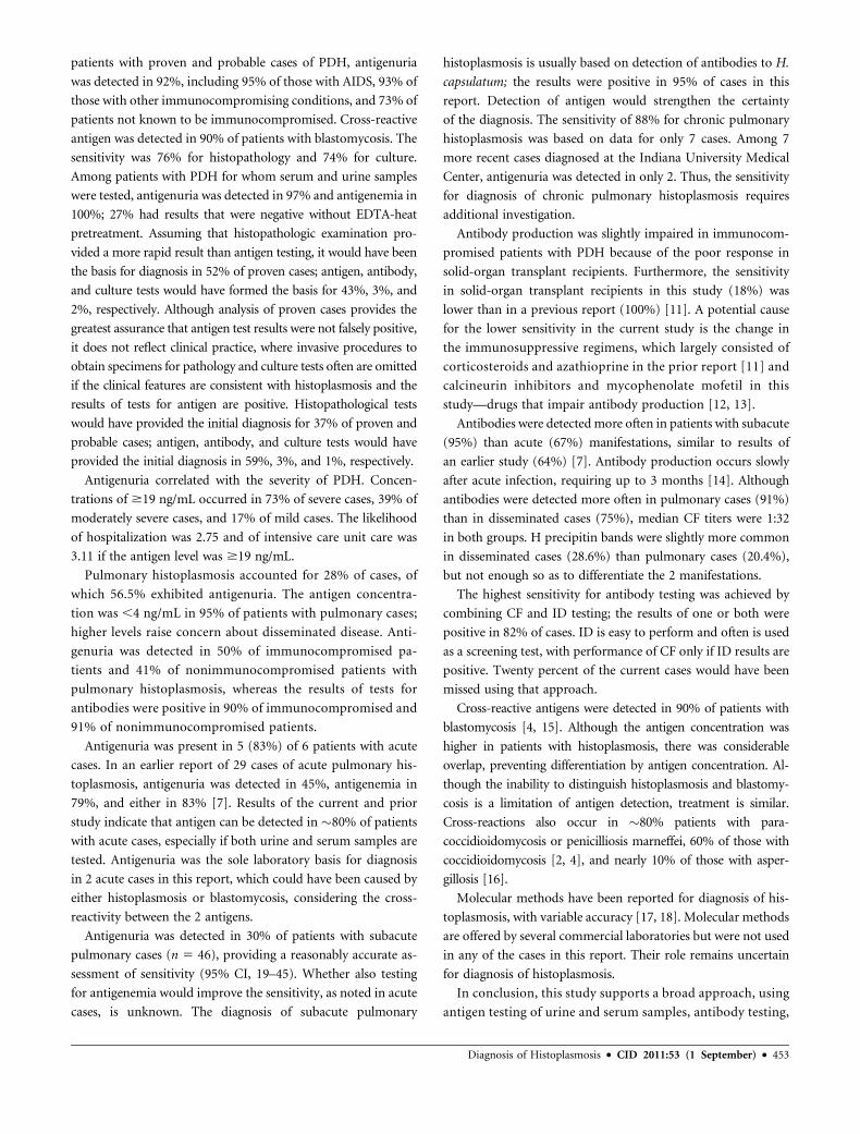
Among immunocompromised patients with pulmonary
histoplasmosis, antigen test results were positive in 7 (50%) of
14, pathology test results were positive in 5 (71.4%) of 7, and
serological test results were positive in 9 (90%) of 10 (Table 4).
The type of pulmonary involvement was acute in 1 case,
chronic in 3 cases, and subacute in 10 cases. Among the non-
immunocompromised patients with pulmonary cases, antigen
test results were positive in 19 (41.3%) of 46, pathology test
results were positive in 6 (33.3%) of 18, and serological test
results were positive in 38 (90.5%) of 42 (P 5 .031). Mean
antigen concentration was 0.91 ng/mL in immunocompro-
mised patients and 0.73 ng/mL in nonimmunocompromised
patients (P 5 .806).
Specificity of Antigen Detection
Cross-reactions occurred in 27 (90%) of 30 patients with proven
pulmonary and/or extrapulmonary blastomycosis (95% CI,
74.4%–96.5%). Concentrations were higher in patients with
proven PDH (mean, 12.10 ng/mL; range, 0.21 to $19.0 ng/mL)
than in those with proven blastomycosis (mean, 7.78 ng/mL;
range, 0.36 to $19.0 ng/mL; P 5 .007). Culture results were
positive in 27 (96.4%) of 28 patients with blastomycosis, and
pathology test results were positive in 21 (84%) of 25 patients
with blastomycosis.
Antibody Detection
Among PDH cases, antibodies were detected in 8 (88.9%)
of 9 nonimmunocompromised patients and 52 (72.2%) of
72 immunocompromised patients (P 5 .889), including 85.7%
of those receiving tumor necrosis factor–inhibitor therapy,
78.9% of those with AIDS, and 18.2% of those who had un-
dergone solid-organ transplantation (Table 1). H and/or M
precipitin bands were detected by ID in 34 (63.2%) of 54
immunocompromised patients versus 6 (85.7%) of 7 non-
immunocompromised patients (P 5 .831). CF titers $1:8 were
detected in 45 (70.3%) of 64 immunocompromised patients
versus 7 (77.8%) of 9 nonimmunocompromised patients (P 5
.932). Elevated antibody levels were present in 41 (73.2%) of
56 patients with proven cases versus 7 (58.3%) of 12 patients
with probable cases (P 5 .854).
Elevated anti-Histoplasma antibody levels were detected in 39
(95.1%) of 41 patients with subacute histoplasmosis, 4 (66.7%)
of 6 with acute histoplasmosis, and 7 (83.3%) of 8 with chronic
pulmonary histoplasmosis (Table 1), as well as in 9 (90%) of
10 immunocompromised patients and 38 (90.5%) of 42 non-
immunocompromised patients (P 5 .806) (Table 4).
Among 101 patients in whom both CF and ID were per-
formed, results of CF alone were positive in 19 (18.8%), results
of ID alone were positive in 8 (7.9%), results of both were
positive in 57 (56.4%), and results of neither were positive in
17 (16.8%). Overall, of 110 patients in whom the ID was per-
formed, M precipitin bands alone were present in 43 (39.1%),
M and H precipitin bands were present in 28 (25.4%), and
neither was present in 39 (35.4%). Among patients in whom
CF was performed, results were positive in 52 (71.2%) of 73
patients with PDH, compared with 40 (80%) of 50 patients
with pulmonary histoplasmosis (P5 .782). The median CF titer
was 1:32 in both groups.
DISCUSSION
To our knowledge, this is the largest multicenter analysis of the
sensitivity of several diagnostic tests for histoplasmosis. Among
Table 4. Findings in Pulmonary Histoplasmosis
Finding
Immunocompromised
patients (n 5 14)
Nonimmunocompromised
patients (n 5 46) P
Culture 7/11 (63.6) 13/24 (54.2) .44
Pathology 5/7 (71.4) 6/18 (33.3) .10
Antigen 7/14 (50)[0.91–1.78]a
19/46 (41.3)[0.73–1.30]
.39
.676b
Antibody 9/10 (90.0) 38/42 (90.5) .806
NOTE. Data are no. of patients with positive results / no. tested (%).a Mean antigen concentration, standard deviation in ng/mL.b P value for comparison of mean antigen level in ng/mL between the
2 groups.
452 d CID 2011:53 (1 September) d Hage et al
patients with proven and probable cases of PDH, antigenuria
was detected in 92%, including 95% of those with AIDS, 93% of
those with other immunocompromising conditions, and 73% of
patients not known to be immunocompromised. Cross-reactive
antigen was detected in 90% of patients with blastomycosis. The
sensitivity was 76% for histopathology and 74% for culture.
Among patients with PDH for whom serum and urine samples
were tested, antigenuria was detected in 97% and antigenemia in
100%; 27% had results that were negative without EDTA-heat
pretreatment. Assuming that histopathologic examination pro-
vided a more rapid result than antigen testing, it would have been
the basis for diagnosis in 52% of proven cases; antigen, antibody,
and culture tests would have formed the basis for 43%, 3%, and
2%, respectively. Although analysis of proven cases provides the
greatest assurance that antigen test results were not falsely positive,
it does not reflect clinical practice, where invasive procedures to
obtain specimens for pathology and culture tests often are omitted
if the clinical features are consistent with histoplasmosis and the
results of tests for antigen are positive. Histopathological tests
would have provided the initial diagnosis for 37% of proven and
probable cases; antigen, antibody, and culture tests would have
provided the initial diagnosis in 59%, 3%, and 1%, respectively.
Antigenuria correlated with the severity of PDH. Concen-
trations of $19 ng/mL occurred in 73% of severe cases, 39% of
moderately severe cases, and 17% of mild cases. The likelihood
of hospitalization was 2.75 and of intensive care unit care was
3.11 if the antigen level was $19 ng/mL.
Pulmonary histoplasmosis accounted for 28% of cases, of
which 56.5% exhibited antigenuria. The antigen concentra-
tion was ,4 ng/mL in 95% of patients with pulmonary cases;
higher levels raise concern about disseminated disease. Anti-
genuria was detected in 50% of immunocompromised pa-
tients and 41% of nonimmunocompromised patients with
pulmonary histoplasmosis, whereas the results of tests for
antibodies were positive in 90% of immunocompromised and
91% of nonimmunocompromised patients.
Antigenuria was present in 5 (83%) of 6 patients with acute
cases. In an earlier report of 29 cases of acute pulmonary his-
toplasmosis, antigenuria was detected in 45%, antigenemia in
79%, and either in 83% [7]. Results of the current and prior
study indicate that antigen can be detected in �80% of patients
with acute cases, especially if both urine and serum samples are
tested. Antigenuria was the sole laboratory basis for diagnosis
in 2 acute cases in this report, which could have been caused by
either histoplasmosis or blastomycosis, considering the cross-
reactivity between the 2 antigens.
Antigenuria was detected in 30% of patients with subacute
pulmonary cases (n 5 46), providing a reasonably accurate as-
sessment of sensitivity (95% CI, 19–45). Whether also testing
for antigenemia would improve the sensitivity, as noted in acute
cases, is unknown. The diagnosis of subacute pulmonary
histoplasmosis is usually based on detection of antibodies to H.
capsulatum; the results were positive in 95% of cases in this
report. Detection of antigen would strengthen the certainty
of the diagnosis. The sensitivity of 88% for chronic pulmonary
histoplasmosis was based on data for only 7 cases. Among 7
more recent cases diagnosed at the Indiana University Medical
Center, antigenuria was detected in only 2. Thus, the sensitivity
for diagnosis of chronic pulmonary histoplasmosis requires
additional investigation.
Antibody production was slightly impaired in immunocom-
promised patients with PDH because of the poor response in
solid-organ transplant recipients. Furthermore, the sensitivity
in solid-organ transplant recipients in this study (18%) was
lower than in a previous report (100%) [11]. A potential cause
for the lower sensitivity in the current study is the change in
the immunosuppressive regimens, which largely consisted of
corticosteroids and azathioprine in the prior report [11] and
calcineurin inhibitors and mycophenolate mofetil in this
study—drugs that impair antibody production [12, 13].
Antibodies were detected more often in patients with subacute
(95%) than acute (67%) manifestations, similar to results of
an earlier study (64%) [7]. Antibody production occurs slowly
after acute infection, requiring up to 3 months [14]. Although
antibodies were detected more often in pulmonary cases (91%)
than in disseminated cases (75%), median CF titers were 1:32
in both groups. H precipitin bands were slightly more common
in disseminated cases (28.6%) than pulmonary cases (20.4%),
but not enough so as to differentiate the 2 manifestations.
The highest sensitivity for antibody testing was achieved by
combining CF and ID testing; the results of one or both were
positive in 82% of cases. ID is easy to perform and often is used
as a screening test, with performance of CF only if ID results are
positive. Twenty percent of the current cases would have been
missed using that approach.
Cross-reactive antigens were detected in 90% of patients with
blastomycosis [4, 15]. Although the antigen concentration was
higher in patients with histoplasmosis, there was considerable
overlap, preventing differentiation by antigen concentration. Al-
though the inability to distinguish histoplasmosis and blastomy-
cosis is a limitation of antigen detection, treatment is similar.
Cross-reactions also occur in �80% patients with para-
coccidioidomycosis or penicilliosis marneffei, 60% of those with
coccidioidomycosis [2, 4], and nearly 10% of those with asper-
gillosis [16].
Molecular methods have been reported for diagnosis of his-
toplasmosis, with variable accuracy [17, 18]. Molecular methods
are offered by several commercial laboratories but were not used
in any of the cases in this report. Their role remains uncertain
for diagnosis of histoplasmosis.
In conclusion, this study supports a broad approach, using
antigen testing of urine and serum samples, antibody testing,
Diagnosis of Histoplasmosis d CID 2011:53 (1 September) d 453
culture, and pathology for diagnosis of disseminated or
pulmonary histoplasmosis. The concentration of antigenuria
in PDH correlates with the severity of disease. Importantly,
the sensitivity of antigen testing is not 100%, even if both
urine and serum samples are tested, and negative results do
not exclude histoplasmosis. Repeated testing is advised for
patients with progressive illness if the initial test results are
negative.
Acknowledgments
Financial support. This work was partly supported by a VA Career
Development Award (CDA-2) to C. A. H.
Potential conflicts of interest. L. J. W., P. C., and M. R. are employees
of MiraVista Diagnostics. All other authors report no conflicts.
All authors have submitted the ICMJE Form for Disclosure of Potential
Conflicts of Interest. Conflicts that the editors consider relevant to the
content of the manuscript have been disclosed in the Acknowledgments
section.
References
1. Wheat LJ, Kohler RB, Tewari RP. Diagnosis of disseminated histo-
plasmosis by detection of Histoplasma capsulatum antigen in serum
and urine specimens. N Engl J Med 1986; 314:83–8.
2. Durkin MM, Connolly PA, Wheat LJ. Comparison of radioimmuno-
assay and enzyme-linked immunoassay methods for detection of
Histoplasma capsulatum var. capsulatum antigen. J Clin Microbiol
1997; 35:2252–5.
3. Wheat LJ, Witt J 3rd, Durkin M, Connolly P. Reduction in false
antigenemia in the second generation Histoplasma antigen assay. Med
Mycol 2007; 45:169–71.
4. Connolly PA, Durkin MM, Lemonte AM, Hackett EJ, Wheat LJ. De-
tection of histoplasma antigen by a quantitative enzyme immunoassay.
Clin Vaccine Immunol 2007; 14:1587–91.
5. Swartzentruber S, LeMonte A, Witt J, et al. Improved detection of
Histoplasma antigenemia following dissociation of immune complexes.
Clin Vaccine Immunol 2009; 16:320–2.
6. Kauffman CA. Diagnosis of histoplasmosis in immunosuppressed
patients. Curr Opin Infect Dis 2008; 21:421–5.
7. Swartzentruber S, Rhodes L, Kurkjian K, et al. Diagnosis of acute
pulmonary histoplasmosis by antigen detection. Clin Infect Dis 2009;
49:1878–2.
8. Wheat J, French ML, Kohler RB, et al. The diagnostic laboratory tests
for histoplasmosis: analysis of experience in a large urban outbreak.
Ann Intern Med 1982; 97:680–5.
9. Wheat LJ, Conces D, Allen SD, Blue-Hnidy D, Loyd J. Pulmonary
histoplasmosis syndromes: recognition, diagnosis, and management.
Semin Respir Crit Care Med 2004; 25:129–44.
10. Agresti A, Coull BA. Approximate is better than ‘‘Exact’’ for interval
estimation of binomial proportions. Am Stat 1998; 52:119–26.
11. Wheat LJ, Smith EJ, Sathapatayavongs B, et al. Histoplasmosis in
renal allograft recipients. Two large urban outbreaks. Arch Intern Med
1983; 143:703–7.
12. Smith KG, Isbel NM, Catton MG, Leydon JA, Becker GJ, Walker RG.
Suppression of the humoral immune response by mycophenolate
mofetil. Nephrol Dial Transplant 1998; 13:160–4.
13. Winslow MM, Gallo EM, Neilson JR, Crabtree GR. The calcineurin
phosphatase complex modulates immunogenic B cell responses. Im-
munity 2006; 24:141–52.
14. Davies SF, Sarosi GA. Serodiagnosis of histoplasmosis and blastomy-
cosis. Am Rev Respir Dis 1987; 136:254–5.
15. Bariola JR, Hage CA, Durkin M, et al. Detection of Blastomyces
dermatitidis antigen in patients with newly diagnosed blastomycosis.
Diagn Microbiol Infect Dis 2011; 69:187–91.
16. Hage CA, Davis TE, Fuller D, et al. Diagnosis of histoplasmosis by
antigen detection in BAL fluid. Chest 2010; 137:623–8.
17. Bialek R, Feucht A, Aepinus C, et al. Evaluation of two nested PCR
assays for detection of Histoplasma capsulatum DNA in human tissue.
J Clin Microbiol 2002; 40:1644–7.
18. Tang YW, Li H, Durkin MM, et al. Urine polymerase chain reaction
is not as sensitive as urine antigen for the diagnosis of disseminated
histoplasmosis. Diagn Microbiol Infect Dis 2006; 54:283–7.
454 d CID 2011:53 (1 September) d Hage et al