A MORPHOLOGICAL STUDY OF THE LINGULA IN SOUTH …
59
1 A MORPHOLOGICAL STUDY OF THE LINGULA IN SOUTH AFRICANS IN RELATION TO SAGITTAL SPLIT OSTEOTOMY Clinton Munsamy A research report submitted to the Faculty of Health Sciences, University of the Witwatersrand, Johannesburg, in partial fulfilment of the requirements for the degree of Master of Dentistry in the branch of Maxillofacial and Oral Surgery Johannesburg, 2013
Transcript of A MORPHOLOGICAL STUDY OF THE LINGULA IN SOUTH …
1
A MORPHOLOGICAL STUDY OF THE
LINGULA IN SOUTH AFRICANS IN
RELATION TO SAGITTAL SPLIT
OSTEOTOMY
Clinton Munsamy
A research report submitted to the Faculty of Health Sciences,
University of the Witwatersrand, Johannesburg, in partial
fulfilment of the requirements for the degree of
Master of Dentistry in the branch of Maxillofacial and Oral
Surgery
Johannesburg, 2013
2
DECLARATION
I, Clinton Munsamy, declare that this research report is my own work. It is being submitted
for the degree of Master of Dentistry in the branch of Maxillofacial and Oral Surgery to the
University of the Witwatersrand, Johannesburg. It has not been submitted before for any
degree or examination at this or any other University.
...........................................
Dr. Clinton Munsamy
......day of ................., 2013
3
ABSTRACT
The sagittal split ramus osteotomy is a common procedure used to correct jaw deformities.
The lingula is an important anatomical landmark that is used as a reference to position the
horizontal osteotomy cut on the medial aspect of the mandible. Knowledge of its position in
relation to surrounding anatomical structures is essential in order to prevent complications
related to the procedure.
The aim of this study was to provide medical and dental practitioners with useful data
regarding the position of the lingula in relation to surrounding anatomical landmarks. Such
data may be of clinical relevance when performing surgery on the mandibular ramus and
when providing anaesthesia for routine dental procedures.
The study involved anthropometric measurements on adult dry mandibles obtained from the
‘Dart Collection’ at the School of Anatomical Sciences, University of the Witwatersrand,
Johannesburg. A total number of 113 adult human dry mandibles were studied. From these
specimens, 201 sides were examined.
The most common shape of lingula noted within the study was that of the truncated type
which was found in 38.8% of cases, followed by the triangular, nodular and assimilated types
which comprised 30.8%, 21.4% and 8.9% respectively. The average distance of the tip of the
lingula from the anterior border, posterior border and sigmoid notch the ramus of the
mandible was approximately 20.15mm, 16.77mm and 16.3mm respectively. The average
distance of the tip of the lingula from the mandibular second molar tooth was found to be
33.3mm. The lingula was above the level of the occlusal plane in 63.7% of cases, by an
average distance of 6.5mm. The width and height of the mandibular foramen exhibited great
variation. Anatomical differences in Caucasian and Black mandibles were noted, with the

4
rami of Caucasian mandibles generally being smaller in dimension compared to black
mandibles.
The anatomic data provided by this study may assist surgeons to locate and identify the
lingula without difficulty, and avoid intraoperative complications. The data presented has a
direct relevance to clinical practice and teaching.
5
DEDICATION
To my dearest wife Thanusha and our beautiful son Matthew,
My parents Stanley and Vasantha
For your love, encouragement, patience and prayers…
The Lord giveth wisdom:
Out of his mouth cometh
knowledge and understanding.
He layeth up sound wisdom for the
righteous:
He is a buckler to them that walk rightly.
Proverbs 2:6-7
6
ACKNOLEDGEMENTS
I wish to express my gratitude to Dr. Ephraim Rikhotso, my supervisor, for his time and
guidance in helping me to complete this research report
Special thanks to Mr Brendan Billings, curator of the ‘Dart Collection’ at the School of
Anatomical Sciences, University of the Witwatersrand, Johannesburg, for his assistance with
the specimens.
7
TABLE OF CONTENTS
DECLARATION…......................................................................................................... 2
ABSTRACT…………..…………………………………………………………………….3
DEDICATION………………………………………………………………………….. 5
ACKNOWLEDGEMENTS................................................................................................ 6
TABLE OF CONTENTS…............................................................................................ 7
LIST OF ABBREVIATIONS............................................................................................. 9
LIST OF FIGURES………………………………………………………………………..10
LIST OF TABLES…..........................................................................................................11
1.0 INTRODUCTION………………………………………………………………… 12
2.0 LITERATURE REVIEW………………………………………………………… 14
2.1 Shape of the lingula………………………………………………………. 14
2.2 Location of the lingula…………………………………………………… 15
2.3 The significance of the lingula in SSRO………………………………… 17
2.3.1 Unfavourable fractures………………………………………. 18
2.3.2 Inferior alveolar nerve injury……………………………….. 20
2.3.3 Haemorrhage ………………………………………………… 21
3.0 AIMS AND OBJECTIVES……………………………………………………… 24
3.1 Aim……………………………………………………………………….. 24
3.2 Objectives………………………………………………………………… 24
4.0 MATERIALS AND METHODS……………………………………………….. 25
4.1 Study setting……………………………………………………………… 25
8
4.2 Inclusion criteria…………………………………………………………. 26
4.3 Exclusion criteria……………………………………………………… 26
4.4 Data collection…………………………………………………………. 26
4.5 Data analysis…………………………………………………………… 29
4.6 Ethical considerations…………………………………………………. 30
5.0 RESULTS……………………………………………………………………… 31
5.1 Shape of the lingula…………………………………………………… 33
5.2 Location of the lingula in relation to ramal landmarks and the mandibular
second molar tooth……………………………………………………. 35
5.3 The relationship of the lingula to the occlusal plane……………….. 36
5.4 The mandibular foramen…………………………………………….. 37
5.5 The position of the lingula in relation to the type of malocclusion… 37
5.6 Racial variations in the anatomical position of the lingula………… 38
6.0 DISCUSSION………………………………………………………………….. 40
7.0 CONCLUSIONS………………………………………………………………. 49
8.0 REFERENCES………………………………………………………………... 52
Appendix 1 (Data collection sheet)…………………………………………… 58
Appendix 2 (Ethics certificate)………………………………………………. 59

9
LIST OF ABBREVIATIONS
SSRO – sagittal split ramus osteotomy
IAN – inferior alveolar nerve
SNAPs – sensory nerve action potentials
10
LIST OF FIGURES
Figure1: Position of the lingula in relation to ramal landmarks……………………… 27
Figure 2: Position of the lingula in relation to the mandibular second molar tooth…. 28
Figure 3: Interobserver testing of data (position of the lingula in relation to ramal
landmarks)……………………………………………………………………………. 30
Figure 4: Age distribution of specimens……………………………………………… 31
Figure 5: Anatomical variations of the lingula……………………………………….. 32
Figure 6: Incidence of the anatomical variations of the lingula…………………….... 33
Figure 7: Percentage distribution of anatomical variations of the lingula amongst males and
females………………………………………………………………………………... 33
11
LIST OF TABLES
Table 1: Location of the lingula in relation to ramal landmarks and the mandibular second
molar tooth……………………………………………………………………….. 34
Table 2: The relationship of the lingula to the occlusal plane…………………… 35
Table 3: The dimensions of the mandibular foramen…………………………… 36
Table 4: The position of the lingula in relation to the type of malocclusion……. 37
Table 5: Differences in the anatomical position of the lingula in Black and Caucasian
specimens………………………………………………………………………… 38
12
1.0 Introduction
The sagittal split ramus osteotomy (SSRO) was first demonstrated by Obwegeser and Trauner
in 1955.1 It provided oral and maxillofacial surgeons with a versatile method for correcting
common dentofacial abnormalities such as prognathism and retrognathism, which are caused
by overgrowth (hyperplasia) or undergrowth (hypoplasia) of the mandible respectively.2
Many modifications of the original technique were made by the likes of Dalpont3, Hunsuck
4,
Bell,5 and Epker.
6 The SSRO allows for an intra-oral approach to mandibular ramus surgery,
rapid healing, and easy rigid internal fixation that promotes early jaw function. Several
anatomic landmarks have been proposed to guide surgeons in locating and avoiding the IAN.
These include various radiographic techniques as well as anthropometric measurements using
dry human skulls, but these are few and have been shown to be population specific.7,8
The SSRO consists of making a horizontal osteotomy on the medial surface of the
mandibular ramus, a sagittal osteotomy along the anterior border of the mandibular ramus
and internal oblique line and a vertical body osteotomy.6,9
An important clinical landmark for
performing the horizontal ramus osteotomy is the lingula. This osteotomy line should be
made just above the lingula and extend posteriorly to it in order to make a safe split, which
minimizes the potential for inferior alveolar nerve injury. 4,5,6,9
The lingula is defined as a tongue shaped bony projection on the medial surface of the ramus
close to posterior margin of the mandibular foramen. It was first described by Johannes
Baptist Spix in 1815, and following his description it was referred to as ‘Spix’s ossicle or
spine.10
It lies in close proximity to the mandibular foramen. Failure to accurately identify the
lingula intra-operatively when performing a sagittal split ramus osteotomy can result in a
number of known complications, such as an unfavourable split, transection of the inferior
13
alveolar vessels which results in haemorrhage and damage to the vessels and nerves within
the vicinity of the osteotomy.11,12,13,14
Both the shape and the position of the lingula on the medial aspect of the ramus of the
mandible are highly variable.15,16,17
Whilst certain shapes, such as the classically described
triangular shape, make for easier identification of the structure intra-operatively, others such
as the nodular and assimilated shapes are much more difficult to recognise. Therefore an
accurate knowledge of the anatomy of the medial aspect of the ramus of the mandible is
imperative in order to prevent complications associated with sagittal split ramus osteotomies.
There exists wide racial and regional variation in the metric characteristics of the mandible as
well as the position of the lingula due to genetic and environmental factors, as well as
functional activity which is thought to play a secondary role in determining the anatomy of
the mandible during the early stages of development.18
Thus the position of the lingula and
anatomy of the medial aspect of the ramus of the mandible has the propensity to demonstrate
variations amongst different populations. Various studies have reported both similarities and
disparities with regard to the position of the lingula and anatomy of the medial aspect of the
ramus of the mandible.9,15,16,17
This makes surgery involving the ramus of the mandible
dependant on population specific parameters.
In South Africa, no study has been conducted on dry mandibles to evaluate the position of the
lingula in relation to surrounding anatomical structures and the lower molar teeth. This study
was conducted to provide an insight on the potential population differences by comparing the
results of the study with data previously published in the literature and to provide clinicians
with reliable clinical landmarks when performing surgery on the mandibular ramus as well as
to provide useful insights to the position of the IAN when delivering local anaesthesia for
various dental procedures.
14
2.0 Literature Review
2.1 The Shape of the Lingula
Nicholson 19
and DuBrul 20
reported the presence of various shapes but did not describe them.
Textbook illustrations often fail to describe the existence of forms other than classic
triangular (or tongue-shaped) form, i.e. the truncated type depicted by Hollinshead,21
the
nodular type shown by Berkovitz et al, 22
Sampson 23
and Williams et al 24
and the
assimilated type shown by Morgan et al.25
The shape of the lingula is important, as surgeons
commonly use this landmark when performing mandibular osteotomies.
The shape of the lingula corresponds to anthropological markers for different populations,26
and can be used to assess different populations and races along with non-metric variants of
the skull.27
Results from a study conducted on 72 dry Thai mandibles (144 sides) found that
the truncated shape (47%) was most common followed by nodular (23%), triangular (17%),
and assimilated (13%) shapes.26
In a similar study conducted in India, 68% of mandibles
demonstrated triangular shaped lingulas, 15.8% truncated, 10.9% nodular and 4.8%
assimilated.17
Hoosein et al also conducted a study on 208 Bangladeshi human mandibles
and found that 70.2% of the lingulas were triangular in shape, 20.2% truncated and 9.6%
assimilated.28
Thus, from these studies it becomes apparent that considerable variation may
exist with regard to the shape of the lingula amongst different populations.
Whilst it is often easier to identify the classically described triangular shape when performing
surgery on the medial aspect of the ramus of the mandible, this shape is not always the most
common. It is, however, often challenging to identify the lingula if it is assimilated or nodular
in shape, and thus knowledge of the anatomical location of the structure is invaluable.
15
2.2 Location of the lingula
The precise location of the lingula is clinically significant as it serves as an important
anatomical landmark when performing surgery involving the medial aspect of the mandibular
ramus. Identification of the lingula intra-operatively may assist in avoiding vital structures,
such as the inferior alveolar nerve (IAN) and vessels, present on the medial aspect of the
ramus of the mandible.
An analysis of reports from various countries around the world has shown that the location
of the lingula varies among different ethnic and racial groups.15,17
In a recent study conducted
in Thailand, investigators found that the lingula was located approximately 20.6mm from the
anterior border of the mandibular ramus, 18.1mm from the posterior border and 16mm from
the mandibular notch.15
The study also found that in the majority of cases, the lingula was
located above the level of the occlusal plane. From their results, the authors were able to
provide medical and dental practitioners with guidelines for efficient anaesthesia for dental
treatment as well as guidelines for mandibular osteotomies in Thai patients. In comparison to
similar studies conducted in other parts of the world, the authors found that the shape and
position of the lingula in relation to surrounding structures in Thais varied from other races.15
Results from a similar study carried out on 242 dry Korean mandibles, of unknown sex and
age; found that the lingula was located approximately 17.4mm from the anterior border of the
mandibular ramus and 15.4mm from the mandibular notch.8
The authors were also able to
measure the distance from the distal side of the mandibular second molar to the tip of the
lingula, which was found to be approximately 28.7mm. According to the authors, this
measurement should prove helpful to clinicians by providing them with guidelines as to
where they should expect to find the lingula, and protect the inferior alveolar vessels and
nerve when performing surgery involving the mandibular ramus.8 The lingula, like in most

16
other studies, was found to be above the occlusal plane with an average vertical distance of
approximately 5.9mm in adult mandibles.8
A number of studies have focused on determining the position of the mandibular foramen and
have determined a relationship of the lingula to the structure.18,19
In one such study conducted
on dry adult Zimbabwean mandibles by Mbajiorgu,18
the height of the lingula was measured
with respect to the position of the mandibular foramen. This study concluded that the
mandibular foramen was highly individualistic, being located 2.56mm (on the right side) and
2.08mm (on the left side) behind the midpoint of the ramus and approximately 3mm above
the midpoint of the ramus on both sides. He also noted that the height of the lingula (distance
from the mandibular foramen to the highest point on the lingula) showed great variation, but
on average was 8.40mm (on the right side) and 8.36mm (on the left side).18
This study refutes
the assumption that the lingula and the mandibular foramen were on the same level above the
occlusal plane.29
The author was lead to conclude from his study that when performing a
mandibular block of the inferior alveolar nerve, the needle be directed 1.5mm to 2.5mm
above the occlusal plane as this will ensure that the needle be directed into the
pterygomandibular space for successful IAN anaesthesia in 76.5% of black Zimbabweans.18
Nicholoson,19
in his determination of the mandibular foramen based on anthropometric
measurements of dry human mandibles of East Indian ethnic origin, found that the foramen
was predominantly located in the anteroposterior midpoint of the ramus halfway between the
mandibular notch and the lower surface of the mandible and two thirds of a line joining the
coronoid process and the angle of the mandible. The study also found that in 75% of cases,
the mandibular foramen was located below the level of the occlusal plane, which is in
contrast to other studies.18
17
Monazzi et al,7 in a study conducted in Brazil on 44 human cadaver dry mandibles, found that
the lingula was approximately 16.5mm from the anterior border of the ramus, 14.6mm from
the posterior border, 16.3mm from the sigmoid notch and approximately 27mm to the lower
border of the mandible. The tip of the lingula was located approximately 5.82mm from the
mandibular foramen.7
In a recent prospective study conducted in Johannesburg, South Africa, investigators
evaluated the effect that impacted third molars have on sagittal split ramus osteotomy.30
The
authors also attempted to define the position of the lingula based on measurements made
from a standard panoramic radiograph. They found that the distance of the lingula from the
anterior border of the ramus ranged from 16mm to 18mm.30
Another South African study
conducted in the mid-seventies attempted to identify the position of the lingula in relation to
structures present on the external surface of the ramus, namely the masseteric apical bump, or
more commonly known as the antilingula.31
This structure, however, is not always present
and cannot be readily identified clinically.31
No study, to date, has been conducted in South
Africa with the aim to identify the position of the lingula in relation to surrounding
anatomical landmarks based on anthropometric measurements of dry human mandibles.
2.3 The Significance of the lingula in SSRO
Due to its relationship to the alveolar nerve, the lingula has been a main anatomic landmark
to guide surgeons.32
Obwegeser and Trauner,1 who first introduced the classical SSRO for
correction of various facial discrepancies, suggested that the medial horizontal osteotomy be
made high ‘just below the mandibular notch.’ However, recent studies indicate that the
osteotomy be placed just above the lingula.9,32,33
This is mainly due to the fact that placement
of the horizontal osteotomy too far above the lingula (which is used as a reference point

18
intraoperatively) places the cut in predominantly cortical bone,9,33
increasing the risk of an
unfavourable fracture.9,32,33,34
In 1968 Hunsuck4 introduced the short medial osteotomy which allowed for less medial soft
tissue reflection and better control of the bone cut because the proximal end is better
visualised. Dal Pont,3
Hunsuck,4 and Epker
6 also noted that the medial horizontal osteotomy
should only extend as far posteriorly as the lingual fossa; the mean length being 18mm.3,4,6
Wolford et al 34
modified the initial surgical technique described by Obwegeser and Trauner.1
The authors suggested that the medial osteotomy be directed perpendicular to the ascending
ramus at the level of the superior aspect of the lingula and carried to the depth of the medial
surface of the buccal cortex. They also noted that by performing the medial horizontal
osteotomy through the superior aspect of the lingula facilitates a less problematic split. The
reason for this was that the more superior the level of the ramus, the less interposed
medullary bone there is, and the greater the incidence of fusion between the buccal and
lingual plates of the ramus of the mandible. Also by keeping the medial osteotomy cut
perpendicular to the ascending ramus (as opposed to parallel to the occlusal plane) keeps the
posterior aspect of the cut lower facilitating easier splitting of the segments.
Complications of sagittal split ramus osteotomies include unwanted fracture of either the
distal or proximal segments (bad split), bleeding, inadequate fixation and relapse.12,13,14
These
complications may occur as a result of variations in the anatomy of the medial aspect of the
mandible, inability to identify vital structures such as the lingula and mandibular foramen
when performing the surgery, inexperience of the surgeon or incorrect technique employed.
2.3.1 Unfavourable fractures (‘bad split’)
Incorrect placement of the horizontal osteotomy may lead to an unfavourable or bad
split.9,14,35
In order to ensure a successful split of the mandible, attention has to be paid not
19
only to the antero-posterior position of the lingula, but also to its supero-inferior position.35,36
Placement of the osteotomy cut too far superiorly above the mandibular foramen positions
the cut in the thinner part of the ramus, increasing the risk of a bad split, as the fracture line
may be within purely cortical bi-laminar bone, whilst not extending the cut beyond the
mandibular foramen may result in the posterior aspect of the sagittal split between the
proximal and distal segments being located anterior to the foramen.9,32
Thus the inferior
alveolar bundle may be contained in the proximal segment, and freeing of the bundle may
result in nerve injury or bleeding.35
Numerous studies have found that placement of the
medial osteotomy at or above the point of fusion may result in an unfavourable fracture.9,33
Smith et al 9 found that in only 2% of cases, there was fusion of the buccal and lingual
cortices of the ramus of the mandible at or below the lingula in the anterior part of the ramus,
and 6.1% in the posterior part of the ramus. They also found that at a distance of 7.5mm to
13.3mm above the lingula, buccal and lingual cortex fusion occurs at a rate of 20% in the
anterior ramus and 39% in the posterior ramus. This study led the author to conclude that the
medial horizontal cut be made at or just above the tip of the lingula because a higher cut may
be associated with increased difficulty in splitting the mandible which may result in an
unfavourable fracture.9 Thus, an intimate knowledge of the position of the lingula as well as
the mandibular foramen is essential when performing a SSRO.
Tom et al.33
conducted a study on 48 cadaver hemimandibles to identify the point of fusion
between the buccal and lingual cortical plates with the aim of better directing the horizontal
medial osteotomy in sagittal split ramus osteotomies. This was done by performing various
vertical sections through the ramus of the mandible. The authors concluded that if the
osteotomy is initiated approximately 5mm or more above the lingula, then the probability of
the cut ending in a point of fusion is increased, which thus increases the probability of a bad
20
split. He therefore advised that the medial osteotomy be placed near the tip of the lingula as
this area has sufficient width of bone with an adequate cancellous layer.33
Placement of the cut too far superiorly, in order to avoid the inferior alveolar vessels and
nerve, increases the risk of a bad split, whilst placement of the osteotomy too far inferiorly on
the ramus increases the risk of damage to these structures.
2.3.2 Inferior alveolar nerve injury
One of the major complications of SSRO includes impairment of the inferior alveolar nerve
(IAN), damage which may result in permanent numbness or altered sensation of the lower lip
and chin area. This can affect a patient’s quality of life due to adverse effects on speech,
eating and drinking which may lead to psychological and social interaction issues.37
The
incidence of inferior alveolar nerve damage following SSRO is reported to range from 2% to
15% .7
Some studies have attempted to evaluate the mechanism of IAN injury based on indirect
evidence from post-operative sensory tests.37,38
There is little data to indicate the actual stage
of SSRO that carries the greatest risk for IAN injury. This prompted Teerijoki et.al.39
to
conduct a study in which sensory nerve action potentials (SNAPs) of the IAN were
continuously monitored in 20 patients undergoing bilateral sagittal ramus osteotomy.
Changes in latency, amplitude and sensory nerve conduction velocity at baseline and at
different stages of the operation were analysed. The results of the study indicated that the
most obvious changes in all parameters and highest risk to damage of the IAN occurred
during preparation of the medial side of the ramus. Risk of injury to the IAN was directly
proportional to the time taken to prepare the medial side of the ramus, with SNAPs
disappearing usually after 10 minutes. On clinical evaluation, the authors also reported that in
7 cases the IAN was lacerated.39
From this study it becomes apparent that the greater
21
knowledge of the anatomy of the ramus of the mandible one has, the shorter the preparation
time of the medial horizontal osteotomy with less risk of IAN injury.
2.3.3 Haemorrhage
Haemorrhage is one of the complications that may occur when performing a sagittal split
ramus osteotomy. The incidence is thought to be between 0.39% - 38%, but there are no
uniform criteria defining bleeding complications within the literature.38
Haemorrhage
following orthognathic surgery is usually the result of insufficient surgical haemostasis after
mechanical disruption of blood vessels, rather than acquired or congenital coagulopathy.40
Blood vessels close to the osteotomy line include the internal carotid artery, retromandibular
vein, maxillary artery, facial artery and vein and the inferior alveolar artery and vein.
Behrman14
reported that 38% of surgeons reported haemorrhage as a complication of sagittal
split ramus osteotomy, with 50% of those indicating that it was a severe problem. According
to the report of the study conducted by Behrman, the principal source of bleeding was the
maxillary artery, followed by the facial artery, inferior alveolar artery and retromandibular
vein.14
Recent reports have not found haemorrhage to be a major problem when performing sagittal
split ramus osteotomies.38,40
According to a study conducted by Tetzrow et.al,38
in which
1274 consecutive patients were reviewed for perioperative complications following sagittal
split ramus osteotomy, 15 patients (1.2%) were reported as having acute haemorrhage as a
complication. The authors reported that the retromandibular vein was most often severed,
followed by the facial artery. In 9 cases the vessel was not identified. The authors also stated
that when the inferior alveolar vessel was severed, achieving haemostasis was usually not a
problem. One patient, however, did develop a massive swelling and haematoma and required
a tracheostomy.38
22
In 8 of the 21 cases of haemorrhage following SSRO or intra-oral vertical ramus osteotomy
reported by Lanigan et al,40
the maxillary artery was severed intra-operatively, most often
resulting from the medial horizontal osteotomy cut. Brisk bleeding was encountered, but no
deaths reported as a result of the complication. However, in two cases the patients were taken
back to the operating theatre to control the bleeding. Four cases required ligation of the
external carotid artery, which often results in increased operating time and more extensive
neck dissection. In one case the inferior alveolar artery and vein were severed during the
malleting of the right sagittal split ramus osteotomy, which resulted in severe bleeding that
was poorly controlled intra-operatively by the attendant surgeons. The patient was transferred
to the ICU intubated, but recovered with no major post-operative haemorrhage, according to
the authors.40
Often bleeding encountered within the maxillofacial area is difficult to visualise, which
makes control of the haemorrhage challenging. Such was the case of a 26 year old female
patient undergoing bilateral sagittal split ramus osteotomy for correction of mandibular
prognathism and facial asymmetry.41
The left internal maxillary artery was inadvertently
severed. The patient developed disseminated intravascular coagulation (DIC) from the
massive haemorrhage that ensued. The patient subsequently required embolization of the
external carotid artery to control the bleeding. Bleeding from the internal maxillary artery is
often difficult to control, due the difficulty in locating and ligating the artery intra-
operatively, as well as the numerous anastomoses possessed by the maxillary artery.42
2.4 The importance of the lingula for dental anaesthesia
Knowledge of the position of the lingula is not only pertinent to those performing surgery on
the ramus of the mandible, but also to dentists who administer local anaesthetic for a
23
mandibular block, with the anaesthetic solution being deposited in the region of the lingula of
the mandible. Incorrect technique or inadequate knowledge of the anatomy of the medial
aspect of the ramus of the mandible may result in failure of the mandibular block, or more
importantly damage to the inferior alveolar or lingual nerves. Local anaesthesia of the inferior
alveolar nerve is achieved most commonly through the Halsted technique, whereby local
anaesthetic is deposited in the pterygomandibular space where the IAN enters the mandibular
foramen.43
The lingula overlies the mandibular foramen, and knowledge of its position is
imperative when performing a mandibular block. It has been shown that racial and regional
differences in functional activity involving the mandible during early stages of development
may affect the size and shape of the mandible and thus the position of the mandibular
foramen.18
Failure rates as high as 29% for IAN anaesthesia have been reported,44
which have
been attributed to a number of factors including anatomic variability.45
Mbajiorgu18
in his
anthropometric study involving dry human mandibles conducted in Zimbabwe found that the
lingula lies on average 5.6mm above the occlusal plane, and so suggested that the needle for
local anaesthesia of the inferior alveolar nerve be directed 1.5mm to 2.5mm above the
occlusal plane to ensure success of the mandibular block.
The etiology of nerve damage following dental injection is unknown.46
In most cases it is
thought to be related to chemical injury from a variety of anaesthetic agents, with articaine
and prilocaine being cited more commonly.46,47
However, direct trauma from the needle
cannot be excluded.46
It is hypothesised that direct trauma may be related to the formation of
an intraneural haematoma caused by the needle striking one of the smaller intraneural blood
vessels.47
Intraneural haematomas are known to be neurotoxic.46
According to Hillerup,47
a
neuroma formed after direct injury to the nerve, may interfere with nerve conduction by
intraneural pressure similar to a situation like compartment syndrome.

24
3.0 Aims and Objectives
3.1 Aim
The aim of this study is to provide medical and dental practitioners with useful data regarding
the position of the lingula in relation to surrounding anatomical landmarks. Such data may be
of clinical relevance when performing surgery on the mandibular ramus, and when providing
anaesthesia for routine dental procedures.
3.2 Objectives
1. To identify the various shapes of the lingula and its occurrence
2. To determine the height and location of the lingula in relation to mandibular ramal
landmarks
3. To determine the position of the lingula in relation to the mandibular second molar
tooth
4. To determine gender and age differences with respect to the above criteria
25
4.0 Materials and Methods
4.1 Study setting
The study involved the use of the Raymond Arthur Dart Collection (commonly referred to as
the “Dart Collection”) which is housed at the School of Anatomical Sciences, University of
The Witwatersrand, Johannesburg, South Africa. It is the largest human skeletal collection
within Southern Africa and comprises a total number of 2605 documented skeletons.
The collection originated in the early 1920’s as a result of the efforts of Raymond Dart, an
Australian born anatomist, who took over Headship of the Anatomy Department of the
recently established University of the Witwatersrand in January 1923. The cadaver derived
skeletons that comprise the Raymond Dart collection have always been collected under the
provision of South African Human Tissue Act (No. 65 of 1983, and by previous Acts eg. The
Anatomy Act No. 20 of 1959). Prior to 1958, specimens within the collection were derived
mainly from unclaimed bodies from Gauteng (previously Transvaal) Provincial Hospitals, but
since then, a bequeathment programme has increasingly contributed to the collection,
providing cadavers for teaching and research in medical sciences. The collection has become
of significant importance for studies of forensic anthropology, population biology, human
variation, dentistry, medicine, clinical anatomy, forensic pathology and
palaeoanthropology.48
The majority of information regarding the cadavers is derived from the death certificates of
the specimens. The collection has a wide selection of ages that extends from 100 years to
neonates, as well as a multitude of populations representing both sexes.
The study was conducted on the dry mandible specimens within the ‘Dart Collection’.
26
4.2 Inclusion Criteria
- Mandibles that were dentate, with dentition up to and including the mandibular
second molar tooth
- mandibles in which the age, sex and race were known (identified according to
historic records)
4.3 Exclusion Criteria
- mandibles that were edentulous, or that exhibited the presence of a bony defect or
deformity or evident pathology
4.4 Data Collection
A digital sliding calliper was used to obtain linear measurements of the specimens
(Mitutoyo R, Japan , capable of measuring to the nearest 0.01mm). All measurements
were carried out by the same investigator.
The mandibles were measured according to predetermined points. All measurements were
determined to the tip of the lingula (defined as the highest point of the lingula). The
following data was obtained:
- Shape of the lingula
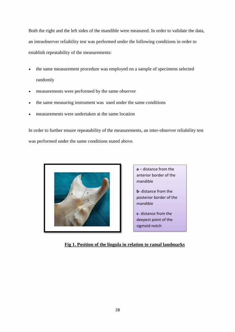
- Position of the lingula in relation to ramal landmarks
o The distance of the lingula from the sigmoid notch was determined by
measuring the distance from the deepest point of the sigmoid notch to the tip
of the lingula (the highest point of the lingula)

27
o The distance of the lingula from anterior border of the ramus of the mandible
was measured by drawing a line from the anterior border of the ramus to the
tip of the lingula perpendicular to the line drawn from the deepest point of the
sigmoid notch to the tip of the lingula
o The distance of the lingula from the posterior border of the ramus of the
mandible was measured by drawing a line from the posterior border of the
ramus of the mandible to the tip of the lingula perpendicular to the line drawn
from the deepest point of the sigmoid notch to the tip of the lingula
- Position of the lingula in relation to the mandibular second molar
o The distance was measured from the tip of the distobuccal cusp of the
mandibular second molar tooth to the tip of the lingula (defined as the highest
point of the lingula)
- Size of the mandibular foramen
o The height of the mandibular foramen was measured as a vertical distance
between the tip of the lingula and lower border of the mandibular foramen
o The width of the mandibular foramen was measured as a distance from the
anterior to the posterior borders of the foramen at the same level.
- Position of the lingula in relation to the occlusal plane
o A flat instrument was used to determine the level of the occlusal plane
o The lingula was defined as being:
At the level of the occlusal plane
Above the occlusal plane
Below the occlusal plane
28
Both the right and the left sides of the mandible were measured. In order to validate the data,
an intraobserver reliability test was performed under the following conditions in order to
establish repeatability of the measurements:
the same measurement procedure was employed on a sample of specimens selected
randomly
measurements were performed by the same observer
the same measuring instrument was used under the same conditions
measurements were undertaken at the same location
In order to further ensure repeatability of the measurements, an inter-observer reliability test
was performed under the same conditions stated above.
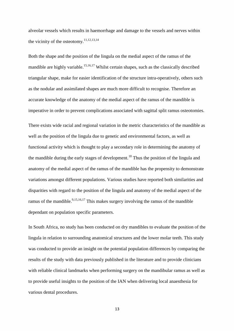
Fig 1. Position of the lingula in relation to ramal landmarks
a – distance from the
anterior border of the
mandible
b- distance from the
posterior border of the
mandible
c- distance from the
deepest point of the
sigmoid notch
29
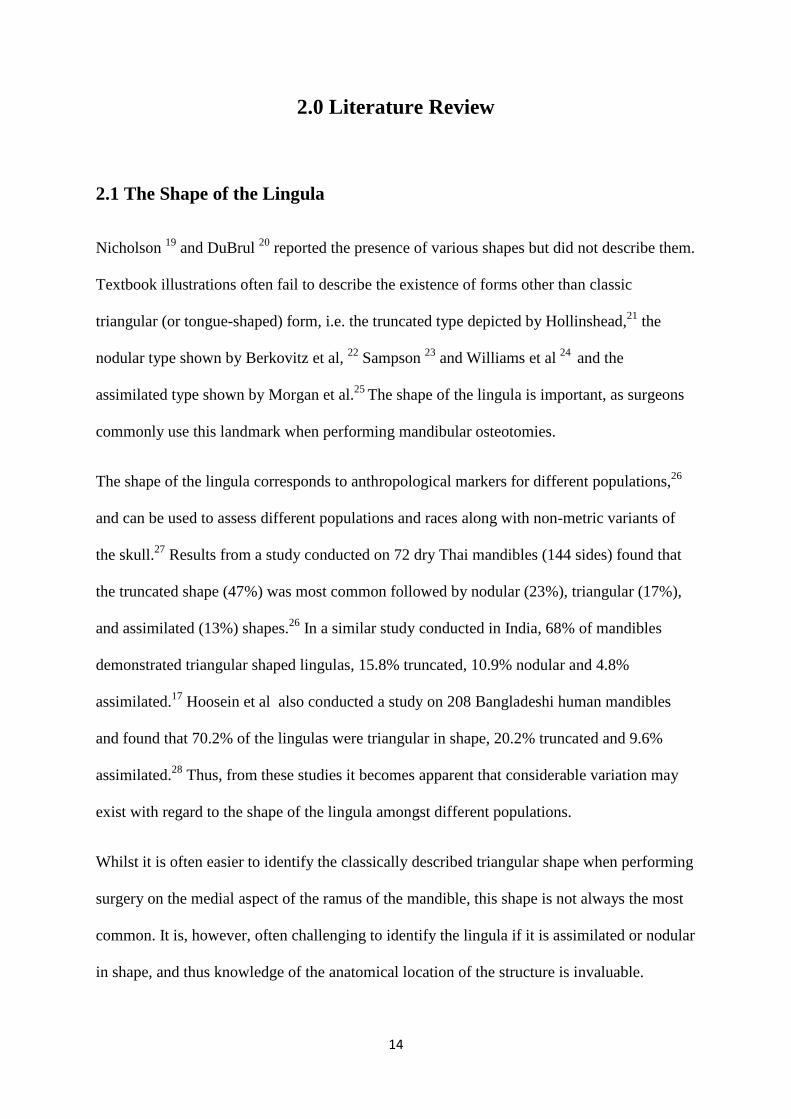
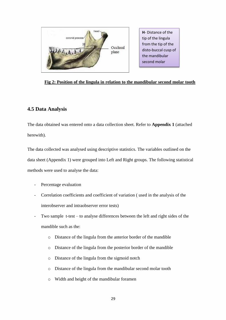
Fig 2: Position of the lingula in relation to the mandibular second molar tooth
4.5 Data Analysis
The data obtained was entered onto a data collection sheet. Refer to Appendix 1 (attached
herewith).
The data collected was analysed using descriptive statistics. The variables outlined on the
data sheet (Appendix 1) were grouped into Left and Right groups. The following statistical
methods were used to analyse the data:
- Percentage evaluation
- Correlation coefficients and coefficient of variation ( used in the analysis of the
interobserver and intraobserver error tests)
- Two sample t-test – to analyse differences between the left and right sides of the
mandible such as the:
o Distance of the lingula from the anterior border of the mandible
o Distance of the lingula from the posterior border of the mandible
o Distance of the lingula from the sigmoid notch
o Distance of the lingula from the mandibular second molar tooth
o Width and height of the mandibular foramen
H- Distance of the
tip of the lingula
from the tip of the
disto-buccal cusp of
the mandibular
second molar
30
- Standard Deviation (to determine how widely values are dispersed from the average
value (mean)
Data was captured using Microsoft Excel 2010 (Microsoft Office Professional Plus 2010).
Data analysis, plotting of graphs and tables was carried out using Microsoft Excel 2010.
4.6 Ethical Considerations
Approval for the use of the human dry mandibles for research purposes has been obtained
from the Department of Anatomy, University of the Witwatersrand from the Human
Research Ethics Committee of South Africa. Ethics reference number- W-CJ-101109-1. See
attached Ethics waiver (Appendix 2).
31
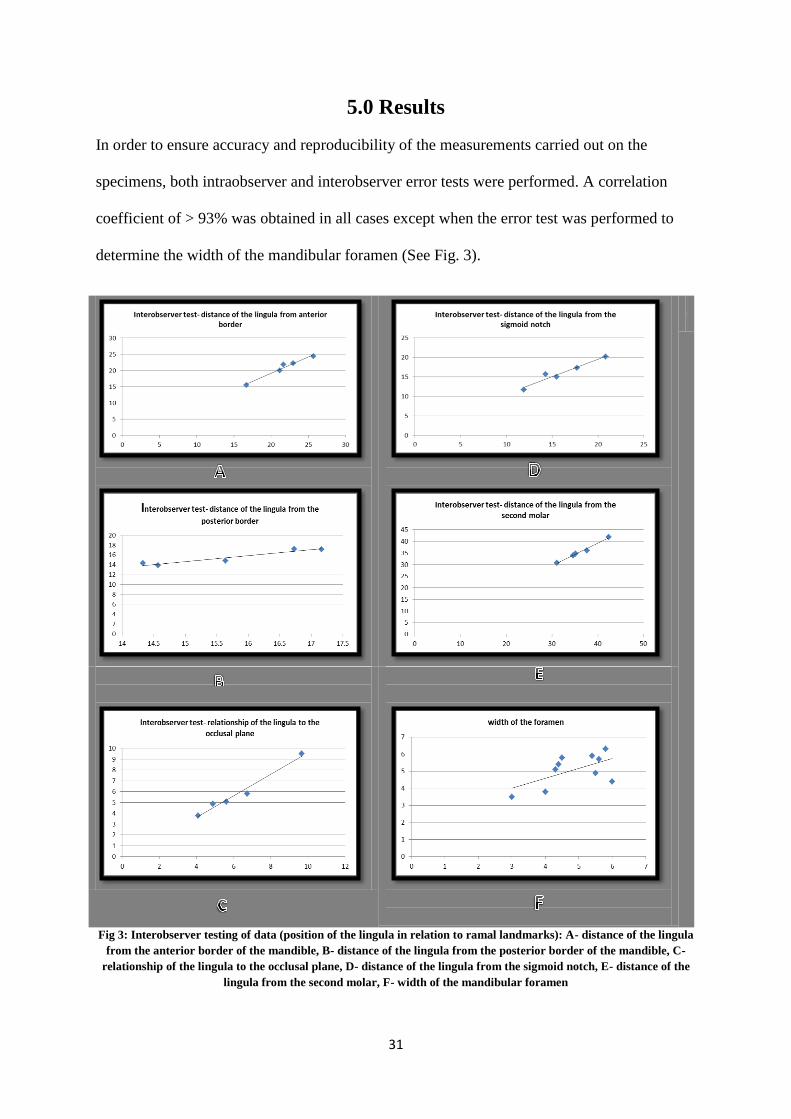
5.0 Results
In order to ensure accuracy and reproducibility of the measurements carried out on the
specimens, both intraobserver and interobserver error tests were performed. A correlation
coefficient of > 93% was obtained in all cases except when the error test was performed to
determine the width of the mandibular foramen (See Fig. 3).
Fig 3: Interobserver testing of data (position of the lingula in relation to ramal landmarks): A- distance of the lingula
from the anterior border of the mandible, B- distance of the lingula from the posterior border of the mandible, C-
relationship of the lingula to the occlusal plane, D- distance of the lingula from the sigmoid notch, E- distance of the
lingula from the second molar, F- width of the mandibular foramen
32
Due to the extensive variation of the mandibular foramen, especially in terms of its width, a
correlation coefficient of 59% was observed. Fig. 1 illustrates a few of the error tests
performed by the interobserver, as well as the error test for determination of the width of the
mandibular foramen.
A total number of 113 adult human dry mandibles were selected from the Raymond Arthur
Dart Collection at the Department of Anatomy, University of the Witwatersrand. From these
specimens, 201 sides were examined. Both the right and the left sides were examined. In 25
cases, only one side was examined, either left or right, due to the omitted side not meeting the
inclusion criteria.
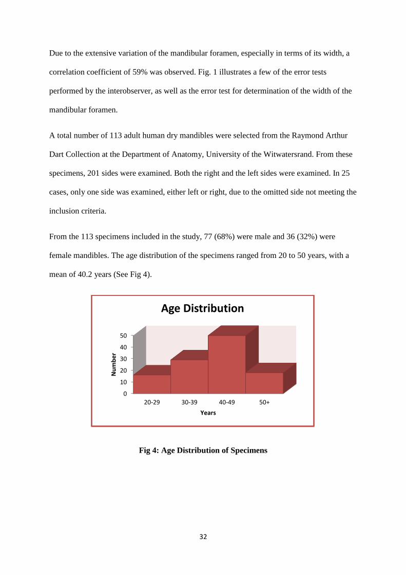
From the 113 specimens included in the study, 77 (68%) were male and 36 (32%) were
female mandibles. The age distribution of the specimens ranged from 20 to 50 years, with a
mean of 40.2 years (See Fig 4).
Fig 4: Age Distribution of Specimens
0
10
20
30
40
50
20-29 30-39 40-49 50+
Nu
mb
er
Years
Age Distribution
33
5.1 Shape of the lingula
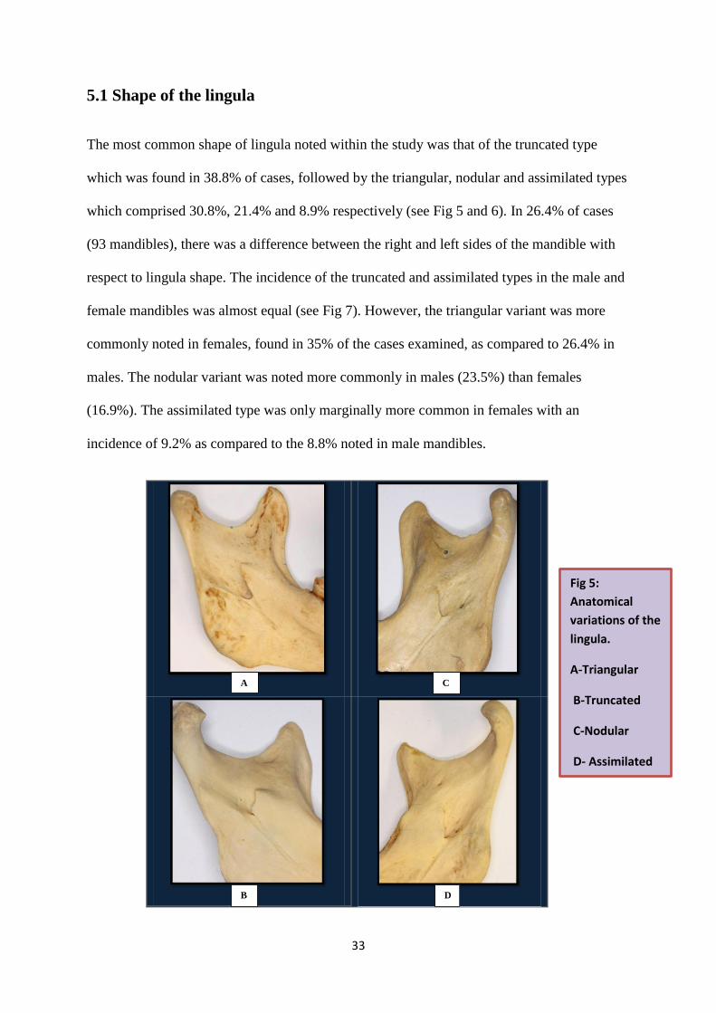
The most common shape of lingula noted within the study was that of the truncated type
which was found in 38.8% of cases, followed by the triangular, nodular and assimilated types
which comprised 30.8%, 21.4% and 8.9% respectively (see Fig 5 and 6). In 26.4% of cases
(93 mandibles), there was a difference between the right and left sides of the mandible with
respect to lingula shape. The incidence of the truncated and assimilated types in the male and
female mandibles was almost equal (see Fig 7). However, the triangular variant was more
commonly noted in females, found in 35% of the cases examined, as compared to 26.4% in
males. The nodular variant was noted more commonly in males (23.5%) than females
(16.9%). The assimilated type was only marginally more common in females with an
incidence of 9.2% as compared to the 8.8% noted in male mandibles.
A
B
C
D
Fig 5:
Anatomical
variations of the
lingula.
A-Triangular
B-Truncated
C-Nodular
D- Assimilated

34
Fig 6: Incidence of the anatomical variations of the lingula
Fig 7: Percentage distribution of anatomical variations of the lingula amongst males
and females
30.80%
38.80%
21.40%
9%
Anatomical variations of the lingula
Triangular
Truncated
Nodular
Assimilated

35
5.2 Location of the lingula in relation to ramal landmarks and the
mandibular 2nd
molar tooth
The position of the lingula in relation to mandibular ramal landmarks is shown in Table 1.
There was statistically no significant difference in position of the lingula between the right
and left sides of the mandible in terms of the distance from the anterior border, posterior
border and sigmoid notch of the ramus of the mandible, with p= 0.18, 0.83 and 0.2
respectively. Measurements indicated that the average horizontal distance of the lingula from
the anterior border of the ramus of the mandible was approximately 20.42mm (right) and
19.88mm (left), the average horizontal distance from the posterior border of the ramus being
16.77mm (right) and 16.84mm (left) whilst the average horizontal distance of the lingula
from the sigmoid notch being 16.53mm (right) and 16.1mm (left).
Average Range Standard Deviation P –value
(t-test)
Parameter Right Left Right Left Right Left
Distance from the
anterior border
20.42 19.88 12.98
to
28.08
13.09
to
27.72
2.85 2.84 p= 0.18
Distance from the
posterior border
16.77 16.84 12.18
to
22.05
10.73
to
22.77
2.05 2.2 p=0.83
Distance from the
sigmoid notch
16.53 16.1 9.25
to
22.74
10.33
to
20.65
2.56 2.2 p= 0.2
Distance of the lingula
from the mandibular
2nd
molar
33.8 32.9 26.4
to
42.41
20.87
to
40.68
3.3 3.9 p= 0.08
Table1: Location of the lingula in relation to ramal landmarks and the mandibular 2nd
molar tooth (millimetres)
36
The horizontal distance from the tip of the disto-buccal cusp of the mandibular second molar
tooth to the tip of the lingula was found to be on average 33.8mm (right) and 32.9mm (left)
(Table 1). This implies that the distance from the second molar to the posterior border of the
ramus of the mandible is approximately 48mm. There was no statistical difference in
measurements between the right and left sides of the mandible.
5.3 The relationship of the lingula to the occlusal plane
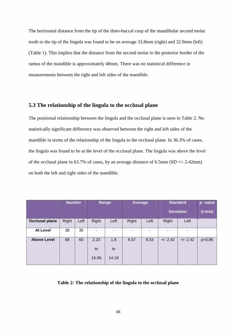
The positional relationship between the lingula and the occlusal plane is seen in Table 2. No
statistically significant difference was observed between the right and left sides of the
mandible in terms of the relationship of the lingula to the occlusal plane. In 36.3% of cases,
the lingula was found to be at the level of the occlusal plane. The lingula was above the level
of the occlusal plane in 63.7% of cases, by an average distance of 6.5mm (SD +/- 2.42mm)
on both the left and right sides of the mandible.
Number Range Average Standard
Deviation
p- value
(t-test)
Occlusal plane Right Left Right Left Right Left Right Left
At Level 38 35 - - - - - -
Above Level 68 60 2.23
to
16.06
1.8
to
14.19
6.57 6.53 +/- 2.42 +/- 2.42 p=0.86
Table 2: The relationship of the lingula to the occlusal plane
37
5.4 The mandibular foramen
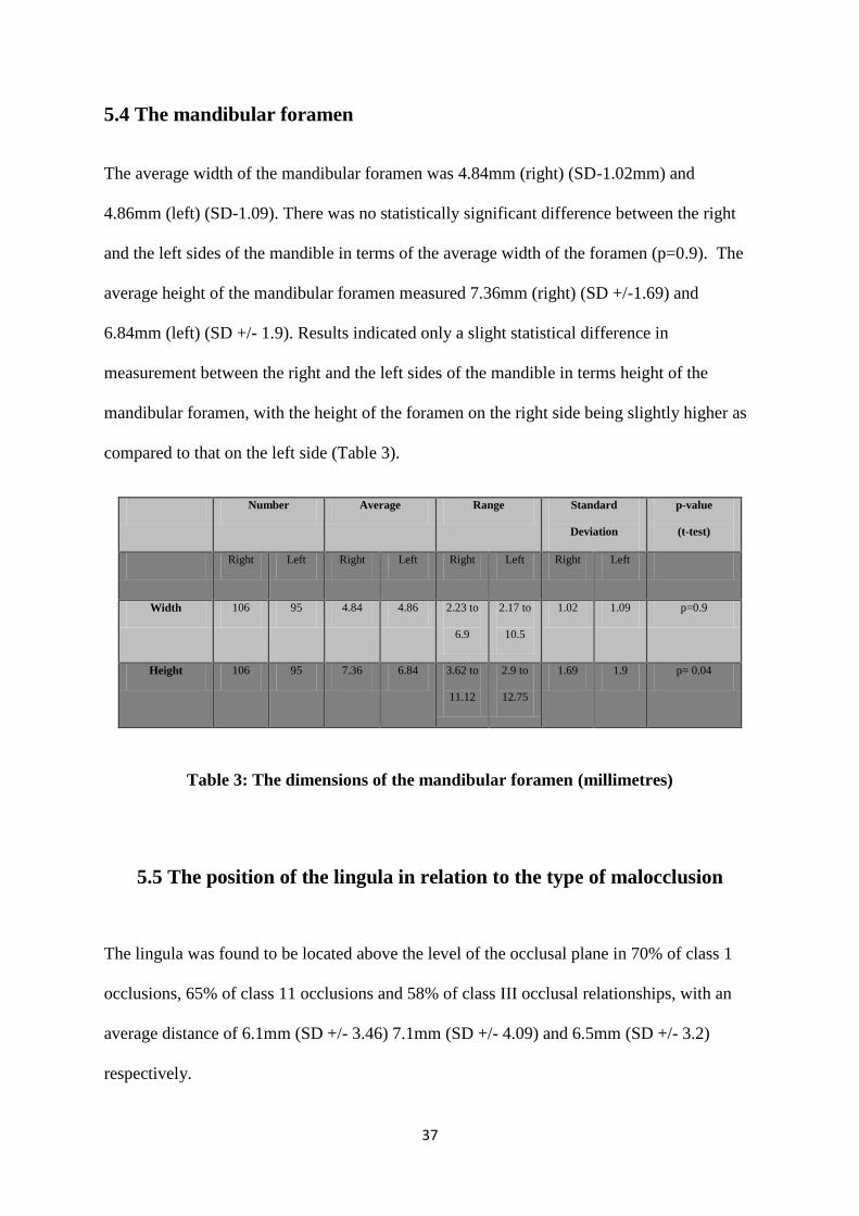
The average width of the mandibular foramen was 4.84mm (right) (SD-1.02mm) and
4.86mm (left) (SD-1.09). There was no statistically significant difference between the right
and the left sides of the mandible in terms of the average width of the foramen (p=0.9). The
average height of the mandibular foramen measured 7.36mm (right) (SD +/-1.69) and
6.84mm (left) (SD +/- 1.9). Results indicated only a slight statistical difference in
measurement between the right and the left sides of the mandible in terms height of the
mandibular foramen, with the height of the foramen on the right side being slightly higher as
compared to that on the left side (Table 3).
Number Average Range Standard
Deviation
p-value
(t-test)
Right Left Right Left Right Left Right Left
Width 106 95 4.84 4.86 2.23 to
6.9
2.17 to
10.5
1.02 1.09 p=0.9
Height 106 95 7.36 6.84 3.62 to
11.12
2.9 to
12.75
1.69 1.9 p= 0.04
Table 3: The dimensions of the mandibular foramen (millimetres)
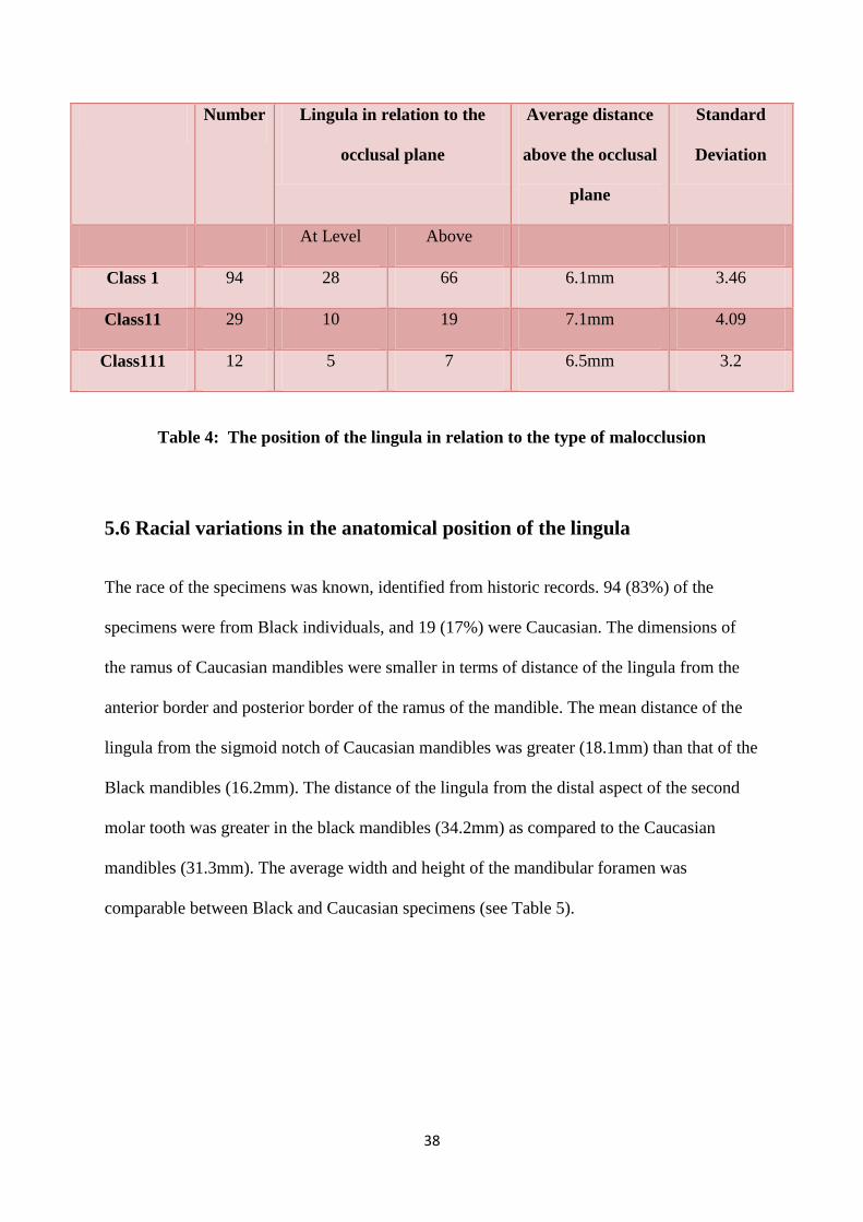
5.5 The position of the lingula in relation to the type of malocclusion
The lingula was found to be located above the level of the occlusal plane in 70% of class 1
occlusions, 65% of class 11 occlusions and 58% of class III occlusal relationships, with an
average distance of 6.1mm (SD +/- 3.46) 7.1mm (SD +/- 4.09) and 6.5mm (SD +/- 3.2)
respectively.
38
Number Lingula in relation to the
occlusal plane
Average distance
above the occlusal
plane
Standard
Deviation
At Level Above
Class 1 94 28 66 6.1mm 3.46
Class11 29 10 19 7.1mm 4.09
Class111 12 5 7 6.5mm 3.2
Table 4: The position of the lingula in relation to the type of malocclusion
5.6 Racial variations in the anatomical position of the lingula
The race of the specimens was known, identified from historic records. 94 (83%) of the
specimens were from Black individuals, and 19 (17%) were Caucasian. The dimensions of
the ramus of Caucasian mandibles were smaller in terms of distance of the lingula from the
anterior border and posterior border of the ramus of the mandible. The mean distance of the
lingula from the sigmoid notch of Caucasian mandibles was greater (18.1mm) than that of the
Black mandibles (16.2mm). The distance of the lingula from the distal aspect of the second
molar tooth was greater in the black mandibles (34.2mm) as compared to the Caucasian
mandibles (31.3mm). The average width and height of the mandibular foramen was
comparable between Black and Caucasian specimens (see Table 5).

39
Number Distance
from the
anterior
border
Distance
from the
posterior
border
Distance
from the
sigmoid
notch
Distance
from the
second
molar
tooth
Width of the
mandibular
foramen
Height of the
mandibular
foramen
Black 94 20.8 17.1 16.2 34.2 4.8 7.2
Caucasian 19 17.1 15.1 18.1 31.3 4.9 7.8
Table 5: Differences in the anatomical position of the lingula in Black and Caucasian
specimens
40
6.0 Discussion
The lingula, as its name implies, is a tongue shaped projection found on the medial aspect of
the ramus of the mandible. It overlies the mandibular foramen, where the inferior alveolar
vessels and nerve enter into the mandibular canal, and has become an important clinical
landmark to surgeons as well as dental practitioners. Knowledge of both its location and
shape is imperative in order to accurately identify the structure clinically, since anatomical
structures in relation to the lingula are subject to injury during a variety of oral and
maxillofacial surgical procedures, such as in orthognathic surgery, mandibular trauma
management, pre-prosthetic surgery and nerve injury during inferior alveolar nerve
blocks.11,12,13,14
Failure to do so may result in an increased risk of complications related to the
procedure being performed. 12,13,14,38
This may include an increase in the occurrence of a ‘bad
split’ or damage to the inferior alveolar vessels and nerve when performing a sagittal split
ramus osteotomy.38,39,42
For too long there has been a dearth of information regarding the shape of the lingula.
Nicholson 19
and Dubrul 20
acknowledged that various shapes may be present, but failed to
describe them. Nowadays, four different shapes are recognised, namely the triangular,
truncated, nodular and assimilated shapes.21,22,23,24
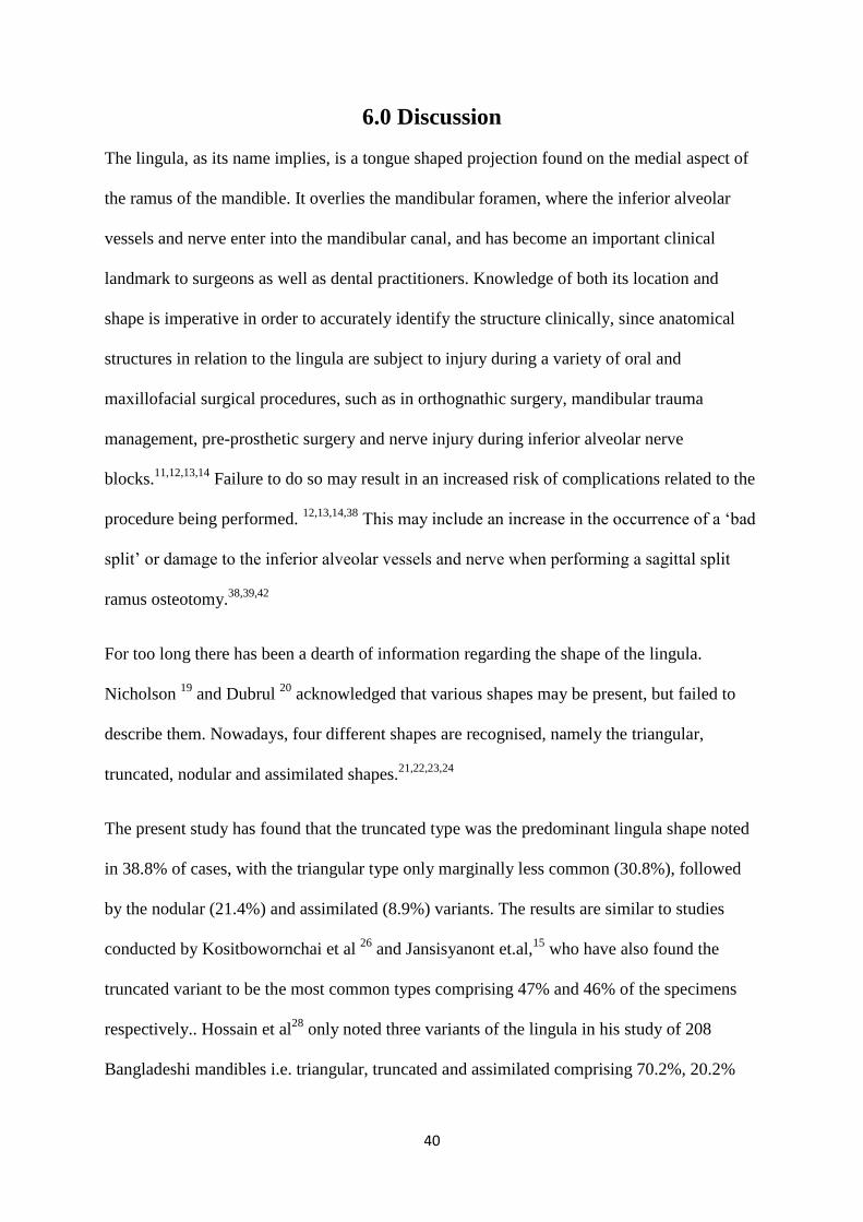
The present study has found that the truncated type was the predominant lingula shape noted
in 38.8% of cases, with the triangular type only marginally less common (30.8%), followed
by the nodular (21.4%) and assimilated (8.9%) variants. The results are similar to studies
conducted by Kositbowornchai et al 26
and Jansisyanont et.al,15
who have also found the
truncated variant to be the most common types comprising 47% and 46% of the specimens
respectively.. Hossain et al28
only noted three variants of the lingula in his study of 208
Bangladeshi mandibles i.e. triangular, truncated and assimilated comprising 70.2%, 20.2%
41
and 9.6% respectively. Other studies have reported the triangular variant to be the most
common.16,17
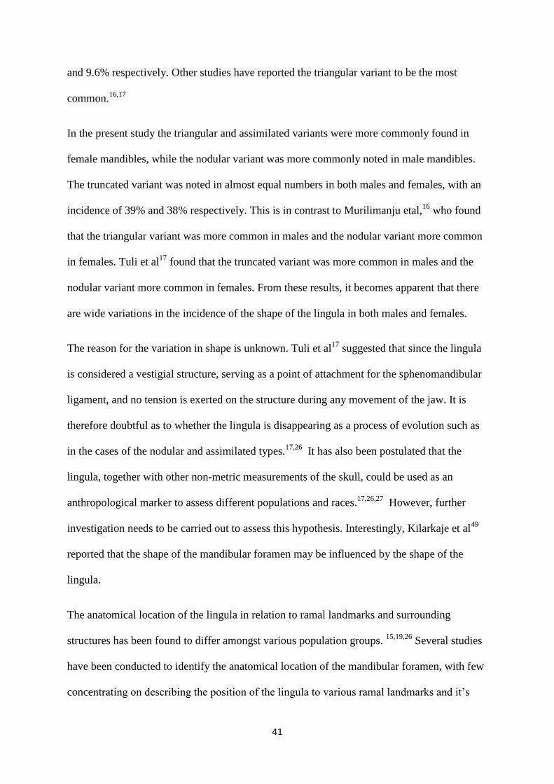
In the present study the triangular and assimilated variants were more commonly found in
female mandibles, while the nodular variant was more commonly noted in male mandibles.
The truncated variant was noted in almost equal numbers in both males and females, with an
incidence of 39% and 38% respectively. This is in contrast to Murilimanju etal,16
who found
that the triangular variant was more common in males and the nodular variant more common
in females. Tuli et al17
found that the truncated variant was more common in males and the
nodular variant more common in females. From these results, it becomes apparent that there
are wide variations in the incidence of the shape of the lingula in both males and females.
The reason for the variation in shape is unknown. Tuli et al17
suggested that since the lingula
is considered a vestigial structure, serving as a point of attachment for the sphenomandibular
ligament, and no tension is exerted on the structure during any movement of the jaw. It is
therefore doubtful as to whether the lingula is disappearing as a process of evolution such as
in the cases of the nodular and assimilated types.17,26
It has also been postulated that the
lingula, together with other non-metric measurements of the skull, could be used as an
anthropological marker to assess different populations and races.17,26,27
However, further
investigation needs to be carried out to assess this hypothesis. Interestingly, Kilarkaje et al49
reported that the shape of the mandibular foramen may be influenced by the shape of the
lingula.
The anatomical location of the lingula in relation to ramal landmarks and surrounding
structures has been found to differ amongst various population groups. 15,19,26
Several studies
have been conducted to identify the anatomical location of the mandibular foramen, with few
concentrating on describing the position of the lingula to various ramal landmarks and it’s
42
relation to the mandibular foramen. In this study we have aimed to identify the anatomical
location of the lingula in dry mandibles, in order to obtain information that could be used
when performing mandibular osteotomies.
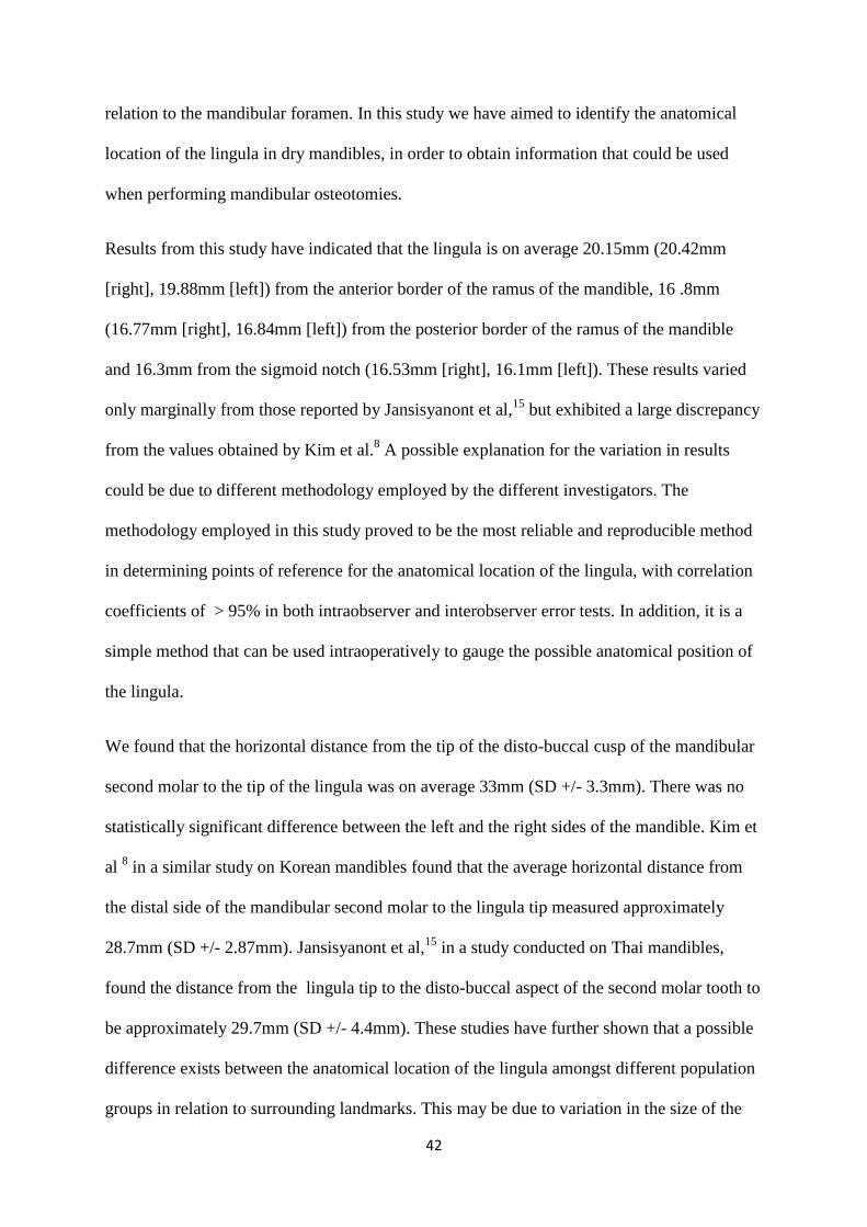
Results from this study have indicated that the lingula is on average 20.15mm (20.42mm
[right], 19.88mm [left]) from the anterior border of the ramus of the mandible, 16 .8mm
(16.77mm [right], 16.84mm [left]) from the posterior border of the ramus of the mandible
and 16.3mm from the sigmoid notch (16.53mm [right], 16.1mm [left]). These results varied
only marginally from those reported by Jansisyanont et al,15
but exhibited a large discrepancy
from the values obtained by Kim et al.8 A possible explanation for the variation in results
could be due to different methodology employed by the different investigators. The
methodology employed in this study proved to be the most reliable and reproducible method
in determining points of reference for the anatomical location of the lingula, with correlation
coefficients of > 95% in both intraobserver and interobserver error tests. In addition, it is a
simple method that can be used intraoperatively to gauge the possible anatomical position of
the lingula.
We found that the horizontal distance from the tip of the disto-buccal cusp of the mandibular
second molar to the tip of the lingula was on average 33mm (SD +/- 3.3mm). There was no
statistically significant difference between the left and the right sides of the mandible. Kim et
al 8 in a similar study on Korean mandibles found that the average horizontal distance from
the distal side of the mandibular second molar to the lingula tip measured approximately
28.7mm (SD +/- 2.87mm). Jansisyanont et al,15
in a study conducted on Thai mandibles,
found the distance from the lingula tip to the disto-buccal aspect of the second molar tooth to
be approximately 29.7mm (SD +/- 4.4mm). These studies have further shown that a possible
difference exists between the anatomical location of the lingula amongst different population
groups in relation to surrounding landmarks. This may be due to variation in the size of the
43
rami of mandibles, as noted in a study by Lee,50
who found that Korean mandibular rami
were the largest among Asians, but smaller than Caucasians. We also found that the ramus of
Caucasian mandibles was smaller in the antero-posterior dimension, when compared to black
mandibles. Further studies, with larger samples, need to be carried out to validate these
findings. When performing surgery, or other procedures, on the ramus of the mandible, it
appears that an understanding of population variations may be essential in preventing
complications.
There are few studies in which the position of the lingula was evaluated with respect to the
level of the occlusal plane.8,15
Most studies have reported on the position of the mandibular
foramen in relation to the occlusal plane.7,18,19,26
Whilst anatomical relationships and
measurements of the mandibular foramen are invaluable, it is often not easily visualised
intraoperatively, and is therefore less likely to be considered a clinical landmark. The lingula,
however, can be readily visualised intraoperatively, given one has a good understanding of its
anatomical location, and therefore is regarded as a better clinical landmark. In this study,
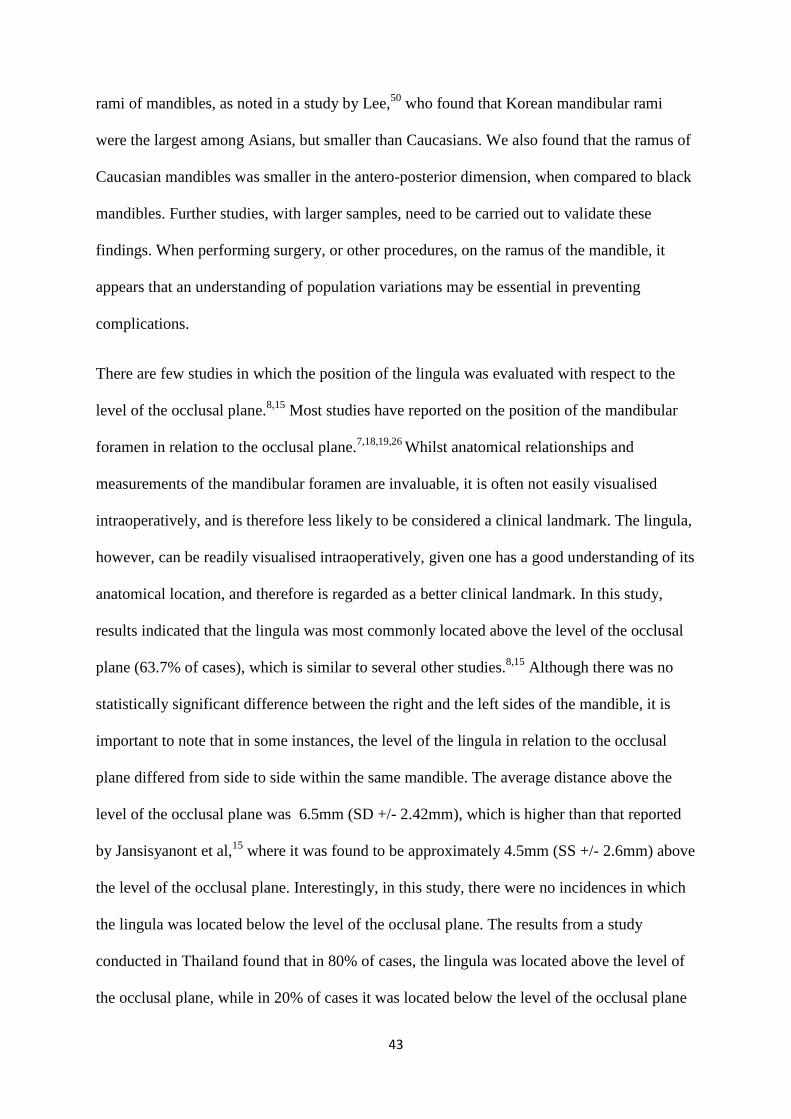
results indicated that the lingula was most commonly located above the level of the occlusal
plane (63.7% of cases), which is similar to several other studies.8,15
Although there was no
statistically significant difference between the right and the left sides of the mandible, it is
important to note that in some instances, the level of the lingula in relation to the occlusal
plane differed from side to side within the same mandible. The average distance above the
level of the occlusal plane was 6.5mm (SD +/- 2.42mm), which is higher than that reported
by Jansisyanont et al,15
where it was found to be approximately 4.5mm (SS +/- 2.6mm) above
the level of the occlusal plane. Interestingly, in this study, there were no incidences in which
the lingula was located below the level of the occlusal plane. The results from a study
conducted in Thailand found that in 80% of cases, the lingula was located above the level of
the occlusal plane, while in 20% of cases it was located below the level of the occlusal plane
44
and in no incidences was the lingula found to be at the level of the occlusal plane.15
From
these studies we were able to conclude that the level of the lingula in relation to the occlusal
plane varies among individuals and can also vary from side to side within the same
individual.
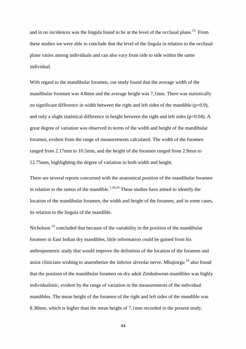
With regard to the mandibular foramen, our study found that the average width of the
mandibular foramen was 4.8mm and the average height was 7,1mm. There was statistically
no significant difference in width between the right and left sides of the mandible (p=0.9),
and only a slight statistical difference in height between the right and left sides (p=0.04). A
great degree of variation was observed in terms of the width and height of the mandibular
foramen, evident from the range of measurements calculated. The width of the foramen
ranged from 2.17mm to 10.5mm, and the height of the foramen ranged from 2.9mm to
12.75mm, highlighting the degree of variation in both width and height.
There are several reports concerned with the anatomical position of the mandibular foramen
in relation to the ramus of the mandible.7,18,19
These studies have aimed to identify the
location of the mandibular foramen, the width and height of the foramen, and in some cases,
its relation to the lingula of the mandible.
Nicholson 19
concluded that because of the variability in the position of the mandibular
foramen in East Indian dry mandibles, little information could be gained from his
anthropometric study that would improve the definition of the location of the foramen and
assist clinicians wishing to anaesthetize the inferior alveolar nerve. Mbajiorgu 18
also found
that the position of the mandibular foramen on dry adult Zimbabwean mandibles was highly
individualistic, evident by the range of variation in the measurements of the individual
mandibles. The mean height of the foramen of the right and left sides of the mandible was
8.38mm, which is higher than the mean height of 7.1mm recorded in the present study.
45
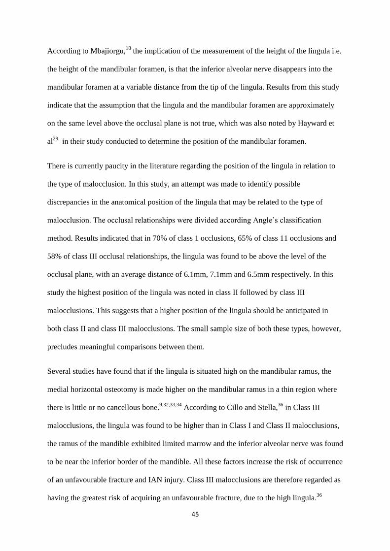
According to Mbajiorgu,18
the implication of the measurement of the height of the lingula i.e.
the height of the mandibular foramen, is that the inferior alveolar nerve disappears into the
mandibular foramen at a variable distance from the tip of the lingula. Results from this study
indicate that the assumption that the lingula and the mandibular foramen are approximately
on the same level above the occlusal plane is not true, which was also noted by Hayward et
al29
in their study conducted to determine the position of the mandibular foramen.
There is currently paucity in the literature regarding the position of the lingula in relation to
the type of malocclusion. In this study, an attempt was made to identify possible
discrepancies in the anatomical position of the lingula that may be related to the type of
malocclusion. The occlusal relationships were divided according Angle’s classification
method. Results indicated that in 70% of class 1 occlusions, 65% of class 11 occlusions and
58% of class III occlusal relationships, the lingula was found to be above the level of the
occlusal plane, with an average distance of 6.1mm, 7.1mm and 6.5mm respectively. In this
study the highest position of the lingula was noted in class II followed by class III
malocclusions. This suggests that a higher position of the lingula should be anticipated in
both class II and class III malocclusions. The small sample size of both these types, however,
precludes meaningful comparisons between them.
Several studies have found that if the lingula is situated high on the mandibular ramus, the
medial horizontal osteotomy is made higher on the mandibular ramus in a thin region where
there is little or no cancellous bone.9,32,33,34
According to Cillo and Stella,36
in Class III
malocclusions, the lingula was found to be higher than in Class I and Class II malocclusions,
the ramus of the mandible exhibited limited marrow and the inferior alveolar nerve was found
to be near the inferior border of the mandible. All these factors increase the risk of occurrence
of an unfavourable fracture and IAN injury. Class III malocclusions are therefore regarded as
having the greatest risk of acquiring an unfavourable fracture, due to the high lingula.36
46
However, according to a recent study in which 2005 sagittal split osteotomies were reviewed,
14 cases resulted in unfavourable fractures.51
The authors concluded that these unfavourable
fractures could not be correlated to a particular type of dental-facial deformity, or skeletal
class according to Angle’s classification.51
Further research is required to determine the
position of the lingula in various occlusal relationships and the possible effect it may have in
determining the location of the medial horizontal osteotomy in sagittal split ramus
osteotomies.
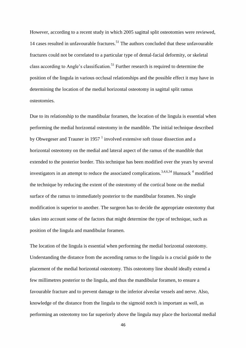
Due to its relationship to the mandibular foramen, the location of the lingula is essential when
performing the medial horizontal osteotomy in the mandible. The initial technique described
by Obwegeser and Trauner in 1957 1 involved extensive soft tissue dissection and a
horizontal osteotomy on the medial and lateral aspect of the ramus of the mandible that
extended to the posterior border. This technique has been modified over the years by several
investigators in an attempt to reduce the associated complications.3,4,6,34
Hunsuck 4 modified
the technique by reducing the extent of the osteotomy of the cortical bone on the medial
surface of the ramus to immediately posterior to the mandibular foramen. No single
modification is superior to another. The surgeon has to decide the appropriate osteotomy that
takes into account some of the factors that might determine the type of technique, such as
position of the lingula and mandibular foramen.
The location of the lingula is essential when performing the medial horizontal osteotomy.
Understanding the distance from the ascending ramus to the lingula is a crucial guide to the
placement of the medial horizontal osteotomy. This osteotomy line should ideally extend a
few millimetres posterior to the lingula, and thus the mandibular foramen, to ensure a
favourable fracture and to prevent damage to the inferior alveolar vessels and nerve. Also,
knowledge of the distance from the lingula to the sigmoid notch is important as well, as
performing an osteotomy too far superiorly above the lingula may place the horizontal medial
47
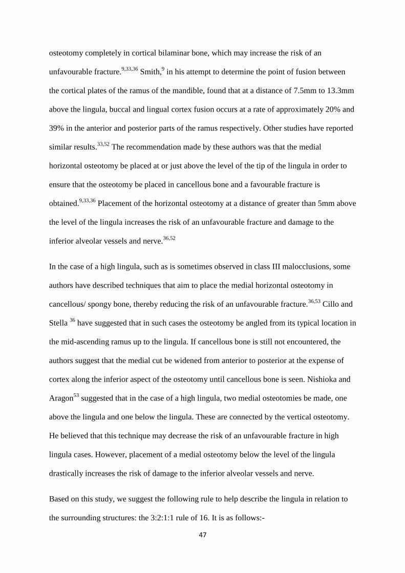
osteotomy completely in cortical bilaminar bone, which may increase the risk of an
unfavourable fracture.9,33,36
Smith,9 in his attempt to determine the point of fusion between
the cortical plates of the ramus of the mandible, found that at a distance of 7.5mm to 13.3mm
above the lingula, buccal and lingual cortex fusion occurs at a rate of approximately 20% and
39% in the anterior and posterior parts of the ramus respectively. Other studies have reported
similar results.33,52
The recommendation made by these authors was that the medial
horizontal osteotomy be placed at or just above the level of the tip of the lingula in order to
ensure that the osteotomy be placed in cancellous bone and a favourable fracture is
obtained.9,33,36
Placement of the horizontal osteotomy at a distance of greater than 5mm above
the level of the lingula increases the risk of an unfavourable fracture and damage to the
inferior alveolar vessels and nerve.36,52
In the case of a high lingula, such as is sometimes observed in class III malocclusions, some
authors have described techniques that aim to place the medial horizontal osteotomy in
cancellous/ spongy bone, thereby reducing the risk of an unfavourable fracture.36,53
Cillo and
Stella 36
have suggested that in such cases the osteotomy be angled from its typical location in
the mid-ascending ramus up to the lingula. If cancellous bone is still not encountered, the
authors suggest that the medial cut be widened from anterior to posterior at the expense of
cortex along the inferior aspect of the osteotomy until cancellous bone is seen. Nishioka and
Aragon53
suggested that in the case of a high lingula, two medial osteotomies be made, one
above the lingula and one below the lingula. These are connected by the vertical osteotomy.
He believed that this technique may decrease the risk of an unfavourable fracture in high
lingula cases. However, placement of a medial osteotomy below the level of the lingula
drastically increases the risk of damage to the inferior alveolar vessels and nerve.
Based on this study, we suggest the following rule to help describe the lingula in relation to
the surrounding structures: the 3:2:1:1 rule of 16. It is as follows:-
48
3 X 16 = the distance from the second molar tooth to the posterior border of the
mandible (48mm)
2 X 16 = the distance from the second molar to the lingula
1 X 16 = the distance from the sigmoid notch to the tip of the lingula
1 X 16 = the distance from the lingula to the posterior border of the mandible
Alternatively, we have also devised the 5:4:4 rule of 4. It is as follows:-
5 X 4mm = 20mm :the distance from the anterior border to the ramus of the mandible
4 X 4mm = 16mm : the distance from the sigmoid notch to the tip of the lingula
4 X 4mm = 16mm : the distance from the lingula to the posterior border
The above rules may serve as guidelines to help locate the lingula more proficiently
intraoperatively.
49
7.0 Conclusions
The shape and metric characteristics of the lingula in relation to surrounding structures in
South Africans vary from other populations. The average distance of the tip of the lingula
from the anterior border, posterior border and sigmoid notch the ramus of the mandible
was approximately 20.15mm, 16.77mm and 16.3mm respectively. This data may assist
surgeons to localize the lingula and avoid intraoperative complications when performing
a SSRO.
The truncated variant of the lingula was observed most commonly, followed by the
triangular, nodular and assimilated shapes. Males were more likely to exhibit a truncated
shaped lingula, whilst females were more likely to exhibit a triangular shape. Knowledge
of the various shapes of the lingula may assist the surgeon to readily identify the structure
intraoperatively, limiting both the amount of time and exposure required to identify the
structure. This may ultimately lead to fewer complications.
The average distance of the tip of the lingula from the mandibular second molar tooth was
found to be 33.3mm. The mandibular second molar tooth may be used as a clinical guide
for the location of the lingula when performing surgery on the medial aspect of the ramus
of the mandible.
50
In the majority of cases (63.7%), the lingula was found to be above the level of the
occlusal plane by an average distance of 6.5mm.
The width and height of the mandibular foramen is highly variable. Results from this
study indicate that the lingula is on average 7.1mm superior to the base of the mandibular
foramen. Thus, by performing the medial horizontal osteotomy cut at the level of the
lingula, or just superior to it, may lead to avoidance of the inferior alveolar neurovascular
bundle.
The lingula was found to be most superior, with respect to the level of the occlusal plane,
in Class II malocclusions (average distance of 7.1mm). This was found to be
contradictory to the assumption that class III malocclusions presented with a high lingula.
The small sample size may be the reason for the discrepancy in findings.
The dimensions of the ramus of Caucasian mandibles were smaller in terms of distance of
the lingula from the anterior and posterior borders of the ramus of the mandible.
However, Black mandibles exhibited, on average, a shorter distance from the deepest
point of the sigmoid notch to the tip of the lingula. This finding may have implications on
the position of the medial horizontal osteotomy cut. However, further investigation is
required to determine if fusion of the buccal and lingual cortices of the ramus of the
mandible occurs at a lower level in Black mandibles as compared to Caucasian
mandibles.

51
When applied to SSRO operations, the anatomic data provided by this study may assist
surgeons to locate and identify the lingula without difficulty, and avoid intraoperative
complications. The data presented has a direct relevance to clinical practice and teaching.
A thorough understanding of the anatomy of the lingula and its surrounding structures is
essential when performing a sagittal split ramus osteotomy.
52
8.0 References
1. Obwegeser H, Trauner R. Zur operationstechnik bei der progenic und anderen
unterkieferanomalien. Deutsch Z Mund Kiefereheilkd 1955;23: H1-H2,1955
2. Archer WH. Oral and Maxillofacial Surgery. Vol II. Philadelphia: WB Saunders,
1975:1448–1449.
3. Dal Pont G. Retromolar osteotomy for the correction of prognathism. J Oral Surg
1961;19: 42–7.
4. Hunsuck E. A modified intraoral sagittal splitting technic for correction of mandibular
prognathism. J Oral Surg 1968; 26:250–3.
5. Bell WH, Proffit WR, White RP. Surgical Correction of Dentofacial
Deformities. Philadelphia: Saunders, 1980: 855–901.
6. Epker BN. Modifications in the sagittal osteotomy of the mandible. J Oral Surg ,
1977; 5:157–159
7. Monnazzi M.S, Passeri L.A, Gabrielli M.F.R, Bollini P.D.A, de Cavalho W.R.S, da
Costa Machodo H. Anatomic study of the mandibular foramen , lingula and
antilingula in dry mandibles, and its statistical relationship between the true lingula
and the antilingula. Int. J Oral Maxillofac. Surg. 2012; 41: 74-78.
8. Kim H.J, Lee H.Y, Chung I.H, Cha I.H, Yi C.K. Mandibular anatomy related to
sagittal split ramus osteotomy in Koreans. Yonsei Medical Journal, 1997; 38 (1): 19-
25.
9. Smith BR, Rajchel JL, Waite DE, Read L. Mandibular ramus anatomy as it relates to
the medial osteotomy of the sagittal split ramus osteotomy. J Oral Maxillofac Surg,
1991; 49:112–116.
10. Dobson J. Anatomical Eponyms, 2nd edn. Edinburgh, London: E. & S.
Livingstone,1962:194
53
11. Turvey TA. Intraoperative complications of sagittal osteotomy of the mandibular
ramus: Incidence and management. J Oral Maxillofac Surg, 1985; 43:504–509.
12. Chrcanovic B.R, Freire-Maia B. Risk factors and prevention of bad splits during
sagittal split osteotomy. Oral Maxillofac Surg, 2012; 16:19-27
13. Kriwalsky M.S, Maurer P, Veras R.B, Eckert A.W, Schubert J. Risk factors for a bad
split during sagittal split osteotomy. Br. J. Oral and Maxillofac Surg, 2008; 46:177-
179
14. Behrman S.J. Complications of sagittal osteotomy of the mandibular ramus. J Oral
Surg, 1972; 30:554
15. Jansisyanont P, Apinhasmit W, Chompoopong S. Shape, height and location of the
lingula for sagittal ramus osteotomy in Thai. Clin Anat, 2009;22:787—93.
16. Murlimanju B.V, Prabhu L.V, Pai M.M, Paul M.T, Saralaya V.V, Kumar C.G.
Morphological study of lingula of the mandibles in South Indian population.
Morphologie, 2012; 96: 16-20.
17. Tuli A, Choudhry R, Choudhry S, Raheja S, Agarwal S. Variation in shape of the
lingula in the adult human mandible. J Anat, 2000;197:313—7.
18. Mbajiorgu E.F. A study of the mandibular foramen in adult black Zimbabwean
mandibles. Cent Afr J Med, 2000; 46(7): 184-90
19. Nicholson ML. A study of the position of the mandibular foramen in the adult human
mandible. Anatomical Record, 1985;212:110-112.
20. Dubrul EL. Sicher and Dubrul’s oral anatomy. 8th ed. New York: Ishiyaku Euro
America, 1988:32—35.
21. Hollinshead WH. Textbook of anatomy. Calcutta: Harper & Row; 1962: 855—856.
22. Berkovitz BKB, Holland GR, Moxham BJ. A colour atlas & textbook of oral
anatomy. London: Wolfe Medical Publications, 1978:15.

54
23. Sampson HW, Montgomery JL, Henryson GL. Atlas of the human skull. Texas:
Texas A & M University Press; 1991:130—131.
24. Williams PL, Bannister LH, Berry MM, Collins P, Dyson M, Dussek JE, et al. Gray’s
anatomy. 38th ed. Edinburgh: Churchill Livingstone, 1995:576.
25. Morgan DH, House LR, Hall WP, Vamvas SJ. Diseases of the temporomandibular
apparatus. 2nd ed. St Louis: C.V. Mosby; 1982:19.
26. Kositbowornchai S, Siritapetawee M, Damrongrungruang T, Khongkankang W, et.al.
Shape of the lingula and its localization by panoramic radiograph versus dry
mandibular measurement. Surg Radiol Anat, 2007; 29: 689-694
27. Berry A.C. Factors affecting the incidence of non-metrical skeletal variants. J Anat,
1975; 120: 519-535
28. Hossain S.M.A, Patwary S.I, Karim M. Variation in shape of the lingulae in the adult
human mandible of Bangladeshi skulls. Pak J Med Sci, 2001;17:233-6
29. Hayward J, Richardson, Malhotra S.K. The mandibular foramen: its anteroposterior
position. Oral Surg Oral Med Oral Pathol, 1977; 44: 837-43
30. Beukes J, Reyneke J.P, Becker P.J. Variations in the anatomical dimensions of the
mandibular ramus and the presence of third molars: its effect on the sagittal split
ramus osteotomy. Int J Oral Maxillofac Surg. In press.
31. Reitzik M, Griffiths R.R, Mirels H. Surgical anatomy of the ascending ramus of the
mandible. Br J Oral Surg, 1976; 14: 150-155
32. de Souza Fernandes A.C, Cardosa P.M, Fernandes I.S, de Moraes M. Anatomic study
for the horizontal cut of the sagittal split ramus osteotomy. J Oral Maxillofac Surg,
2013; 71: 1239-1244
33. Tom W.K, Martone C.H, Mintz S.M: A study of mandibular ramus anatomy and its
significance to sagittal split osteotomy. Int J Oral Maxillofac Surg, 1997; 26: 176-178
55
34. Wolford L.M, Benett M.A, Rafferty C.G. Modification of the mandibular ramus
sagittal split osteotomy. Oral Surg Oral Med Oral Pathol, 1987; 64:146-155
35. Daw J.L Jr, de la Paz M.G, Han H, et al. The mandibular foramen: An anatomical
study and its relevance to the sagittal split ramus osteotomy. J Craniofac Surg,1999;
10:475
36. Cillo J.E. Jr, Stella J.P. Selection of Sagittal Split Ramus Osteotomy Technique Based
on Skeletal Anatomy and Planned Distal Segment Movement: Current Therapy, 2005;
63:109-114
37. Phillips C, Essick G, Blakey 3rd G, Tucker M: Relationship between patients’
perceptions of postsurgical sequelae and altered sensations after bilateral sagittal split
osteotomy. J Oral Maxillofac Surg,2007; 65: 597-607
38. Teltzrow T, Kramer F.J, Schulze A, Baethge C, et.al. Perioperative complications
following sagittal split osteotomy of the mandible. J Craniofac Surg, 2005; 33:307-
313
39. Teerijoki-Oksa T, Ja¨a¨skela¨inen S.K, Forssell K, Forssell H, et.al. Risk factors of
nerve injury during mandibular sagittal split osteotomy. Int J Oral Maxillofac Surg,
2002; 31: 33–39.
40. Lanigan D.T, Hey J, West R.A. Hemorrhage following mandibular osteotomies: A
report of 21 cases. J Oral Maxillofac Surg, 1991; 49:713-724
41. Oh J.S, Kim S.G, Kim H.K, Moon S.Y. Massive haemorrhage following bilateral
sagittal split ramus osteotomy: A case report. J Oral Maxillofac Surg, 2005; 67: 895-
898
42. Yin N.T. Hemorrhage of the initial part of the internal maxillary artery treated by
multiple ligations: Report of four cases. J Oral Maxillofac Surg,1994; 52: 1066
56
43. Roberts D.H, Sowray J.A. Local anaesthesia in dentistry. Bristol. John Wright, 2nd
ed,
1979
44. Levy T.P. Assessment of the Gow-Gates mandibular block for third molar surgery. J
Am Dent Assoc, 1981;103:37-41
45. Wong M.K, Jacobson P.L. Reasons for local anaesthesia failures. J Am Dent Assoc,
1992;123:63-73
46. Pogrel M.A, Thamby S. Permanent nerve involvement resulting from inferior alveolar
nerve blocks. JADA, 2000;131:901-907
47. Hillerup S. Iatrogenic injury to the inferior alveolar nerve: etiology, signs and
symptoms, and observations on recovery. Int J Oral Maxillofac Surg, 2008; 37: 704–
709
48. Dayal R.M, Kegly A.D.T, Strkalj G, Bidmos M.A, Kuykendall K.L. The History and
Composition of the Raymond A. Dart Collection of Human Skeletons at the
University of the Witwatersrand, Johannesburg, South Africa. Am J Phys Anthropol
2009; 140:324-335
49. Kilarkaje N, Nayak SR, Narayan P. Morphology of lingula, coronoid process and the
mandibular notch in Indian dry mandibles. Folia Anat Belgrade 2004; 31:13-6
50. Lee D.S. A study on mandible of Korean. Medical Digest (Korean) 1961,3:971-1005
51. Falter B, Schepers S, Vrielinck L, Lambrichts I, et al. Occurrence of bad splits during
sagittal split osteotomy. Oral Surg Oral Med Oral Pathol Oral Radiol
Endod. 2010;110:430–435
52. Park K.R, Kim S.Y, Kim G.J, Park H.S, Young-Soo Jung Y.S. Anatomic study to
determine a safe surgical reference point for mandibular ramus osteotomy. J Cranio
Maxillofac Surg; 2013: 1-6

57
53. Nishioka G.J, Aragon S.B. Modified sagittal split technique for patients with a high
lingula. J Oral Maxillofac Surg, 1989; 47:426-427

58
Appendix 1: Data Collection Form
A. Number (refers to Specimen number): ____________________________-
Age: ____________________
Sex: ____________________
Race: ____________________
Right Left
Shape (of the lingula)
Width of the foramen
Height of the foramen
Distance of the lingula from
the mandibular notch
Distance of the lingula from
the anterior border of the
mandibular ramus
Distance of the lingula from
the posterior border of the
mandibular ramus
Position of the lingula in
relation to the occlusal plane:
-above/ below/ at level
Distance of the lingula from
the occlusal plane
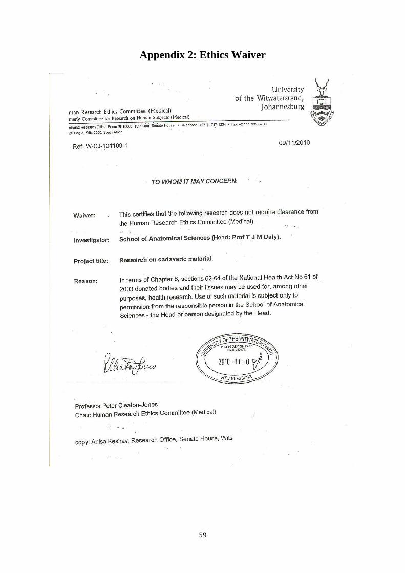
59
Appendix 2: Ethics Waiver