A microRNA‑17‑5p/homeobox B13 axis participates in the ...
11
MOLECULAR MEDICINE REPORTS 24: 731, 2021 Abstract. Vascular smooth muscle cells (VSMCs) serve a deci‑ sive role in intimal hyperplasia, a common pathophysiological process that leads to numerous vascular disorders. The present study aimed to investigate the unknown mechanisms under‑ lying VSMC phenotypic modulation and identified a novel microRNA (miRNA/miR)‑17‑5p/homeobox B13 (HOXB13) axis involved in the phenotypic switching, proliferation and migration of VSMCs. VSMCs were isolated from the thoracic aorta of Sprague‑Dawley rats, cell proliferation was determined by Cell Counting Kit‑8 (CCK‑8) assay, cell migration was examined by Transwell migration assay and gene expression was detected using reverse transcription‑quantitative PCR and western blot analyses. It was firstly found that incubation with platelet‑derived growth factor‑BB (PDGF‑BB) recombinant protein resulted in a significant increase in HOXB13 expression in VSMCs. Using multiple miRNA prediction tools, miR‑17‑5p was identified as a potential regulator for HOXB13, since it had a 7‑base perfect binding site and a 5‑base imperfect binding site with the 3'‑untranslated region of HOXB13 mRNA, and these sequences were highly conserved across species. The regulatory effect of miR‑17‑5p on HOXB13 was validated using luciferase reporter assays. The expression level of miR‑17‑5p was increased in VSMCs under PDGF‑BB stimulation, and was negatively correlated with HOXB13 mRNA and protein expression. Moreover, the miR‑17‑5p mimics significantly inhibited the proliferation and migration of VSMCs, while antagomiR‑17‑5p showed the opposite effects, which could be abolished by HOXB13 knockdown. The miR‑17‑5p/HOXB13 axis also regulated the expression levels of the markers of differentiated VSMCs ( α‑smooth muscle actin, transgelin and smoothelin), proliferating cell nuclear antigen and cell migration proteins, including MMP‑2 and ‑9. In conclusion, the present study demonstrated that miR‑17‑5p inhibited the phenotypic modulation VSMCs stimulated by PDGF‑BB by downregulating HOXB13, indicating that these factors may be potential therapeutic targets for intimal hyperplasia. Introduction Vascular smooth muscle cells (VSMCs) are the main cellular components of blood vessels and serve a key role in intimal hyperplasia, a pathophysiological process that causes numerous vascular disorders, such as atherosclerosis, hypertension and post‑angioplasty restenosis (1). VSMCs are not terminally differentiated and maintain considerable diversity and plas‑ ticity. For instance, these cells undergo phenotypic switching under the action of locally produced growth factors, such as platelet‑derived growth factor‑BB (PDGF‑BB), and migrate to the intima and proliferate to induce intimal disease (1). During the process of neointima formation, VSMCs are char‑ acterized by decreased expression levels of genes associated with cell differentiation and contraction, as well as increased proliferation, migration and synthesis of extracellular matrix components, which lead to the conversion to a synthetic pheno‑ type (2). Therefore, investigating the mechanisms underlying VSMC phenotypic modulation is essential for developing effective treatments in the management of vascular intimal hyperplasia. MicroRNAs (miRNAs/miRs) are a cluster of non‑coding RNAs containing 19‑25 nucleotides that can regulate >60% of human genes involved in almost all the cellular biological behaviors, including proliferation, migration and differentia‑ tion (3). miRNAs exert their biological functions mainly by binding to the 3'‑untranslated region (3'‑UTR) of target genes, leading to the degradation or/and preventing the translation of mRNA, thereby negatively regulating gene expression in a post‑transcriptional manner (4). Accumulating evidence has shown the significance of miRNAs in vascular diseases and their potentials as therapeutic targets (5). For example, some miRNAs, such as miR‑21, miR‑26a and miR‑29b, have been reported to participate in arterial intimal hyperplasia by regu‑ lating the phenotypic switching of VSMCs (6). A microRNA‑17‑5p/homeobox B13 axis participates in the phenotypic modulation of vascular smooth muscle cells TIANCHI YU 1* , TAO WANG 1* , SHIFANG KUANG 2 , GUOPING ZHAO 3 , KUN ZHOU 3 and HUI ZHANG 1 1 Department of Vascular Surgery; 2 Center of Endoscopy; 3 Department of General Surgery, Jiangning Hospital Affiliated to Nanjing Medical University, Nanjing, Jiangsu 211100, P.R. China Received April 13, 2021; Accepted July 26, 2021 DOI: 10.3892/mmr.2021.12370 Correspondence to: Dr Hui Zhang, Department of Vascular Surgery, Jiangning Hospital Affiliated to Nanjing Medical University, 169 Hushan Road, Nanjing, Jiangsu 211100, P.R. China E‑mail: [email protected] * Contributed equally Key words: vascular smooth muscle cells, microRNA‑17‑5p, homeobox B13, phenotypic switching, proliferation, migration
Transcript of A microRNA‑17‑5p/homeobox B13 axis participates in the ...

MOLECULAR MEDICINE REPORTS 24: 731, 2021
Abstract. Vascular smooth muscle cells (VSMCs) serve a deci‑sive role in intimal hyperplasia, a common pathophysiological process that leads to numerous vascular disorders. The present study aimed to investigate the unknown mechanisms under‑lying VSMC phenotypic modulation and identified a novel microRNA (miRNA/miR)‑17‑5p/homeobox B13 (HOXB13) axis involved in the phenotypic switching, proliferation and migration of VSMCs. VSMCs were isolated from the thoracic aorta of Sprague‑Dawley rats, cell proliferation was determined by Cell Counting Kit‑8 (CCK‑8) assay, cell migration was examined by Transwell migration assay and gene expression was detected using reverse transcription‑quantitative PCR and western blot analyses. It was firstly found that incubation with platelet‑derived growth factor‑BB (PDGF‑BB) recombinant protein resulted in a significant increase in HOXB13 expression in VSMCs. Using multiple miRNA prediction tools, miR‑17‑5p was identified as a potential regulator for HOXB13, since it had a 7‑base perfect binding site and a 5‑base imperfect binding site with the 3'‑untranslated region of HOXB13 mRNA, and these sequences were highly conserved across species. The regulatory effect of miR‑17‑5p on HOXB13 was validated using luciferase reporter assays. The expression level of miR‑17‑5p was increased in VSMCs under PDGF‑BB stimulation, and was negatively correlated with HOXB13 mRNA and protein expression. Moreover, the miR‑17‑5p mimics significantly inhibited the proliferation and migration of VSMCs, while antagomiR‑17‑5p showed the opposite effects, which could be abolished by HOXB13 knockdown. The miR‑17‑5p/HOXB13 axis also regulated the expression levels of the markers of
differentiated VSMCs (α‑smooth muscle actin, transgelin and smoothelin), proliferating cell nuclear antigen and cell migration proteins, including MMP‑2 and ‑9. In conclusion, the present study demonstrated that miR‑17‑5p inhibited the phenotypic modulation VSMCs stimulated by PDGF‑BB by downregulating HOXB13, indicating that these factors may be potential therapeutic targets for intimal hyperplasia.
Introduction
Vascular smooth muscle cells (VSMCs) are the main cellular components of blood vessels and serve a key role in intimal hyperplasia, a pathophysiological process that causes numerous vascular disorders, such as atherosclerosis, hypertension and post‑angioplasty restenosis (1). VSMCs are not terminally differentiated and maintain considerable diversity and plas‑ticity. For instance, these cells undergo phenotypic switching under the action of locally produced growth factors, such as platelet‑derived growth factor‑BB (PDGF‑BB), and migrate to the intima and proliferate to induce intimal disease (1). During the process of neointima formation, VSMCs are char‑acterized by decreased expression levels of genes associated with cell differentiation and contraction, as well as increased proliferation, migration and synthesis of extracellular matrix components, which lead to the conversion to a synthetic pheno‑type (2). Therefore, investigating the mechanisms underlying VSMC phenotypic modulation is essential for developing effective treatments in the management of vascular intimal hyperplasia.
MicroRNAs (miRNAs/miRs) are a cluster of non‑coding RNAs containing 19‑25 nucleotides that can regulate >60% of human genes involved in almost all the cellular biological behaviors, including proliferation, migration and differentia‑tion (3). miRNAs exert their biological functions mainly by binding to the 3'‑untranslated region (3'‑UTR) of target genes, leading to the degradation or/and preventing the translation of mRNA, thereby negatively regulating gene expression in a post‑transcriptional manner (4). Accumulating evidence has shown the significance of miRNAs in vascular diseases and their potentials as therapeutic targets (5). For example, some miRNAs, such as miR‑21, miR‑26a and miR‑29b, have been reported to participate in arterial intimal hyperplasia by regu‑lating the phenotypic switching of VSMCs (6).
A microRNA‑17‑5p/homeobox B13 axis participates in the phenotypic modulation of vascular smooth muscle cells
TIANCHI YU1*, TAO WANG1*, SHIFANG KUANG2, GUOPING ZHAO3, KUN ZHOU3 and HUI ZHANG1
1Department of Vascular Surgery; 2Center of Endoscopy; 3Department of General Surgery, Jiangning Hospital Affiliated to Nanjing Medical University, Nanjing, Jiangsu 211100, P.R. China
Received April 13, 2021; Accepted July 26, 2021
DOI: 10.3892/mmr.2021.12370
Correspondence to: Dr Hui Zhang, Department of Vascular Surgery, Jiangning Hospital Affiliated to Nanjing Medical University, 169 Hushan Road, Nanjing, Jiangsu 211100, P.R. ChinaE‑mail: [email protected]
*Contributed equally
Key words: vascular smooth muscle cells, microRNA‑17‑5p, homeobox B13, phenotypic switching, proliferation, migration
YU et al: miR‑17‑5p/HOXB13 IN VSMCs2
Homeobox B13 (HOXB13) belongs to the homeobox family and is a master regulator in cell identity, differentiation, proliferation and migration during embryonic development, tumorigenesis and inflammatory reactions (7,8). In particular, homeobox genes are involved in vasculogenesis and vascular remodeling, which are common events during neointimal lesion formation in atherosclerosis and post‑angioplasty restenosis (9). Moreover, HOXB13 is a well‑known regulator of the terminal differentiation of prostate epithelial cells and represents an important biomarker for prostate cancer (10). However, it remains unknown whether HOXB13 participates in regulating the phenotypic switching, proliferation and migration of VSMCs, and the upstream regulatory miRNAs in VSMCs are yet to be determined. Therefore, present study aimed to investigate the mechanisms underlying VSMC phenotypic modulation.
Materials and methods
Regents and antibodies (Abs). Recombinant PDGF‑BB protein was purchased from Sigma‑Aldrich (Merck KGaA). Abs against HOXB13 (cat. no. sc‑28333), proliferating cell nuclear antigen (PCNA; cat. no. sc‑56), α‑smooth muscle actin (α‑SMA; cat. no. sc‑53142), transgelin (cat. no. sc‑53932), smoothelin (cat. no. sc‑376902), MMP‑2 (cat. no. sc‑13594), MMP‑9 (cat. no. sc‑21733) and β‑actin (cat. no. sc‑47778) were purchased from Santa Cruz Biotechnology, Inc.
Isolation and culture of VSMCs. In total, six male specific pathogen‑free Sprague‑Dawley rats (age, 8 weeks; body weight, 250±20 g) were obtained from the Animal Laboratory of Nanjing Jiangning Hospital (Nanjing, China). Rats were housed at 23±2˚C under a controlled humidity (45‑65%) with free access to food and water under a 12‑h light/dark cycle. Rats were intraperitoneally injected with 3% sodium pentobarbital (100 mg/kg). Immediately after the loss of consciousness, rats underwent thoracotomy and the thoracic aorta was quickly dissected and separated. At this stage, the rats were dead. After being rinsed with PBS, a section of the artery was soaked in sterile Hanks balanced salt solution. The outer membrane was gently peeled, and the artery was cut into small pieces of 0.5‑1 mm3 and placed in a 25‑ml culture flask with a distance of ~0.5 cm. The flask was placed in a 37˚C incubator with a humidified atmosphere of 5% CO2, and 3 h later DMEM (Gibco; Thermo Fisher Scientific, Inc.) supplemented with 10% FBS (Thermo Fisher Scientific, Inc.), 100 U/ml penicillin and 100 µg/ml streptomycin was added. The media were refreshed every 3‑5 days. When cells grew to ~70% conflu‑ence, they were sub‑cultured with trypsinization. Moreover, 5‑6 generations of cells were used for the experiments.
Cell proliferation assay. VSMCs were seeded at a density of 3x10³ cells/well in 96‑well plates and incubated overnight with DMEM supplemented with 10% FBS. The medium was removed and the plates were washed with PBS. The same volume of serum‑free medium was added and cells were cultured for 12 h. After being washed with PBS, cells were incubated at 37˚C for 24 h with recombinant PDGF‑BB protein at 1, 2, 4, 8, 16 or 32 or 15 ng/ml PDGF‑BB protein at 37˚C for 6, 12, 18, 24, 30 or 36 h.
A Cell Counting Kit‑8 (CCK‑8) assay (Dojindo Molecular Technologies, Inc.) was used to determine cell proliferation according to the manufacturer's instructions. The culture medium was replaced with 100 µl fresh serum‑free medium containing 10 µl CCK‑8 solution. The cells were further incu‑bated for 2 h at 37˚C, and the optical density (OD) at 450 nm was measured. The cell proliferation rate (%) was calculated using the following formula: (Experimental OD‑control OD)/control OD x100%.
Migration assay. Transwell chambers (8‑µm pore size) were purchased from BD Bioscience. Cells (2x10⁴) suspended in 200 µl serum‑free medium were seeded on the polycarbonate membrane in the upper Transwell chamber, which was incu‑bated for 60 min at 37˚C with 5% CO2. The lower chamber was then filled with 750 µl medium with 5% FBS. After being incubated for 12, 24 or 36 h at 37˚C in a humidified atmosphere of 5% CO2, the Transwell chamber was washed with PBS and cells on the top surface of the polycarbonate membrane were removed. At room temperature, cells that migrated to the bottom surface of the insert were fixed with 100% methanol for 20 min and stained with 0.25% crystal violet solution for 10 min. Using light microscopy, the cells were counted from digital images, with 10 visual fields captured randomly at a magnification of x200.
Prediction of potential miRNAs. Multiple miRNA prediction tools [miRWalk (version 2.0; http://mirwalk.umm.uni‑heidel‑berg.de/), TargetScan (version 7.1; targetscan.org/), miRanda (version 1.9; omictools.com/miranda‑tool), miRTarBase (version 2.0; mirtarbase.mbc.nctu.edu.tw/) and mirdb (mirdb.org/)] were used to predict the potential upstream miRNAs that can regulate HOXB13 gene by targeting its 3'‑UTR.
Reverse transcription‑quantitative PCR (RT‑qPCR). Total RNA was extracted from cell lysates using TRIzol® reagent (cat. no. 15596026; Thermo Fisher Scientific, Inc.) according to the manufacturer's instructions. RNA quantity and quality were measured using a NanoDrop ND‑1000 system. For amplifying miR‑17‑5p, RNA was reversely transcribed into cDNA using a TaqMan® MicroRNA Reverse Transcription kit (cat no. 4366597; Thermo Fisher Scientific, Inc.) according to the manufacturer's protocol, and a stem‑loop RT primer (5'‑GTC GTA TCC AGT GCA GGT ATT CGC ACT GGA TAC GAC CTA CC‑3'), a sense primer (5'‑TGC AAA GTG CTT ACA GTG CAG ‑3') and an antisense primer (5'‑GTG CAG GGT CCG AGG TAT TC‑3') were used. U6 RNA expression served as a positive control, using a pair of primers: Sense, 5'‑CTC GCT TCG GCA GCA CA‑3' and antisense, 5'‑AAC GCT TCA CGA ATT TGC GT‑3'. For amplifying HOXB13 mRNA, RNA was reversely transcribed into cDNA using TaqMan™ Reverse Transcription Reagents (cat. no. N8080234; Thermo Fisher Scientific, Inc.) according to the manufacturer's protocol, and a pair of primers (sense, 5'‑CAG ATG TGT TGC CAG GGA GA‑3' and antisense, 5'‑AGG CGT CAG GAG GGT GCT ‑3') were used. The expression level of GAPDH served as an internal control, using a pair of primers: Sense, 5'‑GAA ATC CCA TCA CCA TCT TCC AGG ‑3' and antisense: 5'‑GAG CCC CAG CCT TCT CCA TG‑3'. Following RT, qPCR was performed using an Applied Biosystems 7500 Real‑Time PCR system and SYBR
MOLECULAR MEDICINE REPORTS 24: 731, 2021 3
Green (cat. no. S7563; Thermo Fisher Scientific, Inc.) according to the manufacturer's instruction. The thermocycling condi‑tions were as follows: 10 min initial denaturation at 94˚C, 15 sec denaturation at 94˚C and 30 sec of annealing at 55˚C (40 cycles) and final extension for 1 min at 72˚C. Experiments were performed in triplicate, and the relative expression levels were calculated using the 2‑ΔΔCq method (11).
Transfect ion of oligonucleot ides. The miR‑17‑5p mimics (5'‑CAA AGU GCU UAC AGU GCA GGU AG‑3'), antagomiR‑17‑5p (5'‑CUA CCU GCA CUG UAA GCA CUU UG‑3'), mimics negative control (5'‑UUC UCC GAA CGU GUC ACG UTT ‑3'), antagomiR negative control (5'‑UCA CAA CCU CCU AGA AAG AGU AGA ‑3'), small interfering RNA (siRNA) targeting HOXB13 (sense, 5'‑GCG GCC GCA AGA AAC GCA UTT ‑3' and antisense, 5'‑AUG CGU UUU GUG CGG CCG CTT ‑3') and siRNA negative control (sense, 5'‑UUC UCC GAA CGU GUC ACG U‑3' and antisense, 5'‑ACG UGA CAC GUU CGG AGA A‑3') oligonucleotides were purchased from Shanghai GenePharma Co., Ltd. VSMCs (1x10⁵/well) were seeded in 6‑well plates and cultured with DMEM supplemented with 10% FBS. When cells were grown to 60‑70% confluence, they were incubated with oligonucleotides at 100 nM using Lipofectamine® RNAiMAX transfection reagent (Thermo Fisher Scientific, Inc.) in serum‑free media at 37˚C for 48 h, and were then immediately subjected to subsequent assays.
Luciferase reporter assay. The 3'‑UTR fragment of HOXB13 mRNA containing miR‑17‑5p‑targeting sequence was cloned into a pMIR‑REPORT luciferase reporter vector (Ambion; Thermo Fisher Scientific, Inc.) to create wild‑type (WT) and mutated type (MT) pMIR‑luc‑HOXB13‑3'‑UTR vectors. VSMCs (1x10⁴/well) were cultured in 24‑well plates. When at 60‑70% confluence, cells were co‑transfected with the vectors (100 nM), together with 50 nM miR‑175p mimics, 50 nM antagomiR‑17‑5p or 50 nM negative control oligo‑nucleotides using Lipofectamine® 3000 (Thermo Fisher Scientific, Inc.) at 37˚C. A luciferase reporter vector without the miR‑17‑5p targeting sequence was transfected in parallel. After transfection for 48 h, luciferase activities were measured using the Dual‑luciferase Reporter Assay system (Promega Corporation) and normalized to Renilla according to the manufacturer's instructions. The relative luciferase activity in cells was expressed as a percentage of the luciferase activity over that of cells transfected with the vector without miR‑17‑5p targeting sequence.
Western blot analysis. Cells were homogenized in protein lysate buffer (50 mM Tris, pH 7.4, 100 mM EDTA, 0.25 M sucrose, 1% SDS, 1% NP40, 1 mg/ml leupeptin, 1 mg/ml pepstatin A and 100 mM phenyl methyl sulfonyl fluoride), and debris was removed via centrifugation at 10,000 x g for 10 min at 4˚C. Protein concentrations were determined using the Bio‑Rad protein assay (Bio‑Rad Laboratories, Inc.). Protein (30 µg/lane) were resolved on 10% SDS‑polyacrylamide gels and then electrophoretically transferred to PVDF membranes, which were blocked in TBS‑Tween 20 [137 mM NaCl, 20 mM Tris HCl (pH 7.6) and 0.1% (v/v) Tween 20] containing 5% (w/v) non‑fat dry milk at 37˚C for 2 h. The membranes were incubated at 4˚C overnight with primary Abs (Anti‑HOXB13,
1:500; anti‑PCNA, 1:400; anti‑α‑SMA, 1:1,000; anti‑trans‑gelin, 1:1,000; anti‑smoothelin, 1:1,000; anti‑MMP‑2, 1:800; anti‑MMP‑9, 1:1,000; anti‑β‑actin, 1:1,000), followed by an incubation with alkaline phosphatase‑conjugated secondary Abs (1:2,000) for 2 h at room temperature in the dark. Membranes were then developed with 5‑bromo‑4‑chloro‑3‑indolyl phos‑phate/nitro blue tetrazolium (Tiangen Biotech Co., Ltd.). The density of each band was measured using the ChemiDoc XRS system (Version 3.0; Bio‑Rad Laboratories, Inc.) and the semi‑quantitative measurement of band density was repeated thrice.
Statistical analysis. GraphPad Prism 8.02 (GraphPad Software, Inc.) was used for statistical analyses. Data are presented as the mean ± SD. Comparisons were conducted using one‑way ANOVA followed by a Tukey's post hoc test. The correla‑tion between HOXB13 and miR‑17‑5p expression levels was assessed using a Pearson's test. P<0.05 was considered to indicate a statistically significant difference. The experiments were repeated at least three times.
Results
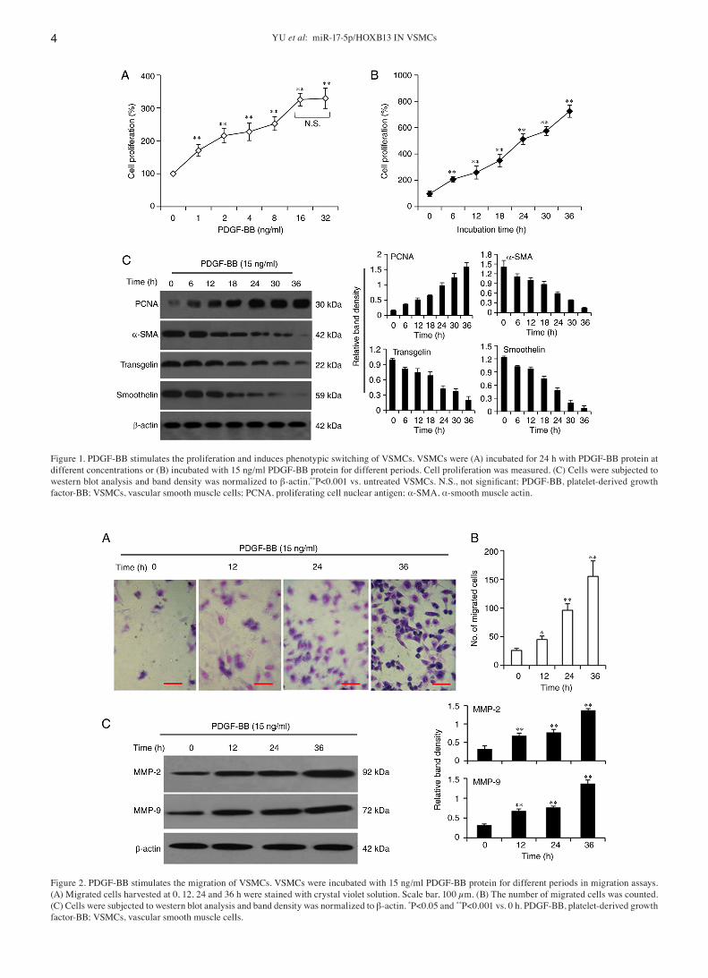
PDGF‑BB stimulates the proliferation and induces pheno‑typic switching of VSMCs. The stimulation of VSMCs with PDGF‑BB protein is widely used as an in vitro functional model for studying vascular intimal hyperplasia (12). VSMCs were first incubated for 24 h with recombinant PDGF‑BB protein at 1, 2, 4, 8, 16 or 32 ng/ml, which were selected based on previous studies (12‑15). The CCK‑8 assay results demon‑strated that incubation with the PDGF‑BB protein stimulated the proliferation of VSMCs in a concentration‑dependent manner (Fig. 1A). Since PDGF‑BB exerted its maximal effect at a concentration of 15 ng/ml, this was used for the subse‑quent experiments. PDGF‑BB stimulation also promoted the proliferation of VSMCs in a time‑dependent manner when VSMCs were incubated with 15 ng/ml PDGF‑BB protein for 6, 12, 18, 24, 30 or 36 h (Fig. 1B). The western blot analysis results identified the reduced expression levels of VSMC differentiation‑related markers, including α‑SMA (16), transgelin (also known as smooth muscle 22α) (17) and smoothelin (18), and increased expression levels of the prolif‑eration marker PCNA, in VSMCs after PDGF‑BB stimulation in a time‑dependent manner (Fig. 1C). These results indicated that PDGF‑BB can induce the phenotypic switch and stimulate the proliferation of VSMCs.
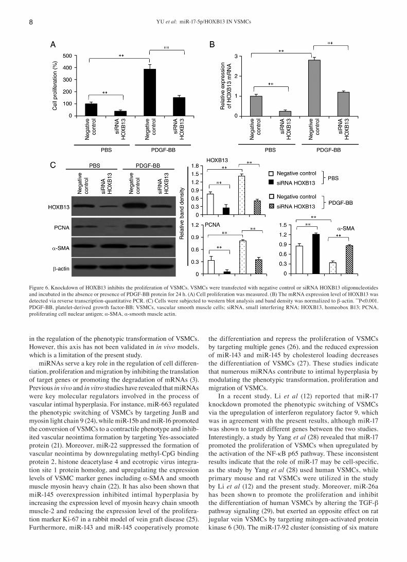
PDGF‑BB promotes the migration of VSMCs. Transwell migration assays revealed that the ability of VSMCs to migrate was significantly enhanced by PDGF‑BB protein in a time‑dependent manner (Fig. 2A and B). Western blot analysis also confirmed that PDGF‑BB stimulation resulted in an upregulation of MMP‑2 and MMP‑9 expression in a time‑dependent manner (Fig. 2C), which are two important mediators of VSMC migration (19).
PDGF‑BB stimulation leads to the upregulation of HOXB13 and the downregulation of miR‑17‑5. It was identified that incubation with 15 ng/ml PDGF‑BB protein increased the mRNA and protein expression levels of HOXB13 in VSMCs
YU et al: miR‑17‑5p/HOXB13 IN VSMCs4
Figure 1. PDGF‑BB stimulates the proliferation and induces phenotypic switching of VSMCs. VSMCs were (A) incubated for 24 h with PDGF‑BB protein at different concentrations or (B) incubated with 15 ng/ml PDGF‑BB protein for different periods. Cell proliferation was measured. (C) Cells were subjected to western blot analysis and band density was normalized to β‑actin.**P<0.001 vs. untreated VSMCs. N.S., not significant; PDGF‑BB, platelet‑derived growth factor‑BB; VSMCs, vascular smooth muscle cells; PCNA, proliferating cell nuclear antigen; α‑SMA, α‑smooth muscle actin.
Figure 2. PDGF‑BB stimulates the migration of VSMCs. VSMCs were incubated with 15 ng/ml PDGF‑BB protein for different periods in migration assays. (A) Migrated cells harvested at 0, 12, 24 and 36 h were stained with crystal violet solution. Scale bar, 100 µm. (B) The number of migrated cells was counted. (C) Cells were subjected to western blot analysis and band density was normalized to β‑actin. *P<0.05 and **P<0.001 vs. 0 h. PDGF‑BB, platelet‑derived growth factor‑BB; VSMCs, vascular smooth muscle cells.

MOLECULAR MEDICINE REPORTS 24: 731, 2021 5
in a time‑dependent manner, as determined via RT‑qPCR and western blot analyses, respectively (Fig. 3A and B). Using multiple miRNA prediction tools, the potential upstream miRNAs that target HOXB13 were examined. miR‑17‑5p was found to have a 7‑base perfect binding site and a 5‑base imper‑fect binding site with the 3'‑UTR of HOXB13 mRNA, and the binding sites were highly conserved across species (Fig. 3C), indicating that miR‑17‑5p may be a potential miRNA to regulate HOXB13. Moreover, a literature search supported the involvement of miR‑17‑5p in the phenotypic modulation of VSMCs (12). It was demonstrated that the expression levels of miR‑17‑5p were significantly decreased in VSMCs upon PDGF‑BB stimulation in a time‑dependent manner (Fig. 3D).
Furthermore, HOXB13 mRNA (Fig. 3E) and protein (Fig. 3F) expression levels were very strongly negatively correlated with those of miR‑17‑5p in VSMCs given PDGF‑BB stimulation for different durations, as determined using a Pearson's test.
miR‑17‑5p negatively regulates the expression level of HOXB13 in VSMCs. Based on the identified binding sites, the WT and MT pMIR‑REPORT luciferase report vectors were constructed (Fig. 4A). VSMCs were co‑transfected with the WT vector and miR‑17‑5p mimics oligonucleotides, and the luciferase assays revealed that miR‑17‑5p inhibited lucif‑erase activities in a concentration‑dependent manner (Fig. 4B). In addition, the miR‑17‑5p mimics significantly decreased,
Figure 3. PDGF‑BB stimulation increases HOXB13 expression and reduces miR‑17‑5p expression in VSMCs. Cells were incubated with 15 ng/ml PDGF‑BB protein for different periods and harvested. (A) RT‑qPCR was used to detect the mRNA expression level of HOXB13. (B) Western blotting was used to detect the protein expression level of HOXB13 and band density was normalized to β‑actin. (C) The binding sites of miR‑17‑5p and the 3'‑untranslated region of HOXB13 are highly conserved across species. (D) RT‑qPCR was used to detect the expression level of miR‑17‑5p. The correlation of the (E) mRNA and (F) protein expression levels of HOXB13 with the expression of miR‑17‑5p was analyzed using a Pearson's test. The correlation coefficient is denoted as ‘r’. *P<0.05 and **P<0.001 vs. 0 h. PDGF‑BB, platelet‑derived growth factor‑BB; VSMCs, vascular smooth muscle cells; miR, microRNA; HOXB13, homeobox B13; RT‑qPCR, reverse transcription‑quantitative PCR.

YU et al: miR‑17‑5p/HOXB13 IN VSMCs6
while antagomiR‑17‑5p significantly increased, the luciferase activity in cells co‑transfected with the WT vector but not the MT vector (Fig. 4C). Moreover, the miR‑17‑5p mimics significantly reduced, while the antagomiR‑17‑5p significantly increased, the mRNA (Fig. 4D) and protein (Fig. 4E) expres‑sion levels of HOXB13. These results indicated that miR‑17‑5p served a negative regulatory role on HOXB13 expression by binding to its 3'‑UTR in VSMCs.
miR‑17‑5p inhibits the proliferation and phenotypic switching of VSMCs. Transfection of mimic negative control had no effect on, while miR‑17‑5p transfection significantly increased, the expression level of miR‑17‑5p in VSMCs (Fig. 5A). Transfection of antagomiR negative control had no effect on, while antagomiR‑17‑5p transfection significantly decreased, the expres‑sion level of miR‑17‑5p in VSMCs (Fig. 5B). Because neither mimic nor antagomiR negative controls affected miR‑17‑5p expression level, mimic negative control was used as a negative control in the subsequent experiments. Transfection of miR‑17‑5p mimics significantly inhibited the proliferation of VSMCs under PDGF‑BB stimulation, while the antagomiR‑17‑5p significantly promoted the proliferation of VSMCs without PDGF‑BB stimu‑lation (Fig. 5C). Furthermore, miR‑17‑5p mimics transfection downregulated the expression levels of HOXB13 and PCNA, and increased the expression levels of α‑SMA, transgelin and smoothelin in VSMCs under PDGF‑BB stimulation. However,
antagomiR‑17‑5p transfection showed the opposite effects in VSMCs without PDGF‑BB stimulation (Fig. 5D).
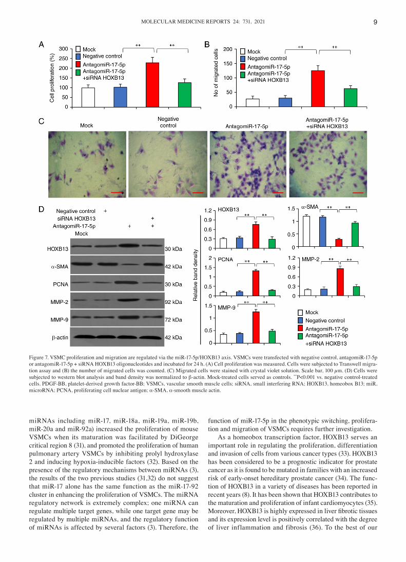
Knockdown of HOXB13 using siRNA inhibits the prolif‑eration and phenotype switching of VSMCs. Knockdown of HOXB13 using siRNA significantly inhibited the proliferation of VSMCs in the presence or absence of PDGF‑BB stimula‑tion, compared with the negative controls (Fig. 6A). siRNA HOXB13 also significantly decreased the mRNA (Fig. 6B) and protein (Fig. 6C) expression levels of HOXB13 in VSMCs in the presence or absence of PDGF‑BB stimulation. In addi‑tion, transfection of siRNA HOXB13 significantly decreased the expression level of PCNA and increased that of α‑SMA, compared with the negative control (Fig. 6C).
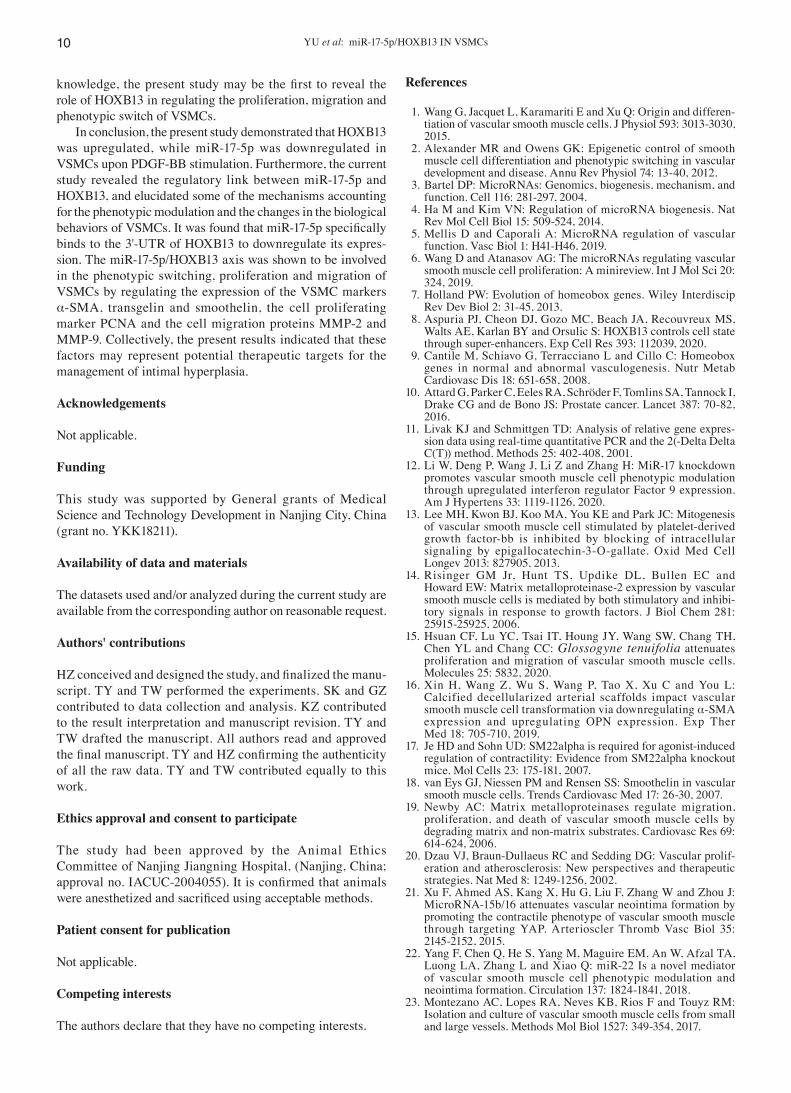
VSMC proliferation, migration and phenotype switching are regulated via the miR‑17‑5p/HOXB13 axis. Compared with the negative control, the antagomiR‑17‑5p significantly promoted the proliferation of VSMCs, while knockdown of HOXB13 using siRNA abolished this effect (Fig. 7A). The antagomiR‑17‑5p also increased the migratory capability of VSMCs but knockdown of HOXB13 abrogated this effect (Fig. 7B and C). Moreover, the antagomiR‑17‑5p significantly increased the expression levels of HOXB13, PCNA, MMP‑2 and MMP‑9, and downregulated α‑SMA expression, while HOXB13 knockdown could reverse these effects (Fig. 7D).
Figure 4. miR‑17‑5p negatively regulates the expression of HOXB13 by specifically binding to its 3'‑UTR in VSMCs. (A) The WT and MT pMIR‑REPORT luciferase report vectors were constructed. (B) VSMCs were either mock‑treated or co‑transfected with the WT luciferase vector and 10 pmol negative control or miR‑17‑5p mimics oligonucleotides at different concentrations. (C) VSMCs were co‑transfected with WT or MT luciferase reporter vectors, and negative control, miR‑17‑5p mimics or antagomiR‑17‑5p oligonucleotides. Luciferase activities are shown in (B) and (C) VSMCs were transfected with negative control, miR‑17‑5p mimics or antagomiR‑17‑5p oligonucleotides, and then subjected to reverse transcription‑quantitative PCR and western blot analyses for detecting the (D) mRNA and (E) protein expression levels of HOXB13, respectively. Band density was normalized to β‑actin in (E) **P<0.001 vs. negative control‑transfected cells. WT, wild‑type; MT, mutated type; PDGF‑BB, platelet‑derived growth factor‑BB; VSMCs, vascular smooth muscle cells; miR, microRNA; HOXB13, homeobox B13; UTR, untranslated region.

MOLECULAR MEDICINE REPORTS 24: 731, 2021 7
Discussion
Previous studies have reported that the transformation of VSMCs from a highly differentiated phenotype to a poorly differentiated phenotype leads to egregious cell proliferation, migration and excessive synthesis of extracellular matrix, resulting in a series of pathological changes, such as vascular intimal hyperplasia or even luminal stenosis (1,2,12,20‑22).
Unfortunately, there are a lack of effective treatment strate‑gies for this disorder. Therefore, it is crucial to elucidate the molecular mechanisms underlying the phenotypic switch, proliferation and migration of VSMCs. A culture of primary VSMCs under PDGF‑BB stimulation is the most commonly used in vitro model to study this specific pathophysiological response (23). By adopting this model, the current study identified a novel miR‑17‑5p/HOXB13 axis that participates
Figure 5. miR‑17‑5p regulates the proliferation and phenotypic switching of VSMCs. (A) VSMCs were mock‑treated, or transfected with mimics negative control or miR‑17‑5p mimics oligonucleotides, and the expression level of miR‑17‑5p was detected via RT‑qPCR. (B) VSMCs were mock‑treated, or transfected with antagomiR negative control or antagomiR‑17‑5p oligonucleotides, and the expression level of miR‑17‑5p was detected RT‑qPCR. VSMCs transfected with negative control, miR‑17‑5p mimics or antagomiR‑17‑5p oligonucleotides were incubated in the absence or presence of PDGF‑BB protein for 24 h. (C) Cell proliferation was measured. (D) Cells were subjected to western blot analysis and band density was normalized to β‑actin. *P<0.05 and **P<0.001. N.S., not significant; PDGF‑BB, platelet‑derived growth factor‑BB; VSMCs, vascular smooth muscle cells; miR, microRNA; RT‑qPCR, reverse transcription‑quantita‑tive PCR; PCNA, proliferating cell nuclear antigen; α‑SMA, α‑smooth muscle actin.
YU et al: miR‑17‑5p/HOXB13 IN VSMCs8
in the regulation of the phenotypic transformation of VSMCs. However, this axis has not been validated in in vivo models, which is a limitation of the present study.
miRNAs serve a key role in the regulation of cell differen‑tiation, proliferation and migration by inhibiting the translation of target genes or promoting the degradation of mRNAs (3). Previous in vivo and in vitro studies have revealed that miRNAs were key molecular regulators involved in the process of vascular intimal hyperplasia. For instance, miR‑663 regulated the phenotypic switching of VSMCs by targeting JunB and myosin light chain 9 (24), while miR‑15b and miR‑16 promoted the conversion of VSMCs to a contractile phenotype and inhib‑ited vascular neointima formation by targeting Yes‑associated protein (21). Moreover, miR‑22 suppressed the formation of vascular neointima by downregulating methyl‑CpG binding protein 2, histone deacetylase 4 and ecotropic virus integra‑tion site 1 protein homolog, and upregulating the expression levels of VSMC marker genes including α‑SMA and smooth muscle myosin heavy chain (22). It has also been shown that miR‑145 overexpression inhibited intimal hyperplasia by increasing the expression level of myosin heavy chain smooth muscle‑2 and reducing the expression level of the prolifera‑tion marker Ki‑67 in a rabbit model of vein graft disease (25). Furthermore, miR‑143 and miR‑145 cooperatively promote
the differentiation and repress the proliferation of VSMCs by targeting multiple genes (26), and the reduced expression of miR‑143 and miR‑145 by cholesterol loading decreases the differentiation of VSMCs (27). These studies indicate that numerous miRNAs contribute to intimal hyperplasia by modulating the phenotypic transformation, proliferation and migration of VSMCs.
In a recent study, Li et al (12) reported that miR‑17 knockdown promoted the phenotypic switching of VSMCs via the upregulation of interferon regulatory factor 9, which was in agreement with the present results, although miR‑17 was shown to target different genes between the two studies. Interestingly, a study by Yang et al (28) revealed that miR‑17 promoted the proliferation of VSMCs when upregulated by the activation of the NF‑κB p65 pathway. These inconsistent results indicate that the role of miR‑17 may be cell‑specific, as the study by Yang et al (28) used human VSMCs, while primary mouse and rat VSMCs were utilized in the study by Li et al (12) and the present study. Moreover, miR‑26a has been shown to promote the proliferation and inhibit the differentiation of human VSMCs by altering the TGF‑β pathway signaling (29), but exerted an opposite effect on rat jugular vein VSMCs by targeting mitogen‑activated protein kinase 6 (30). The miR‑17‑92 cluster (consisting of six mature
Figure 6. Knockdown of HOXB13 inhibits the proliferation of VSMCs. VSMCs were transfected with negative control or siRNA HOXB13 oligonucleotides and incubated in the absence or presence of PDGF‑BB protein for 24 h. (A) Cell proliferation was measured. (B) The mRNA expression level of HOXB13 was detected via reverse transcription‑quantitative PCR. (C) Cells were subjected to western blot analysis and band density was normalized to β‑actin. **P<0.001. PDGF‑BB, platelet‑derived growth factor‑BB; VSMCs, vascular smooth muscle cells; siRNA, small interfering RNA; HOXB13, homeobox B13; PCNA, proliferating cell nuclear antigen; α‑SMA, α‑smooth muscle actin.
MOLECULAR MEDICINE REPORTS 24: 731, 2021 9
miRNAs including miR‑17, miR‑18a, miR‑19a, miR‑19b, miR‑20a and miR‑92a) increased the proliferation of mouse VSMCs when its maturation was facilitated by DiGeorge critical region 8 (31), and promoted the proliferation of human pulmonary artery VSMCs by inhibiting prolyl hydroxylase 2 and inducing hypoxia‑inducible factors (32). Based on the presence of the regulatory mechanisms between miRNAs (3), the results of the two previous studies (31,32) do not suggest that miR‑17 alone has the same function as the miR‑17‑92 cluster in enhancing the proliferation of VSMCs. The miRNA regulatory network is extremely complex; one miRNA can regulate multiple target genes, while one target gene may be regulated by multiple miRNAs, and the regulatory function of miRNAs is affected by several factors (3). Therefore, the
function of miR‑17‑5p in the phenotypic switching, prolifera‑tion and migration of VSMCs requires further investigation.
As a homeobox transcription factor, HOXB13 serves an important role in regulating the proliferation, differentiation and invasion of cells from various cancer types (33). HOXB13 has been considered to be a prognostic indicator for prostate cancer as it is found to be mutated in families with an increased risk of early‑onset hereditary prostate cancer (34). The func‑tion of HOXB13 in a variety of diseases has been reported in recent years (8). It has been shown that HOXB13 contributes to the maturation and proliferation of infant cardiomyocytes (35). Moreover, HOXB13 is highly expressed in liver fibrotic tissues and its expression level is positively correlated with the degree of liver inflammation and fibrosis (36). To the best of our
Figure 7. VSMC proliferation and migration are regulated via the miR‑17‑5p/HOXB13 axis. VSMCs were transfected with negative control, antagomiR‑17‑5p or antagomiR‑17‑5p + siRNA HOXB13 oligonucleotides and incubated for 24 h. (A) Cell proliferation was measured. Cells were subjected to Transwell migra‑tion assay and (B) the number of migrated cells was counted. (C) Migrated cells were stained with crystal violet solution. Scale bar, 100 µm. (D) Cells were subjected to western blot analysis and band density was normalized to β‑actin. Mock‑treated cells served as controls. **P<0.001 vs. negative control‑treated cells. PDGF‑BB, platelet‑derived growth factor‑BB; VSMCs, vascular smooth muscle cells; siRNA, small interfering RNA; HOXB13, homeobox B13; miR, microRNA; PCNA, proliferating cell nuclear antigen; α‑SMA, α‑smooth muscle actin.
YU et al: miR‑17‑5p/HOXB13 IN VSMCs10
knowledge, the present study may be the first to reveal the role of HOXB13 in regulating the proliferation, migration and phenotypic switch of VSMCs.
In conclusion, the present study demonstrated that HOXB13 was upregulated, while miR‑17‑5p was downregulated in VSMCs upon PDGF‑BB stimulation. Furthermore, the current study revealed the regulatory link between miR‑17‑5p and HOXB13, and elucidated some of the mechanisms accounting for the phenotypic modulation and the changes in the biological behaviors of VSMCs. It was found that miR‑17‑5p specifically binds to the 3'‑UTR of HOXB13 to downregulate its expres‑sion. The miR‑17‑5p/HOXB13 axis was shown to be involved in the phenotypic switching, proliferation and migration of VSMCs by regulating the expression of the VSMC markers α‑SMA, transgelin and smoothelin, the cell proliferating marker PCNA and the cell migration proteins MMP‑2 and MMP‑9. Collectively, the present results indicated that these factors may represent potential therapeutic targets for the management of intimal hyperplasia.
Acknowledgements
Not applicable.
Funding
This study was supported by General grants of Medical Science and Technology Development in Nanjing City, China (grant no. YKK18211).
Availability of data and materials
The datasets used and/or analyzed during the current study are available from the corresponding author on reasonable request.
Authors' contributions
HZ conceived and designed the study, and finalized the manu‑script. TY and TW performed the experiments. SK and GZ contributed to data collection and analysis. KZ contributed to the result interpretation and manuscript revision. TY and TW drafted the manuscript. All authors read and approved the final manuscript. TY and HZ confirming the authenticity of all the raw data. TY and TW contributed equally to this work.
Ethics approval and consent to participate
The study had been approved by the Animal Ethics Committee of Nanjing Jiangning Hospital, (Nanjing, China; approval no. IACUC‑2004055). It is confirmed that animals were anesthetized and sacrificed using acceptable methods.
Patient consent for publication
Not applicable.
Competing interests
The authors declare that they have no competing interests.
References
1. Wang G, Jacquet L, Karamariti E and Xu Q: Origin and differen‑tiation of vascular smooth muscle cells. J Physiol 593: 3013‑3030, 2015.
2. Alexander MR and Owens GK: Epigenetic control of smooth muscle cell differentiation and phenotypic switching in vascular development and disease. Annu Rev Physiol 74: 13‑40, 2012.
3. Bartel DP: MicroRNAs: Genomics, biogenesis, mechanism, and function. Cell 116: 281‑297, 2004.
4. Ha M and Kim VN: Regulation of microRNA biogenesis. Nat Rev Mol Cell Biol 15: 509‑524, 2014.
5. Mellis D and Caporali A: MicroRNA regulation of vascular function. Vasc Biol 1: H41‑H46, 2019.
6. Wang D and Atanasov AG: The microRNAs regulating vascular smooth muscle cell proliferation: A minireview. Int J Mol Sci 20: 324, 2019.
7. Holland PW: Evolution of homeobox genes. Wiley Interdiscip Rev Dev Biol 2: 31‑45, 2013.
8. Aspuria PJ, Cheon DJ, Gozo MC, Beach JA, Recouvreux MS, Walts AE, Karlan BY and Orsulic S: HOXB13 controls cell state through super‑enhancers. Exp Cell Res 393: 112039, 2020.
9. Cantile M, Schiavo G, Terracciano L and Cillo C: Homeobox genes in normal and abnormal vasculogenesis. Nutr Metab Cardiovasc Dis 18: 651‑658, 2008.
10. Attard G, Parker C, Eeles RA, Schröder F, Tomlins SA, Tannock I, Drake CG and de Bono JS: Prostate cancer. Lancet 387: 70‑82, 2016.
11. Livak KJ and Schmittgen TD: Analysis of relative gene expres‑sion data using real‑time quantitative PCR and the 2(‑Delta Delta C(T)) method. Methods 25: 402‑408, 2001.
12. Li W, Deng P, Wang J, Li Z and Zhang H: MiR‑17 knockdown promotes vascular smooth muscle cell phenotypic modulation through upregulated interferon regulator Factor 9 expression. Am J Hypertens 33: 1119‑1126, 2020.
13. Lee MH, Kwon BJ, Koo MA, You KE and Park JC: Mitogenesis of vascular smooth muscle cell stimulated by platelet‑derived growth factor‑bb is inhibited by blocking of intracellular signaling by epigallocatechin‑3‑O‑gallate. Oxid Med Cell Longev 2013: 827905, 2013.
14. Risinger GM Jr, Hunt TS, Updike DL, Bullen EC and Howard EW: Matrix metalloproteinase‑2 expression by vascular smooth muscle cells is mediated by both stimulatory and inhibi‑tory signals in response to growth factors. J Biol Chem 281: 25915‑25925, 2006.
15. Hsuan CF, Lu YC, Tsai IT, Houng JY, Wang SW, Chang TH, Chen YL and Chang CC: Glossogyne tenuifolia attenuates proliferation and migration of vascular smooth muscle cells. Molecules 25: 5832, 2020.
16. Xin H, Wang Z, Wu S, Wang P, Tao X, Xu C and You L: Calcified decellularized arterial scaffolds impact vascular smooth muscle cell transformation via downregulating α‑SMA expression and upregulating OPN expression. Exp Ther Med 18: 705‑710, 2019.
17. Je HD and Sohn UD: SM22alpha is required for agonist‑induced regulation of contractility: Evidence from SM22alpha knockout mice. Mol Cells 23: 175‑181, 2007.
18. van Eys GJ, Niessen PM and Rensen SS: Smoothelin in vascular smooth muscle cells. Trends Cardiovasc Med 17: 26‑30, 2007.
19. Newby AC: Matrix metalloproteinases regulate migration, proliferation, and death of vascular smooth muscle cells by degrading matrix and non‑matrix substrates. Cardiovasc Res 69: 614‑624, 2006.
20. Dzau VJ, Braun‑Dullaeus RC and Sedding DG: Vascular prolif‑eration and atherosclerosis: New perspectives and therapeutic strategies. Nat Med 8: 1249‑1256, 2002.
21. Xu F, Ahmed AS, Kang X, Hu G, Liu F, Zhang W and Zhou J: MicroRNA‑15b/16 attenuates vascular neointima formation by promoting the contractile phenotype of vascular smooth muscle through targeting YAP. Arterioscler Thromb Vasc Biol 35: 2145‑2152, 2015.
22. Yang F, Chen Q, He S, Yang M, Maguire EM, An W, Afzal TA, Luong LA, Zhang L and Xiao Q: miR‑22 Is a novel mediator of vascular smooth muscle cell phenotypic modulation and neointima formation. Circulation 137: 1824‑1841, 2018.
23. Montezano AC, Lopes RA, Neves KB, Rios F and Touyz RM: Isolation and culture of vascular smooth muscle cells from small and large vessels. Methods Mol Biol 1527: 349‑354, 2017.
MOLECULAR MEDICINE REPORTS 24: 731, 2021 11
24. Li P, Zhu N, Yi B, Wang N, Chen M, You X, Zhao X, Solomides CC, Qin Y and Sun J: MicroRNA‑663 regulates human vascular smooth muscle cell phenotypic switch and vascular neointimal formation. Circ Res 113: 1117‑1127, 2013.
25. Ohnaka M, Marui A, Yamahara K, Minakata K, Yamazaki K, Kumagai M, Masumoto H, Tanaka S, Ikeda T and Sakata R: Effect of microRNA‑145 to prevent vein graft disease in rabbits by regulation of smooth muscle cell phenotype. J Thorac Cardiovasc Surg 148: 676‑682.e2, 2014.
26. Cordes KR, Sheehy NT, White MP, Berry EC, Morton SU, Muth AN, Lee TH, Miano JM, Ivey KN and Srivastava D: miR‑145 and miR‑143 regulate smooth muscle cell fate and plas‑ticity. Nature 460: 705‑710, 2009.
27. Vengrenyuk Y, Nishi H, Long X, Ouimet M, Savji N, Martinez FO, Cassella CP, Moore KJ, Ramsey SA, Miano JM and Fisher EA: Cholesterol loading reprograms the microRNA‑143/145‑myo‑cardin axis to convert aortic smooth muscle cells to a dysfunctional macrophage‑like phenotype. Arterioscler Thromb Vasc Biol 35: 535‑546, 2015.
28. Yang D, Sun C, Zhang J, Lin S, Zhao L, Wang L, Lin R, Lv J and Xin S: Proliferation of vascular smooth muscle cells under inflammation is regulated by NF‑κB p65/microRNA‑17/RB pathway activation. Int J Mol Med 41: 43‑50, 2018.
29. Leeper NJ, Raiesdana A, Kojima Y, Chun HJ, Azuma J, Maegdefessel L, Kundu RK, Quertermous T, Tsao PS and Spin JM: MicroRNA‑26a is a novel regulator of vascular smooth muscle cell function. J Cell Physiol 226: 1035‑1043, 2011.
30. Tan J, Yang L, Liu C and Yan Z: MicroRNA‑26a targets MAPK6 to inhibit smooth muscle cell proliferation and vein graft neointimal hyperplasia. Sci Rep 7: 46602, 2017.
31. Chen Z, Wu J, Yang C, Fan P, Balazs L, Jiao Y, Lu M, Gu W, Li C, Pfeffer LM, et al: DiGeorge syndrome critical region 8 (DGCR8) protein‑mediated microRNA biogenesis is essential for vascular smooth muscle cell development in mice. J Biol Chem 287: 19018‑19028, 2012.
32. Chen T, Zhou Q, Tang H, Bozkanat M, Yuan JX, Raj JU and Zhou G: miR‑17/20 Controls prolyl hydroxylase 2 (PHD2)/Hypoxia‑Inducible Factor 1 (HIF1) to regulate pulmonary artery smooth muscle cell proliferation. J Am Heart Assoc 5: e004510, 2016.
33. Wang X, Sun Y, Xu T, Qian K, Huang B, Zhang K, Song Z, Qian T, Shi J and Li L: HOXB13 promotes proliferation, migration, and invasion of glioblastoma through transcriptional upregulation of lncRNA HOXC‑AS3. J Cell Biochem 120: 15527‑15537, 2019.
34. Ewing CM, Ray AM, Lange EM, Zuhlke KA, Robbins CM, Tembe WD, Wiley KE, Isaacs SD, Johng D, Wang Y, et al: Germline mutations in HOXB13 and prostate‑cancer risk. N Engl J Med 366: 141‑149, 2012.
35. Nguyen NUN, Canseco DC, Xiao F, Nakada Y, Li S, Lam NT, Muralidhar SA, Savla JJ, Hill JA, Le V, et al: A calcineurin‑Hoxb13 axis regulates growth mode of mamma‑lian cardiomyocytes. Nature 582: 271‑276, 2020.
36. Zuo L, Tan T, Wei C, Wang H, Tan L, Hao Y, Qian J, Chen Y and Wu C: HOXB13 expression is correlated with hepatic inflamma‑tory activity of patients with hepatic fibrosis. J Mol Histol 51: 183‑189, 2020.
This work is licensed under a Creative Commons Attribution-NonCommercial-NoDerivatives 4.0 International (CC BY-NC-ND 4.0) License.