A microchip for quantitative analysis of CNS axon growth under localized biomolecular treatments
9
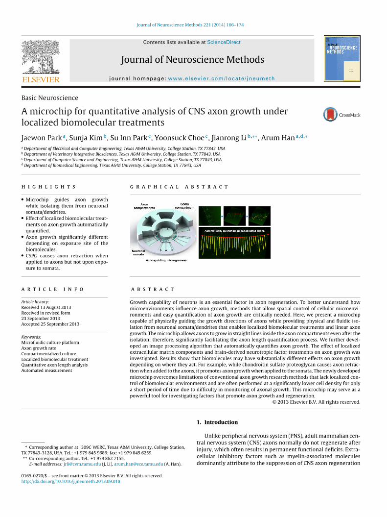
Journal of Neuroscience Methods 221 (2014) 166–174 Contents lists available at ScienceDirect Journal of Neuroscience Methods jou rn al h om epa ge : www.elsevier.com/locate/jneumeth Basic Neuroscience A microchip for quantitative analysis of CNS axon growth under localized biomolecular treatments Jaewon Park a , Sunja Kim b , Su Inn Park c , Yoonsuck Choe c , Jianrong Li b,∗∗ , Arum Han a,d,∗ a Department of Electrical and Computer Engineering, Texas A&M University, College Station, TX 77843, USA b Department of Veterinary Integrative Biosciences, Texas A&M University, College Station, TX 77843, USA c Department of Computer Science and Engineering, Texas A&M University, College Station, TX 77843, USA d Department of Biomedical Engineering, Texas A&M University, College Station, TX 77843, USA h i g h l i g h t s • Microchip guides axon growth while isolating them from neuronal somata/dendrites. • Effect of localized biomolecular treat- ments on axon growth automatically quantified. • Axon growth significantly different depending on exposure site of the biomolecules. • CSPG causes axon retraction when applied to axons but not upon expo- sure to somata. g r a p h i c a l a b s t r a c t a r t i c l e i n f o Article history: Received 13 August 2013 Received in revised form 23 September 2013 Accepted 25 September 2013 Keywords: Microfluidic culture platform Axon growth rate Compartmentalized culture Localized biomolecular treatment Quantitative axon length analysis Automated measurement a b s t r a c t Growth capability of neurons is an essential factor in axon regeneration. To better understand how microenvironments influence axon growth, methods that allow spatial control of cellular microenvi- ronments and easy quantification of axon growth are critically needed. Here, we present a microchip capable of physically guiding the growth directions of axons while providing physical and fluidic iso- lation from neuronal somata/dendrites that enables localized biomolecular treatments and linear axon growth. The microchip allows axons to grow in straight lines inside the axon compartments even after the isolation; therefore, significantly facilitating the axon length quantification process. We further devel- oped an image processing algorithm that automatically quantifies axon growth. The effect of localized extracellular matrix components and brain-derived neurotropic factor treatments on axon growth was investigated. Results show that biomolecules may have substantially different effects on axon growth depending on where they act. For example, while chondroitin sulfate proteoglycan causes axon retrac- tion when added to the axons, it promotes axon growth when applied to the somata. The newly developed microchip overcomes limitations of conventional axon growth research methods that lack localized con- trol of biomolecular environments and are often performed at a significantly lower cell density for only a short period of time due to difficulty in monitoring of axonal growth. This microchip may serve as a powerful tool for investigating factors that promote axon growth and regeneration. © 2013 Elsevier B.V. All rights reserved. ∗ Corresponding author at: 309C WERC, Texas A&M University, College Station, TX 77843-3128, USA. Tel.: +1 979 845 9686; fax: +1 979 845 6259. ∗∗ Co-corresponding author. Tel.: +1 979 862 7155. E-mail addresses: [email protected] (J. Li), [email protected] (A. Han). 1. Introduction Unlike peripheral nervous system (PNS), adult mammalian cen- tral nervous system (CNS) axons normally do not regenerate after injury, which often results in permanent functional deficits. Extra- cellular inhibitory factors such as myelin-associated molecules dominantly attribute to the suppression of CNS axon regeneration 0165-0270/$ – see front matter © 2013 Elsevier B.V. All rights reserved. http://dx.doi.org/10.1016/j.jneumeth.2013.09.018
Transcript of A microchip for quantitative analysis of CNS axon growth under localized biomolecular treatments
B
Al
Ja
b
c
d
h
•
•
•
•
a
ARR2A
KMACLQA
T
0h
Journal of Neuroscience Methods 221 (2014) 166– 174
Contents lists available at ScienceDirect
Journal of Neuroscience Methods
jou rn al h om epa ge : www.elsev ier .com/ locate / jneumeth
asic Neuroscience
microchip for quantitative analysis of CNS axon growth underocalized biomolecular treatments
aewon Parka, Sunja Kimb, Su Inn Parkc, Yoonsuck Choec, Jianrong Lib,∗∗, Arum Hana,d,∗
Department of Electrical and Computer Engineering, Texas A&M University, College Station, TX 77843, USADepartment of Veterinary Integrative Biosciences, Texas A&M University, College Station, TX 77843, USADepartment of Computer Science and Engineering, Texas A&M University, College Station, TX 77843, USADepartment of Biomedical Engineering, Texas A&M University, College Station, TX 77843, USA
i g h l i g h t s
Microchip guides axon growthwhile isolating them from neuronalsomata/dendrites.Effect of localized biomolecular treat-ments on axon growth automaticallyquantified.Axon growth significantly differentdepending on exposure site of thebiomolecules.CSPG causes axon retraction whenapplied to axons but not upon expo-sure to somata.
g r a p h i c a l a b s t r a c t
r t i c l e i n f o
rticle history:eceived 13 August 2013eceived in revised form3 September 2013ccepted 25 September 2013
eywords:icrofluidic culture platform
xon growth rateompartmentalized cultureocalized biomolecular treatment
a b s t r a c t
Growth capability of neurons is an essential factor in axon regeneration. To better understand howmicroenvironments influence axon growth, methods that allow spatial control of cellular microenvi-ronments and easy quantification of axon growth are critically needed. Here, we present a microchipcapable of physically guiding the growth directions of axons while providing physical and fluidic iso-lation from neuronal somata/dendrites that enables localized biomolecular treatments and linear axongrowth. The microchip allows axons to grow in straight lines inside the axon compartments even after theisolation; therefore, significantly facilitating the axon length quantification process. We further devel-oped an image processing algorithm that automatically quantifies axon growth. The effect of localizedextracellular matrix components and brain-derived neurotropic factor treatments on axon growth wasinvestigated. Results show that biomolecules may have substantially different effects on axon growth
uantitative axon length analysisutomated measurement
depending on where they act. For example, while chondroitin sulfate proteoglycan causes axon retrac-tion when added to the axons, it promotes axon growth when applied to the somata. The newly developedmicrochip overcomes limitations of conventional axon growth research methods that lack localized con-trol of biomolecular environments and are often performed at a significantly lower cell density for onlya short period of time due to difficulty in monitoring of axonal growth. This microchip may serve as a
ating
powerful tool for investig∗ Corresponding author at: 309C WERC, Texas A&M University, College Station,X 77843-3128, USA. Tel.: +1 979 845 9686; fax: +1 979 845 6259.∗∗ Co-corresponding author. Tel.: +1 979 862 7155.
E-mail addresses: [email protected] (J. Li), [email protected] (A. Han).
165-0270/$ – see front matter © 2013 Elsevier B.V. All rights reserved.ttp://dx.doi.org/10.1016/j.jneumeth.2013.09.018
factors that promote axon growth and regeneration.© 2013 Elsevier B.V. All rights reserved.
1. Introduction
Unlike peripheral nervous system (PNS), adult mammalian cen-
tral nervous system (CNS) axons normally do not regenerate afterinjury, which often results in permanent functional deficits. Extra-cellular inhibitory factors such as myelin-associated moleculesdominantly attribute to the suppression of CNS axon regeneration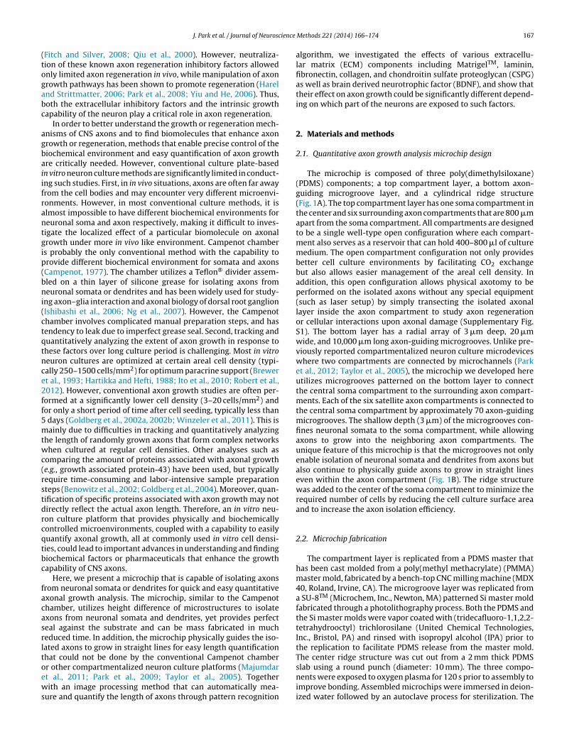
ience
(togabc
agbaiifrantgip(bni(ctqtnce2ff5mtwc(rstdrcqtbc
facasrltoews
J. Park et al. / Journal of Neurosc
Fitch and Silver, 2008; Qiu et al., 2000). However, neutraliza-ion of these known axon regeneration inhibitory factors allowednly limited axon regeneration in vivo, while manipulation of axonrowth pathways has been shown to promote regeneration (Harelnd Strittmatter, 2006; Park et al., 2008; Yiu and He, 2006). Thus,oth the extracellular inhibitory factors and the intrinsic growthapability of the neuron play a critical role in axon regeneration.
In order to better understand the growth or regeneration mech-nisms of CNS axons and to find biomolecules that enhance axonrowth or regeneration, methods that enable precise control of theiochemical environment and easy quantification of axon growthre critically needed. However, conventional culture plate-basedn vitro neuron culture methods are significantly limited in conduct-ng such studies. First, in in vivo situations, axons are often far awayrom the cell bodies and may encounter very different microenvi-onments. However, in most conventional culture methods, it islmost impossible to have different biochemical environments foreuronal soma and axon respectively, making it difficult to inves-igate the localized effect of a particular biomolecule on axonalrowth under more in vivo like environment. Campenot chambers probably the only conventional method with the capability torovide different biochemical environment for somata and axonsCampenot, 1977). The chamber utilizes a Teflon® divider assem-led on a thin layer of silicone grease for isolating axons fromeuronal somata or dendrites and has been widely used for study-
ng axon–glia interaction and axonal biology of dorsal root ganglionIshibashi et al., 2006; Ng et al., 2007). However, the Campenothamber involves complicated manual preparation steps, and hasendency to leak due to imperfect grease seal. Second, tracking anduantitatively analyzing the extent of axon growth in response tohese factors over long culture period is challenging. Most in vitroeuron cultures are optimized at certain areal cell density (typi-ally 250–1500 cells/mm2) for optimum paracrine support (Brewert al., 1993; Hartikka and Hefti, 1988; Ito et al., 2010; Robert et al.,012). However, conventional axon growth studies are often per-ormed at a significantly lower cell density (3–20 cells/mm2) andor only a short period of time after cell seeding, typically less than
days (Goldberg et al., 2002a, 2002b; Winzeler et al., 2011). This isainly due to difficulties in tracking and quantitatively analyzing
he length of randomly grown axons that form complex networkshen cultured at regular cell densities. Other analyses such as
omparing the amount of proteins associated with axonal growthe.g., growth associated protein-43) have been used, but typicallyequire time-consuming and labor-intensive sample preparationteps (Benowitz et al., 2002; Goldberg et al., 2004). Moreover, quan-ification of specific proteins associated with axon growth may notirectly reflect the actual axon length. Therefore, an in vitro neu-on culture platform that provides physically and biochemicallyontrolled microenvironments, coupled with a capability to easilyuantify axonal growth, all at commonly used in vitro cell densi-ies, could lead to important advances in understanding and findingiochemical factors or pharmaceuticals that enhance the growthapability of CNS axons.
Here, we present a microchip that is capable of isolating axonsrom neuronal somata or dendrites for quick and easy quantitativexonal growth analysis. The microchip, similar to the Campenothamber, utilizes height difference of microstructures to isolatexons from neuronal somata and dendrites, yet provides perfecteal against the substrate and can be mass fabricated in mucheduced time. In addition, the microchip physically guides the iso-ated axons to grow in straight lines for easy length quantificationhat could not be done by the conventional Campenot chamber
r other compartmentalized neuron culture platforms (Majumdart al., 2011; Park et al., 2009; Taylor et al., 2005). Togetherith an image processing method that can automatically mea-ure and quantify the length of axons through pattern recognition
Methods 221 (2014) 166– 174 167
algorithm, we investigated the effects of various extracellu-lar matrix (ECM) components including MatrigelTM, laminin,fibronectin, collagen, and chondroitin sulfate proteoglycan (CSPG)as well as brain derived neurotrophic factor (BDNF), and show thattheir effect on axon growth could be significantly different depend-ing on which part of the neurons are exposed to such factors.
2. Materials and methods
2.1. Quantitative axon growth analysis microchip design
The microchip is composed of three poly(dimethylsiloxane)(PDMS) components; a top compartment layer, a bottom axon-guiding microgroove layer, and a cylindrical ridge structure(Fig. 1A). The top compartment layer has one soma compartment inthe center and six surrounding axon compartments that are 800 �mapart from the soma compartment. All compartments are designedto be a single well-type open configuration where each compart-ment also serves as a reservoir that can hold 400–800 �l of culturemedium. The open compartment configuration not only providesbetter cell culture environments by facilitating CO2 exchangebut also allows easier management of the areal cell density. Inaddition, this open configuration allows physical axotomy to beperformed on the isolated axons without any special equipment(such as laser setup) by simply transecting the isolated axonallayer inside the axon compartment to study axon regenerationor cellular interactions upon axonal damage (Supplementary Fig.S1). The bottom layer has a radial array of 3 �m deep, 20 �mwide, and 10,000 �m long axon-guiding microgrooves. Unlike pre-viously reported compartmentalized neuron culture microdeviceswhere two compartments are connected by microchannels (Parket al., 2012; Taylor et al., 2005), the microchip we developed hereutilizes microgrooves patterned on the bottom layer to connectthe central soma compartment to the surrounding axon compart-ments. Each of the six satellite axon compartments is connected tothe central soma compartment by approximately 70 axon-guidingmicrogrooves. The shallow depth (3 �m) of the microgrooves con-fines neuronal somata to the soma compartment, while allowingaxons to grow into the neighboring axon compartments. Theunique feature of this microchip is that the microgrooves not onlyenable isolation of neuronal somata and dendrites from axons butalso continue to physically guide axons to grow in straight lineseven within the axon compartment (Fig. 1B). The ridge structurewas added to the center of the soma compartment to minimize therequired number of cells by reducing the cell culture surface areaand to increase the axon isolation efficiency.
2.2. Microchip fabrication
The compartment layer is replicated from a PDMS master thathas been cast molded from a poly(methyl methacrylate) (PMMA)master mold, fabricated by a bench-top CNC milling machine (MDX40, Roland, Irvine, CA). The microgroove layer was replicated froma SU-8TM (Microchem, Inc., Newton, MA) patterned Si master moldfabricated through a photolithography process. Both the PDMS andthe Si master molds were vapor coated with (tridecafluoro-1,1,2,2-tetrahydrooctyl) trichlorosilane (United Chemical Technologies,Inc., Bristol, PA) and rinsed with isopropyl alcohol (IPA) prior tothe replication to facilitate PDMS release from the master mold.The center ridge structure was cut out from a 2 mm thick PDMS
slab using a round punch (diameter: 10 mm). The three compo-nents were exposed to oxygen plasma for 120 s prior to assembly toimprove bonding. Assembled microchips were immersed in deion-ized water followed by an autoclave process for sterilization. The
168 J. Park et al. / Journal of Neuroscience Methods 221 (2014) 166– 174
F e devi om su
ot
2
opwBlr(tmBagctssgpS
2
(octitomicwwDD
2
at
ig. 1. (A) Schematic illustrations of the top and bottom PDMS layers composing thsolation and guidance via the array of shallow microgrooves patterned on the bott
verall microchip fabrication process is illustrated in Supplemen-ary Fig. S2.
.3. Tissue dissociation and cell preparation
Primary CNS neurons were prepared from forebrains of embry-nic day 16 Sprague-Dawley rats as described in the previousublication (Koito and Li, 2009). Briefly, forebrains free of meningesere dissected in ice-cold dissection buffer (Ca2+/Mg2+-free Hank’salanced Salt Solution containing 10 mM HEPES), dissociated with-cysteine activated papain (10 units/ml) for 5 min at 37 ◦C, andesuspended in dissection medium containing trypsin inhibitor10 mg/ml) for 2–3 min. Following two more washes with therypsin inhibitor solution, the tissue was resuspended in a plating
edium (NBB27 + glutamate: neurobasal medium containing 2%27, 1 mM Glutamine, 25 �M glutamic acid, 100 units/ml penicillin,nd 100 �g/ml streptomycin) and triturated with a fire-polishedlass Pasteur pipette. The cells were then passed through a 70 �mell sieves and live cells were counted using a hemocytometer andrypan blue exclusion assay. The viability of isolated cells was con-tantly greater than 90–95%. Ca2+/Mg2+-free Hank’s balanced saltolution, neurobasal medium, B27, penicillin, streptomycin andoat serum were from Invitrogen (Carlsbad, CA). Poly-d-lysine,apain, trypsin inhibitor, glutamine, and glutamic acid were fromigma–Aldrich (St. Louis, MO).
.4. Microchip cell culture
Dissected primary neurons diluted in 200 �l of plating mediumNBB27 with glutamate) were loaded into the soma compartmentf microchip at an areal density of 500–2000 cells/mm2. 150 �l ofulture medium was added to each of the six axon compartmentso prevent the substrate from drying. The microchip was then leftnside a 37 ◦C humidified 5% CO2 incubator for 30 min to allow cellso settle down and attach to the bottom, followed by adding 600 �lf culture medium to the soma compartment and 200 �l of cultureedium to each of the axon compartment. Cells were then cultured
nside a 37 ◦C humidified 5% CO2 incubator without culture mediumhange for four days. At DIV 4, the culture medium was replacedith NBB27 without glutamate. Culture medium was exchangedith NBB27 without glutamate containing either ECM or BDNF atIV 7 for localized treatment. The growth of axons was analyzed atIV 11 without any additional culture medium exchange.
.5. Localized biomolecular treatment
Fluidic isolation between the neuronal soma/dendrites and thexons was achieved by generating fluidic level difference betweenhe soma and the axon compartments (Park et al., 2012; Taylor
eloped quantitative axon growth analysis microchip. (B) Illustration showing axonbstrate.
et al., 2005). During the localized soma treatment, small but sus-tained medium flow from the axon compartment (higher fluidiclevel) toward the soma compartments (lower fluidic level) preventsbiochemicals applied to the soma compartments from flowing ordiffusing into the axon compartments. In the case of localized axontreatment, the small flow from the soma compartment (higherfluidic level) toward the axon compartments (lower fluidic level)maintains fluidic isolation.
For the localized biomolecular treatments, selected ECMs(MatrigelTM, laminin, fibronectin, collagen, and CSPG) and BDNFwere added directly to either the soma or the axon compartment asan alternative method to coating at DIV 7, a time point when numer-ous axons have already grown across the sealed microgrooves andhave been established inside the axon compartments (Supplemen-tary Fig. S3). This is because the coating of cell culture surfaceswith ECMs often results in blockage of the sealed microgroovesand lead to failure in isolating axons. MatrigelTM was from BD Bio-sciences (San Jose, CA). Fibronectin (F1141) and collagen (C-3867)were from Sigma–Aldrich (St. Louis, MO). Laminin (23017-015) wasfrom Invitrogen (Carlsbad, CA) and BDNF (GF029) and CSPG (CC117)were from Millipore (Billerica, MA).
2.6. Immunocytochemistry
Cells were fixed with 4% paraformaldehyde in phosphatebuffered saline (PBS) for 10–20 min, washed with PBS, and blockedwith TBS-T (50 mM Tris·HCL, pH 7.4, 150 mM NaCl and 0.1% TritonX-100) containing 5% goat serum. The fixed cells were incu-bated overnight at 4 ◦C with antibodies against neurofilament-H(NF) at 1:1000 dilution (Chemicon, Temecula, CA) or microtubule-associated protein 2 (MAP-2) at 1:1000 dilution (Chemicon,Temecula, CA). After washing with TBS-T, secondary antibody con-jugated with either Alexa Fluori 488 or Alexa Fluor 594 (1:1000,Molecular Probes, Inc., Eugene, OR) was incubated with the cells for1 h at room temperature. Cell images were captured using a fluo-rescent microscope (Olympus IX71) equipped with a digital camera(Olympus DP70).
2.7. Axon growth analysis
Calcein-Am (1 �M) was added to the axon compartment at DIV11 for visualization of axons. Isolated axons were imaged witha fluorescent microscope (Olympus IX71) equipped with a digi-
tal camera (Olympus DP70). Effects of localized ECM treatmentson axon growth presented in the paper were manually analyzedand effects of localized BDNF and MatrigelTM treatments on axongrowth were analyzed by the developed algorithm.
J. Park et al. / Journal of Neuroscience Methods 221 (2014) 166– 174 169
racing
2
(sf
2
oa(ta((tsmtttaismlsegalri
2
taapCs
3
3
d
Fig. 2. A schematic illustration showing image processing and axon t
.7.1. Manual axon growth analysisLengths of isolated axons were measured with NIS-Element 2.30
Nikon Instruments, Inc.) software from acquired images by mea-uring the length of the longest grown axon of each microgrooverom the sealed microgroove outlet.
.7.2. Automated axon growth analysisAn image processing and axon tracing algorithm has been devel-
ped to automate the entire axon growth quantification processs shown in Fig. 2 (see Supplementary Information for details).1) For simplicity in later processing, the original image is rotatedo a standard orientation so that the microgrooves, hence thexons within the microgrooves, are aligned to the vertical axis.2) The rotated image is convolved with a Difference-of-GaussianDoG) filter to remove luminance irregularities and to enhancehe edges. (3–4) The baseline, defined as the line connecting thetarting points of all axons where the axons exit from the sealedicrogroove section (sealed microgroove outlets), is detected
hrough the following steps: oriented Gabor filter (horizontal),hresholding, histogram analysis, and finally quadratic curve fit-ing. (5–6) Starting from the bottom part of the image, end pointsre detected through the following steps: thresholding, denois-ng, oriented Gabor filter (vertical), scanning, and candidate pointelection. (7) Among detected end points, candidate axons for eachicrogroove are found by searching around each end point using
ong oriented lines. (8) The candidate axons from step (7) includepurious cases, including redundant axons anchored to the samend point and axons that are out of bound (those that can or dorow beyond the image boundary). These spurious candidate axonsre eliminated and a final sanity check is done by comparing theocation and orientation of the neighboring candidate axons. Afterunning the axon tracing algorithm, the lengths of the axons in allmages are calculated based on their start point and end point.
.8. Statistical analysis
All data presented are mean ± standard deviation from at leasthree independent experiments for each condition. The differencesmong the experimental conditions were analyzed by one-waynalysis of variance (ANOVA) and Bonferroni post-test for multi-le comparisons using Prism Graph Pad (GraphPad Software Inc.,A) and SPSS (IBM Corporation, NY), with p < 0.05 considered astatistically significant.
. Results and discussion
.1. Axon isolation and guidance
After loading and culturing CNS neurons from E16 rats for 11ays inside the soma compartment, isolation of axons was observed
algorithm steps for automated axon growth quantification process.
inside the axon compartment (Fig. 3A). In addition, axons thatcrossed into the axon compartments continued to grow straightdue to the physical guidance of the microgrooves (Fig. 3B). Thedifference in growth morphology of isolated axons with and with-out these axon guiding microgrooves is evident (Fig. 3B-inset). Thisaxon-guiding feature is the key factor that facilitates easy quanti-tative and automated analysis of axon length even for high-densitycell cultures by preventing axons from tangling with those fromneighboring cells.
Having established the isolation and guidance capability of thedeveloped microchip, we wanted to further increase the efficiencyof axon isolation. We have previously demonstrated that minimiz-ing the distance of neuronal somata from the microchannel inletsignificantly improves the axon isolation efficiency (Park et al.,2009). This is still true in this present design as can be seen throughretrograde staining of the isolated axons, where most of the somatawith isolated axons inside the axon compartments are locatedin the vicinity of the sealed microgroove inlets (Fig. 3C). To fur-ther increase the isolation efficiency of the microchip, a cylindricalridge structure was added to the center of the soma compartmentso that the majority of neurons are loaded within 2.5 mm of thesealed microgroove inlets (Fig. 3D). This ridge structure not onlyhelped more cells to be located close to the sealed microgrooveinlets but also reduced the number of neurons needed to achievethe same areal cell density by approximately 56% due to reducedeffective surface area (177 mm2 to 78.5 mm2). After 11 days ofculture at plating density of 1000 cells/mm2, more than 97 ± 2%(n > 3, number of tested microchips) of the microgrooves were filledwith axons when the ridge structure was present, compared to59 ± 15% (n > 3, number of tested microchips) without the ridgestructure (Fig. 3E). To further characterize the relationship betweenthe cell plating density and axon isolation efficiency, neurons wereplated inside the soma compartment with the ridge structureat three different cell densities (500, 1000, and 2000 cells/mm2).The axon isolation efficiency was evaluated by the percentageof channels filled with axons. Cells plated at an areal density of1000 cells/mm2 showed significantly higher isolation efficiencycompared to 500 cells/mm2 (96.8% vs. 84.9%, p = 0.040), while cellsplated at 2000 cells/mm2 showed only a minute increase comparedto 1000 cells/mm2 with no statistical significance (98.0% vs. 96.8%,p = 1.000) (Fig. 3F). This axon isolation efficiency was uniform forall six axon compartments (Supplementary Fig. S4). Based on thisresult, 1000 cells/mm2 was selected as the optimal neuron platingdensity for all subsequent experiments.
3.2. Fluidic isolation between multiple compartments
Since biomolecules applied to the axon compartments are flu-idically confined, the satellite axon compartments are isolatedfrom each other as well as from the soma compartment, thereby

170 J. Park et al. / Journal of Neuroscience Methods 221 (2014) 166– 174
Fig. 3. (A) Sealed microgrooves (3 �m × 20 �m × 800 �m) successfully confined neuronal somata in the soma compartment and prevented dendrites from crossing into theaxon compartment. No dendrites could be observed inside the axon compartment at DIV 11 (axon: NF – green; dendrites: MAP2 – red). White dotted line indicates inlets andoutlets of the sealed microgrooves. (B) Microgrooves formed on the bottom substrate physically guided axons (stained with Calcein-AM) to grow in straight lines once axonscrossed into the axon compartment. Inset shows axons inside a compartmentalized microdevice having similar configuration but without the axon-guiding microgrooves,showing tangling of axons that make quantitative and automatic growth analysis challenging. Scale bar: 50 �m. (C) Retrograde staining of isolated axons by Calcein-AMloaded into the axon compartment shows that most of the somata with axons extending into the axon compartment are located in the vicinity of the inlet area of the sealedmicrogrooves. (D) Illustration and DAPI stained neurons showing the distribution of neurons inside the soma compartment. Well-type open compartment configuration andthe ridge structure enabled most of the sealed microgrooves to have multiple neurons at inlets. (E) Isolated and guided axons inside the axon compartment without (left)and with (right) the cylindrical ridge structure. White dotted lines indicate the boundary between the sealed microgroove and the compartment. (F) Axon isolation efficiencya 6.8 ± 1a erprett
afomeaotoa
t different neuron plating densities measured at DIV 11 (mean ± SD). More than 9t initial density of 1000 cells/mm2 (* p < 0.05 compared to 500 cells/mm2). (For inthe article.)
llowing six different localized biomolecular treatments to be per-ormed simultaneously on the isolated axons in a single device. Inrder to demonstrate the fluidic isolation and the localized treat-ent capability, isolated axons inside the axon compartments were
xposed to CSPG (5 �g/ml), a family of proteoglycan known to neg-tively regulate axon growth and cause retraction/degeneration
f the established CNS axons (Wang et al., 2008). After 4 days ofreatment, most of the CSPG treated axons were degenerated, withnly a few very short axon segments remaining around the outletrea of the sealed microgrooves. In contrast, neurons in the soma.9% of the sealed microgrooves were filled with axons when neurons were platedation of the references to color in text, the reader is referred to the web version of
compartment and axons inside the sealed microgrooves were notaffected by CSPG applied to the axon compartment, suggestingthat CSPG applied to the axon compartment was properly confinedwithin the compartment (Fig. 4A).
Next, the parallel localized biomolecular treatment capabil-ity was tested by applying six different concentrations of CSPG
(0–25 �g/ml) to the isolated axons in each of the six axon com-partments. After 4 days, we found that CSPG at concentrationslower than 250 ng/ml was not sufficient to cause degenerationof pre-established axons. However, at concentrations higher than
J. Park et al. / Journal of Neuroscience Methods 221 (2014) 166– 174 171
F d CSPGc be perc ree in
2a(ctcsTtcbm
3
kFisavtafS
3
taETohluotapcS
ig. 4. (A) Degenerated axons inside the axon compartment after 4 days of localizeonfiguration enabled six different concentrations of localized CSPG treatments to
aused axon degeneration. The images are representative results from more than th
.5 �g/ml, CSPG induced axonal retraction (Fig. 4B). These resultsre consistent with previously reported effective dosage of CSPG3 �g/ml) for inhibition of neurite outgrowth in conventional cellulture (Lingor et al., 2007). Parallel localized treatment feature ofhe microchip opens up the possibility of further developing theurrent multi-compartment microchip into a higher-throughputcreening platform by further segmenting the axon compartments.he microchip also has the potential to minimize the batch-o-batch or replicate-to-replicate variation since a single somaompartment is shared by multiple axon compartments that cane exposed to different localized axonal treatments within a singleicrochip.
.3. Automated axon growth analysis
Results of the image processing and axon tracing algorithm fromey intermediate steps are shown in Fig. 5A–C (see Supplementaryig. S5 for detailed results). Fig. 5D shows the final result of themage processing overlaid on top of the original image for compari-on. The developed algorithm successfully traced the isolated axonsnd quantified the length in significantly reduced time. Fidelity wasalidated by comparing the average length of axons measured byhe automated method against the manual tracing method and theutomatically quantified axon lengths were less than 1% differentrom the manual measurement results (Fig. 5E, Supplementary Fig.6).
.4. Effects of localized ECM treatment on axon growth
To enhance the survival and growth of neurons in vitro, cul-ure substrates such as glass coverslips or multi-well culture platesre often coated with extracellular matrices (ECMs) (Baron-Vanvercooren et al., 1982; Gingras et al., 2008; Serra et al., 2007;aylor et al., 2007; Vyas et al., 2010). However, the direct effectf ECMs on axon growth has not been fully characterized. Weave chosen four ECMs (MatrigelTM, laminin, fibronectin, and col-
agen at protein concentration of 50 �g/ml) that are most widelysed as substrate coating for cell cultures to investigate their effectn axon growth. CSPG (5 �g/ml) was used as a negative con-rol for axon growth. First, the direct effect of ECMs on isolated
xons was investigated by adding ECMs only to the axon com-artment. Average lengths of the isolated axons inside the axonompartments were analyzed 4 days later (Supplementary Fig.7A). All tested ECMs were found to promote the isolated axonstreatment (5 �g/ml). (B) The fluidic isolation feature and the multi-compartmentformed on a single microchip for screening effective CSPG dosage (2.5 �g/ml) thatdependent experiments.
to grow approximately 35–50% longer than that of the control,with MatrigelTM being the most effective and collagen the leasteffective (MatrigelTM - 53.4%, p < 0.001; collagen - 36.1%, p < 0.001)(Fig. 6A). The effects of laminin and fibronectin were comparable tothat of MatrigelTM (laminin - 48.5%, p < 0.001; fibronectin - 53.2%,p < 0.001) and no statistical significance was observed among thesethree treatments (MatrigelTM vs. laminin, p = 0.910; MatrigelTM vs.fibronectin, p = 1.000; laminin vs. fibronectin, p = 0.992). CSPG, onthe other hand, caused all pre-existing axons to degenerate asexpected with no trace of axons left inside the axon compartment.
Next, to investigate how local exposure of neuronal somata toECMs influences axon growth, ECMs were applied to the soma com-partment at the same protein concentration as described above(Supplementary Fig. S7B). MatrigelTM, laminin, and collagen treat-ments to the neuronal somata were found to be more effective thanlocalized axon treatment and promoted axon growth (MatrigelTM
– 74.2%, p < 0.001; laminin – 67.2%, p < 0.001; collagen – 80.3%,p < 0.001) (Fig. 6B). In contrast, although fibronectin promoted axongrowth as effective as MatrigelTM or laminin when added directlyto the isolated axons, it had only minimal effect when added to theneuronal somata (soma vs. axon: 11% vs. 53% with p = 0.016 andp < 0.001, respectively). Interestingly, CSPG had contrasting effectson axon growth depending on where it acts on. Whereas local CSPGtreatment of isolated axons resulted in robust axon retraction anddegeneration (Figs. 4 and 6A), localized neuronal somata exposureto CSPG actually promoted axon growth (27% increase comparedto control, p < 0.001, Fig. 6B). This was unexpected as CSPGs aremainly known for their axon growth inhibitory activities. However,in most previous in vitro studies, CSPGs were used either as sub-stratum or bath-applied to cultured neurons. CSPGs are a complexfamily of proteoglycans that consist of a core protein and covalentlyattached chondroitin sulfate glycosaminoglycan side chains, andare up-regulated in the CNS in response to injury (Laabs et al., 2005).They are the predominant extracellular matrix molecules with axongrowth inhibitory activities in glial scars that impede CNS axonregeneration (Rolls et al., 2009; Silver and Miller, 2004). However,several studies have suggested neuron growth-promoting featuresof CSPGs (Rolls and Schwartz, 2006). Our finding is also consis-tent with a recent study that demonstrated enhanced local proteinsynthesis of RhoA within distal axons underlies CSPG-induced inhi-
bition of axon growth (Walker et al., 2012). It should be noted thatthe CSPG used in our study contains a mixture of large extracel-lular chondroitin sulfate proteoglycans from embryonic chickenbrain and includes neurocan, phosphacan, versican, and aggrecan.
172 J. Park et al. / Journal of Neuroscience Methods 221 (2014) 166– 174
Fig. 5. (A–D) Results from key intermediate steps of the automated axon growth quantification process. (A) Original image. (B) The original image is rotated so that themicrogrooves and the axons are vertically oriented. The boundary between the sealed microgrooves and the axon compartment is located and shown as the red curve (dubbedthe “baseline”). (C) End points of the axons are found by sweeping the image from the bottom toward the top. Identified end points are marked with yellow circles. (D) Finalresult of the axon detection algorithm is shown (yellow lines indicate successfully traced axons). (E) Comparison of the measured axon length by automated (314.3 ± 85.41pixels) and manual measurements (315.1 ± 84.01 pixels) (mean ± SD, n: number of analyzed axon compartments). (For interpretation of the references to color in text, thereader is referred to the web version of the article.)
Localized ECM treatment - Axon
Contr
ol (n =
145
4)
CSP
G (n
= 5
00)
Mat
rigel
(n =
754
)
Lamin
in (n
= 7
03)
Fibro
nectin
(n =
759
)
Colla
gen (n
= 3
93)
0
100
200
300
*** *** ******
Avera
ge a
xo
n len
gth
(%
of
co
ntr
ol)
Contr
ol (n =
145
4)
CSP
G (n
= 3
13)
Mat
rigel
(n =
110
5)
Lamin
in (n
= 1
010)
Fibro
nectin
(n =
526
)
Colla
gen (n
= 8
84)
0
100
200
300
***
***
***
*
***
Avera
ge a
xo
n len
gth
(%
of
co
ntr
ol)
Localized ECM treatment - SomaA B
Contr
ol (n =
611
)
Mat
rigel
(n =
513
)
BDNF (n
= 5
41)
BDNF +
Mat
rigel
(n =
377
)
0
100
200
300
***
******
Contr
ol (n =
611
)
Mat
rigel
(n =
405
)
BDNF (n
= 4
40)
BDNF +
Mat
rigel
(n =
515
)
0
100
200
300
Localized BDNF treatment - Axon
Avera
ge a
xo
n len
gth
(%
of
co
ntr
ol)
Avera
ge a
xo
n len
gth
(%
of
co
ntr
ol)
Localized BDNF treatment - SomaC D
*** ***
***ns
ns
Fig. 6. The effect of localized biomolecular treatments on axon growth. The results shown are obtained from 3 to 6 independent experiments (mean ± SD, n: number ofanalyzed axons). (A) The effect of ECMs and CSPG on axon growth when added only to the axon compartments. (B) The effect of ECMs and CSPG on axon growth when addedonly to the soma compartment. (C) The effect of MatrigelTM with BDNF on axon growth when added only to the axon compartments. (D) The effect of MatrigelTM with BDNFon axon growth when added only to the soma compartments. *p < 0.05, ***p < 0.001 to control.

ience
Aoscuesa
3
tBf2asctobcWgr(Bewoiwapgbcbtar
4
getpTcpocwvpttmlamt
J. Park et al. / Journal of Neurosc
t present, it is not known whether the repelling activity of CSPGn distal axons and the growth-promoting activity on neuronalomata is due to distinct CSPG components or to the sugar sidehains since modifications of the polysaccharide side chains mod-lates biological activities of CSPGs. Nevertheless, the contrastingffect of CSPGs on CNS neurons and distal axons revealed in thistudy underscores the need of spatial dissection of molecular mech-nisms of biofactors that impact axon growth.
.5. Effects of localized BDNF treatment on axon growth
Next, we investigated whether axonal growth could be fur-her enhanced by a combination of an ECM with a growth factor.DNF, which is known to promote the survival, growth and dif-
erentiation of neurons(Acheson et al., 1995; Huang and Reichardt,001), was mixed with MatrigelTM to examine if it further enhancesxon growth. Among the four ECMs tested, MatrigelTM was cho-en because it is one of the most commonly used ECM for neuronultures and because it had similar degree of axon growth promo-ion effect regardless of whether it was applied locally to somatar axons. MatrigelTM (50 �g/ml), BDNF (1 ng/ml), and their com-ination was added to either the soma compartment or the axonompartment at DIV 7, and axonal growth was analyzed at DIV 11.
hen added to axons only, MatrigelTM and BDNF promoted axonrowth by approximately 34.1% (p < 0.001) and 33.7% (p < 0.001)espectively and in combination, by approximately 63% (p < 0.001)Fig. 6C). Significant synergistic effect of combined MatrigelTM andDNF treatment was not observed, however, additive effect wasvident. Interestingly, BDNF, which was as effective as MatrigelTM
hen locally added to the isolated axons, had only a small effectn axon growth when added only to the neuronal somata (9.4%ncrease, p < 0.001) (Fig. 6D). Combination of MatrigelTM with BDNF
as not statistically different from that of MatrigelTM treatedlone (p = 1.000). In summary, our results show that some of thereviously known biomolecules have contrasting effect on axonrowth depending on which part of a neuron is exposed to theiomolecules. The results also demonstrate the need for an in vitroulture platform where localized biomolecular environments cane simply manipulated and axon growth can be readily quantifiedo further understand the mechanism of axonal growth, as wells to find potential factors or therapeutics that can promote axonegeneration.
. Conclusions
We have demonstrated a microchip capable of isolating anduiding axons from neuronal somata as well as dendrites forasy manipulation of cell culture microenvironments for inves-igating effects of localized biomolecular treatment of differentarts of neurons (in this case somata and axons) on axon growth.he microchip resembles the isolation scheme of the Campenothamber but provides perfect fluidic seal throughout the cultureeriod and could be mass fabricated in much reduced time with-ut complicated preparation steps. More importantly, the deviceontains physical axon-guiding features that could not be achievedith the Campenot chamber or other current neuron microde-
ices. Axon guiding feature along with the newly developed imagerocessing algorithm enabled automated quantitative analysis ofhe axonal growth. This is the first step toward utilizing a microchipo better understand the mechanism of CNS axon growth. Our
icrochip overcomes limitations of conventional methods where
ocalized control of biochemical environment and monitoring ofxonal growth over a longer period of time are restricted. Theulti-compartment design can be further developed into a high-hroughput screening platform. Using this microchip, we found a
Methods 221 (2014) 166– 174 173
contrasting effect of CSPGs on axon growth depending on spatialactivation of CSPG signaling pathways in distal axons or cell bodies.While the mechanism by which CSPGs act on cell body to promoteaxon growth remains to be identified, our results are consistentwith recent studies that suggest neuron growth-promoting fea-tures of CSPG. Above all, our observation of an opposing effect ofCSPGs on axon and somata underscores the importance of spatialconsiderations when biological activities of a molecule are investi-gated.
We believe that the microchip described here has the poten-tial to be used not just as a tool for studying axon growth but alsoas a tool for a much broader neuroscience-related research suchas studying axonal transport under tightly controlled biochemicalenvironments or screening of drugs that affects the soma and theaxon differently for finding combinations that are optimal in pro-moting axon growth. We also believe that this capability, combinedwith the simple axotomy capability of the microchip as shownthrough preliminary studies, will allow studying axonal regenera-tion under the influence of different biochemicals. The axon guidingfeature of the microchip along with the automated quantitativeanalysis tool will provide a significant technical framework forstudying axon growth and regeneration in vitro.
Conflicts of interest
The authors declare no conflicts of interest.
Acknowledgements
This work was supported by the National Institutes ofHealth/National Institute of Mental Health grant #1R21MH085267and by the National Institutes of Health/National Institute of Neu-rological Disorders and Stroke grant #NS060017.
Appendix A. Supplementary data
Supplementary material related to this article can befound, in the online version, at http://dx.doi.org/10.1016/j.jneumeth.2013.09.018.
References
Acheson A, Conover JC, Fandl JP, DeChiara TM, Russell M, Thadani A, et al. ABDNF autocrine loop in adult sensory neurons prevents cell death. Nature1995;374:450–3.
Baron-Van Evercooren A, Kleinman HK, Ohno S, Marangos P, Schwartz JP, Dubois-Dalcq ME. Nerve growth factor, laminin, and fibronectin promote neurite growthin human fetal sensory ganglia cultures. J Neurosci Res 1982;8:179–93.
Benowitz LI, Goldberg DE, Irwin N. Inosine stimulates axon growth in vitro and inthe adult CNS. Prog Brain Res 2002;137:389–99.
Brewer G, Torricelli J, Evege E, Price P. Optimized survival of hippocampal neuronsin B27-supplemented neurobasalTM, a new serum-free medium combination. JNeurosci Res 1993;35:567–76.
Campenot RB. Local control of neurite development by nerve growth factor. PNAS1977;74:4516–9.
Fitch M, Silver J. CNS injury, glial scars, and inflammation: inhibitory extracellularmatrices and regeneration failure. Exp Neurol 2008;209:294–301.
Gingras M, Beaulieu M-M, Gagnon V, Durham HD, Berthod F. In vitro study of axonalmigration and myelination of motor neurons in a three-dimensional tissue-engineered model. Glia 2008;56:354–64.
Goldberg JL, Espinosa JS, Xu Y, Davidson N, Kovacs GT, Barres BA. Retinal ganglioncells do not extend axons by default: promotion by neurotrophic signaling andelectrical activity. Neuron 2002a;33:689–702.
Goldberg JL, Klassen MP, Hua Y, Barres BA. Amacrine-signaled loss of intrinsic axongrowth ability by retinal ganglion cells. Science 2002b;296:1860–4.
Goldberg JL, Vargas ME, Wang JT, Mandemakers W, Oster SF, Sretavan DW, et al. Anoligodendrocyte lineage-specific semaphorin, sema5a, inhibits axon growth byretinal ganglion cells. J Neurosci 2004;24:4989–99.
Harel N, Strittmatter S. Can regenerating axons recapitulate developmental guidanceduring recovery from spinal cord injury? Nat Rev Neurosci 2006;7:603–16.
Hartikka J, Hefti F. Development of septal cholinergic neurons in culture: platingdensity and glial cells modulate effects of NGF on survival, fiber growth, andexpression of transmitter-specific enzymes. J Neurosci 1988;8:2967–85.

1 ience
H
I
I
K
L
L
M
N
P
P
P
Q
74 J. Park et al. / Journal of Neurosc
uang EJ, Reichardt LF. Neurotrophins: roles in neuronal development and function.Annu Rev Neurosci 2001;24:677.
shibashi T, Dakin KA, Stevens B, Lee PR, Kozlov SV, Stewart CL, et al. Astrocytes pro-mote myelination in response to electrical impulses. Neuron 2006;49:823–32.
to D, Tamate H, Nagayama M, Uchida T, Kudoh S, Gohara K. Minimum neuron den-sity for synchronized bursts in a rat cortical culture on multi-electrode arrays.Neuroscience 2010;171:50–61.
oito H, Li J. Preparation of rat brain aggregate cultures for neuron and glia devel-opment studies. J Vis Exp 2009;31:1304.
aabs T, Carulli D, Geller HM, Fawcett JW. Chondroitin sulfate proteoglycans inneural development and regeneration. Curr Opin Neurobiol 2005;15:116–20.
ingor P, Teusch N, Schwarz K, Mueller R, Mack H, Bähr M, et al. Inhibition of rhokinase (ROCK) increases neurite outgrowth on chondroitin sulphate proteogly-can in vitro and axonal regeneration in the adult optic nerve in vivo. J Neurochem2007;103:181–9.
ajumdar D, Gao Y, Li D, Webb DJ. Co-culture of neurons and glia in a novel microflu-idic platform. J Neurosci Methods 2011;196:38–44.
g BK, Chen L, Mandemakers W, Cosgaya JM, Chan JR. Anterograde transport andsecretion of brain-derived neurotrophic factor along sensory axons promoteSchwann cell myelination. J Neurosci 2007;27:7597–603.
ark J, Koito H, Li J, Han A. Microfluidic compartmentalized co-culture plat-form for CNS axon myelination research. Biomed Microdevices 2009;11:1145–53.
ark J, Koito H, Li J, Han A. Multi-compartment neuron-glia co-culture platform for
localized CNS axon–glia interaction study. Lab Chip 2012;12:3296–304.ark K, Liu K, Hu Y, Smith P, Wang C, Cai B, et al. Promoting axon regeneration in theadult CNS by modulation of the PTEN/mTOR pathway. Sci STKE 2008;322:963–6.
iu J, Cai D, Filbin M. Glial inhibition of nerve regeneration in the mature mammalianCNS. Glia 2000;29:166–74.
Methods 221 (2014) 166– 174
Robert F, Cloix J-F, Hevor T. Ultrastructural characterization of rat neurons in primaryculture. Neuroscience 2012;200:248–60.
Rolls A, Schwartz M. Chondroitin sulfate proteoglycan and its degradation productsin CNS repair. Adv Pharmacol 2006;53:357–74.
Rolls A, Shechter R, Schwartz M. The bright side of the glial scar in CNS repair. NatRev Neurosci 2009;10:235–41.
Serra M, Leite SB, Brito C, Costa J, Carrondo MJT, Alves PM. Novel culture strategyfor human stem cell proliferation and neuronal differentiation. J Neurosci Res2007;85:3557–66.
Silver J, Miller JH. Regeneration beyond the glial scar. Nat Rev Neurosci2004;5:146–56.
Taylor A, Gifondorwa D, Newbern J, Robinson M, Strupe J, Prevette D, et al. Astrocyteand muscle-derived secreted factors differentially regulate motoneuron surviva.J Neurosci 2007;27:634–44.
Taylor AM, Blurton-Jones M, Rhee SW, Cribbs DH, Cotman CW, Jeon NL. A microflu-idic culture platforms for CNS axonal injury. Regen Transp Nat Methods2005;2:599–605.
Vyas A, Li Z, Aspalter M, Feiner J, Hoke A, Zhou C, et al. An in vitro model of adultmammalian nerve repair. Exp Neurol 2010;223:112–8.
Walker BA, Ji S-J, Jaffrey SR. Intra-axonal translation of rhoa promotes axon growthinhibition by CSPG. J Neurosci 2012;32:14442–7.
Wang H, Katagiri Y, McCann TE, Unsworth E, Goldsmith P, Yu ZX, et al. Chondroitin-4-sulfation negatively regulates axonal guidance and growth. J Cell Sci2008;121:3083–91.
Winzeler AM, Mandemakers WJ, Sun MZ, Stafford M, Phillips CT, Barres BA. Thelipid sulfatide is a novel myelin-associated inhibitor of CNS axon outgrowth. JNeurosci 2011;31:6481–92.
Yiu G, He Z. Glial inhibition of CNS axon regeneration. Nat Rev Neurosci2006;7:617–27.