A Lipid-Anchored NAC Transcription Factor is … · 2 drought stress, resulting in drought...
71
1 RESEARCH ARTICLE A Lipid-Anchored NAC Transcription Factor is Translocated into the Nucleus and Activates Glyoxalase I Expression during Drought Stress Mei Duan, a Rongxue Zhang, a,c Fugui Zhu, a Zhenqian Zhang, a Lanming Gou, a Jiangqi Wen, b Jiangli Dong, a,1 and Tao Wang a,1 a State Key Laboratory of Agrobiotechnology, College of Biological Sciences, China Agricultural University, Beijing 100193, China b Plant Biology Division, Samuel Roberts Noble Research Institute, Ardmore, Oklahoma 73401, United States of America c Crop Research Institute of TIANJIN Academy of Agricultural Sciences, Tianjin 300384, China Corresponding authors: Tao Wang ([email protected]) Jiangli Dong (([email protected]) 1 Address correspondence to [email protected] or [email protected]. Short Title: Regulatory module of a lipid-anchored NAC TF One-sentence summary: Under drought stress, lipid-anchored MfNACsa translocates to the nucleus by thioesterase MtAPT1-dependent de-S-palmitoylation,regulating GlyI gene expression and GSH/GSSG ratio. The author responsible for distribution of materials integral to the findings presented in this article in accordance with the policy described in the Instructions for Authors (www.plantcell.org) are: Tao Wang ([email protected]) and Jiangli Dong (([email protected]). Abstract The plant-specific NAC (NAM, ATAF1/2 and CUC2) transcription factors (TFs) play a vital role in the response to drought stress. Here, we report a lipid-anchored NACsa TF in Medicago falcata. MfNACsa is an essential regulator of plant tolerance to drought stress, resulting in the differential expression of genes involved in oxidation-reduction and lipid transport and localization. MfNACsa is associated with membranes under unstressed conditions, and more specifically, is targeted to the plasma membrane through S-palmitoylation. However, a Cys 26 to Ser mutation or inhibition of S-palmitoylation results in MfNACsa retention in the endoplasmic reticulum/Golgi. Under drought stress, MfNACsa translocates to the nucleus through de-S-palmitoylation mediated by the thioesterase MtAPT1, as co-expression of APT1 results in the nuclear translocation of MfNACsa, whereas mutation of the catalytic site of APT1 results in co-localization with MfNACsa and membrane retention of MfNACsa. Specifically, the nuclear MfNACsa binds the glyoxalase I (MtGlyl) promoter under Plant Cell Advance Publication. Published on July 6, 2017, doi:10.1105/tpc.17.00044 ©2017 American Society of Plant Biologists. All Rights Reserved
Transcript of A Lipid-Anchored NAC Transcription Factor is … · 2 drought stress, resulting in drought...

1
RESEARCH ARTICLE
A Lipid-Anchored NAC Transcription Factor is Translocated
into the Nucleus and Activates Glyoxalase I Expression during
Drought Stress
Mei Duan,a Rongxue Zhang,a,c Fugui Zhu,a Zhenqian Zhang,a Lanming Gou,a Jiangqi Wen,b Jiangli Dong,a,1 and Tao Wanga,1
a State Key Laboratory of Agrobiotechnology, College of Biological Sciences, China Agricultural University, Beijing 100193, China b Plant Biology Division, Samuel Roberts Noble Research Institute, Ardmore, Oklahoma 73401, United States of America c Crop Research Institute of TIANJIN Academy of Agricultural Sciences, Tianjin 300384, China Corresponding authors: Tao Wang ([email protected])
Jiangli Dong (([email protected]) 1 Address correspondence to [email protected] or [email protected].
Short Title: Regulatory module of a lipid-anchored NAC TF
One-sentence summary: Under drought stress, lipid-anchored MfNACsa translocates to the nucleus by thioesterase MtAPT1-dependent de-S-palmitoylation,regulating GlyI gene expression and GSH/GSSG ratio.
The author responsible for distribution of materials integral to the findings presented in this article in accordance with the policy described in the Instructions for Authors (www.plantcell.org) are: Tao Wang ([email protected]) and Jiangli Dong (([email protected]). Abstract The plant-specific NAC (NAM, ATAF1/2 and CUC2) transcription factors (TFs) play a vital role in the response to drought stress. Here, we report a lipid-anchored NACsa TF in Medicago falcata. MfNACsa is an essential regulator of plant tolerance to drought stress, resulting in the differential expression of genes involved in oxidation-reduction and lipid transport and localization. MfNACsa is associated with membranes under unstressed conditions, and more specifically, is targeted to the plasma membrane through S-palmitoylation. However, a Cys26 to Ser mutation or inhibition of S-palmitoylation results in MfNACsa retention in the endoplasmic reticulum/Golgi. Under drought stress, MfNACsa translocates to the nucleus through de-S-palmitoylation mediated by the thioesterase MtAPT1, as co-expression of APT1 results in the nuclear translocation of MfNACsa, whereas mutation of the catalytic site of APT1 results in co-localization with MfNACsa and membrane retention of MfNACsa. Specifically, the nuclear MfNACsa binds the glyoxalase I (MtGlyl) promoter under
Plant Cell Advance Publication. Published on July 6, 2017, doi:10.1105/tpc.17.00044
©2017 American Society of Plant Biologists. All Rights Reserved

2
drought stress, resulting in drought tolerance by maintaining the glutathione pool in a reduced state, and the process is dependent on the APT1-NACsa regulatory module. Our findings reveal a novel mechanism for the nuclear translocation of an S-palmitoylated NAC in response to stress. Introduction
Among the environmental cues, drought stress and water deficits greatly
affect plant growth and productivity, resulting in cellular energy depletion,
redox imbalances and oxidative damage. To tolerate stress, complex signaling
pathways are triggered that regulate cellular homeostasis and promote
survival (Golldack et al., 2014). Drought signal perception and transmission
lead to the activation of transcriptional control. The NAM/ATAF1/2/CUC2
(NAC), AP2/EREBP, MYB/MYC, WRKY and nuclear factor-Y (NF-Y) TFs are
involved in the drought response and drought tolerance (Abe et al., 1997; Dietz
et al., 2010; Krasensky and Jonak, 2012; Ambawat et al., 2013; Baldoni et al.,
2015; Chen et al., 2015; Miao et al., 2015; Singh and Laxmi, 2015). NAC TFs
are one of the largest families of plant-specific TFs, and most of these TFs
exclusively localize to the nucleus to activate responses involved in ROS
homeostasis (Wu et al., 2012; You et al., 2014; Fang et al., 2015; Huang et al.,
2015), osmolyte production (Jeong et al., 2010), and detoxification under
drought stress (Tran et al., 2004; Hu et al., 2006). Moreover, some
membrane-associated NAC members referred to as NTLs (NAC with
transmembrane motif 1-like) are responsible for plasma membrane (PM) or
endoplasmic reticulum (ER) membrane anchoring, which must be activated for
nuclear translocation upon exposure to stress through the post-transcriptional
regulation of alternate splicing (Kim et al., 2010), post-translational regulation of
intramembrane proteolysis (Kim et al., 2006; Kim et al., 2008; Yoon et al., 2008;
Ng et al., 2013; Yang et al., 2014; Liang et al., 2015) or phosphorylation (Kim et
al., 2012). However, it is unclear whether other modifications associated with
the nuclear import of NAC TFs exist.
So far, only one paper has reported a lipid-modified TF in animals

3
(Eisenhaber et al., 2011). The NFAT5α (nuclear factor of activated T-cells 5,
isoform α) TF of Homo sapiens is transported to the PM via the ER and the
Golgi due to both myristoylation and palmitoylation in the resting state. The
nuclear import of NFAT5α upon high salt stress is not based on proteolytic
processing. However, how the reversible palmitoylation mechanism causes
NFAT5α to be released from the membranes under salt stress remains unclear.
In the present study, we identified an S-palmitoylated NAC TF in M. falcata that
was associated with membranes in an uninduced state, and we examined the
mechanism underlying the release of the MfNACsa protein from membranes.
In plants, the receptor-like kinases (RLKs), ABC transporters, soluble NSF
attachment protein receptor (SNAREs), ion channels, various heterotrimeric
G-proteins, Ras-related small GTPases (ROPs) and α-tubulin protein have
been reported to be S-palmitoylated (Lavy and Yalovsky, 2006; Batistic et al.,
2008; Hemsley et al., 2008; Sharma et al., 2008; Singaraja et al., 2009;
Shipston, 2011; Hemsley et al., 2013; Agudo-Ibanez et al., 2015). Most of
these proteins participate in a range of signaling processes involving GTPase
signaling, calcium perception and the responses to pathogens (Hemsley and
Grierson, 2008). However, the regulatory module whereby S-palmitoylated
TFs perform their roles in plants is unknown. In the present study, we found
that MfNACsa is a positive regulator of drought tolerance. Thus we asked
whether the S-palmitoylation of MfNACsa is part of a signaling cascade that
initiates the cellular reaction to drought stress.
M. falcata, a model candidate for studying abiotic stress-responsive
mechanisms in legumes, grows in adverse environments (Zhang et al., 2011).
PI502449, a diploid variety of M. falcata, is more tolerant to drought and cold
stresses than the model legume Medicago truncatula (Miao et al., 2015). Thus,
research on the regulatory mechanisms underlying drought stress in M. falcata
PI502449 may provide information for the breeding of drought resistance in

4
legumes. Here, we provide the first report of a lipid-anchored NAC TF that
translocates to the nucleus under drought stress, and directly or indirectly
activates the expression of stress-, lipid transport- and lipid localization-related
genes in response to drought stress. We found that de-S-palmitoylation
mediated by the thioesterase MtAPT1 is required for the nuclear translocation
of MfNACsa. Moreover, the regulation of nuclear import also applies to other
S-palmitoylated NAC TFs.
Results
The MfNACsa transcription factor is associated with membranes
In a previous study, the changes in the transcriptome of M. falcata cv.
PI502449 in response to dehydration, high salinity or cold stresses were
assessed to characterize the abiotic stress-responsive mechanism in legumes.
A total of 62 NAC transcripts were found to be up-regulated after dehydration
stress (Miao et al., 2015). In the present study, a candidate dehydration
stress-responsive transcript referred to as MfNACsa (accession no. KY673692)
was selected for further analysis. To examine the role played by MfNACsa in
response to dehydration stress, MfNACsa transcript levels were determined
via quantitative real-time PCR (RT-qPCR) and normalized to MfEF1α
expression. Dehydration stress significantly induced the expression
(approximately 36-fold) of MfNACsa at 2 h compared with normal conditions
(see Supplemental Figure 1A). The transcriptional level of MfNACsa in the
leaf was relatively higher than that in the root and stem, and the induction was
also detected in these tissues after 2 h of dehydration (see Supplemental
Figure 1B), indicating that MfNACsa is involved in the dehydration response.
We next cloned full-length MfNACsa for further study. MfNACsa encodes
a 293-amino acid protein belonging to the ATAF NAC domain-containing
protein family, according to the classification of NACs (Ooka et al., 2003) (see
Supplemental Figures 1C and 2). The MfNACsa protein possesses an

5
N-terminal NAC domain structure that is typical of NAC family members, which
are subdivided into five subdomains (A to E) (Tran et al., 2004; Christianson et
al., 2010), including a D subdomain harboring the predicted nuclear
localization signal (NLS), and a C-terminal transcriptional activation region
(TAR) containing a conserved EVQSEPKW motif that is only found in
members of the ATAF group (Ooka et al., 2003, Mendes et al., 2013) (see
Supplemental Figures 1D and 3). To examine the transactivation activity of
MfNACsa, a transient expression assay using a GAL4 DBD (DNA-binding
domain)-responsive reporter system in yeast cells was performed. Cells
harboring the full-length MfNACsa and the positive control grew well in
synthetic dropout (SD) medium lacking tryptophan, histidine, and adenine, and
showed α-galactosidase (α-Gal) activity (see Supplemental Figure 1E (i)). By
contrast, the N-terminal region lacking the TAR region and the negative control
did not show any spot (see Supplemental Figure 1E (ii)), suggesting that
MfNACsa exhibits transactivation activity in vitro.
To determine the subcellular localization of the MfNACsa, we generated
Pro35S:MfNACsa-GFP and ProMfNACsa:MfNACsa-GFP constructs that
expressed MfNACsa from the constitutive cauliflower mosaic virus 35S
promoter and the endogenous promoter up to –1185 bp upstream of the
predicted transcription start site (TSS) that had been amplified from M. falcata
cv. PI502449 genomic DNA using hiTAIL-PCR (Liu and Chen, 2007). We
introduced the two constructs into M. truncatula roots via A.
rhizogenes-mediated hairy root transformation. Surprisingly, confocal imaging
revealed that MfNACsa-GFP protein driven by 35S or native promoter was not
localized to the nucleus (Figure 1A). To identify the exact localization of
MfNACsa, cellular fractionation of Nicotiana benthamiana leaf cells transiently
expressing MfNACsa-GFP fusion protein driven by the native promoter was
performed. After the soluble cytoplasm and insoluble membranes (a mixture of
small membrane vesicles and fragments arising from the PM, tonoplast, ER,

6
and Golgi apparatus) were separated, immunoblot analysis showed that
MfNACsa was present only in the insoluble membrane fraction (Figure 1B (i)).
To determine whether MfNACsa was associated with the nucleus, we
separated the cytoplasmic and nuclear components using
ProMfNACsa:MfNACsa-GFP transient transgenic plants, and immunoblot results
indicated that GFP-tagged MfNACsa was not located in either nuclear or
cytoplasmic fraction (Figure 1B (ii)).
Furthermore, the MfNACsa-eGFP fusion protein transiently expressed in
onion epidermal cells colocalized with A. thaliana PIP2A (Cutler et al., 2000), a
PM aquaporin, with an intensity correlation quotient (ICQ) of 0.363; HDEL
(Gomord et al., 1997), an ER retention motif, with an ICQ of 0.341; and the
endosomal marker FM4-64 (Jennings et al., 2005, Rigal et al., 2015), a cellular
membrane-specific lipophilic dye for tracing endocytosis and vesicle trafficking
in living cells, with an ICQ of 0.239. However, the negative (−0.154) ICQ for the
co-staining of 4',6-diamidino-2-phenylindole (DAPI) (Tarnowski et al., 1991), a
dye employed for nuclear quantitation, showed that MfNACsa protein was not
associated with the nucleus (Figure 1C and see Supplemental Figure 4). To
determine whether MfNACsa targeting could be influenced by the localization
of the GFP reporter, a construct in which the N-terminus of MfNACsa was
tagged with GFP was designed. We observed co-staining of GFP-MfNACsa
with FM4-64 at the membrane, with an ICQ of 0.030, but this fusion was not
associated with the nucleus according to the negative (–0.025) ICQ of DAPI
against GFP-MfNACsa (see Supplemental Figure 5). These data verified that
MfNACsa localized to membranes.
MfNACsa is translocated to the nucleus under dehydration stress
Considering that MfNACsa is a membrane-associated NAC TF, we
investigated whether this protein translocates to the nucleus to function. To
evaluate this hypothesis, we generated transgenic M. truncatula hairy roots

7
that expressed MfNACsa-GFP driven by the native promoter to determine the
subcellular localization of MfNACsa-GFP under dehydration stress. To
scrutinize the fine localization of the MfNACsa-GFP fusion protein under
normal conditions or dehydration stress, we used membrane or nuclear dyes
to obtain insight into the exact subcellular localization patterns. A membrane
distribution pattern of the MfNACsa protein was detected under normal
conditions, which exhibited co-localized with FM4-64 dye for immediate
staining (Figure 2A). However, the nuclear-localized MfNACsa-GFP protein
was more abundant in PEG-treated hairy roots than in untreated roots.
Co-localization with DAPI dye further confirmed the nuclear translocation of
MfNACsa-GFP fusion proteins under 50% PEG-8000 treatment for 2 h (Figure
2B (i)–(ii)). Furthermore, the distribution of cells expressing GFP in the nucleus,
nucleus and membranes, or membranes revealed that the percentage of cells
expressing GFP exclusively in the nucleus or in both membranes and nucleus
was significantly increased following a 2-h treatment with PEG-8000 compared
with mock treatment (nonparametric one-way ANOVA and Duncan multiple
range test, P<0.05) (Figure 2B (iii)), suggesting that dehydration stress
induced mobilization of MfNACsa for nuclear import.
Furthermore, cellular fractionation and immunoblot analysis using
ProMfNACsa:MfNACsa-GFP transient transgenic plants confirmed that MfNACsa
was present only in the membrane fraction under normal conditions, whereas it
was predominantly detected in membrane and nuclear components after 2 h of
30% PEG-8000 treatment (Figure 2D), implying that MfNACsa localization is
affected by dehydration stress. Little cytoplasm-localized MfNACsa protein
was detected, likely because MfNACsa protein driven by the native promoter
was induced by dehydration stress, and resulted in intracellular membrane
shuttling or nuclear translocation of MfNACsa in N. benthamiana cells.
To determine which subdomain was sufficient for the membrane
localization of MfNACsa protein under normal conditions, truncated MfNACsa

8
proteins were respectively fused with eGFP at the C-terminal region and
transiently expressed in onion epidermal cells. Confocal imaging showed that
the forms containing the A subdomain were associated with membrane
(Supplemental Figure 6 (i)–(vi)), whereas the forms lacking the A subdomain
and containing the NLS motif in the D subdomain were exclusively localized to
the nucleus (Supplemental Figure 6 (vii) –(ix)), suggesting that the A
subdomain at N-terminus of MfNACsa were sufficient to mediate plasma
membrane targeting.
Next, we questioned whether proteolytic cleavage in the A subdomain
would release MfNACsa from the membrane into the nucleus during
dehydration stress. It has been reported that ANAC017 was released to the
nucleus upon cleavage of the C-terminal TM region (Ng et al., 2013). As
previously described in Ng et al. (2013), a fusion protein consisting of
full-length ANAC017 with RFP fused to the N-terminal region and GFP fused to
the C-terminal region was constructed to observe the localization of ANAC017.
The authors detected green fluorescence only in the ER, but the RFP-fused
N-terminus of ANAC017 was localized to the nucleus following proteolysis. In
this current study, we generated a similar construct consisting of full-length
MfNACsa with mCherry fused to the N-terminal region and eGFP fused to the
C-terminal region, and transiently transformed the construct in onion cells to
observe the localization of the N and C termini of MfNACsa. If proteolysis
processing indeed occurred, the mCherry-fused N-terminus of MfNACsa that
contained the A subdomain would be attached to membrane, but the
GFP-fused C-terminus harboring the NLS would be localized to the nucleus.
However, we found that mCherry and eGFP fluorescence signals were
co-localized either under normal conditions or PEG treatment. Both
fluorescence signals were associated with the membrane under normal
conditions, but exclusively in the nucleus upon PEG treatment for 2 h (Figure
2C and see Supplemental Figure 7). Therefore, we deduced that the

9
nuclear-relocated MfNACsa protein that was released from the PM was not
dependent on the proteolytic cleavage of the N-terminal A subdomain under
dehydration stress.
MfNACsa plays a positive role in drought stress
To further elucidate the biological functions of MfNACsa under drought
stress, MfNACsa was ectopically expressed in M. truncatula cv. R108 plants.
Given the difficulty of regenerating M. falcata cv. PI502449, ‘R108’ was
efficiently used for routine transformation (Cosson et al., 2006). In the present
study, two stable transgenic lines (MfNACsa-OE23 and MfNACsa-OE33) were
selected by RT-qPCR and immunoblot analysis (see Supplemental Figures 8A
and 8B), and 10-day-old seedlings were submitted to three cycles of drought
stress by withholding the water supply for 12, 14 and 18 d, respectively, until
the leaves of wild-type (WT) plants exhibited primary wilted (Figure 3A). The
WT plants displayed a survival rate of ~20%, while lines in which MfNACsa
was ectopically expressed had survival rates of over 80% (Figure 3B).
Additionally, lines in which MfNACsa was ectopically expressed had
remarkably lower levels of electrolyte leakage, which indicates injury of the
plasma membrane, than did the WT plants (Kruskal-Wallis non-parametric test,
p<0.01) (Figure 3C). These results indicate that ectopic expression of
MfNACsa enhanced drought stress tolerance in M. truncatula.
MtNACsa, a homolog of MfNACsa in M. truncatula, shares 98.29%
identity with MfNACsa. To further determine the function of MfNACsa in
drought stress, we evaluated two homozygous Tnt1 insertion mutants of
NACsa identified from a Tnt1 retrotransposon-tagged mutant population of M.
truncatula (Cheng et al., 2014), NF5250 (designated nacsa-1) and NF9803
(designated nacsa-2), with insertions located in the first intron and third exon,
respectively, via PCR (see Supplemental Figures 8C and 8Di). RT-PCR
verified that the expression of MtNACsa was impaired (see Supplemental

10
Figure 8Dii). The nacsa mutants and WT plants were subjected to three cycles
of drought stress by withholding the water supply for 12, 14 and 14 d,
respectively, until the leaves of the mutants exhibited primary wilting (Figure
3D). After drought stress, statistical analysis of survival rates revealed that ~80%
of the WT plants were still alive, whereas only 20~30% of the nacsa mutants
survived (Figure 3E). The nacsa mutants displayed significantly higher
electrolyte leakage than the WT plants (Kruskal-Wallis non-parametric test,
p<0.01) (Figure 3F), suggesting that the nacsa seedlings were sensitive to
drought stress.
To further identify the function of NACsa, germinated seeds of nacsa-1,
nacsa-2, MfNACsa-OE23, MfNACsa-OE33 and wild-type plants were
dehydrated with 35% PEG-8000 for 14 d and were subsequently rehydrated
for 7 d and the phenotypic characteristics were surveyed (see Supplemental
Figure 10A). The PEG concentrations used were selected based on a gradient
sensitivity assessment in WT plants (see Supplemental Figure 9). After
rehydration, the lines in which MfNACsa was ectopically expressed exhibited
remarkably more green leaf blades and longer primary roots than WT plants
(Kruskal-Wallis non-parametric test, P<0.01) (see Supplemental Figures 10B
and 10C), indicating that ectopic expression of MfNACsa enhanced
dehydration stress tolerance. By contrast, both the nacsa-1 and nacsa-2
mutants exhibited significantly fewer green leaf blades than WT plants
(Kruskal-Wallis non-parametric test, P<0.05) (see Supplemental Figure 10B),
and the primary root length of nacsa-2 plants was markedly shorter than that of
WT plants exposed to 35% PEG-8000 (Kruskal-Wallis non-parametric test,
P<0.01) (see Supplemental Figure 10C), suggesting that the seedlings were
sensitive to dehydration stress when the MtNACsa gene was knocked out.
Based on these phenotypic and physiological observations, we deduced that
MfNACsa plays an essential role in drought stress tolerance.

11
Transcriptional control by MfNACsa under dehydration stress
To determine whether the nuclear translocation of MfNACsa modulates
plant responses to drought stress, we profiled the transcriptome to identify the
target genes regulated by MfNACsa under dehydration stress. The total
number of clean reads was 7,121,390 for the WT plants and 7,248,791 for
ectopic expression of MfNACsa lines at 4 h after 50% PEG-8000 treatment.
Accordingly, a total of 21,418 and 21,317 transcripts were found to be
expressed in these two samples (FPKM>1), respectively, with 20,551 genes
being present in both sets. Among the
significantly differentially expressed transcripts, 40 were up-regulated and 49
transcripts were down-regulated (|log2foldchange|>0.6, P-value<0.05) in lines
ectopically expressing MfNACsa compared with the WT (see Supplemental
Data Sets 3 and 41). Gene Ontology (Go) term enrichment at the biological
process level revealed that the differentially expressed genes were involved in
oxidation-reduction processes, lipid transport and lipid localization (see
Supplemental Figure 11A). The analysis of enriched Kyoto Encyclopedia of
Genes and Genomes (KEGG) pathways showed that the up-regulated genes
were involved in fatty acid degradation (acyl-CoA synthetase, aldehyde
dehydrogenase), carbon fixation in photosynthetic organisms
(fructose-bisphosphate aldolase, ribulose bisphosphate carboxylase small
chain), and pyruvate metabolism (aldehyde dehydrogenase, lactoylglutathione
lyase) (see Supplemental Figure 11B). The down-regulated genes were
enriched in pathways related to starch/sucrose metabolism (cell wall isozyme,
pectin lyase-like superfamily protein), which catalyze the degradation of
sucrose; biosynthesis of secondary metabolism (phenylpropanoid biosynthesis,
tropane/piperidine/pyridine alkaloid biosynthesis), which affected the
biosynthesis of coumarin and lignin; glutathione metabolism (glutathione
S-transferase zeta class), which catalyzes the conversion of GSH into
R-S-glutathione, thereby inhibiting the accumulation of GSH; and

12
ubiquitin-mediated proteolysis (E3 ubiquitin-protein ligase SINA-like 10) (see
Supplemental Figure 11C).
Because MfNACsa possesses transactivation activity in vitro, we focused
on the up-regulated genes. These genes included MtRD22
(dehydration-responsive proteins), and MtBURP3 (BURP domain-containing
protein), which play a vital role in drought stress resistance (Harshavardhan et
al., 2014); MtCOL16 (zinc finger protein CONSTANS-LIKE 16), which has an
important role in the regulation of flowering by photoperiod (An et al., 2004);
MtLTP (non-specific lipid-transfer protein), MtKCS6 (fatty acid
metabolism-associated 3-ketoacyl-CoA synthase 6) and MtLACS (long chain
acyl-CoA synthetase), which show overlapping functions in plant wax and cutin
synthesis, reducing the water loss in response to abiotic stress (Ni and Guo,
2008; Lu et al., 2009; Tapia et al., 2013); and MtGlyI (lactoylglutathione lyase,
also known as glyoxalase I), which converts methyglyoxal (MG) to D-lactate,
playing an important role inMG detoxification and maintaining higher reduced
glutathione levels under abiotic stress (Yadav et al., 2005). To determine
whether these genes were transcriptionally regulated by dehydration stress,
we analyzed their temporal expression patterns. The RT-qPCR results showed
that stress- and lipid metabolism-related genes were induced by dehydration,
while the expression of MtCO16 was inhibited under dehydration stress (see
Supplemental Figure 12).
To analyze whether MfNACsa TF regulates the above genes under
dehydration stress, RT-qPCR analysis was performed and confirmed that the
tested transcripts were increased in lines ectopically expressing MfNACsa
compared with the WT plants under dehydration stress; MtGlyI was particularly
sharply induced (see Supplemental Figure 13A). By ontrast, there was no
significant difference in gene expression between lines ectopically expressing
MfNACsa and WT plants under normal conditions (see Supplemental Figure
13B). These results indicated that ectopic expression of MfNACsa increased

13
the transcription of candidate genes in response to dehydration stress.
MfNACsa transactivates the expression of MtGlyI under dehydration
stress.
To examine whether MfNACsa could bind to the promoter region of MtGlyI,
the 3-kb promoter region of MtGlyI was cloned for analysis. It has been
reported that the CAAATNNNATTTG or [GC]AAA sequence was the specific
NAC binding site of ATAF subfamily (Huh et al., 2012; Wang and Culver, 2012),
An electrophoretic mobility shift assay (EMSA) confirmed the binding of
MfNACsa to the MtGlyI promoter that contained the cis-element
(CAAATNNNCTTTG) in vitro. In all cases, binding was abolished using an
unlabeled competitor probe or was not detected when the predicted NAC
binding site was mutated (Figure 4A).
Furthermore, a chromatin immunoprecipitation (ChIP) experiment was
performed using 2-week-old seedlings ectopically expressing 3x FLAG tagged
MfNACsa upon exposure to the PEG-imposed dehydration stress or mock
treatment. Following ChIP analysis with an anti-FLAG antibody, the MtGlyI
promoter fragment B containing the cis-element (CAAATNNNCTTTG) was
found to be enriched in MfNACsa-3xFLAG immunoprecipitation (IP) from
stressed seedlings compared with non-stressed seedlings (Figure 4B),
whereas the fragments lacking the cis-element (A and C) were not enriched.
These results suggested that MfNACsa directly binds to the promoter of MtGlyI
under drought stress.
Using a transcriptional activity assay to identify whether the MtGlyI
promoter region (–488 bp to –38 bp) harboring the MfNACsa binding site
(CAAATNNNCTTTG) was induced by MfNACsa, the Pro35S:MfNACsa-GFP
effector plasmid and ProGlyI:GUS reporter plasmid were transiently expressed
in N. benthamiana leaf cells together with the negative control vector. A clear
enhancement of GUS activity driven by cotransformation of the MfNACsa

14
protein with the MtGlyI promoter region was detected under 10% PEG
treatment for 2 h, whereas there was no remarkable difference in GUS activity
under normal conditions (Figure 4C), suggesting that MfNACsa directly binds
the Glyl promoter to activate its transcription under dehydration stress.
MtGlyI is necessary for maintaining glutathione homeostasis in response
to drought stress
To investigate the role of MtGlyI in drought stress, two Tnt1 insertion
mutants of the MtGlyI gene, NF20885 (designated glyI-1) and NF10049
(designated glyI-2), were identified from the Tnt1 retrotransposon-tagged
mutant population of M. truncatula. These mutants exhibited insertions in the
second exon and the sixth exon of MtGlyI, respectively (see Supplemental
Figure 14A), which were confirmed via PCR (see Supplemental Figures 14B (i)
and 14C (i)), and RT-PCR analysis revealed that the expression of MtGlyI was
impaired (see Supplemental Figures 14B (ii) and 14C (ii)). We evaluated the
effect of three cycles of drought stress by withholding the water supply for 12,
14 and 14 d respectively between glyI mutants and WT plants, until the leaves
of mutants exhibited primary wilting (Figure 5A). Notably, after drought stress,
WT plants exhibited a survival rate of ~86.67%, while the glyI mutants showed
survival rates of 26.67% and 13.33%, respectively (Figure 5B), suggesting that
the glyI mutants were sensitive to drought stress.
Elevated glyoxalase I (GlyI) activity increases the GSH/GSSG ratio (Jain
et al., 2002). To identify the redox state of glutathione associated withMtGlyI
under dought stress, we measured the changes in the GSH/GSSG ratio in glyI
mutants and WT plants under PEG-imposed dehydration stress. In the WT
seedlings, the GSH/GSSG ratio continuously decreased during 4 h of 50%
PEG-8000 treatment, but the glyI mutants showed a significantly lower
GSH/GSSG ratio than WT plants under normal conditions or PEG-imposed
dehydration stress (Figure 5C), indicating that MtGlyI is involved in the

15
regulation of the GSH/GSSG redox potential under drought stress.
S-palmitoylation at Cys26 is critical for targeting MfNACsa to the PM
In the present study, we were surprised to find that the anchoring of the
MfNACsa TF to membranes does not depend on transmembrane domains,
while the A subdomain of MfNACsa was sufficient for membrane targeting; the
nuclear translocation of the MfNACsa protein was not dependent on the
proteolytic cleavage; moreover, MfNACsa can translocate to the nucleus to
activate MtGlyI transcription and positively regulate drought stress tolerance.
Therefore, we initially investigated the interaction manner of membrane and
MfNACsa protein. First, an online tool
(http://lipid.biocuckoo.org/webserver.php) was used to predict the potential for
lipidation (palmitoylation, N-myristoylation, farnesylation and
geranylgeranylation) of MfNACsa, which is a post-translational modification for
targeting proteins to membranes. In the present study, a threshold with high
stringency was chosen for the prediction of lipid modification. MfNACsa was
predicted to undergo S-palmitoylation at Cys26 of the N-terminal A subdomain
(Figure 6A). S-palmitoylation is crucial for proteins from the ER/Golgi
apparatus to undergo PM trafficking via a novel cellular targeting pathway
(Batistic et al., 2008). We validated the S-palmitoylation of MfNACsa in the
plant extract of M. truncatula cv. R108. In the reaction system, the purified
His-tagged wild type MfNACsa protein or the mutated version of Cys to Ser
(C26S) protein was incubated with biotin-modified palmitic acid for 4 h in the
R108 extract of the cytoplasm or membrane fraction, respectively. Native
protein gel electrophoresis was used to separate MfNACsa proteins, and
labeled signals were detected using the streptavidin-HRP system. The
immunoblotting results showed that a main band of approximately twice the
molecular weight of the His-tagged MfNACsa monomer (approximately 35 kDa)
was biotin-palmitate labeled in the R108 extract of the membrane fraction,

16
whereas this band was not labeled when His-tagged C26S mutant protein was
added or in the ‘R108’ extract of cytoplasm extraction (Figure 6B). We
hypothesize that some specific palmityl transferases from the M. truncatula
R108 membrane extraction catalyze the S-palmitoylation of MfNACsa.
To determine whether the N-terminal Cys26 plays a vital role in the PM
localization of MfNACsa, the C26S-eGFP fusion protein was transiently
expressed in onion cells. Confocal imaging revealed spots distributed around
the nucleus, and these spots merged with the Golgi and ER marker protein
MAN1 from soybean (Glycine max) (Traverso et al., 2013). By contrast, the
wild-type MfNACsa protein localized to the membrane (Figure 6C and
Supplemental Figure 15A), showing that the C26S mutation caused MfNACsa
ER/Golgi apparatus retention. Additionally, in the presence of a palmitoyl
inhibitor (2-bromopalmitate dissolved in EtOH) (Lavy et al., 2002), the
MfNACsa-eGFP proteins were colocalized with the ER/Golgi marker protein
and absent at the PM. Whereas the control treatment (EtOH alone) did not
affect the PM localization of MfNACsa (Figure 6D and Supplemental Figure
15B), indicating that inhibition of MfNACsa palmitoylation blocked its PM
targeting.
MfNACsa is translocated to the nucleus by de-S-palmitoylation
Unlike other lipid modifications that mediate membrane associations,
S-palmitoylation is reversible (Conibear and Davis, 2010; Konrad et al., 2014).
We speculated that the nuclear translocation of MfNACsa was based on
de-S-palmitoylation. To verify this hypothesis, we detected the subcellular
localization of MfNACsa upon hydroxylamine (NH2OH) treatment, which
revealed protein de-S-palmitoylation through the cleavage of thioester linkages
between cysteine residues and palmitate chains (Morrison et al., 1991).
Transgenic M. truncatula hairy roots expressing MfNACsa-GFP fusion protein
were treated with 0.05 M NH2OH. Membrane-localized MfNACsa-GFP fusion

17
proteins were observed in the control. A small amount of the proteins were
detected in the nucleus after a 0.5-h NH2OH treatment, and
the nuclear-localized MfNACsa-GFP remarkably increased after treatment for
2 h, which was confirmed by DAPI staining (Figure 7A). Additionally, cellular
fractionation and immunoblotting analysis showed that MfNACsa proteins
were mainly localized in the nuclear component after 2 h of 0.05 M NH2OH
treatment (Figure 7C). These results suggest that the mobilization mechanism
of MfNACsa is dependent on de-S-palmitoylation.
In mammalian cells, only the PM-localized fraction of the NFAT5α TF that
was previously palmitoylated is imported to the nucleus (Eisenhaber et al.,
2011). To further determine whether this is also the case in plants, theC26S
mutant protein fused with GFP was transiently expressed in M. truncatulahairy
roots , which were treated with NH2OH. As expected, when C26S-GFP fusion
proteins expressed in M. truncatula hairy root cells were subjected to NH2OH
treatment for 2 h, we did not observe the nuclear translocation of C26S mutant
protein as the negative (–0.006) ICQ of C26S-GFP against DAPI dye (Figure
7B). Moreover, the expressed mutant C26S-eGFP protein in onion cells was
retained in the ER/Golgi and not translocated to the nucleus under NH2OH or
PEG treatment (Figure 7D). On the basis of the significant decrease in
PM-localized wild-type MfNACsa protein (Figures 2B, 2C and 7A), we deduced
that de-S-palmitoylation is actively used to regulate the nuclear translocation of
PM-localized MfNACsa.
MtAPT1 mediates the nuclear translocation of MfNACsa
The reversal of S-palmitoylation in vivo is catalyzed by thioesterases that
cleave the thioester bond between the protein and the acyl group (Camp and
Hofmann, 1993). In mammalian cells, increasing evidence suggests that
acyl-protein thioesterases APT1 and APT2 are the major APTs that mediate
the depalmitoylation of diverse cellular substrates (Lin and Conibear, 2015). In

18
the present study, we generated a phylogenetic tree of potential
thioesterase-like proteins from the Medicago JCVI database, and identified a
cluster consisting of MtAPT1, Medtr5g056230.1, Medtr5g056300.1 and
Medtr5g056390.2, shares a relatively high degree of amino acid sequence
identity with AtALT family members, which belong to a group of single hotdog
fold fatty acyl-ACP thioesterase that generate fatty acids and β-ketofatty acids
(Pulsifer et al., 2014) (see Supplemental Figure 16), and possess conserved
active site residues (aspartate, glycine, and valine in the DXXGXV motif) (see
Supplemental Figure 17). Furthermore, the relative expression of the three
indicated genes under 50% PEG-8000 treatment was monitored via RT-qPCR,
which demonstrated that MtAPT1 was rapidly and strongly induced in
response to dehydration stress (Figure 8A).
To investigate whether MtAPT1 interacts with MfNACsa, a yeast
two-hybrid assay was performed. We co-transformed the GAL4 DBD–MtAPT1
construct, as bait, and the full-length MfNACsa protein fused with the GAL4
activation domain (GAL4 AD-MfNACsa) into the AH109 yeast strain. The
co-existence of the GAL4AD-MfNACsa and GAL4DBD-MtAPT1 fusion
proteins induced growth on SD minimal base medium lacking tryptophan,
histidine, leucine, and adenine in the presence of 9 mM 3-AT (3-amino triazole)
and showed α-Gal activity. By contrast, none of the empty vectors grew in the
same medium (Figure 8B). Thus, MtAPT1 interacted with MfNACsa in vitro.
To identify whether MtAPT1 was colocalized with MfNACsa in vivo, we
contransformed MfNACsa-eGFP and MtAPT1-RFP in onion cells. We found
that GFP and RFP fluorescence signals were detected at the membranes and,
in addition, GFP fluorescence was observed in the nucleus (Figure 8C (i)).
When the active site residues of MtAPT1 were mutated (DXXGXV-AXXAXA),
MtAPT1m was co-localized with MfNACsa only at membranes (Figure 8C(ii)),
suggesting that MtAPT1 induced the nuclear accumulation of MfNACsa,
whereas mutation of the MtAPT1 catalytic site resulted in no cleavage activity,

19
despite co-localization. Taken together, we speculated that GFP fluorescence
observed in the nucleus was probably related to the activity of wild-type
MtAPT1.
Considering the localization of MfNACsa was affected by active MtAPT1,
the mutation of MtAPT1 at the active site residues (DXXGXV-AXXAXA) was
used for a bimolecular fluorescence complementation (BiFC) assay to
determine the interaction locations of MfNACsa and MtAPT1. YFP
fluorescence was only detected at the membranes of cells co-transformed with
MfNACsa-nYFP and MtAPT1m-cYFP (Figure 8D). Combining the results of
colocalization, we deduced that wild-type MtAPT1 physically interacted with
MfNACsa and possibly accelerated the release of MfNACsa from the PM.
To further investigate the role of MtAPT1 in the activation of MfNACsa
nuclear translocation, the cells of N. benthamiana leaves co-transformed with
MfNACsa-GFP and wild-type MtAPT1 or MtAPT1m mutant protein were used
to generate extracts after separating the total, nuclear and membrane
components. Immunoblot analysis revealed that GFP-tagged MfNACsa was
detectable in both the nuclear and membrane components following
cotransformation with MfNACsa and wild-type MtAPT1, but was only
detectable in the membrane component following cotransformation with
MfNACsa and the mutated form of MtAPT1m (Figure 8E). Based on these
results, we deduced that the acyl-protein thioesterase MtAPT1 is required to
regulate MfNACsa nuclear translocation.
APT1 and NACsa are required for GlyI expression and glutathione
homeostasis in response to drought stress
As APT1 mediates NACsa nuclear translocation, we were prompted to
examine whether the activation of GlyI expression by NACsa was dependent
on APT1. To this end, Tnt1 insertion mutants of APT1 were identified from the
Tnt1 retrotransposon-tagged mutant population of M. truncatula. Only

20
NF15130 (designated apt1) was screened. The Tnt1 retrotransposon was
inserted in the second exon of APT1 (see Supplemental Figure 19A), which
was confirmed by PCR (see Supplemental Figure 19B). The expression of
APT1 transcript was significantly impaired, as confirmed by RT-PCR (see
Supplemental Figure 19C). We further examined the temporal expression
pattern of GlyI in the apt1 mutant and WT plants. PEG-imposed dehydration
stress enhanced GlyI expression to peak levels after 2 h in WT plants,
whereas the GlyI transcript was not induced in the apt1 mutant under stress
(Figure 9A), suggesting that APT1 is also essential for the activation of GlyI
under dehydration stress.
To identify the redox state of glutathione associated with the APT1 gene,
we measured the ratio of GSH/GSSG in the apt1 mutant under dehydration
stress. The GSH/GSSG ratio continuously decreased over 4 h upon 50%
PEG-8000 treatment in WT plants, while the GSH/GSSG ratio markedly
decreased in the apt1 mutant compared with that in WT plants (Figure 9B),
suggesting that APT1 is necessary for the activation of GlyI expression and the
regulation of the GSH/GSSG ratio mediated by MtGlyI in M. truncatula.
We speculated that the potential mechanism by which NACsa enhances
drought tolerance involves alteration of the GSH/GSSG ratio. First, we
determined the temporal expression pattern of GlyI in the nacsa mutant and
WT plants under PEG-imposed dehydration stress. GlyI was significantly
induced in WT plants, but was not induced in the nacsa mutant under 50%
PEG-8000 treatment (Figure 9C). Furthermore, we determined the changes in
the GSH/GSSG ratio in ectopic expression of NACsa lines and the nacsa
mutant under dehydration stress. The result showed that the GSH/GSSG ratio
decreased continuously over 4 h under 50% PEG-8000 treatment in WT plants.
This decrease was more apparent in the nacsa mutant, suggesting that
knockout of NACsa decreased the GSH/GSSG ratio under dehydration stress.
By contrast, the GSH/GSSG ratio continuously increased, peaking after 2 h of

21
stress in the ectopic expression of NACsa lines (Figure 9D), further confirming
that NACsa affects the changes in the GSH/GSSG ratio under dehydration
stress. We speculated that the decreased GSH/GSSG ratio observed in the
ectopic expression of MfNACsa lines after 4 h compared with 2 h of drought
stress may have been due to the serious oxidative damage caused by
prolonged stress. These results further confirmed that the effect of GlyI on the
GSH/GSSG redox potential was dependent on the nuclear translocation of
NACsa.
In conclusion, S-palmitoylation modified MfNACsa was translocated into
the nucleus upon dehydration stress, which was mediated by the thioesterase
APT1, and this regulation mode played a vital role in GlyI expression and
glutathione homeostasis.
Discussion
Drought-induced changes in the cellular glutathione pool are correlated
with the regulatory module of NACsa/APT1/GlyI
In this study, we characterized a lipid-anchored NAC domain-containing
protein, NACsa, as a positive regulator of the glutathione redox balance
mediated by activation of GlyI. The thioesterase APT1 is involved in this
process. This conclusion is supported by the following observations. First,
APT1 was rapidly and strongly induced in response to dehydration stress
(Figure 8A). Second, co-expression of APT1 accelerated the nuclear
translocation of NACsa, suggesting that de-S-palmitoylation mediated by
APT1 is required for the nuclear import of NACsa (Figure 8D). Third, the
co-existence of NACsa and APT1 is necessary for the activation of GlyI under
drought stress. This notion was confirmed in nacsa and apt1 mutants (Figures
9A and 9C). Fourth, glutathione homeostasis under drought stress requires the
activation of GlyI expression, which was confirmed in nacsa, apt1 and glyI
mutants (Figures 5C, 9B and 9D).

22
Based on the findings of this study, we propose a hypothetical working
model for the nuclear translocation of lipid-anchored NACsa in response to
drought tolerance (Figure 10). S-palmitoylated MfNACsa is transported to the
PM via the ER and Golgi in an uninduced state. Under drought stress,
MfNACsa is translocated to the nucleus after de-S-palmitoylation by the
acyl-protein thioesterase APT1, where it activates the transcription of a
glyoxylase GlyI, which maintains GSH homeostasis under drought stress.
It is worth noting that the model we proposed is focused on the regulation
mechanism of de-S-palmitoylation for nuclear import of MfNACsa TF. However,
little is known about the defined distribution between the ER/Golgi and the PM
of MfNACsa, which are continuous with endosomes, although it is known that
S-palmitoylation is crucial for ER-to-PM trafficking (Batistic et al., 2008), and it
was reported that S-palmitoylated substrates were randomly and rapidly
distributed over all membranes in mammalian cells (Rocks et al., 2010). We
next examined why MfNACsa is retained in the ER/Golgi when protein
palmitoylation is blocked. There are two hypotheses to address this issue: (1)
MfNACsa initially undergoes an irreversible hydrophobic modification in the
cytosol, and then reversibly associates with the ER and Golgi membranes,
where S-palmitoylation is processed for MfNACsa PM trafficking as GAD65
(GABA-synthesizing enzyme) that is palmitoylated in Golgi membranes and
targeted to a post-Golgi vesicular pathway (Kanaani et al., 2008). (2) Other
lipidations existed at the N-terminal of MfNACsa. It was reported that an early
cotranslational modification of N-terminal Met excision and myristoylation
occur in the ribosomal tunnel to target calcineurin B-like protein 1 (CBL1) to the
ER, and the consensus sequence of myristoylation is (MGXXX(S/T)) (Batistic
et al., 2008). In our study, we analyzed the amino acid residues of MfNACsa
and focused on the MQGXXX motif. The mutant form of G3A-eGFP was
transiently expressed in onion cells to observe its subcellular localization. The
confocal images revealed that G3A-eGFP was localized in the nucleus (see
23
Supplemental Figure 20). MfNACsaC26S was retained in the ER/Golgi when
S-palmitoylation was prevented (Figures 6C and 6D). Taken together, we
speculated that Gly3 is a probable key residue for N-myristoylation of
MfNACsa, which is a possible prerequisite for ER membrane tethering and the
subsequent S-palmitoylation of MfNACsa. However, these possibilities need to
be further validated.
We next wanted to know whether N-myristoylation at the Gly3 residue of
MfNACsa was affected by dehydration stress treatment. In our study, we found
that PM-localized wild-type MfNACsa disappeared with prolonged
PEG/NH2OH treatment (Figures 2B, 2C and 7A), but the
endomembrane-localized MfNACsaC26S mutant protein was not translocated
into the nucleus upon stress signals (Figures 7B and 7D), suggesting that
N-myristoylation of MfNACsa was not affected by the above stress treatment.
These results indicated that de-S-palmitoylation played a vital role in regulating
the mobilization of MfNACsa for nuclear import under dehydration stress. We
speculate that, under dehydration stress, MfNACsa is released from the PM
and the nuclear localization signal (NLS) is exposed, probably because of the
protein conformational change, which makes MfNACsa translocate into the
nucleus.
Therefore, the hypothesis model uncovered the regulatory module of
NACsa/APT1/GlyI upon drought stress, whereas the possible influence on
intracellular lipidation modification (N-myristoylation or S-palmitoylation) of
MfNACsa under prolonged and extensive drought stress merits further study.
A general mechanism for the nuclear translocation of S-palmitoylated
NAC TFs
In the present study, we found that the N-terminal Cys26 of MfNACsa was
important for the translocalization and function of the protein. However, it
remains to be determined whether S-palmitoylation of NAC TFs that are

24
imported into the nucleus is a more general phenomenon. To further
investigate this possibility, we compared the phylogenetic trees with
Arabidopsis, soybean and M. truncatula to identify the homologous NACs of
MfNACsa. Four Arabidopsis NACs cluster with MfNACsa and were most
closely related to the ATAF group: ATAF1, ATAF2, ANAC102, and ANAC032
(see Supplemental Figure 21). The online GPS-Lipid predictor was used to
predict potential lipidation, and a threshold with high stringency was chosen.
Only ATAF1 and ATAF2 were predicted to undergo S-palmitoylation (see
Supplemental Figure 23). In soybean, GmNAC18, Glyma.04G208300.1,
Glyma.05G195000.1, Glyma.04G249000.1, GmNAC2, Glyma.13G030900.1,
and Glyma.14G152700.1 belong to the ATAF subfamily (see Supplemental
Figure 22), GmNAC18, Glyma.04G249000.1, GmNAC2, Glyma.13G030900.1
and Glyma.14G152700.1 were predicted to be S-palmitoylated, while
Glyma.05G195000.1 was predicted to undergo both N-myristoylation and
S-palmitoylation (see Supplemental Figure 23). In M. truncatula, MtNACsa,
Medtr3g096920.1, Medtr8g094580.1 and Medtr3g088110.1 belong to the
ATAF subfamily (see Supplemental Figure 2). MtNACsa, Medtr3g096920.1
and Medtr3g088110.1 were predicted to be S-palmitoylated, whereas there
was no lipidation modification predicted for the Medtr8g094580.1 protein (see
Supplemental Figure 23).
Among these proteins, MtNACsa, ATAF2 and GmNAC2 were selected to
determine the effect of S-palmitoylation/de-S-palmitoylation on their
subcellular partitioning according to the GPS-lipid prediction (see
Supplemental Figure 24A). Confocal imaging showed that all three proteins
were colocalized with the lipophilic dye FM4-64 (see Supplemental Figure 24B
(i)). After 2 h of NH2OH treatment, the proteins translocated to the nucleus
(see Supplemental Figure 24B (ii)). These results suggested that
S-palmitoylation is vital for membrane association and de-S-palmitoylation is
required for the nuclear translocation of ATAF subfamily members, which

25
provided a general mechanism for the nuclear translocation of S-palmitoylated
ATAF TFs, and supplemented our current knowledge of the protein
modifications of NAC TFs.
The role of the lipid-anchored NACsa transcription factor
Whether the S-palmitoylated membrane-associated MfNACsa TF is only
dormant or performs certain functions in the membrane system is
an interesting question. In the present study, we wanted to determine the role
of PM-targeted MfNACsa. Palmitoylation of the soluble proteins is critical for
lipid raft associations of the PM (Li et al., 2002; Fragoso et al., 2003), and the
segregation of PM proteins into the lipid rafts is important for regulating many
cellular signaling events and often requires palmitoylation (Levental et al.,
2010). Such localization promotes interaction with functional partners and
protein activity. Additionally, wild-type MfNACsa was significantly decreased at
the PM and translocated into nucleus after PEG-simulated drought stress or
NH2OH treatment, whereas the C26S mutant protein was retained at the
endomembrane during PEG or NH2OH treatment, suggesting that only
PM-localized/S-palmitoylated MfNACsa is translocated into the nucleus, which
is consistent with the reported findings regarding the NFAT5α TF from Homo
sapiens (Eisenhaber et al., 2011). Therefore, we speculated that PM-localized
MfNACsa specifically functions to rapidly respond and transduce the drought
stress signal.
Interestingly, based on the transcriptome data, we found that some lipid
transport/localization-related genes were differentially expressed in lines
ectopically expressing MfNACsa (see Supplemental Figure 13). Hence, we
sought to decipher the role of the palmitic group released from the membrane:
does it return to the ER/Golgi for re-palmitoylation, or is it involved in fatty acid
metabolism under abiotic stress? These points merit further research.

26
The role of acyl-protein thioesterase in the regulation of drought signal
transduction
Protein-bound palmitate is added post-translationally and turns over more
rapidly than the protein itself (Loisel et al., 1999; Conibear and Davis, 2010),
suggesting that the regulation of protein de-S-palmitoylation rapidly modulates
stress responses and cellular signal transduction. The signaling regulators in
the animal system, such as H/N-Ras, heterotrimeric G-protein subunit Gi and
the non-receptor tyrosine kinase Fyn, are de-S-palmitoylated to be released
and returned to the Golgi for another round of re-palmitoylation. The Golgi and
PM pools of these proteins activate different downstream signaling cascades
(Fehrenbacher et al., 2009; Conibear and Davis, 2010). Additionally,
S-palmitoylated proteins that eventually undergo degradation and disposal in
the lysosome are de-S-palmitoylated through the palmitoyl-protein
thioesterase (PPT1) to cleave fatty acids from cysteine residues in proteins (Lu
and Hofmann, 2006). Until recently, little was known about the role of
de-S-palmitoylation in TFs during signal transduction in plant.
In animals, acyl-protein thioesterase 1 (APT1) was initially identified as a
lysophospholipase that catalyzes fatty acid release (Sugimoto et al., 1998;
Satou et al., 2010). Subsequently, APT1 was the first authentic participant in
the regulated depalmitoylation of intracellular signaling proteins to be identified
(Duncan and Gilman, 1998, 2002), and increasing evidence suggests that
APT1 and APT2 are the major APTs that mediate the depalmitoylation of
diverse cellular substrates (Lin and Conibear, 2015). In plants, acyl-protein
thioesterase ALT-like genes, which are present in diverse plant taxa, are
important for regulating lipid metabolism and lipid trafficking (Pulsifer et al.,
2014). However, little is known about the role of acyl-protein thioesterases in
signal transduction in plants. In the present study, we demonstrate that a
newly characterized acyl-protein thioesterase 1 (MtAPT1), regulates MfNACsa
translocation, suggesting a role for MtAPT1 in the regulation of drought signal

27
transduction. However, whether MtAPT1 is the only enzyme responsible for
regulating the state of S-palmitoylation in Medicago and which factors regulate
the access of APT1 to NACsa merit further investigation.
Methods
Plant materials
Medicago falcata ecotype (accession number PI502449) is a diploid
cultivar of M. falcata, and the seeds originated from Russia; M. truncatula cv.
R108 seeds were provided from BRC (UMR 1097, INRA, Montpellier, France).
The Tnt1 insertion mutant lines, NF5250 (nacsa-1 mutant) and NF9803
(nacsa-2 mutant), NF15130 (apt1 mutant), NF20885 (glyI-1 mutant) and
NF10049 (glyI-2 mutant) were screened from a Tnt1 retrotransposon-tagged
mutant population of M. truncatula (Tadege et al., 2008; Cheng et al., 2014).
The Pro35S:MfNACsa-3×FLAG plasmid construct was generated using
the full-length MfNACsa gene amplified from M. falcata cDNA. The plant
expression construct was transformed into the A. tumefaciens EHA105 strain
and used for the transformation of M. truncatula cv. R108 as previously
described (Cosson et al., 2006).
Pro35S:MfNACsa-GFP and ProMfNACsa:MfNACsa-GFP carried by the A.
rhizogenes strain ARqua1, were transformed into M. truncatula hairy roots as
described in the Medicago Truncatula Handbook (http://
www.noble.org/Global/medicagohandbook/pdf/AgrobacteriumRhizogenes.pdf).
Approximately 4 w after A. rhizogenes inoculation, plants expressing the
MfNACsa-GFP fusion protein were subjected to 50% PEG-8000 or 0.05 M
NH2OH treatment. Under treatment, the hair roots were divided into different
portions, one was imposed into deionized water (mock), and others were
soaked in PEG-8000 or NH2OH solution for various hours, respectively. The
numerous cells/tissues were observed. The GFP fluorescence was excited at
488 nm, DAPI was used to stain nuclei and excited at 385 nm, and FM4-64

28
staining was observed at a 546 nm fluorescence excitation wavelength under a
confocal microscope (Olympus FluoView™ FV1000), and the single slice
image mode was performed because of the thickness of the hairy root tissues.
Seeds were surfaced-sterilized with 1 mL of H2SO4 for 8 min and sterilized
5 times with water, and then sterilized with 1 mL of 5% sodium hypochlorite
containing 0.05% (v:v) Tween 20 for 15 min and subsequently washed 5 times
with sterile water (Clean Bench). The sterilized seeds were plated on 0.8%
agarose medium and allowed to grow for approximately 3 d at 4°C, after which
they were placed in the dark at 24°C for 1 d. The germinated seeds were
subsequently planted in a soil/vermiculite (1:3, v/v) mixture and grown in a
controlled culture room under a 22°C/18°C day/night regime, with 70% relative
humidity, dim light (0.7-0.8 μmol s−1 m−2) (PHILIPS, TL5 Essential Super 80)
and a 14 h light/10 h dark light cycle.
Dehydration treatment of M. falcata and RT-qPCR assay
M. falcata cv. PI502449 seeds were germinated as described above, and
4-week-old seedlings were planted in the soil-oriented plates for dehydration
treatment according to previous work (Seki et al., 2002; Lee et al., 2012; Miao
et al., 2015). Briefly, the seedlings were air-dried on 3 MM Whatman filter
paper for various durations (1, 2, 3 and 4 h) under dim light (0.7-0.8 μmol s−1
m−2) (PHILIPS, TL5 Essential Super 80), a relative humidity of 70% and a
22°C/18°C day/night regime during the stress period. Whole plants were
employed for the extraction of total RNA using the TRIzol reagent (Invitrogen,
USA), and first-strand cDNA synthesis was performed using 2.0 μg of total
RNA using oligo-dT (18) and M-MLV Reverse Transcriptase (Promega).
Quantitative real-time RT-PCR (RT-qPCR) analysis was performed in a
CFX-96 real-time system (Bio-Rad) using SYBR Premix Ex Taq (TaKaRa). To
validate the presumed stable expression of reference genes under dehydration,
the stability of four candidate reference genes (MfActin, MfGAPDH, MfEFla,

29
and MfeIF4A) during various dehydration periods was ranked according to a
comparison of Ct values using GeNorm software, which defined the internal
control gene-stability measure (M) as the average pairwise variation of a
particular gene with all other control genes (Vandesompele et al., 2002). The
expression of MfEF1a was found to be stable at the lowest M value
(M=0.035<0.5) and was selected as the reference gene for PI502449 under
dehydration treatment. For the temporal expression of the MfNACsa transcript,
the relative expression data were normalized with MfEF1a. The mean values
and SE were calculated from the results of two independent experiments. The
primers that were used in this study are listed in Supplemental Data Set 8.
For the spatial analysis of MfNACsa expression after dehydration for 2 h,
the roots, stems and leaves of three individual plants were sampled. The
experiments were independently repeated twice. Total RNA extraction,
reverse transcription and RT-qPCR analysis were performed as previously
described.
Phenotypic assay, electrolyte leakage and GSH/GSSG ratio
determination under soil drought stress treatment
Seedlings of lines ectopically expressing MfNACsa, nacsa mutants, glyI
mutants and WT (R108) were grown under normal watering, in individual pots
containing an equal weight of a dry soil/vermiculite (1:3, v/v) mixture in a
controlled culture room. Ten-day-old seedlings were subjected to three cycles
of water-deficit treatments by withholding water for 12, 14 or more than 14 d,
respectively. After the first and second cycles of drought stress treatment, the
seedlings were rewatered with 600 mL H2O/plot for the next drought stress.
The plants were photographed and the survival rates and relative electrical
conductivity in plants were investigated until the WT or mutants leaves showed
primary wilting. The mock-treated plants were watered once a week, according
to standard procedures.

30
The seedlings were evaluated to determine the survival percentage based
on the observation of actively regrowing seedlings as survivors, and
non-growing or wilted seedlings, as non-survivors. The experiments were
performed using 15 seedlings of each line, and the vertical bars represent the
standard error of three individual experiments.
Electrolyte leakage was assayed as previously described (Zhang et al.,
2011) with some modifications. Briefly, three leaf discs detached from the
drought-stressed plants described above were immersed in 50 mL tubes
containing 25 mL of deionized water. After 15 min of vacuum treatment and
shaking at 230 rpm for 2 h at 25°C, the electrical conductivity of the samples
(C1) was determined with an electrical conductivity meter (HI8733 HANNA
Instruments, www.hannainst.com). The samples were then boiled for 15 min.
After cooling to room temperature, the electrical conductivity of the samples
(C2) and deionized water (C0) were determined again. The relative ion
leakage%=(C1-C0/C2-C0)×100% was subsequently calculated. The
experiments were performed using 15 seedlings from each line, and the
vertical bars represent the standard error of three individual experiments.
Total glutathione (GSH+GSSG) and GSSG were measured according the
GSH and GSSG Assay Kit (Beyotime) using kinetic determination methods
under a 405 nm wavelength (BIO-RAD, Model 680), and the GSH/GSSG ratio
(reduced /oxidized glutathione) was calculated. The vertical bars represent the
standard error of two individual samples.
PEG-simulated dehydration stress treatment
The wild-type (WT; R108) plants grown under treatment with different
concentrations of PEG-8000 were germinated as described above. The
sterilized seeds of WT plants were plated on 0.8% agarose medium and grown
for 3 d at 4°C, followed by growth in darkness for 1 d at 24°C. The germinated
seedlings were subsequently transferred to the agar-solidified basal medium

31
[half-strength Murashige and Skoog (MS) salts] or 10% (w:v),35% (w:v) and
70% (w:v) PEG-infused agar plates for 14 d. The PEG-infused plate system
was described previously (Verslues and Bray, 2004). Then, the plants were
transferred to half-strength MS salts for recovery for 14 d, and the relative
length of the primary root and numbers of lateral roots were quantified. For
each repeated experiment, approximately 15 WT seedlings were used. The
mean values and SE were calculated from the results of two independent
biological replicate .
The MfNACsa ectopic expression lines, nacsa mutants and WT (R108)
plants were plated on half-strength MS saltssolid medium that supplemented
with 35% (w:v) PEG-8000 as described above.The germinated seedlings were
subsequently plated on half-strength MS salts solid medium that
supplemented with 35% (w:v) PEG-8000 for 14 d. The seedlings were then
transferred to half-strength MS salts solid medium for recovery for 7 d. In the
mock control, the plants were cultured in normal half-strength MS salt solid
medium for 21 d. The number of green leaf blades and the relative length of
the primary roots were quantified. For each repeated experiment,
approximately 10 seedlings from the WT, the MfNACsa ectopic expression
lines and the nacsa mutants were used. The mean values and SE were
calculated from the results of two independent biological replicates.
Transcriptome analysis
Seedlings of lines ectopically expressing MfNACsa (OE23 and OE33) and
the WT were grown in half-strength MS liquid medium under a
16-h-light/8-h-dark cycle (PHILIPS, TL5 Essential Super 80). Total RNA was
extracted from 2-week-old seedlings treated with 50% PEG-8000 for 4 h. Total
RNA was isolated from whole plants using TRIzol reagent (Invitrogen, USA).
RNA quality was monitored based on the absorbance (Abs) at 260 nm and 280
nm (NanoVue plus), and the integrity of all RNA samples

32
was examined via 1.2% agarose gel electrophoresis. The total RNA samples
were pooled into four samples for RNA-seq analysis (Novogene, China),
designated PWT-1, PWT-2, PMfNACsa (PMfNMASA)-1, and PMfNACsa
(PMfNMASA)-2 (two genotypes and two biological replicates each). Then, 3 μg
of total RNA from each sample was used to isolate poly (A) mRNA and
prepare a non-directional Illumina RNA-Seq library. For each library, the SE50
single-read sequencing method was applied using the Illumina HiSeq 2500
platform. Based on the RNA-seq data, 30 of the most significantly enriched GO
terms, analyzed with Goseq software, are presented in Supplemental Figure
11A. GO terms with a corrected P-value of <0.05 were considered significantly
enriched (Young et al., 2010). We used KOBAS software to test for statistical
enrichment of differentially expressed genes in KEGG pathways (Mao et al.,
2005), and 20 of the most significantly enriched pathway terms were
assessed.
To determine the relative expression of up-regulated genes based on
RNA-seq analysis, dehydration stress treatment of WT plants and of plants
ectopically expressing MfNACsa was performed as described previously in the
RNA-seq assay. The MtActin gene (GenBank: JQ028731.1) was used as the
reference gene, and all primers employed in this study are listed in
Supplemental Data Set 8.
To determine the temporal expression patterns of MtNACsa, MtAPT1 and
MtGlyI under 50% PEG-8000 treatment in M. truncatula cv.R108, total RNA
was extracted from leaf tissues of 2-week-old seedlings treated with or without
50% PEG-8000 for 0.5, 1, 2 and 4 h. Real-time RT-PCR analysis was
performed with the CFX-96 real-time system (Bio-Rad) using SYBR Premix Ex
Taq (TaKaRa). MtActin was used as the reference gene, and the primers
employed in this assay are listed in Supplemental Data Set 84.
hiTAIL- PCR

33
Genomic DNAs were prepared from M. falcata cv. PI502449 leaf tissues.
The LAD primers, universal primer AC1 and known sequence-specific primers
are listed in Supplemental Data Set 8. The pre-amplification, primary
TAIL-PCR and secondary TAIL-PCR reactions and the amplification procedure
were performed as previously described (Liu and Chen, 2007).
Phylogenetic analysis
NAC domains in the N-terminal region were classified into two large
groups: Groups I and II, and each group could be divided into several
subgroups on the basis of similarities in NAC domain structures (Ooka et al.,
2003). Group I was divided into 14 subgroups (TERN, ONAC022, SENU5,
NAP, AtNAC3, ATAF, OsNAC3, NAC2, ANAC011, TIP, OsNAC8, OsNAC7,
NAC1, and NAM), and Group II was divided into 4 subgroups (ANAC001,
ONAC003, ONAC001, and ANAC063). The N-terminal consensus NAC
domains (including subdomains A–E) for each subfamily from Ooka et al.
(2003) Fig. 3 were used as an input for the phylogenetic tree analysis (see
supplemental Data Set 1). According to the classification of NACs in medicago
(see Supplemental Figure 1C), MfNACsa belonged to the ATAF subgroup of
proteins that the N-terminal consensus NAC domain was subdivided into the
five subdomains on N-terminal NAC domain (A–E). The nuclear localization
signal (NLS: IKKALVFYAGKAPKGVKTN) was located in the D motif; and the
conserved TAR (EVQSEPKW) that is only found in ATAF group was
previously reported in GmNAC30 from soybean (Mendes et al., 2013). A
phylogenetic tree was constructed using MEGA 5.0 software through the
neighbor-joining method with 1,000 bootstrap replicates.
For the phylogenetic analysis of thioesterase-like proteins, sequences
were obtained from the JCVI database (Mt genome v 4.0) (see Supplemental
Table 1 and Supplemental Data Set 5). The phylogenetic tree was constructed
using MEGA 5.0 software through the neighbor-joining method with 1,000

34
bootstrap replicates.
For the phylogenetic analysis of MfNACsa and homologous NACs in M.
truncatula, Arabidopsis and soybean, sequences were obtained from
Phytozome v11.0 (http://www.phytozome.net) (see supplemental Data Sets 2,
6 and 7). The multiple sequence alignment of NAC amino acids was analyzed
using CLUSTAL W2, and the phylogenetic tree was constructed by the
neighbor-joining method. The numbers beside the branches represent
bootstrap values based on 1000 replicates.
Transactivation assays in yeast cells and Nicotiana benthamiana leaves
The functional transactivation domain of MfNACsa was determined using
a yeast transactivation assay as previously described (Shan et al., 2014). The
coding region of MfNACsa was fused to the GAL4 binding domain (DBD).
According to the manufacturer’s instructions, DBD-MfNACsa,
DBD-MfNACsa-N (harboring N-terminal region without TAR), the positive
control DBD-p53 + AD-RecT, and the negative control DBD-Lam + AD-RecT
were employed to transform the AH109 yeast strain using the lithium acetate
method. The transformed strains were then spotted onto SD medium lacking
leucine/histidine/adenine, and cultured for 3 d at 28°C. α-Gal activity was
colorimetrically analyzed based on x-α-Gal staining.
The reporter construct ProGlyI:GUS was generated using the sequence of
the promoter region harboring the MfNACsa binding site (CAAATNNNCTTTG)
of MtGlyI (–488bp to –38bp) fused with the GUS reporter gene through
restriction digestion, using the EcoRI and HindIII sites of the pCAMBIA1381
vector harboring the minimal 35S promoter. The Pro35S:MfNACsa-GFP
effector construct and ProGlyI:GUS reporter construct were co-infiltrated into N.
benthamiana leaves using the Agrobacterium tumefaciens EHA105 strain.
GUS staining and GUS activity were assayed according to previously
described methods with some modifications (Topping and Lindsey, 1997;

35
Blazquez, 2007). The plants were incubated at 22°C for 3 d and were
subjected to treatment with 10% PEG-8000 for 2 h or not prior to staining with
X-Gluc (5-bromo-4-chloro-3-indolyl-β-D-glucuronic acid) for 12 h. To
determine the GUS activity, the crude protein was extracted from the leaves of
infiltrated plants that were subjected to PEG treatment or not, using GUS
extraction buffer (50 mM sodium phosphate, pH 7.0, 10 mM EDTA, 0.1%
Triton X-100, 0.1% sodium lauryl/sarcosine, β-mercaptoethanol). Enzymatic
reactions were carried out using 0.8 mg of total protein extract with
4-methylumbelliferyl-β-D-glucuronide (M-5664, Sigma Chemical) as substrate
at 37°C for 30 min using an F4500 microtiter fluorometer.
Biolistic bombardment in onion epidermal cells
To examine subcellular localization, MfNACsa fused with eGFP at its N- or
C-terminus, respectively, MfNACsa fused with mCherry at its N-terminus and
eGFP at its C-terminus, the associated variations (C26S, G3A, G10A),
MtNACsa, the truncated forms of MfNACsa, ATAF2, and GmNAC2 fused with
eGFP at their C-terminus, and MtAPT1/MtAPT1m, AtPIP2A (PM marker), and
GlyMan1 (ER/Golgi marker) fused with RFP at their C terminus were
transiently expressed or co-expressed in onion cells. The transformed onion
cells were cultured on MS medium for at least 2 h and bombarded with
DNA-coated gold particles using a PDS-1000/He system (Bio-Rad, Hercules,
CA, USA) at 1100 p.s.i. The transformed cells were cultured on MS medium at
25°C for 18 to 24 h and subsequently observed under an Olympus FluoView™
FV1000 confocal fluorescence microscope. Confocal Z-stack, 1-3 μm/slice
images were captured. The eGFP fluorescence was excited at 488 nm, and
red RFP and mCherry were excited at 546 nm. DAPI was used to stain nuclei
and excited at 385 nm. For staining with the cellular membrane-specific
lipophilic dye FM4-64 (Lam et al., 2007; Zhou et al., 2013), transformed cells
were stained with 30 μg/mL FM4-64 for more than 5 min, and were observed

36
at a 546 nm fluorescence excitation wavelength. For various treatments, such
as PEG-8000, 2-bromopalmitate and homologous control, the solution was
added to the onion cells, and the same cells were imaged before and after
PEG treatment. Concentration gradient experiments were repeated at least
twice using samples derived from different specimens.
Intensity correlation analysis
Co-localization analysis of the fluorescence signals or dyes in the
transformed onion cells or Medicago hairy roots cells was conducted using
ImageJ. The intensity correlation analysis (ICA) plug-in was employed as
previously described (Li et al., 2004). When the intensities of two images
varied synchronously, they varied around their respective mean image
intensities together. The intensity correlation quotient (ICQ) is based on
non-parametric sign-test analysis of the product of the differences from the
mean (PDM) values: dependent staining 0<ICQ≤+0.5; Random staining:
ICQ~0; Segregated staining: 0>ICQ≥-0.5.
Cellular fractionation and immunoblot assays
A. tumefaciens EHA105 cells carrying the Pro35S:MfNACsa-GFP or
ProMfNACsa:MfNACsa-GFP plasmid were infiltrated into N. benthamiana leaves.
The leaves of transformed N. benthamiana plants were harvested at 3 d after
infiltration and the fusion proteins were extracted as previously described (Lei
et al., 2015) with some modifications. To separate the soluble cytoplasm and
insoluble membrane components, ProMfNACsa:MfNACsa-GFP and
Pro35S:MfNACsa-GFP fusion proteins under treatment with 50% PEG-8000
or 0.05 M hydroxylamine or normal conditions were extracted from N.
benthamiana by homogenization in buffer I (50 mM HEPES-KOH, pH 7.5, 10%
sucrose, 50 Mm NaCl, 5 mM MgCl2, 1 mM 2-mercaptoethanol, protease
inhibitor cocktail, and 2 mM phenylmethanesulfonyl fluoride). The

37
homogenates were centrifuged for 10 min at 4°C at 25,000 g, and the
supernatants and the pellets were collected, respectively. The supernatants
were centrifuged again at 100,000 g for 1 h at 4°C, and the supernatant
samples were collected as the soluble fraction. The collected pellets were
incubated at 4°C for 30 min in buffer II (50 mM HEPES-KOH, pH 7.5, 10%
sucrose, 50 mM NaCl, 5 mM MgCl2, 1 mM 2-mercaptoethanol, 0.2% Triton
X-100, plant protease inhibitor cocktail, and 2 mM phenylmethanesulfonyl
flouride) and were centrifuged for 10 min at 4°C at 25,000 g, The supernatants
were collected as the insoluble membrane fraction. Protein concentrations
were measured using a Coomassie (Bradford) Protein Assay Kit (Thermo).
The soluble fractions were 1×concentrated compared with the membrane
component. The soluble fractions and insoluble membrane fraction were
brought up to equal concentrations, and the fractions (1 mg) were analyzed by
10% SDS-PAGE (Liu et al., 2017), and immunoblotted using a 1:2000 dilution
of GFP monoclonal antibody (M20004M, Abmart, approximately 27 kDa). A
1:5000 dilution of H+-ATPase (Agrisera, approximately 95 kDa) and a 1:5000
dilution of cFBPase (Agrisera, approximately 37 kDa) were used as plasma
membrane and cytosolic fraction markers, respectively.
To isolate the cytoplasm and nuclei, the above fusion proteins were
extracted from N. benthamiana using the nuclear/cytoplasmic method (Du et
al., 2015). The 1.5 g powder was homogenized with 3 mL lysis buffer (20 mM
Tris-HCl, pH 7.4, 20 mM KCl, 2 mM EDTA, 2.5 mM MgCl2, 25% glycerol, 5 mM
DTT, 1×protease inhibitor cocktail), the supernatant was recentrifuged and
collected as the cytoplasmic fraction, and the pellet was washed gently 5 times
with 5 mL precooled NRBT buffer (20 mM Tris-HCl, pH 7.4, 2.5 mM MgCl2, 25%
glycerol, 0.2% Triton X-100). Then, the pellet was resuspended in 300 μL
extraction buffer II (10 mM Tris-HCl, pH 8.0, 250 mM sucrose, 10 mM MgCl2, 5
mM β-mercaptoethanol, 1%Triton X-100, and protease inhibitor cocktail) and
gently placed on a suspension of 300 μL extraction buffer III (10 mM Tris-HCl,

38
pH 8.0, 1.7 M sucrose, 2 mM MgCl2, 5 mM β-mercaptoethanol, 0.15% Triton
X-100, and 1×protease inhibitor cocktail), The pellet was resuspended in 100
μL lysis buffer and used as the nuclear fraction. Protein concentrations were
measured with a Coomassie (Bradford) Protein Assay Kit (Thermo). The
nuclear fractions were 30-fold more concentrated than the soluble fractions.
The samples were brought up to equal concentrations and the fractions (1 mg)
were analyzed by 10% SDS-PAGE (Liu et al., 2017),and immunoblotted using
a 1:2000 of dilution of GFP monoclonal antibody (M20004M Abmart), 1:5000
dilution of Histone H3 (Agrisera, approximately 17 kDa) and 1:5000 dilution of
cFBPase as nuclear and cytoplasmic fraction markers, respectively.
Palmitoylation of the MfNACsa protein in vitro
The pET-30a(+)-His-MfNACsa and pET-30a(+)-His-C26S (Cys26-Ser26)
constructs were transformed into Escherichia coli strain BL21 (DE3),
respectively, and the proteins were purified via Ni affinity chromatography
according to the manufacturer’s instructions. The synthesis of biotin-modified
palmitate (pal-Lys (biotin)) was carried out at Synpeptide Co., Ltd, with 98%
purity. For dissolution, 10% HAC+90% CAN was employed, and the
concentration was 200 ng/mL. M. truncatula cv. R108 plant extracts were
obtained from the insoluble membrane fraction and soluble cytoplasmic
fraction as describedpreviously (Lei et al., 2015).
The pET-30a(+)-His-MfNACsa and pET-30a(+)-His-C26S protein (200 ng)
were incubated at 25°C for 4 h with pal-Lys(biotin) (0.1 μg) and M. truncatula
cv. R108 plant extracts, respectively. Protein expression was subsequently
confirmed through native protein gel electrophoresis and immunoblot analysis.
Labeled signals were detected using the streptavidin-HRP system.
Yeast two-hybrid assay
pGBKT7-MtAPT1 was generated to be used as a bait vector, and

39
MfNACsa was cloned into the pGADT7 prey vector, and the positive control
pGBKT7-p53 + pGADT7-RecT, and the negative control pGBKT7-Lam +
pGADT7-RecT were used to transform the AH109 yeast strain using the
lithium acetate method according to the manufacturer’s protocol (Clontech).
The colonies were assayed on SD medium lacking leucine /histidine/adenine
supplemented with 9 mM 3-AT and cultured for 3 d at 28°C. The
α-galactosidase activity was colorimetrically analyzed based on α-Gal staining.
Bimolecular fluorescence complementation (BiFC) assay
For BiFC visualization of the interaction between MfNACsa and
MtAPT1/MtAPT1m when transiently co-expressed in onion epidermal cells,
one group of vectors harbored the eYFP N-terminus encompassing amino
acids 1–173, and the target genes were cloned into the N-terminus of eYFP
(pSPYNE173) or the C-terminus of eYFP (pSPYNE(R)173). The other group
of vectors harbored the eYFP C-terminus encompassing amino acids 156–239
and the point mutation A206K in the eYFP C-terminus, and the target genes
were cloned into the N-terminus of eYFP (pSPYCE(M) or C-terminus of eYFP
(pSPYCE(MR) (Waadt et al., 2008). To evaluate the interaction between
MfNACsa and MtAPT1, the pSPYNE(R)173MfNACsa and
pSPYCE(M)-MtAPT1/pSPYCE(MR)-MtAPT1; pSPYNE173-MfNACsa and
pSPYCE (M)-MtAPT1/pSPYCE (MR-MtAPT1; pSPYNE(R)173-MtAPT1 and
pSPYCE(M)-MfNACsa/pSPYCE(MR)-MfNACsa; pSPYNE173::MtAPT1 and
pSPYCE(M)-MfNACsa/pSPYCE(MR)-MfNACsa of combined constructs were
used to identify the physical interaction. The MfNACsa and MtAPT1m
constructs were similarly combined and analyzed. All of the constructs and the
respective negative controls were co-expressed in onion cells via gene-gun
bombardment transient transformation and YFP fluorescence signals were
analyzed via confocal fluorescence microscopy with excitation wavelength at
513 nm or 488 nm, and the emission wavelength at 527 nm or 507 nm .

40
.
Electrophoretic mobility shift assay (EMSA)
MfNACsa was fused with a His tag and expressed in the Escherichia coli
BL21 strain using the pET-30a (+) vector. The proteins were purified through
Ni affinity chromatography according to the manufacturer’s instructions. The
EMSA experiments were performed using a LightShift Chemiluminescent
EMSA Kit (Thermo). The reaction mixture was loaded into an 8%
non-denatured polyacrylamide gel and blotted onto a nylon membrane under
UV cross-linking. The labeled signals were detected using the
streptavidin-HRP system. The DNA probes employed in the EMSA are listed in
Supplemental Table 2.
Chromatin immunoprecipitation (ChIP) assay
The ChIP experiments were performed as previously described (Saleh et
al., 2008) with some modifications. Two-week-old seedlings ectopically
expressing MfNACsa were treated with 50% PEG-8000 for 4 h or not. The
DNA-bound proteins from leaves were cross-linked with cross-linking buffer I
(0.4 M sucrose,10 mM Tris-HCl, pH 8.0, 1 mM EDTA) and resuspended in
nuclei isolation buffer (0.25 M sucrose, 15 mM PIPES, pH 6.8, 5 mM MgCl2, 60
mM KCl, 15 mM NaCl, 1 mM CaCl2, 0.9% Triton X-100). After centrifugation,
the pellets (nuclei component) were resuspended in cold nuclei lysis buffer (50
mM HEPES, pH7.5, 150 mM NaCl, 1 mM EDTA, pH 8.0, 0.1% SDS, 0.1%
deoxycholate, 1% Triton X-100), followed by sonication for 7 m at 0ºC
(Bioruptor TM Next Gen, Diagenode: high power, 15 s on, 15 s off, 14 cycles).
An anti-FLAG antibody (F3165 Sigma) was used to immunoprecipitate the
target MfNACsa-3×FLAG fusion protein. RT-qPCR was performed to identify
enriched DNA fragments in the immunoprecipitations (IPs) compared with
inputs. The degenerated primers are listed in Supplemental Data Set 8. To
analyze the relative IPs of samples, mock samples (mock-actin, mock-A,
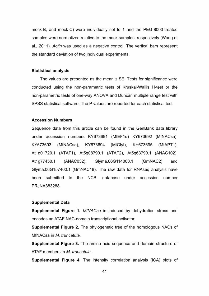
41
mock-B, and mock-C) were individually set to 1 and the PEG-8000-treated
samples were normalized relative to the mock samples, respectively (Wang et
al., 2011). Actin was used as a negative control. The vertical bars represent
the standard deviation of two individual experiments.
Statistical analysis
The values are presented as the mean ± SE. Tests for significance were
conducted using the non-parametric tests of Kruskal-Wallis H-test or the
non-parametric tests of one-way ANOVA and Duncan multiple range test with
SPSS statistical software. The P values are reported for each statistical test.
Accession Numbers
Sequence data from this article can be found in the GenBank data library
under accession numbers KY673691 (MfEF1α) KY673692 (MfNACsa),
KY673693 (MtNACsa), KY673694 (MtGlyI), KY673695 (MtAPT1),
At1g01720.1 (ATAF1), At5g08790.1 (ATAF2), At5g63790.1 (ANAC102),
At1g77450.1 (ANAC032), Glyma.06G114000.1 (GmNAC2) and
Glyma.06G157400.1 (GmNAC18). The raw data for RNAseq analysis have
been submitted to the NCBI database under accession number
PRJNA383288.
Supplemental Data
Supplemental Figure 1. MfNACsa is induced by dehydration stress and
encodes an ATAF NAC-domain transcriptional activator.
Supplemental Figure 2. The phylogenetic tree of the homologous NACs of
MfNACsa in M. truncatula.
Supplemental Figure 3. The amino acid sequence and domain structure of
ATAF members in M. truncatula.
Supplemental Figure 4. The intensity correlation analysis (ICA) plots of

42
co-localization of MfNACsa with membrane markers and membrane/nuclear
dyes.
Supplemental Figure 5. Subcellular localization of N-terminal GFP fusion
MfNACsa protein.
Supplemental Figure 6. A subdomain at the N-terminus of MfNACsa was
sufficient to mediate plasma membrane targeting.
Supplemental Figure 7. The intensity correlation analysis of MfNACsa
translocation under PEG-imposed dehydration stress.
Supplemental Figure 8. Molecular identification of the lines ectopically
expressing MfNACsa or Tnt1 insertion.
Supplemental Figure 9. The phenotype of wild-type R108 under PEG-8000
treatment.
Supplemental Figure 10. NACsa positively regulates plant dehydration
tolerance.
Supplemental Figure 11. Gene Ontology (GO) and pathway enrichment
analysis for differentially expressed genes regulated by MfNACsa.
Supplemental Figure 12. The temporal expression pattern of stress-, lipid
transport- and localization-related genes under dehydration stress.
Supplemental Figure 13. MfNACsa positively regulates the stress-, lipid
transport- and localization-related genes expression under PEG-imposed
drought stress.
Supplemental Figure 14. Molecular identification of glyI mutants.
Supplemental Figure 15. The intensity correlation analysis of mutant
C26S-eGFP or MfNACsa-eGFP subjected to 2-BP treatment.
Supplemental Figure 16. MtAPT1 belongs to a single Hotdog fold fatty
acyl-ACP thioesterase.
Supplemental Figure 17. MtAPT1 possesses conserved active site residues
(aspartate, glycine, and valine) in the DXXGXV motif of acyl-ACP thioesterase
members.

43
Supplemental Figure 18. The intensity correlation analysis of MfNACsa
co-localized with wild-type MtAPT1 and mutant MtAPT1m.
Supplemental Figure 19. Molecular identification of the apt1 mutant.
Supplemental Figure 20. Subcellular localization of MfNACsaG3A-eGFP
fusion protein.
Supplemental Figure 21. The phylogenetic tree of the homologous NACs of
MfNACsa in Arabidopsis.
Supplemental Figure 22. The phylogenetic tree of the homologous NACs of
MfNACsa in soybean.
Supplemental Figure 23. The lipidation prediction of ATAF family members in
A. thaliana, Glycine max and M. trunctula.
Supplemental Figure 24. A general mechanism for the nuclear relocation of
S-palmitoylated ATAF transcription factors.
Supplemental Table 1. List of potential thioesterase-like proteins (JCVI
database, Mt genome v 4.0).
Supplemental Table 2. Oligonucleotide probes used in EMSA.
Supplemental Data Set 1. Text file of the amimo acid sequence of MfNACsa
and the N-terminal consensus NAC domains from Ooka et al. (2003) that were
used as an input for the phylogenetic tree analysis.
Supplemental Data Set 2. Text file of the homologous NACs of MfNACsa in M.
truncatula.
Supplemental Data Set 3. Up-regulated genes in lines ectopically expressing
MfNACsa under PEG-simulated drought stress.
Supplemental Data Set 4. Down-regulated genes in lines ectopically
expressing MfNACsa under PEG-simulated drought stress.
Supplemental Data Set 5. Text file of the amimo acid sequences of potential
thioesterase-like proteins in M. truncatula.
Supplemental Data Set 6. Text file of the homologous NACs of MfNACsa in A.
thaliana,

44
Supplemental Data Set 7. Text file of the homologous NACs of MfNACsa in
Glycine max.
Supplemental Data Set 8. Primers used in this study.
Acknowledgements
We thank Dr. Jean Marie Prosperi and Magalie Delalande (BRC for Medicago
truncatula, UMR 1097, INRA, Montpellier, France) for providing seeds of
Medicago truncatula cv. R108. We thank the US National Plant Germplasm
System (NPGS) of the US Department of Agriculture (USDA) for providing
Medicago falcata cv. PI502449 seeds. This work was supported by grants from
the National Natural Science Foundation of China (31571587and 31371689).
Authors’ contributions
T.W. and J.D. led the study and helped revise the manuscript; M.D. performed
the main experiments and written the manuscript; R.Z. participated in the plant
materials preparation, stress treatment and data analysis; F.Z.participated in
the modifications of some constructs and genetic transformation; Z.Z.
participated in the ChIP assay; L.G. participated in genetic transformation and
figures arrange; J.W. provided the Tnt1 mutants. All authors read and
approved the final manuscript.
Competing financial interests
The authors declare no competing financial interests.
References Abe, H., Yamaguchi-Shinozaki, K., Urao, T., Iwasaki, T., Hosokawa, D., and
Shinozaki, K. (1997). Role of Arabidopsis MYC and MYB homologs in drought- and abscisic acid-regulated gene expression. Plant Cell 9, 1859-1868.
Agudo-Ibanez, L., Herrero, A., Barbacid, M., and Crespo, P. (2015). H-Ras distribution and signaling in plasma membrane microdomains are regulated by acylation and deacylation events. Mol. Cell. Biol. 35,

45
1898-1914. Ambawat, S., Sharma, P., Yadav, N.R., and Yadav, R.C. (2013). MYB
transcription factor genes as regulators for plant responses: an overview. Physiol Mol Biol Plants 19, 307-321.
An, H., Roussot, C., Suarez-Lopez, P., Corbesier, L., Vincent, C., Pineiro, M., Hepworth, S., Mouradov, A., Justin, S., Turnbull, C., and Coupland, G. (2004). CONSTANS acts in the phloem to regulate a systemic signal that induces photoperiodic flowering of Arabidopsis. Development 131, 3615-3626.
Baldoni, E., Genga, A., and Cominelli, E. (2015). Plant MYB transcription factors: their role in drought response mechanisms. Int J Mol Sci 16, 15811-15851.
Batistic, O., Sorek, N., Schultke, S., Yalovsky, S., and Kudla, J. (2008). Dual fatty acyl modification determines the localization and plasma membrane targeting of CBL/CIPK Ca2+ signaling complexes in Arabidopsis. Plant Cell 20, 1346-1362.
Blazquez, M. (2007). Quantitative GUS Activity Assay of Plant Extracts. CSH protoc 2007, pdb prot4690.
Camp, L.A., and Hofmann, S.L. (1993). Purification and properties of a palmitoyl-protein thioesterase that cleaves palmitate from H-Ras. J. Biol. Chem. 268, 22566-22574.
Chen, M., Zhao, Y., Zhuo, C., Lu, S., and Guo, Z. (2015). Overexpression of a NF-YC transcription factor from bermudagrass confers tolerance to drought and salinity in transgenic rice. Plant Biotechnol. J. 13, 482-491.
Cheng, X., Wang, M., Lee, H.K., Tadege, M., Ratet, P., Udvardi, M., Mysore, K.S., and Wen, J. (2014). An efficient reverse genetics platform in the model legume Medicago truncatula. New Phytol. 201, 1065-1076.
Christianson, J.A., Dennis, E.S., Llewellyn, D.J., and Wilson, I.W. (2010). ATAF NAC transcription factors: regulators of plant stress signaling. Plant Signal Behav. 5, 428-432.
Conibear, E., and Davis, N.G. (2010). Palmitoylation and depalmitoylation dynamics at a glance. J. Cell.Sci. 123, 4007-4010.
Cosson, V., Durand, P., d'Erfurth, I., Kondorosi, A., and Ratet, P. (2006). Medicago truncatula transformation using leaf explants. Methods Mol. Biol. 343, 115-127.
Cutler, S.R., Ehrhardt, D.W., Griffitts, J.S., and Somerville, C.R. (2000). Random GFP::cDNA fusions enable visualization of subcellular structures in cells of Arabidopsis at a high frequency. Proc. Natl. Acad. Sci. U.S.A. 97, 3718-3723.
Dietz, K.J., Vogel, M.O., and Viehhauser, A. (2010). AP2/EREBP transcription factors are part of gene regulatory networks and integrate metabolic, hormonal and environmental signals in stress acclimation and retrograde signalling. Protoplasma. 245, 3-14.
Du, J.L., Zhang, S.W., Huang, H.W., Cai, T., Li, L., Chen, S., and He, X.J.

46
(2015). The splicing factor PRP31 is involved in transcriptional gene silencing and stress response in Arabidopsis. Mol Plant 8, 1053-1068.
Duncan, J.A., and Gilman, A.G. (1998). A cytoplasmic acyl-protein thioesterase that removes palmitate from G protein alpha subunits and p21 (RAS). J. Biol. Chem. 273, 15830-15837.
Duncan, J.A., and Gilman, A.G. (2002). Characterization of Saccharomyces cerevisiae acyl-protein thioesterase 1, the enzyme responsible for G protein alpha subunit deacylation in vivo. J. Biol. Chem. 277, 31740-31752.
Eisenhaber, B., Sammer, M., Lua, W.H., Benetka, W., Liew, L.L., Yu, W., Lee, H.K., Koranda, M., Eisenhaber, F., and Adhikari, S. (2011). Nuclear import of a lipid-modified transcription factor: mobilization of NFAT5 isoform a by osmotic stress. Cell Cycle 10, 3897-3911.
Fang, Y., Liao, K., Du, H., Xu, Y., Song, H., Li, X., and Xiong, L. (2015). A stress-responsive NAC transcription factor SNAC3 confers heat and drought tolerance through modulation of reactive oxygen species in rice. J. Exp. Bot. 66, 6803-6817.
Fehrenbacher, N., Bar-Sagi, D., and Philips, M. (2009). Ras/MAPK signaling from endomembranes. Mol Oncol 3, 297-307.
Fragoso, R., Ren, D., Zhang, X., Su, M.W., Burakoff, S.J., and Jin, Y.J. (2003). Lipid raft distribution of CD4 depends on its palmitoylation and association with Lck, and evidence for CD4-induced lipid raft aggregation as an additional mechanism to enhance CD3 signaling. J. Immunol. 170, 913-921.
Golldack, D., Li, C., Mohan, H., and Probst, N. (2014). Tolerance to drought and salt stress in plants: Unraveling the signaling networks. Front Plant Sci 5, 151.
Gomord, V., Denmat, L.A., Fitchette-Laine, A.C., Satiat-Jeunemaitre, B., Hawes, C., and Faye, L. (1997). The C-terminal HDEL sequence is sufficient for retention of secretory proteins in the endoplasmic reticulum (ER) but promotes vacuolar targeting of proteins that escape the ER. Plant J. 11, 313-325.
Harshavardhan, V.T., Van Son, L., Seiler, C., Junker, A., Weigelt-Fischer, K., Klukas, C., Altmann, T., Sreenivasulu, N., Baumlein, H., and Kuhlmann, M. (2014). AtRD22 and AtUSPL1, members of the plant-specific BURP domain family involved in Arabidopsis thaliana drought tolerance. PLoS ONE 9, e110065.
Hemsley, P.A., and Grierson, C.S. (2008). Multiple roles for protein palmitoylation in plants. Trends Plant Sci. 13, 295-302.
Hemsley, P.A., Taylor, L., and Grierson, C.S. (2008). Assaying protein palmitoylation in plants. Plant Methods 4, 2.
Hemsley, P.A., Weimar, T., Lilley, K.S., Dupree, P., and Grierson, C.S. (2013). A proteomic approach identifies many novel palmitoylated proteins in Arabidopsis. New Phytol. 197, 805-814.

47
Hu, H., Dai, M., Yao, J., Xiao, B., Li, X., Zhang, Q., and Xiong, L. (2006). Overexpressing a NAM, ATAF, and CUC (NAC) transcription factor enhances drought resistance and salt tolerance in rice. Proc. Natl. Acad. Sci. U.S.A. 103, 12987-12992.
Huang, J., Sun, S.J., Xu, D.Q., Yang, X., Bao, Y.M., Wang, Z.F., Tang, H.J., and Zhang, H. (2009). Increased tolerance of rice to cold, drought and oxidative stresses mediated by the overexpression of a gene that encodes the zinc finger protein ZFP245. Biophys. Res. Commun. 389, 556-561.
Huang, Q., Wang, Y., Li, B., Chang, J., Chen, M., Li, K., Yang, G., and He, G. (2015). TaNAC29, a NAC transcription factor from wheat, enhances salt and drought tolerance in transgenic Arabidopsis. BMC Plant Biol. 15, 268.
Huh, S.U., Lee, S.B., Kim, H.H., and Paek, K.H. (2012). ATAF2, a NAC transcription factor, binds to the promoter and regulates NIT2 gene expression involved in auxin biosynthesis. Mol. Cells 34, 305-313.
Ilyas, M., Rasheed, A., and Mahmood, T. (2016). Functional characterization of germin and germin-like protein genes in various plant species using transgenic approaches. Biotechnol. Lett. 38, 1405-1421.
Jain, M., Choudhary, D., Kale, R.K., and Bhalla-Sarin, N. (2002). Salt- and glyphosate-induced increase in glyoxalase I activity in cell lines of groundnut (Arachis hypogaea). Physol. Plant 114, 499-505.
Jennings, P., Bertocchi, C., Frick, M., Haller, T., Pfaller, W., and Dietl, P. (2005). Ca2+ induced surfactant secretion in alveolar type II cultures isolated from the H-2Kb-tsA58 transgenic mouse. Cell. Physiol. Biochem. 15, 159-166.
Jeong, J.S., Kim, Y.S., Baek, K.H., Jung, H., Ha, S.H., Do Choi, Y., Kim, M., Reuzeau, C., and Kim, J.K. (2010). Root-specific expression of OsNAC10 improves drought tolerance and grain yield in rice under field drought conditions. Plant Physiol. 153, 185-197.
Kanaani, J., Patterson, G., Schaufele, F., Lippincott-Schwartz, J., and Baekkeskov, S. (2008). A palmitoylation cycle dynamically regulates partitioning of the GABA-synthesizing enzyme GAD65 between ER-Golgi and post-Golgi membranes. J. Cell. Sci. 121, 437-449
Kim, M.J., Park, M.J., Seo, P.J., Song, J.S., Kim, H.J., and Park, C.M. (2012). Controlled nuclear import of the transcription factor NTL6 reveals a cytoplasmic role of SnRK2.8 in the drought-stress response. Biochem. J. 448, 353-363.
Kim, S.G., Lee, A.K., Yoon, H.K., and Park, C.M. (2008). A membrane-bound NAC transcription factor NTL8 regulates gibberellic acid-mediated salt signaling in Arabidopsis seed germination. Plant J. 55, 77-88.
Kim, S.G., Lee, S., Seo, P.J., Kim, S.K., Kim, J.K., and Park, C.M. (2010). Genome-scale screening and molecular characterization of membrane-bound transcription factors in Arabidopsis and rice.

48
Genomics. 95, 56-65. Kim, Y.S., Kim, S.G., Park, J.E., Park, H.Y., Lim, M.H., Chua, N.H., and Park,
C.M. (2006). A membrane-bound NAC transcription factor regulates cell division in Arabidopsis. Plant Cell 18, 3132-3144.
Krasensky, J., and Jonak, C. (2012). Drought, salt, and temperature stress-induced metabolic rearrangements and regulatory networks. J. Exp. Bot. 63, 1593-1608.
Lam, S.K., Siu, C.L., Hillmer, S., Jang, S., An, G., Robinson, D.G., and Jiang, L. (2007). Rice SCAMP1 defines clathrin-coated, trans-golgi-located tubular-vesicular structures as an early endosome in tobacco BY-2 cells. Plant Cell 19, 296-319.
Lavy, M., and Yalovsky, S. (2006). Association of Arabidopsis type-II ROPs with the plasma membrane requires a conserved C-terminal sequence motif and a proximal polybasic domain. Plant J. 46, 934-947.
Lavy, M., Bracha-Drori, K., Sternberg, H., and Yalovsky, S. (2002). A cell-specific, prenylation-independent mechanism regulates targeting of type II RACs. Plant Cell 14, 2431-2450.
Lee, S., Seo, P.J., Lee, H.J., and Park, C.M. (2012). A NAC transcription factor NTL4 promotes reactive oxygen species production during drought-induced leaf senescence in Arabidopsis. Plant J. 70, 831-844.
Lei, M.J., Wang, Q., Li, X., Chen, A., Luo, L., Xie, Y., Li, G., Luo, D., Mysore, K.S., Wen, J., Xie, Z.P., Staehelin, C., and Wang, Y.Z. (2015). The Small GTPase ROP10 of Medicago truncatula is required for both tip growth of root hairs and nod factor-induced root hair deformation. Plant Cell. 27, 806-822.
Levental, I., Lingwood, D., Grzybek, M., Coskun, U., and Simons, K. (2010). Palmitoylation regulates raft affinity for the majority of integral raft proteins. Proc. Natl. Acad. Sci. U.S.A. 107, 22050-22054.
Li, M., Yang, C., Tong, S., Weidmann, A., and Compans, R.W. (2002). Palmitoylation of the murine leukemia virus envelope protein is critical for lipid raft association and surface expression. J. Virol. 76, 11845-11852.
Li, Q., Lau, A., Morris, T.J., Guo, L., Fordyce, C.B., and Stanley, E.F. (2004). A syntaxin 1, Galpha(o), and N-type calcium channel complex at a presynaptic nerve terminal: analysis by quantitative immunocolocalization. J. neurosci. 24, 4070-4081.
Liang, M., Li, H., Zhou, F., Li, H., Liu, J., Hao, Y., Wang, Y., Zhao, H., and Han, S. (2015). Subcellular distribution of NTL transcription factors in Arabidopsis thaliana. Traffic 16, 1062-1074.
Lin, D.T., and Conibear, E. (2015). Enzymatic protein depalmitoylation by acyl protein thioesterases. Biochem. Soc. Trans. 43, 193-198.
Liu, Y.G., and Chen, Y. (2007). High-efficiency thermal asymmetric interlaced PCR for amplification of unknown flanking sequences. BioTechniques 43, 649-650, 652, 654 passim.

49
Liu, Z., Jia, Y., Ding, Y., Shi, Y., Li, Z., Guo, Y., Gong, Z., and Yang, S. (2017). Plasma membrane CRPK1-Mediated phosphorylation of 14-3-3 proteins induces their nuclear import to fine-tune CBF signaling during cold response. Molecular cell 66, 117-128 e115.
Loisel, T.P., Ansanay, H., Adam, L., Marullo, S., Seifert, R., Lagace, M., and Bouvier, M. (1999). Activation of the β2-adrenergic receptor-GαS
complex leads to rapid depalmitoylation and inhibition of repalmitoylation of both the receptor and GαS. J. Biol. Chem. 274, 31014-31019.
Lu, J.Y., and Hofmann, S.L. (2006). Thematic review series: lipid posttranslational modifications. Lysosomal metabolism of lipid-modified proteins. J. Lipid Res. 47, 1352-1357.
Lu, S., Song, T., Kosma, D.K., Parsons, E.P., Rowland, O., and Jenks, M.A. (2009). Arabidopsis CER8 encodes long-chain acyl-CoA synthetase 1 (LACS1) that has overlapping functions with LACS2 in plant wax and cutin synthesis. Plant J. 59, 553-564.
Mendes, G.C., Reis, P.A., Calil, I.P., Carvalho, H.H., Aragao, F.J., and Fontes, E.P. (2013). GmNAC30 and GmNAC81 integrate the endoplasmic reticulum stress- and osmotic stress-induced cell death responses through a vacuolar processing enzyme. Proc. Natl. Acad. Sci. U.S.A. 110, 19627-19632.
Miao, Z., Xu, W., Li, D., Hu, X., Liu, J., Zhang, R., Tong, Z., Dong, J., Su, Z., Zhang, L., Sun, M., Li, W., Du, Z., Hu, S., and Wang, T. (2015). De novo transcriptome analysis of Medicago falcata reveals novel insights about the mechanisms underlying abiotic stress-responsive pathway. BMC Genomics 16, 818.
Morrison, D.F., O'Brien, P.J., and Pepperberg, D.R. (1991). Depalmitylation with hydroxylamine alters the functional properties of rhodopsin. J. Biol. Chem. 266, 20118-20123.
Ng, S., Ivanova, A., Duncan, O., Law, S.R., Van Aken, O., De Clercq, I., Wang, Y., Carrie, C., Xu, L., Kmiec, B., Walker, H., Van Breusegem, F., Whelan, J., and Giraud, E. (2013). A membrane-bound NAC transcription factor, ANAC017, mediates mitochondrial retrograde signaling in Arabidopsis. Plant Cell 25, 3450-3471.
Ni, Y., and Guo, Y.J. (2008). Progress in the study on genes encoding enzymes involved in biosynthesis of very long chain fatty acids and cuticular wax in plants. Yi Chuan 30, 561-567.
Ooka, H., Satoh, K., Doi, K., Nagata, T., Otomo, Y., Murakami, K., Matsubara, K., Osato, N., Kawai, J., Carninci, P., Hayashizaki, Y., Suzuki, K., Kojima, K., Takahara, Y., Yamamoto, K., and Kikuchi, S. (2003). Comprehensive analysis of NAC family genes in Oryza sativa and Arabidopsis thaliana. DNA Res. 10, 239-247.
Pulsifer, I.P., Lowe, C., Narayaran, S.A., Busuttil, A.S., Vishwanath, S.J., Domergue, F., and Rowland, O. (2014). Acyl-lipid thioesterase1-4 from

50
Arabidopsis thaliana form a novel family of fatty acyl-acyl carrier protein thioesterases with divergent expression patterns and substrate specificities. Plant Mol.Biol. 84, 549-563.
Puranik, S., Sahu, P.P., Srivastava, P.S., and Prasad, M. (2012). NAC proteins: regulation and role in stress tolerance. Trends Plant Sci. 17, 369-381.
Rigal, A., Doyle, S.M., and Robert, S. (2015). Live cell imaging of FM4-64, a tool for tracing the endocytic pathways in Arabidopsis root cells. Methods in molecular biology 1242, 93-103.
Rocks, O., Gerauer, M., Vartak, N., Koch, S., Huang, Z.P., Pechlivanis, M., Kuhlmann, J., Brunsveld, L., Chandra, A., Ellinger, B., et al. (2010). The palmitoylation machinery is a spatially organizing system for peripheral membrane proteins. Cell 141, 458-471.
Saleh, A., Alvarez-Venegas, R., and Avramova, Z. (2008). An efficient chromatin immunoprecipitation (ChIP) protocol for studying histone modifications in Arabidopsis plants. Nat Protoc 3, 1018-1025.
Satou, M., Nishi, Y., Yoh, J., Hattori, Y., and Sugimoto, H. (2010). Identification and characterization of acyl-protein thioesterase 1/lysophospholipase I as a ghrelin deacylation/lysophospholipid hydrolyzing enzyme in fetal bovine serum and conditioned medium. Endocrinology 151, 4765-4775.
Seki, M., Narusaka, M., Ishida, J., Nanjo, T., Fujita, M., Oono, Y., Kamiya, A., Nakajima, M., Enju, A., Sakurai, T., Satou, M., Akiyama, K., Taji, T., Yamaguchi-Shinozaki, K., Carninci, P., Kawai, J., Hayashizaki, Y., and Shinozaki, K. (2002). Monitoring the expression profiles of 7000 Arabidopsis genes under drought, cold and high-salinity stresses using a full-length cDNA microarray. Plant J. 31, 279-292.
Shan, W., Kuang, J.F., Lu, W.J., and Chen, J.Y. (2014). Banana fruit NAC transcription factor MaNAC1 is a direct target of MaICE1 and involved in cold stress through interacting with MaCBF1. Plant Cell Environ. 37, 2116-2127.
Sharma, C., Yang, X.H., and Hemler, M.E. (2008). DHHC2 affects palmitoylation, stability, and functions of tetraspanins CD9 and CD151. Mol. Biol. Cell 19, 3415-3425.
Shipston, M.J. (2011). Ion channel regulation by protein palmitoylation. J. Biol. Chem. 286, 8709-8716.
Singaraja, R.R., Kang, M.H., Vaid, K., Sanders, S.S., Vilas, G.L., Arstikaitis, P., Coutinho, J., Drisdel, R.C., El-Husseini Ael, D., Green, W.N., Berthiaume, L., and Hayden, M.R. (2009). Palmitoylation of ATP-binding cassette transporter A1 is essential for its trafficking and function. Circ. Res. 105, 138-147.
Singh, D., and Laxmi, A. (2015). Transcriptional regulation of drought response: a tortuous network of transcriptional factors. Front Plant Sci. 6, 895.
Sugimoto, H., Odani, S., and Yamashita, S. (1998). Cloning and expression of cDNA encoding rat liver 60-kDa lysophospholipase containing an asparaginase-like region and ankyrin repeat. J. Biol. Chem. 273,

51
12536-12542. Tadege, M., Wen, J., He, J., Tu, H., Kwak, Y., Eschstruth, A., Cayrel, A., Endre,
G., Zhao, P.X., Chabaud, M., Ratet, P., and Mysore, K.S. (2008). Large-scale insertional mutagenesis using the Tnt1 retrotransposon in the model legume Medicago truncatula. Plant J. 54, 335-347.
Tapia, G., Morales-Quintana, L., Parra, C., Berbel, A., and Alcorta, M. (2013). Study of nsLTPs in Lotus japonicus genome reveal a specific epidermal cell member (LjLTP10) regulated by drought stress in aerial organs with a putative role in cutin formation. Plant Mol. Biol. 82, 485-501.
Tarnowski, B.I., Spinale, F.G., and Nicholson, J.H. (1991). DAPI as a useful stain for nuclear quantitation. Biotech Histochem 66, 297-302.
Topping, J.F., and Lindsey, K. (1997). Promoter trap markers differentiate structural and positional components of polar development in Arabidopsis. Plant Cell 9, 1713-1725.
Tran, L.S., Nakashima, K., Sakuma, Y., Simpson, S.D., Fujita, Y., Maruyama, K., Fujita, M., Seki, M., Shinozaki, K., and Yamaguchi-Shinozaki, K. (2004). Isolation and functional analysis of Arabidopsis stress-inducible NAC transcription factors that bind to a drought-responsive cis-element in the early responsive to dehydration stress 1 promoter. Plant Cell 16, 2481-2498.
Traverso, J.A., Micalella, C., Martinez, A., Brown, S.C., Satiat-Jeunemaitre, B., Meinnel, T., and Giglione, C. (2013). Roles of N-terminal fatty acid acylations in membrane compartment partitioning: Arabidopsis h-type thioredoxins as a case study. Plant Cell 25, 1056-1077.
Vandesompele, J., De Preter, K., Pattyn, F., Poppe, B., Van Roy, N., De Paepe, A., and Speleman, F. (2002). Accurate normalization of real-time quantitative RT-PCR data by geometric averaging of multiple internal control genes. Genome Biol. 3, RESEARCH0034.
Waadt, R., Schmidt, L.K., Lohse, M., Hashimoto, K., Bock, R., and Kudla, J. (2008). Multicolor bimolecular fluorescence complementation reveals simultaneous formation of alternative CBL/CIPK complexes in planta. Plant J. 56, 505-516.
Wang, L., Hua, D., He, J., Duan, Y., Chen, Z., Hong, X., and Gong, Z. (2011). Auxin Response Factor2 (ARF2) and its regulated homeodomain gene HB33 mediate abscisic acid response in Arabidopsis. PLoS genetics 7, e1002172.
Wang, X., and Culver, J.N. (2012). DNA binding specificity of ATAF2, a NAC domain transcription factor targeted for degradation by Tobacco mosaic virus. BMC Plant Biol. 12, 157.
Wu, A., Allu, A.D., Garapati, P., Siddiqui, H., Dortay, H., Zanor, M.I., Asensi-Fabado, M.A., Munne-Bosch, S., Antonio, C., Tohge, T., Fernie, A.R., Kaufmann, K., Xue, G.P., Mueller-Roeber, B., and Balazadeh, S. (2012). JUNGBRUNNEN1, a reactive oxygen species-responsive NAC transcription factor, regulates longevity in Arabidopsis. Plant Cell 24,

52
482-506. Yadav, S.K., Singla-Pareek, S.L., Ray, M., Reddy, M.K., and Sopory, S.K.
(2005). Methylglyoxal levels in plants under salinity stress are dependent on glyoxalase I and glutathione. Biochem. Biophys. Res.Commun. 337, 61-67.
Yang, Z.T., Wang, M.J., Sun, L., Lu, S.J., Bi, D.L., Sun, L., Song, Z.T., Zhang, S.S., Zhou, S.F., and Liu, J.X. (2014). The membrane-associated transcription factor NAC089 controls ER-stress-induced programmed cell death in plants. PLoS Genet. 10, e1004243.
Yoon, H.K., Kim, S.G., Kim, S.Y., and Park, C.M. (2008). Regulation of leaf senescence by NTL9-mediated osmotic stress signaling in Arabidopsis. Mol. Cells 25, 438-445.
You, J., Zong, W., Hu, H., Li, X., Xiao, J., and Xiong, L. (2014). A stress-responsive NAC1-regulated protein phosphatase gene rice protein phosphatase18 modulates drought and oxidative stress tolerance through abscisic acid-independent reactive oxygen species scavenging in rice. Plant Physiol. 166, 2100-2114.
Zhang, L.L., Zhao, M.G., Tian, Q.Y., and Zhang, W.H. (2011). Comparative studies on tolerance of Medicago truncatula and Medicago falcata to freezing. Planta 234, 445-457.
Zhou, L.Z., Li, S., Feng, Q.N., Zhang, Y.L., Zhao, X., Zeng, Y.L., Wang, H., Jiang, L., and Zhang, Y. (2013). Protein S-ACYL Transferase10 is critical for development and salt tolerance in Arabidopsis. Plant Cell 25, 1093-1107.
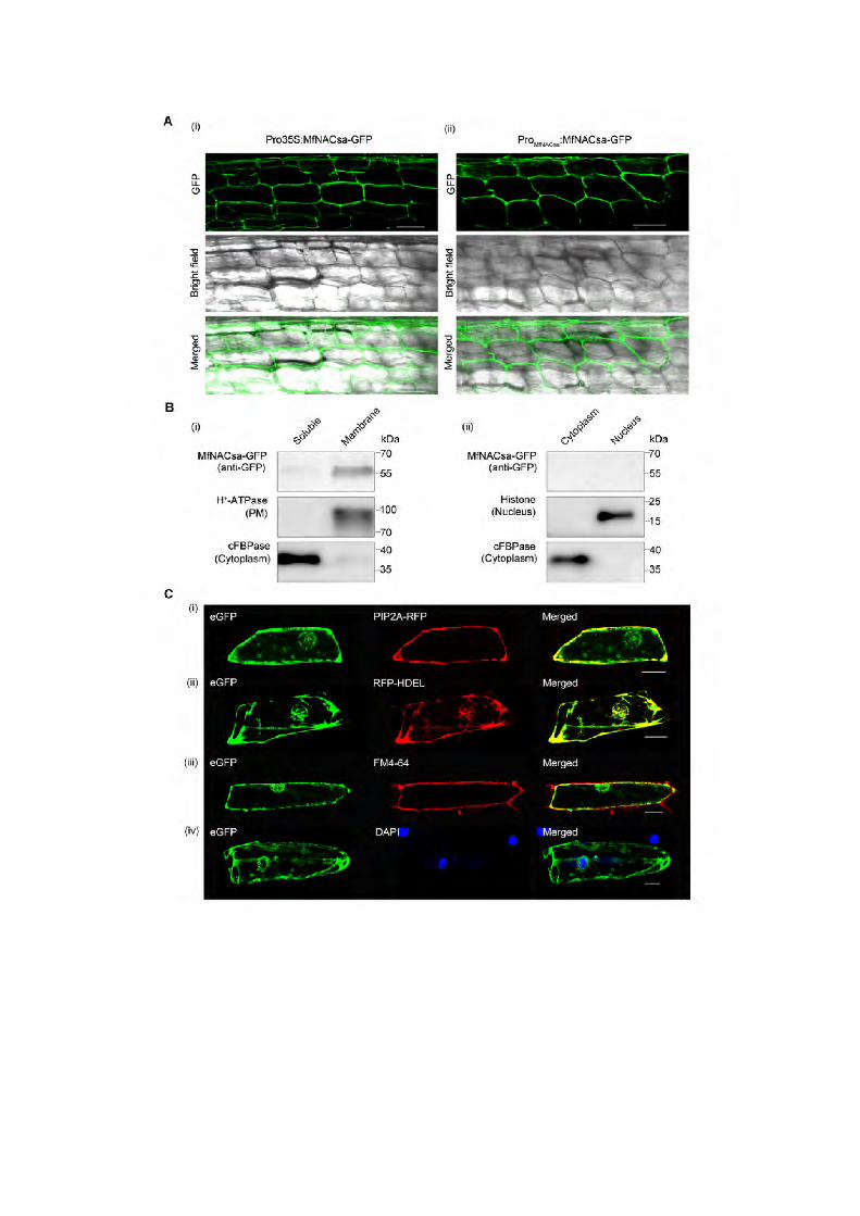

Figure1. MfNACsa is associated with membranes. (A) Confocal imaging of transgenic M. truncatula hairy roots expressing MfNACsa-GFP fusion proteins driven by the constitutive CaMV 35S promoter (i) or the MfNACsa endogenous promoter (ii). GFP signal was detected with an Olympus FluoView™ FV1000 laser scanning confocal microscope. Bars = 50 μm. (B) Cellular fractionation and immunoblot analysis of ProMfNACsa:MfNACsa-GFP protein in N. benthamiana leaves. Soluble and membrane proteins (i), Cytoplasmic and nuclear proteins were separated and subjected the immunoblot analysis (ii). H+-ATPase, Histone H3 and cFBPase were used as plasma membrane (PM), nuclear and cytosolic fraction markers, respectively. (C) MfNACsa localizes to the PM and ER compartment in onion epidermal cells. Co-localization of MfNACsa-eGFP fusion proteins driven by CaMV35S promoter with the RFP-tagged PIP2A PM marker (i) or the HDEL ER marker (ii), and co-staining with the endocytic dye FM4-64 (iii) or nuclear dye DAPI (iv). The signal patterns were observed at 488 nm (eGFP: green), 546 nm (RFP or FM4-64: magenta) or 358 nm (DAPI: blue) fluorescence excitation wavelength by confocal imaging (Olympus FluoView™ FV1000). Bars = 50 μm. The eGFP-labeled MfNACsa is green, RFP-labeled PIP2A or HDEL or FM4-64 dye is magenta, and merged image is yellow. The intensity correlation quotients (ICQs) of MfNACsa-eGFP against PIP2A-RFP, RFP-HDEL, FM4-64 dye and DAPI are 0.363, 0.341, 0.239 and –0.154, respectively.


Figure 2. MfNACsa is translocated to the nucleus under PEG-imposed dehydration stress. (A) Transgenic M. truncatula hairy roots expressing the MfNACsa-GFP fusion proteins driven by native promoter were stained with the membrane dye FM4-64 for 5 min under normal conditions (no stress), with an ICQ of 0.114. (B) After hairy roots transiently expressing MfNACsa-GFP driven by the native promoter were treated with 50% PEG-8000 for 2 h (dehydration stress), the steady-state locations of MfNACsa-GFP protein were imaged and stained with DAPI dye (pseudo-color, red) (i)–(ii). Bars = 50 μm. The percentage of cells expressing GFP at the nucleus (N), nucleus and membranes (N+M), or membranes (M) in transgenic hairy root cells were counted respectively (iii). The data represent the mean and SE of three independent replicates (n>15) (nonparametric one-way ANOVA and Duncan multiple range test, P<0.05). (C) Nucleus translocation of the mCherry-MfNACsa-eGFP fusion protein under 10% PEG-8000 treatment in onion epidermal cells. The mCherry and eGFP signal patterns were observed at 0 (i) and 2 h (ii) after 10% PEG-8000 treatment by confocal imaging (Olympus FluoView™ FV1000). The same cells were imaged before and after treatment. Bars = 50 μm. (D) Cellular fractionation and immunoblot detection of transiently expressed ProMfNACsa:MfNACsa-GFP in N. benthamiana leaves under normal conditions (i) or 30% PEG-8000 treatment for 2 h (ii). MfNACsa-GFP is shown. cFBPase, H+-ATPase and Histone H3 mark the cytoplasm, PM and nuclear fraction, respectively.


Figure 3. MfNACsa plays a positive role in drought stress tolerance. (A) Ten-day-old seedlings of the wild-type (WT, R108) and lines ectopically expressing MfNACsa (OE23 and OE33) were subjected to three cycles of drought stress by withholding the water supply for 12, 14 and 18 d, respectively, until the leaves of WT plants exhibited primary wilting (ii). The mock plants were watered once a week, according to standard procedures (i). (B) The survival rate of plants in (Aii). The mean values and SE were calculated from three independent experiments. The asterisk indicates a significant difference between ectopic expression of MfNACsa lines and WT plants (Kruskal-Wallis non-parametric test, **P < 0.01). (C) The leaf relative electrical conductivity of the plants in (Aii). The mean values and SE were calculated from three independent experiments. The asterisk indicates a significant difference between ectopic expression of MfNACsa lines and WT plants (Kruskal-Wallis non-parametric test, **P < 0.01). (D) The phenotypes of the wild-type (WT, R108) and nacsa mutants (nacsa-1 and nacsa-2) subjected to three cycles of drought stress by withholding the water supply for 12, 14 and 14 d, respectively. Seedling were photographed when the leaves of mutants exhibited primary wilting (ii). The mock plants were watered once a week, according to standard procedure (i). (E) The survival rate of plants in (Dii). The mean values and SE were calculated from three independent experiments. The asterisk indicates a significant difference between nacsa mutants and WT plants (Kruskal-Wallis non-parametric test, **P < 0.01). (F) The leaf relative electrical conductivity of plants in (Dii). The mean values and SE were calculated from three independent experiments. The asterisk indicates a significant difference between nacsa mutants and WT plants (Kruskal-Wallis non-parametric test, **P < 0.01).


Figure 4. MfNACsa directly binds to the MtGlyI gene promoter to activate expression under drought stress. (A) In vitro protein-DNA binding assay showing that His-MfNACsa protein interacted with a cis-element (CAAATNNNCTTTG) in the promoter region of MtGlyI. A purified His-MfNACsa protein (200 ng) was incubated with a biotin-labeled probe (20 fM) and a mutant probe (20 fM). For the competition assay, a non-labeled probe (1000 times) was added. The cis-element (CAAATNNNCTTTG) of GlyI-P Wt and mutant element (CCCCCNNNCCCCC) of GlyI-P mut were showed in red font. (B) In vivo ChIP assay showing binding of MfNACsa to the promoter of MtGlyI under PEG-imposed dehydration stress. The ChIP experiment was performed using 2-week-old Pro35S:MfNACsa-3xFLAG plants treated with 50% PEG-8000 for 4 h. Plants ectopically expressing MfNACsa without stress treatment were used as the negative control. The MtGlyI promoter fragment B containing the cis-element (CAAATNNNCTTTG) (–346~–175 bp), fragment A located upstream of cis-element (–1897~–1714bp), and fragment C located downstream of cis-element (+550~+718 bp). Because of the different Ct values of the A, B and C fragment, mock samples (mock-actin, mock-A, mock-B, and mock-C) individually set to 1 and the PEG-8000 samples normalized relative to them, respectively, and actin used as a negative control. The mean values and SE were calculated from the two independent experiments (Kruskal-Wallis non-parametric test, **P < 0.01). (C) The Pro35S:MfNACsa-GFP effector plasmid and ProGlyI+Min35S:GUS reporter plasmid harboring the promoter region (–488bp~–38bp) of MtGlyI, a 35S min promoter and GUS reporter gene were transiently expressed in N. benthamiana leaf cells, together with a negative control vector, respectively. Before starting with sampling, the plants were submitted to 10% PEG treatment for 2 h (dehydration) or not (mock). The GUS staining and GUS activity were assayed according to a previously described method with some modifications (Topping and Lindsey, 1997; Blazquez, 2007). The mean values and SE were calculated from the two independent experiments (Kruskal-Wallis non-parametric test, **P < 0.01).
Figure 5. MtGlyI is necessary for altering the GSH/GSSG ratio in response to drought tolerance. (A) The wild-type (WT, R108) plants and glyI mutants (glyI-1 and glyI-2) were subjected to three cycles of drought stress by withholding the water supply for 12, 14 and 14 d, respectively. Seedlings were photographed when the leaves of mutants exhibited primary wilting (ii). The mock plants were watered once a week,according to standard procedure (i). (B) The survival rate of the plants in (Aii). The mean values and SE were calculated from three independent experiments. The asterisk indicates a significant difference between glyI mutants and WT plants (Kruskal-Wallis non-parametric test, **P < 0.01). (C) The GSH/GSSG ratio of glyI mutants and WT plants under PEG-imposed dehydration stress. Total glutathione (GSH+GSSG) and GSSG were measured in glyI mutants and WT plants under 50% PEG-8000 treatment for 0, 0.5, 1, 2 and 4 h. The error bars represent the standard error of two samples.The asterisk indicates a significant difference between glyI mutants and WT plants under PEG-imposed dehydration stress (Kruskal-Wallis non-parametric test, **P < 0.01).


Figure 6. S-palmitoylation at Cys26 determines PM targeting of MfNACsa protein. (A) Cys26 in the N-terminus of MfNACsa is predicted to be S-palmitoylated based on the GPS-lipid predictor analysis. The S-palmitoylation amino acid residue was shown in red font. (B) Palmitoylation of MfNACsa in vitro. pET-30a(+)-His-MfNACsa and pET-30a(+)-His-C26S (200 ng) were incubated at 25°C for 4 h with pal-Lys (biotin) (0.1 μg) and the plant extracts of M. truncatula cv. R108 (1 mg) from the cytoplasmic or membrane fraction, respectively. Protein expression was subsequently confirmed through native protein gel electrophoresis and immunoblot analysis. Labeled signals were detected through the streptavidin-HRP system. (C) Subcellular localization of MfNACsa protein harboring the C26S substitution in the N-terminus. C26S mutant protein was colocalized with the RFP-labeled Golgi and ER marker protein MAN1 from soybean, with the ICQ of 0.173, and not associated with the nucleus, as the negative (–0.021) ICQ between C26S-GFP and DAPI. The left panel is the subcellular localization of wild-type MfNACsa protein. The signal patterns were observed by fluorescence microscopy. Bars=50 μm. (D) Onion epidermal cells expressing MfNACsa-eGFP fusion proteins were treated with 2-bromopalmitate (2-BP), an inhibitor of S-acyl transferase. After treatment with EtOH (Control, left panel) or 100 μM 2-BP for 20 h, the individual cells were imaged. Upon 2-BP treatment, MfNACsa was co-localized with the RFP-labeled Golgi and ER marker protein MAN1, with the ICQ of 0.18, and not associated with nucleus as the negative (–0.029) ICQ between MfNACsa-eGFP and DAPI. The signal patterns were observed by fluorescence microscopy (Olympus FluoView™ FV1000). Bars = 50 μm.


Figure 7. MfNACsa is translocated to the nucleus through de-S-palmitoylation. (A) Transgenic M. truncatula hairy roots expressing the Pro35S:MfNACsa-GFP fusion proteins were treated with 0.05 M hydroxylamine (NH2OH). The GFP signal pattern was detected by confocal imaging at 0 (control), 0.5 or 2 h after NH2OH treatment. After 0.05 M NH2OH treatment for 2 h, the nucleus was stained with DAPI (pseudo-color, red), with the ICQ of 0.436. Bars = 50 μm. (B) Transgenic M. truncatula hairy roots expressing the Pro35S:C26S -GFP mutant proteins were treated with 0.05 M NH2OH. The GFP signal pattern was detected by confocal imaging (Olympus FluoView™ FV1000) under control conditions, and the negative (–0.037) ICQ of C26S-GFP against FM4-64. After 0.05 M NH2OH treatment for 2 h, the nucleus was stained with DAPI (pseudo-color, red), and the negative (–0.006) ICQ of C26S-GFP against DAPI dye. Bars = 50 μm. (C) Cellular fraction analysis of transient transgenic N. benthamiana leaves expressing MfNACsa-GFP proteins at 0 and 2 h after NH2OH treatment. Presence of MfNACsa-GFP was shown. cFBPase, H+-ATPase and Histone H3 are used as cytoplasm, PM and nuclear fraction markers, respectively. (D) Onion epidermal cells expressing C26S-eGFP mutant proteins were treated with 0.05 M NH2OH or 10% PEG-8000 for 2 h, respectively. The signal patterns were observed by fluorescence microscopy (Olympus FluoView™ FV1000). Bars = 50 μm.


Figure 8. MtAPT1 interacts with MfNACsa and mediates the nuclear translocation of MfNACsa. (A) The relative expression of MtAPT1, Medtr5g056230 and Medtr5g056390, which belong to a hotdog fold fatty acyl-ACP thioesterase in M. truncatula, was monitored by RT-qPCR under 50% PEG-8000 treatment for 8 h. Gene expression was calculated using the 2-ΔΔCT
method and the MtActin as an endogenous control. The mean values and SE were calculated from three independent experiments. (B) The interaction of MfNACsa with MtAPT1 was tested by yeast two-hybrid analysis. The transformed yeast cultures were spotted onto SD plates without tryptophan, histidine, leucine and adenine, but supplemented with 9 mM 3AT. The plates were incubated for 3 d and an α-Gal assay was performed. Positive control: DBD-p53 + AD-RecT, Negative control: DBD- Lam + AD-RecT. (C) Co-transformation of MfNACsa-eGFP with MtAPT1-RFP (i) or MtAPT1m-RFP ( DXXGXV-AXXAXA) mutant protein (ii) in onion epidermal cells. The signal patterns were observed at 488 nm (eGFP), 546 nm (RFP) and 358 nm (DAPI) fluorescence excitation wavelength by confocal imaging (Olympus FluoView™ FV1000). Bars = 50 μm. The ICQ value of MfNACsa-eGFP against DAPI is 0.210 (i), and against MtAPT1m-RFP is 0.174 (ii). (D) The BiFC assay of SPYNE173-::MfNACsa with SPYCE(MR) - MtAPT1m (i), SPYNE173-MfNACsa with SPYCE(MR) (ii) and SPYNE173 with SPYCE(MR)-MtAPT1m (iii), which were respectively co-expressed transiently in onion epidermal cells, and the YFP protein was visualized by confocal imaging (Olympus FluoView™ FV1000). Bars = 50 μm. (E) Immunoblot assay of total (T), nuclear (N) and membrane (M) components separated from N. benthamiana leaf cells that were co-transformed with Pro35S:MfNACsa-GFP and Pro35S:MtAPT1-HA (i) or Pro35S:MtAPT1m-HA (ii). H+-ATPase and Histone H3 were used as PM and nuclear fraction markers, respectively.
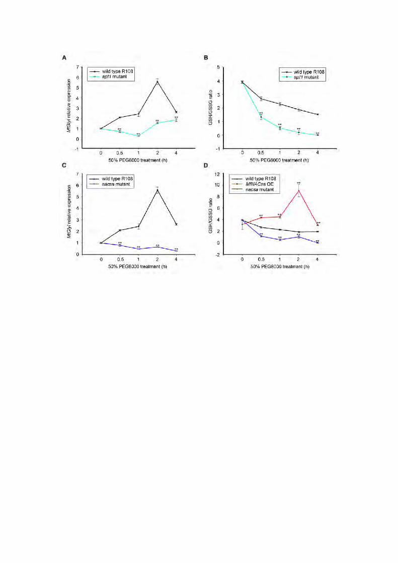

Figure 9. APT1 and NACsa are required for GlyI expression and GSH/GSSG ratio regulation. (A) Transcript levels of MtGlyI in two-week-old of WT (R108) plants or apt1 mutant subjected to 50% PEG-8000 treatment for 0, 0.5, 1, 2 and 4 h. The error bars represent the standard error of two samples. The asterisk indicates a significant difference between apt1 mutants and WT plants under PEG-imposed dehydration stress (Kruskal-Wallis non-parametric test, **P < 0.01). (B) The apt1 mutant and wild-type plant tissues (0.1 g) were used to measure the total glutathione (GSH+GSSG) and GSSG, and the GSH/GSSG ratio was calculated under 50% PEG-8000 treatment for 0, 0.5, 1, 2 and 4 h. The error bars represent standard error of two samples. The asterisk indicates a significant difference between apt1 mutants and WT plants under PEG-imposed dehydration stress (Kruskal-Wallis non-parametric test, **P < 0.01). (C) Transcript levels of MtGlyI in two-week-old of WT (R108) plants or nacsa-1 mutant with treated with 50% PEG-8000 for 0, 0.5, 1, 2, and 4 h. The error bars represent the standard error of two samples. The asterisk indicates a significant difference between nacsa mutants and WT plants under PEG-imposed dehydration stress (Kruskal-Wallis non-parametric test, **P < 0.01). (D) The GSH/GSSG ratio of ectopic expression of MfNACsa, nacsa-1 mutant and wild-type R108 plants under PEG-imposed dehydration stress. Total glutathione (GSH+GSSG) and GSSG were measured in leaf tissues of tested plants (0.1 g) subjected to 50% PEG-8000 treatment for 0, 0.5, 1, 2 and 4 h. The error bars represent the standard error of two samples. The asterisk indicates a significant difference between lines ectopically expressing MfNACsa or nacsa-1 mutant compared with WT plants under PEG-imposed dehydration stress (Kruskal-Wallis non-parametric test, **P < 0.01).
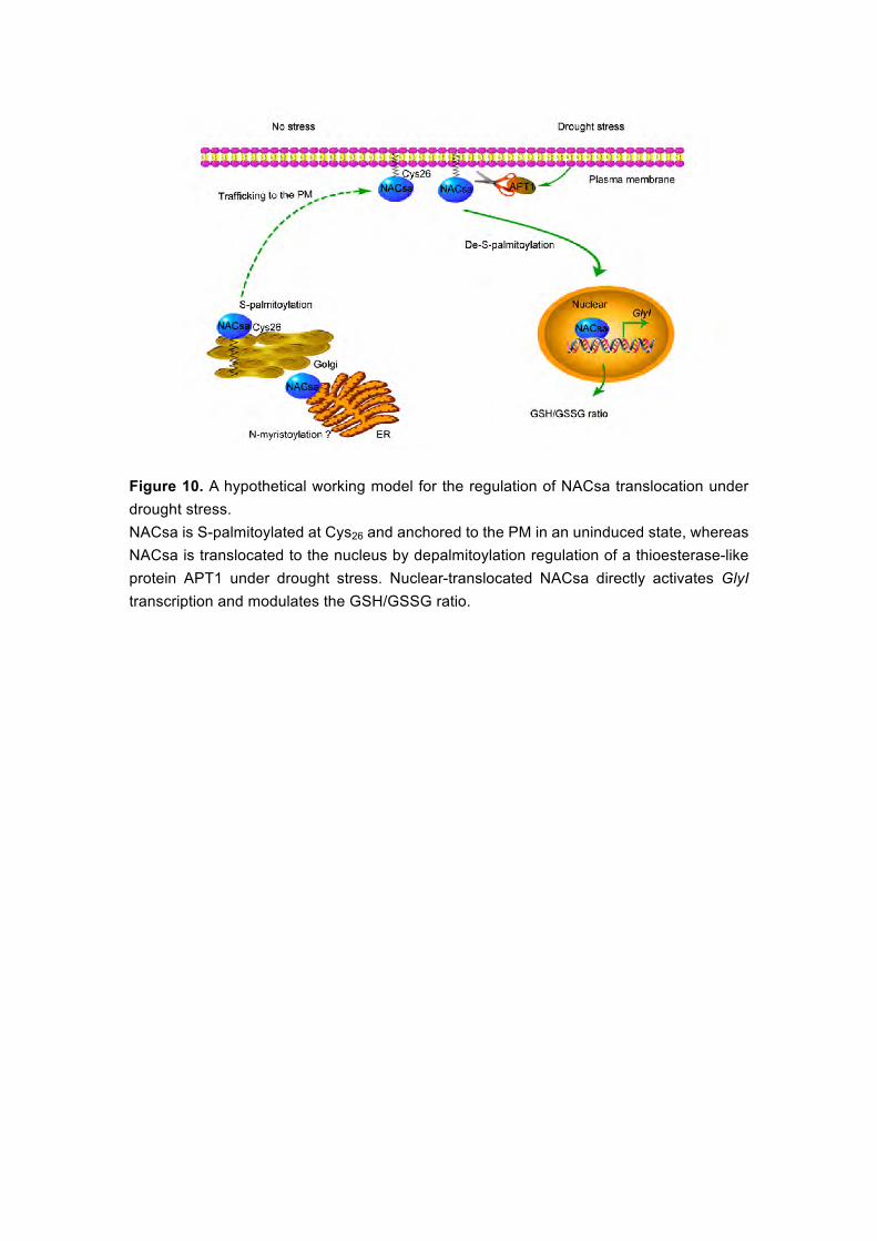
Figure 10. A hypothetical working model for the regulation of NACsa translocation under drought stress. NACsa is S-palmitoylated at Cys26 and anchored to the PM in an uninduced state, whereas NACsa is translocated to the nucleus by depalmitoylation regulation of a thioesterase-like protein APT1 under drought stress. Nuclear-translocated NACsa directly activates GlyI transcription and modulates the GSH/GSSG ratio.

DOI 10.1105/tpc.17.00044; originally published online July 6, 2017;Plant Cell
and Mei DuanTao Wang, Jiangli Dong, Rongxue Zhang, Fugui Zhu, Zhenqian Zhang, Lanming Gou, Jiangqi Wen
expression involved in drought stressA lipid-anchored NAC transcription factor translocates into nucleus to activate GlyI gene
This information is current as of August 31, 2018
Supplemental Data /content/suppl/2017/07/06/tpc.17.00044.DC1.html
Permissions X
https://www.copyright.com/ccc/openurl.do?sid=pd_hw1532298X&issn=1532298X&WT.mc_id=pd_hw1532298
eTOCs http://www.plantcell.org/cgi/alerts/ctmain
Sign up for eTOCs at:
CiteTrack Alerts http://www.plantcell.org/cgi/alerts/ctmain
Sign up for CiteTrack Alerts at:
Subscription Information http://www.aspb.org/publications/subscriptions.cfm
is available at:Plant Physiology and The Plant CellSubscription Information for
ADVANCING THE SCIENCE OF PLANT BIOLOGY © American Society of Plant Biologists


![Review Article Role of Glutathione in Cancer Progression ...downloads.hindawi.com/journals/omcl/2013/972913.pdf · GCL and glutathione S-transferases [ ]. 2. GSH Biosynthesis Glutathione](https://static.fdocuments.in/doc/165x107/5edbd12aad6a402d666637cd/review-article-role-of-glutathione-in-cancer-progression-gcl-and-glutathione.jpg)